Embed Size (px)
Citation preview
A
dbtee©
K
C
1
splo
r
1d
Seminars in Cell & Developmental Biology 19 (2008) 224–233
Review
Yeast and fungal morphogenesis from an evolutionary perspective
Roland Wedlich-Soldner a,∗, Rong Li b,∗∗a Max-Planck Institute of Biochemistry, Am Klopferspitz 18, 82152 Martinsried, Germany
b The Stowers Institute for Medical Research, 1000 East 50th Street, Kansas City, MO 64110, United States
Available online 20 January 2008
bstract
Cellular morphogenesis is a complex process and molecular studies in the last few decades have amassed a large amount of information that isifficult to grasp in any completeness. Fungal systems, in particular the budding and fission yeasts, have been important players in unravelling theasic structural and regulatory elements involved in a wide array of cellular processes. In this article, we address the design principles underlying
he various processes of yeast and fungal morphogenesis. We attempt to explain the apparent molecular complexity from the perspective of thevolutionary theory of “facilitated variation”. Following a summary of some of the most studied morphogenetic phenomena, we discuss, using recentxamples, the underlying core processes and their associated “weak” regulatory linkages that bring about variation in morphogenetic phenotypes.2008 Elsevier Ltd. All rights reserved.
eywords: Yeast; Fungi; Morphogenesis; Cell polarity; Facilitated variation
ontents
1. Introduction . . . . . . . . . . . . . . . . . . . . . . . . . . . . . . . . . . . . . . . . . . . . . . . . . . . . . . . . . . . . . . . . . . . . . . . . . . . . . . . . . . . . . . . . . . . . . . . . . . . . . . . . . . . . 2242. Fungal morphogenesis—an overview. . . . . . . . . . . . . . . . . . . . . . . . . . . . . . . . . . . . . . . . . . . . . . . . . . . . . . . . . . . . . . . . . . . . . . . . . . . . . . . . . . . . . . 2253. The conserved core process in the generation of cell polarity . . . . . . . . . . . . . . . . . . . . . . . . . . . . . . . . . . . . . . . . . . . . . . . . . . . . . . . . . . . . . . . . . 226
3.1. Rho family GTPases . . . . . . . . . . . . . . . . . . . . . . . . . . . . . . . . . . . . . . . . . . . . . . . . . . . . . . . . . . . . . . . . . . . . . . . . . . . . . . . . . . . . . . . . . . . . . . 2263.2. Cytoskeletal polymers . . . . . . . . . . . . . . . . . . . . . . . . . . . . . . . . . . . . . . . . . . . . . . . . . . . . . . . . . . . . . . . . . . . . . . . . . . . . . . . . . . . . . . . . . . . . . 2263.3. Exocytosis . . . . . . . . . . . . . . . . . . . . . . . . . . . . . . . . . . . . . . . . . . . . . . . . . . . . . . . . . . . . . . . . . . . . . . . . . . . . . . . . . . . . . . . . . . . . . . . . . . . . . . . 2273.4. The core process in the establishment of cell polarity . . . . . . . . . . . . . . . . . . . . . . . . . . . . . . . . . . . . . . . . . . . . . . . . . . . . . . . . . . . . . . . . . 227
4. Regulatory linkages affecting the Cdc42 module . . . . . . . . . . . . . . . . . . . . . . . . . . . . . . . . . . . . . . . . . . . . . . . . . . . . . . . . . . . . . . . . . . . . . . . . . . . 2285. Regulatory linkages downstream of Cdc42 . . . . . . . . . . . . . . . . . . . . . . . . . . . . . . . . . . . . . . . . . . . . . . . . . . . . . . . . . . . . . . . . . . . . . . . . . . . . . . . . . 2296. Scaffolds as potential flexible linkages . . . . . . . . . . . . . . . . . . . . . . . . . . . . . . . . . . . . . . . . . . . . . . . . . . . . . . . . . . . . . . . . . . . . . . . . . . . . . . . . . . . . 230
Acknowledgements . . . . . . . . . . . . . . . . . . . . . . . . . . . . . . . . . . . . . . . . . . . . . . . . . . . . . . . . . . . . . . . . . . . . . . . . . . . . . . . . . . . . . . . . . . . . . . . . . . . . . 230References . . . . . . . . . . . . . . . . . . . . . . . . . . . . . . . . . . . . . . . . . . . . . . . . . . . . . . . . . . . . . . . . . . . . . . . . . . . . . . . . . . . . . . . . . . . . . . . . . . . . . . . . . . . . . 230
. Introduction
Fungi exist in nature in a rich variety of shapes and formsuitable to their distinctive life styles. How these relatively sim-
fission yeasts have brought forth an era where most of the genesand the pathways they constitute are known, but the emergingmolecular networks exhibit a daunting complexity, and the logicand design principles are not apparent. As all living systems are
le organisms develop diverse morphologies at the single cellevel is of significant interest to both cell and evolutionary biol-gists. Powerful molecular genetics techniques in budding and
∗ Corresponding author. Tel.: +49 89 8578 3410; fax: +49 89 8578 3430.∗∗ Corresponding author. Tel.: +1 816 926 4340; fax: +1 816 926 4660.
E-mail addresses: [email protected] (R. Wedlich-Soldner),[email protected] (R. Li).
bbfbg
tv
084-9521/$ – see front matter © 2008 Elsevier Ltd. All rights reserved.oi:10.1016/j.semcdb.2008.01.003
uilt through an evolutionary process, the ability to evolve muste a fundamental design principle. Thus, a useful perspectiveor understanding complex cellular systems is the relationshipetween the molecular network architecture and the ability to
enerate rapid phenotypic variation.The recent “Theory of Facilitated Variation” [1] proposeshat variation in form and function is brought about throughariation in the regulatory linkages that impinge upon highly
l & D
cccrsttutpmcocdywbcwoac
2
tcriatnttbsto
aTfttsatsssybtfl
aafiospotifoohwfp
osuilmem[dmdhtfmtccfIoitostuta
Ittwb
R. Wedlich-Soldner, R. Li / Seminars in Cel
onserved core processes. The premise of this theory is that theore processes of biological systems are built from conservedore elements in a highly adaptable way, which enables “weakegulatory linkages” to release their output in response to diverseignal input. The term “weak” refers to those regulatory connec-ions that are not directly involved in output generation and canhus be changed with little constraints in the course of evolutionnder different selective conditions. In this review, we intendo use this theory as a guiding principle to analyse and inter-ret some of the recent data from the study of yeast and fungalorphogenesis. We focus on the budding yeast Saccharomyces
erevisae, as this model organism has provided the majorityf the basic insights into the establishment of cell polarity—aore process required for all morphogenetic processes. However,ue to the limited range of morphogenetic features in buddingeast, we will broaden the scope to include other types of fungihere new molecular details of cellular morphogenesis haveeen unravelled in recent years. Our purpose is not to provide aomprehensive review on all the detailed molecular interactions,hich have been expertly covered in a recent article [2]. More-ver, due to the large volume of the literature touched upon, wepologize for often citing review articles where primary citationsan be found instead of the primary articles themselves.
. Fungal morphogenesis—an overview
The kingdom of fungi is visible to cell biologists mostlyhrough groundbreaking discoveries in the model organisms S.erevisiae and Schizosaccharomyces pombe (fission yeast). It isarely appreciated that fungi constitute a large group of organ-sms that diverged from animals around one billion years agond encompass a vast range of morphological forms [3]. In addi-ion to the aforementioned budding and fission yeasts there are aumber of other fungal models currently studied to address suchopics as plant and animal pathogenicity, and antibiotics biosyn-hesis [4]. Furthermore, a growing number of studies addressasic cell biological questions in diverse fungal species. Thesetudies exploit the powerful genetic and molecular manipula-ions possible in fungi and often uncover fascinating variationsn common themes as well as novel regulatory mechanisms.
A defining property of fungi is that they are surrounded bystable cell wall made up of proteins, glucans and chitin [5].he presence of a cell wall puts significant constraint on the way
ungal cells grow but also provides a rigid stabilizing scaffoldo the cell shape that forms through polarized growth [6,7]. Thewo major modes of growth in fungi are filamentous growth,uch as formation of hyphae, and yeast-like growth [8]. Hyphaere typical for most fungi and are characterized by continuousip growth leading to the formation of an elongated tube con-isting of a chain of single nucleate cells divided by septa. Inome fungi, the cells along the hyphae communicate througheptal pores that result from incomplete cytokinesis [9,10]. Theeast forms are characterized by discrete cells that divide either
y budding or by fission where daughter cells dissociate fromhe mother cell after division. Many human or plant pathogenicungi, such as Candida albicans or the corn smut fungus Usti-ago maydis, are capable of a “dimorphic switch” between yeastcfti
evelopmental Biology 19 (2008) 224–233 225
nd hyphal growth forms, but the filamentous form is most oftenssociated with pathogenicity [11]. A distinct growth mode inlamentous fungi is the simultaneous maintenance of more thanne axis of polarized growth during sub-apical branching or tipplitting [12]. In addition there are several more specialized mor-hological states in fungi such as pseudohyphae, various typesf spores and mating projections, which develop in responseo well defined environmental cues [13]. Even more special-zed morphologies are adopted during interaction of pathogenicungi with their host plants or animals. For example, infectionf corn plants by U. maydis occurs through a single dikary-tic, highly elongated cell. This cell results after two compatibleaploid yeast-like sporidia form mating protrusions and fuseith each other [14]. Thus, fungal species are a rich plat-
orm for studying the development of cell shape in response tohysiological cues.
The budding yeast S. cerevisiae is naturally found in soilr the bark of trees [15] but has probably adopted new hostsuch as grapes or rice as a result of domestication and itsse in the production of wine and sake [16]. Nearly all stud-es of budding yeast are limited to laboratory strains that haveost morphogenetic features important in the natural environ-
ent. In particular, naturally occurring S. cerevisae coloniesxhibit a structured pattern that seems related to an extracellularatrix material and is rapidly lost under laboratory conditions
17]. Despite their smooth colony morphology in the lab, bud-ing yeast cells are also capable of forming biofilms [18] andulticellular stalk-like structures that might be linked to spore
issemination [19,20]. Budding yeast can stably exist as eitheraploid or diploid cells, which, under favourable growth condi-ions, divide by budding. In this process a daughter cell (bud)orms through polarized growth from a particular site on theother cell surface and remains connected to the mother cell
hrough the bud neck that is stabilized by a ring of chitin in theell wall [21]. After nuclear division, cytokinesis occurs throughonstriction of an actomyosin ring at the bud neck coupled withormation of the primary septum catalyzed by chitin synthaseI [22,23]. Cell separation is accomplished through a cohortf cell wall remodelling enzymes [24,25]. The position of anncipient bud site on the cortex is specified through a processermed “bud site selection” and depends on the previous sitesf cell division known as bud scars (on mother cells) or birthcars (on daughter cells) [26]. Budding is also temporally con-rolled by the cell cycle: buds are initiated at the G1–S transitionpon establishment of growth polarity [27]. The bud continueso enlarge through polarized growth during S and G2 phases,nd cytokinesis occurs subsequent to mitotic exit [23].
Haploid yeast cells can be either of a or � mating type.f cells of opposing mating types are in sufficient proximityo each other, a distinct morphogenetic program is engagedhrough pheromone signalling [28]. Both cells form shmoos,hich are mating projections that grow in a direction guidedy the pheromone gradient elicited by the opposing mating
ell. Cell fusion occurs at the shmoo tip, followed by nuclearusion (karyogamy). The direction of shmoo formation is guidedhrough the pheromone gradient elicited by the opposing mat-ng cell. Diploid S. cerevisiae cells of the a/� type cannot form
2 ll & D
sgpcgtm
mbacbrTiras
3p
phwliaepaocidi
3
nGhGict(iRtfsab
WinCc
3
pfifiihwbanbpaaTsntmu
pmopsilatfincpctistm[rmr
26 R. Wedlich-Soldner, R. Li / Seminars in Ce
hmoos but undergo another growth form, termed pseudohyphalrowth, when the cells are starved for nitrogen [29]. Pseudohy-hae are chains of slightly elongated cells that have separatedytoplasms but are connected through their cell walls. Thisrowth mode enables the cells to cover long distances and ishought to aid in the search for an improved nutritional environ-
ent.The fission yeast S. pombe cells have a regular, rod-shaped
orphology. In contrast to budding yeast, they do not formuds but grow by polarized tip elongation, first at the old endnd then also at the new end that resulted from the previousell division [30]. Cells divide in the middle through mem-rane constriction driven by an actomyosin-based contractileing, followed by septum formation and cell separation [31].here has been considerable work on the regulation of polar-
zed growth and cell division in S. pombe. One major differenceelative to S. cerevisiae is the prominent role for microtubulesnd microtubule-associated motors in the determination of cellhape.
. The conserved core process in the generation of cellolarity
Whereas many studies are focused on specific morphogeneticrocesses in yeast or fungi, it may be difficult to understandow regulatory linkages affect specific morphogenetic outcomesithout insights into the core processes upon which regulatory
inkages exert their effects. A hallmark of the core processess the highly conserved structural or regulatory elements thatre shared not just among fungal species but also among otherukaryotic kingdoms such as animals and plants. However, corerocesses are also adaptable, enabling their own regulation byvariety of signals. Whether in budding or hyphal growth, therigin of fungal morphogenesis, as in most morphogenetic pro-esses, is the establishment of cell polarity. This core processs constructed through several conserved core elements, as firstiscussed below, and enables cell surface growth to be localizedn defined patterns.
.1. Rho family GTPases
The central regulator of cell polarity in S. cerevisiae – andearly all other eukaryotic organisms studied so far – is a smallTPase of the Rho family called Cdc42 [32]. Rho GTPasesydrolyze GTP and cycle between two states, either bound toDP (“off” state), or to GTP (“on” state). The latter enables
nteraction with downstream effector molecules. This GTPaseycle is slow on its own, but can be drastically accelerated bywo types of enzymes: the guanine nucleotide exchange factorsGEF), which assists the release of GDP and the GTPase activat-ng proteins (GAP), which stimulate the rate of GTP hydrolysis.ho GTPases are anchored into the membrane through C-
erminal prenylation but can also exist in a soluble cytosolic
orm due to binding to GDP dissociation inhibitors (GDI), whichhield the hydrophobic prenyl group and at the same time blockccess of GEFs and GAPs. Hence, Rho GTPases are also capa-le of cycling between membrane and cytosolic compartments.pt
a
evelopmental Biology 19 (2008) 224–233
hile Cdc42 is essential for cell polarization in S. cerevisiae,n other fungi such as U. maydis [33] Cdc42 has a less promi-ent role, partly due to other Rho type GTPases taking overdc42’s functions, in particular in the organization of the actinytoskeleton.
.2. Cytoskeletal polymers
While the GTPase module is central to the control of cellolarity, structures containing polar cytoskeletal elements, actinlaments and microtubules, are almost always the workhorseor this process. In budding yeast, only the actin cytoskeleton isnvolved in cell polarity, while microtubules are mainly involvedn chromosome segregation and nuclear migration. S. cerevisiaeave three types of F-actin structures [34]: (1) actin patches,hich contain a dendritic network of actin filaments nucleatedy the Arp2/3 complex and are the major sites of endocytosis; (2)ctin cables, made up of parallel bundles of short actin filamentsucleated by formin family proteins, which are directly activatedy Rho family GTPases, and responsible for intracellular trans-ort of organelles and RNAs through type V myosin motors;nd (3) the actomyosin ring, which contains formin-nucleatedctin filaments and type II myosin and is involved in cytokinesis.hese three types of structures are comprised largely of distinctets of actin-associated proteins but are regulated in a coordi-ated fashion. The Rho GTPases are known to exert control overhe assembly of all three types of actin structures; however, the
olecular mechanisms underlying this control remain poorlynderstood and are still an area of active research.
In contrast to the situation in budding yeast, microtubuleslay an important role in polarized growth and cell shape deter-ination in the fission yeast S. pombe and in many filamentous
r dimorphic fungi, such as U. maydis. These fungal speciesossess the same actin structures as budding yeast and thesetructures are equally important for polarized growth. However,n the case of fission yeast, microtubules exert control over theocation of actin assembly, thus dictating the pattern of growthnd cell shape [35], whereas in U. maydis microtubules haveaken over part of actin’s role in vesicular transport [36]. Inssion yeast, anti-parallel interphase microtubule arrays origi-ating from the interphase microtubule organizing centres at theell centre, deliver factors, such as the Kelch-repeat-containingrotein Tea1 and its interacting proteins, which restrict actinable nucleating proteins to the growing cell cortex [30]. In ordero ensure growth only at cell ends, the organization and dynam-cs of microtubules must be precisely controlled. Recent workuggests that the antiparallel microtubule arrays are establishedhrough coordinated actions of a kinesin family microtubule
otor Klp2 and a conserved microtubules bundling protein Ase130,37]. Microtubules grow continuously until the plus endseach the polar cortex (but not the lateral cortex) where depoly-erization is triggered. This pattern of microtubule dynamics
equires several highly conserved microtubule plus end-tracking
roteins (Tip1/CLIP170, Mal3/EB1), a kinesin-like motor pro-ein (Tea2), as well as a nuclear rim protein Amo1 [30].In U. maydis, while polarized growth still happens in thebsence of microtubules or microtubule dependent motors, the
l & D
rrwpstiipnRUgaadotbcbc
oamabrfmplfsadiborra
3
fsofEgcoSo
Ot[topsicwEmSS(rGst[
tTpaetpp[efaBtacbht[
3
oncFafto
R. Wedlich-Soldner, R. Li / Seminars in Cel
emaining actin cytoskeleton can only support growth, at aeduced rate, up to 40–60 �m compared to 100–150 �m longildtype hyphae [36]. The role of microtubules in efficientolarized growth may be three fold. First, like in all othertudied filamentous fungi, nuclei in U. maydis are exclusivelyransported via microtubules and they fail to enter the hyphaen the absence of microtubules [38]. Therefore, if any signals generated from the nucleus to sustain tip growth, nuclearositioning in the hyphae would be required for fast sig-al transmission. Second, it was shown recently that severalNA binding proteins are transported along microtubules in. maydis and some of these seem to be involved in polarizedrowth [39,40]. Finally, endosomes are transported bidirection-lly along microtubules, which is required for efficient recyclingnd the generation of fast polar growth [41,42]. Interestingly,epletion of a type V myosin (essential in budding yeast) alsonly reduces hyphal growth rates, and only the combined disrup-ion of actin- and microtubule-dependent transport completelylocks growth [43]. This is an example of multiple pathwaysontributing to a similar goal, where individual pathways mighte non-essential but their collaboration results in a higher effi-iency, robustness or fine-tuning of the outcome.
In addition to actin and microtubules, the septins are a groupf conserved GTP-binding proteins that assemble into filamentsnd play important roles in morphogenesis, cytokinesis andembrane trafficking [44,45]. In budding yeast the septins formring around the bud neck and define the typical morphology of audding yeast cell [46]. The septins also serve as a diffusion bar-ier between the bud and mother cortex, which in turn is requiredor the asymmetric distribution of several proteins involved initotic exit and cell polarization [47,48]. Interestingly, a recent
aper reported that the septins are localized at the bases of “bud-ike” dendritic spines in hippocampal neurons and are importantor dendritic spine development [49]. This finding suggests thateptins’ role in morphogenesis may be conserved between fungind animal cells. In S. pombe, septins are not essential for cellivision but are required for efficient cell separation. Interest-ngly, haploid U. maydis cells, which are elongated but divide byudding, switch to a fission-like division process upon deletionf the septin Sep3 [50]. The septins also seem to have importantoles in hyphal growth [51] and the localization of the septining can be used as morphological distinction between hyphaend pseudohyphae [52].
.3. Exocytosis
Cell surface growth ultimately requires addition of new sur-ace materials. Exocytosis is the process of delivering newlyynthesized or recycled components to the plasma membraner the extracellular milieu, and as such, encompasses many dif-erent steps, from trafficking of exocytic vesicles through theR and Golgi to cytoskeleton-based transport to the sites ofrowth, and finally, fusion with the plasma membrane. This dis-
ussion emphasizes the final vesicle docking and fusion stepnce delivery via the actin cytoskeleton has been accomplished.ix subunits of the so-called exocyst complex are preassembledn secretory vesicles that contain the Rab GTPase Sec4 [53].ru“p
evelopmental Biology 19 (2008) 224–233 227
ther exocyst components, Sec3 and Exo70, are localized athe target site possibly via direct interaction with Rho GTPases54,55], although the extent to which this localization contributeso polarized secretion is still being debated [56]. Completionf exocyst formation could thus tether secretory vesicles at thelasma membrane to enable fusion. There appears to be someeparation of duty between Sec3 and Exo70, where the latters required for secretion of a specific class of secretory vesi-les that are important for early steps in bud formation [57],hereas Sec3 has an important role in inheritance of the corticalR [55]. In the vesicle fusion reaction Sec4 facilitates the for-ation of a SNARE-complex between the v-SNAREs Snc1 andnc2 and the t-SNAREs Sso1, Sso2 and Sec9 [58]. Recently,ro7 and Sro77, members of the conserved Lethal Giant LarvaeLgl) family, were shown to play an important role in SNAREegulation. Sro7 and Sro77 interact with the exocyst and Sec4-TP, as well as the t-SNARE Sec9 [58–60]. A new structural
tudy suggests that Sro7 allosterically regulates the forma-ion of the SNARE complex through its interaction with Sec961].
Many filamentous fungi contain an intriguing structure inheir growing hyphal tips called the “Spitzenkorper” (spk) [62].his structure has been proposed to function as a vesicle sup-ly centre that supports sustained and rapid polar growth at thepex of the highly elongated hyphal tip cell [63]. It has beenstimated that in fast growing hyphae of Neurospora crassa upo 38,000 vesicles per minute fuse with the apical area of thelasma membrane [64]. As this study did not take recyclingrocesses into account this may even be an underestimation65]. Markers localized to the spk in C. albicans include thendocytic dye FM4-64, the myosin light chain Mlc1 and theormin Bni1 [66]. The integrity of the spk is dependent on thectin cytoskeleton and on several proteins that interact withni1 [66]. One possible interpretation of the spk is simply as
he terminal site of secretory vesicle transport. Vesicles mightccumulate there because they get delivered faster than theyan fuse with their target sites in the hyphal tip. This woulde consistent with observations that the spk is most visible inyphae that grow rapidly (and where more vesicles get deliveredo the tip) and is often absent from slow or non-growing hyphae62].
.4. The core process in the establishment of cell polarity
From an evolutionary perspective, an intrinsic core processf symmetry breaking that allows polarity establishment, and isot completely dependent on external or developmental signals,an be highly advantageous for diverse morphogenetic purposes.rom the standpoint of fungi, the most crucial and perhapsncient function is vegetative proliferation through either budormation or hyphal elongation. Additional morphogenetic pat-erns, such as bud site selection, chemotropic shmoo formationr pseudohyphal formation, are likely to be related to sexual
eproduction or nutrient scavenging and might have evolvednder different selective conditions through the emergence ofweak regulatory linkages” that could bias the existing corerocess.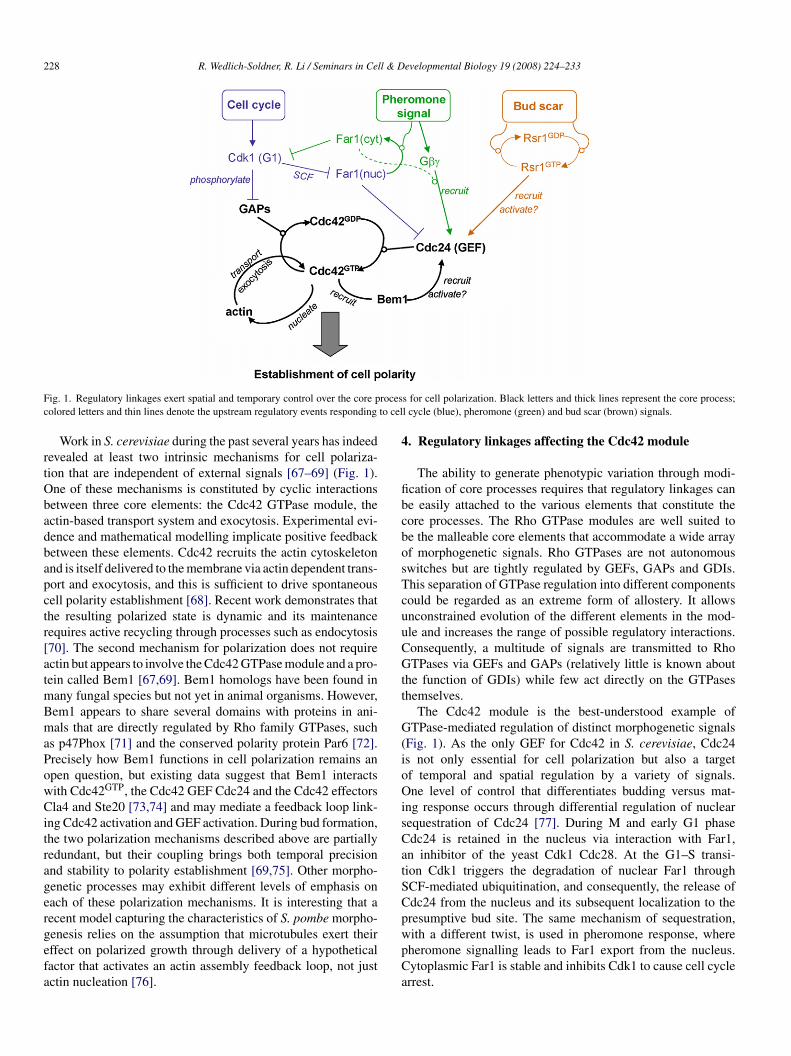
228 R. Wedlich-Soldner, R. Li / Seminars in Cell & Developmental Biology 19 (2008) 224–233
F rocesc to cel
rtObadbapctr[atmBmaPowCitragergefa
4
fibcbosTcuuCGtt
G(ioOisCatSCp
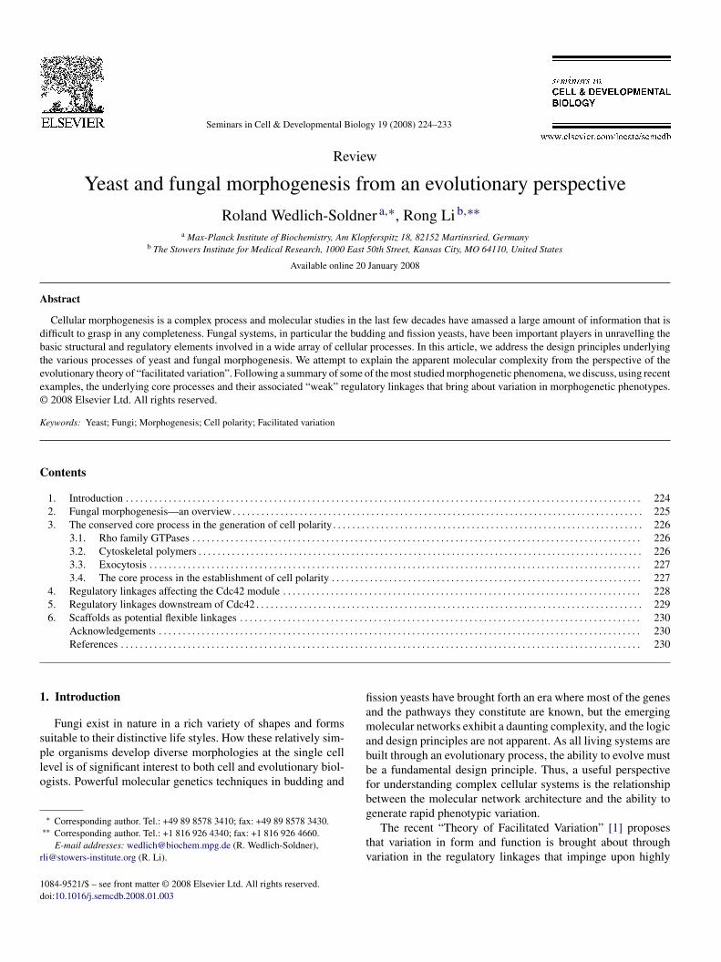
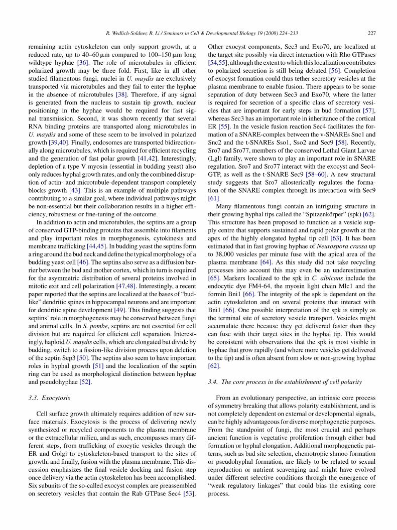
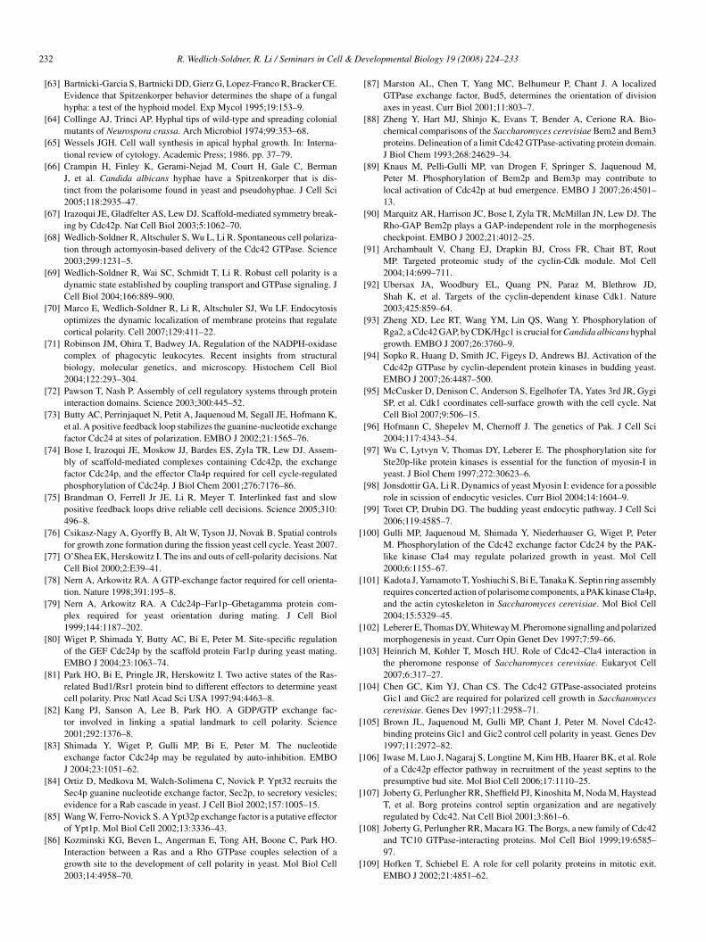
ig. 1. Regulatory linkages exert spatial and temporary control over the core polored letters and thin lines denote the upstream regulatory events responding
Work in S. cerevisiae during the past several years has indeedevealed at least two intrinsic mechanisms for cell polariza-ion that are independent of external signals [67–69] (Fig. 1).ne of these mechanisms is constituted by cyclic interactionsetween three core elements: the Cdc42 GTPase module, thectin-based transport system and exocytosis. Experimental evi-ence and mathematical modelling implicate positive feedbacketween these elements. Cdc42 recruits the actin cytoskeletonnd is itself delivered to the membrane via actin dependent trans-ort and exocytosis, and this is sufficient to drive spontaneousell polarity establishment [68]. Recent work demonstrates thathe resulting polarized state is dynamic and its maintenanceequires active recycling through processes such as endocytosis70]. The second mechanism for polarization does not requirectin but appears to involve the Cdc42 GTPase module and a pro-ein called Bem1 [67,69]. Bem1 homologs have been found inany fungal species but not yet in animal organisms. However,em1 appears to share several domains with proteins in ani-als that are directly regulated by Rho family GTPases, such
s p47Phox [71] and the conserved polarity protein Par6 [72].recisely how Bem1 functions in cell polarization remains anpen question, but existing data suggest that Bem1 interactsith Cdc42GTP, the Cdc42 GEF Cdc24 and the Cdc42 effectorsla4 and Ste20 [73,74] and may mediate a feedback loop link-
ng Cdc42 activation and GEF activation. During bud formation,he two polarization mechanisms described above are partiallyedundant, but their coupling brings both temporal precisionnd stability to polarity establishment [69,75]. Other morpho-enetic processes may exhibit different levels of emphasis onach of these polarization mechanisms. It is interesting that aecent model capturing the characteristics of S. pombe morpho-
enesis relies on the assumption that microtubules exert theirffect on polarized growth through delivery of a hypotheticalactor that activates an actin assembly feedback loop, not justctin nucleation [76].wpCa
s for cell polarization. Black letters and thick lines represent the core process;l cycle (blue), pheromone (green) and bud scar (brown) signals.
. Regulatory linkages affecting the Cdc42 module
The ability to generate phenotypic variation through modi-cation of core processes requires that regulatory linkages cane easily attached to the various elements that constitute theore processes. The Rho GTPase modules are well suited toe the malleable core elements that accommodate a wide arrayf morphogenetic signals. Rho GTPases are not autonomouswitches but are tightly regulated by GEFs, GAPs and GDIs.his separation of GTPase regulation into different componentsould be regarded as an extreme form of allostery. It allowsnconstrained evolution of the different elements in the mod-le and increases the range of possible regulatory interactions.onsequently, a multitude of signals are transmitted to RhoTPases via GEFs and GAPs (relatively little is known about
he function of GDIs) while few act directly on the GTPaseshemselves.
The Cdc42 module is the best-understood example ofTPase-mediated regulation of distinct morphogenetic signals
Fig. 1). As the only GEF for Cdc42 in S. cerevisiae, Cdc24s not only essential for cell polarization but also a targetf temporal and spatial regulation by a variety of signals.ne level of control that differentiates budding versus mat-
ng response occurs through differential regulation of nuclearequestration of Cdc24 [77]. During M and early G1 phasedc24 is retained in the nucleus via interaction with Far1,n inhibitor of the yeast Cdk1 Cdc28. At the G1–S transi-ion Cdk1 triggers the degradation of nuclear Far1 throughCF-mediated ubiquitination, and consequently, the release ofdc24 from the nucleus and its subsequent localization to theresumptive bud site. The same mechanism of sequestration,
ith a different twist, is used in pheromone response, whereheromone signalling leads to Far1 export from the nucleus.ytoplasmic Far1 is stable and inhibits Cdk1 to cause cell cyclerrest.
l & Developmental Biology 19 (2008) 224–233 229
vheguptGGAltmodctiBbclwb(lct
tBipisngtsccieCafmgtiaoBrSG
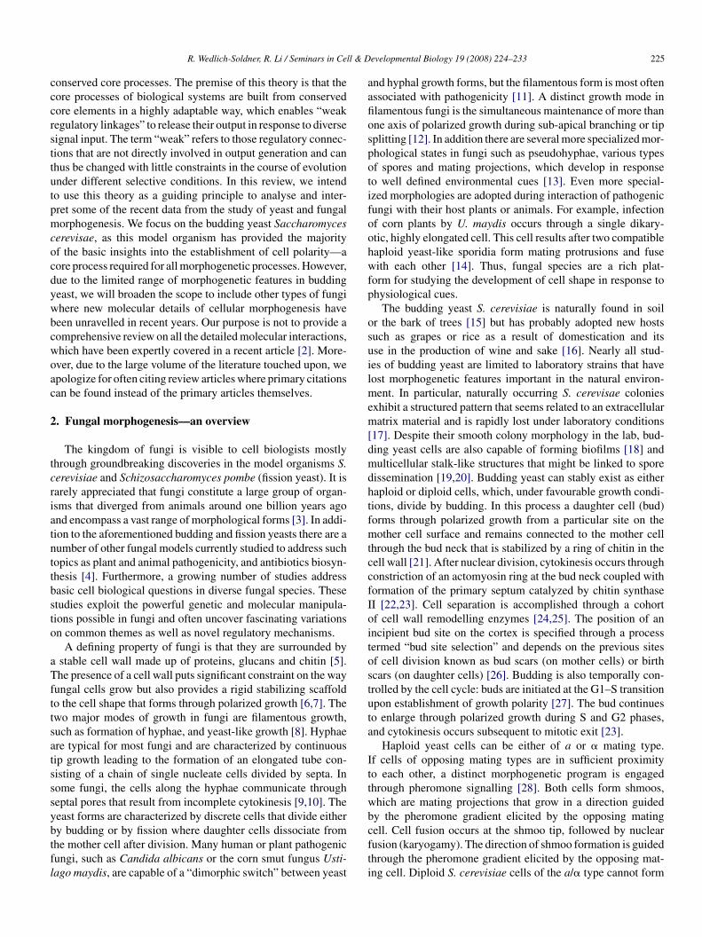
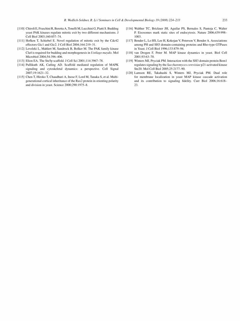
Fig. 2. Regulatory linkages downstream of Cdc42 GTPase cycle elicit a mul-titude of events in signaling and structure assembly required for variousmco
5
p(aTpdpwtttc[esratppod
aCrrasc
R. Wedlich-Soldner, R. Li / Seminars in Cel
Recruitment of Cdc24 to the site of pheromone receptor acti-ation through complex formation with Far1 and subunits of theeterotrimeric G protein serves the important function of ori-nting the direction of shmoo formation toward the pheromoneradient [78–80]. During budding, Cdc24 is also a target of reg-lation dictating the orientation of bud formation relative to therevious bud scar (Fig. 1). Cdc24 is activated and recruitedo the plasma membrane partly through interaction with theTP-bound Ras type GTPase Rsr1 [81–83]. Involvement ofTPase cascades is a recurring theme in spatial organization.nother example in yeast is the Rab GTPase cascade that regu-
ates exocytosis [84,85]. As with the Rsr1GTP–Cdc24 interactionhe connection between the different GTPase modules occurs
ainly between the GTPase of one module and a regulator (GAPr GEF) of the other module [84]. Interestingly, Rsr1GTP alsoirectly binds Cdc42 [86], which might further increase the effi-iency of local Cdc42 activation through Cdc24. Importantly,he function of Rsr1 in activation of the Cdc42 module requirests GTPase cycle, which is in turn regulated through the GEFud5 and GAP Bud2 [87]. Rapid cycling may enable multipleinding and release events, allowing the GTPase to act catalyti-ally to promote the downstream reactions. Bud5 and Bud2 areocalized in turn through interactions with proteins associatedith the bud scars. Thus the Rsr1/Bud5/Bud2 GTPase moduleiases Cdc42-driven polarization toward the cortical landmarkbud scar). Consistent with the concept of “weak regulatoryinkages”, Rsr1 is not essential for Cdc24 activation or corti-al recruitment and may be partially redundant with Bem1 inhese activities [73,83].
Whereas budding yeast only has a single GEF for Cdc42,hree GAPs have been identified so far as Rga1, Rga2 andem3. Another GAP, Bem2, shows GAP activity towards Rho1
nstead of Cdc42 in vitro [88]. However, genetic analysis of cellolarity indicates that Bem2 might still act on Cdc42 in liv-ng cells without relying on its GAP activity [89,90]. Genetictudies showed that the Cdc42 GAPs have overlapping buton-identical functions. Recent work from several labs sug-ests that the GAPs are important targets for cell cycle kinaseso control the timing of polarization (Fig. 1). Two proteomicsearches for proteins associated with specific Cdc28-cyclinomplexes identified Rga1 and Bem3 as targets of Cdc28 inomplex with the G1 cyclin Cln2 [91,92]. Three recent reportsnvolving S. cerevisae and one involving C. albicans providedvidence that the GAPs are phosphorylated by Cdc28 and Pho85dks in late G1 [89,93–95]. Interestingly, overexpression ofGAP-deficient allele of Bem3 acts in a dominant-negative
ashion and induces spontaneous polarization of G1 cells in aanner similar to constitutively active Cdc42 [89]. This sug-
ests that Cdc42’s intrinsic nucleotide exchange is sufficiento achieve a threshold level of Cdc42GTP once the GAPs arenactivated. A non-phosphorylatable mutant of Rga2 acts in
dominant-active fashion and inhibits cell polarization uponverexpression [94]. The same holds true for the respective
em3 mutants even for low levels of expression [89]. Theseesults provide compelling evidence that Cdk1 activation at G1-induces polarization at least in part through inhibition of Cdc42AP.
suii
orphogenetic processes. The events that lead to actin cable formation and exo-ytosis, denoted with thick lines, are part of the core process in the establishmentf cell polarity.
. Regulatory linkages downstream of Cdc42
Variation in morphogenetic responses may also be accom-lished through diverse and often redundant Cdc42 effectorsFig. 2). A conserved family of Cdc42 effectors are the p21-ctivated kinases (PAK), including Cla4, Ste20 and Skm1 [96].hese kinases, again, are partially redundant, but many resultsoint to both shared and divergent roles of these kinases in bud-ing yeast morphogenesis. Ste20 and Cla4 have been shown tohosphorylate and activate type I myosins Myo3 and Myo5 [97],hich in turn have a role in the internalization step of endocy-
osis via actin patches [98,99]. Cla4 is also the major kinasehat phosphorylates the GEF Cdc24, although the exact role forhis phosphorylation is still controversial [74,100]. Cla4 plays aritical role in the assembly of the septin ring at the bud neck101]. Whereas Cla4’s function is more important during veg-tative growth, Ste20 is essential during pheromone signalling,tress response and pseudohyphal growth [96,102]. The majorole for Ste20 during these processes is to transmit signal inputt the plasma membrane to the MAP kinase cascades that leado transcriptional activation of the downstream morphogeneticrograms. Whereas Ste20 has its own set of interacting partnersromoting these processes, Cla4 appears to have a negative effectn the pheromone response [103], further indicating functionalivergence of these homologous kinases.
Cdc42 interacts with a pair of yeast-specific effectors, Gic1nd Gic2, which bind active Cdc42 through the conservedRIB domain [104,105]. These homologous proteins are largely
edundant and are important for polarized actin assembly and theecruitment and assembly of the septin ring at elevated temper-tures [106]. Both proteins interact specifically with the Cdc12eptin. Interestingly, Borg3, a Cdc42 effector in mammalianells that bears no similarity to the Gic proteins, also regulates
eptin assembly [107,108]. Although the exact mechanism isnclear in either process, this might be another example of flex-ble regulatory linkages between conserved core elements tomplement specific morphogenetic responses. In addition to the
2 ll & D
rao
CeUshooRait[
6
sfcnmitSarcst3[mB
sTmubfCBstbttobapif
et
tpSfDcblomptaprm[gfl
A
ct
R
30 R. Wedlich-Soldner, R. Li / Seminars in Ce
ole in bud formation, the Gic proteins, as well as the PAKs,lso play a role in mitotic exit control, which senses spindlerientation relative to the axis of polarity [109–111].
While S. cerevisiae and S. pombe only express the Rho anddc42 classes of Rho GTPases most filamentous fungi alsoxpress the third type, Rac1. Interestingly, in one of these fungi,. maydis, Cdc42 is not essential but limited to a function in
eptum formation and cell separation [33]. Instead, the Rac1omologue is required for proper bud formation and cells with-ut Rac1 divide by fission instead [33]. Rac1 overexpression,n the other hand, results in hypha formation suggesting thatac1 specifically induces downstream events required for fil-mentous growth [33]. Fitting the concept of flexible linkagest seems that Rac1 in U. maydis partially acts through some ofhe same effectors used by Cdc42 in S. cerevisiae, namely Cla4112].
. Scaffolds as potential flexible linkages
The use of protein scaffolds to link regulators within definedignalling pathways or at particular locations in the cell is arequently inferred mechanism to explain the role of proteinomplex formation [113,114]. However, the term scaffold hasot been used in a consistent manner and can imply distinctechanistic scenarios. In processes that emphasize spatial local-
zation, scaffold often implies more static structures that serveo restrict mobility of interacting proteins. For example, the. cerevisiae bud scars are scaffolds that immobilize associ-ted proteins, such as Rax2, to form a cortical landmark. Rax2emains immobilized for several consecutive cell divisions andan be used to trace the history of a cell [115]. Similarly, eiso-omes are immobile structures in the yeast plasma membranehat exhibit no lateral motion over time spans of more than0 min and could therefore act as scaffolds for other proteins116]. Static scaffolds may be large multiprotein complexes oracromolecular assemblies that are not subject to the effects ofrownian diffusion.
In contrast, most proposed scaffold proteins implicated inignal transduction or during cell polarization are dynamic.hrough multiple protein interaction domains, these proteinsay facilitate transient protein complex formation but are
nlikely to act as static reference site for the localization of theound proteins. One example is Bem1, which has binding sitesor a number of proteins involved in cell polarization, includingdc42GTP, Cdc24, Cla4 and Boi1,2 proteins [67,117]. Whereasem1 may engage in hetero-oligomeric interactions, it has been
hown to rapidly cycle on and off the plasma membrane and isherefore unlikely to provide any significant anchorage for itsinding partners [69]. Similarly the MAP kinase scaffold pro-ein Ste5 has been shown to dynamically interact with the shmooip as well as the MAP kinase cascade proteins [118]. In bothf these examples, significant interactions also exist among theinding partners of the scaffold proteins. Thus, it may be reason-
ble to speculate that the function of such dynamic scaffolds is toromote cooperative protein complex assembly, enabling weaknteracting partners to form strong and possibly self-enhancingunctional units under a variety of conditions. Through suchevelopmental Biology 19 (2008) 224–233
ffects, dynamic scaffolds can be particularly versatile linkageso generate pathway diversity.
Bem1 is potentially such an example: whereas during vegeta-ive growth it links Cla4 to active Cdc42 and the GEF Cdc24 toromote bud formation, during pheromone response Bem1 bindste20 through the same domain and this interaction is requiredor optimal signalling through the MAP kinase cascade [119].ynamic scaffolding could also account for assembly of polarity
omplexes on the cortex without the involvement of transmem-rane domains. Cdc42 and other Rho proteins are reversiblyinked to the membrane via their prenylated C-termini. A numberf polar cortical components contain weak membrane bindingotifs, including Cdc24, Cla4, Bem1, and Bem1-interacting
roteins Boi1 and Boi2, GAP proteins for Cdc42 and Rho andhe exocyst component Exo84. Dynamic scaffolding to allowssembly of protein complex on the membrane through multi-le weak interactions may provide a flexible means for polarityegulation and adaptation to changing conditions. A similarechanism has been proposed for Ste5 in MAPK signalling
120]. Studies of dynamic scaffolds in other fungal morpho-enetic systems could lend further insights into their role asexible linkages in the evolution of morphological diversity.
cknowledgements
The authors thank Michael Sixt and Susanne Wedlich forritical reading of the manuscript. This work was supported byhe Max-Planck Society (RWS) and NIH RO1-057063 (RL).
eferences
[1] Gerhart J, Kirschner M. The theory of facilitated variation. Proc NatlAcad Sci USA 2007;1(Suppl 104):8582–9.
[2] Park HO, Bi E. Central roles of small GTPases in the development ofcell polarity in yeast and beyond. Microbiol Mol Biol Rev 2007;71:48–96.
[3] James TY, Kauff F, Schoch CL, Matheny PB, Hofstetter V, Cox CJ, et al.Reconstructing the early evolution of fungi using a six-gene phylogeny.Nature 2006;443:818–22.
[4] Hamer L, Tanzer M. Fungal role models: a bouquet of foes and friends.Proceedings of the fifth European conference on fungal genetics. FungalGenet Biol 2000;30:163–5.
[5] Bartnicki-Garcia S. Cell wall chemistry, morphogenesis, and taxonomyof fungi. Annu Rev Microbiol 1968;22:87–108.
[6] Harold FM. To shape a cell: and inquiry into the causes of morphogenesisof microorganisms. Microbiol Rev 1990;54:381–431.
[7] Slaughter B, Li R. Toward a molecular interpretation of the surface stresstheory for yeast morphogenesis. Curr Opin Cell Biol 2006;18:47–53.
[8] Sudbery P, Gow N, Berman J. The distinct morphogenic states of Candidaalbicans. Trends Microbiol 2004;12:317–24.
[9] van Driel KG, Boekhout T, Wosten HA, Verkleij AJ, Muller WH.Laser microdissection of fungal septa as visualised by scanning electronmicroscopy. Fungal Genet Biol 2007;44:466–73.
[10] Shepherd VA, Orlovich DA, Ashford AE. Cell-to-cell transport via motiletubules in growing hyphae of a fungus. J Cell Sci 1993;105(Pt 4):1173–8.
[11] Klosterman SJ, Perlin MH, Garcia-Pedrajas M, Covert SF, Gold SE.Genetics of morphogenesis and pathogenic development of Ustilago may-
dis. Adv Genet 2007;57:1–47.[12] Momany M. Polarity in filamentous fungi: establishment, maintenanceand new axes. Curr Opin Microbiol 2002;5:580–5.
[13] Whiteway M, Bachewich C. Morphogenesis in Candida albicans. AnnuRev Microbiol 2007.
l & D
R. Wedlich-Soldner, R. Li / Seminars in Cel[14] Feldbrugge M, Kamper J, Steinberg G, Kahmann R. Regulation of matingand pathogenic development in Ustilago maydis. Curr Opin Microbiol2004;7:666–72.
[15] Sniegowski PD, Dombrowski PG, Fingerman E. Saccharomyces cere-visiae and Saccharomyces paradoxus coexist in a natural woodland sitein North America and display different levels of reproductive isolationfrom European conspecifics. FEMS Yeast Res 2002;1:299–306.
[16] Fay JC, Benavides JA. Evidence for domesticated and wild populationsof Saccharomyces cerevisiae. PLoS Genet 2005;1:66–71.
[17] Kuthan M, Devaux F, Janderova B, Slaninova I, Jacq C, Palkova Z.Domestication of wild Saccharomyces cerevisiae is accompanied bychanges in gene expression and colony morphology. Mol Microbiol2003;47:745–54.
[18] Reynolds TB, Fink GR. Bakers’ yeast, a model for fungal biofilm forma-tion. Science 2001;291:878–81.
[19] Engelberg D, Mimran A, Martinetto H, Otto J, Simchen G, Karin M,et al. Multicellular stalk-like structures in Saccharomyces cerevisiae. JBacteriol 1998;180:3992–6.
[20] Scherz R, Shinder V, Engelberg D. Anatomical analysis of Saccharomycescerevisiae stalk-like structures reveals spatial organization and cell spe-cialization. J Bacteriol 2001;183:5402–13.
[21] Cabib E, Roh DH, Schmidt M, Crotti LB, Varma A. The yeast cell walland septum as paradigms of cell growth and morphogenesis. J Biol Chem2001;276:19679–82.
[22] Silverman SJ, Sburlati A, Slater ML, Cabib E. Chitin synthase 2 is essen-tial for septum formation and cell division in Saccharomyces cerevisiae.Proc Natl Acad Sci USA 1988;85:4735–9.
[23] Tolliday N, Bouquin N, Li R. Assembly and regulation of the cytokineticapparatus in budding yeast. Curr Opin Microbiol 2001;4:690–5.
[24] Colman-Lerner A, Chin TE, Brent R. Yeast Cbk1 and Mob2 activatedaughter-specific genetic programs to induce asymmetric cell fates. Cell2001;107:739–50.
[25] Weiss EL, Kurischko C, Zhang C, Shokat K, Drubin DG, Luca FC.The Saccharomyces cerevisiae Mob2p–Cbk1p kinase complex promotespolarized growth and acts with the mitotic exit network to facilitate daugh-ter cell-specific localization of Ace2p transcription factor. J Cell Biol2002;158:885–900.
[26] Casamayor A, Snyder M. Bud-site selection and cell polarity in buddingyeast. Curr Opin Microbiol 2002;5:179–86.
[27] Lew DJ, Reed SI. Morphogenesis in the yeast cell cycle: regulation byCdc28 and cyclins. J Cell Biol 1993;120:1305–20.
[28] Thorner J. Pheromonal regulation of development in Saccharomyces cere-visiae. In: Strathern JN, Jones EW, Broach JR, editors. The molecularbiology of the yeast saccharomyces: life cycle and inheritance. ColdSpring Harbor, NY: Cold Spring Harbor Laboratory Press; 1981. p.143–80.
[29] Gancedo JM. Control of pseudohyphae formation in Saccharomyces cere-visiae. FEMS Microbiol Rev 2001;25:107–23.
[30] La Carbona S, Le Goff C, Le Goff X. Fission yeast cytoskeletons and cellpolarity factors: connecting at the cortex. Biol Cell 2006;98:619–31.
[31] Feierbach B, Chang F. Cytokinesis and the contractile ring in fission yeast.Curr Opin Microbiol 2001;4:713–9.
[32] Johnson DI. Cdc42: an essential Rho-type GTPase controlling eukaryoticcell polarity. Microbiol Mol Biol Rev 1999;63:54–105.
[33] Mahlert M, Leveleki L, Hlubek A, Sandrock B, Bolker M. Rac1 andCdc42 regulate hyphal growth and cytokinesis in the dimorphic fungusUstilago maydis. Mol Microbiol 2006;59:567–78.
[34] Moseley JB, Goode BL. The yeast actin cytoskeleton: from cel-lular function to biochemical mechanism. Microbiol Mol Biol Rev2006;70:605–45.
[35] Chang F, Feierbach B, Martin S. Regulation of actin assembly bymicrotubules in fission yeast cell polarity. Novartis Found Symp2005;269:59–66 [discussion-72, 223–30].
[36] Steinberg G. Tracks for traffic: microtubules in the plant pathogen Usti-lago maydis. New Phytol 2007;174:721–33.
[37] Janson ME, Loughlin R, Loiodice I, Fu C, Brunner D, Nedelec FJ, et al.Crosslinkers and motors organize dynamic microtubules to form stablebipolar arrays in fission yeast. Cell 2007;128:357–68.
evelopmental Biology 19 (2008) 224–233 231
[38] Fuchs U, Manns I, Steinberg G. Microtubules are dispensable for the ini-tial pathogenic development but required for long-distance hyphal growthin the corn smut fungus Ustilago maydis. Mol Biol Cell 2005;16:2746–58.
[39] Zarnack K, Feldbrugge M. mRNA trafficking in fungi. Mol GenetGenomics 2007;278:347–59.
[40] Becht P, Konig J, Feldbrugge M. The RNA-binding protein Rrm4 is essen-tial for polarity in Ustilago maydis and shuttles along microtubules. J CellSci 2006;119:4964–73.
[41] Wedlich-Soldner R, Bolker M, Kahmann R, Steinberg G. A putative endo-somal t-SNARE links exo- and endocytosis in the phytopathogenic fungusUstilago maydis. EMBO J 2000;19:1974–86.
[42] Wedlich-Soldner R, Straube A, Friedrich MW, Steinberg G. A balance ofKIF1A-like kinesin and dynein organizes early endosomes in the fungusUstilago maydis. EMBO J 2002;21:2946–57.
[43] Schuchardt I, Assmann D, Thines E, Schuberth C, Steinberg G. Myosin-V, Kinesin-1, and Kinesin-3 cooperate in hyphal growth of the fungusUstilago maydis. Mol Biol Cell 2005;16:5191–201.
[44] Lindsey R, Momany M. Septin localization across kingdoms: threethemes with variations. Curr Opin Microbiol 2006;9:559–65.
[45] Kinoshita M. Diversity of septin scaffolds. Curr Opin Cell Biol2006;18:54–60.
[46] Longtine MS, Bi E. Regulation of septin organization and function inyeast. Trends Cell Biol 2003;13:403–9.
[47] Faty M, Fink M, Barral Y. Septins: a ring to part mother and daughter.Curr Genet 2002;41:123–31.
[48] Castillon GA, Adames NR, Rosello CH, Seidel HS, Longtine MS, CooperJA, et al. Septins have a dual role in controlling mitotic exit in buddingyeast. Curr Biol 2003;13:654–8.
[49] Tada T, Simonetta A, Batterton M, Kinoshita M, Edbauer D, Sheng M.Role of Septin cytoskeleton in spine morphogenesis and dendrite devel-opment in neurons. Curr Biol 2007;17:1752–8.
[50] Boyce KJ, Chang H, D’Souza CA, Kronstad JW. An Ustilago maydisseptin is required for filamentous growth in culture and for full symptomdevelopment on maize. Eukaryot Cell 2005;4:2044–56.
[51] Warenda AJ, Konopka JB. Septin function in Candida albicans morpho-genesis. Mol Biol Cell 2002;13:2732–46.
[52] Berman J, Sudbery PE. Candida albicans: a molecular revolution builton lessons from budding yeast. Nat Rev 2002;3:918–30.
[53] Boyd C, Hughes T, Pypaert M, Novick P. Vesicles carry most exocystsubunits to exocytic sites marked by the remaining two subunits, Sec3pand Exo70p. J Cell Biol 2004;167:889–901.
[54] Zhang X, Zajac A, Zhang J, Wang P, Li M, Murray J, et al. The critical roleof Exo84p in the organization and polarized localization of the exocystcomplex. J Biol Chem 2005;280:20356–64.
[55] Wiederkehr A, Du Y, Pypaert M, Ferro-Novick S, Novick P. Sec3p isneeded for the spatial regulation of secretion and for the inheritance ofthe cortical endoplasmic reticulum. Mol Biol Cell 2003;14:4770–82.
[56] Roumanie O, Wu H, Molk JN, Rossi G, Bloom K, Brennwald P. RhoGTPase regulation of exocytosis in yeast is independent of GTP hydroly-sis and polarization of the exocyst complex. J Cell Biol 2005;170:583–94.
[57] He B, Xi F, Zhang J, TerBush D, Zhang X, Guo W. Exo70p mediates thesecretion of specific exocytic vesicles at early stages of the cell cycle forpolarized cell growth. J Cell Biol 2007;176:771–7.
[58] Novick P, Medkova M, Dong G, Hutagalung A, Reinisch K, GrosshansB. Interactions between Rabs, tethers, SNAREs and their regulators inexocytosis. Biochem Soc Trans 2006;34:683–6.
[59] Grosshans BL, Andreeva A, Gangar A, Niessen S, Yates 3rd JR, Bren-nwald P, et al. The yeast lgl family member Sro7p is an effector of thesecretory Rab GTPase Sec4p. J Cell Biol 2006;172:55–66.
[60] Zhang X, Wang P, Gangar A, Zhang J, Brennwald P, TerBush D, et al.Lethal giant larvae proteins interact with the exocyst complex and areinvolved in polarized exocytosis. J Cell Biol 2005;170:273–83.
[61] Hattendorf DA, Andreeva A, Gangar A, Brennwald PJ, Weis WI. Structureof the yeast polarity protein Sro7 reveals a SNARE regulatory mechanism.Nature 2007;446:567–71.
[62] Virag A, Harris SD. The Spitzenkorper: a molecular perspective. MycolRes 2006;110:4–13.
2 ll & D
32 R. Wedlich-Soldner, R. Li / Seminars in Ce[63] Bartnicki-Garcia S, Bartnicki DD, Gierz G, Lopez-Franco R, Bracker CE.Evidence that Spitzenkorper behavior determines the shape of a fungalhypha: a test of the hyphoid model. Exp Mycol 1995;19:153–9.
[64] Collinge AJ, Trinci AP. Hyphal tips of wild-type and spreading colonialmutants of Neurospora crassa. Arch Microbiol 1974;99:353–68.
[65] Wessels JGH. Cell wall synthesis in apical hyphal growth. In: Interna-tional review of cytology. Academic Press; 1986. pp. 37–79.
[66] Crampin H, Finley K, Gerami-Nejad M, Court H, Gale C, BermanJ, et al. Candida albicans hyphae have a Spitzenkorper that is dis-tinct from the polarisome found in yeast and pseudohyphae. J Cell Sci2005;118:2935–47.
[67] Irazoqui JE, Gladfelter AS, Lew DJ. Scaffold-mediated symmetry break-ing by Cdc42p. Nat Cell Biol 2003;5:1062–70.
[68] Wedlich-Soldner R, Altschuler S, Wu L, Li R. Spontaneous cell polariza-tion through actomyosin-based delivery of the Cdc42 GTPase. Science2003;299:1231–5.
[69] Wedlich-Soldner R, Wai SC, Schmidt T, Li R. Robust cell polarity is adynamic state established by coupling transport and GTPase signaling. JCell Biol 2004;166:889–900.
[70] Marco E, Wedlich-Soldner R, Li R, Altschuler SJ, Wu LF. Endocytosisoptimizes the dynamic localization of membrane proteins that regulatecortical polarity. Cell 2007;129:411–22.
[71] Robinson JM, Ohira T, Badwey JA. Regulation of the NADPH-oxidasecomplex of phagocytic leukocytes. Recent insights from structuralbiology, molecular genetics, and microscopy. Histochem Cell Biol2004;122:293–304.
[72] Pawson T, Nash P. Assembly of cell regulatory systems through proteininteraction domains. Science 2003;300:445–52.
[73] Butty AC, Perrinjaquet N, Petit A, Jaquenoud M, Segall JE, Hofmann K,et al. A positive feedback loop stabilizes the guanine-nucleotide exchangefactor Cdc24 at sites of polarization. EMBO J 2002;21:1565–76.
[74] Bose I, Irazoqui JE, Moskow JJ, Bardes ES, Zyla TR, Lew DJ. Assem-bly of scaffold-mediated complexes containing Cdc42p, the exchangefactor Cdc24p, and the effector Cla4p required for cell cycle-regulatedphosphorylation of Cdc24p. J Biol Chem 2001;276:7176–86.
[75] Brandman O, Ferrell Jr JE, Li R, Meyer T. Interlinked fast and slowpositive feedback loops drive reliable cell decisions. Science 2005;310:496–8.
[76] Csikasz-Nagy A, Gyorffy B, Alt W, Tyson JJ, Novak B. Spatial controlsfor growth zone formation during the fission yeast cell cycle. Yeast 2007.
[77] O’Shea EK, Herskowitz I. The ins and outs of cell-polarity decisions. NatCell Biol 2000;2:E39–41.
[78] Nern A, Arkowitz RA. A GTP-exchange factor required for cell orienta-tion. Nature 1998;391:195–8.
[79] Nern A, Arkowitz RA. A Cdc24p–Far1p–Gbetagamma protein com-plex required for yeast orientation during mating. J Cell Biol1999;144:1187–202.
[80] Wiget P, Shimada Y, Butty AC, Bi E, Peter M. Site-specific regulationof the GEF Cdc24p by the scaffold protein Far1p during yeast mating.EMBO J 2004;23:1063–74.
[81] Park HO, Bi E, Pringle JR, Herskowitz I. Two active states of the Ras-related Bud1/Rsr1 protein bind to different effectors to determine yeastcell polarity. Proc Natl Acad Sci USA 1997;94:4463–8.
[82] Kang PJ, Sanson A, Lee B, Park HO. A GDP/GTP exchange fac-tor involved in linking a spatial landmark to cell polarity. Science2001;292:1376–8.
[83] Shimada Y, Wiget P, Gulli MP, Bi E, Peter M. The nucleotideexchange factor Cdc24p may be regulated by auto-inhibition. EMBOJ 2004;23:1051–62.
[84] Ortiz D, Medkova M, Walch-Solimena C, Novick P. Ypt32 recruits theSec4p guanine nucleotide exchange factor, Sec2p, to secretory vesicles;evidence for a Rab cascade in yeast. J Cell Biol 2002;157:1005–15.
[85] Wang W, Ferro-Novick S. A Ypt32p exchange factor is a putative effector
of Ypt1p. Mol Biol Cell 2002;13:3336–43.[86] Kozminski KG, Beven L, Angerman E, Tong AH, Boone C, Park HO.Interaction between a Ras and a Rho GTPase couples selection of agrowth site to the development of cell polarity in yeast. Mol Biol Cell2003;14:4958–70.
evelopmental Biology 19 (2008) 224–233
[87] Marston AL, Chen T, Yang MC, Belhumeur P, Chant J. A localizedGTPase exchange factor, Bud5, determines the orientation of divisionaxes in yeast. Curr Biol 2001;11:803–7.
[88] Zheng Y, Hart MJ, Shinjo K, Evans T, Bender A, Cerione RA. Bio-chemical comparisons of the Saccharomyces cerevisiae Bem2 and Bem3proteins. Delineation of a limit Cdc42 GTPase-activating protein domain.J Biol Chem 1993;268:24629–34.
[89] Knaus M, Pelli-Gulli MP, van Drogen F, Springer S, Jaquenoud M,Peter M. Phosphorylation of Bem2p and Bem3p may contribute tolocal activation of Cdc42p at bud emergence. EMBO J 2007;26:4501–13.
[90] Marquitz AR, Harrison JC, Bose I, Zyla TR, McMillan JN, Lew DJ. TheRho-GAP Bem2p plays a GAP-independent role in the morphogenesischeckpoint. EMBO J 2002;21:4012–25.
[91] Archambault V, Chang EJ, Drapkin BJ, Cross FR, Chait BT, RoutMP. Targeted proteomic study of the cyclin-Cdk module. Mol Cell2004;14:699–711.
[92] Ubersax JA, Woodbury EL, Quang PN, Paraz M, Blethrow JD,Shah K, et al. Targets of the cyclin-dependent kinase Cdk1. Nature2003;425:859–64.
[93] Zheng XD, Lee RT, Wang YM, Lin QS, Wang Y. Phosphorylation ofRga2, a Cdc42 GAP, by CDK/Hgc1 is crucial for Candida albicans hyphalgrowth. EMBO J 2007;26:3760–9.
[94] Sopko R, Huang D, Smith JC, Figeys D, Andrews BJ. Activation of theCdc42p GTPase by cyclin-dependent protein kinases in budding yeast.EMBO J 2007;26:4487–500.
[95] McCusker D, Denison C, Anderson S, Egelhofer TA, Yates 3rd JR, GygiSP, et al. Cdk1 coordinates cell-surface growth with the cell cycle. NatCell Biol 2007;9:506–15.
[96] Hofmann C, Shepelev M, Chernoff J. The genetics of Pak. J Cell Sci2004;117:4343–54.
[97] Wu C, Lytvyn V, Thomas DY, Leberer E. The phosphorylation site forSte20p-like protein kinases is essential for the function of myosin-I inyeast. J Biol Chem 1997;272:30623–6.
[98] Jonsdottir GA, Li R. Dynamics of yeast Myosin I: evidence for a possiblerole in scission of endocytic vesicles. Curr Biol 2004;14:1604–9.
[99] Toret CP, Drubin DG. The budding yeast endocytic pathway. J Cell Sci2006;119:4585–7.
[100] Gulli MP, Jaquenoud M, Shimada Y, Niederhauser G, Wiget P, PeterM. Phosphorylation of the Cdc42 exchange factor Cdc24 by the PAK-like kinase Cla4 may regulate polarized growth in yeast. Mol Cell2000;6:1155–67.
[101] Kadota J, Yamamoto T, Yoshiuchi S, Bi E, Tanaka K. Septin ring assemblyrequires concerted action of polarisome components, a PAK kinase Cla4p,and the actin cytoskeleton in Saccharomyces cerevisiae. Mol Biol Cell2004;15:5329–45.
[102] Leberer E, Thomas DY, Whiteway M. Pheromone signalling and polarizedmorphogenesis in yeast. Curr Opin Genet Dev 1997;7:59–66.
[103] Heinrich M, Kohler T, Mosch HU. Role of Cdc42–Cla4 interaction inthe pheromone response of Saccharomyces cerevisiae. Eukaryot Cell2007;6:317–27.
[104] Chen GC, Kim YJ, Chan CS. The Cdc42 GTPase-associated proteinsGic1 and Gic2 are required for polarized cell growth in Saccharomycescerevisiae. Genes Dev 1997;11:2958–71.
[105] Brown JL, Jaquenoud M, Gulli MP, Chant J, Peter M. Novel Cdc42-binding proteins Gic1 and Gic2 control cell polarity in yeast. Genes Dev1997;11:2972–82.
[106] Iwase M, Luo J, Nagaraj S, Longtine M, Kim HB, Haarer BK, et al. Roleof a Cdc42p effector pathway in recruitment of the yeast septins to thepresumptive bud site. Mol Biol Cell 2006;17:1110–25.
[107] Joberty G, Perlungher RR, Sheffield PJ, Kinoshita M, Noda M, HaysteadT, et al. Borg proteins control septin organization and are negativelyregulated by Cdc42. Nat Cell Biol 2001;3:861–6.
[108] Joberty G, Perlungher RR, Macara IG. The Borgs, a new family of Cdc42and TC10 GTPase-interacting proteins. Mol Cell Biol 1999;19:6585–97.
[109] Hofken T, Schiebel E. A role for cell polarity proteins in mitotic exit.EMBO J 2002;21:4851–62.
l & D
R. Wedlich-Soldner, R. Li / Seminars in Cel[110] Chiroli E, Fraschini R, Beretta A, Tonelli M, Lucchini G, Piatti S. Buddingyeast PAK kinases regulate mitotic exit by two different mechanisms. JCell Biol 2003;160:857–74.
[111] Hofken T, Schiebel E. Novel regulation of mitotic exit by the Cdc42effectors Gic1 and Gic2. J Cell Biol 2004;164:219–31.
[112] Leveleki L, Mahlert M, Sandrock B, Bolker M. The PAK family kinaseCla4 is required for budding and morphogenesis in Ustilago maydis. MolMicrobiol 2004;54:396–406.
[113] Elion EA. The Ste5p scaffold. J Cell Sci 2001;114:3967–78.[114] Pullikuth AK, Catling AD. Scaffold mediated regulation of MAPK
signaling and cytoskeletal dynamics: a perspective. Cell Signal2007;19:1621–32.
[115] Chen T, Hiroko T, Chaudhuri A, Inose F, Lord M, Tanaka S, et al. Multi-generational cortical inheritance of the Rax2 protein in orienting polarityand division in yeast. Science 2000;290:1975–8.
evelopmental Biology 19 (2008) 224–233 233
[116] Walther TC, Brickner JH, Aguilar PS, Bernales S, Pantoja C, WalterP. Eisosomes mark static sites of endocytosis. Nature 2006;439:998–1003.
[117] Bender L, Lo HS, Lee H, Kokojan V, Peterson V, Bender A. Associationsamong PH and SH3 domain-containing proteins and Rho-type GTPasesin Yeast. J Cell Biol 1996;133:879–94.
[118] van Drogen F, Peter M. MAP kinase dynamics in yeast. Biol Cell2001;93:63–70.
[119] Winters MJ, Pryciak PM. Interaction with the SH3 domain protein Bem1regulates signaling by the Saccharomyces cerevisiae p21-activated kinase
Ste20. Mol Cell Biol 2005;25:2177–90.[120] Lamson RE, Takahashi S, Winters MJ, Pryciak PM. Dual rolefor membrane localization in yeast MAP kinase cascade activationand its contribution to signaling fidelity. Curr Biol 2006;16:618–23.


![Distinct Steps in Yeast Spore Morphogenesis Require ... · multiple and distinct aberrant spore wall patterns are Detroit, MI), 2% glucose], or SA (0.67% yeast nitrogen base observed](https://img.pdfslide.us/doc/110x75/60bb3b09add0b53ad5487487/distinct-steps-in-yeast-spore-morphogenesis-require-multiple-and-distinct-aberrant.jpg)