Embed Size (px)
Citation preview
Name:
Science Class:
Teacher:
Hand in day:
Y8 Science
Term 1 Homework Booklet
Biology
Hand in Date Parents Signature
Digestion
Homework 1
Homework 2
Homework 3
Homework 4
Circulation
Homework 1
Homework 2
Homework 3
Homework 4
Digestion: Homework 1
Nutrients
Learn the different food groups and try to remember what each group is used
for in the human body:
Carbohydrates: Two types: starch and sugar. They provide energy – an excess
cause’s weight increase.
Protein: Important for growth and repair of cells and tissues.
Lipids (fats and oils): Stored as a reserve energy supply. A layer under the skin
provides insulation against cold. An excess cause’s weight gain and can lead to
other health issues.
Minerals: Tiny amounts are needed – e.g. iron for red blood cells and calcium for
teeth and bones.
Vitamins: small amounts are needed – e.g. vitamin C for repair of the skin and
vitamin D for taking up calcium.
Dietary fibre: needed to keep the large intestine working well.
Water: Needed to stop a person becoming dehydrated.
Questions
1. What are the two types of carbohydrate?
2. What does the body use carbohydrates for?
3. Why is protein important?
4. Give some examples of foods that contain protein.
5. What role do lipids play in the body?
6. Give an example of a mineral needed as part of a healthy diet.
7. Give an example of a vitamin needed as part of a healthy diet.
8. What can a lack of dietary fibre cause?
Digestion: Homework 2
Testing for Nutrients
Exam Question:
Describe how a student could test cow’s milk to show whether it contains protein and different types of carbohydrate (sugar and starch).
______________________________________________________
______________________________________________________
______________________________________________________
______________________________________________________
______________________________________________________
______________________________________________________
______________________________________________________
______________________________________________________
______________________________________________________
______________________________________________________
______________________________________________________
______________________________________________________
______________________________________________________
______________________________________________________
______________________________________________________
______________________________________________________
______________________________________________________
______________________________________________________
(6)
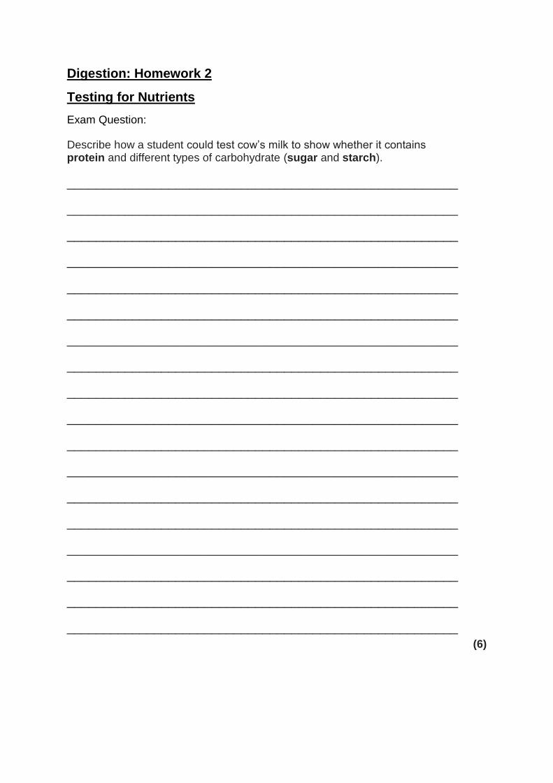
Digestion: Homework 3
The Digestive System
1. Learn the spelling and function of each part of the digestive system:
Mouth: Teeth break down food into smaller pieces.
Salivary gland: Produces saliva which moistens food and contains enzymes to help
digest food.
Oesophagus: Has muscular walls that move food from the mouth to the stomach by
an action called peristalsis.
Stomach: Has strong muscular walls that allow food to be mixed, also produces
hydrochloric acid and enzymes. The acid kills harmful microbes and provides the
optimum pH for stomach enzymes to work. The enzymes help to digest food.
Small intestine: Food is absorbed into the blood in the small intestine. It has a very
large surface area.
Large intestine: Absorbs water which solidifies waste.
Rectum: The final section of the large intestine. Where waste is stored before being
released.
Anus: A strong muscle that opens to release waste from the rectum.
2. Label the diagram below:
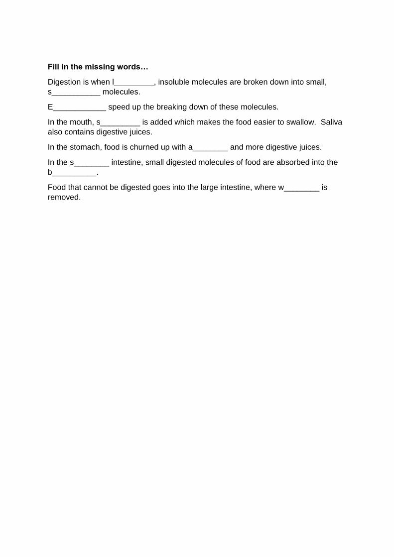
Fill in the missing words…
Digestion is when l_________, insoluble molecules are broken down into small,
s___________ molecules.
E____________ speed up the breaking down of these molecules.
In the mouth, s_________ is added which makes the food easier to swallow. Saliva
also contains digestive juices.
In the stomach, food is churned up with a________ and more digestive juices.
In the s________ intestine, small digested molecules of food are absorbed into the
b__________.
Food that cannot be digested goes into the large intestine, where w________ is
removed.
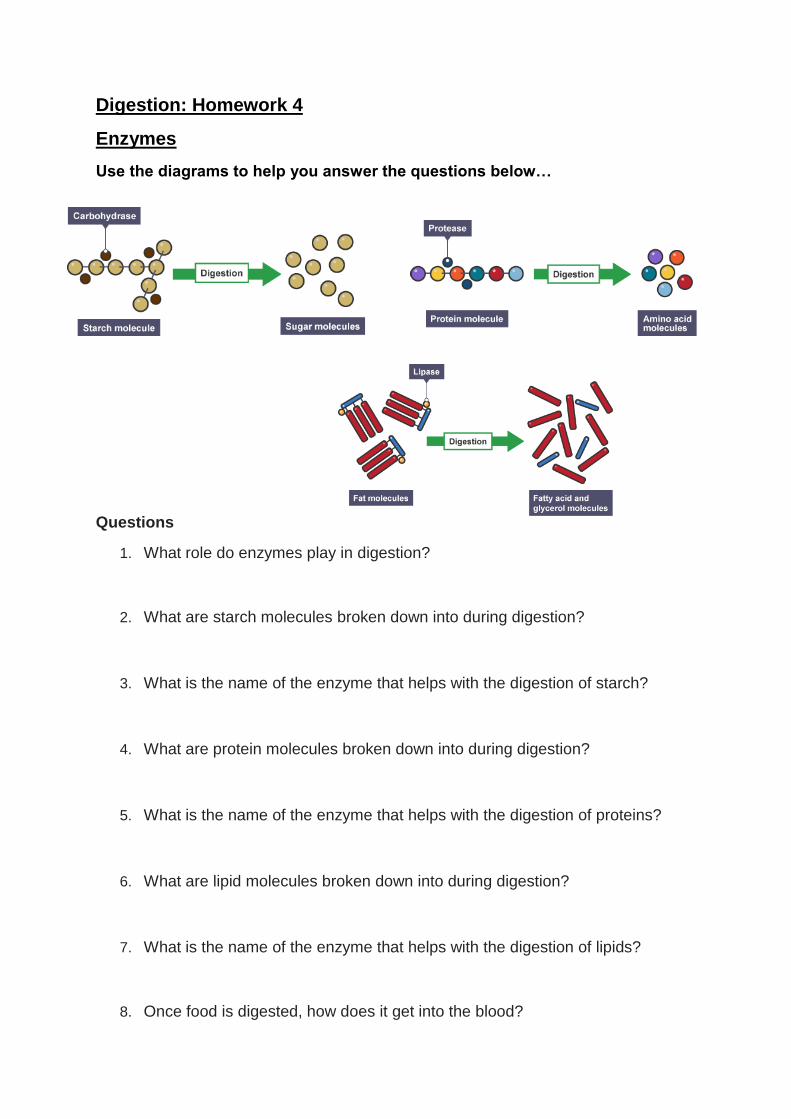
Digestion: Homework 4
Enzymes
Use the diagrams to help you answer the questions below…
Questions
1. What role do enzymes play in digestion?
2. What are starch molecules broken down into during digestion?
3. What is the name of the enzyme that helps with the digestion of starch?
4. What are protein molecules broken down into during digestion?
5. What is the name of the enzyme that helps with the digestion of proteins?
6. What are lipid molecules broken down into during digestion?
7. What is the name of the enzyme that helps with the digestion of lipids?
8. Once food is digested, how does it get into the blood?
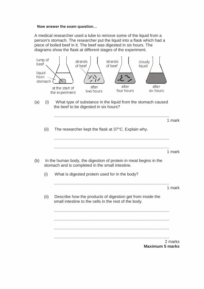
Now answer the exam question…
A medical researcher used a tube to remove some of the liquid from a person's stomach. The researcher put the liquid into a flask which had a piece of boiled beef in it. The beef was digested in six hours. The diagrams show the flask at different stages of the experiment.
(a) (i) What type of substance in the liquid from the stomach caused the beef to be digested in six hours?
..................................................................................................... 1 mark
(ii) The researcher kept the flask at 37°C. Explain why.
.....................................................................................................
..................................................................................................... 1 mark
(b) In the human body, the digestion of protein in meat begins in the stomach and is completed in the small intestine.
(i) What is digested protein used for in the body?
..................................................................................................... 1 mark
(ii) Describe how the products of digestion get from inside the small intestine to the cells in the rest of the body.
.....................................................................................................
.....................................................................................................
.....................................................................................................
..................................................................................................... 2 marks
Maximum 5 marks
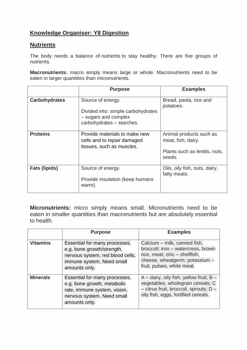
Knowledge Organiser: Y8 Digestion
Nutrients
The body needs a balance of nutrients to stay healthy. There are five groups of nutrients.
Macronutrients: macro simply means large or whole. Macronutrients need to be eaten in larger quantities than micronutrients.
Purpose Examples
Carbohydrates Source of energy.
Divided into: simple carbohydrates – sugars and complex carbohydrates – starches.
Bread, pasta, rice and potatoes.
Proteins Provide materials to make new
cells and to repair damaged
tissues, such as muscles.
Animal products such as
meat, fish, dairy.
Plants such as lentils, nuts, seeds.
Fats (lipids) Source of energy.
Provide insulation (keep humans warm).
Oils, oily fish, nuts, dairy, fatty meats.
Micronutrients: micro simply means small. Micronutrients need to be eaten in smaller quantities than macronutrients but are absolutely essential to health.
Purpose Examples
Vitamins Essential for many processes,
e.g. bone growth/strength,
nervous system, red blood cells,
immune system. Need small
amounts only.
Calcium – milk, canned fish, broccoli; iron – watercress, brown rice, meat; zinc – shellfish, cheese, wheatgerm; potassium – fruit, pulses, white meat.
Minerals Essential for many processes,
e.g. bone growth, metabolic
rate, immune system, vision,
nervous system. Need small
amounts only.
A – dairy, oily fish, yellow fruit; B – vegetables, wholegrain cereals; C – citrus fruit, broccoli, sprouts; D – oily fish, eggs, fortified cereals.
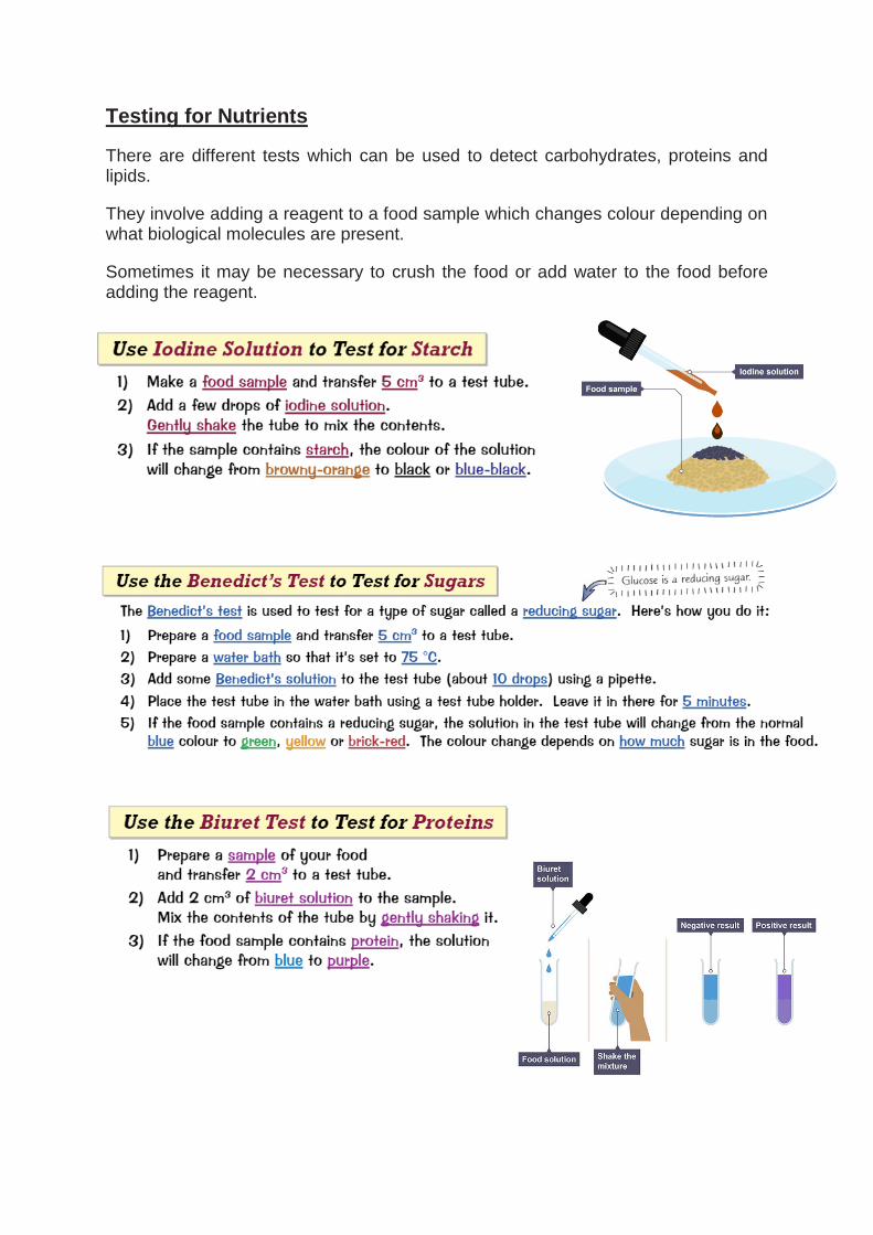
Testing for Nutrients
There are different tests which can be used to detect carbohydrates, proteins and lipids.
They involve adding a reagent to a food sample which changes colour depending on what biological molecules are present.
Sometimes it may be necessary to crush the food or add water to the food before adding the reagent.
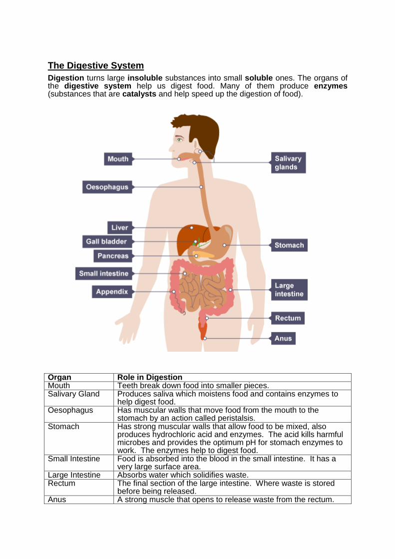
The Digestive System
Digestion turns large insoluble substances into small soluble ones. The organs of the digestive system help us digest food. Many of them produce enzymes (substances that are catalysts and help speed up the digestion of food).
Organ Role in Digestion Mouth Teeth break down food into smaller pieces. Salivary Gland Produces saliva which moistens food and contains enzymes to
help digest food. Oesophagus Has muscular walls that move food from the mouth to the
stomach by an action called peristalsis. Stomach Has strong muscular walls that allow food to be mixed, also
produces hydrochloric acid and enzymes. The acid kills harmful microbes and provides the optimum pH for stomach enzymes to work. The enzymes help to digest food.
Small Intestine Food is absorbed into the blood in the small intestine. It has a very large surface area.
Large Intestine Absorbs water which solidifies waste. Rectum The final section of the large intestine. Where waste is stored
before being released. Anus A strong muscle that opens to release waste from the rectum.
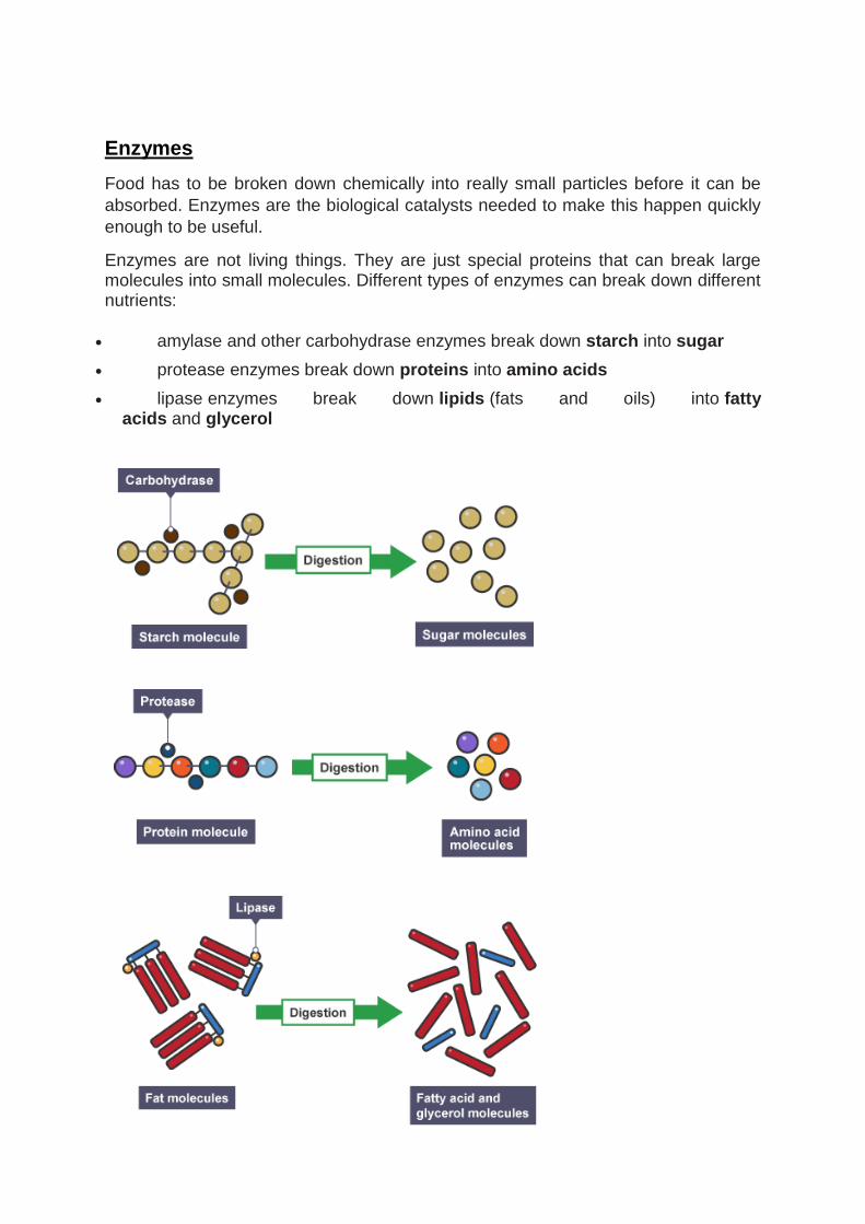
Enzymes
Food has to be broken down chemically into really small particles before it can be
absorbed. Enzymes are the biological catalysts needed to make this happen quickly
enough to be useful.
Enzymes are not living things. They are just special proteins that can break large molecules into small molecules. Different types of enzymes can break down different nutrients:
amylase and other carbohydrase enzymes break down starch into sugar
protease enzymes break down proteins into amino acids
lipase enzymes break down lipids (fats and oils) into fatty acids and glycerol
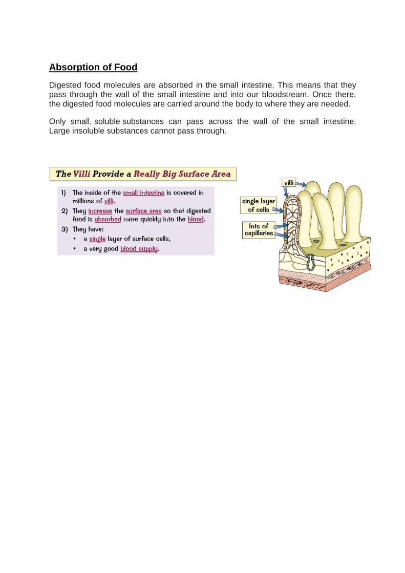
Absorption of Food Digested food molecules are absorbed in the small intestine. This means that they pass through the wall of the small intestine and into our bloodstream. Once there, the digested food molecules are carried around the body to where they are needed.
Only small, soluble substances can pass across the wall of the small intestine. Large insoluble substances cannot pass through.
Circulation: Homework 1
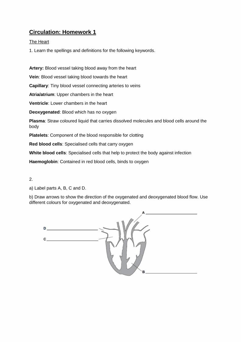
The Heart
1. Learn the spellings and definitions for the following keywords.
Artery: Blood vessel taking blood away from the heart
Vein: Blood vessel taking blood towards the heart
Capillary: Tiny blood vessel connecting arteries to veins
Atria/atrium: Upper chambers in the heart
Ventricle: Lower chambers in the heart
Deoxygenated: Blood which has no oxygen
Plasma: Straw coloured liquid that carries dissolved molecules and blood cells around the
body
Platelets: Component of the blood responsible for clotting
Red blood cells: Specialised cells that carry oxygen
White blood cells: Specialised cells that help to protect the body against infection
Haemoglobin: Contained in red blood cells, binds to oxygen
2.
a) Label parts A, B, C and D.
b) Draw arrows to show the direction of the oxygenated and deoxygenated blood flow. Use
different colours for oxygenated and deoxygenated.
Key words test:
1.
2.
3.
4.
5.
6.
7.
8.
9.
10.
Circulation: Homework 2
Journey of a Red Blood Cell
Describe how the blood travels from the body to the heart and then back to the body. Use
the following key words: aorta, vena cava, atrium, ventricle, lung, body, arteries, veins,
oxygenated blood, and deoxygenated blood.
________________________________________________________
________________________________________________________
________________________________________________________
________________________________________________________
________________________________________________________
________________________________________________________
________________________________________________________
________________________________________________________
________________________________________________________
________________________________________________________
________________________________________________________
________________________________________________________
________________________________________________________
________________________________________________________
________________________________________________________
________________________________________________________
________________________________________________________
________________________________________________________
Circulation: Homework 3
Blood Vessels
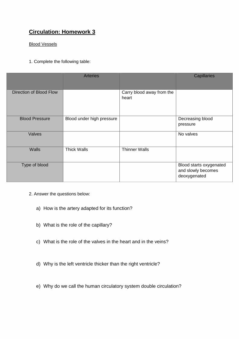
1. Complete the following table:
2. Answer the questions below:
a) How is the artery adapted for its function?
b) What is the role of the capillary?
c) What is the role of the valves in the heart and in the veins?
d) Why is the left ventricle thicker than the right ventricle?
e) Why do we call the human circulatory system double circulation?
Arteries Capillaries
Direction of Blood Flow
Carry blood away from the
heart
Blood Pressure Blood under high pressure Decreasing blood
pressure
Valves No valves
Walls Thick Walls Thinner Walls
Type of blood Blood starts oxygenated
and slowly becomes
deoxygenated
Circulation: Homework 4
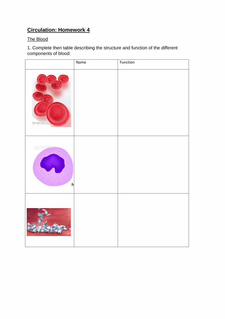
The Blood
1. Complete then table describing the structure and function of the different
components of blood:
Name Function
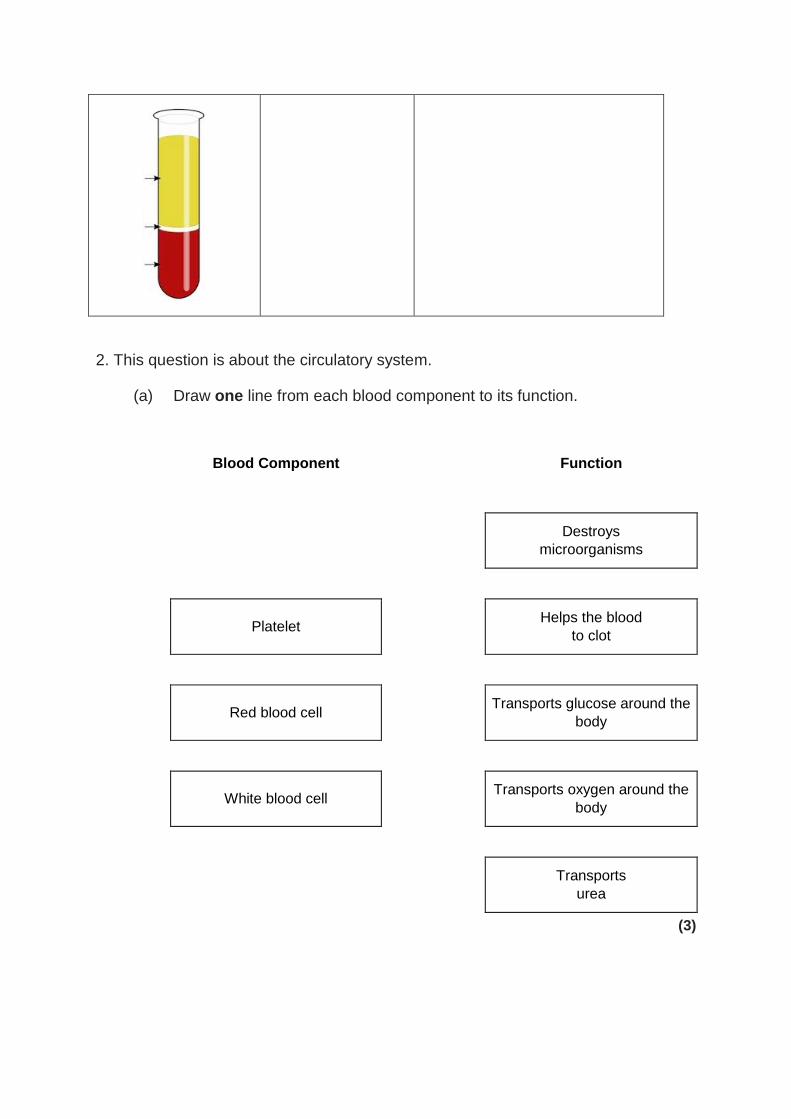
2. This question is about the circulatory system.
(a) Draw one line from each blood component to its function.
Blood Component Function
Destroys
microorganisms
Platelet Helps the blood
to clot
Red blood cell Transports glucose around the
body
White blood cell Transports oxygen around the
body
Transports
urea
(3)
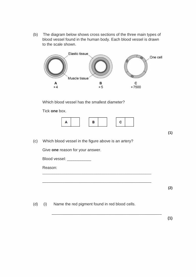
(b) The diagram below shows cross sections of the three main types of
blood vessel found in the human body. Each blood vessel is drawn
to the scale shown.
Which blood vessel has the smallest diameter?
Tick one box.
(1)
(c) Which blood vessel in the figure above is an artery?
Give one reason for your answer.
Blood vessel: ___________
Reason: ______________________________________________________
______________________________________________________
(2)
(d) (i) Name the red pigment found in red blood cells.
__________________________________________________
(1)
(ii) Describe, in detail, the function of this red pigment.
__________________________________________________
__________________________________________________
__________________________________________________
__________________________________________________
(2)
(e) Describe one other way in which the structure of a red blood cell is different from the structure of a white blood cell.
______________________________________________________
______________________________________________________
(1)
(Total 10 marks)
Knowledge Organiser: Circulation
The Circulatory System
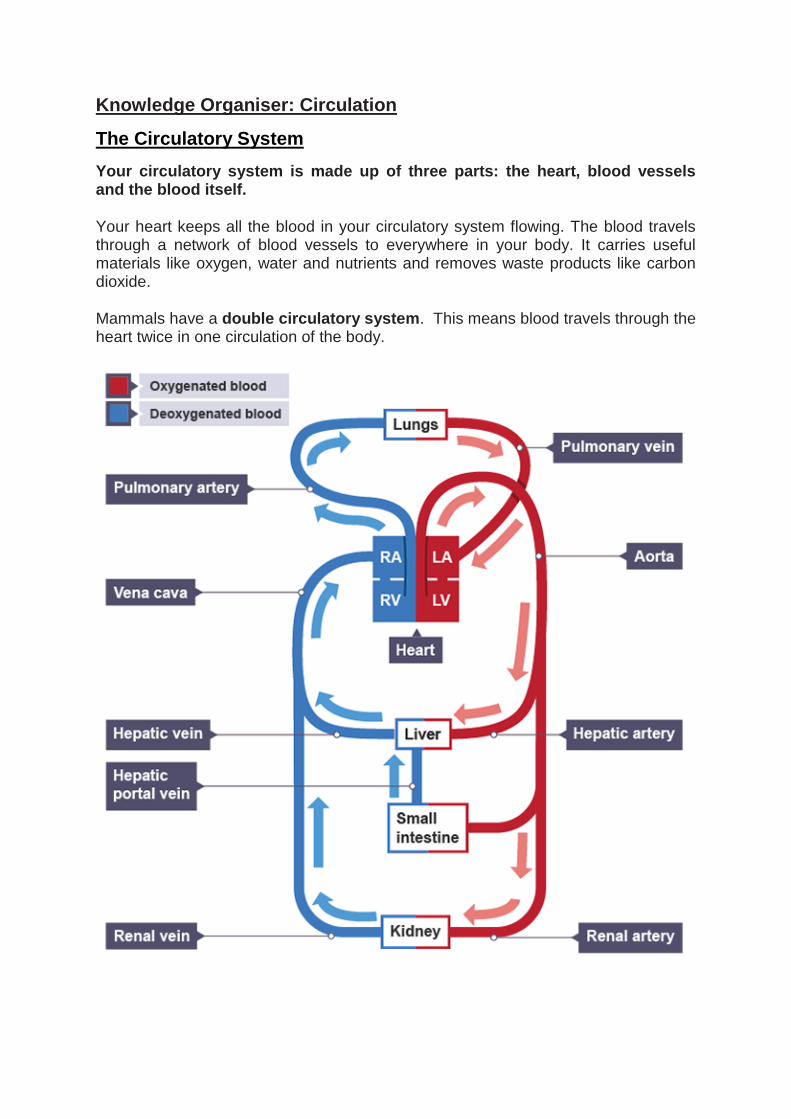
Your circulatory system is made up of three parts: the heart, blood vessels and the blood itself.
Your heart keeps all the blood in your circulatory system flowing. The blood travels through a network of blood vessels to everywhere in your body. It carries useful materials like oxygen, water and nutrients and removes waste products like carbon dioxide.
Mammals have a double circulatory system. This means blood travels through the heart twice in one circulation of the body.
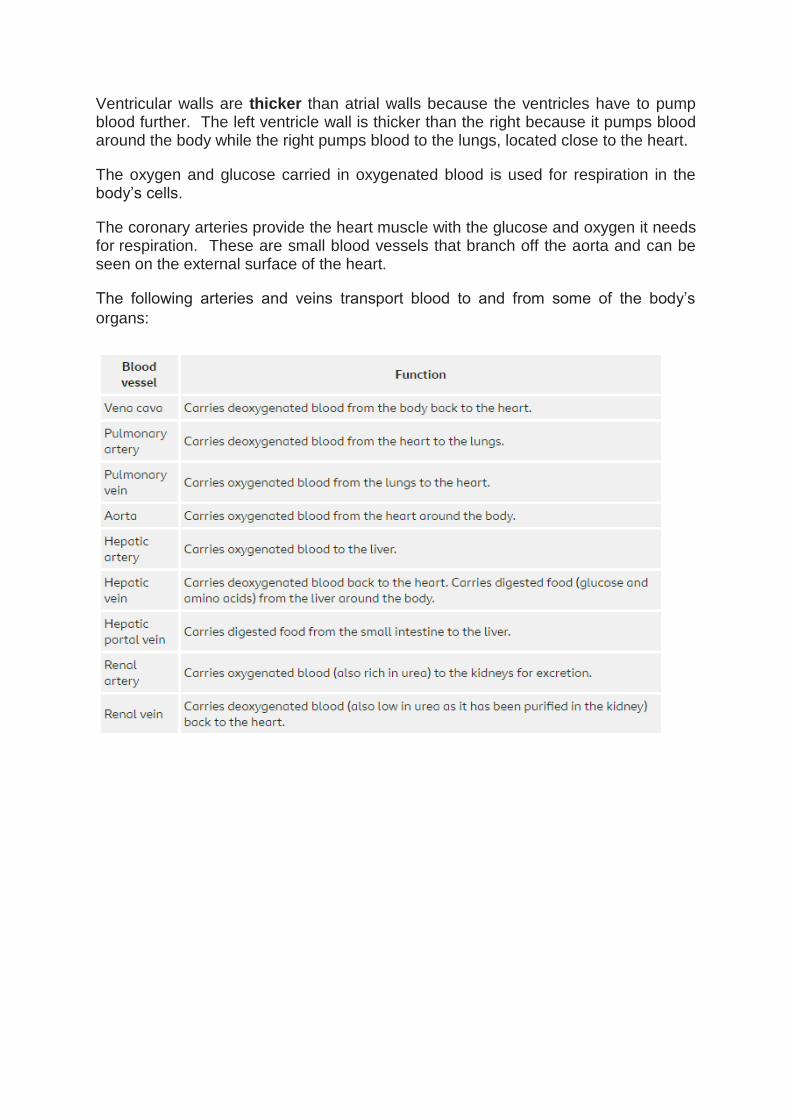
Ventricular walls are thicker than atrial walls because the ventricles have to pump blood further. The left ventricle wall is thicker than the right because it pumps blood around the body while the right pumps blood to the lungs, located close to the heart.
The oxygen and glucose carried in oxygenated blood is used for respiration in the body’s cells.
The coronary arteries provide the heart muscle with the glucose and oxygen it needs for respiration. These are small blood vessels that branch off the aorta and can be seen on the external surface of the heart.
The following arteries and veins transport blood to and from some of the body’s
organs:
The Heart
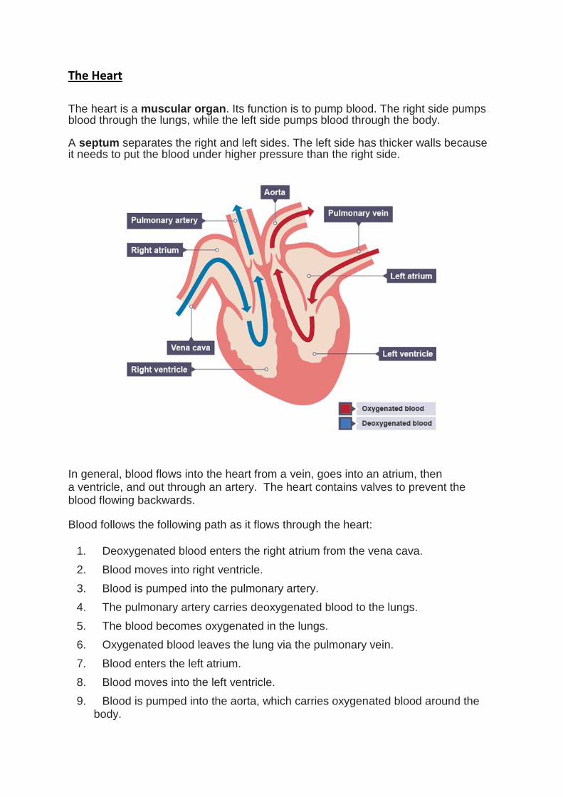
The heart is a muscular organ. Its function is to pump blood. The right side pumps blood through the lungs, while the left side pumps blood through the body. A septum separates the right and left sides. The left side has thicker walls because it needs to put the blood under higher pressure than the right side. In general, blood flows into the heart from a vein, goes into an atrium, then a ventricle, and out through an artery. The heart contains valves to prevent the blood flowing backwards.
Blood follows the following path as it flows through the heart:
1. Deoxygenated blood enters the right atrium from the vena cava.
2. Blood moves into right ventricle.
3. Blood is pumped into the pulmonary artery.
4. The pulmonary artery carries deoxygenated blood to the lungs.
5. The blood becomes oxygenated in the lungs.
6. Oxygenated blood leaves the lung via the pulmonary vein.
7. Blood enters the left atrium.
8. Blood moves into the left ventricle.
9. Blood is pumped into the aorta, which carries oxygenated blood around the body.
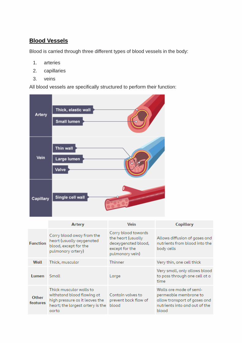
Blood Vessels Blood is carried through three different types of blood vessels in the body:
1. arteries
2. capillaries
3. veins
All blood vessels are specifically structured to perform their function:
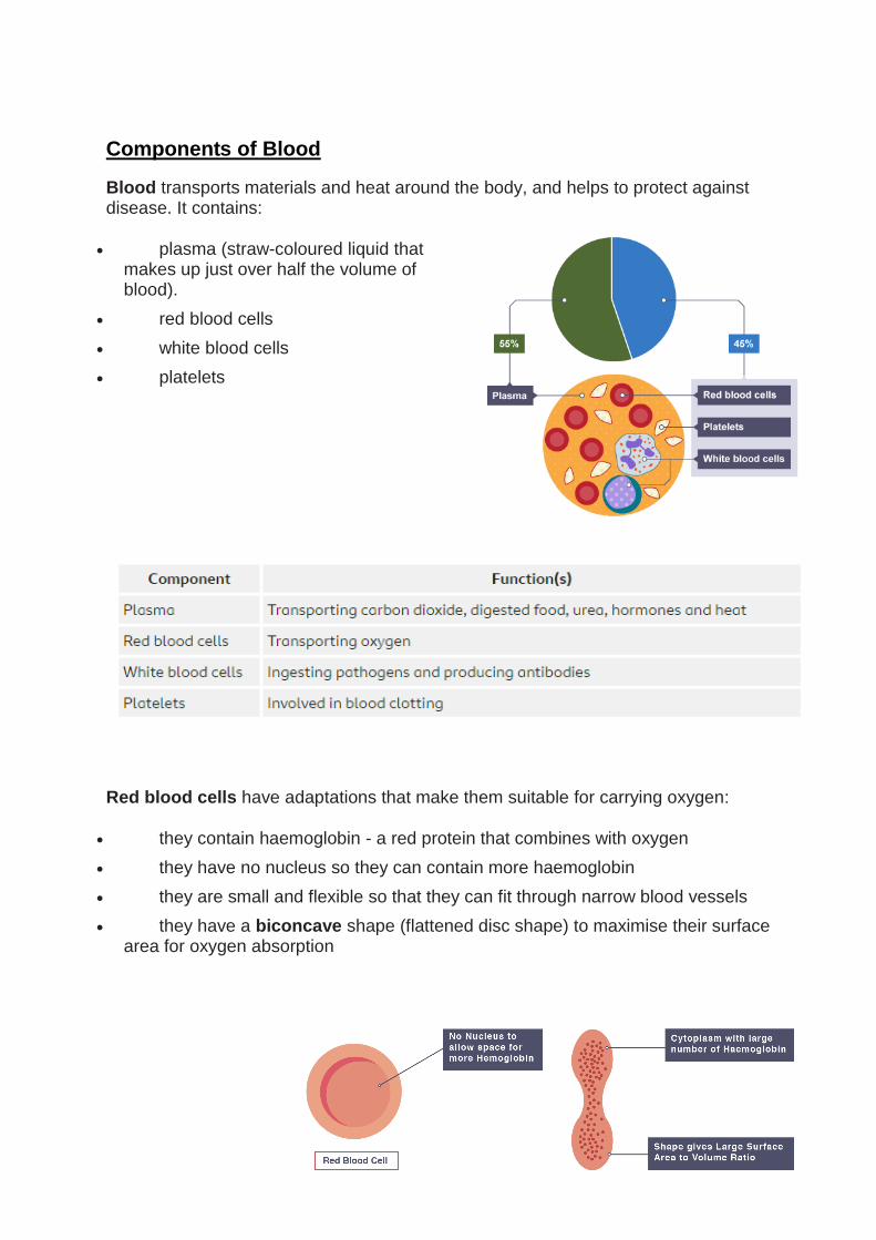
Components of Blood Blood transports materials and heat around the body, and helps to protect against disease. It contains:
plasma (straw-coloured liquid that makes up just over half the volume of blood).
red blood cells
white blood cells
platelets
Red blood cells have adaptations that make them suitable for carrying oxygen:
they contain haemoglobin - a red protein that combines with oxygen
they have no nucleus so they can contain more haemoglobin
they are small and flexible so that they can fit through narrow blood vessels
they have a biconcave shape (flattened disc shape) to maximise their surface area for oxygen absorption

Heart Disease Coronary heart disease
The heart is a muscular pump. Like all muscles, the heart needs oxygen to carry out aerobic respiration which provides the energy it needs to contract. The coronary arteries supply blood, and therefore oxygen, to the heart muscle.
Risk factors for cardiovascular disease
The risk of developing cardiovascular disease is increased by several factors, including:
smoking
high blood pressure
high levels of salt in the diet
high levels of saturated fat in the diet
High levels of salt in the diet can lead to increased blood pressure. This may damage the blood vessels, making it easier for fatty deposits to build up.
Heart attacks
A heart attack can happen after a sequence of events:
1. high levels of saturated fats in the diet are linked to an increase in levels of cholesterol in the blood
2. high levels of cholesterol cause fatty deposits to build up in the coronary arteries
3. a blood clot can form on a fatty deposit
4. the blood clot can block a coronary artery
5. some heart muscle cells do not get the oxygen and nutrients they need
6. the person develops chest pain
7. if left untreated then the cells start to die
8. this leads to a heart attack


![Homework Booklet [B]](https://img.pdfslide.us/doc/110x75/55cf926d550346f57b9672b7/homework-booklet-b.jpg)