Embed Size (px)
Citation preview
See discussions, stats, and author profiles for this publication at: https://www.researchgate.net/publication/237632176
XRD characterization of cobalt-based historical pigments and glazes
Article in Chemija · January 2009
CITATIONS
17READS
2,578
4 authors:
Some of the authors of this publication are also working on these related projects:
Layered double hydroxides - NANOCONCOR View project
Piezoelectric materials, multiferroic materials, La based chromates, manganates View project
Jonas Kiuberis
Vilnius University
7 PUBLICATIONS 69 CITATIONS
SEE PROFILE
Aivaras Kareiva
Vilnius University
401 PUBLICATIONS 4,869 CITATIONS
SEE PROFILE
Remigijus Juškėnas
Center for Physical Sciences and Technology
157 PUBLICATIONS 2,045 CITATIONS
SEE PROFILE
R. Ramanauskas
Center for Physical Sciences and Technology
89 PUBLICATIONS 1,412 CITATIONS
SEE PROFILE
All content following this page was uploaded by Aivaras Kareiva on 04 January 2014.
The user has requested enhancement of the downloaded file.

chemija. 2009. vol. 20. No. 1. P. 10–18© lietuvos mokslų akademija, 2009© lietuvos mokslų akademijos leidykla, 2009
* corresponding author. e-mail: [email protected]
XRD characterization of cobalt-based historical pigments and glazes
Dalia Jonynaitė1,
Jūratė Senvaitienė1,
Jonas Kiuberis1,
Aivaras Kareiva1*,
Remigijus Juškėnas2,
Rimantas Ramanauskas2
1 Faculty of Chemistry, Vilnius University, Naugarduko 24, LT-03225 Vilnius, Lithuania
2 Institute of Chemistry, A. Goštauto 9, LT-01108 Vilnius, Lithuania
This article covers the results of application of X-ray powder diffraction (XRD) in assess-ing the chemical and phase composition of historical pigments and their glazes. In order to demonstrate the reliability of the above technique, XRD analyses were performed on representative samples of the cobalt-based pigments and glazes. Ten individual pigments (Kremer Pigmente) were used for X-ray diffraction characterization: cobalt yellow, cobalt violet brilliant light, cobalt violet dark, cobalt cerulean blue, cobalt blue dark, cobalt blue light, cobalt blue greenish, cobalt green bluish, and two specimens of smalt with different grind. The same pigments, along with lead oxide (Pb3O4) and silica (SiO2), were used for the preparation of cobalt-based glazes. XRD analysis has proven to be a useful tool for the qualitative determination of the composition of glazes. However, in some cases many problems concerning the identification of separate phases are still to be solved using other analytical techniques.
Key words: pigments, glazes, cobalt-based, X-ray diffraction characterization
INTRODUCTION
Cobalt-based ceramic pigments are widely used for col-oured glazes in the ceramic industry for floor or wall white-wares, and also in the bulk coloration of polished, unglazed, porcelainized stoneware. They are characterized by a high resistance with respect to light, environment, high tempera-ture and chemicals. These pigments are also used in many industries because of their different colour, fine particle size, good hiding power, acid acceptance and compatibility with many organic and inorganic systems. The pallete of their colours is very large: blue, green, yellow, violet, brown and black [1–3].
Recently, new cobalt-based ceramic pigments have been synthesized using different synthetic approaches. A poly-crystalline material with the qualities of a blue pigment has been obtained at low temperatures in the CoO–ZnO–SiO2 system by the sol-gel technology [4]. The classical Co olivine blue pigment (Co2SiO4) and Co-doped willemite
(Co0.05Zn1.95SiO4) were prepared by the traditional solid state reaction method [5, 6]. Blue cobalt aluminate (CoAl2O4) and purple pyroborate (Co2B2O5) were prepared by the solution combustion method [7]. Recently, a blue pearlescent pig-ment was obtained by coating microemulsion-synthesized CoAl2O4 nanoparticles onto mica titania [8]. Cobalt-doped alumina powders were synthesized by the polymeric precur-sor method to obtain a ceramic pigment [9]. The Co3(PO4)2 and FePO4 solid solutions (Co3-xFexP2O8+x/2) were synthe-sized by the chemical coprecipitation method [10]. The pos-sibility of using cobalt molybdophosphates as pigments was also demonstrated [11].
There is a wide range of various historical cobalt pig-ments. Some of them were more important in the history of painting, others were more often used for decorating ceramic works or producing ceramic glaze [1, 3, 12–18]. For example, cobalt yellow (aureolin, K3[Co(NO2)6]) appeared in trade as a pigment in 1860. In those years it was the only steady bright yellow glazing colour used in the colour assortment together with the Indian yellow. Because of the remarkable effect this pigment is perfectly suitable for various painting techniques:

11XRD characterization of cobalt-based historical pigments and glazes
aquarelle, tempera and oil. Also, it is used in glass, porce-lain painting, and enamel. The most important violet cobalt pigments are different phosphates: cobalt violet brilliant light (CoNH4PO4 · H2O) and cobalt violet dark (Co3(PO4)2). These pigments have been known since the end of the 18th century. Cobalt cerulean blue (CoO · nSnO2) was discovered in 1789. This pigment was used in enamel art and porcelain painting. Cobalt blue dark and cobalt blue light (CoO · Al2O3) pigments were already used in ancient Egypt for decorat-ing pottery. Cobalt blue greenish (Cr2O3 · CoO · Al2O3) was produced in the middle of the 19th century and used for decorating porcelain. The colour of pigment varies from blu-ish green to greenish blue depending on the molar ratio of aluminium and chromium. Cobalt green bluish (CoO · ZnO) was synthesized in 1778 and became more widely used in the 19th century. The pigment is resistant to light and suit-able for all painting techniques. The blue cobalt pigment smalt (CoO · nSiO2) is also of significant importance in art history. It was known and used as a pigment already in the ancient world, in Egypt and Mesopotamia. In the 17th cen-tury, the smalt became one of the basic blue pigments in ba-roque style painting.
Recently, in order to demonstrate the reliability of the X-ray powder diffraction (XRD) technique, XRD analyses were performed on representative samples of lead oxide (Pb3O4 or PbCO3 · Pb(OH)2) based pigments and glazes [19]. The glaze compositions contained silica and calcite as the main constituents and pigments lead-tin yellow, smalt, Ve-rona green, manganese black, Naples yellow and malachite as secondary phases. XRD analysis has proven to be a useful tool for the qualitative determination of the composition of glazes. In the present study, attention has been focused on the characterization of cobalt-based pigments and glazes using X-ray diffraction analysis. The identification of pigments in their mixtures or on unknown ceramic samples is very im-portant not only for the characterization of materials, but also for non-destructive conservation and successful restora-tion, dating and authentication [20–22].
EXPERIMENTAL
Ten analytical grade individual pigments (Kremer Pig-mente) were used for the X-ray diffraction characteriza-tion: K3[Co(NO2)6] · 3H2O (cobalt yellow, Aureolin), CoNH4-PO4 · H2O (cobalt violet brilliant light), Co3(PO4)2 (cobalt violet dark), CoO · nSnO2 (cobalt cerulean blue), CoO · Al2O3 (cobalt blue dark), CoO · Al2O3 (cobalt blue light), Cr2O3 · CoO · Al2O3 (cobalt blue greenish), CoO · ZnO (cobalt green bluish), CoO · nSiO2 (smalt with different grind: 120 µm and 80 µm). The same pigments along with lead oxide (Pb3O4) and silica (SiO2) were used for the preparation of cobalt-based glazes. In all cases the same molar ratio of ingredients has been se-lected Pb3O4 : SiO2 : pigment = 2.85 : 1.9 : 0.25.
The pigments and prepared glazes were characterized by X-ray powder diffraction (XRD) analysis. The XRD was per-formed with a D8 Bruker AXS powder diffractometer using CuKα1 radiation.
RESULTS AND DISCUSSION
The X-ray diffraction pattern of the cobalt yellow (aureo-lin) pigment is shown in Fig. 1. All diffraction peaks corre-sponds to a standard diffractogram of K3[Co(NO2)6] (PDF [9–404]. The XRD pattern of cobalt yellow based glaze is shown in Fig. 2. Most of the diffraction lines seen in Fig. 2 could be attributed to the Pb3O4 phase (PDF [41–1493]). Be-sides, the most intensive line from silica at 2θ ≈ 20.8° (PDF [34–717]) is also seen. However, no signals from cobalt yellow (aureolin) pigment, likely at 2θ ≈ 34.6°, 24.5° and 42.8°, could be detected in the XRD pattern of the cobalt yellow based glaze.
The X-ray diffraction pattern of the cobalt violet bril-liant light pigment is a typical XRD pattern of CoNH4-PO4 · H2O (Fig. 3) (PDF [21–793]). The XRD pattern of the cobalt violet brilliant light based glaze is shown in Fig. 4. The main diffraction peak of the cobalt violet bril-liant light pigment located at 2θ ≈ 10.0° is seen in Fig. 4.
Fig. 1. X-ray diffraction pattern of cobalt yellow (aureolin) pigment

Dalia Jonynaitė, Jūratė Senvaitienė, Jonas Kiuberis, Aivaras Kareiva, Remigijus Juškėnas, Rimantas Ramanauskas12
Fig. 3. X-ray diffraction pattern of cobalt violet brilliant light pigment
Fig. 2. X-ray diffraction pattern of cobalt yellow based glaze
Fig. 4. X-ray diffraction pattern of cobalt violet brilliant light based glaze. (∗) – CoNH4PO4 · H2O
13XRD characterization of cobalt-based historical pigments and glazes
Therefore, we can conclude that, contrary to aureolin, the cobalt violet brilliant light pig-ment could be distinguished in its glaze by X-ray diffraction.
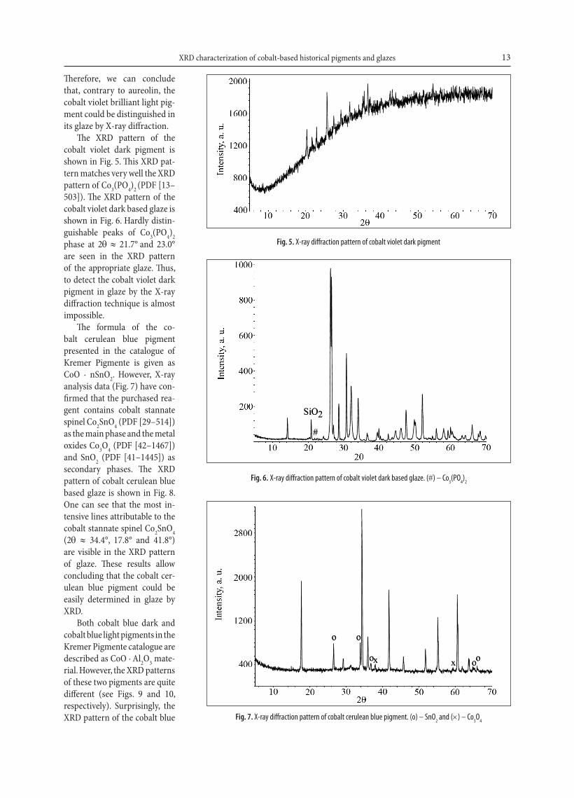
The XRD pattern of the cobalt violet dark pigment is shown in Fig. 5. This XRD pat-tern matches very well the XRD pattern of Co3(PO4)2 (PDF [13–503]). The XRD pattern of the cobalt violet dark based glaze is shown in Fig. 6. Hardly distin-guishable peaks of Co3(PO4)2 phase at 2θ ≈ 21.7° and 23.0°
are seen in the XRD pattern of the appropriate glaze. Thus, to detect the cobalt violet dark pigment in glaze by the X-ray diffraction technique is almost impossible.
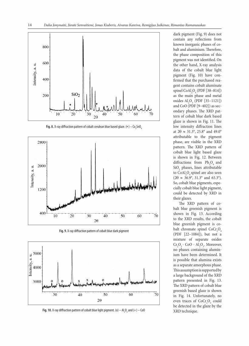
The formula of the co-balt cerulean blue pigment presented in the catalogue of Kremer Pigmente is given as CoO · nSnO2. However, X-ray analysis data (Fig. 7) have con-firmed that the purchased rea-gent contains cobalt stannate spinel Co2SnO4 (PDF [29–514]) as the main phase and the metal oxides Co3O4 (PDF [42–1467]) and SnO2 (PDF [41–1445]) as secondary phases. The XRD pattern of cobalt cerulean blue based glaze is shown in Fig. 8. One can see that the most in-tensive lines attributable to the cobalt stannate spinel Co2SnO4 (2θ ≈ 34.4°, 17.8° and 41.8°) are visible in the XRD pattern of glaze. These results allow concluding that the cobalt cer-ulean blue pigment could be easily determined in glaze by XRD.
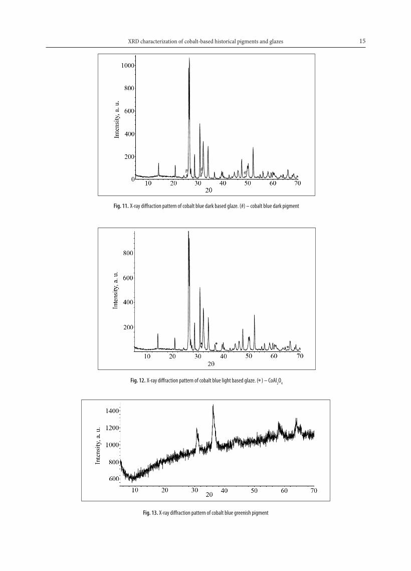
Both cobalt blue dark and cobalt blue light pigments in the Kremer Pigmente catalogue are described as CoO · Al2O3 mate-rial. However, the XRD patterns of these two pigments are quite different (see Figs. 9 and 10, respectively). Surprisingly, the XRD pattern of the cobalt blue Fig. 7. X-ray diffraction pattern of cobalt cerulean blue pigment. (o) – SnO2 and (×) – Co3O4
Fig. 5. X-ray diffraction pattern of cobalt violet dark pigment
Fig. 6. X-ray diffraction pattern of cobalt violet dark based glaze. (#) – Co3(PO4)2
Dalia Jonynaitė, Jūratė Senvaitienė, Jonas Kiuberis, Aivaras Kareiva, Remigijus Juškėnas, Rimantas Ramanauskas14
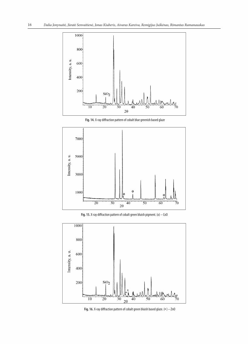
dark pigment (Fig. 9) does not contain any reflections from known inorganic phases of co-balt and aluminium. Therefore, the phase composition of this pigment was not identified. On the other hand, X-ray analysis data of the cobalt blue light pigment (Fig. 10) have con-firmed that the purchased rea-gent contains cobalt aluminate spinel CoAl2O4 (PDF [38–814]) as the main phase and metal oxides Al2O3 (PDF [35–1121]) and CoO (PDF [9–402]) as sec-ondary phases. The XRD pat-tern of cobalt blue dark based glaze is shown in Fig. 11. The low intensity diffraction lines at 2θ ≈ 31.3°, 25.8° and 49.0° attributable to the pigment phase, are visible in the XRD pattern. The XRD pattern of cobalt blue light based glaze is shown in Fig. 12. Between diffractions from Pb3O4 and SiO2 phases, lines attributable to CoAl2O4 spinel are also seen (2θ ≈ 36.9°, 31.3° and 65.3°). So, cobalt blue pigments, espe-cially cobalt blue light pigment, could be detected by XRD in their glazes.
The XRD pattern of co-balt blue greenish pigment is shown in Fig. 13. According to the XRD results, the cobalt blue greenish pigment is co-balt chromate spinel CoCr2O4 (PDF [22–1084]), but not a mixture of separate oxides Cr2O3 ∙ CoO ∙ Al2O3. Moreover, no phases containing alumin-ium have been determined. It is possible that alumina exists as a separate amorphous phase. This assumption is supported by a large background of the XRD pattern presented in Fig. 13. The XRD pattern of cobalt blue greenish based glaze is shown in Fig. 14. Unfortunately, no even traces of CoCr2O4 could be detected in the glaze by the XRD technique.Fig. 10. X-ray diffraction pattern of cobalt blue light pigment. (o) – Al2O3 and (×) – CoO
Fig. 8. X-ray diffraction pattern of cobalt cerulean blue based glaze. (∗) – Co2SnO4
Fig. 9. X-ray diffraction pattern of cobalt blue dark pigment
15XRD characterization of cobalt-based historical pigments and glazes
Fig. 13. X-ray diffraction pattern of cobalt blue greenish pigment
Fig. 11. X-ray diffraction pattern of cobalt blue dark based glaze. (#) – cobalt blue dark pigment
Fig. 12. X-ray diffraction pattern of cobalt blue light based glaze. (∗) – CoAl2O4
Dalia Jonynaitė, Jūratė Senvaitienė, Jonas Kiuberis, Aivaras Kareiva, Remigijus Juškėnas, Rimantas Ramanauskas16
Fig. 15. X-ray diffraction pattern of cobalt green bluish pigment. (o) – CoO
Fig. 14. X-ray diffraction pattern of cobalt blue greenish based glaze
Fig. 16. X-ray diffraction pattern of cobalt green bluish based glaze. (∗) – ZnO
17XRD characterization of cobalt-based historical pigments and glazes
The XRD pattern of cobalt green bluish pigment cor-responds to the XRD pattern of ZnO (PDF [36–1451]) as the main phase along with diffraction lines from the CoO (PDF [43–1004]) phase (Fig. 15). These results are in good agreement with the nominal composition of the pigment (CoO ∙ ZnO). The XRD pattern of cobalt green bluish based glaze is shown in Fig. 16. The marked diffraction lines at 2θ ≈ 36.3° and 34.4° correspond to the ZnO phase. These results suggest that, contrary to the cobalt blue greenish pig-ment, the cobalt green bluish pigment could be distinguished in its glaze by X-ray diffraction.
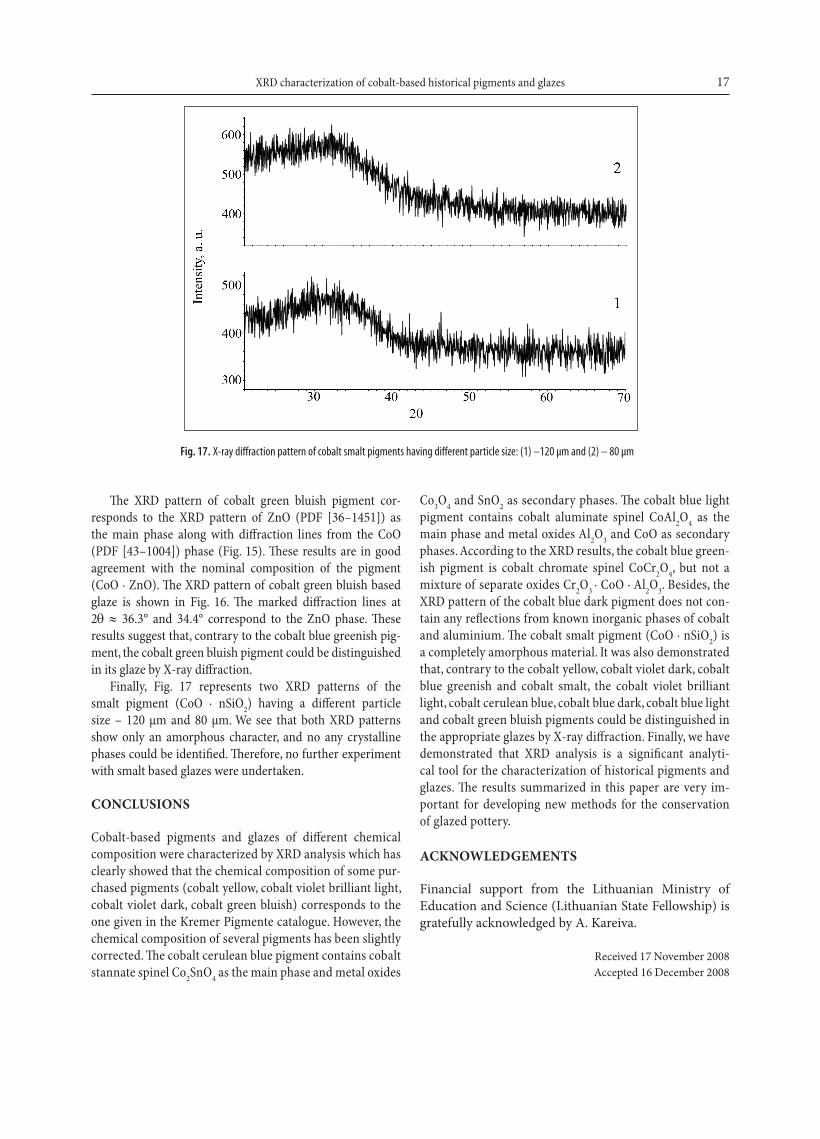
Finally, Fig. 17 represents two XRD patterns of the smalt pigment (CoO ∙ nSiO2) having a different particle size – 120 µm and 80 µm. We see that both XRD patterns show only an amorphous character, and no any crystalline phases could be identified. Therefore, no further experiment with smalt based glazes were undertaken.
CONCLUSIONS
Cobalt-based pigments and glazes of different chemical composition were characterized by XRD analysis which has clearly showed that the chemical composition of some pur-chased pigments (cobalt yellow, cobalt violet brilliant light, cobalt violet dark, cobalt green bluish) corresponds to the one given in the Kremer Pigmente catalogue. However, the chemical composition of several pigments has been slightly corrected. The cobalt cerulean blue pigment contains cobalt stannate spinel Co2SnO4 as the main phase and metal oxides
Co3O4 and SnO2 as secondary phases. The cobalt blue light pigment contains cobalt aluminate spinel CoAl2O4 as the main phase and metal oxides Al2O3 and CoO as secondary phases. According to the XRD results, the cobalt blue green-ish pigment is cobalt chromate spinel CoCr2O4, but not a mixture of separate oxides Cr2O3 ∙ CoO ∙ Al2O3. Besides, the XRD pattern of the cobalt blue dark pigment does not con-tain any reflections from known inorganic phases of cobalt and aluminium. The cobalt smalt pigment (CoO ∙ nSiO2) is a completely amorphous material. It was also demonstrated that, contrary to the cobalt yellow, cobalt violet dark, cobalt blue greenish and cobalt smalt, the cobalt violet brilliant light, cobalt cerulean blue, cobalt blue dark, cobalt blue light and cobalt green bluish pigments could be distinguished in the appropriate glazes by X-ray diffraction. Finally, we have demonstrated that XRD analysis is a significant analyti-cal tool for the characterization of historical pigments and glazes. The results summarized in this paper are very im-portant for developing new methods for the conservation of glazed pottery.
ACKNOwLEDgEMENTS
Financial support from the lithuanian ministry of education and Science (lithuanian State Fellowship) is gratefully acknowledged by a. Kareiva.
Received 17 November 2008 accepted 16 December 2008
Fig. 17. X-ray diffraction pattern of cobalt smalt pigments having different particle size: (1) –120 µm and (2) – 80 µm
Dalia Jonynaitė, Jūratė Senvaitienė, Jonas Kiuberis, Aivaras Kareiva, Remigijus Juškėnas, Rimantas Ramanauskas18
References
1. h. endriss, Aktuelle anorganische Bunt-Pigmente, vincentz Network, hannover (1974).
2. N. m. ahmed, a. attia, m. m. Selim, Anti-Corros. Methods Mater., 52, 353 (2005).
3. a. Bouquillon, i. Katona, a. D’allesandro, a. Zucchiatti, Int. J. PIXE, 15, 317 (2005).
4. S. Djambazov, Y. ivanova, a. Yoleva, N. Nedelchev, Ceram. Int., 24, 281 (1998).
5. m. llusar, a. Fores, j. a. Badenes, j. calbo, m. a. Tena, G. monros, J. Europ. Ceram. Soc., 21, 1121 (2001).
6. a. Fores, m. llusar, j. a. Badenes, j. calbo, m. a. Tena, G. monros, Green Chem., 2, 93 (2000).
7. T. mimani, S. Ghosh, Current Sci., 78, 892 (2000). 8. j. chen, X. h. Shi, Dyes Pigm., 75, 766 (2007). 9. S. cava, S. m. Tebcherani, S. a. Pianaro, c. a. Paskocimas,
e. longo, j. a. varela, Mater. Chem. Phys., 97, 102 (2006).
10. S. meseguer, m. a. Tena, c. Gargori, j. a. Badenes, m. llu-sar, G. monros, Ceram. Int., 33, 843 (2007).
11. N. i. Radishevskaya, l. a. egorova, a. m. Shul’pekov, v. i. vereshchagin, Glass Ceram., 60, 21 (2003).
12. R. j. Gettens, G. l. Stout, Painting Materials. A Short Encyc-lopedia, Dover Publications, New York (1966).
13. j. Riederer, Archaeometry, 16, 102 (1974). 14. j. Riederer, Naturwissenschaften, 69, 82 (1982). 15. T. Rehren, Archaeometry, 43, 483 (2001). 16. m.-c. corbeil, j.-P. charland, e. a. moffatt, Studies in Con-
servation, 47, 237 (2002). 17. S. Bruhlmann, Cobaltblau Werkstoffgeschichte und Werkstof-
ftechnologie, hochschule der Kunste Bern, Bern (2003).
18. a. Zucchiatti, a. Bouquillon, i. Katona, a. D’alessandro, Archaeometry, 48, 131 (2006).
19. j. Senvaitiene, j. Smirnova, a. Beganskiene, a. Kareiva, Acta Chimica Slovenica, 54, 185 (2007).
20. l. Burgio, R. j. h. clark, S. Firth, Analyst, 126, 222 (2001). 21. G. D. Smith, R. j. h. clark, J. Cult. Herit., 3, 101 (2002). 22. m. a. legodi, D. de Waal, cryst. eng., 6, 287 (2003).
Dalia Jonynaitė, Jūratė Senvaitienė, Jonas Kiuberis, Aivaras Kareiva, Remigijus Juškėnas, Rimantas Ramanauskas
KOBALTO ISTORINIŲ PIgMENTŲ IR gLAZŪRŲ APIBŪDINIMAS RENTgENO SPINDULIŲ DIFRAKCINĖS ANALIZĖS METODU
S a n t r a u k aParodyta, kad Rentgeno spindulių difrakcinė analizė yra pakanka-mai efektyvus metodas kobalto pigmentams bei glazūroms apibū-dinti. Kobalto pigmentų (Kremer Pigmente, Vokietija) Rentgeno spindulių difrakcinė analizė akivaizdžiai parodė, kad kai kurių pig-mentų kataloguose pateikta cheminė sudėtis skiriasi nuo nusta-tytosios. Kobalto pigmentas ceruleumas yra Co2SnO4 (pagrindinė fazė), SnO2
ir Co3O4 mišinys, o ne CoO · nSnO2; kobalto mėlynasis šviesusis – CoAl2O4 (pagrindinė fazė), Al2O3 ir CoO mišinys, o ne CoO · Al2O3; kobalto melsvai žaliasis – CoCr2O4 (pagrindinė fazė), o ne Cr2O3 ∙ CoO ∙ Al2O3. Kobalto mėlynojo tamsiojo fazinė sudė-tis nieko bendro neturi su CoO · Al2O3. Nustatyta, kad Rentgeno spindulių difrakcinės analizės metodas tinka identifikuoti kobal-to šviesiai violetinį, ceruleumą, kobalto mėlynąjį šviesųjį, kobalto mėlynąjį tamsųjį ir kobalto žalsvai mėlynąjį jų glazūrose, tačiau netinkamas aureolino, kobalto tamsiai violetinio, kobalto melsvai žaliojo ir smaltos identifikavimui.
View publication statsView publication stats


![[1].pdf - ResearchGate](https://img.pdfslide.us/doc/110x75/62129a6cbbf9242e6965a6a7/1pdf-researchgate.jpg)