Embed Size (px)
Citation preview

XRAY SCENARIOS
Lisa Gow
February 2012
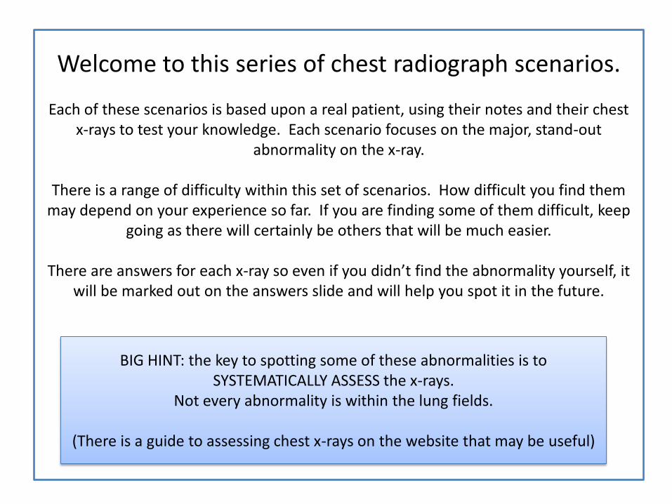
Welcome to this series of chest radiograph scenarios.
Each of these scenarios is based upon a real patient, using their notes and their chest x-rays to test your knowledge. Each scenario focuses on the major, stand-out
abnormality on the x-ray.
There is a range of difficulty within this set of scenarios. How difficult you find them may depend on your experience so far. If you are finding some of them difficult, keep
going as there will certainly be others that will be much easier.
There are answers for each x-ray so even if you didn’t find the abnormality yourself, it will be marked out on the answers slide and will help you spot it in the future.
BIG HINT: the key to spotting some of these abnormalities is to SYSTEMATICALLY ASSESS the x-rays.
Not every abnormality is within the lung fields.
(There is a guide to assessing chest x-rays on the website that may be useful)
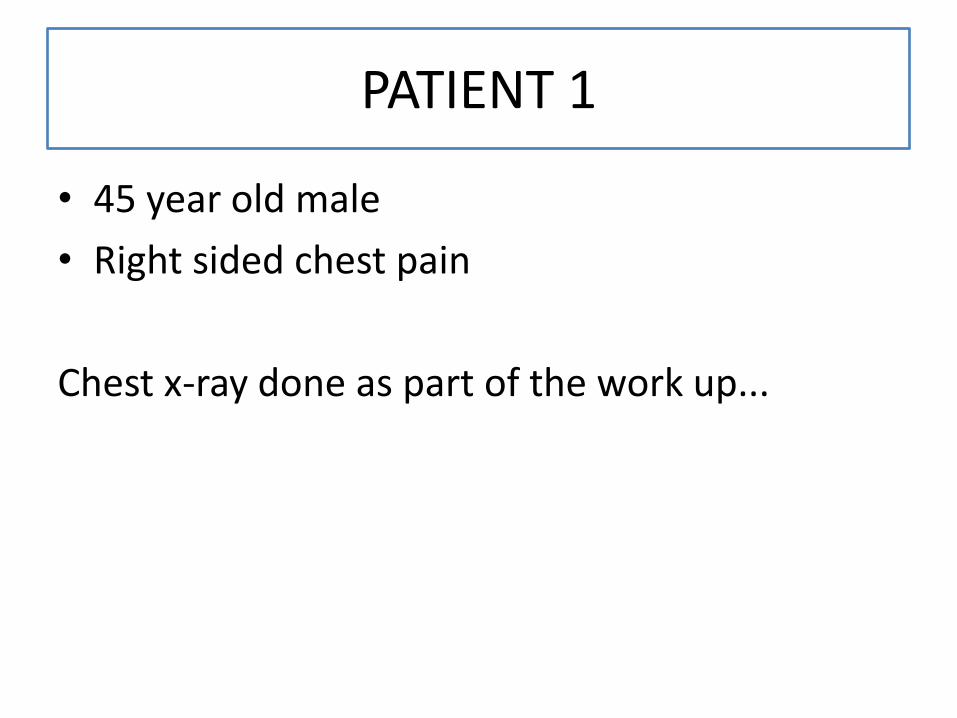
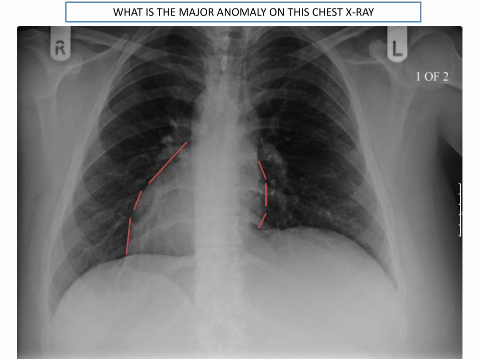
PATIENT 1
• 45 year old male
• Right sided chest pain
Chest x-ray done as part of the work up...

WHAT IS THE MAJOR ANOMALY ON THIS CHEST X-RAY
What does the x-ray show?
• Pneumonia
• Dextracardia
• Pneumothorax
• Right sided mass
What does the x-ray show?
X Pneumonia
Dextrocardia
X Pneumothorax
X Right sided mass
WHAT IS THE MAJOR ANOMALY ON THIS CHEST X-RAY
PATIENT 2
• 32 year old male
• Well
• New entrant to UK
• Mantoux screening test on entrance: 12mm
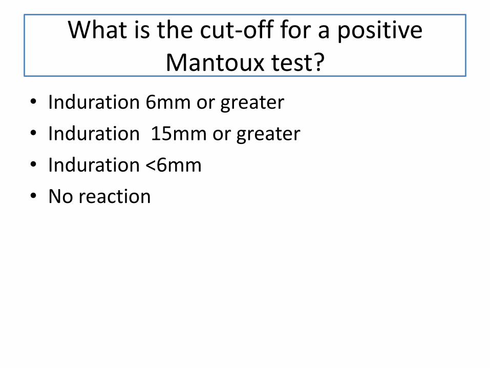
What is the cut-off for a positive Mantoux test?
• Induration 6mm or greater
• Induration 15mm or greater
• Induration <6mm
• No reaction
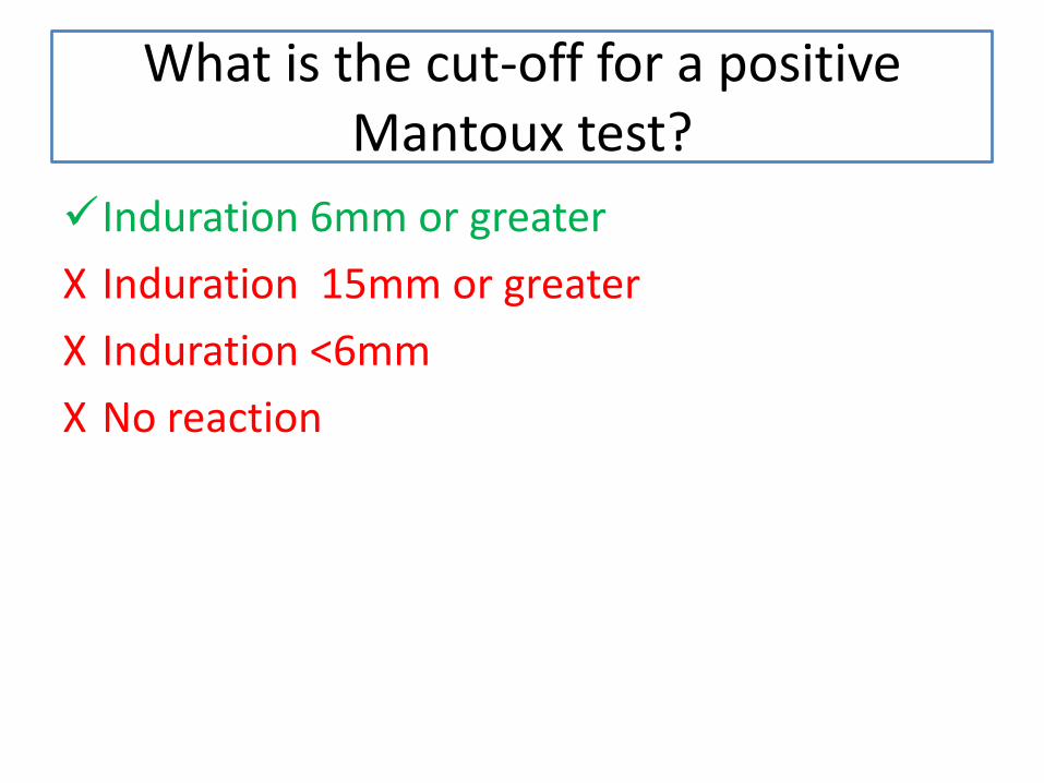
What is the cut-off for a positive Mantoux test?
Induration 6mm or greater
X Induration 15mm or greater
X Induration <6mm
X No reaction
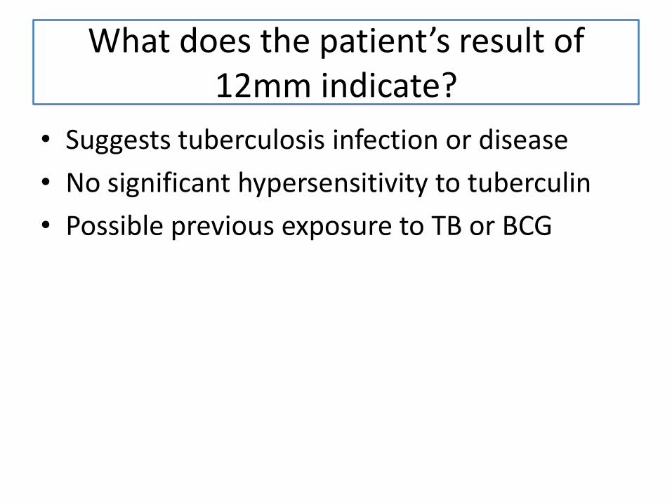
What does the patient’s result of 12mm indicate?
• Suggests tuberculosis infection or disease
• No significant hypersensitivity to tuberculin
• Possible previous exposure to TB or BCG
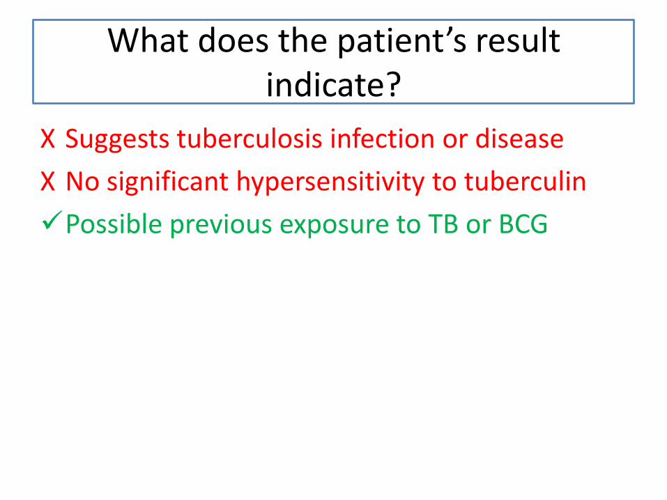
What does the patient’s result indicate?
X Suggests tuberculosis infection or disease
X No significant hypersensitivity to tuberculin
Possible previous exposure to TB or BCG
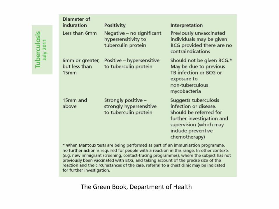
The Green Book, Department of Health
The patient was referred for a CXR as part of the new immigrant screening program.

Normal or abnormal?
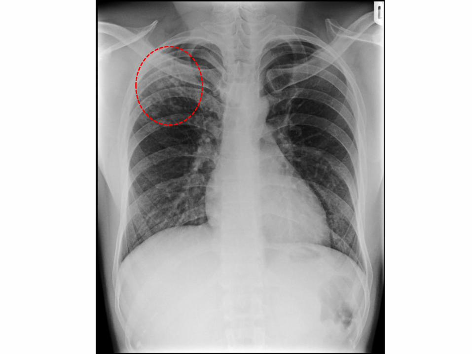
REPORT:
• Infiltration seen in the right upper lobe• Underlying tuberculosis is possible• Advice: chest clinic referral
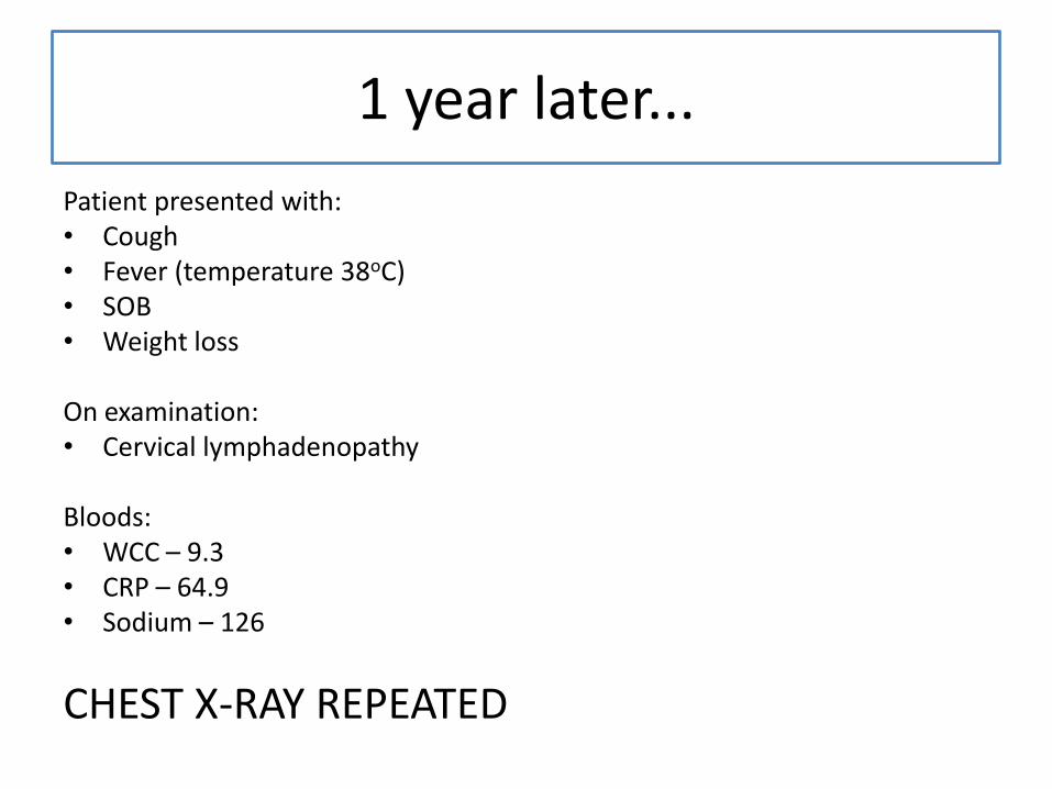
1 year later...
Patient presented with:• Cough• Fever (temperature 38oC)• SOB• Weight loss
On examination:• Cervical lymphadenopathy
Bloods:• WCC – 9.3• CRP – 64.9• Sodium – 126
CHEST X-RAY REPEATED

What does the x-ray show?
• Pulmonary oedema
• Primary lung cancer
• Metastatic lung cancer
• TB
• Fibrosis
• Pneumonia

What is the appropriate treatment?
• Chemotherapy
• Radiotherapy
• Piperacillin and gentamicin
• Isoniazid, rifampicin, pyrazinamine and ethambutol
• Furosemide
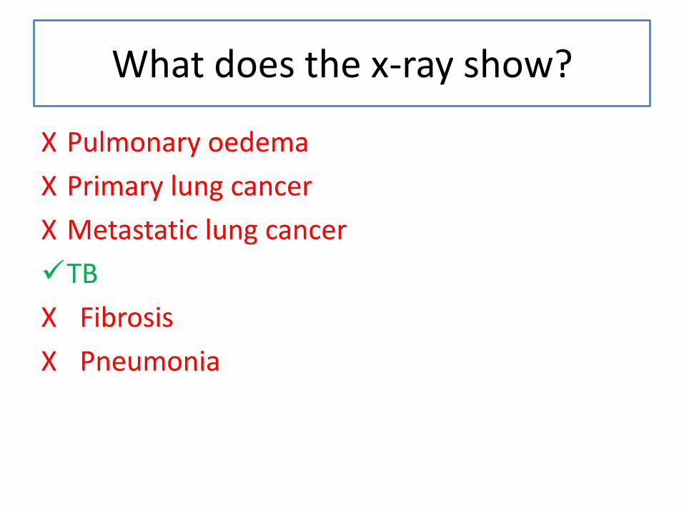
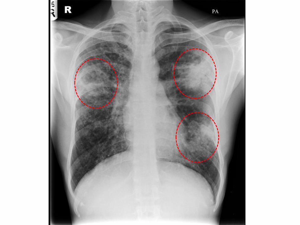
What does the x-ray show?
X Pulmonary oedema
X Primary lung cancer
X Metastatic lung cancer
TB
X Fibrosis
X Pneumonia

X Chemotherapy
X Radiotherapy
X Piperacillin and gentamicin
Isoniazid, rifampicin, pyrazinamine and ethambutol
X Furosemide
What is the appropriate treatment?
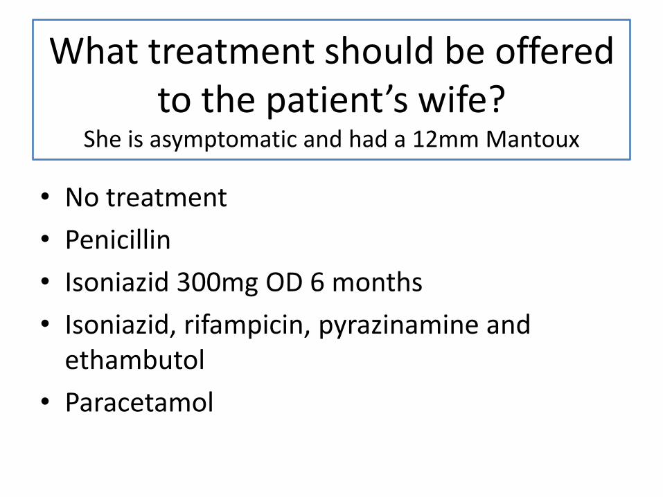
What treatment should be offered to the patient’s wife?
She is asymptomatic and had a 12mm Mantoux
• No treatment
• Penicillin
• Isoniazid 300mg OD 6 months
• Isoniazid, rifampicin, pyrazinamine and ethambutol
• Paracetamol

What treatment should be offered to the patient’s wife?
She is asymptomatic and had a 12mm Mantoux
X No treatment
X Penicillin
Isoniazid 300mg OD 6 months
X Isoniazid, rifampicin, pyrazinamine and ethambutol
X Paracetamol
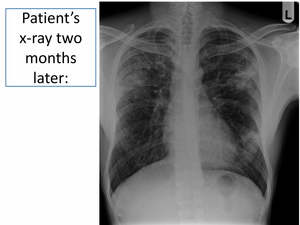
Patient’s x-ray two months
later:
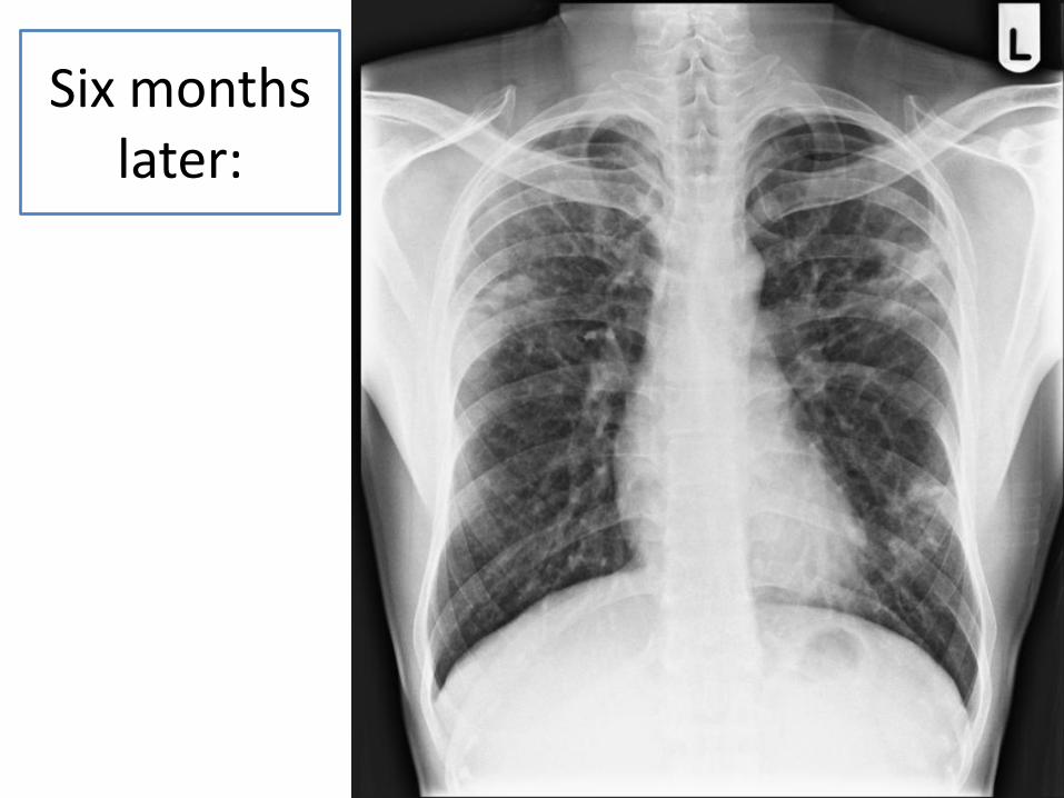
Six months later:
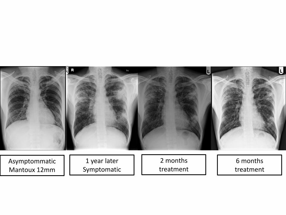
AsymptommaticMantoux 12mm
1 year laterSymptomatic
2 months treatment
6 months treatment

PATIENT 3
• 42 year old female
• Presented to A&E
• Severe left sided chest pain
• Fell 2m from a ladder
Chest x-ray taken...
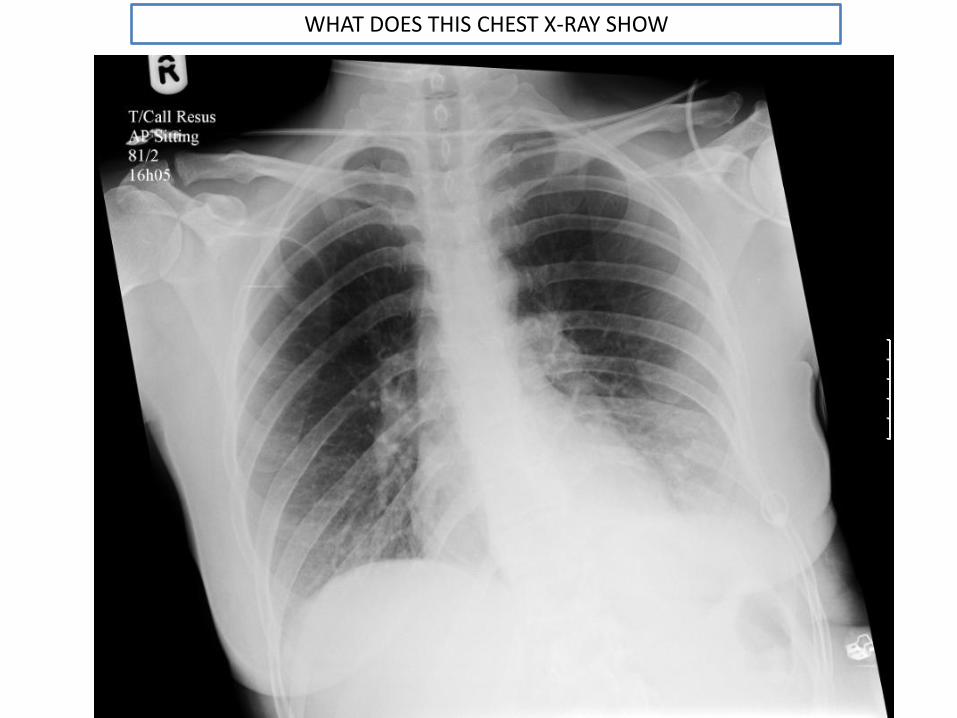
WHAT DOES THIS CHEST X-RAY SHOW
What does the x-ray show?Select all that apply
• Tension pneumothorax
• Pneumonia
• Fractured ribs on the right
• Fractured ribs on the left
• Haemothorax on the right
• Haemothorax on the left
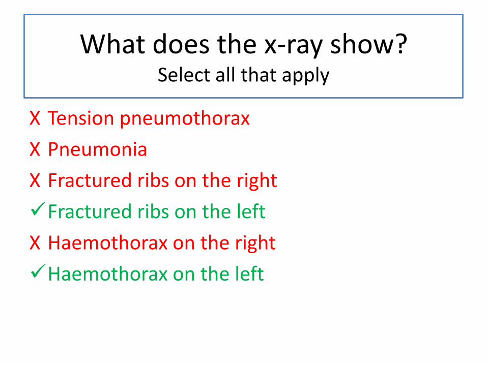
What does the x-ray show?Select all that apply
X Tension pneumothorax
X Pneumonia
X Fractured ribs on the right
Fractured ribs on the left
X Haemothorax on the right
Haemothorax on the left
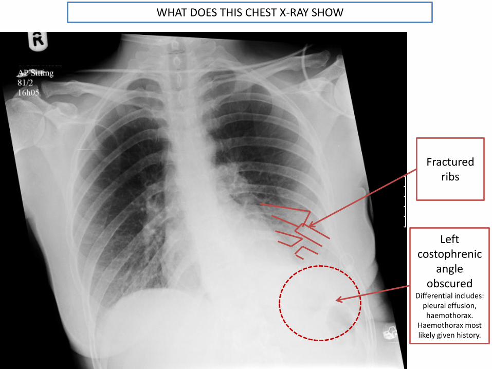
Fractured ribs
Left costophrenic
angle obscured
Differential includes: pleural effusion, haemothorax.
Haemothorax most likely given history.
WHAT DOES THIS CHEST X-RAY SHOW
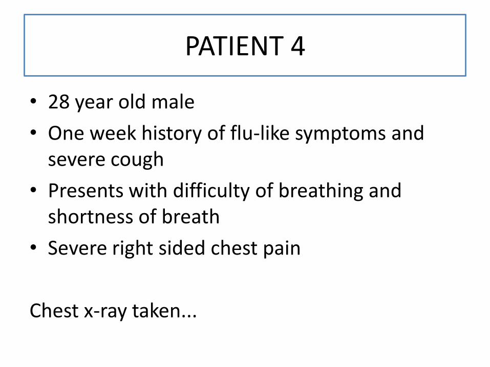
PATIENT 4
• 28 year old male
• One week history of flu-like symptoms and severe cough
• Presents with difficulty of breathing and shortness of breath
• Severe right sided chest pain
Chest x-ray taken...
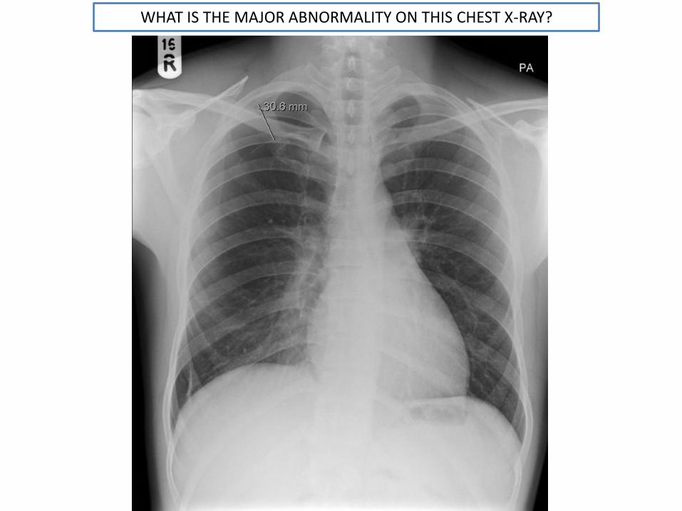
WHAT IS THE MAJOR ABNORMALITY ON THIS CHEST X-RAY?
What does the x-ray show?
• Foreign body on the right
• Foreign body on the left
• Pneumothorax on the right
• Pneumothorax on the left
• Fractured ribs on the right
• Fractured ribs on the left

What does the x-ray show?
X Foreign body on the right
X Foreign body on the left
Pneumothorax on the right
X Pneumothorax on the left
X Fractured ribs on the right
X Fractured ribs on the left

WHAT IS THE MAJOR ABNORMALITY ON THIS CHEST X-RAY?

WHAT KIND OF PNEUMOTHORAX IS THIS?
• Tension pneumothorax
• Simple pneumothorax
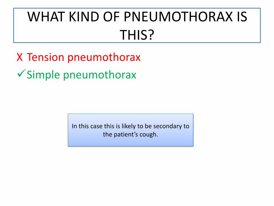
WHAT KIND OF PNEUMOTHORAX IS THIS?
X Tension pneumothorax
Simple pneumothorax
In this case this is likely to be secondary to the patient’s cough.
TREATMENT
• A chest drain was inserted and 900ml air was drained from the right chest.
• A chest x-ray was taken following drainage to assess response.....
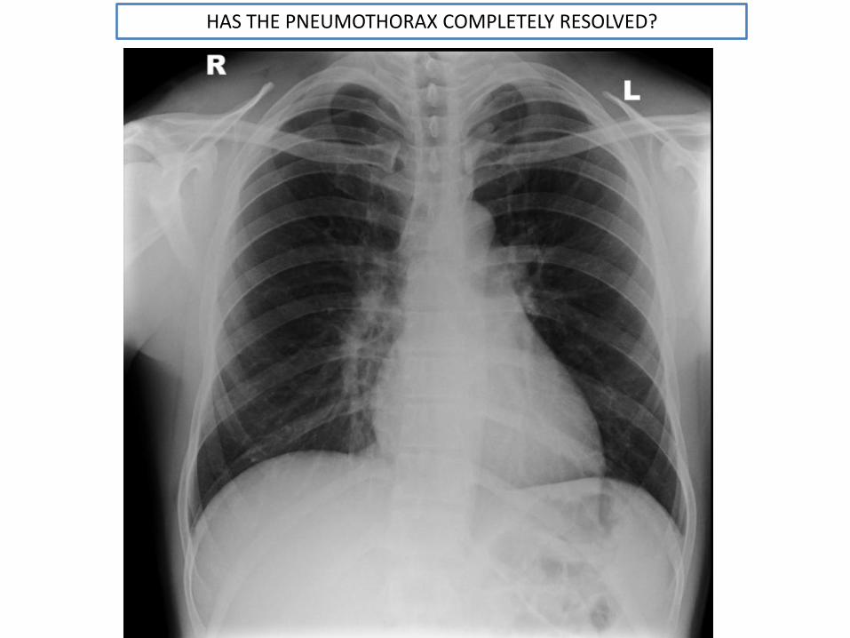
HAS THE PNEUMOTHORAX COMPLETELY RESOLVED?
HAS THE PNEUMOTHORAX COMPLETELY RESOLVED FOLLOWING
DRAINAGED?
• Yes
• No
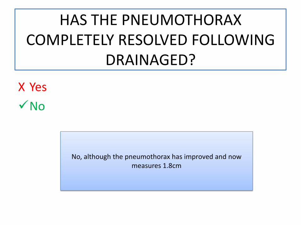
HAS THE PNEUMOTHORAX COMPLETELY RESOLVED FOLLOWING
DRAINAGED?
X Yes
No
No, although the pneumothorax has improved and now measures 1.8cm
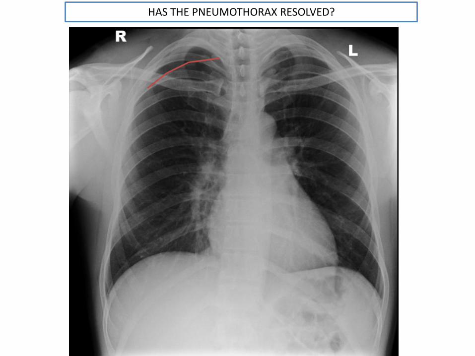
HAS THE PNEUMOTHORAX RESOLVED?
What sign on the chest x-ray would indicate a tension pneumothorax
• Mediastinal shift
• Air under the diaphragm
• Loss of the costophrenic angle
• Air in the mediastinum
What sign on the chest x-ray would indicate a tension pneumothorax
Mediastinal shift
X Air under the diaphragm
X Loss of the costophrenic angle
X Air in the mediastinum
WHAT IS THE EMERGENCY TREATMENT OF A TENSION PNEUMOTHORAX
• Needle thoracostomy in the 2nd intercostal space, mid-clavicular line
• Needle thoracostomy in the 2nd intercostal space, mid-axillary line
• Needle thoracostomy in the 5th intercostal space, mid-clavicular line
• Chest drain insertion
• Midline sternotomy
• Adrenaline IV
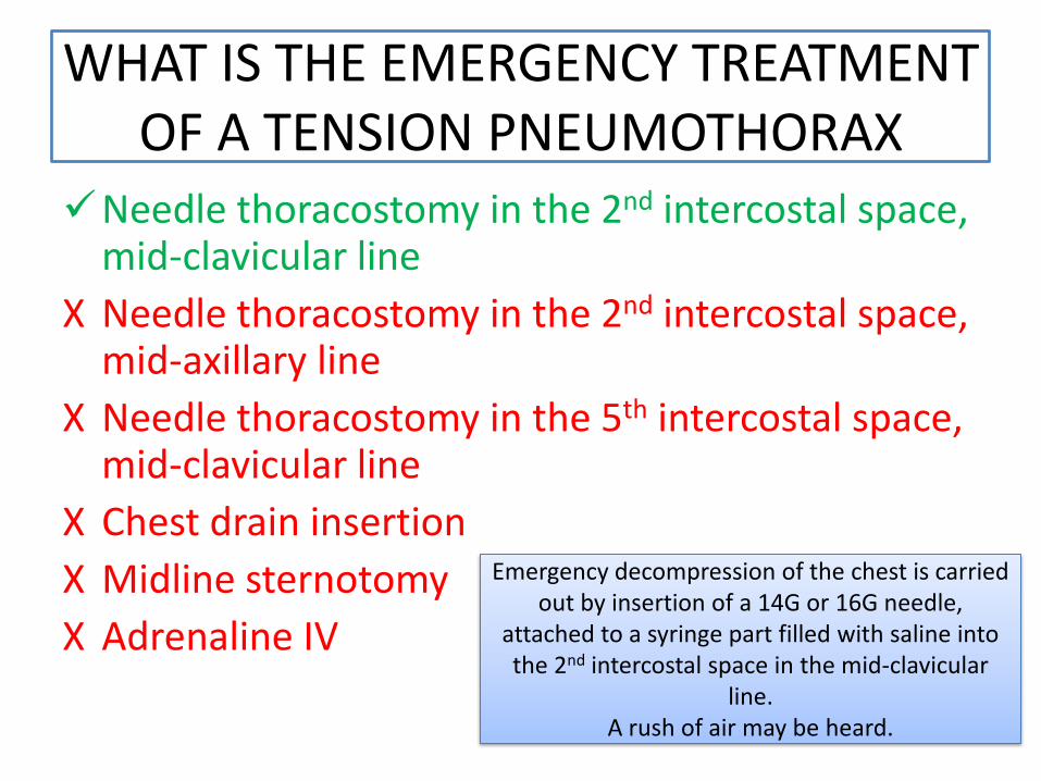
WHAT IS THE EMERGENCY TREATMENT OF A TENSION PNEUMOTHORAX
Needle thoracostomy in the 2nd intercostal space, mid-clavicular line
X Needle thoracostomy in the 2nd intercostal space, mid-axillary line
X Needle thoracostomy in the 5th intercostal space, mid-clavicular line
X Chest drain insertion
X Midline sternotomy
X Adrenaline IV
Emergency decompression of the chest is carried out by insertion of a 14G or 16G needle,
attached to a syringe part filled with saline into the 2nd intercostal space in the mid-clavicular
line.A rush of air may be heard.
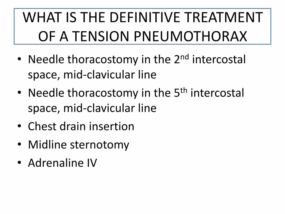
WHAT IS THE DEFINITIVE TREATMENT OF A TENSION PNEUMOTHORAX
• Needle thoracostomy in the 2nd intercostalspace, mid-clavicular line
• Needle thoracostomy in the 5th intercostalspace, mid-clavicular line
• Chest drain insertion
• Midline sternotomy
• Adrenaline IV

WHAT IS THE DEFINITIVE TREATMENT OF A TENSION PNEUMOTHORAX
X Needle thoracostomy in the 2nd intercostalspace, mid-clavicular line
X Needle thoracostomy in the 5th intercostalspace, mid-clavicular line
Chest drain insertion
X Midline sternotomy
X Adrenaline IVEmergency decompression of the chest by needle
thoracostomy converts a tension pneumothorax to a simple pneumothorax. This is a short term measure.
PATIENT 5
• 64 year old female
• Admitted following a fall
Chest x-ray taken....
Systematically assess this x-ray
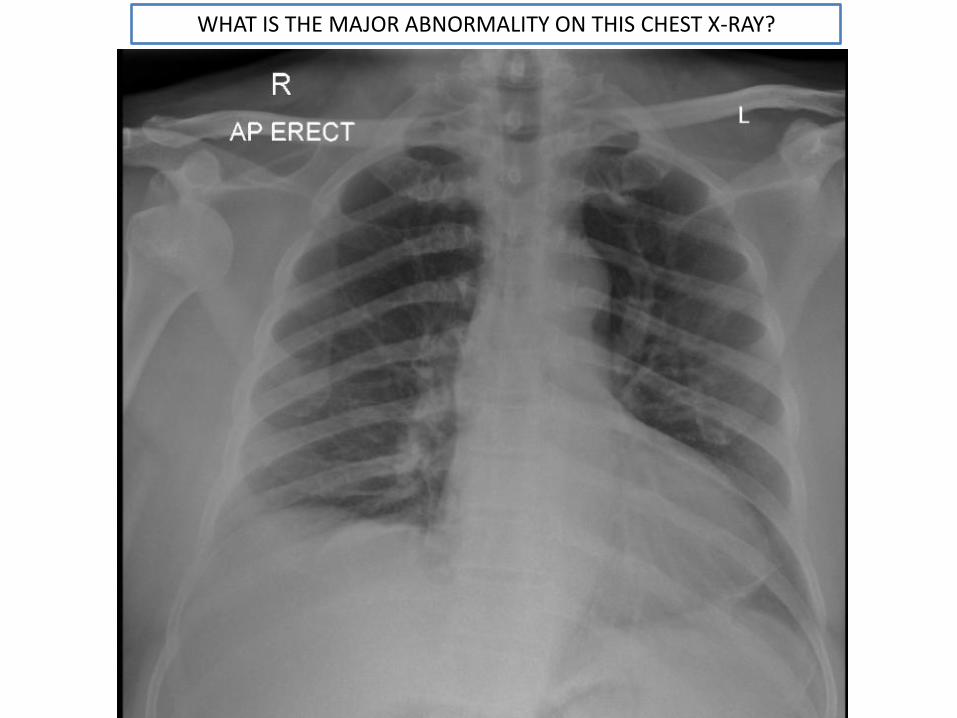
WHAT IS THE MAJOR ABNORMALITY ON THIS CHEST X-RAY?
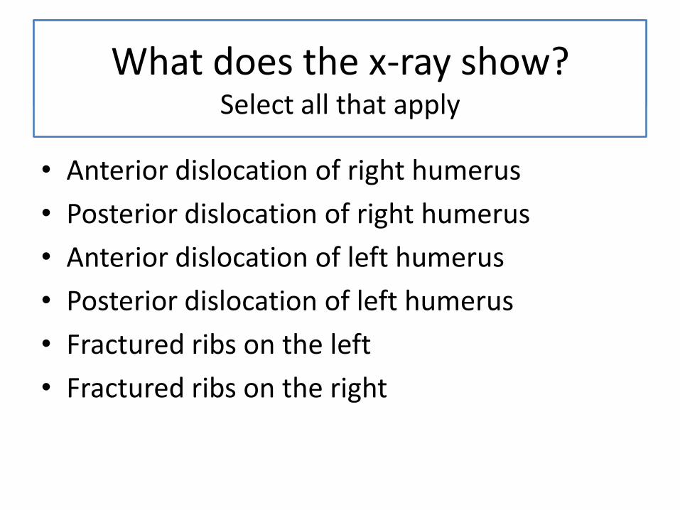
What does the x-ray show?
• Anterior dislocation of right humerus
• Posterior dislocation of right humerus
• Anterior dislocation of left humerus
• Posterior dislocation of left humerus
• Fractured ribs on the left
• Fractured ribs on the right
What does the x-ray show?Select all that apply
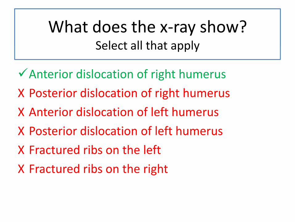
What does the x-ray show?
Anterior dislocation of right humerus
X Posterior dislocation of right humerus
X Anterior dislocation of left humerus
X Posterior dislocation of left humerus
X Fractured ribs on the left
X Fractured ribs on the right
What does the x-ray show?Select all that apply

WHAT IS THE MAJOR ABNORMALITY ON THIS CHEST X-RAY?
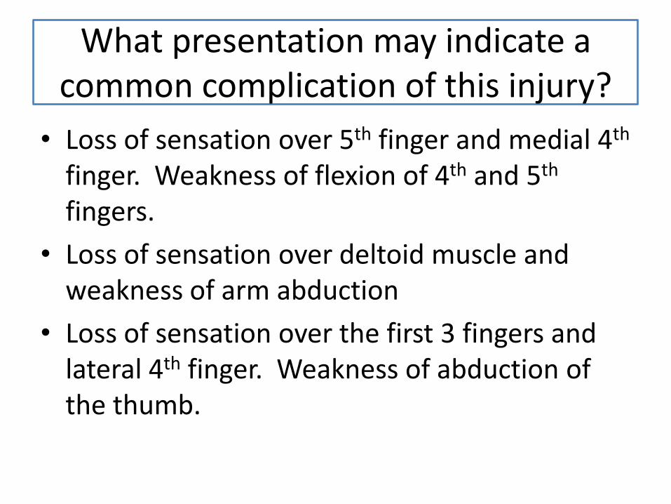
What presentation may indicate a common complication of this injury?
• Loss of sensation over 5th finger and medial 4th
finger. Weakness of flexion of 4th and 5th
fingers.
• Loss of sensation over deltoid muscle and weakness of arm abduction
• Loss of sensation over the first 3 fingers and lateral 4th finger. Weakness of abduction of the thumb.
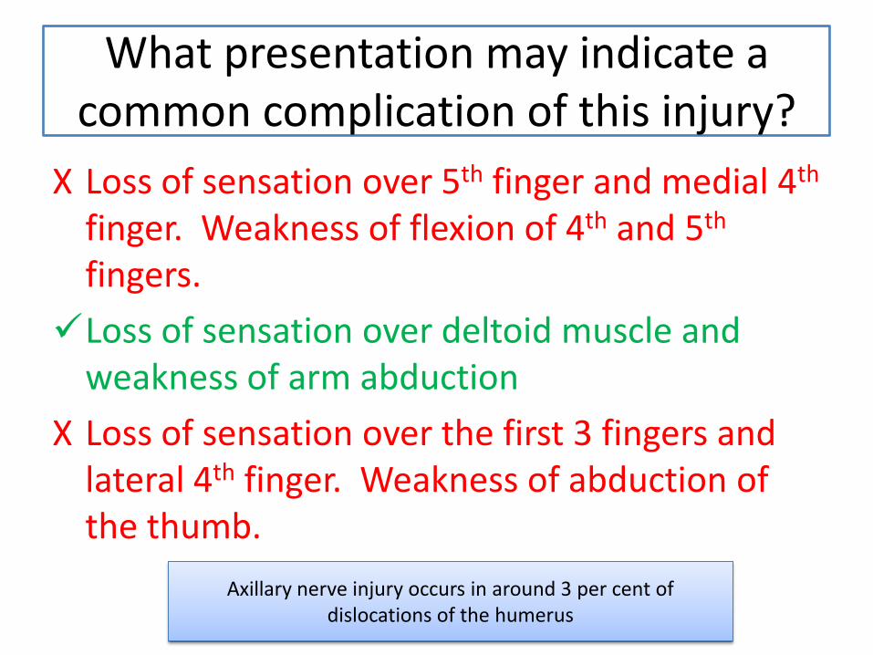
What presentation may indicate a common complication of this injury?
X Loss of sensation over 5th finger and medial 4th
finger. Weakness of flexion of 4th and 5th
fingers.
Loss of sensation over deltoid muscle and weakness of arm abduction
X Loss of sensation over the first 3 fingers and lateral 4th finger. Weakness of abduction of the thumb.
Axillary nerve injury occurs in around 3 per cent of dislocations of the humerus

PATIENT 6
INITIAL PRESENTATION• GP referral• 60 year old female• Smoker – 40 pack years• 1 month history of cough and malaise• 1 stone weight loss over past 3 months• Clubbing
The patient was investigated, diagnosed and treatment undertaken.
Following treatment, a routine CXR was taken....
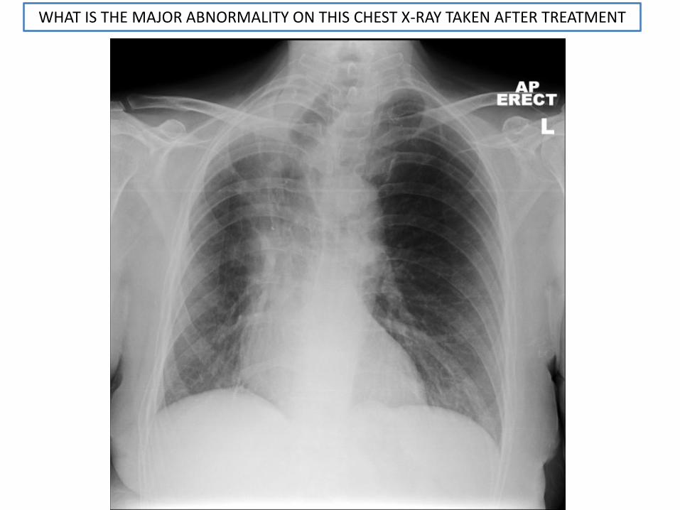
WHAT IS THE MAJOR ABNORMALITY ON THIS CHEST X-RAY TAKEN AFTER TREATMENT
WHAT IS THE MAJOR ABNORMALITY ON THIS CHEST X-RAY?
• Abdominal perforation
• Fractured ribs
• Tracheal deviation
• Cardiomegaly
• Dextrocardia

WHAT IS THE MAJOR ABNORMALITY ON THIS CHEST X-RAY?
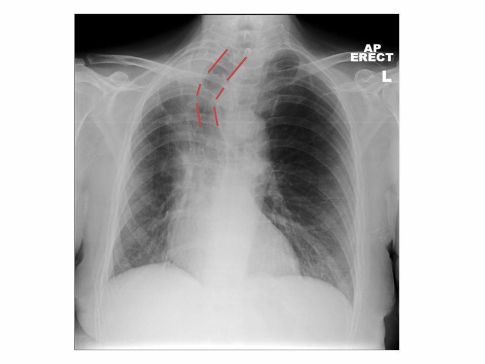
X Abdominal perforation
X Fractured ribs
Tracheal deviation
X Cardiomegaly
X Dextrocardia
What is the most likely cause of this patient’s post-treatment tracheal deviation in view of
the history?
• Pleural effusion
• Collapse
• Fibrosis
• Pneumothorax
• Retrosternal goitre
• Lobe resection
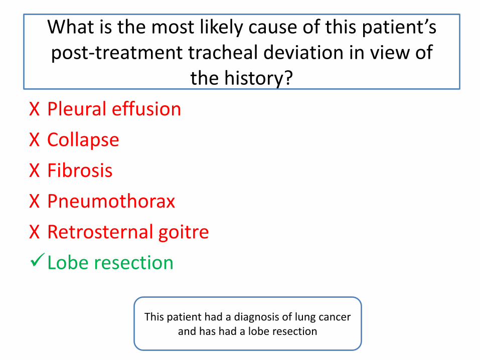
X Pleural effusion
X Collapse
X Fibrosis
X Pneumothorax
X Retrosternal goitre
Lobe resection
This patient had a diagnosis of lung cancer and has had a lobe resection
What is the most likely cause of this patient’s post-treatment tracheal deviation in view of
the history?
• 24 year old female
• Well
• Chest x-ray for occupational health check
PATIENT 7
Systematically assess this x-ray
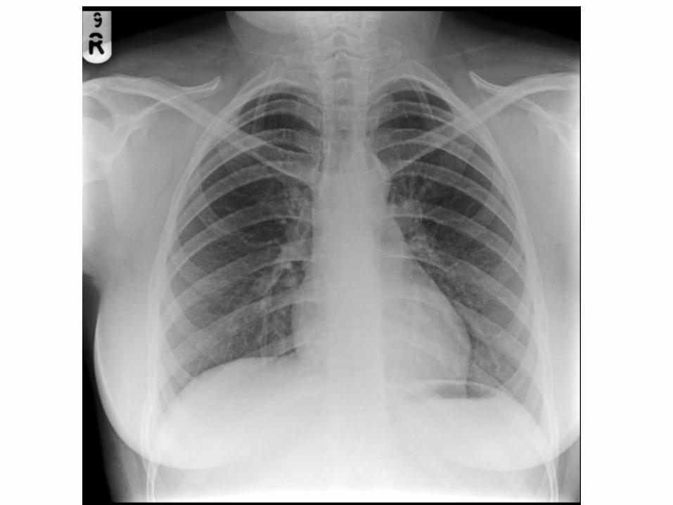
Is there an abnormality?
• No abnormality
• Abnormality within the lung fields
• Mediastinal abnormality
• Soft tissue abnormality
• Bony abnormality
Is there an abnormality?
X No abnormality
X Abnormality within the lung fields
X Mediastinal abnormality
X Soft tissue abnormality
Bony abnormality


CERVICAL RIBS
• Congenital abnormality
• 1 in 500 population
• Arise from 7th cervical vertebra
Potential complication: thoracic outlet obstruction. Compression of subclavianartery/ branches of brachial plexus.
PATIENT 8
• 39 year old male
• Retrosternal chest pain
• No radiation
• Worse on bending, stooping and lying down
• Worse with hot drinks or alcohol
• Relieved by antacids
Chest x-ray carried out as part of work-up....
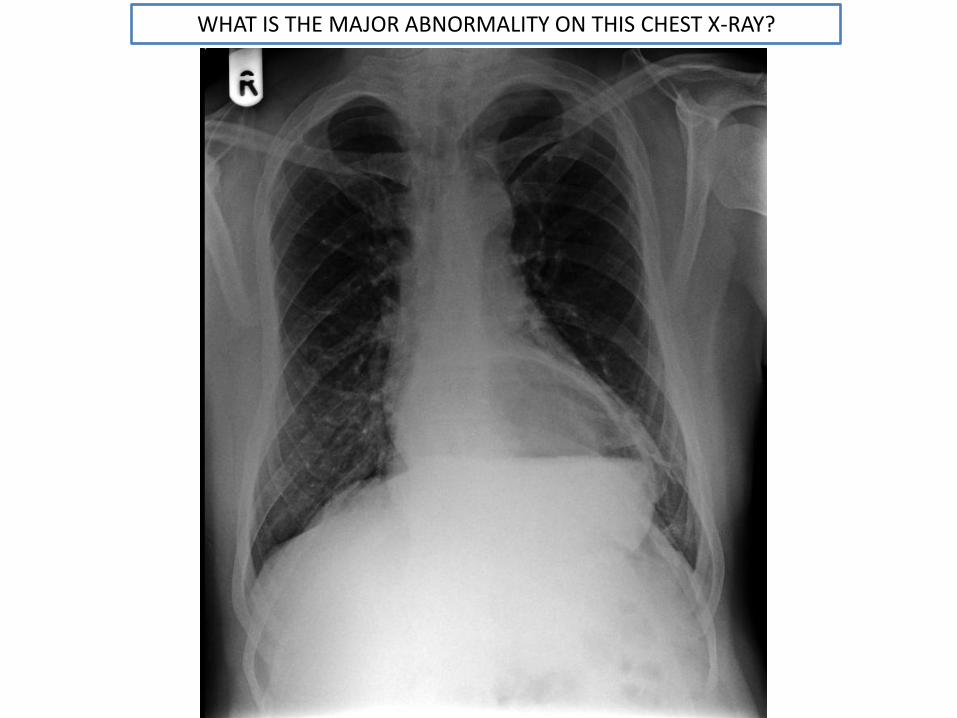
WHAT IS THE MAJOR ABNORMALITY ON THIS CHEST X-RAY?
WHAT IS THE DIAGNOSIS BASED ON THE HISTORY AND CHEST X-RAY?
• Pericarditis
• Cardiac tamponade
• Large hiatus hernia
• Aortic aneurysm
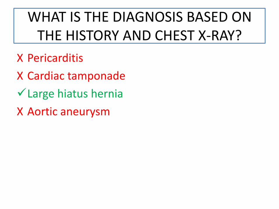
X Pericarditis
X Cardiac tamponade
Large hiatus hernia
X Aortic aneurysm
WHAT IS THE DIAGNOSIS BASED ON THE HISTORY AND CHEST X-RAY?
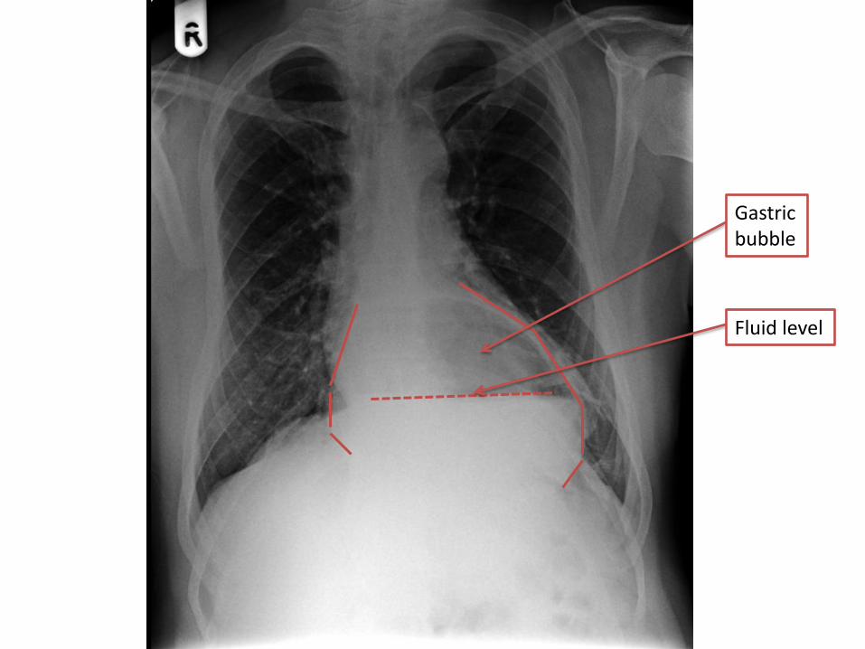
Gastric bubble
Fluid level
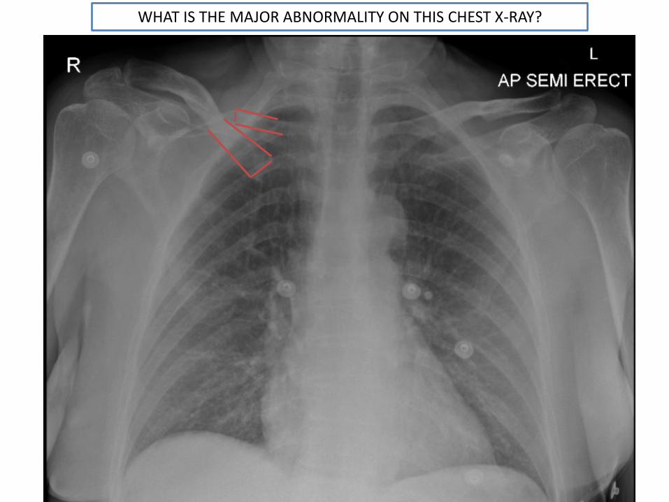
PATIENT 9
• 59 year old female
• Assessed in A&E following a road traffic accident
Chest x-ray taken....
Systematically assess this x-ray
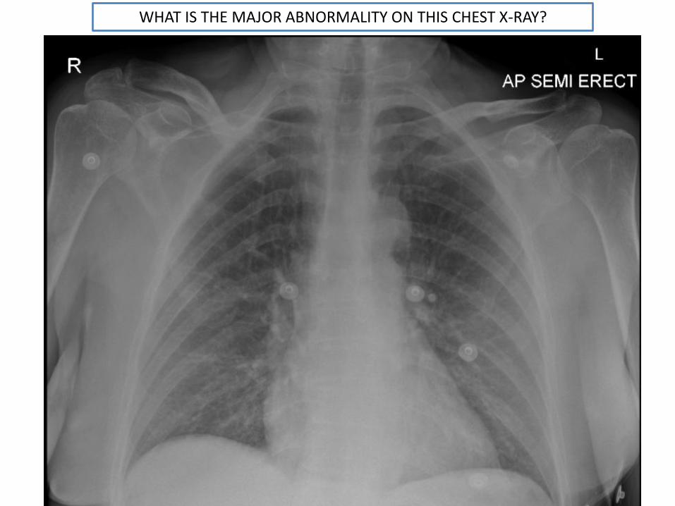
WHAT IS THE MAJOR ABNORMALITY ON THIS CHEST X-RAY?
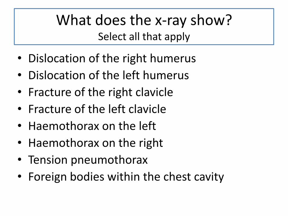
• Dislocation of the right humerus
• Dislocation of the left humerus
• Fracture of the right clavicle
• Fracture of the left clavicle
• Haemothorax on the left
• Haemothorax on the right
• Tension pneumothorax
• Foreign bodies within the chest cavity
What does the x-ray show?Select all that apply
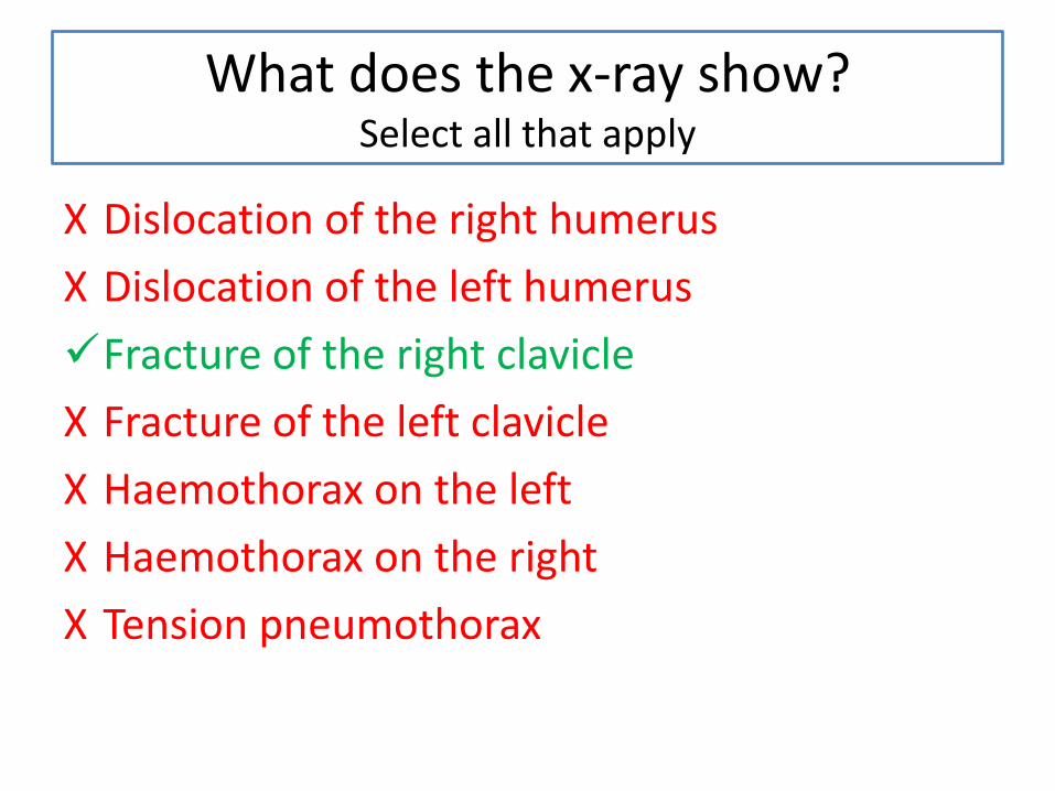
X Dislocation of the right humerus
X Dislocation of the left humerus
Fracture of the right clavicle
X Fracture of the left clavicle
X Haemothorax on the left
X Haemothorax on the right
X Tension pneumothorax
What does the x-ray show?Select all that apply
WHAT IS THE MAJOR ABNORMALITY ON THIS CHEST X-RAY?

WHAT ARE THESE?

WHAT ARE THESE?
• Internal foreign bodies within the thorax
• External foreign objects

WHAT ARE THESE?
X Internal foreign bodies within the thorax
External foreign objects
ECG CLIPS!
PATIENT 10
• 85 year old male
• Admitted with collapse
• On examination: right hemiparesis, expressive aphasia, up-going right plantar reflex.
• Swallow judged unsafe and nasogastric tube inserted
• PMHx: TB left lung
• Chest x-ray taken as part of the work up.....
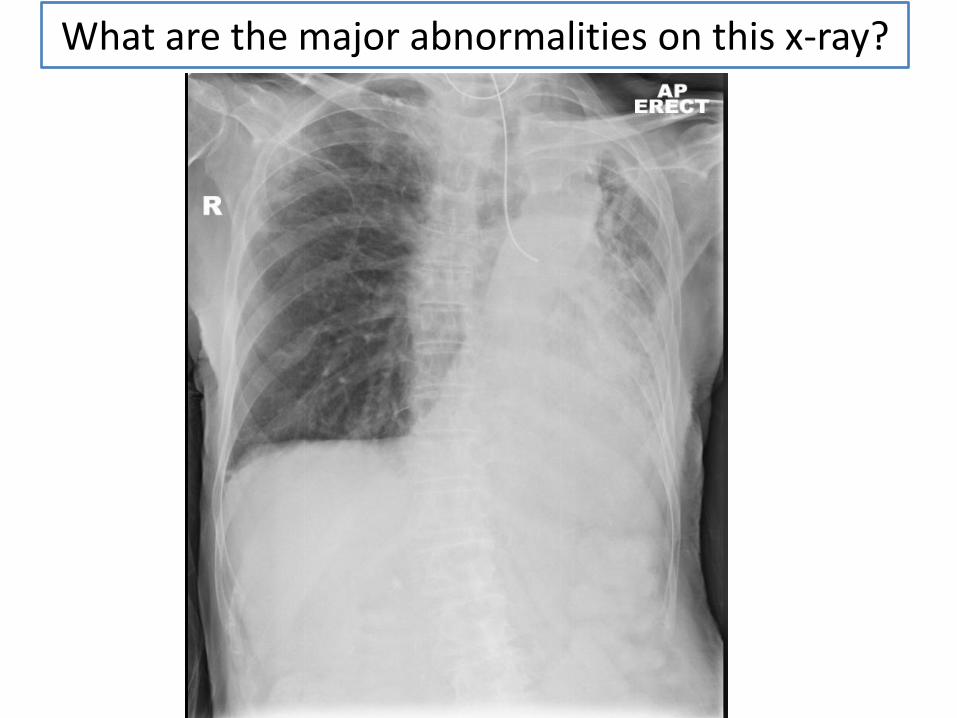
What are the major abnormalities on this x-ray?
WHAT DOES THIS CHEST X-RAY SHOW?Select all that apply
• Mediastinal shift to the left
• Mediastinal shift to the right
• Correctly placed NG-tube
• Incorrectly placed NG tube
• Rib fractures on the left
• Rib fractures on the right
WHAT DOES THIS CHEST X-RAY SHOW?Select all that apply
Mediastinal shift to the left
X Mediastinal shift to the right
X Correctly placed NG-tube
Incorrectly placed NG tube
X Rib fractures on the left
X Rib fractures on the right
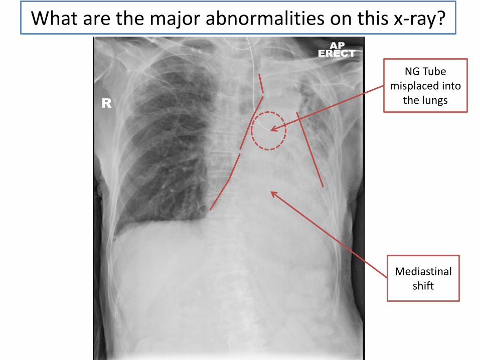
What are the major abnormalities on this x-ray?
NG Tube misplaced into
the lungs
Mediastinalshift
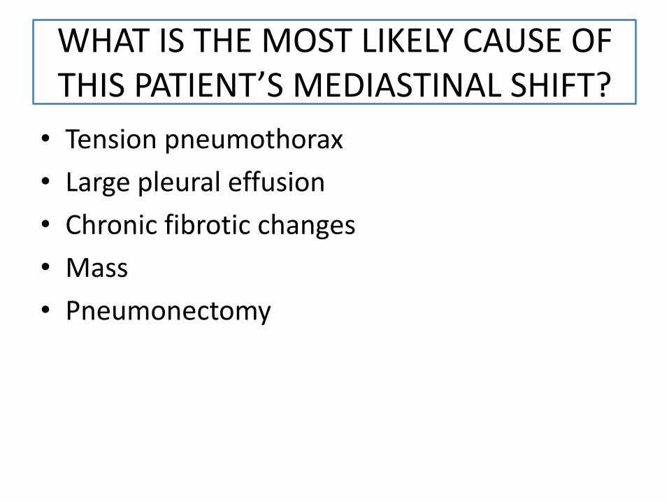
WHAT IS THE MOST LIKELY CAUSE OF THIS PATIENT’S MEDIASTINAL SHIFT?
• Tension pneumothorax
• Large pleural effusion
• Chronic fibrotic changes
• Mass
• Pneumonectomy
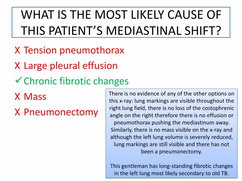
WHAT IS THE MOST LIKELY CAUSE OF THIS PATIENT’S MEDIASTINAL SHIFT?
X Tension pneumothorax
X Large pleural effusion
Chronic fibrotic changes
X Mass
X Pneumonectomy
There is no evidence of any of the other options on this x-ray: lung markings are visible throughout the right lung field, there is no loss of the costophrenicangle on the right therefore there is no effusion or
pneumothorax pushing the mediastinum away. Similarly, there is no mass visible on the x-ray and although the left lung volume is severely reduced,
lung markings are still visible and there has not been a pneumonectomy.
This gentleman has long-standing fibrotic changes in the left lung most likely secondary to old TB.
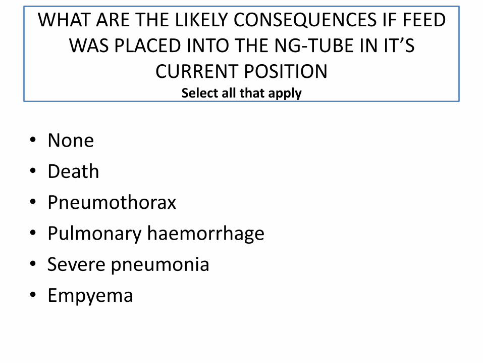
WHAT ARE THE LIKELY CONSEQUENCES IF FEED WAS PLACED INTO THE NG-TUBE IN IT’S
CURRENT POSITIONSelect all that apply
• None
• Death
• Pneumothorax
• Pulmonary haemorrhage
• Severe pneumonia
• Empyema
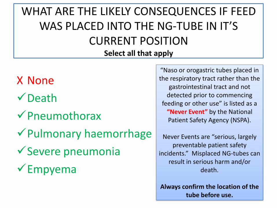
WHAT ARE THE LIKELY CONSEQUENCES IF FEED WAS PLACED INTO THE NG-TUBE IN IT’S
CURRENT POSITIONSelect all that apply
X None
Death
Pneumothorax
Pulmonary haemorrhage
Severe pneumonia
Empyema
“Naso or orogastric tubes placed in the respiratory tract rather than the
gastrointestinal tract and not detected prior to commencing
feeding or other use” is listed as a “Never Event” by the National Patient Safety Agency (NSPA).
Never Events are “serious, largely preventable patient safety
incidents.” Misplaced NG-tubes can result in serious harm and/or
death.
Always confirm the location of the tube before use.
The incorrect placement of the NG-tube was recognised and the tube was removed.
A new NG-tube was inserted and another chest x-ray taken...
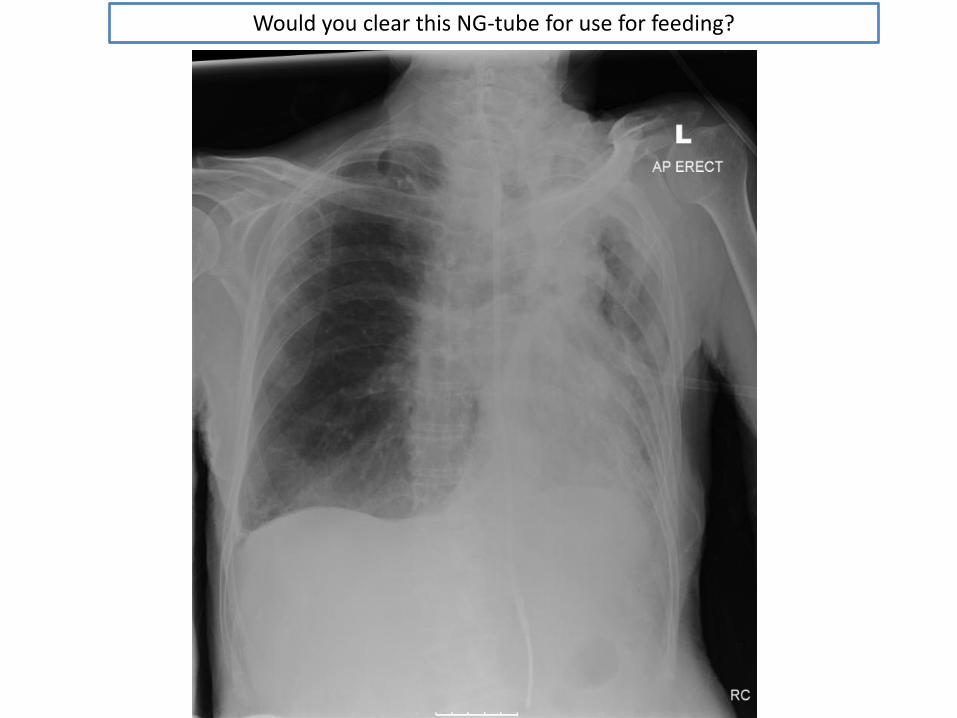
Would you clear this NG-tube for use for feeding?

Would you use this NG-tube for feeding?
• Yes
• No
Would you use this NG-tube for feeding?
Yes
X No
The tip of this NG-tube is clearly visible in the upper left quadrant of the abdomen.
There is no danger that this is in the lungs.
It is therefore safe to use.
PATIENT 11
• 91 year old male
• Presented with increased shortness of breath
• 3 week history of cough productive of yellow sputum
• On examination:– Reduced chest expansion on the right
– Dullness to percussion on the right
– Reduced vocal resonance on the right
– Decreased breath sounds at the right base
A chest x-ray was taken...

WHAT IS THE MAJOR ABNORMALITY ON THIS X-RAY

WHAT DOES THE X-RAY SHOW
• Pleural effusion
• Mass
• Consolidation
• Pneumothorax
• Cardiomegaly
Plus bonus points for the smaller abnormality on the left....
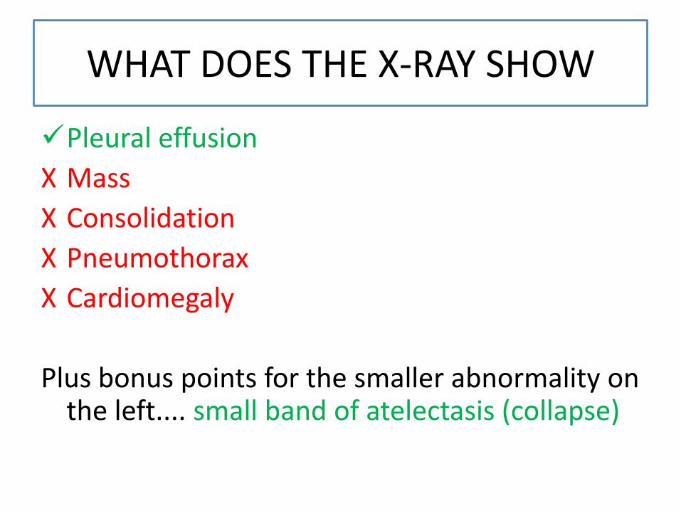
WHAT DOES THE X-RAY SHOW
Pleural effusion
X Mass
X Consolidation
X Pneumothorax
X Cardiomegaly
Plus bonus points for the smaller abnormality on the left.... small band of atelectasis (collapse)
WHAT IS THE MAJOR ABNORMALITY ON THIS X-RAY
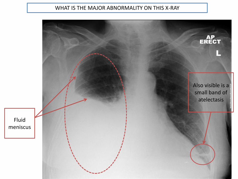
Fluid meniscus
Also visible is a small band of
atelectasis
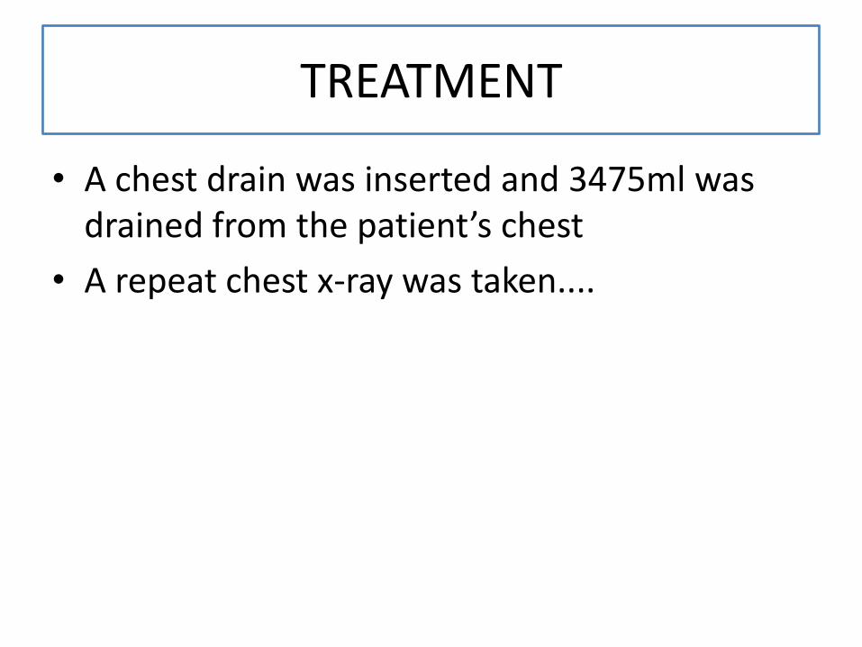
TREATMENT
• A chest drain was inserted and 3475ml was drained from the patient’s chest
• A repeat chest x-ray was taken....

Q 1. WHAT DOES THE X-RAY SHOWQ 2. WHAT HOSPTIAL KIT IS VISIBLE ON THIS X-RAY

Q1. WHAT DOES THE CHEST X-RAY SHOW?SELECT ALL THAT APPLY
• Effusion completely resolved
• Small effusion on the right
• Consolidation
• Mass
• Pneumothorax

Q2. WHAT HOSPITAL KIT IS VISIBLE ON THE X-RAY?SELECT ALL THAT APPLY
• NG tube
• ET tube
• Chest drain (atypical location)
• Nasal cannula
• Pacemaker

Q1. WHAT DOES THE CHEST X-RAY SHOW?SELECT ALL THAT APPLY
X Effusion completely resolved
Small effusion on the right
Consolidation
X Mass
X Pneumothorax
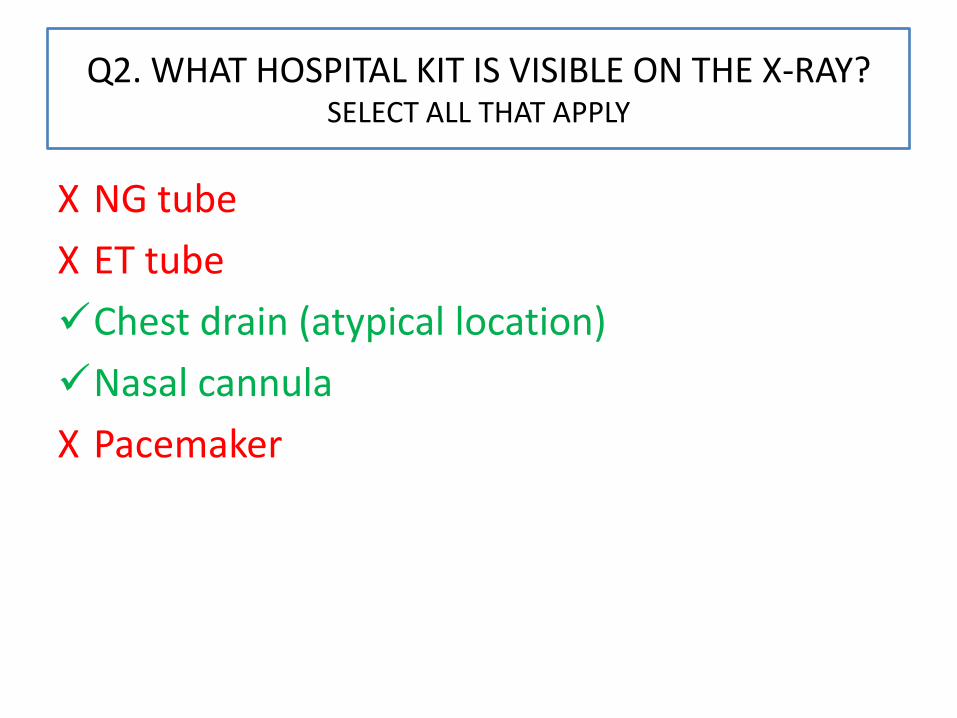
Q2. WHAT HOSPITAL KIT IS VISIBLE ON THE X-RAY?SELECT ALL THAT APPLY
X NG tube
X ET tube
Chest drain (atypical location)
Nasal cannula
X Pacemaker

Q 1. WHAT DOES THE X-RAY SHOWQ 2. WHAT HOSPTIAL KIT IS VISIBLE ON THIS X-RAY
NG Tube
Chest drain
Area of consolidation
Loss of R costophrenicangle. Fluid meniscus.
Also visible is a small band of
atelectasis
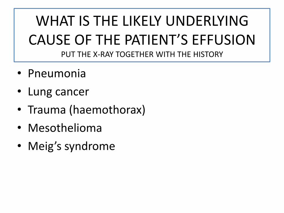
WHAT IS THE LIKELY UNDERLYING CAUSE OF THE PATIENT’S EFFUSION
PUT THE X-RAY TOGETHER WITH THE HISTORY
• Pneumonia
• Lung cancer
• Trauma (haemothorax)
• Mesothelioma
• Meig’s syndrome
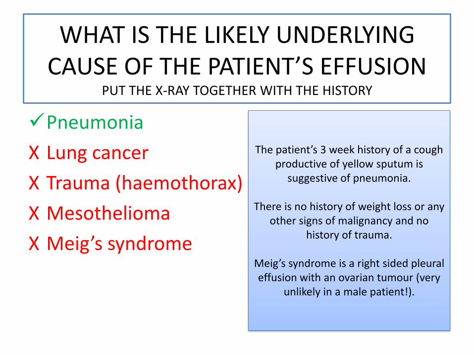
WHAT IS THE LIKELY UNDERLYING CAUSE OF THE PATIENT’S EFFUSION
PUT THE X-RAY TOGETHER WITH THE HISTORY
Pneumonia
X Lung cancer
X Trauma (haemothorax)
X Mesothelioma
X Meig’s syndrome
The patient’s 3 week history of a cough productive of yellow sputum is
suggestive of pneumonia.
There is no history of weight loss or any other signs of malignancy and no
history of trauma.
Meig’s syndrome is a right sided pleural effusion with an ovarian tumour (very
unlikely in a male patient!).

THE PATIENT IS ALLERGIC TO PENICILLIN.
WHICH ANTIBIOTIC(S) WOULD BE SAFE TO USE TO TREAT HIS PNEUMONIA?
• Amoxicillin
• Tazocin
• Gentamicin
• Co-amoxiclav
• Augmentin
• Clarithromycin
• Benzylpenicillin

THE PATIENT IS ALLERGIC TO PENICILLIN.
WHICH ANTIBIOTIC(S) WOULD BE SAFE TO USE TO TREAT HIS PNEUMONIA?
X Amoxicillin
X Tazocin
X Ampicillin
X Co-amoxiclav
X Augmentin
Clarithromycin
X Flucoxacillin
Clarithromycin is the only safe antibiotic for treating penicillin allergic patients on this list.
All of the other antibiotics contain penicillins.