Embed Size (px)
Citation preview
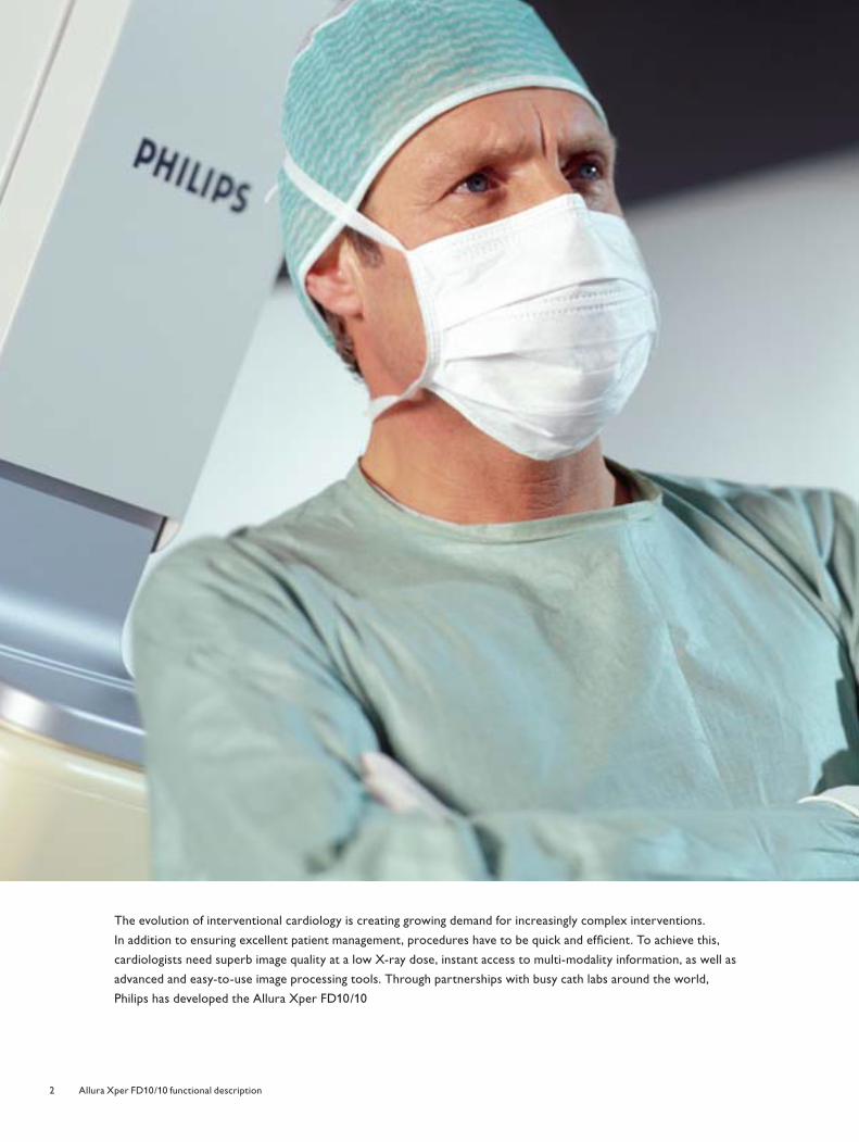
Xperience the futurePhilips Allura Xper FD10/10 Functional description
Nieuw logo.indd 1 3/31/08 10:04:54 AM4522_962_22291.indd 14522_962_22291.indd 1 4/9/08 4:06:05 PM4/9/08 4:06:05 PM
2 Allura Xper FD10/10 functional description
The evolution of interventional cardiology is creating growing demand for increasingly complex interventions.
In addition to ensuring excellent patient management, procedures have to be quick and effi cient. To achieve this,
cardiologists need superb image quality at a low X-ray dose, instant access to multi-modality information, as well as
advanced and easy-to-use image processing tools. Through partnerships with busy cath labs around the world,
Philips has developed the Allura Xper FD10/10
4522_962_22291.indd 24522_962_22291.indd 2 4/9/08 4:06:09 PM4/9/08 4:06:09 PM
3Allura Xper FD10/10 functional description
Contents
4 Speed with fl at detector image quality
5 Bi-plane viewing power and safety Safety is critical for pediatric use Integration6 Xper provides excellent
customization to meet your needs DoseWise Xper Module6 Xper in the examination room On-Screen Display7 Xper Geometry Module Xper Imaging Module7 Xper in the control room Xper Review Module Xper data monitor and Xper review
monitor8 The image quality you want with
low X-ray dose Dynamic Flat Detector Xres MRC tube SpectraBeam fi ltration Monitors MultiVision video switch Fluoro storage Xper Beam Shaping Rotational Scan9 Integration features that enhance
workfl ow Xper DICOM Image Interface Continuous autopush RIS/CIS DICOM Interface MultiSwitch / Xper Window Switch Quantitative Analysis Packages12 Technical information - Geometry G-shaped Gantry Double C-arc (LARC) BodyGuard Patient Protection 13 Rotational Scan Xper table
14 Technical information - Geometry Table tilt Table tilt & cradle Table Automatic Position Controller15 Pivot PAN handle Monitor Ceiling Suspension Ceiling Suspended Radiation Shield Table Mounted Radiation Shield17 Technical information -
User Interface17 Xper User Interface in the examination
room Xper Module18 Technical information -
User Interface Xper Biplane Geometry Module19 Xper Biplane Imaging Module19 Xper User Interface in the control
room Xper Review Module20 Technical information -
User Interface Xper data monitor Scheduling21 Technical information -
User Interface options Xper review monitor Second Xper Biplane Imaging Module Second Xper Biplane Geometry Module Second or third Xper Module22 Technical information -
Integration Storage capacity MultiSwitch, option Lab Reporting, option23 Technical information -
Image Detection Dynamic Flat Detector Xres Fluoroscopy Subtraction package
24 Technical information - X-ray Generation
Velara X-ray generator Xper Beam Shaping X-ray tube MRC-GS 0508 SpectraBeam25 Technical information - Viewing26 Technical information - Options MultiVision Physio Viewing Continuous autopush DICOM Print Intercom RIS/CIS DICOM Interface Biplane standard line rate video
input/output Real-time digital link27 Quantitative Analysis Packages Biplane Left Ventricular Quantifi cation
software package Right Ventricular Quantifi cation
software package Coronary Quantifi cation
software package Vascular Quantifi cation
software package Autocall28 StentBoost Allura 3D-CA CT TrueView Allura 3D-RA EP navigator Examination Light29 Accessories30 Technical information -
Dimensions31 Technical information -
Room Layout
4522_962_22291.indd 34522_962_22291.indd 3 4/9/08 4:06:18 PM4/9/08 4:06:18 PM
4 Allura Xper FD10/10 functional description
Speed with fl at detector image quality
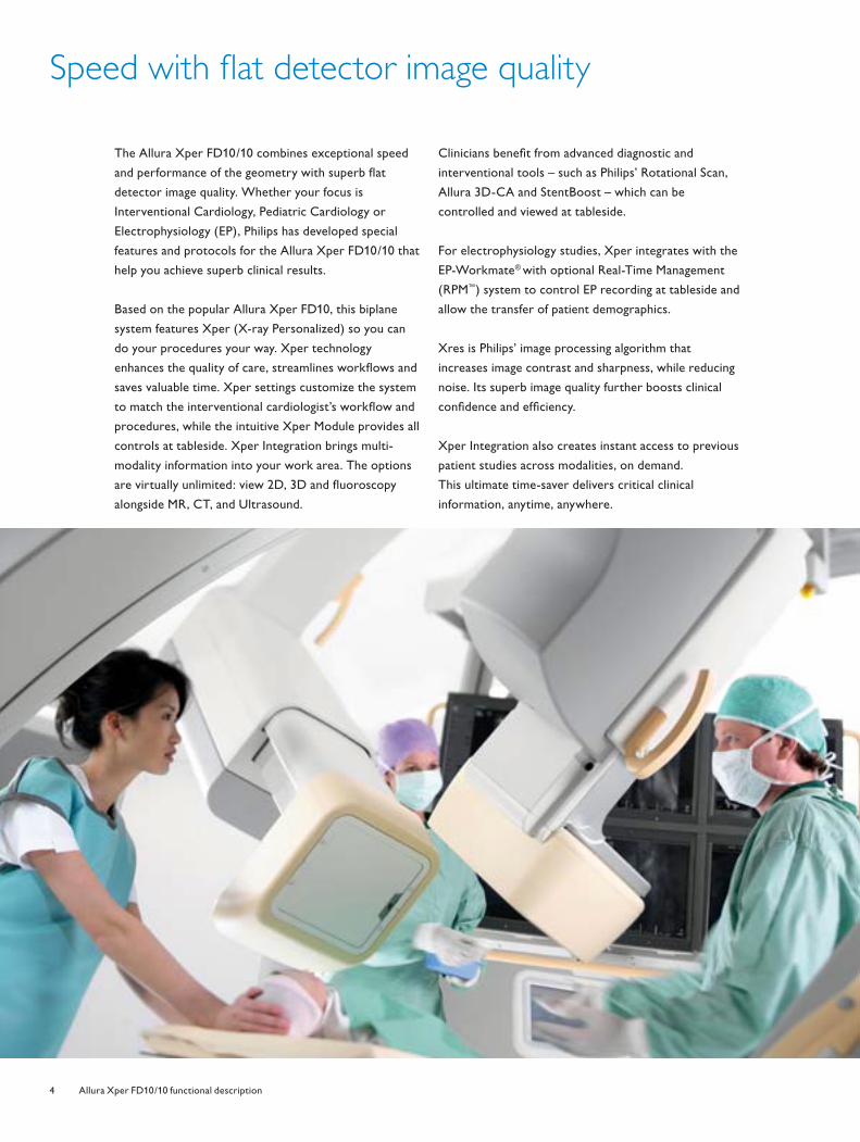
The Allura Xper FD10/10 combines exceptional speed
and performance of the geometry with superb fl at
detector image quality. Whether your focus is
Interventional Cardiology, Pediatric Cardiology or
Electrophysiology (EP), Philips has developed special
features and protocols for the Allura Xper FD10/10 that
help you achieve superb clinical results.
Based on the popular Allura Xper FD10, this biplane
system features Xper (X-ray Personalized) so you can
do your procedures your way. Xper technology
enhances the quality of care, streamlines workfl ows and
saves valuable time. Xper settings customize the system
to match the interventional cardiologist’s workfl ow and
procedures, while the intuitive Xper Module provides all
controls at tableside. Xper Integration brings multi-
modality information into your work area. The options
are virtually unlimited: view 2D, 3D and fl uoroscopy
alongside MR, CT, and Ultrasound.
Clinicians benefi t from advanced diagnostic and
interventional tools – such as Philips’ Rotational Scan,
Allura 3D-CA and StentBoost – which can be
controlled and viewed at tableside.
For electrophysiology studies, Xper integrates with the
EP-Workmate® with optional Real-Time Management
(RPM™) system to control EP recording at tableside and
allow the transfer of patient demographics.
Xres is Philips’ image processing algorithm that
increases image contrast and sharpness, while reducing
noise. Its superb image quality further boosts clinical
confi dence and effi ciency.
Xper Integration also creates instant access to previous
patient studies across modalities, on demand.
This ultimate time-saver delivers critical clinical
information, anytime, anywhere.
4522_962_22291.indd 44522_962_22291.indd 4 4/9/08 4:06:21 PM4/9/08 4:06:21 PM
5Allura Xper FD10/10 functional description
Bi-plane viewing power and safety
The Allura Xper FD10/10 brings fl at detector technology
to biplane viewing. This system delivers superb image
quality in both the frontal and lateral plane, enabling
cardiologists to view them side-by-side. The Allura Xper
FD10/10 saves valuable time when capturing accurate 3D
information while also reducing x-ray dose and contrast
medium.
Safety is critical for pediatric use
In pediatric applications where cardiac anomalies are the
norm, biplane imaging provides tremendous benefi ts.
It delivers twice the information with a single contrast
injection. Moreover, the system offers full patient access
to larger clinical teams. The Allura Xper FD10/10 also
offers special pediatric programs and settings developed
in partnership with pediatric cardiologists. The Xper
table offers optional Tilt and Cradle functionality as well.
Imaging tools optimize care and effi ciency. For
Interventional Cardiology, the Allura Xper FD10/10
combines multi-modality information and a unique
package of diagnostic and interventional tools. Philips’
Rotational Scan gives you multi-dimensional views in
real-time for more precise diagnosis of vessels.
StentBoost improves visualization of stents in coronary
arteries while the guide wire is still in place. StentBoost
images help to confi rm stent expansion in relation to the
vessel lumen and visualize nearby objects, enabling the
interventional cardiologist to take any corrective action
that is required while the patient is still in the exam room.
Allura 3D-CA, available only from Philips, uses two slices
from a Rotational Scan acquisition to instantly construct
a 3D model of the heart’s vasculature. This model can
help the clinician in assessing optimal viewing/working
angles and in determining the accurate lesion length. CT
Trueview, also a unique Philips’ option, provides identical
clinical results based on a Philips’ Brilliance CT scan.
Allura 3D-RA provides extensive, three-dimensional
insight into vascular pathologies from a single Rotational
Scan acquisition. It allows the development of better
treatment strategies and the selection of the best stand
projections for treatment.
Integration
Multi-modality integration saves time and lives. Integration
of the Allura Xper FD10/10 system with Xcelera (Image
Management System), CT, MR, and Ultrasound means that
clinicians and other members of the care team can get the
information they need from the exam room and control
room to their offi ce or laptop – anytime and anywhere.
Saving space is another critical issue. The Xper Window
Switch and MultiSwitch option enable you to share the
control room workspot with RIS/CIS, PACS, and
Interventional Tools. MultiVision allows images from other
modalities to be viewed on the LCD monitors in the exam
room, eliminating the need for additional monitors.
4522_962_22291.indd Sec1:54522_962_22291.indd Sec1:5 4/9/08 4:06:27 PM4/9/08 4:06:27 PM
6 Allura Xper FD10/10 functional description
Xper provides excellent customization to meet your needs
DoseWise
Philips’ DoseWise philosophy is the foundation of the
Allura Xper FD10/10’s design. The legendary MRC X-ray
tube with SpectraBeam fi ltration achieves optimum
image quality at a low X-ray dose. To further reduce
dose, Xper Beam Shaping positions shutters and
wedges on the last image without using radiation. Xper
fluoro storage allows recording of fl uoro sequences
for recall and/or review, eliminating the need for
additional runs. The unique dose display makes users
much more aware of dose that is used in relation to
control of the system, thus protecting patients against
radiation skin burns.
• Easy to understand graphical dose data display
• Provides predictive and actual DAP dose rate indication
• Provides AirKerma patient X-ray dose per body zone:
- Aimed to help prevent skin burns
- 10 Cardiac zones defi ned
- Graphical AK dose level indication for the actual
zone, related to the 2 Gy critical dose level
Other safety features include Philips’ BodyGuard
technology, which senses the patient’s position so the
stand can rotate safely at high speeds. Also, Philips’
unique patient support system is designed so that you
can instantly apply CPR to the patient with the tabletop
in any position.
The more demanding your cath lab environment,
the more you need the Allura Xper FD10/10. It features
Xper technology which is designed to improve your
personal and departmental effi ciency.
• Xper settings provide an advanced level of
customization so users can create an interventional lab
that meets their individual needs and preferences
• The Xper User Interface provides intuitive system
controls and all relevant functionality at the tableside
to enhance ease of use
• Xper Integration provides bi-directional information
exchange
Xper Module
Available in both the examination and/or control room,
the Xper Module communicates user preferences for
acquisition settings, automatic position control and
processing. An additional option also allows very easy
tableside control of quantitative analysis, Allura
3D-CA / StentBoost / Xcelera PACS / Allura 3D-RA and
hemodynamics (Xper IM) via the touch screen controls
or integrated joystick.
Xper in the examination room
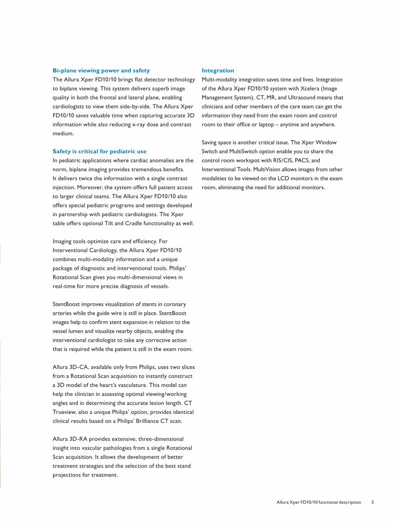
On-Screen Display
The On-Screen Display provides information that
includes X-ray indication, rotation and angulation stand
positions, Source Image Distance, Air Kerma (rate and
accumulated dose per body zone, as well as a predictive
value), frame speed, and fl uoroscopy mode.
4522_962_22291.indd Sec1:64522_962_22291.indd Sec1:6 4/9/08 4:06:28 PM4/9/08 4:06:28 PM
7Allura Xper FD10/10 functional description
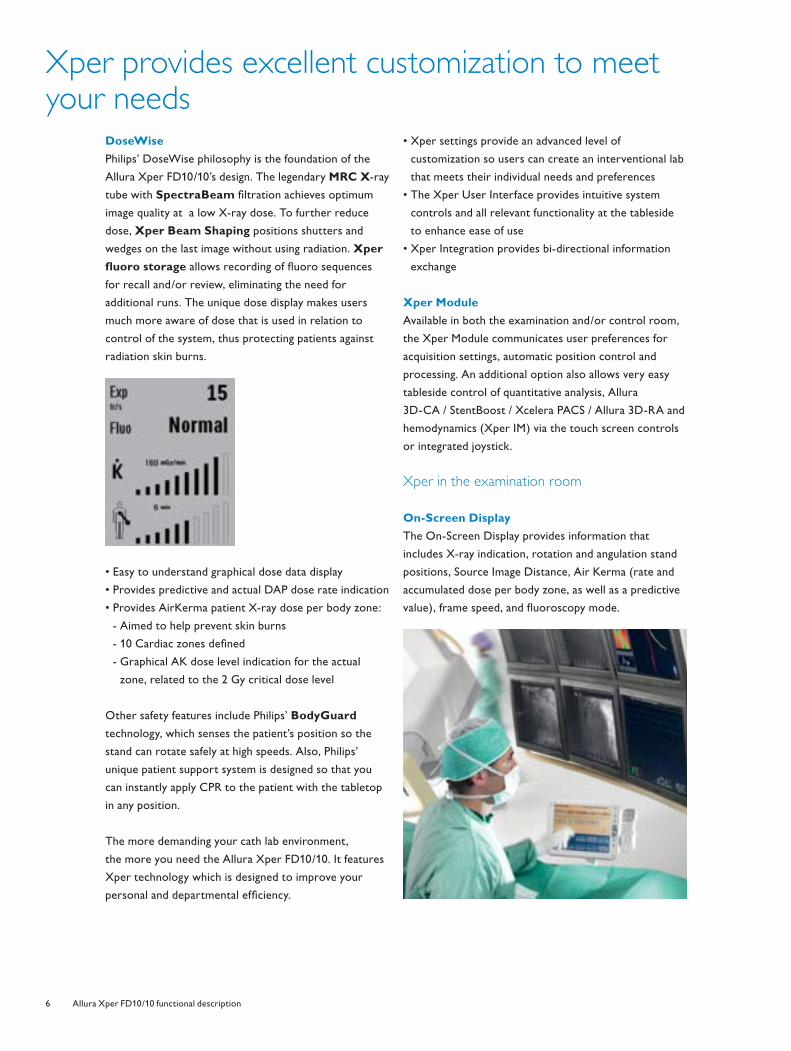
On the reference monitor, the On-Screen Display
contains the user interface of the Xper ViewPad, which is
used to carry out functions like run and image selection,
review speed, active fi le selection and digital zoom.
Xper Geometry Module
To make operation as convenient as possible, the Xper
Geometry Module can be positioned on all sides of the
patient table. The Geometry Module automatically
adjusts itself to the position to retain the intuitive button
operation. It controls tabletop fl oat, table height, Source
Image Distance, stand positioning (including memory
positions) and optionally, tilt and cradle functionality.
Xper Imaging Module
Like the Xper Geometry Module, the Xper Imaging
Module can be positioned on all sides of the patient
table, while retaining its intuitive button operation.
The Xper Imaging Module allows the user to activate
shutter and wedge positioning, fl uoroscopy mode as defi ned
via Xper settings, detector fi eld size and beam width.
Both Xper Modules have a removable protection bar
that prevents unintended activation of the system.
Xper in the control room
Xper Review Module
The Xper Review Module serves cardiovascular viewing
needs. It offers direct control of basic viewing controls
like exam and run cycle, contrast, brightness, edge
enhancement, and viewing speed (tagarno wheel).
Xper data monitor and Xper review monitor
The Xper data monitor and Xper review monitor use a
shared screen and the mouse can be moved over the
two monitors. The data monitor provides patient and
exam data to assist with all stages of workfl ow, including
scheduling, preparation, acquisition, reviewing, reporting,
and archiving. System information is displayed on the
bottom of the data monitor.
The intuitive review monitor enables effi cient review of
exams and control of image processing and Quantitative
Analysis Programs.
4522_962_22291.indd Sec1:74522_962_22291.indd Sec1:7 4/9/08 4:06:29 PM4/9/08 4:06:29 PM
8 Allura Xper FD10/10 functional description
The image quality you want with low X-ray dose
The Allura Xper FD10/10 features advanced algorithms,
a next generation fl at detector, and Philips’ renowned
imaging chain to ensure superb image quality at a low
patient X-ray dose.
Dynamic Flat Detector
Philips’ 14-bit virtually distortion-free dynamic fl at
detector offers 184 micron pixels for higher resolution
and a DQE(O) of 75% that provides better image quality,
especially for low dose fl uoroscopy. The compact design
with a very large fi eld of view of 25 cm (10 in.) is the
optimal size for dedicated cardiology and EP applications.
It also offers a refresh light that provides temporal
virtually artifact-free imaging by “blanking” the detector,
thereby eliminating image glow during dynamic studies.
Xres
Xres is Philips’ real-time image processing algorithm.
Xres was developed by the Philips Research Laboratories
and has been applied in several Philips products, e.g.
Ultrasound and MRI. This image processing algorithm
provides billions of calculations per frame and is applied to
each clinical image in real-time. Xres provides excellent
image quality through improved contrast and sharpness.
It exploits the benefi ts of the fully digital detector to
reduce noise in clinical images. Each user can customize
Xres via Xper settings according to their preferred
image quality settings.
Xres also harmonizes the background of an image to
provide excellent visualization of coronary arteries in
complex projections.
MRC tube
The Allura Xper FD10/10 is equipped with the legendary
high power MRC-GS 0508 X-ray tube. The tube’s
exceptional design provides long life and allows it to
withstand high continuous loads, while maximizing heat
dissipation. This enables virtually unlimited X-ray
sessions without forced cool down delays.
SpectraBeam fi ltration
The MRC tube works in tandem with SpectraBeam
fi ltration to allow increased X-ray output with better
fi ltration of soft radiation. SpectraBeam offers four levels
of fi ltration - up to one mm Cu equivalent - to reduce
patient X-ray dose, while maintaining image quality.
The fi ltration level can be programmed via Xper settings.
The fl uoroscopy mode can be selected at tableside.
Monitors
The LCD progressive display monitors are virtually
fl icker-free to prevent physician eyestrain. In the control
room, the 19-inch LCD color monitor and two 18-inch
LCD black and white monitors are standard. In the exam
room, four 18-inch LCD black and white monitors are
standard. For each plane it provides the live monitors
and the reference monitors.
MultiVision video switch
MultiVision allows images from different image sources
to be viewed on the monitor in the exam room,
eliminating the need for multiple monitors.
Fluoro storage
Xper fl uoro storage lets you store and review the last
fl uoroscopy run (service confi gurable time).
Xper Beam Shaping
Xper Beam Shaping allows the wedges and shutters to
be positioned without using X-ray radiation.
Rotational Scan
Rotational Scan saves time, contrast medium and X-ray
dose by creating real-time 3D impressions of complex
vasculature and coronary arteries with multiple
projections – all from just one contrast injection.
The Rotational Scans can be sent to an interventional
tool for a 3D reconstruction.
4522_962_22291.indd Sec1:84522_962_22291.indd Sec1:8 4/9/08 4:06:32 PM4/9/08 4:06:32 PM
9Allura Xper FD10/10 functional description
Integration features that enhance workfl ow
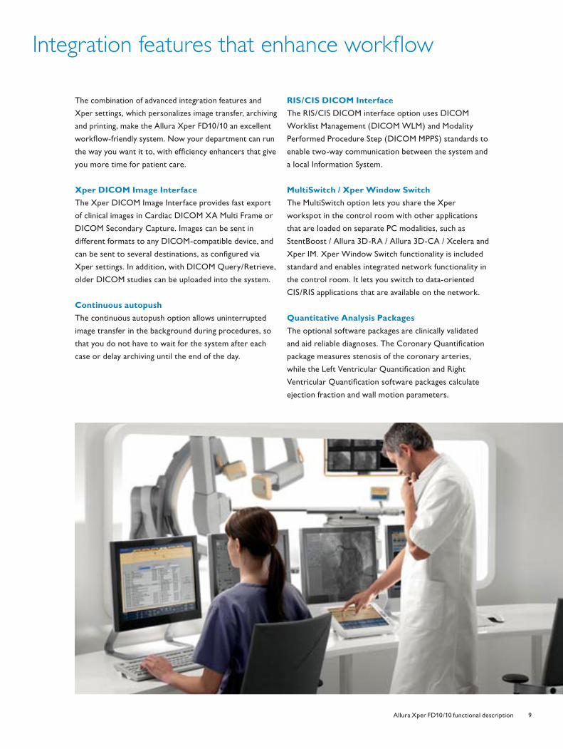
The combination of advanced integration features and
Xper settings, which personalizes image transfer, archiving
and printing, make the Allura Xper FD10/10 an excellent
workfl ow-friendly system. Now your department can run
the way you want it to, with effi ciency enhancers that give
you more time for patient care.
Xper DICOM Image Interface
The Xper DICOM Image Interface provides fast export
of clinical images in Cardiac DICOM XA Multi Frame or
DICOM Secondary Capture. Images can be sent in
different formats to any DICOM-compatible device, and
can be sent to several destinations, as confi gured via
Xper settings. In addition, with DICOM Query/Retrieve,
older DICOM studies can be uploaded into the system.
Continuous autopush
The continuous autopush option allows uninterrupted
image transfer in the background during procedures, so
that you do not have to wait for the system after each
case or delay archiving until the end of the day.
RIS/CIS DICOM Interface
The RIS/CIS DICOM interface option uses DICOM
Worklist Management (DICOM WLM) and Modality
Performed Procedure Step (DICOM MPPS) standards to
enable two-way communication between the system and
a local Information System.
MultiSwitch / Xper Window Switch
The MultiSwitch option lets you share the Xper
workspot in the control room with other applications
that are loaded on separate PC modalities, such as
StentBoost / Allura 3D-RA / Allura 3D-CA / Xcelera and
Xper IM. Xper Window Switch functionality is included
standard and enables integrated network functionality in
the control room. It lets you switch to data-oriented
CIS/RIS applications that are available on the network.
Quantitative Analysis Packages
The optional software packages are clinically validated
and aid reliable diagnoses. The Coronary Quantifi cation
package measures stenosis of the coronary arteries,
while the Left Ventricular Quantifi cation and Right
Ventricular Quantifi cation software packages calculate
ejection fraction and wall motion parameters.
4522_962_22291.indd Sec1:94522_962_22291.indd Sec1:9 4/9/08 4:06:32 PM4/9/08 4:06:32 PM
10 Allura Xper FD10/10 functional description
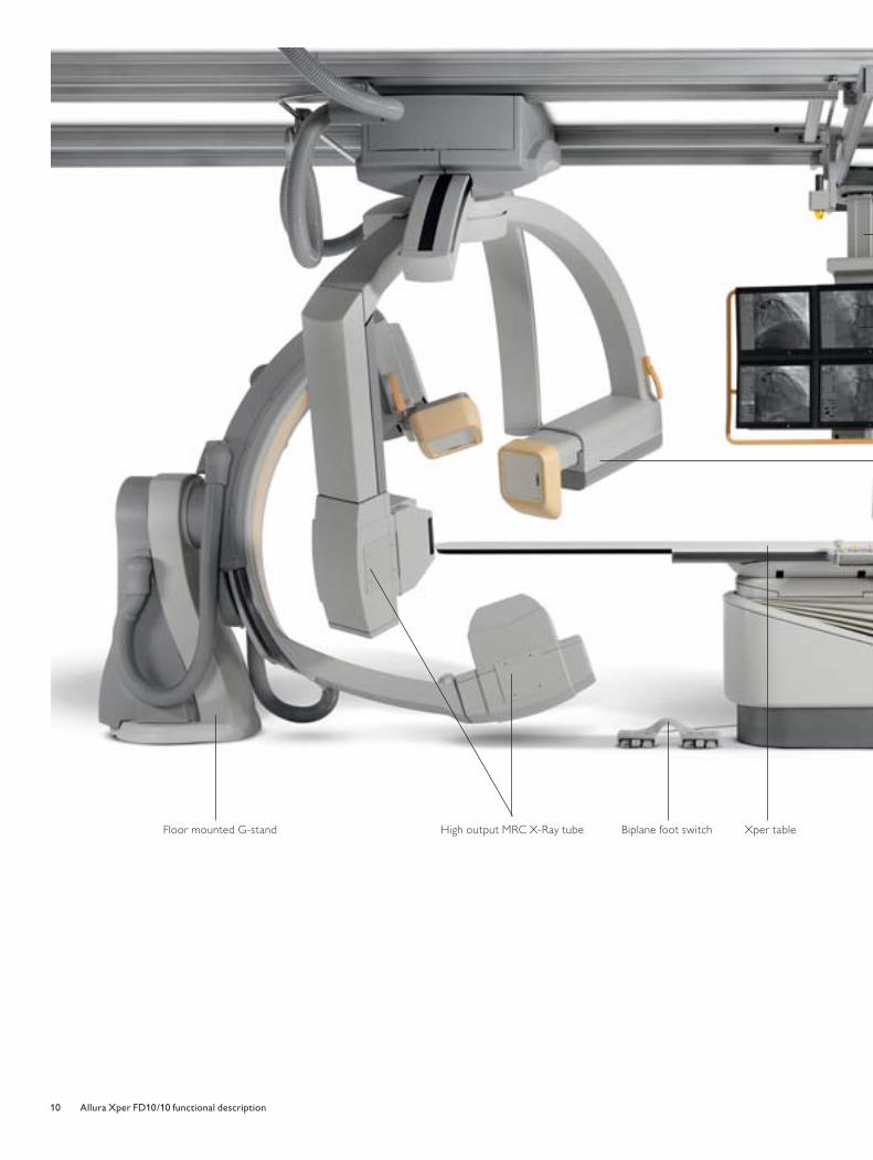
Floor mounted G-stand High output MRC X-Ray tube Xper tableBiplane foot switch
4522_962_22291.indd Sec1:104522_962_22291.indd Sec1:10 4/9/08 4:06:34 PM4/9/08 4:06:34 PM
11Allura Xper FD10/10 functional description
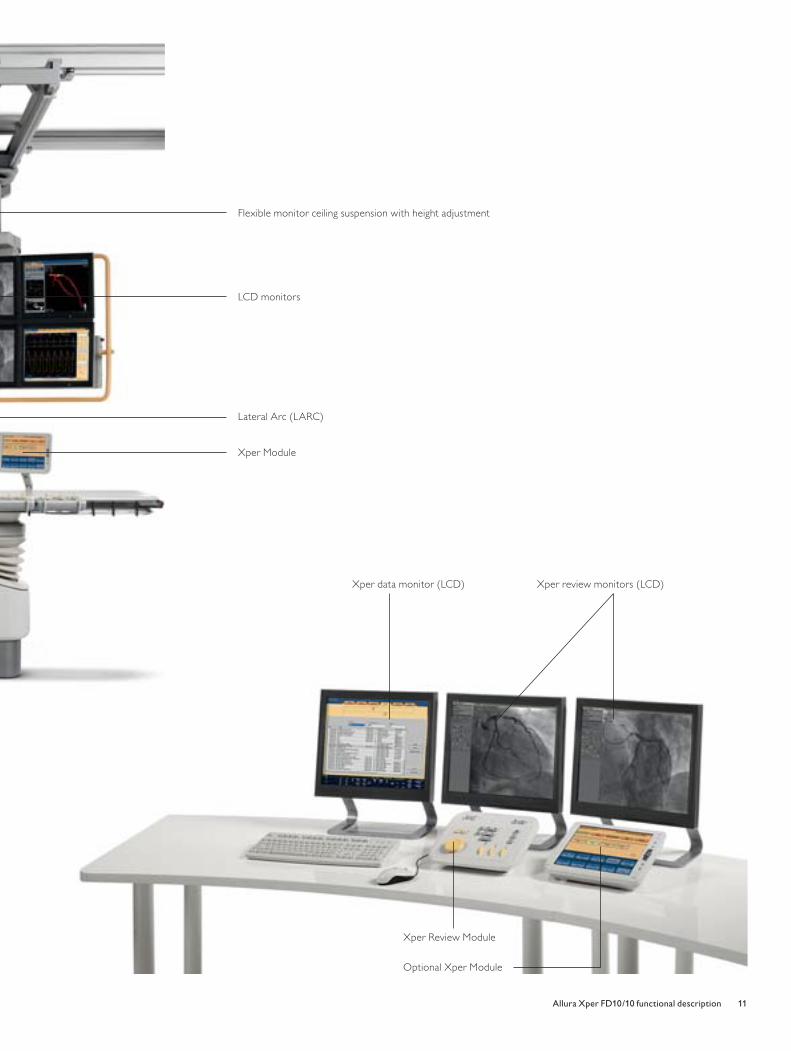
Lateral Arc (LARC)
Xper Review Module
Flexible monitor ceiling suspension with height adjustment
LCD monitors
Xper Module
Xper data monitor (LCD) Xper review monitors (LCD)
Optional Xper Module
4522_962_22291.indd Sec1:114522_962_22291.indd Sec1:11 4/9/08 4:06:40 PM4/9/08 4:06:40 PM
12 Allura Xper FD10/10 functional description
Technical information - Geometry
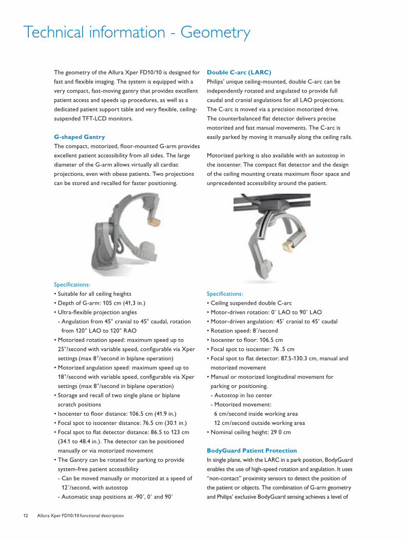
The geometry of the Allura Xper FD10/10 is designed for
fast and fl exible imaging. The system is equipped with a
very compact, fast-moving gantry that provides excellent
patient access and speeds up procedures, as well as a
dedicated patient support table and very fl exible, ceiling-
suspended TFT-LCD monitors.
G-shaped Gantry
The compact, motorized, fl oor-mounted G-arm provides
excellent patient accessibility from all sides. The large
diameter of the G-arm allows virtually all cardiac
projections, even with obese patients. Two projections
can be stored and recalled for faster positioning.
Specifi cations:
• Suitable for all ceiling heights
• Depth of G-arm: 105 cm (41,3 in.)
• Ultra-fl exible projection angles
- Angulation from 45° cranial to 45° caudal, rotation
from 120° LAO to 120° RAO
• Motorized rotation speed: maximum speed up to
25°/second with variable speed, confi gurable via Xper
settings (max 8°/second in biplane operation)
• Motorized angulation speed: maximum speed up to
18°/second with variable speed, confi gurable via Xper
settings (max 8°/second in biplane operation)
• Storage and recall of two single plane or biplane
scratch positions
• Isocenter to fl oor distance: 106.5 cm (41.9 in.)
• Focal spot to isocenter distance: 76.5 cm (30.1 in.)
• Focal spot to fl at detector distance: 86.5 to 123 cm
(34.1 to 48.4 in.). The detector can be positioned
manually or via motorized movement
• The Gantry can be rotated for parking to provide
system-free patient accessibility
- Can be moved manually or motorized at a speed of
12˚/second, with autostop
- Automatic snap positions at -90 ,̊ 0˚ and 90˚
Double C-arc (LARC)
Philips’ unique ceiling-mounted, double C-arc can be
independently rotated and angulated to provide full
caudal and cranial angulations for all LAO projections.
The C-arc is moved via a precision motorized drive.
The counterbalanced fl at detector delivers precise
motorized and fast manual movements. The C-arc is
easily parked by moving it manually along the ceiling rails.
Motorized parking is also available with an autostop in
the isocenter. The compact fl at detector and the design
of the ceiling mounting create maximum fl oor space and
unprecedented accessibility around the patient.
Specifi cations:
• Ceiling suspended double C-arc
• Motor-driven rotation: 0˚ LAO to 90˚ LAO
• Motor-driven angulation: 45˚ cranial to 45˚ caudal
• Rotation speed: 8˚/second
• Isocenter to fl oor: 106.5 cm
• Focal spot to isocenter: 76 .5 cm
• Focal spot to fl at detector: 87.5-130.3 cm, manual and
motorized movement
• Manual or motorized longitudinal movement for
parking or positioning.
- Autostop in Iso center
- Motorized movement:
6 cm/second inside working area
12 cm/second outside working area
• Nominal ceiling height: 29 0 cm
BodyGuard Patient Protection
In single plane, with the LARC in a park position, BodyGuard
enables the use of high-speed rotation and angulation. It uses
“non-contact” proximity sensors to detect the position of
the patient or objects. The combination of G-arm geometry
and Philips’ exclusive BodyGuard sensing achieves a level of
4522_962_22291.indd Sec1:124522_962_22291.indd Sec1:12 4/9/08 4:06:44 PM4/9/08 4:06:44 PM
13Allura Xper FD10/10 functional description
control that is not possible with conventional high-speed
motorized C-arm confi gurations. These high-speed stand
designs use a pre-set “one-size-fi ts-all” program, resulting in
a so-called “safety envelope” that is too large to be practical.
With BodyGuard’s continual capacitive sensing, the
system adapts to individual patient size. The system
slows down or stops moving when a patient or object
gets too close. BodyGuard has an override function to
allow full gantry positioning control at all times. In
addition, BodyGuard uses motor current sensing (the
electronic equivalent of a slip clutch) to safeguard all
stand movements.
The counterbalanced fl at detector incorporates these
sensing technologies, along with a mechanical slip clutch,
to control motorized and manual movements.
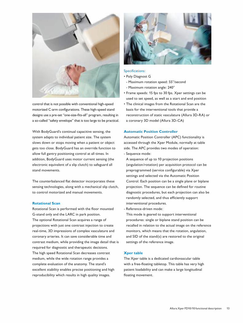
Rotational Scan
Rotational Scan is performed with the fl oor mounted
G-stand only and the LARC in park position.
The optional Rotational Scan acquires a range of
projections with just one contrast injection to create
real-time, 3D impressions of complex vasculature and
coronary arteries. It can save considerable time and
contrast medium, while providing the image detail that is
required for diagnostic and therapeutic decisions.
The high speed Rotational Scan decreases contrast
medium, while the wide rotation range provides a
complete evaluation of the anatomy. The stand's
excellent stability enables precise positioning and high
reproducibility which results in high quality images.
Specifi cations:
• Poly Diagnost G
- Maximum rotation speed: 55°/second
- Maximum rotation angle: 240°
• Frame speeds: 15 fps to 30 fps. Xper settings can be
used to set speed, as well as a start and end position
• The clinical images from the Rotational Scan are the
basis for the interventional tools that provide a
reconstruction of static vasculature (Allura 3D-RA) or
a coronary 3D model (Allura 3D-CA)
Automatic Position Controller
Automatic Position Controller (APC) functionality is
accessed through the Xper Module, normally at table
side. The APC provides two modes of operation:
- Sequence mode:
A sequence of up to 10 projection positions
(angulation/rotation) per acquisition protocol can be
preprogrammed (service confi gurable) via Xper
settings and selected via the Automatic Position
Control. Each position can be a single plane or biplane
projection. The sequence can be defi ned for routine
diagnostic procedures, but each projection can also be
randomly selected, and thus effi ciently support
interventional procedures.
- Reference-driven mode:
This mode is geared to support interventional
procedures: single or biplane stand position can be
recalled in relation to the actual image on the reference
monitors, which means that the rotation, angulation,
and SID of the stand(s) are restored to the original
settings of the reference image.
Xper table
The Xper table is a dedicated cardiovascular table
with a free-fl oating tabletop. This table has very high
patient loadability and can make a large longitudinal
fl oating movement.
4522_962_22291.indd Sec1:134522_962_22291.indd Sec1:13 4/9/08 4:06:46 PM4/9/08 4:06:46 PM
14 Allura Xper FD10/10 functional description
Technical information - Geometry
Specifi cation:
• Radio translucent carbon fi ber tabletop
• Tabletop length: 319 cm
• Tabletop width: 50 cm
• Motorized height movement: From 79 – 107 cm
• Tabletop metal free overhang: 125 cm
• Free fl oat at 0 degrees tilt
• Longitudinal fl oat: 120 cm
• Transversal fl oat: 36 cm
• Maximum allowable patient weight: 250 kg (550 lbs)
with additional force of 500 N (100 kg/220 lbs) allowed
in case of CPR. CPR can be performed while the
tabletop is set in any longitudinal position
• Pivot over 270 degrees
• Comfortable patient mattress
• The Xper Module, Xper Imaging, and Xper Geometry
Modules can be positioned on three sides of the patient
support
• Cables incorporated in the table to allow maximum
operational fl exibility
Table tilt
The optional isocentric table tilt enhances the accuracy
and effi ciency of gravity-oriented procedures. It is ideal for
interventional, myelography, phlebography and head-down
procedures because it provides more precise imaging of
contrast medium, blood, or objects in the body. As the
table tilts, the X-ray beam automatically coordinates to the
movement to keep the region of interest in the isocenter
of rotation and angulation of the stand. If the longitudinal
position of the stand changes, the tilt isocenter is changed
to match with the new stand position. As a result, the
region of interest is always centered. As the table tilts, the
X-ray beam automatically coordinates to the movement.
Specifi cations:
Maximum tilt range: -17˚ (head-down) to +17˚
(head-up). Tilt speed: 2 degrees/second
• Automatic safeguarding system with manual override
• Panning range in tilted plane: equal to standard
panning range
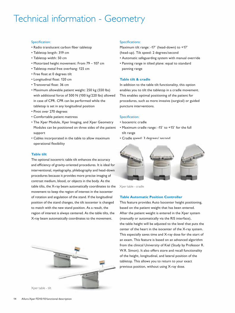
Table tilt & cradle
In addition to the table tilt functionality, this option
enables you to tilt the tabletop in a cradle movement.
This enables optimal positioning of the patient for
procedures, such as more invasive (surgical) or guided
puncture interventions.
Specifi cation:
• Isocentric cradle
• Maximum cradle range: -15˚ to +15˚ for the full
tilt range
• Cradle speed: 3 degrees/ second
Table Automatic Position Controller
This feature provides Auto Isocenter height positioning,
based on the patient weight that has been entered.
After the patient weight is entered in the Xper system
(manually or automatically via the RIS interface),
the table height will be adjusted to the level that puts the
center of the heart in the isocenter of the X-ray system.
This especially saves time and X-ray dose for the start of
an exam. This feature is based on an advanced algorithm
from the clinical University of Kiel (Study by Professor R.
W.R. Simon). It also offers store and recall functionality
of the height, longitudinal, and lateral position of the
tabletop. This allows you to return to your exact
previous position, without using X-ray dose.
racy
deal for
-downn
ng of
the
pee : 3 eg ees/ secop
Xper table - cradle
Xper table - tilt
4522_962_22291.indd Sec1:144522_962_22291.indd Sec1:14 4/9/08 4:06:48 PM4/9/08 4:06:48 PM
15Allura Xper FD10/10 functional description
Pivot
The table-based pivot option is designed for angiographic
and interventional procedures of the upper peripherals.
It provides improved table access for patient transfer.
This option also enables the table to pivot around its
vertical axes. The pivot range moves from -90˚ to +180˚
(or -180˚ to +90˚) with locked positions at 0 ,̊ -13 ,̊ and +31˚
(to facilitate arm angiography) and -90 ,̊ +90 ,̊ and 180 .̊
PAN handle
The PAN handle is a tabletop fl oat control extension,
which can be attached to any side of the table. This
additional PAN handle works in a master/slave confi guration.
Table Mounted Radiation ShieldMonitor Ceiling Suspension
Pivot
Ergonomic PAN handle
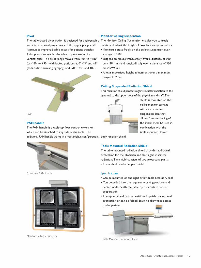
Monitor Ceiling Suspension
The Monitor Ceiling Suspension enables you to freely
rotate and adjust the height of two, four or six monitors.
• Monitors rotate freely on the ceiling suspension over
a range of 350˚
• Suspension moves transversely over a distance of 300
cm (118.1 in.) and longitudinally over a distance of 330
cm (129.9 in.)
• Allows motorized height adjustment over a maximum
range of 32 cm
Ceiling Suspended Radiation Shield
This radiation shield protects against scatter radiation to the
eyes and to the upper body of the physician and staff. The
shield is mounted on the
ceiling monitor carriage
with a two-section
suspension arm that
allows free positioning of
the shield. It can be used in
combination with the
table mounted, lower
body radiation shield.
Table Mounted Radiation Shield
The table mounted radiation shield provides additional
protection for the physician and staff against scatter
radiation. The shield consists of two protective parts:
a lower shield and an upper shield.
Specifi cations:
• Can be mounted on the right or left table accessory rails
• Can be pulled into the required working position and
parked underneath the tabletop to facilitate patient
preparation
• The upper shield can be positioned upright for optimal
protection or can be folded down to allow free access
to the patient
4522_962_22291.indd Sec1:154522_962_22291.indd Sec1:15 4/9/08 4:06:51 PM4/9/08 4:06:51 PM
16 Allura Xper FD10/10 functional description
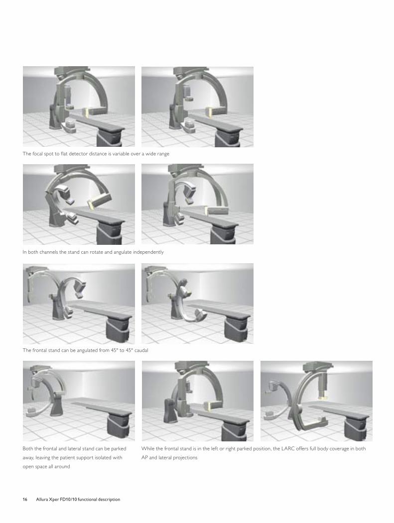
While the frontal stand is in the left or right parked position, the LARC offers full body coverage in both
AP and lateral projections
The focal spot to fl at detector distance is variable over a wide range
In both channels the stand can rotate and angulate independently
The frontal stand can be angulated from 45° to 45° caudal
Both the frontal and lateral stand can be parked
away, leaving the patient support isolated with
open space all around
4522_962_22291.indd Sec1:164522_962_22291.indd Sec1:16 4/9/08 4:06:54 PM4/9/08 4:06:54 PM
17Allura Xper FD10/10 functional description
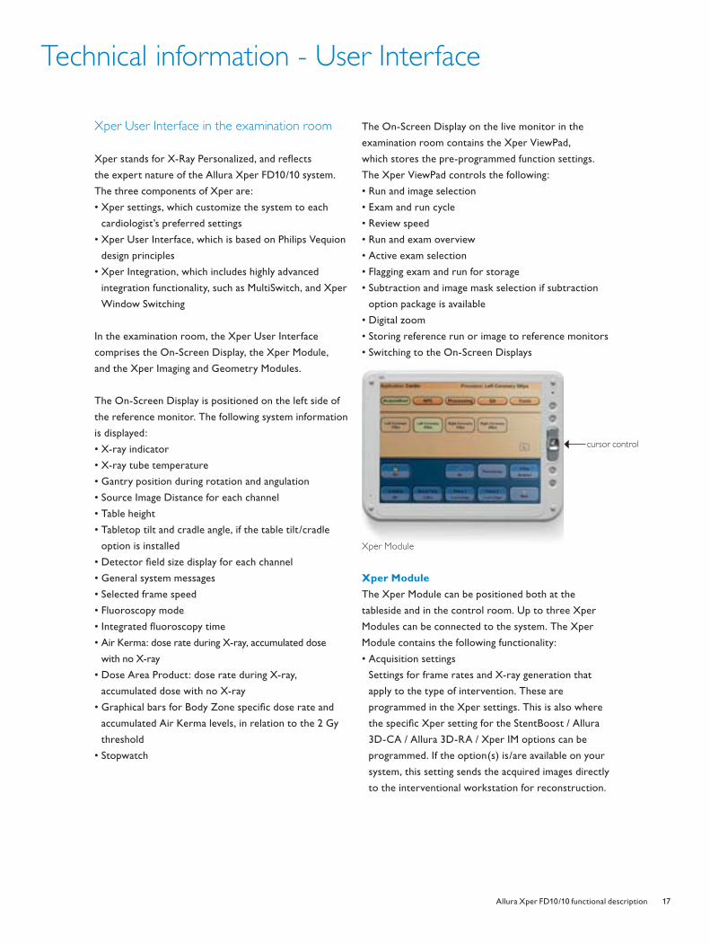
cursor control
Technical information - User Interface
Xper User Interface in the examination room
Xper stands for X-Ray Personalized, and refl ects
the expert nature of the Allura Xper FD10/10 system.
The three components of Xper are:
• Xper settings, which customize the system to each
cardiologist’s preferred settings
• Xper User Interface, which is based on Philips Vequion
design principles
• Xper Integration, which includes highly advanced
integration functionality, such as MultiSwitch, and Xper
Window Switching
In the examination room, the Xper User Interface
comprises the On-Screen Display, the Xper Module,
and the Xper Imaging and Geometry Modules.
The On-Screen Display is positioned on the left side of
the reference monitor. The following system information
is displayed:
• X-ray indicator
• X-ray tube temperature
• Gantry position during rotation and angulation
• Source Image Distance for each channel
• Table height
• Tabletop tilt and cradle angle, if the table tilt/cradle
option is installed
• Detector fi eld size display for each channel
• General system messages
• Selected frame speed
• Fluoroscopy mode
• Integrated fl uoroscopy time
• Air Kerma: dose rate during X-ray, accumulated dose
with no X-ray
• Dose Area Product: dose rate during X-ray,
accumulated dose with no X-ray
• Graphical bars for Body Zone specifi c dose rate and
accumulated Air Kerma levels, in relation to the 2 Gy
threshold
• Stopwatch
The On-Screen Display on the live monitor in the
examination room contains the Xper ViewPad,
which stores the pre-programmed function settings.
The Xper ViewPad controls the following:
• Run and image selection
• Exam and run cycle
• Review speed
• Run and exam overview
• Active exam selection
• Flagging exam and run for storage
• Subtraction and image mask selection if subtraction
option package is available
• Digital zoom
• Storing reference run or image to reference monitors
• Switching to the On-Screen Displays
Xper Module
The Xper Module can be positioned both at the
tableside and in the control room. Up to three Xper
Modules can be connected to the system. The Xper
Module contains the following functionality:
• Acquisition settings
Settings for frame rates and X-ray generation that
apply to the type of intervention. These are
programmed in the Xper settings. This is also where
the specifi c Xper setting for the StentBoost / Allura
3D-CA / Allura 3D-RA / Xper IM options can be
programmed. If the option(s) is/are available on your
system, this setting sends the acquired images directly
to the interventional workstation for reconstruction.
Xper Module
4522_962_22291.indd Sec1:174522_962_22291.indd Sec1:17 4/9/08 4:06:57 PM4/9/08 4:06:57 PM
18 Allura Xper FD10/10 functional description
Technical information - User Interface
• Automatic Position Control (APC), optional
• Image Processing
Image Processing parameters, like contrast, brightness,
edge enhancement and image invert can be adjusted.
• Quantitative Analysis (QA), optional
If QA packages are available on the system, the analysis
can be performed on the Xper Module. The package
may contain Quantitative Coronary Analysis, Left and
Right Ventricular Analysis.
• StentBoost on Xper module, optional
Allows operation of StentBoost via the Xper Module in
the examination room during the examination.
• Allura 3D-RA on Xper Module, optional
Allows operation of Allura 3D-RA via the Xper Module
in the examination room during the examination.
• Allura 3D-CA on Xper Module, optional
Allows operation of Allura 3D-CA via the Xper Module
in the examination room during the examination.
• Xcelera on Xper Module, optional
Integrates the Xcelera network application in the
Allura Xper system. It allows operation of the Xcelera
Viewer with the Xper Module in the examination room
during the examination.
• Hemo on Xper Module
Integrates Xper IM Physiomonitoring in the Allura Xper
system. It allows the physician and staff to perform a
complete hemodynamic study tableside via the Xper
Module. The “Hemo” menu contains the following
subset of Xper IM physiomonitoring features:
- SNAP (Auto record)
- Obtain/Capture and store hemodynamic waveforms
and ECG's
- Cardiac output measurements
- Monitor scale and sweep speed
- NIBP measurement
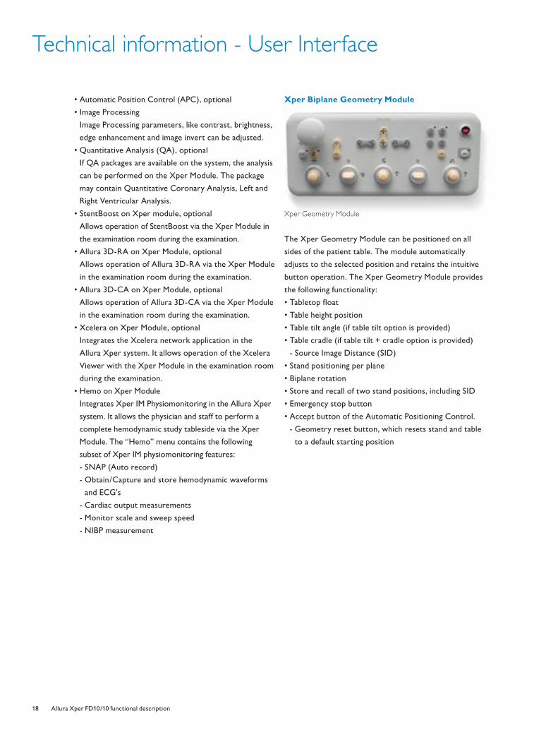
Xper Biplane Geometry Module
The Xper Geometry Module can be positioned on all
sides of the patient table. The module automatically
adjusts to the selected position and retains the intuitive
button operation. The Xper Geometry Module provides
the following functionality:
• Tabletop fl oat
• Table height position
• Table tilt angle (if table tilt option is provided)
• Table cradle (if table tilt + cradle option is provided)
- Source Image Distance (SID)
• Stand positioning per plane
• Biplane rotation
• Store and recall of two stand positions, including SID
• Emergency stop button
• Accept button of the Automatic Positioning Control.
- Geometry reset button, which resets stand and table
to a default starting position
Xper Geometry Module
4522_962_22291.indd Sec1:184522_962_22291.indd Sec1:18 4/9/08 4:06:57 PM4/9/08 4:06:57 PM
19Allura Xper FD10/10 functional description
Xper User Interface in the control room
In the control room, the Xper User Interface includes an
Xper Review Module, two LCD monitors, the keyboard,
a mouse, and monitor pedestal to align the monitor
heights. The monitors have shared screens: the left color
screen is the data monitor, and the black and white
screen is the review monitor.
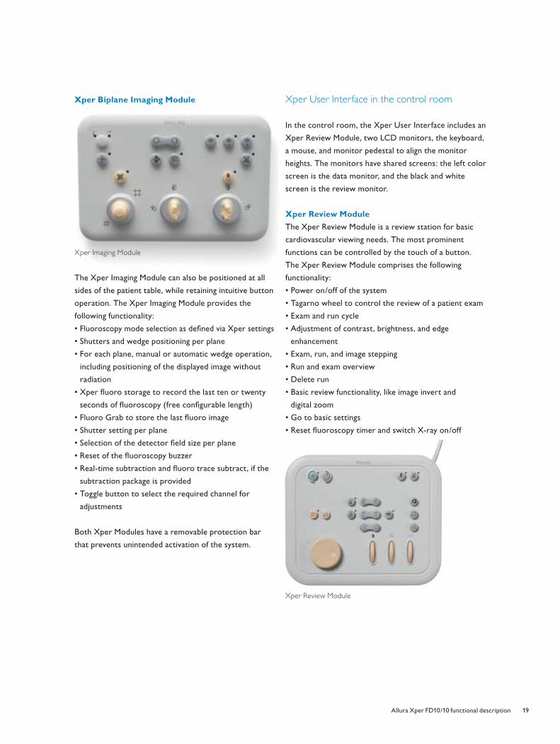
Xper Review Module
The Xper Review Module is a review station for basic
cardiovascular viewing needs. The most prominent
functions can be controlled by the touch of a button.
The Xper Review Module comprises the following
functionality:
• Power on/off of the system
• Tagarno wheel to control the review of a patient exam
• Exam and run cycle
• Adjustment of contrast, brightness, and edge
enhancement
• Exam, run, and image stepping
• Run and exam overview
• Delete run
• Basic review functionality, like image invert and
digital zoom
• Go to basic settings
• Reset fl uoroscopy timer and switch X-ray on/off
Xper Review Module
Xper Biplane Imaging Module
The Xper Imaging Module can also be positioned at all
sides of the patient table, while retaining intuitive button
operation. The Xper Imaging Module provides the
following functionality:
• Fluoroscopy mode selection as defi ned via Xper settings
• Shutters and wedge positioning per plane
• For each plane, manual or automatic wedge operation,
including positioning of the displayed image without
radiation
• Xper fl uoro storage to record the last ten or twenty
seconds of fl uoroscopy (free confi gurable length)
• Fluoro Grab to store the last fl uoro image
• Shutter setting per plane
• Selection of the detector fi eld size per plane
• Reset of the fl uoroscopy buzzer
• Real-time subtraction and fl uoro trace subtract, if the
subtraction package is provided
• Toggle button to select the required channel for
adjustments
Both Xper Modules have a removable protection bar
that prevents unintended activation of the system.
Xper Imaging Module
4522_962_22291.indd Sec1:194522_962_22291.indd Sec1:19 4/9/08 4:06:58 PM4/9/08 4:06:58 PM
20 Allura Xper FD10/10 functional description
Technical information - User Interface
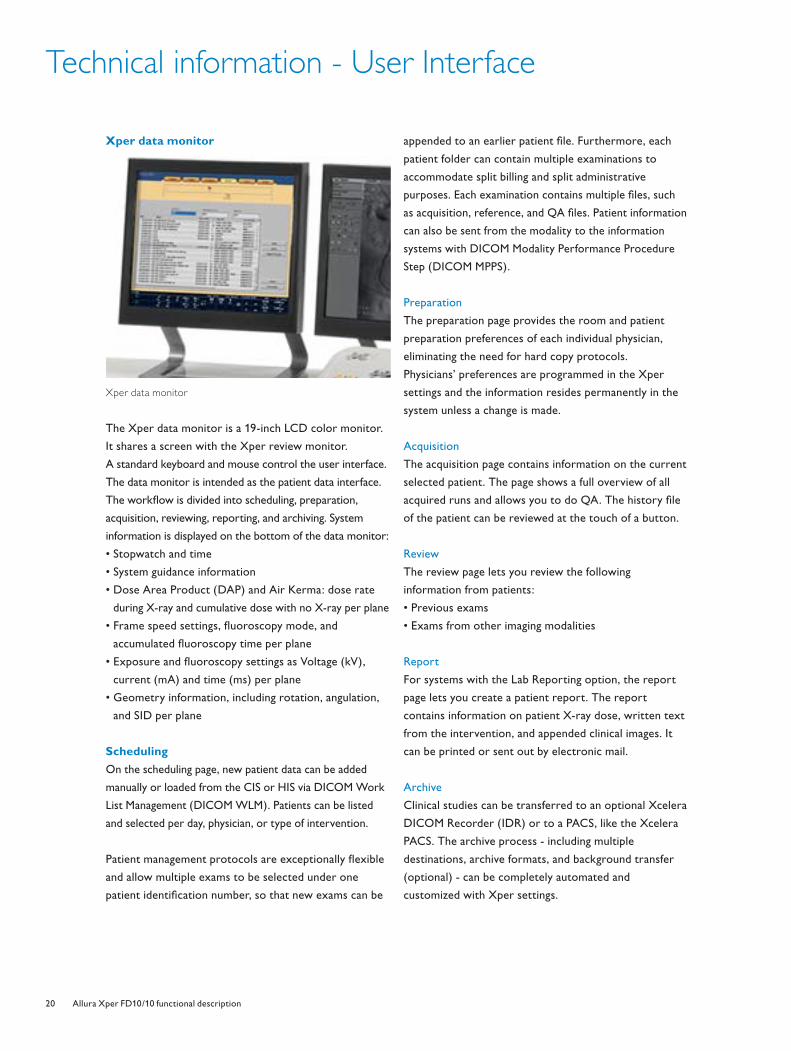
Xper data monitor
Xper data monitor
The Xper data monitor is a 19-inch LCD color monitor.
It shares a screen with the Xper review monitor.
A standard keyboard and mouse control the user interface.
The data monitor is intended as the patient data interface.
The workfl ow is divided into scheduling, preparation,
acquisition, reviewing, reporting, and archiving. System
information is displayed on the bottom of the data monitor:
• Stopwatch and time
• System guidance information
• Dose Area Product (DAP) and Air Kerma: dose rate
during X-ray and cumulative dose with no X-ray per plane
• Frame speed settings, fl uoroscopy mode, and
accumulated fl uoroscopy time per plane
• Exposure and fl uoroscopy settings as Voltage (kV),
current (mA) and time (ms) per plane
• Geometry information, including rotation, angulation,
and SID per plane
Scheduling
On the scheduling page, new patient data can be added
manually or loaded from the CIS or HIS via DICOM Work
List Management (DICOM WLM). Patients can be listed
and selected per day, physician, or type of intervention.
Patient management protocols are exceptionally fl exible
and allow multiple exams to be selected under one
patient identifi cation number, so that new exams can be
appended to an earlier patient fi le. Furthermore, each
patient folder can contain multiple examinations to
accommodate split billing and split administrative
purposes. Each examination contains multiple fi les, such
as acquisition, reference, and QA fi les. Patient information
can also be sent from the modality to the information
systems with DICOM Modality Performance Procedure
Step (DICOM MPPS).
Preparation
The preparation page provides the room and patient
preparation preferences of each individual physician,
eliminating the need for hard copy protocols.
Physicians’ preferences are programmed in the Xper
settings and the information resides permanently in the
system unless a change is made.
Acquisition
The acquisition page contains information on the current
selected patient. The page shows a full overview of all
acquired runs and allows you to do QA. The history fi le
of the patient can be reviewed at the touch of a button.
Review
The review page lets you review the following
information from patients:
• Previous exams
• Exams from other imaging modalities
Report
For systems with the Lab Reporting option, the report
page lets you create a patient report. The report
contains information on patient X-ray dose, written text
from the intervention, and appended clinical images. It
can be printed or sent out by electronic mail.
Archive
Clinical studies can be transferred to an optional Xcelera
DICOM Recorder (IDR) or to a PACS, like the Xcelera
PACS. The archive process - including multiple
destinations, archive formats, and background transfer
(optional) - can be completely automated and
customized with Xper settings.
4522_962_22291.indd Sec1:204522_962_22291.indd Sec1:20 4/9/08 4:07:00 PM4/9/08 4:07:00 PM

21Allura Xper FD10/10 functional description
Technical information - User Interface options
Xper review monitor
The review monitor is an 18-inch black and white LCD
monitor that is shared with the color data monitor.
Xper review monitor
The Graphical User Interface on the black and white
monitor has the following features:
• Step through exam, run, or images
• Exam and run overview
• Image processing features, such as contrast, brightness,
and edge enhancement
• Flagging runs or images for transfer
• Exam annotation
• Automatic printing
• Quantitative Analysis Packages, if available
• Subtraction functionality, if available
Second Xper Biplane Imaging Module
Extension of the imaging controls in the control room
with a second module in a master-slave confi guration.
Second Xper Biplane Geometry Module
Extension of the geometry controls in the control room
with a second module in a master-slave confi guration.
Second or third Xper Module
Additional Xper Modules can be connected in the
control room or in the examination room at tableside.
Up to two Xper Modules can be connected in the
control room, but only one Xper Module can be
connected in the examination room.
The specifi cations and information on the Xper Module
are similar for all Xper Modules connected to the
system. If more than one Xper Module is connected
to the system, each Xper Module can be operated
independently.
Product Security
McAfee Virus-Scan software has been validated
for use with our CV products. Please refer to
Philips' service and security documentation for
specifi c product details regarding McAfee usage
and installation.
Philips offers:
• Online security resources to address your
privacy, security, and regulatory concerns
• Access to security professionals who can assist
with your IT department's compliance efforts
and risk assessment
• Vulnerability monitoring and 24x7 incident
response to help ensure that cyber security
threats to medical devices and systems do not
interfere with patient care
4522_962_22291.indd Sec1:214522_962_22291.indd Sec1:21 4/9/08 4:07:03 PM4/9/08 4:07:03 PM
22 Allura Xper FD10/10 functional description
Technical information - Integration
The Xper DICOM Image Interface enables the export
of clinical images to a destination like a CD-Medical
station or a PACS server. The export formats are based
on DICOM 3.0 protocols. The system exports clinical
studies in Cardiac DICOM XA Multi-Frame or DICOM
Secondary Capture formats:
• The Xper DICOM Image Interface transfers through its
fast ethernet link, making images available on-line within
seconds. The archiving process can be confi gured in the
Xper settings
• The images are sent out, either in the background or
manually, upon completion of the examination
• The export format can be confi gured to a 512x512 or
1024x1024 matrix, at 8- or 10 bit resolution
• The examination can be sent to multiple destinations
for archiving and reviewing purposes
• The Xper DICOM Imaging Interface provides DICOM
Storage and DICOM Storage Commitment Services.
The DICOM Query/Retrieve function allows older
DICOM XA MF and DICOM SC studies to be uploaded
to the system. Additional information can be appended
to a study without changing the patient identifi cation
Storage capacity
The Allura Xper FD 10/10 has a standard storage capacity
of approximately 100 cardiac examinations, which can
store up to 100,000 images in a 10242 10-bit matrix.
MultiSwitch, option
MultiSwitch enables the Xper workspot in the control
room to be shared with other applications that are
loaded on separate PC modalities. The MultiSwitch
enables you to switch to the color LCD data monitor,
keyboard and mouse that are normally connected to the
Allura Xper system. This saves signifi cant space in the
control room by enabling only one monitor and
keyboard to be used for multiple applications, like
StentBoost, Allura 3D- RA, Allura 3D-CA, Xcelera,
Xper IM and ViewForum.
MultiSwitch includes Window Switch functionality.
Xper Window Switch is a web based-browser (HTML)
or X-window (Exceed) application that allows the
Xper Viewing Console to be switched to Radiology/
Cardiology Information Systems. The Xper Window
Switch option makes full use of the available RIS/CIS
facilities and existing support for automatic handling of
logistic tasks (e.g., automatic tracking, purchasing
supplies, and billing).
Lab Reporting, option
This option allows the clinical user to generate and print
a report in modality stand-alone situations. The user can
incorporate free text, clinical images, and X-ray dose
information. The report is sent out via Dicom MPPS
and contains:
• Total Fluoroscopy Time in minutes
• Radiation dose
• Total number of Exposures in numbers
• Accumulated Fluoroscopy Dose in mGy
• Accumulated Exposure Dose in mGy
• Total Dose in mGy
• Total Number of Frames in numbers
• Image Area Dose Product in mGy
• Entrance dose and Air Kerma in mGy
Detailed exposure information:
• Number of Exposure Results
• Exposure-related information, including Exposure
Channel, Exposure Start Time, KVP, Distance Source to
Detector (SID), Exposure Time, X-ray Tube Current,
Positioner Primary Angle, Positioner Secondary Angle,
and Frame Rate
Part of the report is generated automatically from
administrative data (e.g., patient/exam data, hospital
name), and acquired data (e.g., run-log and event-log).
4522_962_22291.indd Sec1:224522_962_22291.indd Sec1:22 4/9/08 4:07:07 PM4/9/08 4:07:07 PM
23Allura Xper FD10/10 functional description
Technical information - Image Detection
The Allura Xper FD10/10 is equipped with the latest
generation dynamic fl at detector, whose compact size can
easily handle complex projections. Image quality and X-ray
dose reduction are further enhanced by Xres techniques.
Dynamic Flat Detector
Philips’ next generation dynamic fl at detector provides
excellent quality at a low patient X-ray dose.
Specifi cations for each plane:
• Size of detector housing, including BodyGuard: 37 cm
(14 in.) diagonal
• Field Of View: 25 cm (10 in.), diagional square
• Detector zoom fi elds: 19 and 15 cm (8 and 6 in.)
diagonal square format
• Pixel size: 184 x 184 microns to allow visualization of
the smallest details
• Detective Quantum Effi ciency DQE(0): 75%
• Output digital video frame: 10242 at 14-bit depth
resolution
• Acquisition speeds can not be customized per system:
3.75, 7.5, 15 and 30 frames/second
• Digital video frame out for archiving purposes is
customizable via Xper settings in different formats:
10242, 5122, and 8 or 10 bit
Xres
Xres is a real-time image processing algorithm originally
developed by Philips Research. Xres exploits the benefi ts
of the fully digital detector to reduce noise in clinical
images. It uses spatial fi ltering and does not compromise
in image quality. Xres provides excellent visualization of
coronary arteries in complex projections by harmonizing
the background image. Plus, it improves the visualization
of the region of interest. For example, it visualizes the
fi ne details in the coronary arteries in situations where
the arteries are projected over the diaphragm or spine.
Specifi cations:
• Real-time processing for X-ray fl uoro and exposure
speeds of up to 30 fps
• Xres can be customized for different image profi les via
Xper settings. This customization allows each clinical
user to choose their preferred image quality
Fluoroscopy
Three fl uoro modes are available at tableside and these
can be programmed via Xper settings. Each mode can be
programmed with a different composition of X-ray dose
rate, digital processing, and fi lter settings.
Specifi cations:
• Fluoroscopy image processing: recursive fi ltering,
localized contrast-adaptive contour enhancement and
Xres algorithm
• Pulsed X-ray modes with a Fast Fluoro Reset function,
which quickly returns the system to fl uoroscopy if
there is an unexpected system reset
• Pulse rates: system-customized at 3.75, 7.5, 15 and 30
pulses per second
• Choice of Last Image Hold during fl uoroscopy or a loop
of the last fl uoroscopy run (service confi gurable time)
• Frame grabbing of static fl uoroscopy images or Xper
fl uoro storage to store the fl uoroscopy run (service
confi gurable time) for reference or archiving
Subtraction package
The Digital Subtraction Angiography (DSA) option
extends the vascular applicational functionality of the
Allura Xper system. DSA features real-time digital
subtraction at low frame speeds of 0.5, 1, 2, 3, or 6
frames per second. The DSA programs can be selected
via the Xper settings.
This option’s exposure technique provides exceptional
image quality for Subtracted images. It also offers run-
subtract to perform subtraction per run.
This feature can be applied in the Rotational Scan and
Bolus Chase Subtract options. DSA includes the
following functionality: Fluoro-Trace, Fluoro-Subtract,
Exposure Subtract on individual images or runs,
mask selection, landmarking, and pixel shift.
4522_962_22291.indd Sec1:234522_962_22291.indd Sec1:23 4/9/08 4:07:07 PM4/9/08 4:07:07 PM
24 Allura Xper FD10/10 functional description
Technical information - X-ray Generation
X-ray generation consists of the following elements: X-
ray generator, X-ray tube, collimator (including
SpectraBeam beam fi ltration), and dose protection
mechanism. The complete dose protection mechanism is
part of the DoseWise program.
Velara X-ray generator
The Velara generator is optimized for the latest
cardiovascular needs.
Specifi cations for each plane:
• Microprocessor-controlled, 100 kW high-frequency
converter generator
• Quartz-controlled IGBT-power-switch, with a
minimum switching time of 1 ms
• Voltage range: 40 to 125 kV
• Maximum current: 1250 mA at 80 kV
• Maximum continuous power: 2.5 kW for 0.5 hours,
2kW for 0.8 hours
• Nominal power (highest electrical power): 100 kW
(1000 mA at 100 kV)
The Xper settings for X-ray generation control can be
selected on the Xper Module.
Xper Beam Shaping
Xper Beam Shaping provides virtual collimation of the
shutters and wedges on the last X-ray image, eliminating
additional X-ray dose during collimation changes.
X-ray tube
The Allura Xper FD10/10 features the legendary high
power MRC-GS 0508 X-ray tube. In the last seven years,
Philips has installed more than 5000 MRC X-ray tubes
with customers around the world. Data shows that on
average, the MRC X-ray tube lasts signifi cantly longer
than conventional tubes.
MRC-GS 0508X-ray tube
MRC-GS 0508
The powerful MRC-GS 0508 X-ray tube allows very high
heat dissipation, enabling SpectraBeam fi ltration to
reduce patient dose signifi cantly.
Specifi cations:
• 0.5/0.8 nominal focal spot values with maximum
loadability of 45 kW and 85 kW
• Grid Switching with pulsed fl uoroscopy
• Anode heat dissipation in continuous mode: 3200 W
• Fluoro power for 10 minutes: 4500 W
• Fluoro power for 20 minutes: 3500 W
• Maximum heat dissipation of assembly: 3500 W
• SpectraBeam dose management
• Oil-cooled X-ray tube with thermal safety switch
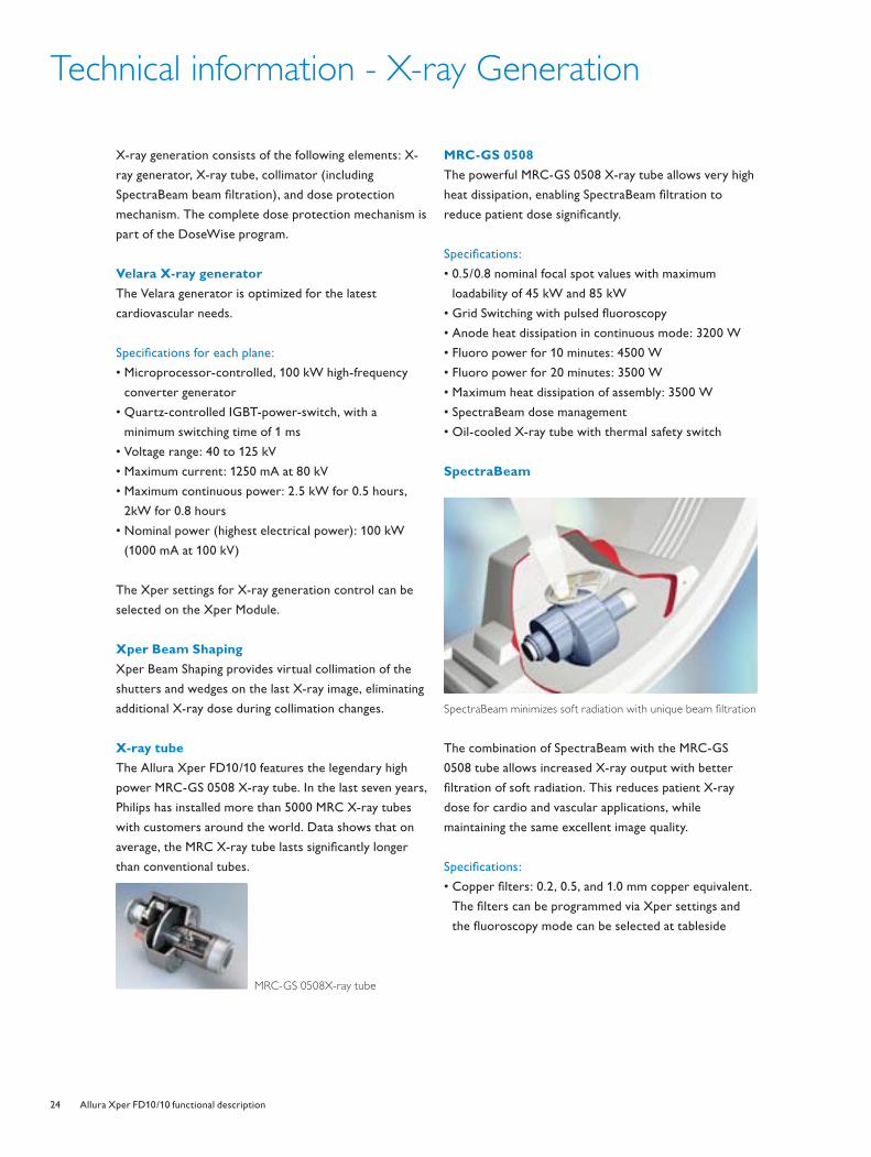
SpectraBeam
Spectrabeam
SpectraBeam minimizes soft radiation with unique beam fi ltration
The combination of SpectraBeam with the MRC-GS
0508 tube allows increased X-ray output with better
fi ltration of soft radiation. This reduces patient X-ray
dose for cardio and vascular applications, while
maintaining the same excellent image quality.
Specifi cations:
• Copper fi lters: 0.2, 0.5, and 1.0 mm copper equivalent.
The fi lters can be programmed via Xper settings and
the fl uoroscopy mode can be selected at tableside
4522_962_22291.indd Sec1:244522_962_22291.indd Sec1:24 4/9/08 4:07:07 PM4/9/08 4:07:07 PM

25Allura Xper FD10/10 functional description
Technical information - Viewing
The system is delivered standard with four black and white
18-inch LCD monitors in the examination room. One 19-
inch LCD color monitor and two 18-inch black and white
LCD monitors are standard in the control room.
Specifi cations of color LCD monitor:
• 19-inch color LCD display
• Native format: 1280 x 1024 SXGA
• Wide viewing angle: approximately 170°
• Controlled brightness: typically 270 Cd/ m2,
with ambient light dependent brightness control
• On-screen display of control functions operated via
push buttons
- Audio output 0.5 Watt
- Contrast typically 800 on 1
Specifi cations of monochrome LCD monitor:
• 18-inch monochrome LCD display with a native format
of 1280 x 1024 SXGA
• 10 bit grey-scale resolution with grey-scale correction
• Wide viewing angle: approximately 160°
• High brightness: maximum 600 Cd/ m2, with ambient
light dependent brightness control
• On-screen display of control functions operated via
push buttons
• Examination room LCD monitors include protection
screen and motorized height adjustment
LCD monitors in the control room LCD monitors in the examination room
Delivering clinical excellence with Philips CUSTOMerCARE service agreements
Philips CUSTOMerCARE service agreements deliver the fl exibility and choice for you to succesfully
manage your business. We offer customized solutions that enhance the quality of your care, increase your
productivity, and improve your fi nancial performance.
Philips Comprehensive service agreements meet the intensive requirements of your high performance
environment and offer advanced total solution support, increased uptime, clinical education, and exceptional
overall value.
Philips Alliance agreements fully leverage the value of your in-house biomedical and clinical engineering staff.
Philips Basic coverage plans are available for essential needs, such as planned maintenance or equipment repair.
4522_962_22291.indd Sec1:254522_962_22291.indd Sec1:25 4/9/08 4:07:08 PM4/9/08 4:07:08 PM

26 Allura Xper FD10/10 functional description
Technical information - Options
MultiVision
The MultiVision video switch is the integrated video
switch for high quality, progressive display video sources.
It can switch either black and white or color signals, and
supports up to four inputs to one output. MultiVision
enables an extra color monitor in the ceiling suspension in
the examination room to be shared between the system
and other sources, such as a DICOM viewer, an
Ultrasound system, StentBoost, a Allura 3D-RA or Allura
3D-CA interventional tool, etc. The switch is controlled
via the acquisition manual on the Xper Module.
Physio Viewing
Physio Viewing provides acquisition, storage and display
of physiological signals on the Allura Xper FD10/10
system. Four physiological data signals can be acquired
and stored. One signal of choice can be displayed when
reviewing images.
Continuous autopush
The continuous autopush option provides additional
processor boards that are dedicated to archiving to
minimize interruptions caused by other functions that
require the image processor, such as patient review.
Using this option speeds up archiving and the availability
of clinical images for reviewing at other PACS destinations.
DICOM Print
DICOM Print provides an interface to any DICOM
Printer. It provides Print Preview, Print Manual
Overrides, Print Job submission, and Print Job
management via automated printing protocols.
Intercom
Remote Intercom is used for communication between
the examination and control room.
RIS/CIS DICOM Interface
This interface option enables two-way communication
between the system, a local Information System (CIS or
RIS), or hemodynamic system. The interface uses the
DICOM Worklist Management (DICOM WLM) and
Modality Performed Procedure Step (DICOM MPPS)
standards. If a hospital has an information system, it is
possible to receive patient and examination (request)
information and to report the examination results.
This option provides the following benefi ts:
• Eliminates the need to retype patient information on
the system
• Prevents errors in typing patient name or registration
number, which ensures consistency of information
throughout the department to prevent problems in
archive clusters
• Provides information to and from the information
system about the acquired images and radiation dose
Upon request from the system, the complete worklist
with all relevant patient and examination data is returned
to the system.
Biplane standard line rate video input/output
The standard line rate video output is 625 (525) lines for a
50 (60) Hz video interface board. This option is required
to connect standard line rate video peripherals for each
plane, such as a VCR and/or video printer. The interface
provides control for automatic biplane recording of fl uoro
and exposures with a VCR medical DVD recorder, and for
replay of VCR or DVD images (on any SLR video source)
on the system monitors.
Real-time digital link
The real-time digital link is a dedicated image link to an
interventional tool, such as Allura 3D-RA, StentBoost,
and/or Allura 3D-CA. This dedicated digital link sends raw
or processed image data (depending on the application) in
real-time during exposures to the connected interventional
tool. This provides instant results of the applicable
reconstruction after the exposure run.
4522_962_22291.indd Sec1:264522_962_22291.indd Sec1:26 4/9/08 4:07:10 PM4/9/08 4:07:10 PM
27Allura Xper FD10/10 functional description
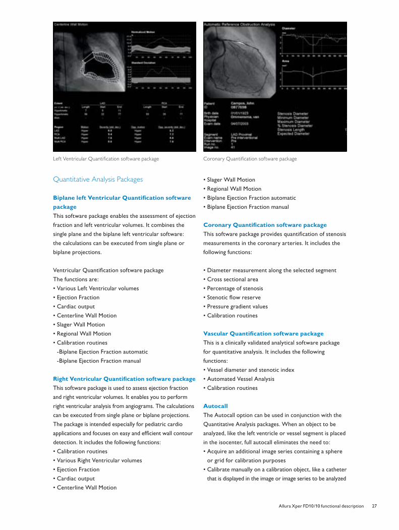
Left Ventricular Quantifi cation software package
Quantitative Analysis Packages
Biplane left Ventricular Quantifi cation software
package
This software package enables the assessment of ejection
fraction and left ventricular volumes. It combines the
single plane and the biplane left ventricular software:
the calculations can be executed from single plane or
biplane projections.
Ventricular Quantifi cation software package
The functions are:
• Various Left Ventricular volumes
• Ejection Fraction
• Cardiac output
• Centerline Wall Motion
• Slager Wall Motion
• Regional Wall Motion
• Calibration routines
-Biplane Ejection Fraction automatic
-Biplane Ejection Fraction manual
Right Ventricular Quantifi cation software package
This software package is used to assess ejection fraction
and right ventricular volumes. It enables you to perform
right ventricular analysis from angiograms. The calculations
can be executed from single plane or biplane projections.
The package is intended especially for pediatric cardio
applications and focuses on easy and effi cient wall contour
detection. It includes the following functions:
• Calibration routines
• Various Right Ventricular volumes
• Ejection Fraction
• Cardiac output
• Centerline Wall Motion
• Slager Wall Motion
• Regional Wall Motion
• Biplane Ejection Fraction automatic
• Biplane Ejection Fraction manual
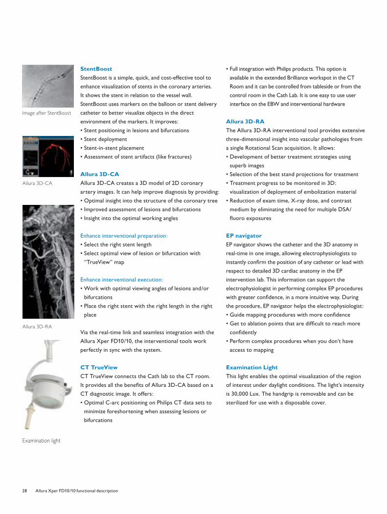
Coronary Quantifi cation software package
This software package provides quantifi cation of stenosis
measurements in the coronary arteries. It includes the
following functions:
• Diameter measurement along the selected segment
• Cross sectional area
• Percentage of stenosis
• Stenotic fl ow reserve
• Pressure gradient values
• Calibration routines
Vascular Quantifi cation software package
This is a clinically validated analytical software package
for quantitative analysis. It includes the following
functions:
• Vessel diameter and stenotic index
• Automated Vessel Analysis
• Calibration routines
Autocall
The Autocall option can be used in conjunction with the
Quantitative Analysis packages. When an object to be
analyzed, like the left ventricle or vessel segment is placed
in the isocenter, full autocall eliminates the need to:
• Acquire an additional image series containing a sphere
or grid for calibration purposes
• Calibrate manually on a calibration object, like a catheter
that is displayed in the image or image series to be analyzed
Coronary Quantifi cation software package
4522_962_22291.indd Sec1:274522_962_22291.indd Sec1:27 4/9/08 4:07:10 PM4/9/08 4:07:10 PM
28 Allura Xper FD10/10 functional description
StentBoost
StentBoost is a simple, quick, and cost-effective tool to
enhance visualization of stents in the coronary arteries.
It shows the stent in relation to the vessel wall.
StentBoost uses markers on the balloon or stent delivery
catheter to better visualize objects in the direct
environment of the markers. It improves:
• Stent positioning in lesions and bifurcations
• Stent deployment
• Stent-in-stent placement
• Assessment of stent artifacts (like fractures)
Allura 3D-CA
Allura 3D-CA creates a 3D model of 2D coronary
artery images. It can help improve diagnosis by providing:
• Optimal insight into the structure of the coronary tree
• Improved assessment of lesions and bifurcations
• Insight into the optimal working angles
Enhance interventional preparation:
• Select the right stent length
• Select optimal view of lesion or bifurcation with
“TrueView” map
Enhance interventional execution:
• Work with optimal viewing angles of lesions and/or
bifurcations
• Place the right stent with the right length in the right
place
Via the real-time link and seamless integration with the
Allura Xper FD10/10, the interventional tools work
perfectly in sync with the system.
CT TrueView
CT TrueView connects the Cath lab to the CT room.
It provides all the benefi ts of Allura 3D-CA based on a
CT diagnostic image. It offers:
• Optimal C-arc positioning on Philips CT data sets to
minimize foreshortening when assessing lesions or
bifurcations
• Full integration with Philips products. This option is
available in the extended Brilliance workspot in the CT
Room and it can be controlled from tableside or from the
control room in the Cath Lab. It is one easy to use user
interface on the EBW and interventional hardware
Allura 3D-RA
The Allura 3D-RA interventional tool provides extensive
three-dimensional insight into vascular pathologies from
a single Rotational Scan acquisition. It allows:
• Development of better treatment strategies using
superb images
• Selection of the best stand projections for treatment
• Treatment progress to be monitored in 3D:
visualization of deployment of embolization material
• Reduction of exam time, X-ray dose, and contrast
medium by eliminating the need for multiple DSA/
fl uoro exposures
EP navigator
EP navigator shows the catheter and the 3D anatomy in
real-time in one image, allowing electrophysiologists to
instantly confi rm the position of any catheter or lead with
respect to detailed 3D cardiac anatomy in the EP
intervention lab. This information can support the
electrophysiologist in performing complex EP procedures
with greater confi dence, in a more intuitive way. During
the procedure, EP navigator helps the electrophysiologist:
• Guide mapping procedures with more confi dence
• Get to ablation points that are diffi cult to reach more
confi dently
• Perform complex procedures when you don’t have
access to mapping
Examination Light
This light enables the optimal visualization of the region
of interest under daylight conditions. The light’s intensity
is 30,000 Lux. The handgrip is removable and can be
sterilized for use with a disposable cover.
Examination light
Allura 3D-RA
Allura 3D-CA
Image after StentBoost
4522_962_22291.indd Sec1:284522_962_22291.indd Sec1:28 4/9/08 4:07:13 PM4/9/08 4:07:13 PM

29Allura Xper FD10/10 functional description
Accessories
• Peripheral X-ray fi lter
• Cath arm support (adjustable)
• Ratchet compressor
• Table X-ray protection
• Pulse cath arm support
• Pan handle
• Ceiling-suspended radiation shield
• MCS bracket ceiling Radiation Shield
• Examination light
• Drip stand
• Arm support
• Mattress
• Neuro Mattress
• Set of arm supports
• Table clamp
• Patient straps
• Head support
• Set hand grips & clamps
• Cerebral fi lter
• Neuro wedge
• Cable holders (15 pieces)
Head support
Arm support
Pulse cath arm support
Patient straps
4522_962_22291.indd Sec1:294522_962_22291.indd Sec1:29 4/9/08 4:07:16 PM4/9/08 4:07:16 PM
30 Allura Xper FD10/10 functional description
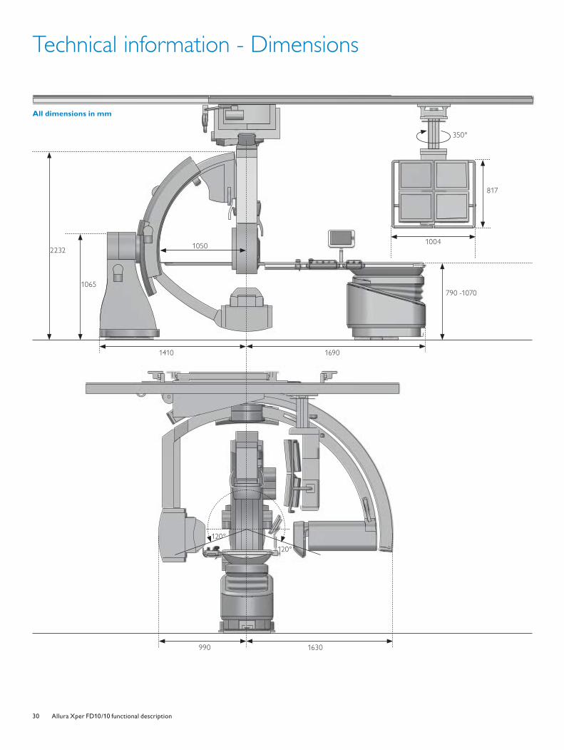
Technical information - Dimensions
All dimensions in mm
1410
1065
2232
350°
817
790 -1070
120°
1050
120°
1004
1690
990 1630
4522_962_22291.indd Sec1:304522_962_22291.indd Sec1:30 4/9/08 4:07:18 PM4/9/08 4:07:18 PM
31Allura Xper FD10/10 functional description
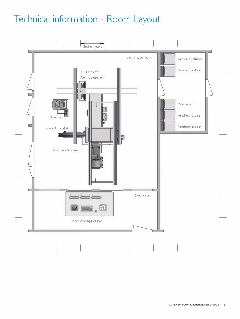
Technical information - Room Layout
Generator cabinet
Generator cabinet
Main cabinet
Peripheral cabinet
Peripheral cabinet
Grid in meters
LCD Monitor
Ceiling Suspension
Lateral Arc (LARC)
Floor mounted G-stand
Examination room
Control room
Xper Viewing Console
Generator cabinet
Generator cabinet
Main cabinet
Peripheral cabinet
Peripheral cabinet
Grid in meters
LCD Monitor
Ceiling Suspension
Floor mounted G-stand
Examination room
Control room
Xper Viewing Console
Injector
Lateral Arc (LARC)
4522_962_22291.indd Sec1:314522_962_22291.indd Sec1:31 4/9/08 4:07:27 PM4/9/08 4:07:27 PM
Philips Healthcare is part of
Royal Philips Electronics
Interested?
Would you like to know more about our imaginative
products? Please do not hesitate to contact us.
We would be glad to hear from you.
On the web
www.philips.com/healthcare
Via email
By fax
+31 40 27 64 887
By mail
Philips Healthcare
Global Information Center
P.O. Box 1286
5602 BG Eindhoven
The Netherlands
By phone
Asia
Tel: +852 2821 5888
Europe, Middle East, Africa
Tel: +49 7031 463 2254
Latin America
Tel: +55 11 2125 0764
North America
Tel: +1 425 487 7000
800 285 5585 (toll free, US only)
© 2008 Koninklijke Philips Electronics N.V.
All rights are reserved.
Philips Healthcare reserves the right to make changes in specifi cations and/or to discontinue any product at any time without notice or obligation and
will not be liable for any consequences resulting from the use of this publication.
Printed in The Netherlands.
4522 962 22291/722 * APR 2008
Nieuw logo.indd 2 3/31/08 10:04:55 AM4522_962_22291.indd Sec1:324522_962_22291.indd Sec1:32 4/9/08 4:07:28 PM4/9/08 4:07:28 PM