Embed Size (px)
Citation preview
Xin Qi, PhD, Daniel J. Medina, PhD, Laura Zheng, MS, Lei Cong, MS, Hana Aviv, PhD, Lauri A. Goodell, MD,
Roger K. Strair,MD PhD, David J. Foran, PhD
Presentor: Wenjin Chen, Ph.D.Center for Biomedical Imaging and Informatics
The Cancer Institute of New Jersey
Pathology Informatics 2010 Scientific SectionBoston, September 20, 2010
Background Non-Hodgkin lymphomas (NHLs) are a diverse group of blood
cancers that include any kind of lymphoma except Hodgkin's lymphomas. Types of NHL vary significantly in their severity, from indolent to very aggressive.
Lymphomas are types of cancer derived from lymphocytes, a type of white blood cell. Lymphomas are treated by combinations of chemotherapy monoclonal antibodies, immunotherapy, radiation, and hematopoietic stem cell transplantation.
Our team previously reported the development of tools for performing grid-enabled, content-based image retrieval and caBIG compliant data management.
We have since begun to investigate the potential use of these tools for evaluating non-Hodgkin’s lymphoma histology specimens to determine the staining and expression patterns of markers CD20, CD21, CD138, and FDC.
Motivation Enhance our understanding of biology of non-
Hodgekin’s lymphomas Simultaneously visualize protein expressions
and chromosomal changes from tissue sections
Multispectual imaging potencially allows for analysing multiple proteins/DNA probes on single cells or on sub-cellular compartments
Preserve tissue resources
Challenges
Sample preparation protocols (FICTION)
Multispectral Imaging protocols
Unmixing Signals
Data processing
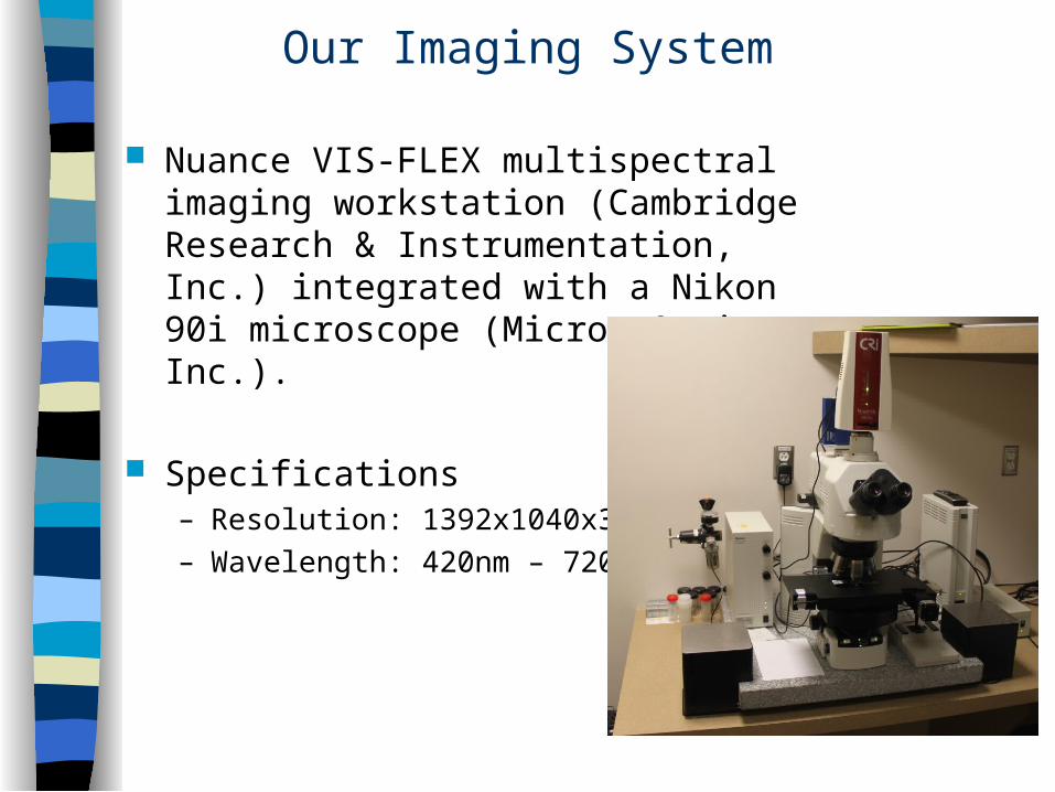
Our Imaging System
Nuance VIS-FLEX multispectral imaging workstation (Cambridge Research & Instrumentation, Inc.) integrated with a Nikon 90i microscope (Micron Optics, Inc.).
Specifications– Resolution: 1392x1040x31– Wavelength: 420nm – 720 nm
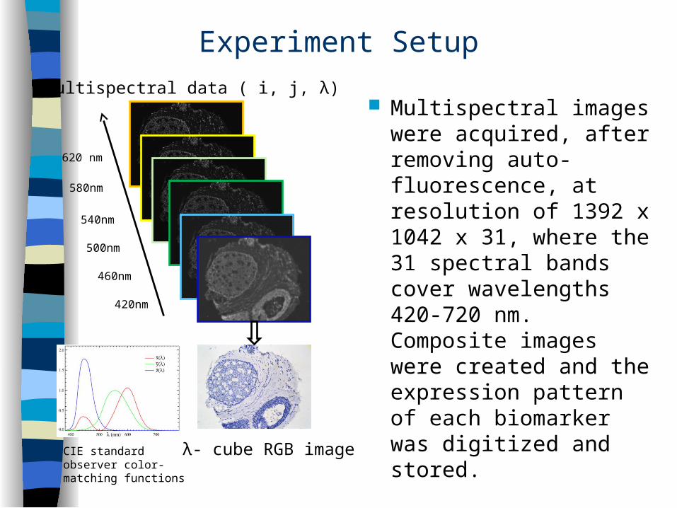
Experiment Setup
Multispectral images were acquired, after removing auto-fluorescence, at resolution of 1392 x 1042 x 31, where the 31 spectral bands cover wavelengths 420-720 nm. Composite images were created and the expression pattern of each biomarker was digitized and stored.
420nm
460nm
500nm
540nm
580nm
620 nm
Multispectral data ( i, j, λ)
CIE standard observer color-matching functions
λ- cube RGB image
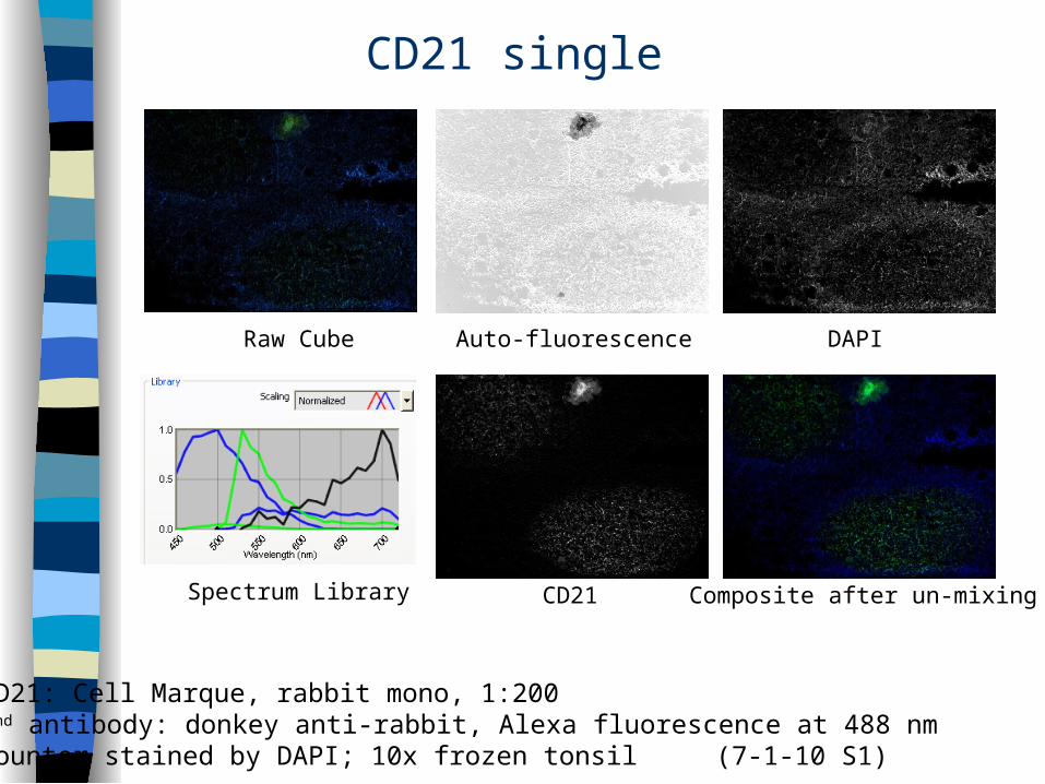
CD21 single
CD21: Cell Marque, rabbit mono, 1:2002nd antibody: donkey anti-rabbit, Alexa fluorescence at 488 nm Counter stained by DAPI; 10x frozen tonsil (7-1-10 S1)
Auto-fluorescence DAPI
CD21 Composite after un-mixing
Raw Cube
Spectrum Library

FDC+CD21
FDC+CD21 (7-1-10 S8)
DAPI
CD21(488) Composite after un-mixing
Raw Cube FDC(594)
Spectrum Library
CD21 (488) + FDC (594)10x; Frozen Tonsil
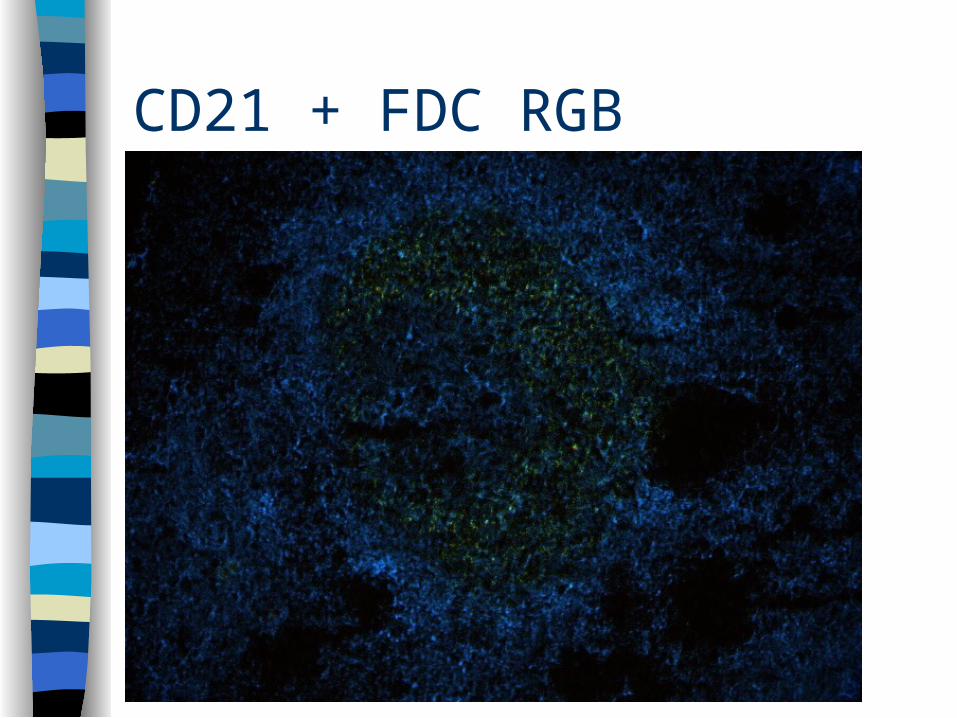
CD21 + FDC RGB
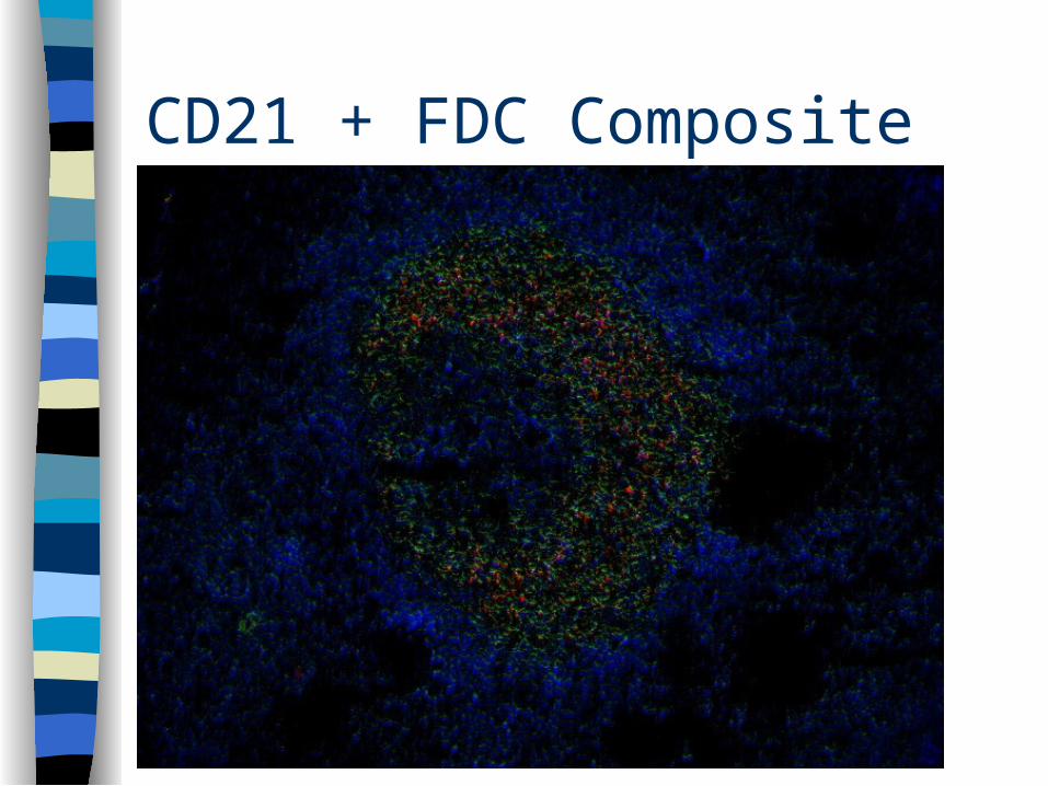
CD21 + FDC Composite
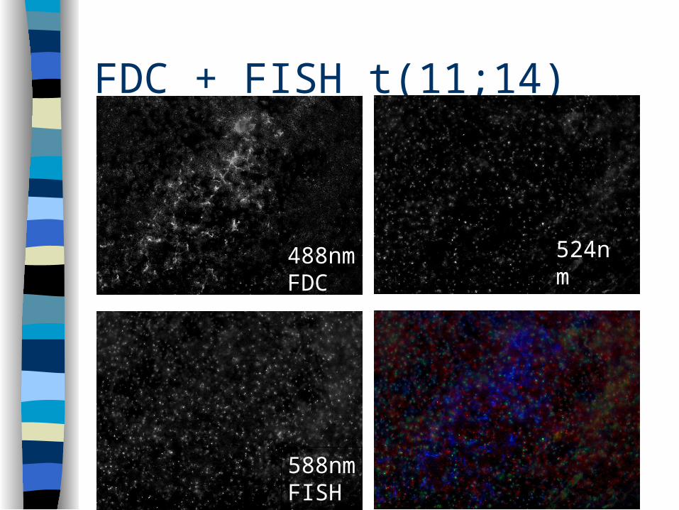
FDC + FISH t(11;14)
488nmFDC
524nmFISH
588nmFISH
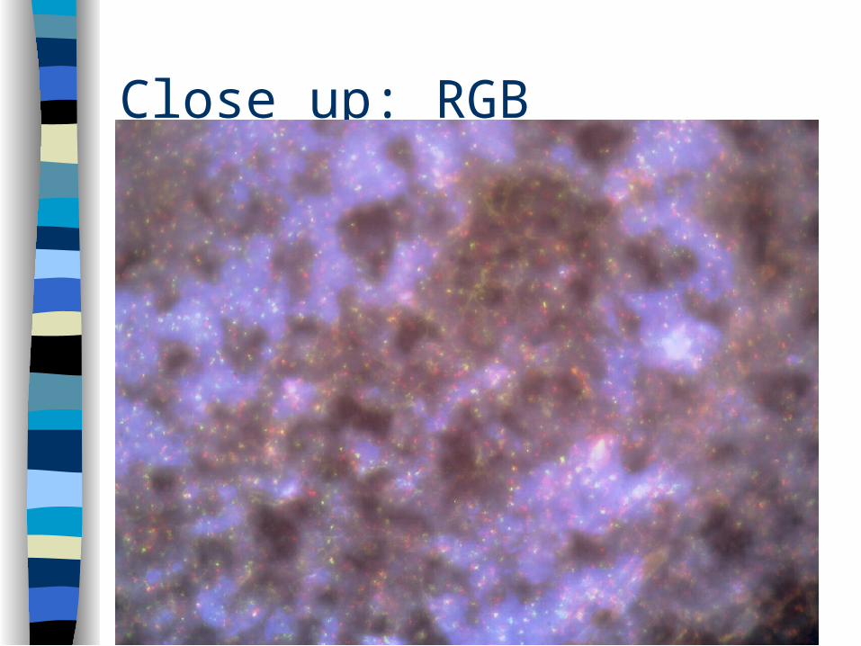
Close up: RGB
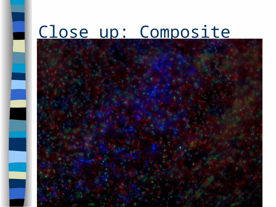
Close up: Composite
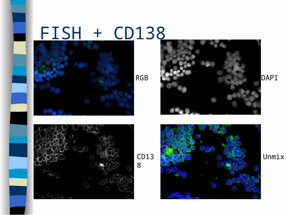
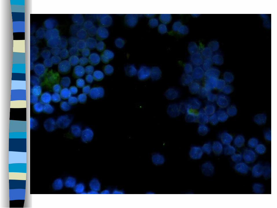
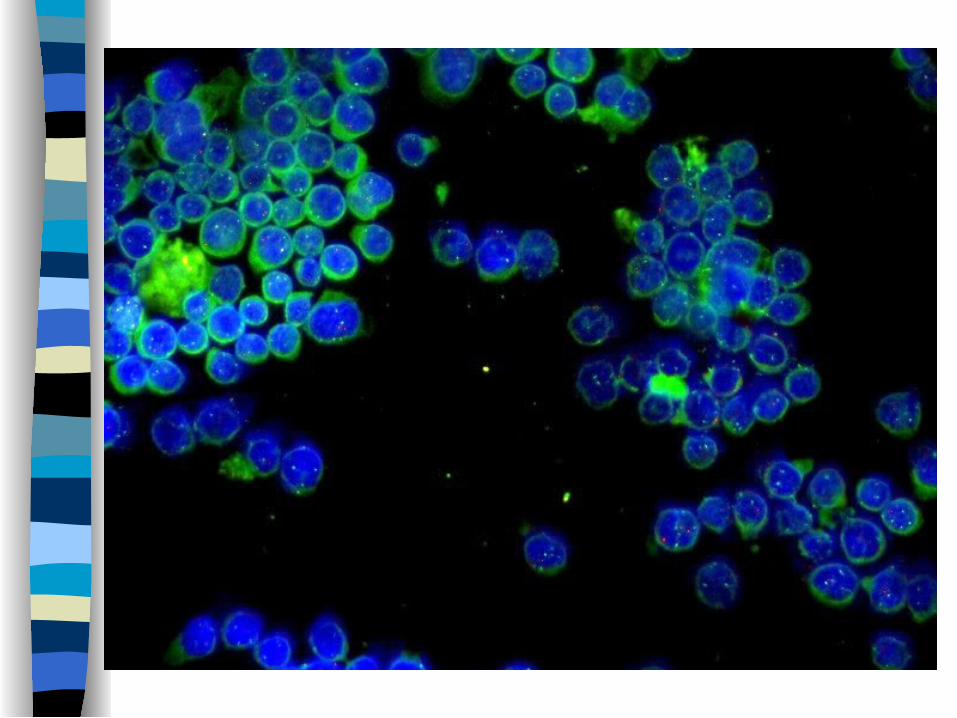
FISH + CD138
RGB DAPI
CD138 Unmix
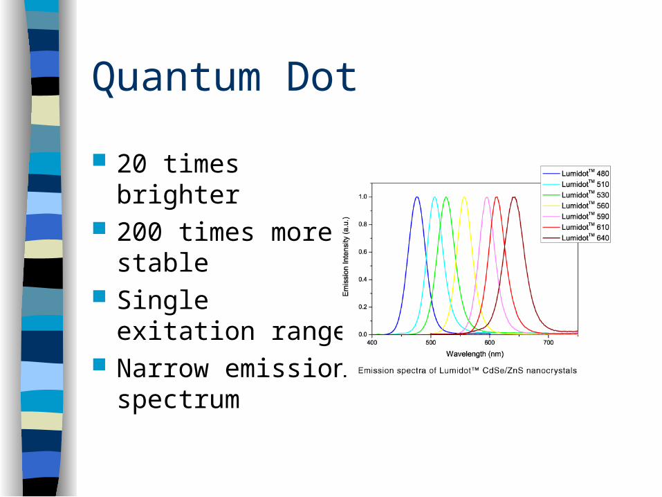
Quantum Dot
20 times brighter 200 times more
stable Single exitation
range Narrow emission
spectrum
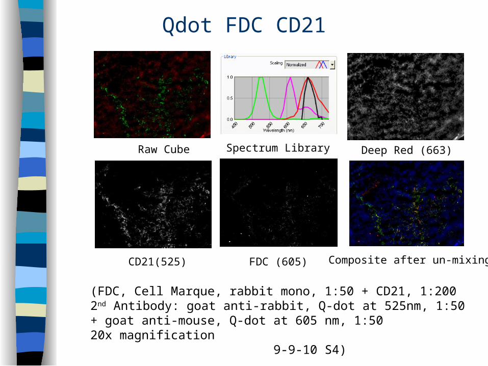
Qdot FDC CD21
Deep Red (663)
CD21(525) Composite after un-mixing
Raw Cube
FDC (605)
(FDC, Cell Marque, rabbit mono, 1:50 + CD21, 1:2002nd Antibody: goat anti-rabbit, Q-dot at 525nm, 1:50 + goat anti-mouse, Q-dot at 605 nm, 1:50 20x magnification 9-9-10 S4)
Spectrum Library
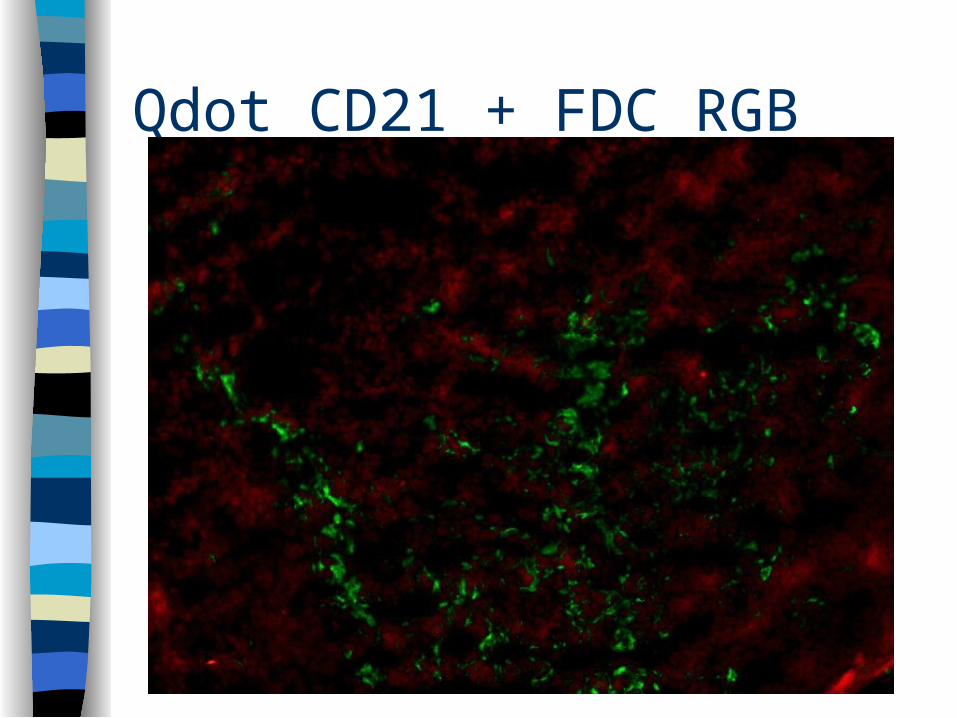
Qdot CD21 + FDC RGB
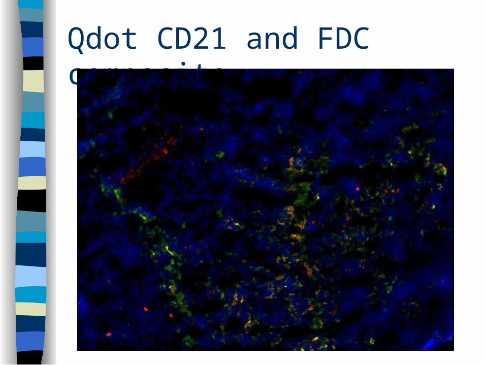
Qdot CD21 and FDC composite
Conclusion
Both the chromosome FISH probes and the surface expression of CD20, CD21, CD138 and FDA could be strongly detected using MSI even though the signals were faint using standard fluorescent imaging.
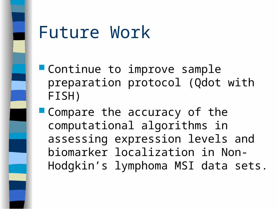
Future Work
Continue to improve sample preparation protocol (Qdot with FISH)
Compare the accuracy of the computational algorithms in assessing expression levels and biomarker localization in Non-Hodgkin’s lymphoma MSI data sets.
Acknowledgement
David J. Foran, Ph. D Daniel J. Medina, Ph. D
Wenjin Chen, Ph.D. Laura Zheng, M.S.
Lauri Goodell, M.D Lei Cong, M.S.
Lin Yang, Ph.D Hana Aviv, Ph.D
Fuyong Xing, M.S. Roger K. Strair, Ph.D., M.D.
Will Cukierski Bekah Gensure
Vicky Chu, M.S.
Acknowledgement This research was funded, in part, by grants from the National
Institutes of Health through contract 5R01EB003587-04 from the National Institute of Biomedical Imaging and Bioengineering and contracts 5R01LM009239-03 and 3R01LM009239-03S2 from the National Library of Medicine. The content is solely the responsibility of the authors and does not necessarily represent the official views of the National Cancer Institute or the National Institutes of Health.
Additional funds were provided by IBM through a Shared University Research Award. UMDNJ also wants to thank and acknowledge IBM for providing free computational power and technical support for this research through World Community Grid.
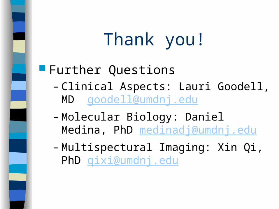
Thank you!
Further Questions– Clinical Aspects: Lauri Goodell, MD
[email protected]– Molecular Biology: Daniel Medina, PhD
[email protected]– Multispectural Imaging: Xin Qi, PhD
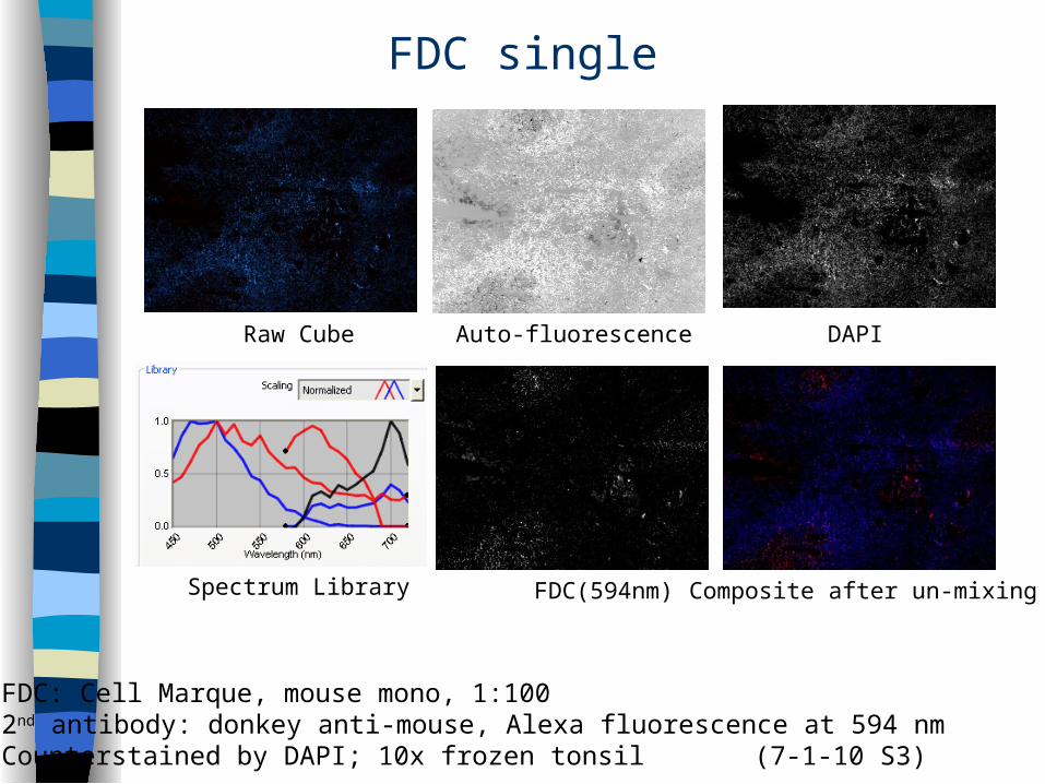
FDC single
Auto-fluorescence DAPI
FDC(594nm) Composite after un-mixing
Raw Cube
Spectrum Library
FDC: Cell Marque, mouse mono, 1:1002nd antibody: donkey anti-mouse, Alexa fluorescence at 594 nmCounterstained by DAPI; 10x frozen tonsil (7-1-10 S3)