Embed Size (px)
Citation preview
X-Ray Rounds: X-Ray Rounds: (Plain) Radiographic Evaluation (Plain) Radiographic Evaluation
of the Ankleof the Ankle
Garry W. K. Ho, M.D.Garry W. K. Ho, M.D.VCU / Fairfax Family PracticeVCU / Fairfax Family Practice
Sports Medicine FellowSports Medicine FellowSeptember 2006September 2006
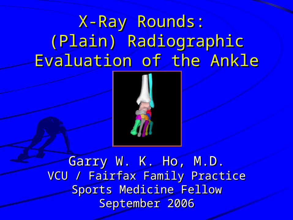
AnatomyAnatomy
Complex hinge jointComplex hinge joint
Articulations among:Articulations among:– FibulaFibula– TibiaTibia– TalusTalus
Tibial “plafond”Tibial “plafond”– Distal tibial articular Distal tibial articular
surfacesurface
Complex ligamentous Complex ligamentous systemsystem
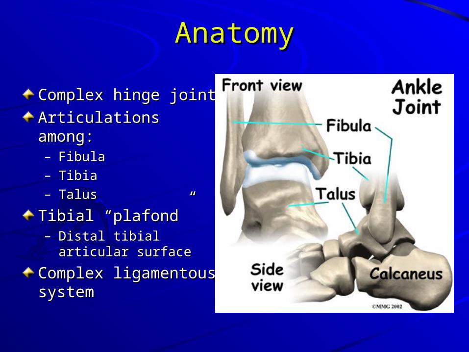
AnatomyAnatomy
Medial malleolusMedial malleolus– Distal tibiaDistal tibia– Medial supportMedial support
Lateral malleolusLateral malleolus– Distal fibulaDistal fibula– Lateral supportLateral support
TalusTalus– Trapezoid-shapedTrapezoid-shaped
Mortise (tibial plafond, medial & lateral malleoli)Mortise (tibial plafond, medial & lateral malleoli)
- Constrained articulation with the talar dome- Constrained articulation with the talar dome
AnatomyAnatomy
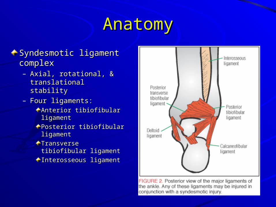
Syndesmotic ligament Syndesmotic ligament complexcomplex– Axial, rotational, & Axial, rotational, &
translational stabilitytranslational stability– Four ligaments:Four ligaments:
Anterior tibiofibular Anterior tibiofibular ligamentligament
Posterior tibiofibular Posterior tibiofibular ligamentligament
Transverse tibiofibular Transverse tibiofibular ligamentligament
Interosseous ligamentInterosseous ligament
AnatomyAnatomy
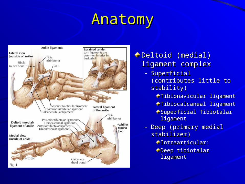
Deltoid (medial) Deltoid (medial) ligament complexligament complex– Superficial (contributes Superficial (contributes
little to stability)little to stability)Tibionavicular ligamentTibionavicular ligament
Tibiocalcaneal Tibiocalcaneal ligamentligament
Superficial Tibiotalar Superficial Tibiotalar ligamentligament
– Deep (primary medial Deep (primary medial stabilizer)stabilizer)
Intraarticular:Intraarticular:
Deep tibiotalar Deep tibiotalar ligamentligament
AnatomyAnatomy
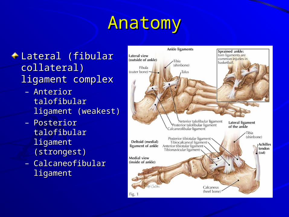
Lateral (fibular Lateral (fibular collateral) ligament collateral) ligament complexcomplex– Anterior talofibular Anterior talofibular
ligament (weakest)ligament (weakest)– Posterior talofibular Posterior talofibular
ligament (strongest)ligament (strongest)– Calcaneofibular Calcaneofibular
ligamentligament
Indications for Ankle RadiographsIndications for Ankle Radiographs
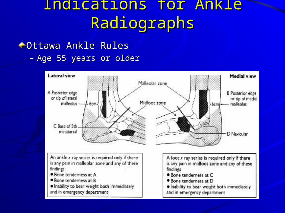
Ottawa Ankle RulesOttawa Ankle Rules– Age 55 years or olderAge 55 years or older
Indications for Ankle RadiographsIndications for Ankle Radiographs
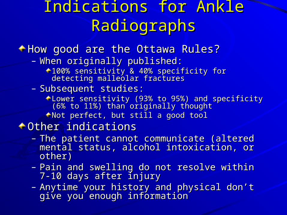
How good are the Ottawa Rules?How good are the Ottawa Rules?– When originally published:When originally published:
100% sensitivity & 40% specificity for detecting 100% sensitivity & 40% specificity for detecting malleolar fracturesmalleolar fractures
– Subsequent studies: Subsequent studies: Lower sensitivity (93% to 95%) and specificity (6% to Lower sensitivity (93% to 95%) and specificity (6% to 11%) than originally thought11%) than originally thoughtNot perfect, but still a good toolNot perfect, but still a good tool
Other indicationsOther indications– The patient cannot communicate (altered The patient cannot communicate (altered
mental status, alcohol intoxication, or other)mental status, alcohol intoxication, or other)– Pain and swelling do not resolve within 7-10 Pain and swelling do not resolve within 7-10
days after injury days after injury – Anytime your history and physical don’t give Anytime your history and physical don’t give
you enough informationyou enough information
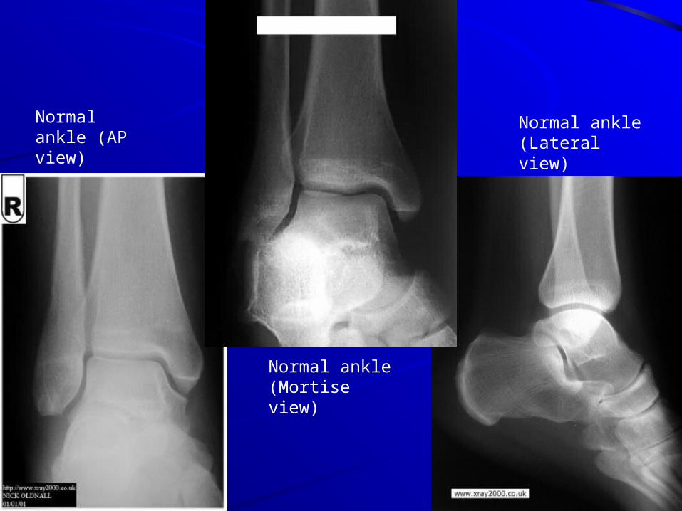
Normal ankle (AP view)
Normal ankle (Mortise view)
Normal ankle (Lateral view)
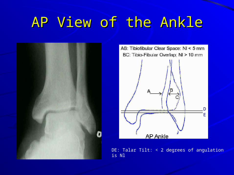
AP View of the AnkleAP View of the Ankle
DE: Talar Tilt: < 2 degrees of angulation is Nl
DE
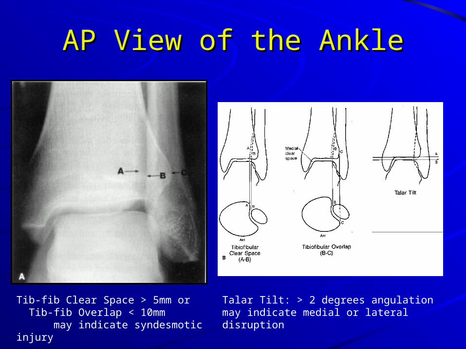
AP View of the AnkleAP View of the Ankle
Talar Tilt: > 2 degrees angulation may indicate medial or lateral disruption
Tib-fib Clear Space > 5mm or Tib-fib Overlap < 10mm may indicate syndesmotic injury
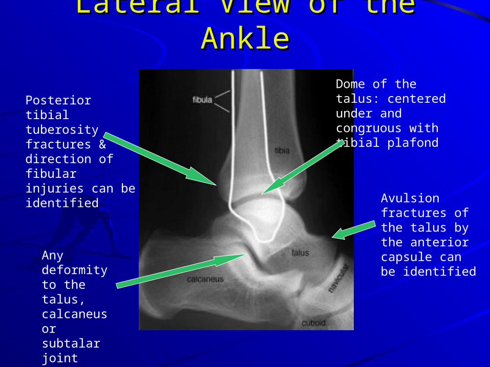
Lateral View of the AnkleLateral View of the AnkleDome of the talus: centered under and congruous with tibial plafond
Posterior tibial tuberosity fractures & direction of fibular injuries can be identified
Avulsion fractures of the talus by the anterior capsule can be identified
Any deformity to the talus, calcaneus or subtalar joint
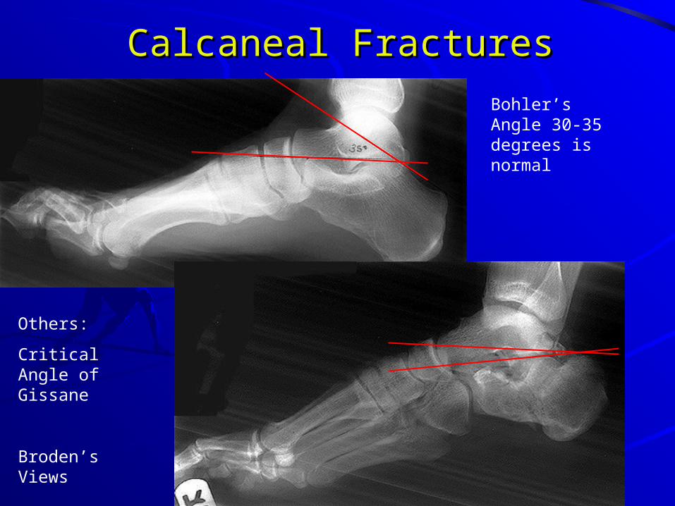
Calcaneal FracturesCalcaneal Fractures
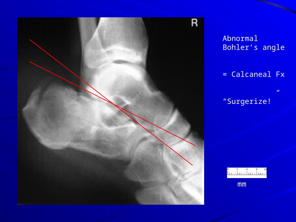
Bohler’s Angle 30-35 degrees is normal
Others:
Critical Angle of Gissane
Broden’s Views
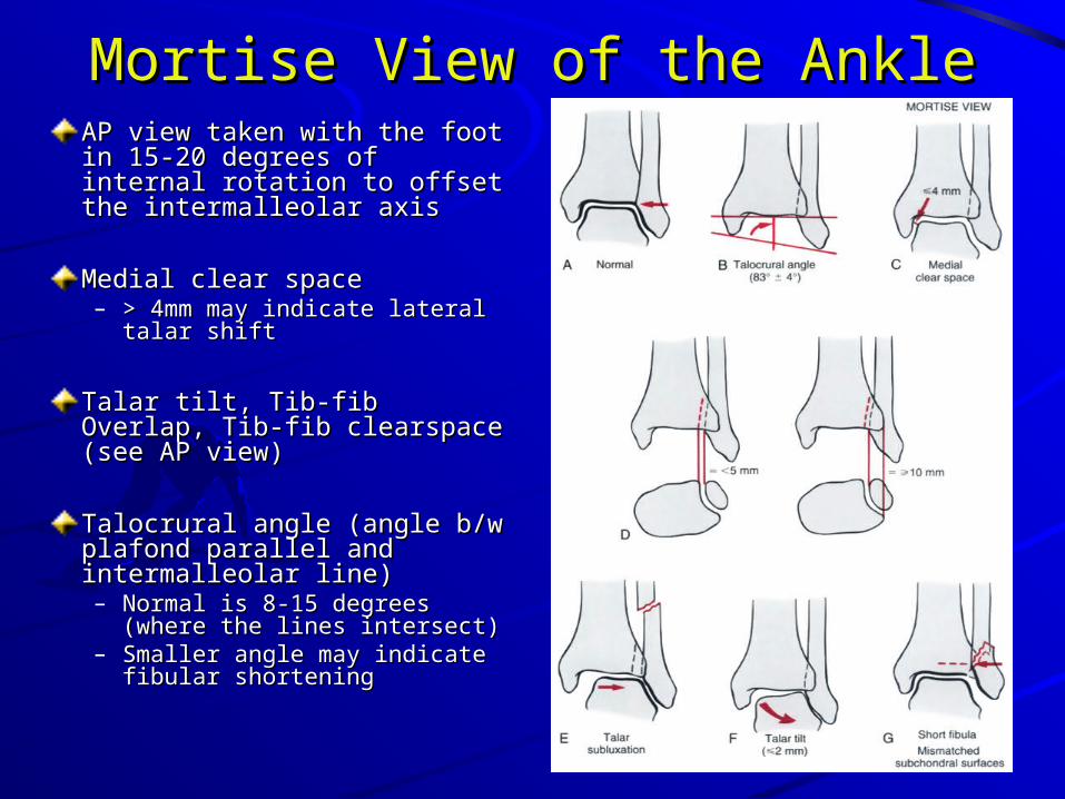
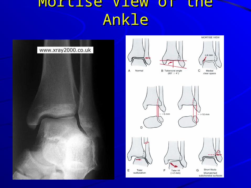
Mortise View of the AnkleMortise View of the AnkleAP view taken with the foot AP view taken with the foot in 15-20 degrees of internal in 15-20 degrees of internal rotation to offset the rotation to offset the intermalleolar axisintermalleolar axis
Medial clear spaceMedial clear space– > 4mm may indicate > 4mm may indicate
lateral talar shiftlateral talar shift
Talar tilt, Tib-fib Overlap, Talar tilt, Tib-fib Overlap, Tib-fib clearspace (see AP Tib-fib clearspace (see AP view)view)
Talocrural angle (angle b/w Talocrural angle (angle b/w plafond parallel and plafond parallel and intermalleolar line)intermalleolar line)– Normal is 8-15 degrees Normal is 8-15 degrees
(where the lines intersect)(where the lines intersect)– Smaller angle may indicate Smaller angle may indicate
fibular shorteningfibular shortening
Mortise View of the AnkleMortise View of the Ankle
mm
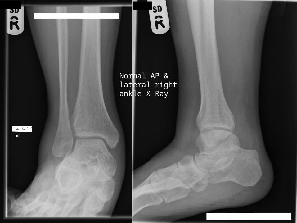
Normal AP & lateral right ankle X Ray
mm
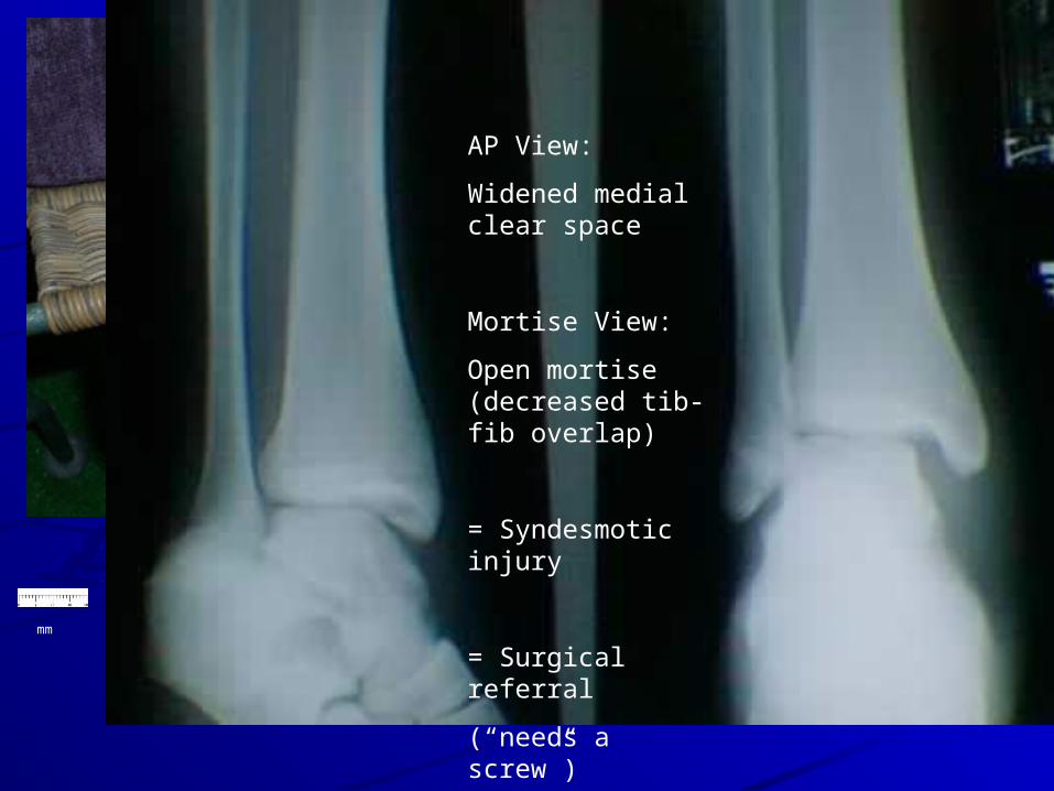
AP View:
Widened medial clear space
Mortise View:
Open mortise (decreased tib-fib overlap)
= Syndesmotic injury
= Surgical referral
(“needs a screw”)
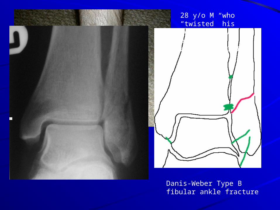
28 y/o M who “twisted” his left ankle while playing basketball 1 day ago
Danis-Weber Type B fibular ankle fracture
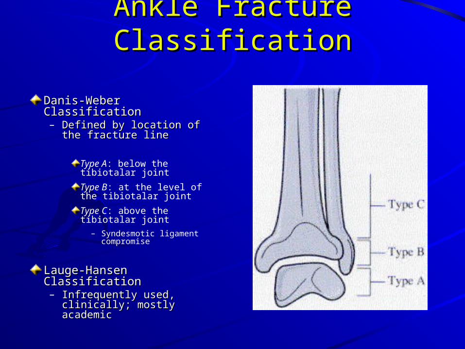
Ankle Fracture ClassificationAnkle Fracture Classification
Danis-Weber ClassificationDanis-Weber Classification– Defined by location of the Defined by location of the
fracture linefracture line
Type A: below the tibiotalar joint
Type B: at the level of the tibiotalar joint
Type C: above the tibiotalar joint
– Syndesmotic ligament compromise
Lauge-Hansen Lauge-Hansen ClassificationClassification– Infrequently used, Infrequently used,
clinically; mostly academicclinically; mostly academic
mm
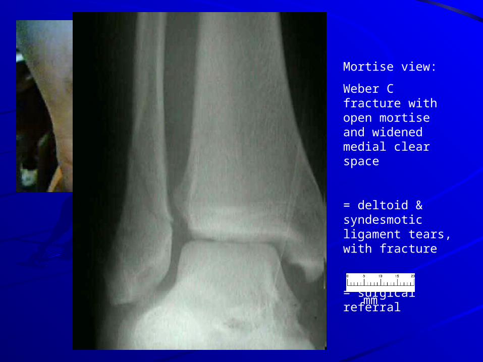
Mortise view:
Weber C fracture with open mortise and widened medial clear space
= deltoid & syndesmotic ligament tears, with fracture
= surgical referral
mm
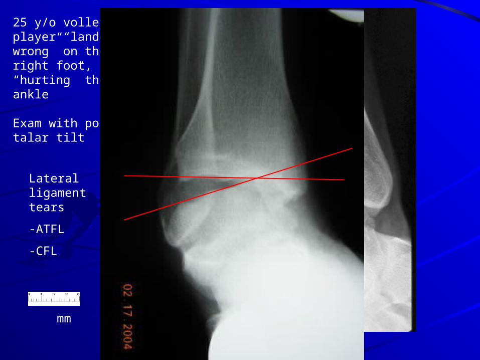
25 y/o volleyball player “landed wrong” on the right foot, “hurting” the ankle
Exam with positive talar tilt
Lateral ligament tears
-ATFL
-CFL
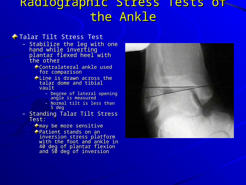
Radiographic Stress Tests of the AnkleRadiographic Stress Tests of the Ankle
Talar Tilt Stress Test– Stabilize the leg with one Stabilize the leg with one
hand while inverting plantar hand while inverting plantar flexed heel with the otherflexed heel with the other
Contralateral ankle used for Contralateral ankle used for comparisoncomparisonLine is drawn across the Line is drawn across the talar dome and tibial vaulttalar dome and tibial vault
– Degree of lateral opening Degree of lateral opening angle is measuredangle is measured
– Normal tilt is less than 5 Normal tilt is less than 5 degdeg
– Standing Talar Tilt Stress Standing Talar Tilt Stress Test:Test:
may be more sensitivemay be more sensitivePatient stands on an Patient stands on an inversion stress platform inversion stress platform with the foot and ankle in with the foot and ankle in 40 deg of plantar flexion 40 deg of plantar flexion and 50 deg of inversionand 50 deg of inversion
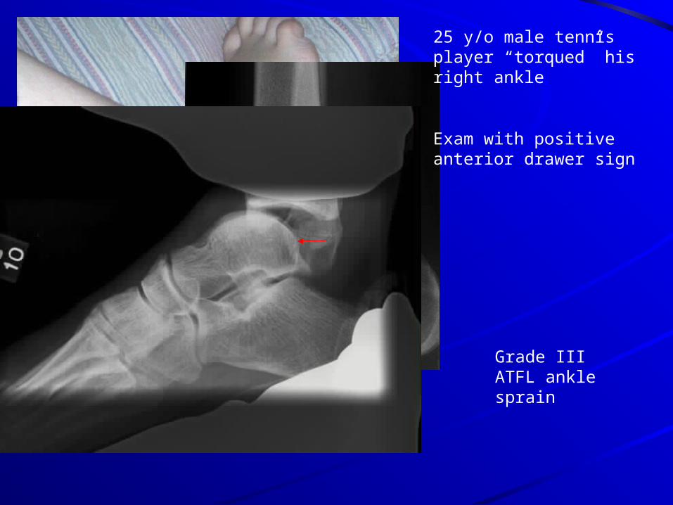
Grade III ATFL ankle sprain
25 y/o male tennis player “torqued” his right ankle
Exam with positive anterior drawer sign
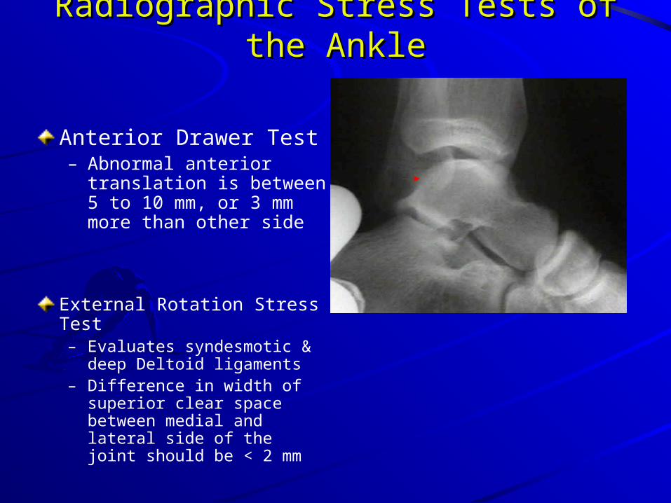
Radiographic Stress Tests of the AnkleRadiographic Stress Tests of the Ankle
Anterior Drawer Test– Abnormal anterior
translation is between 5 to 10 mm, or 3 mm more than other side
External Rotation Stress Test– Evaluates syndesmotic &
deep Deltoid ligaments– Difference in width of
superior clear space between medial and lateral side of the joint should be < 2 mm
mm
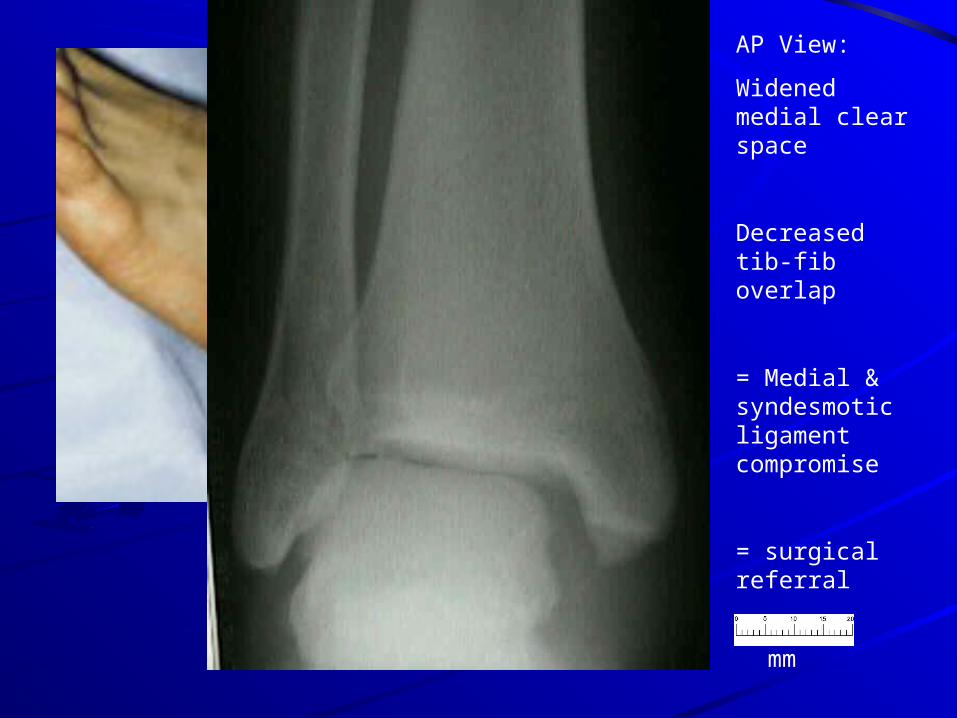
AP View:
Widened medial clear space
Decreased tib-fib overlap
= Medial & syndesmotic ligament compromise
= surgical referral
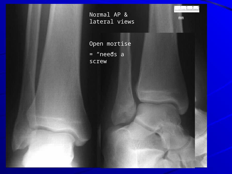
mmNormal AP & lateral views
Open mortise
= “needs a screw”
mm
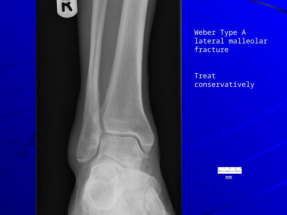
Weber Type A lateral malleolar fracture
Treat conservatively
mm
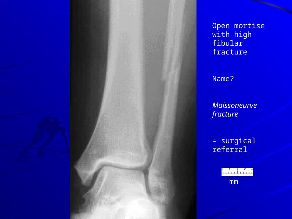
Open mortise with high fibular fracture
Name?
Maissoneurve fracture
= surgical referral
mm
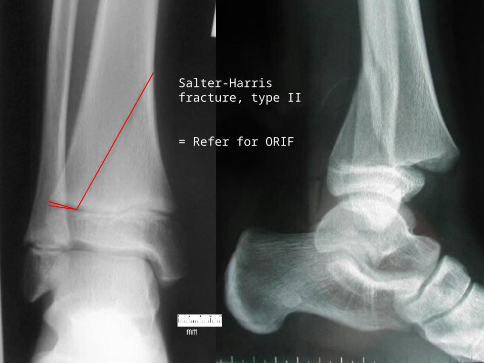
Salter-Harris fracture, type II
= Refer for ORIF
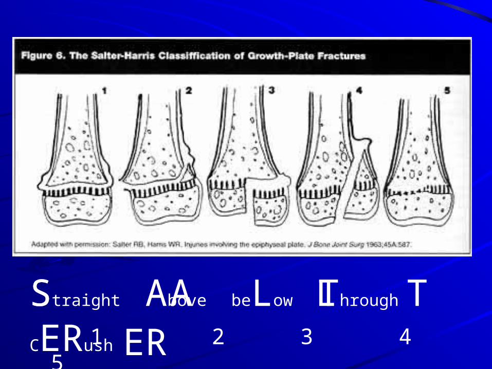
S A L T ER Straight Above beLow Through CERush
1 2 3 4 5
mm
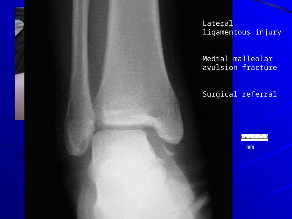
Lateral ligamentous injury
Medial malleolar avulsion fracture
Surgical referral
mm
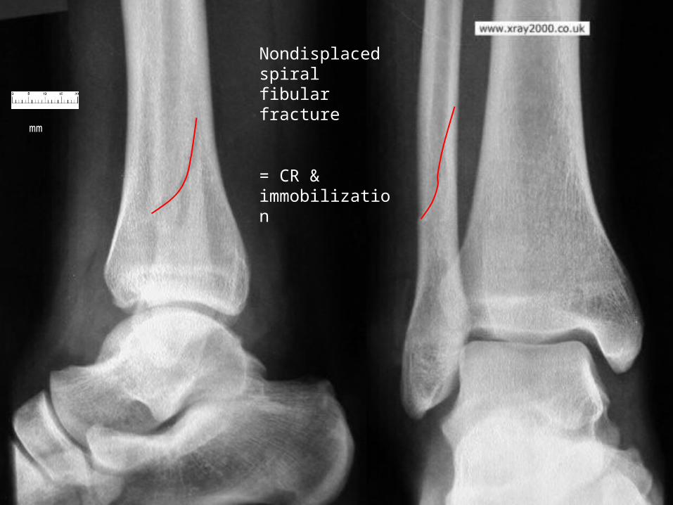
Nondisplaced spiral fibular fracture
= CR & immobilization
mm
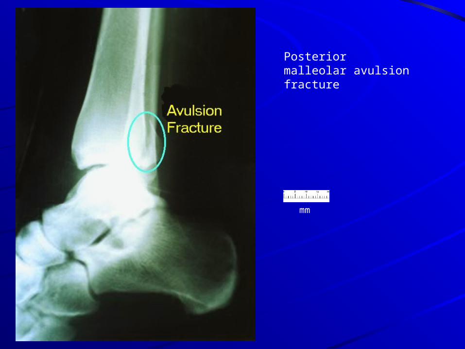
Posterior malleolar avulsion fracture
mm
Abnormal Bohler’s angle
= Calcaneal Fx
“Surgerize!”
mm
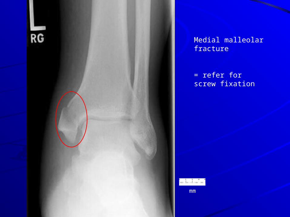
Medial malleolar fracture
= refer for screw fixation
mm
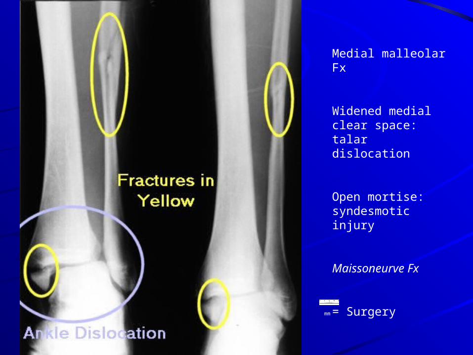
Medial malleolar Fx
Widened medial clear space: talar dislocation
Open mortise: syndesmotic injury
Maissoneurve Fx
= Surgery
mm
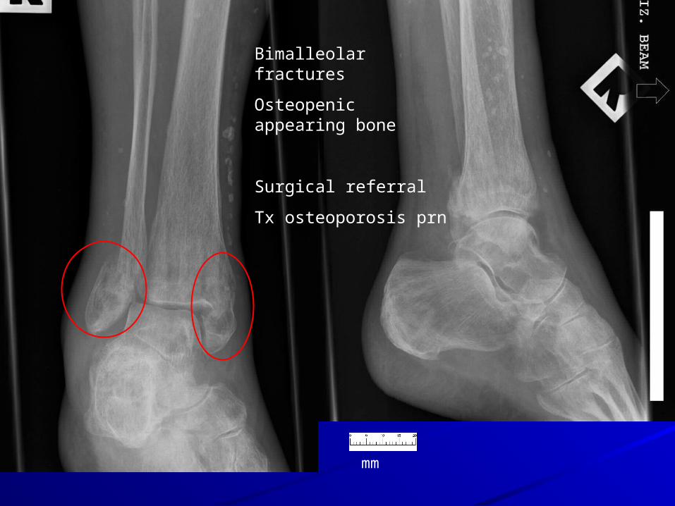
Bimalleolar fractures
Osteopenic appearing bone
Surgical referral
Tx osteoporosis prn
mm
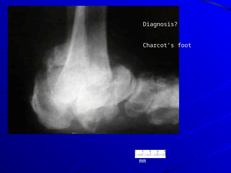
Diagnosis?
Charcot’s foot
mm
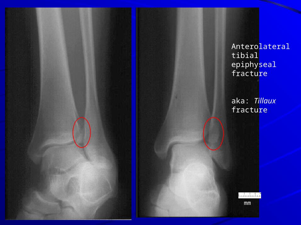
Anterolateral tibial epiphyseal fracture
aka: Tillaux fracture
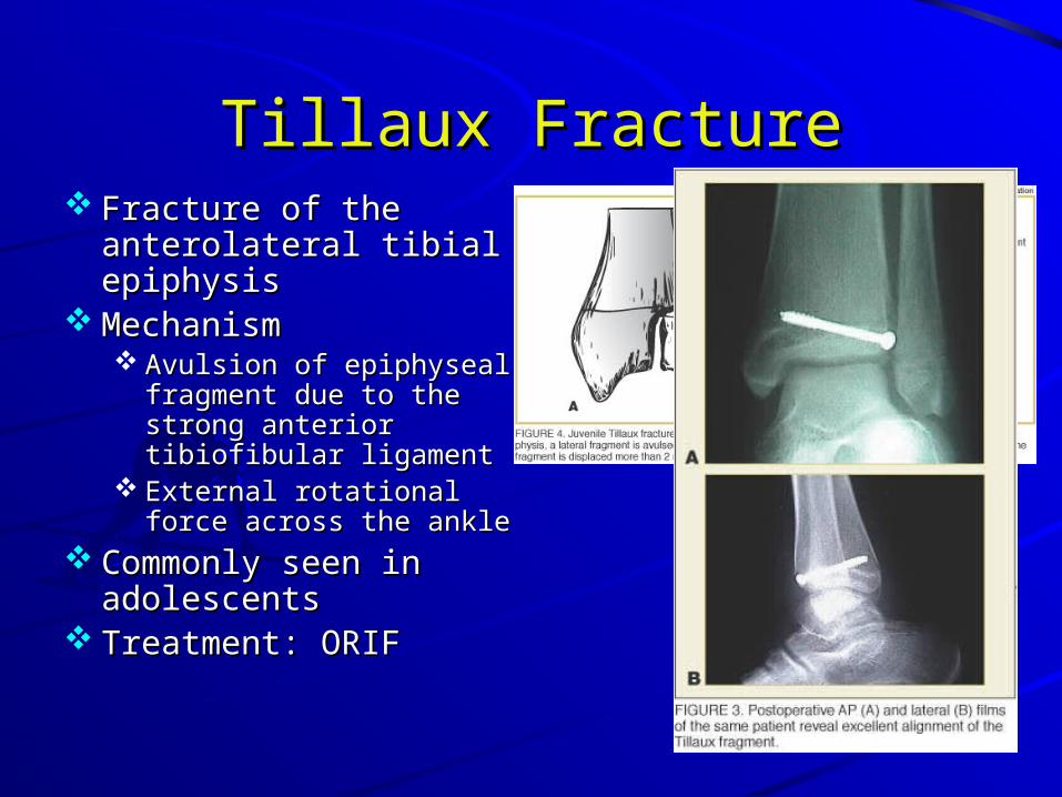
Tillaux FractureTillaux Fracture Fracture of the Fracture of the
anterolateral tibial anterolateral tibial epiphysis epiphysis
MechanismMechanism Avulsion of epiphyseal Avulsion of epiphyseal
fragment due to the fragment due to the strong anterior strong anterior tibiofibular ligament tibiofibular ligament
External rotational force External rotational force across the ankleacross the ankle
Commonly seen in Commonly seen in adolescentsadolescents
Treatment: ORIFTreatment: ORIF
mm
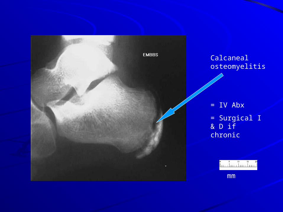
Calcaneal osteomyelitis
= IV Abx
= Surgical I & D if chronic
mm
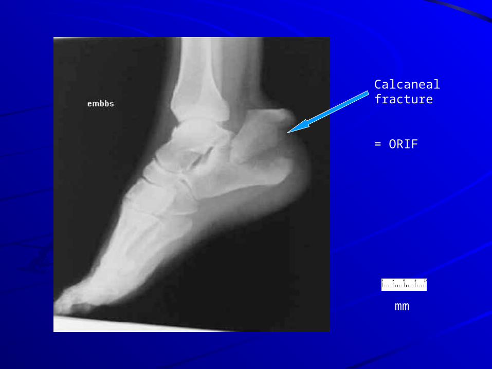
Calcaneal fracture
= ORIF
mm
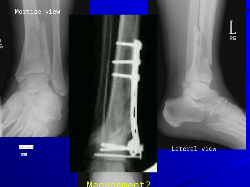
Mortise view
AP view
Lateral view
Pilon fracture(Comminuted tibial
plafond compression fracture)
Management?
mm
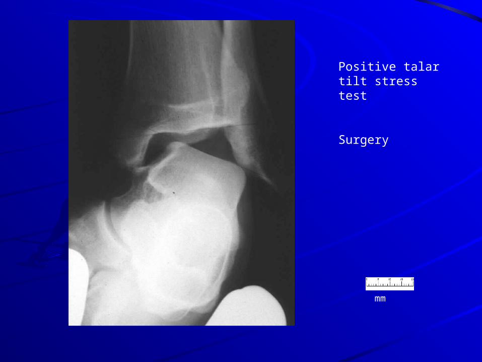
Positive talar tilt stress test
Surgery
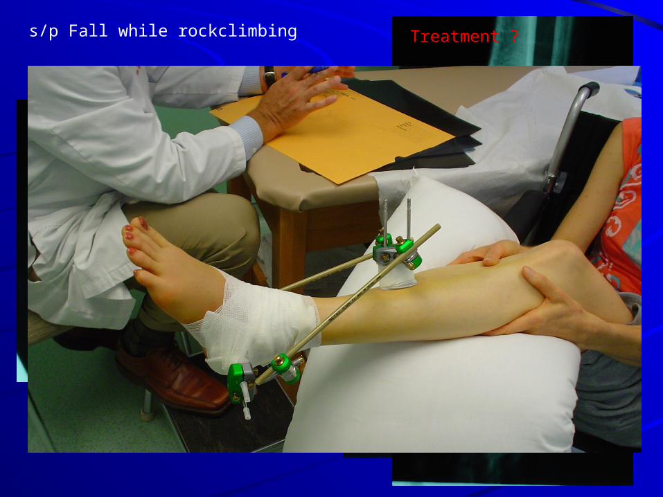
s/p Fall while rockclimbing Treatment ?
ConclusionConclusion
Plain radiographic Plain radiographic anatomy of the anatomy of the ankleankle
Indications for Indications for plain radiographs plain radiographs of the ankleof the ankle
Direct and indirect Direct and indirect signs of injury on signs of injury on plain radiographsplain radiographs
The End
Thanks!