Embed Size (px)
Citation preview
RESEARCH ARTICLE
X-linked Christianson syndrome: heterozygous female Slc9a6knockout mice develop mosaic neuropathological changes andrelated behavioral abnormalitiesJakub Sikora1,2,*, Jennifer Leddy1, Maria Gulinello1 and Steven U. Walkley1,*
ABSTRACTChristianson syndrome (CS) is an X-linked neurodevelopmental andneurological disorder characterized in males by core symptoms thatinclude non-verbal status, intellectual disability, epilepsy, truncalataxia, postnatal microcephaly and hyperkinesis. CS is caused bymutations in the SLC9A6 gene, which encodes a multipasstransmembrane sodium (potassium)-hydrogen exchanger 6(NHE6) protein, functional in early recycling endosomes. Theextent and variability of the CS phenotype in femaleheterozygotes, who presumably express the wild-type and mutantSLC9A6 alleles mosaically as a result of X-chromosome inactivation(XCI), have not yet been systematically characterized. Slc9a6knockout mice (Slc9a6 KO) were generated by insertion of thebacterial lacZ/β-galactosidase (β-Gal) reporter into exon 6 of theX-linked gene. Mutant Slc9a6 KO male mice have been shown todevelop late endosomal/lysosomal dysfunction associated withglycolipid accumulation in selected neuronal populations andpatterned degeneration of Purkinje cells (PCs). In heterozygousfemale Slc9a6 KO mice, β-Gal serves as a transcriptional/XCIreporter and thus facilitates testing of effects of mosaic expression ofthe mutant allele on penetrance of the abnormal phenotype. Usingβ-Gal, we demonstrated mosaic expression of the mutant Slc9a6allele and mosaically distributed lysosomal glycolipid accumulationand PC pathology in the brains of heterozygous Slc9a6 KO femalemice. At the behavioral level, we showed that heterozygous femalemice suffer from visuospatial memory and motor coordinationdeficits similar to but less severe than those observed inX-chromosome hemizygous mutant males. Our studies inheterozygous Slc9a6 KO female mice provide important clues forunderstanding the likely phenotypic range of Christianson syndromeamong females heterozygous for SLC9A6 mutations and mightimprove diagnostic practice and genetic counseling by helping tocharacterize this presumably underappreciated patient/carriergroup.
KEYWORDS: Christianson syndrome,Slc9a6, NHE6 protein, Femaleheterozygotes, X-chromosome inactivation, Mosaicism
INTRODUCTIONChristianson syndrome (CS, OMIM 300243) is a rare,X-chromosome-linked neurodevelopmental and neurologicaldisorder characterized in males by non-verbal status, intellectualdisability, epilepsy, craniofacial dysmorphologywith microcephaly,truncal ataxia and hyperkinesis. These core phenotypic features(present in >85% of patients) can be accompanied by secondarysymptoms, such as signs of autism and behavioral abnormalitiesmimicking Angelman syndrome, eye movement problems,hypotonia or gastroesophageal reflux disease (for furtherinformation see Pescosolido et al., 2014). Magnetic resonanceimaging studies have suggested hippocampal and/or progressivecerebellar atrophy in male CS patients (Gilfillan et al., 2008; Schroeret al., 2010; Mignot et al., 2013). Furthermore, neuropathologicalreports on male CS brains have demonstrated widespreadneurodegeneration, including loss of Purkinje cells (PCs),dystrophic neuritic changes, gliosis and tau deposition(Christianson et al., 1999; Garbern et al., 2010).
CS develops as a result of mutations in the solute-carrier 9A6gene (SLC9A6, Xq26.3; Gilfillan et al., 2008). SLC9A6 codes amultipass transmembrane protein (NHE6) that is believed to co-regulate the luminal pH of early/recycling endosomes by its sodium(potassium)-hydrogen antiporter activity (Ohgaki et al., 2011;Kondapalli et al., 2014). A significant fraction of the reportedSLC9A6 mutations are nonsense or shift the open reading frame ofSLC9A6 and result in introduction of premature stop codons(reviewed by Pescosolido et al., 2014).
As a result of X-chromosome inactivation (XCI), femaleheterozygotes presumably express SLC9A6 mutations in their cellsand tissues mosaically. Although several female heterozygotes havepresented with clinical symptoms reminiscent of those identified intheir CS-affected male relatives (Christianson et al., 1999; Gilfillanet al., 2008; Schroer et al., 2010; Pescosolido et al., 2014),conclusive information about the range of the probably mitigatedand/or variable clinical phenotype in this particular group stillremains to be considered systematically.
Previous studies by us and others have demonstrated therelevance of the knockout of the murine Slc9a6 gene (Slc9a6 KO)for studies exploring the human CS phenotype. Our analyses ofmutant Slc9a6 KO males (Slc9a6−/Y ‘mutant’ males) andhomozygous mutant Slc9a6 KO females (Slc9a6−/− ‘mutant’females), both of which serve as models with uniform tissuedistribution of the (transcriptionally) active mutant Slc9a6 allele,indicated late endosomal/lysosomal dysfunction characterized byintraneuronal accumulation of GM2 ganglioside and unesterifiedcholesterol in the amygdala and the CA3/CA4 and fascia dentataregions of the hippocampus (Stromme et al., 2011). In addition, bothmutant males and mutant females expressed progressive, patternedPC degeneration associated with axonal spheroid formation.Received 15 August 2015; Accepted 25 October 2015
1Dominick P. Purpura Department of Neuroscience, Rose F. Kennedy Intellectualand Developmental Disabilities Research Center, Albert Einstein College ofMedicine, Bronx, NY 10461, USA. 2Institute of Inherited Metabolic Disorders,Charles University in Prague – 1st Faculty of Medicine, Ke Karlovu 2, Praha 2160 00, Czech Republic.
*Authors for correspondence ( [email protected];[email protected])
This is an Open Access article distributed under the terms of the Creative Commons AttributionLicense (http://creativecommons.org/licenses/by/3.0), which permits unrestricted use,distribution and reproduction in any medium provided that the original work is properly attributed.
13
© 2016. Published by The Company of Biologists Ltd | Disease Models & Mechanisms (2016) 9, 13-23 doi:10.1242/dmm.022780
Disea
seModels&Mechan
isms
Behavioral testing in mutant males revealed mild but significantlyincreased locomotor activity and motor coordination deficits,suggesting further overlap with the human CS clinical condition(Pescosolido et al., 2014). Importantly, a subsequent study usingin vitro experimental approaches in neuronal cultures derived fromthe Slc9a6 KO model proposed that abnormal endosomalacidification caused by the NHE6 deficit attenuates tropomyosinrelated kinase B (TrkB) signaling and results in underdevelopedcortical and hippocampal neuritic arborization (Ouyang et al., 2013).Although different in specific molecular details from the human
situation, the random XCI and its propagation in the tissues of thedeveloping embryo are replicated in mice (Deng et al., 2014). In themurine female brain, XCI topography generates intra- and inter-individual diversity that ranges from individual cells to the entireorgan. Crucially, however, it was shown that specific neuronalpopulations in female mice can tend, as a result of the complexneurodevelopment, to be inactivated non-randomly on a functionallyrelevant spatial scale (Wu et al., 2014). Crucial for our studies, themurine KO model carries an insertion of the lacZ-Neo cassette intoexon 6 of the Slc9a6 gene (X.A5; Stromme et al., 2011). lacZ, whichcodes nuclear-targeted β-galactosidase (β-Gal) from E.coli, thusserves both as a mutagen that obliterates the Slc9a6 open readingframe and as a transcriptional reporter that allows effective tracing ofthe cellular expression of the mutant Slc9a6 allele. Important forutility of the β-Gal reporter, its expression patterns correspond to theendogenous expression of the protein as identified by mRNAexpression studies (Kondapalli et al., 2013) and/or a specific anti-NHE6 antibody (Deane et al., 2013; Ouyang et al., 2013). As afurther important prerequisite, the murine Slc9a6 gene was notpreviously identified, similarly to humans (Cotton et al., 2015),among X-linked genes that escape XCI (Yang et al., 2010).Therefore, crucial for the present study, β-Gal expression can alsobe considered to reflect the inactivation of thewild-type (WT) Slc9a6allele in tissues of heterozygous Slc9a6KO female (Slc9a6−/+) mice.Given the relevance of the murine model for CS and with the
purpose of providing insights into the potential disease phenotype inhuman female heterozygotes carrying SLC9A6 mutations, weperformed a study to evaluate the neuropathology and behavioralpresentation in heterozygous Slc9a6 KO female mice. Here, wefocused on delineating the mosaic patterns of abnormalintraneuronal lysosomal GM2 ganglioside accumulation inamygdala and hippocampus, characterizing the extent ofcerebellar PC degeneration and comparing the range of motorcoordination and cognitive deficits with the abnormalities observedin mutant Slc9a6 KO males.
RESULTSExpression of the mutant Slc9a6 allele is mosaic in thebrains of heterozygous female miceCellular and tissue expression of the mutant Slc9a6 allele can betracked by the lacZ-encoded β-Gal reporter. Using the histochemicalX-GAL-based approach, we previously documented the brain-specific expression patterns of β-Gal in mutant males and mutantfemales (Stromme et al., 2011). Being aware of the limitations of thehistochemical technique and aiming to assess β-Gal expressionqualitatively and quantitatively in the brains of heterozygousfemales, we tested the utility of a specific antibody for detectionof the reporter protein. To allow multi-immunofluorescence (IF)tissue-labeling experiments, we selected a specific anti-E.coli β-Galantibody raised in chickens.In order to assess the progression of disease at later ages than
previously reported (Stromme et al., 2011) and to fully document
the reference neuropathology resulting from uniform tissuedistribution of the (transcriptionally) active mutant Slc9a6 allele,we optimized IF staining conditions in the brains of X-chromosomehemizygous mutant males (aged 4, 20-22 and 32-34 weeks). Wecompared the results with negative findings in age-matched WTmales (shown in the cerebellar PC layer in Fig. S1C) and WTfemales (data not shown). The anti-β-Gal antibody generated anuclear staining pattern in the brains of mutant males, allowing us toconfirm that the expression patterns of β-Gal by IF corresponded tothose previously identified by X-GAL histochemistry (Strommeet al., 2011; Ouyang et al., 2013).
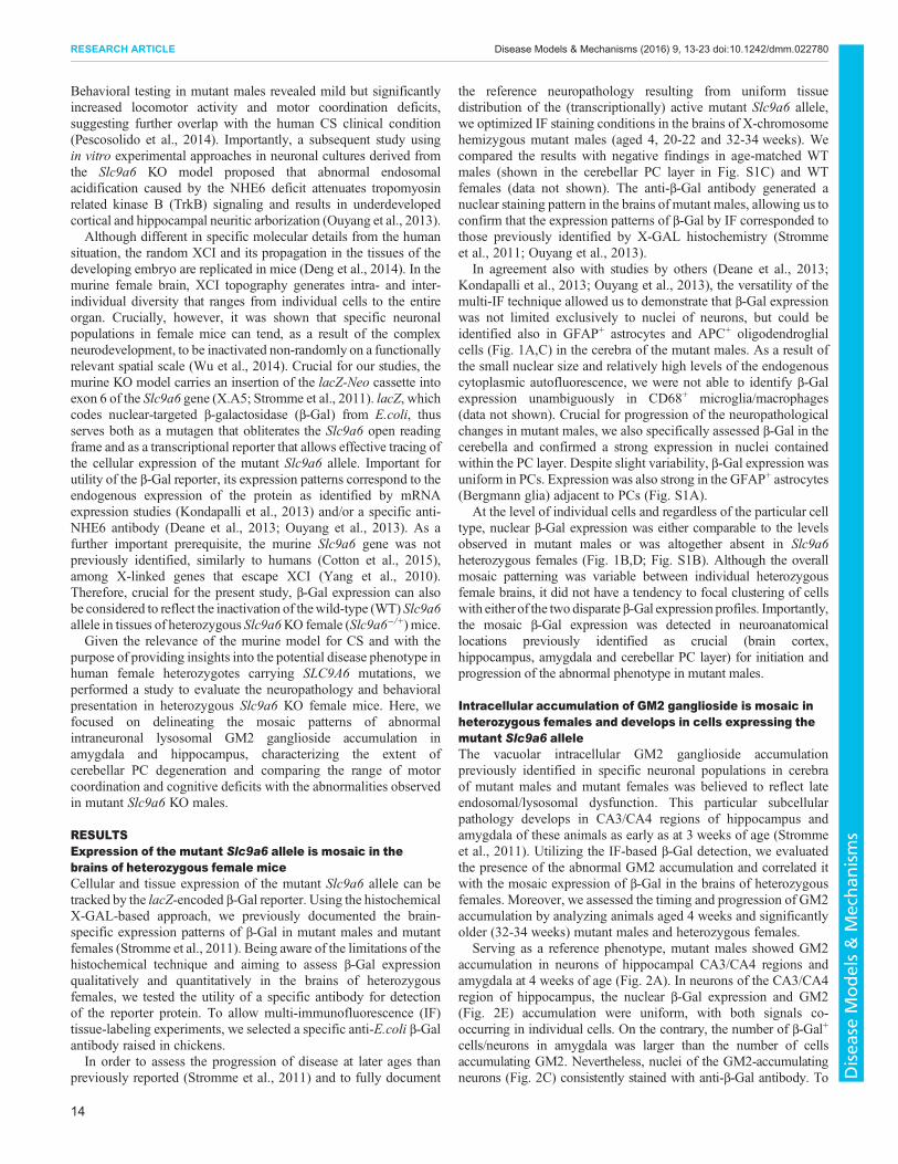
In agreement also with studies by others (Deane et al., 2013;Kondapalli et al., 2013; Ouyang et al., 2013), the versatility of themulti-IF technique allowed us to demonstrate that β-Gal expressionwas not limited exclusively to nuclei of neurons, but could beidentified also in GFAP+ astrocytes and APC+ oligodendroglialcells (Fig. 1A,C) in the cerebra of the mutant males. As a result ofthe small nuclear size and relatively high levels of the endogenouscytoplasmic autofluorescence, we were not able to identify β-Galexpression unambiguously in CD68+ microglia/macrophages(data not shown). Crucial for progression of the neuropathologicalchanges in mutant males, we also specifically assessed β-Gal in thecerebella and confirmed a strong expression in nuclei containedwithin the PC layer. Despite slight variability, β-Gal expression wasuniform in PCs. Expression was also strong in the GFAP+ astrocytes(Bergmann glia) adjacent to PCs (Fig. S1A).
At the level of individual cells and regardless of the particular celltype, nuclear β-Gal expression was either comparable to the levelsobserved in mutant males or was altogether absent in Slc9a6heterozygous females (Fig. 1B,D; Fig. S1B). Although the overallmosaic patterning was variable between individual heterozygousfemale brains, it did not have a tendency to focal clustering of cellswith eitherof the two disparate β-Gal expression profiles. Importantly,the mosaic β-Gal expression was detected in neuroanatomicallocations previously identified as crucial (brain cortex,hippocampus, amygdala and cerebellar PC layer) for initiation andprogression of the abnormal phenotype in mutant males.
Intracellular accumulation of GM2 ganglioside is mosaic inheterozygous females and develops in cells expressing themutant Slc9a6 alleleThe vacuolar intracellular GM2 ganglioside accumulationpreviously identified in specific neuronal populations in cerebraof mutant males and mutant females was believed to reflect lateendosomal/lysosomal dysfunction. This particular subcellularpathology develops in CA3/CA4 regions of hippocampus andamygdala of these animals as early as at 3 weeks of age (Strommeet al., 2011). Utilizing the IF-based β-Gal detection, we evaluatedthe presence of the abnormal GM2 accumulation and correlated itwith the mosaic expression of β-Gal in the brains of heterozygousfemales. Moreover, we assessed the timing and progression of GM2accumulation by analyzing animals aged 4 weeks and significantlyolder (32-34 weeks) mutant males and heterozygous females.
Serving as a reference phenotype, mutant males showed GM2accumulation in neurons of hippocampal CA3/CA4 regions andamygdala at 4 weeks of age (Fig. 2A). In neurons of the CA3/CA4region of hippocampus, the nuclear β-Gal expression and GM2(Fig. 2E) accumulation were uniform, with both signals co-occurring in individual cells. On the contrary, the number of β-Gal+
cells/neurons in amygdala was larger than the number of cellsaccumulating GM2. Nevertheless, nuclei of the GM2-accumulatingneurons (Fig. 2C) consistently stained with anti-β-Gal antibody. To
14
RESEARCH ARTICLE Disease Models & Mechanisms (2016) 9, 13-23 doi:10.1242/dmm.022780
Disea
seModels&Mechan
isms
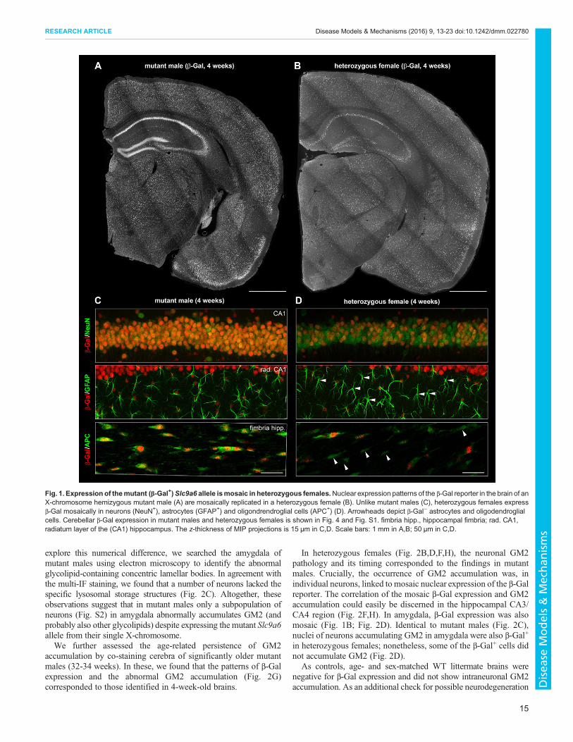
explore this numerical difference, we searched the amygdala ofmutant males using electron microscopy to identify the abnormalglycolipid-containing concentric lamellar bodies. In agreement withthe multi-IF staining, we found that a number of neurons lacked thespecific lysosomal storage structures (Fig. 2C). Altogether, theseobservations suggest that in mutant males only a subpopulation ofneurons (Fig. S2) in amygdala abnormally accumulates GM2 (andprobably also other glycolipids) despite expressing the mutant Slc9a6allele from their single X-chromosome.We further assessed the age-related persistence of GM2
accumulation by co-staining cerebra of significantly older mutantmales (32-34 weeks). In these, we found that the patterns of β-Galexpression and the abnormal GM2 accumulation (Fig. 2G)corresponded to those identified in 4-week-old brains.
In heterozygous females (Fig. 2B,D,F,H), the neuronal GM2pathology and its timing corresponded to the findings in mutantmales. Crucially, the occurrence of GM2 accumulation was, inindividual neurons, linked to mosaic nuclear expression of the β-Galreporter. The correlation of the mosaic β-Gal expression and GM2accumulation could easily be discerned in the hippocampal CA3/CA4 region (Fig. 2F,H). In amygdala, β-Gal expression was alsomosaic (Fig. 1B; Fig. 2D). Identical to mutant males (Fig. 2C),nuclei of neurons accumulating GM2 in amygdala were also β-Gal+
in heterozygous females; nonetheless, some of the β-Gal+ cells didnot accumulate GM2 (Fig. 2D).
As controls, age- and sex-matched WT littermate brains werenegative for β-Gal expression and did not show intraneuronal GM2accumulation. As an additional check for possible neurodegeneration
Fig. 1. Expression of themutant (β-Gal+)Slc9a6 allele ismosaic in heterozygous females.Nuclear expression patterns of the β-Gal reporter in the brain of anX-chromosome hemizygous mutant male (A) are mosaically replicated in a heterozygous female (B). Unlike mutant males (C), heterozygous females expressβ-Gal mosaically in neurons (NeuN+), astrocytes (GFAP+) and oligondrendroglial cells (APC+) (D). Arrowheads depict β-Gal− astrocytes and oligodendroglialcells. Cerebellar β-Gal expression in mutant males and heterozygous females is shown in Fig. 4 and Fig. S1. fimbria hipp., hippocampal fimbria; rad. CA1,radiatum layer of the (CA1) hippocampus. The z-thickness of MIP projections is 15 µm in C,D. Scale bars: 1 mm in A,B; 50 µm in C,D.
15
RESEARCH ARTICLE Disease Models & Mechanisms (2016) 9, 13-23 doi:10.1242/dmm.022780
Disea
seModels&Mechan
isms
in the regions affected by the glycolipid accumulation inmutantmalesand heterozygous females,we IF co-stained cerebra of animals of boththese genotypes with antibodies targeting neurons (anti-NeuN) andmicroglia/macrophages (anti-CD68). In contrast to the cerebellum(see the section below) and when compared with WT controls, noneof these stains directly or indirectly suggested widespreadneurodegeneration at 4 and/or 32-34 weeks of age (data not shown).
Purkinje cells in Slc9a6 KO heterozygous females exhibitdegeneration that is ameliorated compared with mutantmalesDegeneration of PCs that is associated with spheroid formation ontheir axons is another feature consistently discernible in mutantmales. The spatial patterning of this process follows cerebellarzonal organization with Zebrin II-positive PCs being the most
Fig. 2. The abnormal intraneuronal accumulation of GM2 ganglioside in amygdala and CA3 region of hippocampus is found in the neurons expressingthe mutant (β-Gal+) Slc9a6 allele in heterozygous females. At 4 weeks of age, GM2 accumulation is conspicuously present in the neurons of CA3 region ofhippocampus (white arrowheads) and amygdala (blue arrowheads) of mutant males (A) and in heterozygous females (B). In mutant males, only a fraction ofβ-Gal+ neurons in amygdala accumulates GM2. The intracellular glycolipid accumulation adopts the form of concentric lamellar bodies by electron microscopy(C; Fig. S2). In heterozygous females, β-Gal+ and β-Gal+/GM2+ neurons are less frequent than inmutant males. The lysosomal ultrastructural abnormalities foundin heterozygous females (D) are equivalent to findings in mutant males. Whereas CA3 neurons in mutant males express β-Gal and accumulate GM2 uniformly(E,G), GM2 accumulation in CA3 neurons of heterozygous females is mosaic and reflects the expression of the mutant (β-Gal+) Slc9a6 allele (F,H). amyg.,amygdala; CA3, CA3 region of hippocampus. Scale bars: 1 mm in A,B; 200 µm (IF images) and 5 µm (electron micrographs) in C,D; 100 µm in E-H. Panels A,Bshowing GM2 accumulation originate from sections co-labeled for β-Gal shown in Fig. 1A,B.
16
RESEARCH ARTICLE Disease Models & Mechanisms (2016) 9, 13-23 doi:10.1242/dmm.022780
Disea
seModels&Mechan
isms
resistant to decay (Stromme et al., 2011). Transformed to (para)sagittal projection (Sillitoe and Joyner, 2007), PCs in the anterior(lobules I-V) and posterior (lobule VIII) cerebellar zones tend todegenerate earlier than those localized to the central (lobule VI/VII)
and nodular (lobules IX/X) zones. The molecular anddevelopmental basis of this pattern remains to be understoodfully; nonetheless, as observed in mutant males, it is replicatedin other murine disease models that develop PC degeneration
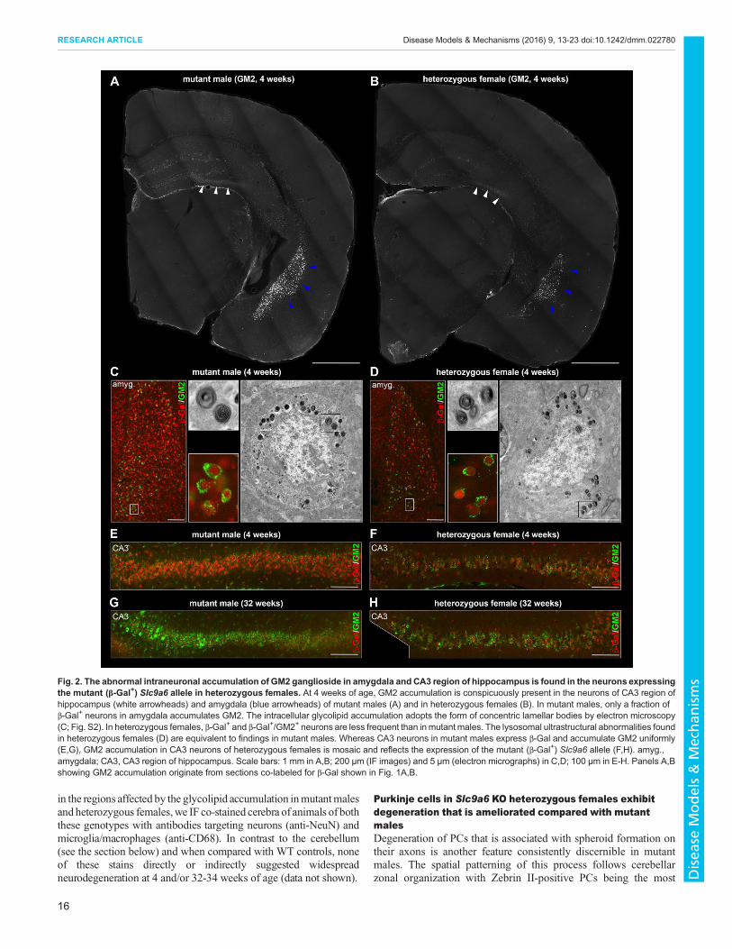
Fig. 3. Cerebellar pathology in heterozygous females. Degeneration of Purkinje cells (visualized by calbindin IF staining) in anterior and posterior zones isinitiated in mutant males (A) and heterozygous females (B) as early as at 4 weeks of age and is highlighted by the abnormal population of CD68+ microglia inboth genotypes (arrowheads). (C,D) Neuronophagy of PC cell bodies (arrowheads) and dendrites in the molecular cerebellar layer can be readily identified at thisage by confocal microscopy in both sex/genotype groups by clusters of CD68+ microglia/macrophages. (E) At 32 weeks of age, mutant males present withadvanced and widespread loss of Purkinje cells in anterior (lobule I-V) and posterior (lobule VIII) cerebellar zones (arrowheads). (F) In heterozygous females, PCdegenerative patterns are not as evident as in mutant males and vary among individual animals. (G,H) PC density quantification in lobules I-II (representative ofthe anterior zone) and X (representative of the nodular zone) showed significant PC loss in lobule I-II of 32-week-old mutant males and heterozygous females(n=4 WT and 5 mutant males and 6 WT and 17 heterozygous females) in contrast to lobule X (n=4 WT and 4 mutant males and 6 WT and 14 heterozygousfemales). *P<0.0001. The z-thickness of MIP projections in C,D is 15 µm, and these images correspond to areas outlined by white rectangles in A,B. Scale bars:1 mm in A,B,E,F; 50 µm in C,D. Panel A reviews the numbering of individual cerebellar lobules.
17
RESEARCH ARTICLE Disease Models & Mechanisms (2016) 9, 13-23 doi:10.1242/dmm.022780
Disea
seModels&Mechan
isms
(e.g. Niemann–Pick disease type A/B or C; Sarna et al., 2001;Macauley et al., 2008).We evaluated cerebellar pathology both in 4- and 32- to 34-week-
old animals. Neurono/dendritophagic clustering of CD68+
macrophages/microglia in the PC and molecular layers ofcerebellar lobules I-II, III and VIII (Fig. 3A,C) suggested ongoingPC loss (visualized by calbindin IF staining) in these areas alreadyin 4-week-old mutant males. Identical but less profound changesthan those identified in mutant males were also detected in 4-week-old heterozygous females (Fig. 3B,D).At 32 weeks, cerebella of mutant males showed extensive PC
loss in anterior and posterior zones (Fig. 3E). This pathology wasalso associated with atrophy and gliosis of the cerebellar molecularlayer in these zones (shown for lobule I-II in Fig. S1A). CD68+
cells were frequent in these cerebellar cortical areas; nonetheless,they did not form PC neuronophagic clusters comparable to4-week-old animals (data not shown). To document the majorquantitative difference in a cohort of 32-week-old mutant males,we sampled the density of PCs in lobule I-II and lobule X selectedas representative of zones affected and spared from PCdegeneration, respectively. The density of PCs in lobule I-II wassignificantly lower in mutant compared with WT males(F(1,7)=227.4, P<0.0001; Fig. 3G).Crucial for 32-week-old heterozygous females, the extent of PC
degeneration was not histologically as discernible (Fig. 3F) as inmutant males. The secondary gliosis in the molecular layer oflobules affected by PC degeneration was also less profound inheterozygous females (Fig. S1B) than in mutant males. Despitethese mitigated histological findings, the density of PCs in lobule
I-II in heterozygous compared with WT females was significantlylower (F(1,21)=24.9, P<0.0001; Fig. 3H).
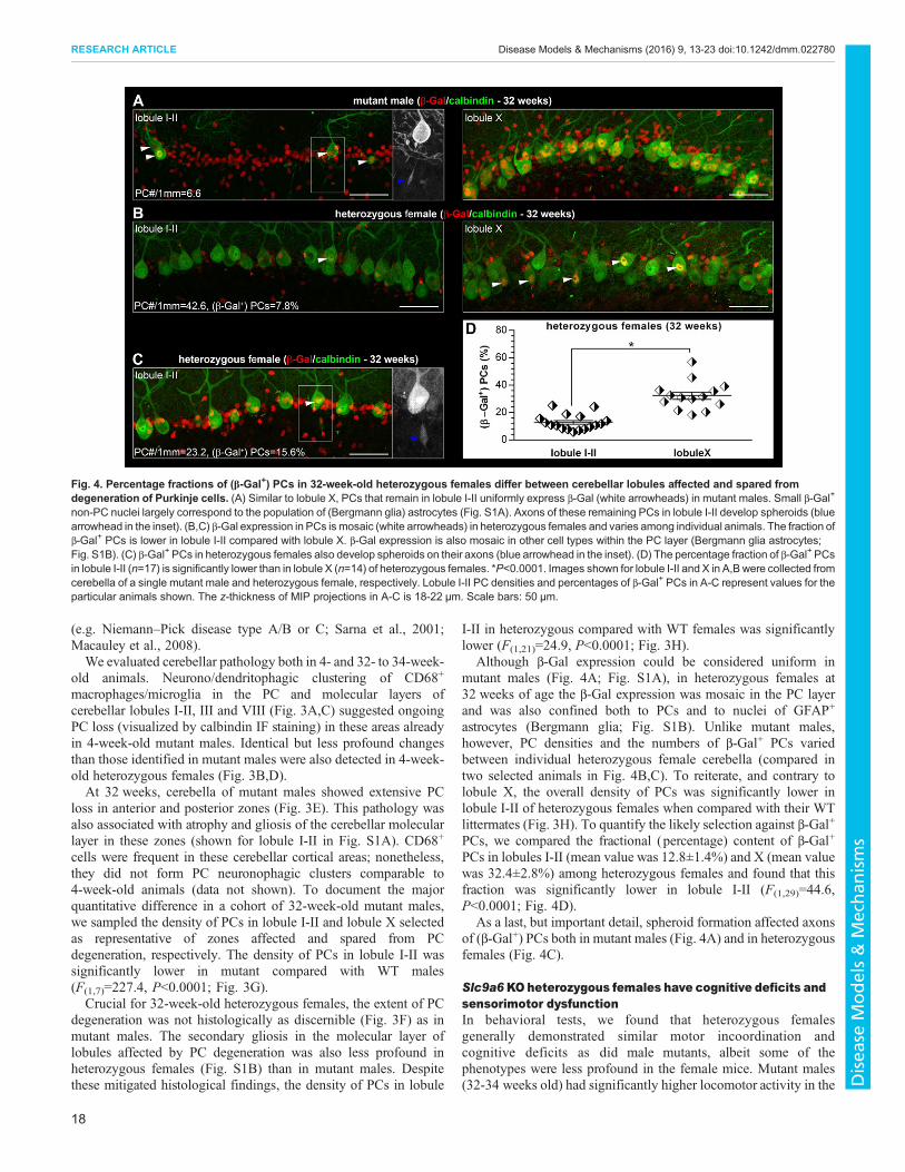
Although β-Gal expression could be considered uniform inmutant males (Fig. 4A; Fig. S1A), in heterozygous females at32 weeks of age the β-Gal expression was mosaic in the PC layerand was also confined both to PCs and to nuclei of GFAP+
astrocytes (Bergmann glia; Fig. S1B). Unlike mutant males,however, PC densities and the numbers of β-Gal+ PCs variedbetween individual heterozygous female cerebella (compared intwo selected animals in Fig. 4B,C). To reiterate, and contrary tolobule X, the overall density of PCs was significantly lower inlobule I-II of heterozygous females when compared with their WTlittermates (Fig. 3H). To quantify the likely selection against β-Gal+
PCs, we compared the fractional (percentage) content of β-Gal+
PCs in lobules I-II (mean value was 12.8±1.4%) and X (mean valuewas 32.4±2.8%) among heterozygous females and found that thisfraction was significantly lower in lobule I-II (F(1,29)=44.6,P<0.0001; Fig. 4D).
As a last, but important detail, spheroid formation affected axonsof (β-Gal+) PCs both in mutant males (Fig. 4A) and in heterozygousfemales (Fig. 4C).
Slc9a6KOheterozygous females have cognitive deficits andsensorimotor dysfunctionIn behavioral tests, we found that heterozygous femalesgenerally demonstrated similar motor incoordination andcognitive deficits as did male mutants, albeit some of thephenotypes were less profound in the female mice. Mutant males(32-34 weeks old) had significantly higher locomotor activity in the
Fig. 4. Percentage fractions of (β-Gal+) PCs in 32-week-old heterozygous females differ between cerebellar lobules affected and spared fromdegeneration of Purkinje cells. (A) Similar to lobule X, PCs that remain in lobule I-II uniformly express β-Gal (white arrowheads) in mutant males. Small β-Gal+
non-PC nuclei largely correspond to the population of (Bergmann glia) astrocytes (Fig. S1A). Axons of these remaining PCs in lobule I-II develop spheroids (bluearrowhead in the inset). (B,C) β-Gal expression in PCs ismosaic (white arrowheads) in heterozygous females and varies among individual animals. The fraction ofβ-Gal+ PCs is lower in lobule I-II compared with lobule X. β-Gal expression is also mosaic in other cell types within the PC layer (Bergmann glia astrocytes;Fig. S1B). (C) β-Gal+ PCs in heterozygous females also develop spheroids on their axons (blue arrowhead in the inset). (D) The percentage fraction of β-Gal+ PCsin lobule I-II (n=17) is significantly lower than in lobule X (n=14) of heterozygous females. *P<0.0001. Images shown for lobule I-II and X in A,B were collected fromcerebella of a single mutant male and heterozygous female, respectively. Lobule I-II PC densities and percentages of β-Gal+ PCs in A-C represent values for theparticular animals shown. The z-thickness of MIP projections in A-C is 18-22 µm. Scale bars: 50 µm.
18
RESEARCH ARTICLE Disease Models & Mechanisms (2016) 9, 13-23 doi:10.1242/dmm.022780
Disea
seModels&Mechan
isms
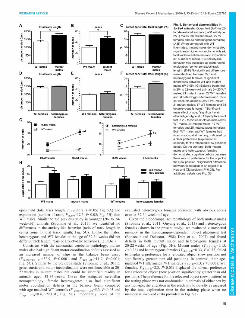
open field (total track length, F(1,59)=5.7, P<0.05; Fig. 5A) andexploration (number of rears, F(1,59)=12.5, P<0.05; Fig. 5B) thanWT males. Similar to the previous study in younger (20- to 24-week-old) animals (Stromme et al., 2011), we identified nodifferences in the anxiety-like behavior (ratio of track length incenter zone to total track length; Fig. 5C). Unlike the males,heterozygous and WT females at the age of 32-34 weeks did notdiffer in track length, rears or anxiety-like behavior (Fig. 5D-F).Consistent with the substantial cerebellar pathology, mutant
males also had significant motor coordination deficits assessed asan increased number of slips in the balance beam assay(F(genotype 1,84)=32.9, P<0.0001 and F(age 1,86)=11.9, P<0.001;Fig. 5G). Similar to the previous study (Stromme et al., 2011),gross ataxia and motor incoordination were not detectable at 20-22 weeks in mutant males but could be identified readily inanimals aged 32-34 weeks. Given the mitigated cerebellarneuropathology, female heterozygotes also had significantmotor coordination deficits in the balance beam comparedwith age-matched WT controls (F(genotype 1,105)=5.3, P<0.05 andF(age 1,105)=8.4, P<0.01; Fig. 5G). Importantly, none of the
evaluated heterozygous females presented with obvious ataxiaeven at 32-34 weeks of age.
Given the hippocampal neuropathology of both mutant males(Stromme et al., 2011; Ouyang et al., 2013) and heterozygousfemales (shown in the present study), we evaluated visuospatialmemory in the hippocampus-dependent object placement test(Ennaceur and Delacour, 1988; Dere et al., 2007) and founddeficits in both mutant males and heterozygous females at20-22 weeks of age (Fig. 5H). Mutant males (T(d.f.=23)=1.15,P<0.26) and heterozygous females (T(d.f.=24)=0.33, P<0.75) failedto display a preference for a relocated object (new position notsignificantly greater than old position). In contrast, their age-matched WT littermates (WT males, T(d.f.=14)=5.3, P<0.0001; WTfemales, T(d.f.=16)=2.3, P<0.05) displayed the normal preferencefor a relocated object (new position significantly greater than oldposition). The preference for the relocated object (new position) inthe testing phase was not confounded in animals of either sex byany non-specific alteration in the reactivity to novelty as assessedby the total exploration time in the training phase when nomemory is involved (data provided in Fig. S3).
Fig. 5. Behavioral abnormalities inSlc9a6 animals. Open field (A-F) in 32-to 34-week-old animals [n=31 wild-type(WT) males, 30 mutant males, 22 WTfemales and 33 heterozygous females].(A,B) When compared with WTlittermates, mutant males demonstratedsignificantly higher locomotor activity (A;total track in centimeters) and exploration(B; number of rears). (C) Anxiety-likebehavior was assessed as center zoneexploration (center zone/total tracklength). (D-F) No significant differenceswere identified between WT andheterozygous females. *Significantdifferences between WT and mutantmales (P<0.05). (G) Balance beam testin 20- to 22-week-old animals (n=25 WTmales, 21 mutant males, 22 WT femalesand 34 heterozygous females) and 32- to34-week-old animals (n=25 WT males,21 mutant males, 17 WT females and 38heterozygous females). *Significantmain effect of age; #significant maineffect of genotype. (H) Object placementtest in 20- to 22-week-old animals (n=15WT males, 24 mutant males, 17 WTfemales and 25 heterozygous females).Both WT males and WT females hadintact visuospatial memory, indicated bya clear preference (exploration inseconds) for the relocated (New position)object. On the contrary, both mutantmales and heterozygous femalesdemonstrated cognitive deficits becausethere was no preference for the object inthe New position. *Significant differencebetween exploration of an object in aNew and Old position (P<0.05). Foradditional details see Fig. S3.
19
RESEARCH ARTICLE Disease Models & Mechanisms (2016) 9, 13-23 doi:10.1242/dmm.022780
Disea
seModels&Mechan
isms
DISCUSSIONOur study demonstrates that expression of the mutant Slc9a6allele in heterozygous female mice mosaically follows theneuroanatomical distribution and cell-type-specific patternsidentified in the brains of X-chromosome hemizygous mutantmales. As a consequence, heterozygous females develop behavioraland neuropathological abnormalities that are, albeit milder,comparable to defects identified in mutant males. As a follow-upto our previous studies, by analyzing animals up to 8 months old wedocument that specifically the sensorimotor dysfunction and thecerebellar PC degenerative pathology are progressive with age bothin mutant males and in heterozygous females.Overall, we propose that Slc9a6 KO heterozygous female mice
represent a relevant and crucial model for future studies aimed atunderstanding the pathogenesis of human CS. As the secondpathology-expressing genotype relevant to human X-linked CS,heterozygous female mice should complement the experimental useof mutant males. As a result of the mosaic expression of the mutantSlc9a6 allele, we expect heterozygous females to becomeparticularly useful for testing the cell-autonomous nature ofspecific (neuro)pathologies and/or exploring the roles ofintercellular interactions (neuronal circuitry dysfunction included)in the onset and progression of the abnormal phenotype. Similarlyimportant will be studies aimed at exploring the embryonic anddevelopmental impacts of XCI on overall and site-specific brain(dys)function in heterozygous female mice.The bacterial β-galactosidase that reports the expression of the
mutant Slc9a6 allele can be identified in murine tissues by a specificantibody. With IF, we confirmed cerebral and cerebellar β-Galexpression in large populations of neurons and astrocytes (Strommeet al., 2011; Deane et al., 2013; Kondapalli et al., 2013; Ouyanget al., 2013) in mutant males and identified the mosaic presence ofβ-Gal in these cell types and locations in heterozygous females.Knowing the expression patterns in mutant males, presuming thatthe murine Slc9a6 gene does not escape XCI (Yang et al., 2010) andcomparing our results with the recently published XCI topographymaps of the female murine brain (Wu et al., 2014), we explain themosaic β-Gal expression patterns in the brains of heterozygousfemale mice by XCI. Previously unreported, we found β-Gal inAPC+ oligodendrocytes of both mutant males and heterozygousfemales. In fact, this suggests that another glial cell type istheoretically compromised by the Slc9a6 deficit both in the mousemodel and in CS patients.Although the neuropathological findings in human hippocampi
are relatively mild (Garbern et al., 2010) and hippocampal atrophywas identified by neuroimaging studies only in some maleindividuals (Schroer et al., 2010), dysfunction of hippocampusand amygdala can be presumed based on the clinical CS phenotype(Stromme et al., 2011). Lysosomal accumulation of GM2 in neuronsof hippocampal CA3/CA4 regions and basolateral amygdala is oneof the early occurring abnormalities in Slc9a6mutant male mice. Inthe present study, we found that only a subpopulation of neurons inamygdala accumulates GM2. Molecular, structural and, mostimportantly, functional/connectivity characteristics (Capogna,2014; Janak and Tye, 2015) of these neurons, the determinants oftheir sphingolipid pathology and its contribution to the overallphenotype remain to be established. Crucially, however, inheterozygous female mice, the timing, neuroanatomicaldistribution and ultrastructural appearance of GM2 pathology arereplicated. As an anticipated effect, GM2 accumulation in neuronsof both hippocampus and amygdala were correlated with themosaically expressed mutant Slc9a6 allele in heterozygous females.
The early-onset degeneration of PCs and ataxia present in oldermutant male mice correspond to the clinical, neuroimaging andcerebellar pathology in male CS patients (Christianson et al., 1999;Gilfillan et al., 2008; Garbern et al., 2010; Schroer et al., 2010;Mignot et al., 2013). More specifically, PC loss and consequentatrophic changes in the cerebellar cortex of mutant males follow apreviously described and conserved cerebellar developmental/gene-expression pattern (Stromme et al., 2011). In heterozygous femalemice, the mosaic β-Gal expression profiles in the cerebellar cortexcorrespond at the cellular level to the uniform expression found inmutant males. PC degeneration in heterozygous females is initiatedat the same age as in mutant males, and a selection against (β-Gal+)PCs is likely in heterozygous females in cerebellar cortical zonesthat are affected by degeneration in mutant males. Importantly, themean values (and distribution of individual values amongheterozygous females) of the percentage fraction of β-Gal+ PCs inthe cerebellar zone(s) not affected by degeneration even in older(32- to 34-week-old) mice suggest random XCI in this particularneuronal population. This result shows that direct detection of theβ-Gal reporter in the brains of heterozygous females allows Slc9a6allelic expression quantification even in numerically limited oranatomically restricted specific cellular types/populations. Besidesneurons, we also found that GFAP+ cells (Bergmann glia) in thecerebellar cortex express β-Gal. Aware of the intimatedevelopmental, structural and functional association between PCsand Bergmann glia (Bellamy, 2006; Wang et al., 2012) thatencompasses regulation of synaptic transmission and/or plasticityand buffering of extracellular K+ and neurotrophin levels byastrocytes (Kondapalli et al., 2014), we hypothesize that the PCdegeneration in mutant male or heterozygous female mice might notbe an exclusively cell-autonomous event. If valid, the variableextent of XCI in PCs and cerebellar cortical astrocytes wouldprobably, in conjunction with the microdomain and overall zonalorganization of cerebellar cortex, represent a crucial determinant ofthe cerebellar degenerative pathology in heterozygous females.
Crucial for human CS, dominance and recessivity allelic relationsvalid for autosomal genes do not apply to X-chromosome-linkedtraits (Dobyns et al., 2004). Whereas X-linked phenotypes arepenetrant in X-chromosome hemizygous male CS patients, theclinical presentation in heterozygous female patients/carriers ismitigated and often variable. One of the principal underlyingmechanisms for such variability in females is XCI in their tissues(Yang et al., 2011; Deng et al., 2014). Unlike in males, data on thefrequency of the mutations in X-linked genes associated withintellectual disability (ID) among females is largely unavailable as alikely result of the diversity of their clinical phenotype(s).Interestingly, it was shown that female heterozygotes in familieswith X-linked ID have significantly skewed XCI ratios in peripheralwhite blood cells (Plenge et al., 2002). However, the relation of theskewed XCI status in leukocytes to the range and severity of theneurodevelopmental, neurological and psychiatric disease(s) stillremains to be understood fully (Tzschach et al., 2015). Consistentwith cellular abnormalities, Slc9a6 heterozygous female miceshowed the same cognitive and sensorimotor abnormalities,albeit mitigated, that were compromised in mutant males. Itremains for future studies, however, to attempt to establishneuroanatomical XCI thresholds for the behavioral pathologiesand determine the sources of inter-individual variability inheterozygous female mice.
Pescosolido et al. (2014) defined the core and secondaryclinical features of CS by evaluating the largest cohort of affectedfamilies and male patients. Although reported inconsistently, a
20
RESEARCH ARTICLE Disease Models & Mechanisms (2016) 9, 13-23 doi:10.1242/dmm.022780
Disea
seModels&Mechan
isms
variable and mitigated phenotype seems likely also in femaleheterozygotes for SLC9A6 mutations. Here, some femaleshave presented with neurodevelopmental delays, problems withspeech, learning and/or behavioral difficulties, aggressiveness inchildhood and adolescence, hyperkinesis or truncal ataxia(Christianson et al., 1999; Gilfillan et al., 2008; Schroer et al.,2010; Pescosolido et al., 2014). Intriguingly for interpretation ofthese observations, Gilfillan et al. (2008) partly excluded XCI as acontributor to the clinical CS presentation because several femaleheterozygotes in their pedigrees presented with normal XCI ratiosin lymphocytes.Several recent genomic projects (Tarpey et al., 2009; Schuurs-
Hoeijmakers et al., 2013; Tzschach et al., 2015) demonstrated thatCS is one of the most frequent X-linked neurodevelopmental IDdisorders. However, the patient cohorts in these studies eitherconsisted exclusively of male ID patients and their female relativesor included females with penetrant ID phenotypes. As a possibleresult of disqualifying individuals with mitigated or variableabnormalities (applicable specifically to female heterozygotes),the CS population frequency that was estimated based on some ofthese data sets as 1 in 16,000-100,000 (Pescosolido et al., 2014)could, in fact, be higher. Also relevant to the occurrence of CS, asubstantial fraction of male CS probands develop the SLC9A6mutations de novo (Schroer et al., 2010; Pescosolido et al., 2014).This lack of maternal inheritance in CS pedigrees has not yet beenspecifically explored for post-zygotic mutagenic events or low-levelmaternal somatic/germinal mosaicism, both of which result in variablenon-homogeneous tissue distribution of SLC9A6 mutations. Asthese phenomena are relatively frequent in the general population(Acuna-Hidalgo et al., 2015), they could theoretically represent anadditional and disparate source of phenotypic variability contributingto the likely under-diagnosis of CS.Here, we have provided evidence for a behavioral and
neuropathological phenotype in Slc9a6 KO heterozygous femalemice. Such findings, we believe, indicate a crucial need to gaindetailed insight into the range of potential phenotypes inheterozygous female CS patients/carriers. Although weacknowledge that the homogenous genetic background of murineSlc9a6 KO model might not necessarily reflect the complex andheterogeneous human genomics and X-linked epigenetics, wehypothesize that the expected XCI-driven expression (Cotton et al.,2015) of SLC9A6 mutations in the brains of human femaleheterozygotes contributes to the diversity of their resultant clinicalpresentation. Defining the full phenotypic spectrum in thispotentially underappreciated patient group is, to us, a keyprerequisite to delineate the population frequency of CS, avoiddiagnostic neglect and allow efficient genetic counseling andscreening in affected families. Moreover, an evidence-basedrecognition of female CS heterozygotes as at risk or affected by avariable or mitigated disease could be an additional strong argumentadvocating further experimental and clinical research, including thedevelopment of therapy for this disorder.
MATERIALS AND METHODSMice and tissue collectionAll procedures used in experiments involving animals were approved by theInstitutional Animal Care and Use Committee of the Albert Einstein Collegeof Medicine. The study used B6.129P2-Slc9a6tm1Dgen/J (Slc9a6 KO) miceoriginally acquired from the Jackson Laboratory (Bar Harbor, ME, USA).Genotyping of litters was performed as previously reported (Stromme et al.,2011). In this study, we used Slc9a6 wild-type male (Slc9a6+/Y or ‘WT’males) and female (Slc9a6+/+ or ‘WT’ females) mice, X-chromosome
hemizygous (mutant) Slc9a6 KO male mice (Slc9a6−/Y or ‘mutant’ males)and X-chromosome heterozygous Slc9a6 KO females (Slc9a6−/+ or‘heterozygous’ females). Homozygous (mutant) Slc9a6 KO females(Slc9a6−/− or ‘mutant’ females) were not tested. If not stated otherwise,results were compared between genotypes and separately for sexes of theanimals (mutant males×WTmales and heterozygous females×WT females).For tissue collection, mice were deeply anesthetized with ketamine andxylazine and perfused transcardially with saline and subsequently with 4%paraformaldehyde (PFA). Dissected organs were further immersion fixedovernight in 4% PFA and then transferred to ice-cold phosphate buffer andstored at 4°C. This study did not use any data or material generated in ourprevious study (Stromme et al., 2011). Behavioral and tissue studies wereperformed in parallel and in identical conditions in all sex and genotypegroups.
Antibodies and immunofluorescenceFor immunofluorescence (IF) labeling, brains were divided along themidline and then split into cerebral and cerebellar-brainstem parts at themesencephalic level. The separated fragments were embedded into 8.0%sucrose and 3.5% agarose, and serial sections 35 µm thick (coronal incerebrum and sagittal in cerebellum) were cut using a Leica VT-1000SVibratome (Leica Microsystems, Wetzlar, Germany). Sections representingrostral-caudal bregma (−1.94 to −2.30) and lateral (0.40-0.84) ranges(Franklin and Paxinos, 2008) were selected from cerebra and cerebella,respectively. Matched sections were stained by multi-IF protocols asdescribed before (McGlynn et al., 2004). The following primary antibodieswere used for staining of specific epitopes: chicken anti-E.coliβ-galactosidase (β-Gal) Ab (ab9361, 1:2000; Abcam, Cambridge, MA,USA), mouse anti-calbindin (Purkinje cells) mAb D-28K (C9848, 1:2000;Sigma-Aldrich, St Louis, MO, USA), rabbit anti-calbindin pAb (AB1778,1:800; Chemicon, Temecula, CA, USA), rat anti-CD68 mAb (MCA1957,1:1000; AbD Serotec, Kidlington, UK), mouse anti-glial fibrillary acidicprotein (GFAP) mAb G-A-5 (G3893, 1:3000; Sigma-Aldrich), mouse anti-neurofilament (anti-NF) medium chain mAb (NB300-134, 1:500; NovusBiologicals, Littleton, CO, USA), mouse anti-APC (oligodendroglial cells;OP80, 1:100; Calbiochem, San Diego, CA, USA), mouse anti-NeuN(neuronal nuclei) IgG mAb (MAB377, 1:1000; Chemicon) and mouse anti-GM2 ganglioside IgM mAb (mab 10-11, 1:15; cell culture supernatant wasproduced in-house from the 10-11 hybridoma line by ProgenicsPharmaceuticals, Tarrytown, NY, USA). Species-specific secondaryantibodies conjugated to Alexa Fluor (AF) 488, 546 and 633 dyes(Invitrogen, Carlsbad, CA, USA) were used for detection of primaryantibodies. A minimum of three animals were evaluated for all sex andgenotype groups aged 4 and 32-34 weeks. The number of cerebella analyzedin the quantitative studies in 32-week-old animals is listed in the legends toFigs 3,4 and Fig. S1.
Light microscopyOverview images (Fig. 1A,B; Fig. 2A,B; Fig. 3A,B,E,F) of the cerebral andcerebellar IF-labeled sections were acquired using an IX70 microscope(Olympus, Tokyo, Japan) equipped with an HQ2 camera (Photometrics,Britannia, AZ, USA) and a Proscan II-encoded xyz stage (Prior Scientific,Rockland, MA, USA) equipped with 10 position excitation-emissionSmartShutters filter wheels (Sutter, Novato, CA, USA). The exposuretime for all antibody combinations was 750 ms (gain 2×) per channel.Excitation/emission conditions for the AF488 and AF546 dyes wereexcitation (exc.) 490/20/beamsplitter (b.s.) 480-513/emission (em.) 535/40and exc.572/23/b.s.555-588/em.630/60 nm, respectively. Individual butpartly overlapping double-channeled 14-bit images (downsampled to 8-bit)were acquired with a Plan 10× (NA 0.25) objective and digitally stitched inMetamorph/MetaFluor (Molecular Devices, Sunnyvale, CA, USA) andsubsequently in Photoshop CS6 (Adobe, San Jose, CA, USA) software.Selected areas of the IF-labeled sections (Figs 1-4; Fig. S1) were imaged bylaser scanning confocal microscope (Zeiss Meta Duo V2, Oberkochen,Germany) using a Plan Apochromat 20× (NA 0.8) objective. Conditions ofimage acquisition (pinhole size, excitation laser intensity, scanning speed,dichroic mirrors, excitation and emission (band)pass filters andphotomultiplier gains) were kept constant for individual combinations of
21
RESEARCH ARTICLE Disease Models & Mechanisms (2016) 9, 13-23 doi:10.1242/dmm.022780
Disea
seModels&Mechan
isms
primary and secondary antibodies. Excitation and emission filtering was set tominimize crosstalk, and individual dyes were excited and their emission wascollected sequentially. The final z-stacks (thickness is reported in the legendsof the individual figures) were transformed into single-plane maximalintensity projection (MIP) images using the Zeiss LSM Image Browser(Zeiss). Images of amygdala and CA3 brain regions (Fig. 2C-H) representscans with an open confocal pinhole, allowing collection of the fluorescenceinformation from the entire thickness of the section (35 µm).
Electron microscopyFor electron microscopy, cerebra of 4- to 5-week-old WT and mutant malesand heterozygous females were coronally sectioned (250 µm) using aVibratome. Sections containing hippocampal CA3 and amygdala regionswere selected, and these regions were manually dissected. The tissue blockswere first transferred to 0.1 M cacodylate buffer and post-fixed in 2%glutaraldehyde. Samples were further washed and post-fixed in osmium (1%osmium in 0.1% cacodylate buffer), dehydrated and embedded in Epon-araldite. Ultrathin sections were stained with uranyl acetate and lead citrateand examined with a Philips CM10 electron microscope (Philips ElectronOptics, Eindhoven, The Netherlands).
Purkinje cell density countsSerial cerebellar sagittal sections (35 µm thick) were visually searched forthe first occurrence of the deep cerebellar nuclei (∼0.48 mm lateral;Fig. 105 of Franklin and Paxinos 2008). Such a section was considered assection +1. This and three consecutive sections in lateral sequence (+3, +5and +7) were stained with a combination of anti-calbindin (PC marker) andanti-β-Gal antibodies and secondary antibodies conjugated to AF488 andAF546, respectively (protocol modified for IF from Praggastis et al., 2015).The total number of Purkinje cells (PC#), the number of PCs with β-Gal-positive nuclei (β-Gal+ PCs) and the overall lengths of PC layers (inmillimetres) were manually counted/traced in at least three out of the fourselected sections in lobules I-II and X. The number of 32-week-old animalsanalyzed is provided in legends to Figs 3,4 and Fig. S1. Stereo Investigatorsoftware (MBF Bioscience, Williston, VT, USA) installed in an OlympusBx51 microscope was used for outlining the PC layers and counting PCs.PC# and β-Gal+ PC# were assessed using a PlanFL 20× (NA 0.5) objective.Although nuclei were co-detected by DAPI in all the double-IF-stainedsections, the parallel triple fluorescent quantification proved technicallydifficult because of unbalanced intensities and fluorescence bleaching ofthe three dyes. The total number of PCs with structurally identifiable nucleiin lobules I-II and X was first assessed using the fluorescence filter setdiscerning AF488 (exc.480/40/b.s.510LP/em.505LP). β-Gal+ PCs weresubsequently quantified by changing the excitation and emission to a tripleexcitation/emission (DAPI/FITC/TRITC) filter cube (Chroma set#61000v2, Bellows Falls, VT, USA). The overall PC density (PC#/1 mmof the length of the PC layer) and percentage fractions of β-Gal+ PCs inindividual animals were calculated in lobules I-II and X from values in the35-µm-thick sections.
Behavioral studiesThe strategy for behavioral testing was designed according to our previousanalyses performed in mutant andWTmales aged ∼20-24 weeks (Strommeet al., 2011). Expecting a mitigated, delayed and also variable phenotypein heterozygous females compared with the reference abnormality inX-chromosome hemizygous mutant males, we decided to increasesubstantially the number of tested animals in all compared sex andgenotype groups and to perform some of the behavioral tests at an olderage (weeks 32-34). Animals were tested for their voluntary locomotor andexploratory activities and anxiety-like (exploration of the center zone)behavior in an open field. Motor coordination was assessed by the balancebeam test. Spatial memory of the animals was tested in the object placementtest. The timeline of the tests and ages of animals when tested were asfollows: balance beam at 20-22 weeks, repeated at 32-34 weeks; objectplacement at 20-22 weeks; and open field at 32-34 weeks.
Motor coordination was evaluated by counting the number of slips madewhile crossing a round balance beam (16 mm diameter; 120 cm long;Stanley et al., 2005). Before each testing session, mice were trained to walk
over a 6-cm-wide, flat wooden plank to diminish anxiety. Owing to the timescale of the study (8 months) and tissue collection, some animals were testedonly once (at either 20-22 or 32-34 weeks old), whereas most were testedlongitudinally.
Locomotor activity, exploration and thigmotaxis (anxiety-like behavior)of the animals were assayed in the open-field arena (37 cm×42 cm) for6 min. The total track length traveled in the entire arena was used as ameasure of locomotor activity. Anxiety-like behavior was assessed as theproportion of the center zone (15 cm×15 cm) exploration (center zone track)from the total track traveled. Both measures were assessed automaticallyusing Viewer software (Biobserve, Bonn, Germany). Rears (exploration)were counted manually.
For the object placement test (Ennaceur and Delacour, 1988; Dere et al.,2007), animals were first allowed to explore a pair of identical, non-toxicobjects for 5 min (training phase) in an open field with high-contrast visualcues placed on each wall of the arena. Animals were then placed back intotheir home cages for 16 min (retention interval). Subsequently, one of theobjects wasmoved to a new position, whereas the position of the other objectremained unchanged (old position), and the animals were allowed to exploreboth objects for 3 min (testing phase). Exploration of the objects was definedas any physical contact with an object (whisking, sniffing, rearing on ortouching the object) or orienting to the object from within 5 cm. Viewersoftware (Biobserve) was used to record the sessions. Care was taken toensure that the intrinsic relationship between the objects and the relativeposition of the objects to the visual cues was altered. The total times (inseconds) spent exploring the objects in the old and new positions during thetesting phase were compared. Animals with cumulative exploration times<2 s in either training or testing phases were excluded from further analyses.
Statistical analyses and figure preparationJMP statistical software (v.11; SAS, Cary, NC, USA) was used to analyzethe results of the behavioral and PC quantification analyses. The balancebeam data were analyzed by a mixed-effects analysis including random andfixed factors, similar to a standard repeated-measures analysis, but with theadvantage that missing data can be accommodated in a mixed-effects modelthat thus analyzed both between (fixed)- and within (random)-subjectvariability while preserving the correct sample sizes and degrees of freedom(d.f.) when some animals were tested twice (Fig. 5G). The results of theobject placement test require analysis of the time exploring the object in boththe new and the old position by each mouse (measured during the testingphase). Thus, to preserve the sample size and because the values representtwo data points from each individual animal, these data were matched andanalyzed by paired t-tests in each sex/genotype group. The rest of thestatistical analyses (total track lengths, ratios of center and total tracklengths, number of rears, total exploration time in the training phase of theobject placement test, PC densities and comparisons of fractions of β-Gal+
PCs in lobules I-II and X) were evaluated by one-way ANOVA. P-values<0.05 were considered statistically significant. Data in Figs 3,4 (shown alsoas individual values) and 5, in the text and supplementary material, arepresented as arithmetic mean values±s.e.m. Figures were prepared inPhotoshop CS6 (Adobe, San Jose, CA, USA) and graphs were plotted inGraphPad Prism version 6.05 software (GraphPad Software, La Jolla, CA,USA). For better contrast and visibility without altering the biologicalinformation, images were stretched to fill the full dynamic 8-bit ranges.
AcknowledgementsThe authors would like to acknowledge Kostantin Dobrenis and Cristin Davidson forcrucial discussions and Gloria Stephney and Bin Cui for technical assistance.Behavioral studies were conducted at the Behavioral Core Facility DominickP. Purpura Department of Neuroscience (director M.G.), Rose F. KennedyIntellectual and Developmental Disabilities Research Center, Albert EinsteinCollege of Medicine.
Competing interestsThe authors declare no competing or financial interests.
Author contributionsJ.S. conceived and designed the study, performed light and electron microscopicimaging, participated in the cell quantification studies, analyzed the data, wrote the
22
RESEARCH ARTICLE Disease Models & Mechanisms (2016) 9, 13-23 doi:10.1242/dmm.022780
Disea
seModels&Mechan
isms
first draft and co-edited the final version of the manuscript. J.L. performed thebehavioral testing, tissue staining and cell quantification analyses and co-edited themanuscript. M.G. designed and supervised the behavioral testing and statisticalanalyses and co-edited the manuscript. S.U.W. conceived and designed the study,analyzed the data and co-edited and submitted the final version of the manuscript.
FundingThis project was supported by National Institute of Child Health and HumanDevelopment grants [R01 HD045561 and P30 HD071593 to S.U.W.]. J.S. wassupported by National Institute of Neurological Disorders and Stroke Award [1F05NS074790] and by the research project [IGAMZ NT14015-3/2013] from the Ministryof Health of the Czech Republic.
Supplementary informationSupplementary information available online athttp://dmm.biologists.org/lookup/suppl/doi:10.1242/dmm.022780/-/DC1
ReferencesAcuna-Hidalgo, R., Bo, T., Kwint, M. P., van de Vorst, M., Pinelli, M., Veltman,J. A., Hoischen, A., Vissers, L. E. L. M. and Gilissen, C. (2015). Post-zygoticpoint mutations are an underrecognized source of de novo genomic variation.Am. J. Hum. Genet. 97, 67-74.
Bellamy, T. C. (2006). Interactions between Purkinje neurones and Bergmann glia.Cerebellum 5, 116-126.
Capogna, M. (2014). GABAergic cell type diversity in the basolateral amygdala.Curr. Opin. Neurobiol. 26, 110-116.
Christianson, A. L., Stevenson, R. E., van der Meyden, C. H., Pelser, J., Theron,F. W., van Rensburg, P. L., Chandler, M. and Schwartz, C. E. (1999). X linkedsevere mental retardation, craniofacial dysmorphology, epilepsy, ophthalmoplegia,and cerebellar atrophy in a large South African kindred is localised to Xq24-q27.J. Med. Genet. 36, 759-766.
Cotton, A. M., Price, E. M., Jones, M. J., Balaton, B. P., Kobor, M. S. and Brown,C. J. (2015). Landscape of DNA methylation on the X chromosome reflects CpGdensity, functional chromatin state and X-chromosome inactivation. Hum. Mol.Genet. 24, 1528-1539.
Deane, E. C., Ilie, A. E., Sizdahkhani, S., Das Gupta, M., Orlowski, J. andMcKinney, R. A. (2013). Enhanced recruitment of endosomal Na+/H+ exchangerNHE6 into Dendritic spines of hippocampal pyramidal neurons during NMDAreceptor-dependent long-term potentiation. J. Neurosci. 33, 595-610.
Deng, X., Berletch, J. B., Nguyen, D. K. and Disteche, C. M. (2014). Xchromosome regulation: diverse patterns in development, tissues and disease.Nat. Rev. Genet. 15, 367-378.
Dere, E., Huston, J. P. and De Souza Silva, M. A. (2007). The pharmacology,neuroanatomy and neurogenetics of one-trial object recognition in rodents.Neurosci. Biobehav. Rev. 31, 673-704.
Dobyns, W. B., Filauro, A., Tomson, B. N., Chan, A. S., Ho, A. W., Ting, N. T.,Oosterwijk, J. C. and Ober, C. (2004). Inheritance of most X-linked traits is notdominant or recessive, just X-linked. Am. J. Med. Genet. 129A, 136-143.
Ennaceur, A. and Delacour, J. (1988). A new one-trial test for neurobiologicalstudies of memory in rats. 1: behavioral data. Behav. Brain. Res. 31, 47-59.
Franklin, K. B. J. and Paxinos, G. (2008). The Mouse Brain in StereotaxicCoordinates. New York: Elsevier, Inc.
Garbern, J. Y., Neumann, M., Trojanowski, J. Q., Lee, V. M., Feldman, G., Norris,J. W., Friez, M. J., Schwartz, C. E., Stevenson, R. and Sima, A. A. F. (2010). Amutation affecting the sodium/proton exchanger, SLC9A6, causes mentalretardation with tau deposition. Brain 133, 1391-1402.
Gilfillan, G. D., Selmer, K. K., Roxrud, I., Smith, R., Kyllerman, M., Eiklid, K.,Kroken, M., Mattingsdal, M., Egeland, T., Stenmark, H. et al. (2008). SLC9A6mutations cause X-linked mental retardation, microcephaly, epilepsy, and ataxia,a phenotype mimicking Angelman syndrome. Am. J. Hum. Genet. 82, 1003-1010.
Janak, P. H. and Tye, K. M. (2015). From circuits to behaviour in the amygdala.Nature 517, 284-292.
Kondapalli, K. C., Hack, A., Schushan, M., Landau, M., Ben-Tal, N. and Rao, R.(2013). Functional evaluation of autism-associated mutations in NHE9. Nat.Commun. 4, 2510-2522.
Kondapalli, K. C., Prasad, H. and Rao, R. (2014). An inside job: how endosomalNa(+)/H(+) exchangers link to autism and neurological disease. Front. Cell.Neurosci. 8, 172-193.
Macauley, S. L., Sidman, R. L., Schuchman, E. H., Taksir, T. and Stewart, G. R.(2008). Neuropathology of the acid sphingomyelinase knockout mouse model of
Niemann-Pick A disease including structure-function studies associated withcerebellar Purkinje cell degeneration. Exp. Neurol. 214, 181-192.
McGlynn, R., Dobrenis, K. and Walkley, S. U. (2004). Differential subcellularlocalization of cholesterol, gangliosides, and glycosaminoglycans in murinemodels of mucopolysaccharide storage disorders. J. Comp.Neurol. 480, 415-426.
Mignot, C., Heron, D., Bursztyn, J., Momtchilova, M., Mayer, M., Whalen, S.,Legall, A., Billette de Villemeur, T. and Burglen, L. (2013). Novel mutation inSLC9A6 gene in a patient with Christianson syndrome and retinitis pigmentosum.Brain. Dev. 35, 172-176.
Ohgaki, R., van IJzendoorn, S. C. D., Matsushita, M., Hoekstra, D. andKanazawa, H. (2011). Organellar Na+/H+ exchangers: novel players in organellepH regulation and their emerging functions. Biochemistry 50, 443-450.
Ouyang, Q., Lizarraga, S. B., Schmidt, M., Yang, U., Gong, J., Ellisor, D., Kauer,J. A. and Morrow, E. M. (2013). Christianson syndrome protein NHE6 modulatesTrkB endosomal signaling required for neuronal circuit development. Neuron 80,97-112.
Pescosolido, M. F., Stein, D. M., Schmidt, M., El Achkar, C. M., Sabbagh, M.,Rogg, J. M., Tantravahi, U., McLean, R. L., Liu, J. S., Poduri, A. et al. (2014).Genetic and phenotypic diversity of NHE6 mutations in Christianson syndrome.Ann. Neurol. 76, 581-593.
Plenge, R. M., Stevenson, R. A., Lubs, H. A., Schwartz, C. E. and Willard, H. F.(2002). Skewed X-chromosome inactivation is a common feature of X-linkedmental retardation disorders. Am. J. Hum. Genet. 71, 168-173.
Praggastis, M., Tortelli, B., Zhang, J., Fujiwara, H., Sidhu, R., Chacko, A., Chen,Z., Chung, C., Lieberman, A. P., Sikora, J. et al. (2015). A murine Niemann-PickC1 I1061T knock-in model recapitulates the pathological features of the mostprevalent human disease allele. J. Neurosci. 35, 8091-8106.
Sarna, J., Miranda, S. R. P., Schuchman, E. H. and Hawkes, R. (2001). Patternedcerebellar Purkinje cell death in a transgenic mouse model of Niemann Pick typeA/B disease. Eur. J. Neurosci. 13, 1873-1880.
Schroer, R. J., Holden, K. R., Tarpey, P. S., Matheus, M. G., Griesemer, D. A.,Friez, M. J., Fan, J. Z., Simensen, R. J., Strømme, P., Stevenson, R. E. et al.(2010). Natural history of Christianson syndrome. Am. J. Med. Genet. A 152A,2775-2783.
Schuurs-Hoeijmakers, J. H. M., Vulto-van Silfhout, A. T., Vissers, L. E. L. M.,van de Vondervoort, I. I. G. M., II, van Bon, B. W. M., de Ligt, J., Gilissen, C.,Hehir-Kwa, J. Y., Neveling, K., del Rosario, M. et al. (2013). Identification ofpathogenic gene variants in small families with intellectually disabled siblings byexome sequencing. J. Med. Genet. 50, 802-811.
Sillitoe, R. V. and Joyner, A. L. (2007). Morphology, molecular codes, and circuitryproduce the three-dimensional complexity of the cerebellum. Annu. Rev. Cell.Dev. Biol. 23, 549-577.
Stanley, J. L., Lincoln, R. J., Brown, T. A., McDonald, L. M., Dawson, G. R. andReynolds, D. S. (2005). The mouse beam walking assay offers improvedsensitivity over the mouse rotarod in determining motor coordination deficitsinduced by benzodiazepines. J. Psychopharmacol. 19, 221-227.
Stromme, P., Dobrenis, K., Sillitoe, R. V., Gulinello, M., Ali, N. F., Davidson, C.,Micsenyi, M. C., Stephney, G., Ellevog, L., Klungland, A. et al. (2011). X-linkedAngelman-like syndrome caused by Slc9a6 knockout in mice exhibits evidence ofendosomal-lysosomal dysfunction. Brain 134, 3369-3383.
Tarpey, P. S., Smith, R., Pleasance, E., Whibley, A., Edkins, S., Hardy, C.,O’Meara, S., Latimer, C., Dicks, E., Menzies, A. et al. (2009). A systematic,large-scale resequencing screen of X-chromosome coding exons in mentalretardation. Nat. Genet. 41, 535-543.
Tzschach, A., Grasshoff, U., Beck-Woedl, S., Dufke, C., Bauer, C., Kehrer, M.,Evers, C., Moog, U., Oehl-Jaschkowitz, B., Di Donato, N. et al. (2015). Next-generation sequencing in X-linked intellectual disability. Eur. J. Hum. Genet. 23,1513-1518.
Wang, F., Xu, Q., Wang, W., Takano, T. and Nedergaard, M. (2012). Bergmannglia modulate cerebellar Purkinje cell bistability via Ca2+-dependent K+ uptake.Proc. Natl. Acad. Sci. USA 109, 7911-7916.
Wu, H., Luo, J., Yu, H., Rattner, A., Mo, A.,Wang, Y., Smallwood, P.M., Erlanger,B., Wheelan, S. J. and Nathans, J. (2014). Cellular resolution maps of Xchromosome inactivation: implications for neural development, function, anddisease. Neuron 81, 103-119.
Yang, F., Babak, T., Shendure, J. and Disteche, C. M. (2010). Global survey ofescape from X inactivation by RNA-sequencing in mouse. Genome Res. 20,614-622.
Yang, C., Chapman, A. G., Kelsey, A. D., Minks, J., Cotton, A. M. and Brown,C. J. (2011). X-chromosome inactivation: molecular mechanisms from the humanperspective. Hum. Genet. 130, 175-185.
23
RESEARCH ARTICLE Disease Models & Mechanisms (2016) 9, 13-23 doi:10.1242/dmm.022780
Disea
seModels&Mechan
isms