Embed Size (px)
Citation preview
NHS HIGHLAND WOUND
MANAGEMENT GUIDELINES AND FORMULARY
Policy Reference: Date of Issue: 05 March 2013 Prepared by: Tissue Viability Leadership Group Date of Review: 05 March 2014 Lead Reviewer: Tissue Viability Leadership Group
Version: 2:0
Authorised by: NHSH Senior Management Team
Date: 05 March 2013
Distribution • Executive Directors • Associate Directors • Clinical Directors • General Managers • Clinical Leads • Assistant General Managers • Lead Nurses/Nurse
Managers • Clinical Governance
Committee • Risk Management Steering
Group • Head of Clinical Governance
and Risk Management Health and Safety
• Professional Heads of Service/ AHP
Professional Leads • Network Managers • Head of Public Involvement • Head of eHealth • Director of Occupational Health • Director of Pharmacy • Lead Pharmacists • Clinical Dental Manager • Clinical Governance Support Team Managers • All Clinical Staff
Method
CD Rom E-mail X � Paper X � Intranet X �
Warning – Document uncontrolled when printed
2
NHS HIGHLAND WOUND MANAGEMENT GUIDELINES AND FORMULARY
CONTENTS PAGE NO Section One GENERAL INFORMATION
Contents 2 Introduction 4 Accountability and Responsibility 5
Section Two WOUND MANAGEMENT
The Physiology of Wound Healing 6 Moist Wound Healing 7 Factors Which Affect Wound Healing 8 Wound Assessment 11 Wound Cleansing 13 Care of the Surrounding Skin 14 Interventions to Manage Exudate 15 Management of Odour 17 Infection 18 Wound Swabbing 20 Pain Control in Wound Management 21
Section Three WOUND CLASSIFICATION AND TREATMENT
Surgical Wound 23 Abrasions 24 Epithelialising Wound 25 Granulating Wound 26 Over Granulating Wound 27 Sloughy Wound 28 Necrotic Wound 29 Cavity Wound 30 Sinus Wound 31 Fungating Wound 32 Bites 33 Flap Lacerations 35 Blisters / Bullae 37 Thermal Injuries / Burns 38 Diabetic Foot 41 Avulsion / Ablation of Toenails 43 Leg Ulcers 44 Skin Grafts 46 Pressure Ulcers 47
Section Four PRODUCT INFORMATION
Wound Management (Dressing) Products 49 Secondary Dressings and Sundries 59
Section Five SPECIALIST PRODUCTS
Iodine (Cadexomer Iodine) 63 Silver Products 66 Honey Products 67 Permafoam for Toe Dressings 68 Charcoal Dressing 69 Prontosan 70 Polyurethane Foam Film Dressings 71 Specialist Primary Wound Contact Layer 72 Larval (Maggot) Therapy 73 Negative Pressure Wound Therapy 77
3
Section Six APPENDICES
1. Referral Guidance for Diabetes Foot Ulcer Clinic 78 2. Offloading the Diabetic Foot Ulcer 79 3. Texas Diabetic Foot Ulcer Classification Tool 80 4. Assessment Chart for Wound Management 81 5. Pressure Ulcer Grading Tool 84 6. Formulary Product Feedback Form 85 7. Photograph Consent Form 86
Section Seven GLOSSARY
87
Section Eight FORMULARY PRODUCT LISTS
89
4
SECTION ONE
1. Introduction The NHS Highland Wound Management Guidelines and Formulary have been developed by the Tissue Viability Leadership Group. This is a multidisciplinary group of professionals working across NHS Highland and NHS Western Isles. The aim of the Wound Management Guidelines and Formulary is to provide practitioners with guidance and a selection of products which are preferred for use in NHS Highland, based on effectiveness, suitability, acceptability and cost-effectiveness. Practitioners should aim to use a product included in the Formulary in most cases and only use a non-Formulary product when there is a good clinical reason for doing so. We have provided, for a broad range of wound types, descriptions, treatment aims and advice on the most appropriate product(s) to use. Because of the diversity of care settings across NHS Highland we have tried to provide practitioners with generic and concise, but flexible, guidelines and formulary. In the vast majority of cases, the products included in the formulary are both on the Scottish National Procurement Contract (which controls products purchased by Scottish Hospitals) and The Scottish Drug Tariff (which states what can be prescribed on NHS prescription forms in primary care settings). The specialist products section should only be used where there is no suitable product for a particular wound type/clinical condition available from the standard primary and secondary product sections. If you wish a product to be considered for inclusion within the formulary, or you wish to report back on existing products, you should complete a Formulary Product Feedback Form (SEE APPENDIX 6) and submit to the Tissue Viability Leadership Group. Contact Kathryn Bell at [email protected] .
5
2. Accountability And Responsibility
As healthcare professionals using this formulary yo u must:
• Use your professional knowledge, judgement and skills to make a decision based on evidence for best practice and the person’s best interests. You need to be able to justify the decisions that you make
• Ensure any advice you give is evidence based when suggesting healthcare products or services
• Have the knowledge and skills for safe and effective practice when working without direct supervision
• Recognise and work within the limits of your competence • Keep your knowledge and skills up to date throughout your working life • Take part in appropriate learning and practice activities that maintain and develop your
competence and performance • Keep clear and accurate records of the discussions you have, the assessments you make, the
treatment and medicines you give and how effective these have been • Complete records as soon as possible after an event has occurred • Ensure any entries made in someone's paper records are clearly and legibly signed, dated and
timed • Ensure any entries made in someone's electronic records are clearly attributable to you • Where wound care is multi-professional and shared ensure all involved are informed of any
significant change is status and/or dressing regime as soon as possible after the contact has occurred
6
SECTION TWO:
3. The Physiology Of Wound Healing Acute and chronic wounds have distinct differences. Some of the basic differences (excluding the microbiological/cellular differences) are:
ACUTE WOUNDS CHRONIC WOUNDS • Short duration • No underlying pathology • Normal inflammatory stage • Usually heals without
complication • Acute wound fluid supports cell
proliferation.
• Unhealed within 6 weeks of formation
• Underlying pathology • Prolonged inflammatory stage • Variety of complications may
arise • Chronic wound fluid does not
support cell proliferation. (Cutting & Tong 2003)
The literature cites many descriptive models of healing. Whichever model is followed, it is essential to have an understanding of the basic process as this will influence decisions made in the day to day management of the wound. Most models suggest that the mechanics of dermal wound healing fall largely into four overlapping phases: 1. Haemostasis Bleeding starts the process of haemostasis. Blood vessels contract, platelets aggregate and a clot is formed. Leucocytes are attracted to the injured area. 2. Inflammation Prostaglandins and proteins are released, which cause vasodilation and inflammation. Neutrophils (whose function is phagocytosis of bacteria) and macrophages (which control the healing process) proliferate in the wound. 3. Granulation
New supporting tissue is formed like a scaffold, along with new blood vessel development, which is known as angiogenesis, and the wound begins to contract. 4. Epithelialisation
New skin cells emerge from the dermal edge and hair follicles, slowly bringing the wound edges together. Healing By Primary Or Secondary Intention
Wound healing by primary intention is when the edges of the wound can be brought together, eg a surgical wound which has been sutured, clipped or glued. The first three phases of healing are usually short but scar maturation may take a few months. Wound healing by secondary intention occurs when the edges of a wound cannot be approximated, eg a leg ulcer. This type of wound heals by a combination of proliferation and wound contraction. The granulation and epithelialisation phases of this type of wound may take months to complete.
7
Moist Wound Healing This concept dates back to the 1940s but did not gain credibility until 1962 when George Winter’s now infamous experiment examined the healing time of wounds exposed to air, compared with wounds covered with polyurethane. The wounds which were covered healed almost twice as fast as those exposed to air. Although this theory was applied to acute wounds, the significance of these findings in chronic wounds has been debated with little agreement about healing rates in the literature (Miller, 1998; Parnham, 2002). However, other benefits for creating a moist environment in chronic wound healing have been cited, such as enhancement of autolytic debridement and reduction in pain during wear and on removal of dressings (Hollinworth, 2005). Maceration may occur where there is excessive moisture on the wound bed. Excessive moisture can excoriate the surrounding skin and cause extension of the wound. Correct choice of dressing is essential to achieve a balance between a wound that is too wet and one that is too dry. Wound fluid contains essential growth factors necessary for epidermal growth. Proteolytic enzymes found in wound fluid have been shown to be beneficial to wound healing but are thought to be present in excessive numbers in chronic wounds (Wysocki et al. 1993). At present, there is no biochemical test to measure an excess of proteases in order to prove this is the cause of delayed healing. Moist Wound Healing in Ischaemic Wounds It is important, when attempting to promote moist wound healing in ischaemic wounds, to be aware that wounds with an underlying ischaemic cause are prone to infection. The presence of necrotic /sloughy tissue, which contain greater quantities of bacteria, increase the risk of infection (Leaper & Ellis, 2002) when moistened and rehydration of the tissue is attempted. Where there is underlying ischaemic disease, and revascularization or restoration of the blood supply is not suitable, moist wound healing may not be appropriate. Devitalised necrotic tissue has a propensity to continually accumulate and may be impossible to resolve (Falanga, 2002) particularly with additional pathophysiology such as Diabetes. The bacterial release can overwhelm the wound, causing deterioration and expansion to the wound itself as well as risking systemic infection. Where individuals have severe arterial impairment, moist wound healing is often best avoided and the area kept as dry as possible. In the case of ulceration to digits, it is advisable to separate digits from one another to prevent the spread of inter-digit ulceration particularly between the toes. REFERENCES Cutting, K. & Tong, A. (2003) Wound Physiology and Moist Wound Healing. Holsworthy: Medical Communications UK Ltd. Falanga, V. (2002) Wound bed preparation and the role of enzymes: a case for multiple actions of therapeutic agents. Wounds: A Compendium of Clinical Research and Practice 2002; 14:2. Hollinworth, H (2005)The management of patients’ pain in wound care. Nursing Standard Tissue Viability Supplement.20(7) 65-73. Leaper, D. and Ellis, S. (2002) “Managing Infection”. In: Harding, K. and Harker, J. (2002) Essential Wound Management for Day –To-Day Practice, Medical Education Partnership, London: Halcyon Print. Miller, M. (1998) Moist wound healing: the evidence. Nursing Times 94, 74-76. Parnham, A. (2002) Moist wound healing: does the theory apply to chronic wounds? Journal of Wound Care 11, 143-146. Wysocki, A.B., Staianocoico, L., Grinnell, F. (1993) Wound fluid from chronic leg ulcers contains elevated levels of metallopreinases MMP-2 and MMP-9. Journal of Investigative Dermatology 101, 64-68.
8
3.2 Factors Which Affect Wound Healing Extrinsic Factors 3.2.1 Nutrition
Nutritional status plays a critical role in the wound healing process. Neglecting the nutritional health of the individual may totally compromise all wound management to be carried out. The Essential Nutrients for Wound Healing Protein, Vitamins C, B and A, Zinc, Iron and Copper are essential for wound healing. In addition to these nutrients, it is essential that adequate energy (calories) is obtained from fats and carbohydrates to prevent tissue protein being used as a source of energy. Protein Aim for 1.0-1.5g/kg/day which is equivalent to 60-90g for a 60kg individual. Protein is required for healing tissues and an inadequate intake inhibits normal protein synthesis and wound healing. The immune response is diminished and there is a delay in matrix formation. Sources include meat, fish, eggs, milk, cheese, yoghurt, pulses and nuts. Energy Aim for a minimum of 30kcal/kg/day which is equivalent to 1800kcal for a 60kg individual. An adequate energy intake is essential to prevent dietary and tissue protein being used as a source of energy rather than for wound healing. All foods provide energy and preserve tissue protein. Carbohydrate sources include bread, potatoes, breakfast cereal, rice and pasta. Fat sources include oils, butter, margarine, fried foods. Fluid Aim for a minimum of 30-35ml/kg/day which is equivalent to 1800-2100ml for a 60kg individual. Adequate fluids are required to prevent skin dehydration. Vitamin C Aim for a minimum of 60mg per day. Vitamin C is required for collagen synthesis and aids iron absorption. Because it is not stored in the body deficiency can occur rapidly. Supplementation should be considered if there is a suspected or confirmed deficiency. Sources include all fruit and vegetables, citrus fruits and juices, blackcurrant juice drinks and fortified fruit squashes. Vitamin A Promotes epithelialisation and granulation of healing wounds. Sources include liver, dairy products, oily fish, carrots and dried fruits. Vitamin B Complex Co-factor for enzyme systems in protein, fat and carbohydrate metabolism. Sources include liver, kidney, meat, poultry, fortified breakfast cereals, wholemeal bread, yeast extract, eggs, and green vegetables. Zinc Deficiency is associated with poor wound healing because it plays an essential role in collagen synthesis, epithelialisation and cell proliferation. Sources include liver, meat, fish, eggs, pulses including baked beans, wholegrain cereals. Iron Anaemia will result in decreased transport of oxygen to damaged tissue and may delay wound healing. Iron is also required for collagen formation. Sources include liver, meat, poultry, oily fish, egg yolk, pulses and dried fruits.
9
Copper Is necessary for collagen formation and essential for red blood cells formation. Sources include meat, fish, cereals and pulses, green vegetables. Nutrition Assessment There are numerous factors to consider when assessing nutrition including reduced access to food, poor appetite, dysphagia, malabsorption and increased metabolism all of which can contribute to a deprivation of nutrients and delayed wound healing. It is essential to consider the nutritional status of all patients including those with wounds by screening each individual using the Malnutrition Universal Screening Tool (MUST). Once an individual’s MUST Score has been calculated the appropriate MUST Care Plan should be implemented and where appropriate referral to a dietician should then be made. Information regarding MUST can be found on the NHS Highland Intranet using the following link. http://intranet.nhsh.scot.nhs.uk/Org/DHS/SSU/ClinicalServicesDir/NutritionandDietetics/Nutrition%20%20Dietetics%20Documents/Information%20for%20Adult%20Wards/must.pdf MUST Care Plans can also be found on the NHS Highland Intranet. Patients identified as having a poor nutritional intake should receive basic nutritional care including:
• Help and advice on menu choices • Between meal snacks • Nourishing drinks e.g. milk • Assistance with eating and drinking if required • Use of Red Tray or equivalent • Monitoring of intake using food record charts
Nutritional Supplements These should be prescribed according to the NHS Highland Guide to Prescribing Nutritional Sip Feeds which can be found using the following link. http://intranet.nhsh.scot.nhs.uk/Org/DHS/SSU/ClinicalServicesDir/NutritionandDietetics/Nutrition%20%20Dietetics%20Documents/Prescribing%20Guidelines%20for%20Nutritional%20Sip%20Feeds%20NOV%2009.pdf For hospital inpatients Fortisip Compact can be given as part of the MUST Care Plan for individuals with a score of 1 or more when the above does not adequately improve food intake. Any patients on supplements should be regularly reviewed both in hospital and following discharge to ensure appropriate use and discontinuation when no longer required. 3.2.2.Drug Therapies • Cytotoxic drugs interfere with cell proliferation and may cause neutropenia, making the
patient more susceptible to wound infection • Long-term use of corticosteroids may suppress fibroblast and collagen synthesis • Non-steroidal anti-inflammatory drugs (NSAIDs) suppress the normal inflammatory response
and may affect healing by causing vasoconstriction (Bale, Harding & Leaper 2000).
10
3.2.3 Poor Wound Management
• Surgical techniques such as inadequate skin closure, rough handling and prolonged theatre time have been shown to delay healing (Bale & Jones 1997; Morison et al. 1997) • Failure to accurately identify abnormalities of healing • Inappropriate use of antiseptics, hypochlorites and antibiotics • Poor dressing choice: high exudate levels, which are not managed effectively by the dressing, cause maceration and subsequent breakdown (Cutting & White 2002). Conversely, if the wound surface is too dry, the cells will become desiccated and may die causing further delay • Failure to provide appropriate pressure relief will contribute to tissue breakdown
3.2.4 Radiotherapy Wounds situated near the treated area may heal slowly or fail to heal. 3.2.5 Smoking Nicotine inhibits epithelialisation, macrophage activity and wound contraction (Siana et al. 1992).
3.2.6 Infection See separate section on infected wounds. Intrinsic Factors
3.2.7 Ageing
• General slowing of the metabolic process • Reduced collagen synthesis • Decline of immune system.
3.2.8 Disease
• Anaemia • Arteriosclerosis • Cancer • Cardiovascular disorders • Diabetes • Immune disorders • Inflammatory diseases • Jaundice/liver failure • Rheumatoid arthritis • Uraemia. (Bale, Harding & Leaper, 2000)
Consider relevant laboratory investigations for these disease processes 3.2.9 Psychological Factors
• Both depression and anxiety can affect wound healing. (Cole-King et al 2001)
REFERENCES
Bale, S., Harding, K., Leaper, D. (2000) An Introduction to Wounds. London: Emap Healthcare. Bale, S., Jones, V. (1997) Wound Care Nursing: A patient-centred approach. London: Ballière Tindall. Cole-King, A Harding K (2001) Psychological Factors and Delayed Healing in Chronic Wounds. Psychosomatic Medicine 63:216-220 (2001). Cutting, K.F. & White, R.J. (2002) Maceration of the skin and wound bed 1; its nature and causes. Journal of Wound Care 11, 275-282. Morison, M.J., Moffatt, C., Bridel-Nixon, J., Bale, S. (1997) A Colour Guide to the Nursing Management of Chronic Wounds. London: Mosby NHS Highland Nutritional Guidelines(2003). . Siana, J.E., Frankild, B.S., Gottrup, F. (1992) The effect of smoking on tissue function. Journal of Wound Care 1, 37-46.

11
4. Wound Assessment A holistic person-centred approach to care should be considered at all times. The wound assessment must be completed by a registered nurse or other healthcare professional with appropriate knowledge and experience. Standard Infection Control Precautions (SICPs) should be applied at ALL times when providing healthcare when there is a risk of exposure to blood, other body fluids, secretions or excretions (except sweat), non-intact skin or mucous membranes. (See http://www.hps.scot.nhs.uk/haiic/index.aspx ) For more information on the key precautions and management principles in tissue viability an educational workbook is available at http://www.nes.scot.nhs.uk/hai/ulcers/ Step 1 • Does the wound need cleansing? • Only cleanse if there is debris on the wound bed that needs removed. Step 2 • Measure wound length, width, depth and undermining. • Do not estimate. • Use a scale such as:
- tracing, disposable ruler for length and/or width - wound swab stick, wound probe for depth and/or undermining
Step 3 • What tissue type and levels of exudate does the wound have? • Dressing choice must accommodate tissue type, exudate level, odour, expected wear time, peri-wound skin, area to be dressed, pain at dressing change and patient/client need. • Select secondary dressing if required. Step 4 • Document in wound chart. • A wound chart must be completed for every patient/client with a wound. • An example of a wound chart can be found in Appendix 4 and at www.tissueviabilityonline.com
Points to remember: • Know the action and possible side effects of any dressing you apply. • Know how to apply and remove any dressing correctly, eg safe and atraumatic removal of all
dressings. • Know how long a dressing can stay in place and indication(s) for dressing change. • Do not mix different primary and secondary types of dressing together, eg hydrogel and
hydrofibre. • Select a dressing that is the correct size for the wound. A dressing that is too big or too small
can be detrimental to the wound. • If in doubt seek advice from appropriate healthcare professional, ie tissue viability nurse,
dermatology nurse, podiatrist.
12
4(a) Wound Photography
Photographs are an important component of effective wound assessment. The value of clinical photography in wound management lies in the ability to achieve repeated views over time, adding objective visual confirmation to the written record, and can provide evidence of rates of healing, capturing therapeutic efficacy. When seeking to take photographs for the purpose of monitoring wounds informed consent should always be sought from the patient, or parent/guardian in the case of children under the age of 16. Although patients under 16 deemed to understand the procedure can give their own consent. Current legal opinion recommends that written informed consent should be obtained from patients prior to any imaging. If the patient consents verbally, but is unable to write, this should be recorded in the case notes. See appendix 7 for consent form. The patient has the right to withdraw consent for wound photography at any time. The withdrawal should be fully documented in the patient records and any historic images should be struck through so as to make it clear that they may not be used. Consideration needs to be given to ensure all images remain confidential and are stored in such a way as confidentiality is not breached, this includes sharing of images with the wider team. If an inpatient at Raigmore the clinical photography dept can be contacted to photograph wounds. Personal cameras, phone and personal memory cards should not be used for photography. All images need uploading to medical illustration dept. This can be done by e mailing high- [email protected]. Photograph the consent form too and send this with other images. This does not apply to GP surgeries. When photographing a wound it is important to be able to assess the dimensions so disposable measuring tapes should be placed on the skin around the wound and then the photograph taken. The patients CHI number can be written on the tape as well as date and explanation of which body part is being photographed, including an arrow to indicate head. Infection control measures should be maintained at all times.
REFERENCES www.tissueviabilityonline.com
Institute of Medical Illustrators , Clinical photography in wound management guidelines 2007 NHS Community Care Western Cheshire, Clinical guideline for Digital Photography in woundcare 2009 Heywood Middleton and Rochdale NHS Trust, Woundcare photography guidelines 2008
5. Asepsis: The use of sterile dressing packs should be restricted to clinical procedures which require asepsis. i.e.:- • insertion of urinary catheter • wound drain • IV line • acute surgical wounds/burns • wound suturing • skin grafts • bypass graft sites • infection/osteomylitis • immunosuppressed patients, • exposed bone • wounds caused by trauma where dirt or grit may be within wound bed. Clinical judgment should be used in deciding whether a sterile dressing pack is required. Clean technique is a safe approach to chronic wound management. For information on Aseptic Technique and a Review of the Literature please refer to the following web sites: http://www.wounds-uk.com/pdf/content_9437.pdf http://www.wounds-uk.com/pdf/content_9436.pdf
13
6. Wound Cleansing As a general rule, routine cleansing of wounds to remove bacteria or to reduce infection is unlikely to be effective (Miller and Gilchrist 1997). Wound cleansing may be advocated to remove contaminants in the following instances: • To remove visible debris after a wound has occurred to aid assessment • To remove excess slough and exudate • To remove any remaining dressing material • Prior to obtaining a microbiology swab Frequent washing of wounds is unnecessary and undes irable. 6.1 Cleansing Solutions In the past wounds were cleansed with antibacterial solutions. Studies comparing the effectiveness of antibacterial solutions to tap water, normal sodium chloride 0.9% and distilled water have found no difference in lowering bacterial count and no increased incidents of infection (Dire & Welsh 1990; Rodeheaver et al. 1982) Antiseptic solutions have been reported to cause tissue damage and hinder the healing process and are unlikely to be effective (Hellewell et al. 1997). One study found the infection rate lowest in wounds cleansed with tap water. Tap water is more common as a cleansing agent in clinical settings (particularly community). It is cost-efficient, copious and accessible and is the recommended wound cleansing solution of choice. Routine use of sterile sodium chloride 0.9% results in a significant waste of resources. Sterile sodium chloride 0.9%, which is an isotonic solution, does not impede the healing process, cause allergic reactions or alter the bacterial flora of the skin. It should be used in the following situations, where tap water is not recommended: • On exposed bone or tendon • On skin or bypass graft • For severely immunosuppressed patients 6.2 Methods Of Cleansing Irrigation is the cleansing mechanism recommended for removal of contaminants. Scrubbing causes pain and local tissue oedema, which decreases host defences. Vigorous cleansing may however be necessary, in some instances, to remove grease and dirt from traumatic wounds which, if left in situ, can cause unsightly tattooing of the skin (Miller & Glover 1999). 6.3 Summary • Does the wound need to be cleansed? If not, don’t do it. • Always warm the irrigation fluid being used. Cooling the wound inhibits cell mitosis. • Never use cotton wool or gauze swabs to clean wounds as they damage granulating tissue and
shed fibres, which increase the risk of infection. REFERENCES Dire, D.J. & Welsh, A.P. (1990) A comparison of wound irrigating solutions used in the emergency department. Annals of Emergency Medicine 19, 704-708. Hellewell, T.B., Major, D.A., Foresman, P.A., Rodeheaver, G.T. (1997) A cytotoxicity evaluation of antimicrobial and non-antimicrobial wound cleansers. Wounds 9, 15-20. Miller, M. & Gilchrist, B. (1997) Understanding Wound Cleaning and Infection. London: Macmillan. Miller, M. & Glover, D. (1999) Wound Management: theory and practice. London: Emap Healthcare Ltd. Rodeheaver, G., Bellamy, W., Kody, M. et al. (1982) Bactericidal activity and toxicity of iodine-containing solutions in wounds. Archives of Surgery 117, 181-186.
14
7. Care Of The Surrounding Skin
The state of the skin surrounding a wound should be assessed at each dressing change. Observe for signs of: • Dry skin which may break down and provide a portal for infection • Maceration caused by poor management of exudate • Contact sensitivity to dressing. The principles of good skin care depend on: • Keeping the skin clean and dry • Avoiding the excessive use of soap • Using showers in preference to baths where possible • Keeping the skin moisturised. 7.1 Emollients Emollients are moisturisers that soothe and hydrate the skin. They are indicated for all dry or scaling disorders but their effects are short-lived so they must be applied frequently and regularly to maintain improvement. Most are best applied after a shower or bath. They should continue to be applied even after improvement occurs for future prevention. There are different types of product available. Effectiveness depends upon the correct choice of product and correct use. Choice will depend upon: • The severity of the condition • Patient preference • The site of application • Cost of preparation.
7.1.1 Treatment The NHS Highland Formulary lists appropriate preparations, and those that are to be used first line. The Highland Formulary can be accessed at: http://intranet.nhsh.scot.nhs.uk/Clinical/Formulary/HJF/Highland%20Formulary%20
4E.pdf Emollients should be applied in the direction of hair growth. Some ingredients may rarely cause sensitisation and this should be suspected if an eczematous reaction occurs. 7.1.2 Ointments Ointments are recommended as the first choice of formulation in most conditions and are particularly useful for chronic dry conditions. Ointments are greasy and generally insoluble in water so can be difficult to wash off, and do not suit all patients. 7.1.3 Creams Creams are emulsions of oil and water, they often contain an antimicrobial preservative and are, therefore, more likely to cause both irritant and allergic reactions. For this reason creams are best avoided first line but can be better than ointments for acute conditions due to a cooling effect as they evaporate, and may be more cosmetically acceptable for some patients. 7.1.4 Lotions Lotions also have a cooling effect, and may be preferable for treating hairy sites. They can be either water or alcohol based. The latter will sting if applied to broken skin.
7.1.5 Gels Have high water content, and are suitable for face and scalp.
15
8. Interventions To Manage Exudate
• The appropriate management of wound exudate requires an understanding of the underlying processes that lead to its production. • Exudate can present in a variety of forms, indicating the need to assess it by volume, viscosity and colour. • The selection of management options should be based on the characteristics of the wound and the needs of the patient. • Dressings may not always be the most appropriate option for exudate management. Consideration should also be given to physical methods of exudate control (White and Cutting 2006).
AREA MANAGEMENT OPTIONS
PERI-WOUND SKIN CLEANSE SKIN
PROTECT THE SKIN
Use warm tap water to remove excess exudate unless sterile sodium chloride 0.9% indicated (see Wound Cleansing section). Be careful not to rub the wound bed as this can destroy healthy granulation tissue. Cavilon® cream, spray or lotion is highly recommended. Consider a topical steroid to reduce inflammation and excoriation. Also consider the following:
• Stoma/Wound bags • Bed rest • Elevation/gentle compression
Topical Negative Pressure
WOUND BED
DRESSINGS:
ANTIMICROBIAL
DRESSINGS
Dressings achieve wound exudate management by absorbing, gelling and transferring the fluid away from the wound bed. When choosing a dressing product it is important to be aware of the fluid handling properties or how the dressing will deal with fluid. Dressings with an antimicrobial component are intended for the control of wound bio burden in critical colonisation. Antimicrobial dressings are therefore useful where raised exudate levels are attributed to bacterial cause.
REFERENCE White, R: Cutting KF (2006) Modern exudate management – a review of wound treatments. World Wide Wounds http://www.worldwidewounds.com/2006/september/White/Modern-Exudate-Mgt.html
16
8.1 Describing Exudate Appearance:
TERM CLINICAL
APPREARANCE REASON
SEROUS
CLEAR WATERY CONSISTENCY
Possibly a sign of infection if profuse. Some bacteria produce fibrinolysins, which degrade fibrin clots or coagulated plasma.
FIBRINOUS CLOUDY Contains fibrin protein strands PURULENT PRODUCING OR
CONTAINING PUS Contains pyogenic organisms and other inflammatory
HAEMOPURULENT
BLOOD STAINED PUS Contains neutrophils, dead and dying bacteria and inflammatory cells. Infection may be present Consequent damage to dermal capillaries leads to blood leakage
HAEMORRHAGIC
BLOODY Capillaries are so friable they readily breakdown, and spontaneous bleeding occurs. Not to be confused with bloody exudates from over enthusiastic debridement
(Cutting 2004)
8.2 Volume Of Exudate And Wound Appearance:
NONE
Wound tissue dry.
SCANT Wound tissue moist.
SMALL Wound tissue wet. Moisture evenly distributed in the wound.
MODERATE Wound tissues saturated. Drainage may or may not be evenly distributed in the wound.
COPIOUS Wound tissues bathed in fluid. Drainage freely expressed.
(Bates-Jensen 1999)
NB: In patients with ‘dampened’ inflammatory response, an increased level of exudate may be an
indication of infection.
REFERENCES
Bates-Jensen, B.M. (1999) Chronis Wound Assessment, Nursing Clinics of North America 34, 799-844
Cutting KF. Exudate: composition and functions. In: White RJ, editor. Trends in Wound Care Volume III. London: Quay Books, 2004
17
9. Management Of Odour Wound odour is a normal characteristic of healing. The fact that an occlusive dressing has been covering a wound and then removed, will emit an odour. It is when this odour becomes related to infection, or an underlying patho-physiological response, or most importantly, if it effects the patient, that it must be addressed. 9.1 Activated Charcoal Absorbent Dressings Properties • Activated charcoal reduces the concentration of offensive odour Wound Types • Discharging, purulent and contaminated wounds complicated by bacterial infection and
offensive odour, e.g. fungating carcinomas, leg ulcers, pressure ulcers, gangrenous lesions etc. How To Use/When To Change • Change when required, e.g. when strike through of exudate occurs or when odour is no longer
being controlled. • Apply directly to wound or over primary dressing. (See Specialist Products Section for further inform ation)
18
10. Infection Infection may be defined as the invasion of living tissue by micro-organisms. The number of micro-organisms and their degree of pathogenicity determine the establishment of infection. Infection delays healing. Nosocomial (hospital-acquired) infections are associated with virulent organisms and are a great cause for concern. Misuse or overuse of antibiotics leads to resistance of these and to the emergence of new bacterial strains (Bale, Harding & Leaper 2000). Host defences usually resist all but the most pathogenic organisms but such defences are often depressed by systemic factors such as shock, immunosuppression, poor nutrition, and local factors such as ischaemia, trauma or implantation of foreign material. Rodeheaver (2001) stated that the single most important parameter to reduce the level of bacterial contamination in the chronic wound is the removal of devitalised tissue.
This may be carried out by: • Surgical debridement which is fast and effective but may be complicated by local pain • Autolytic debridement using moist interactive dressings which liquefy slough and simultaneously promote granulation tissue. This process may be slow to achieve debridement. • Biosurgical debridement which uses sterile larvae to breakdown and remove dead tissue. This is a fairly fast, effective method of debridement but may not be accepted by some patients (see specialist product section).
10.1 Bacterial Colonisation The mere presence of bacteria does not always indicate that a wound is infected. All chronic wounds are colonised with bacteria, usually of more than one species, and often in very large numbers (Hutchinson 1992). When healing progresses normally, these wound inhabitants rarely attract attention. • Many patients who have chronic wounds which are colonised by bacteria progress to complete healing without any setbacks • Some colonised wounds may become ‘indolent’ (where there is delayed healing) although there is no visible deterioration • Over-use of systemic antibiotics has resulted in resistance and this has prompted a return to the debate of using topical antiseptics. Iodine and silver in their contemporary formats appear
to be of clinical benefit particularly where there is heavy or ‘critical’ colonisation and delayed healing (White et al. 2001). Critical colonisation refers to the point where a wound is unable to maintain a balance between the number of microbes and the defence systems available (White et al. 2001). Kingsley (2001) incorporates this notion into a wound infection continuum, extending from sterility to infection.
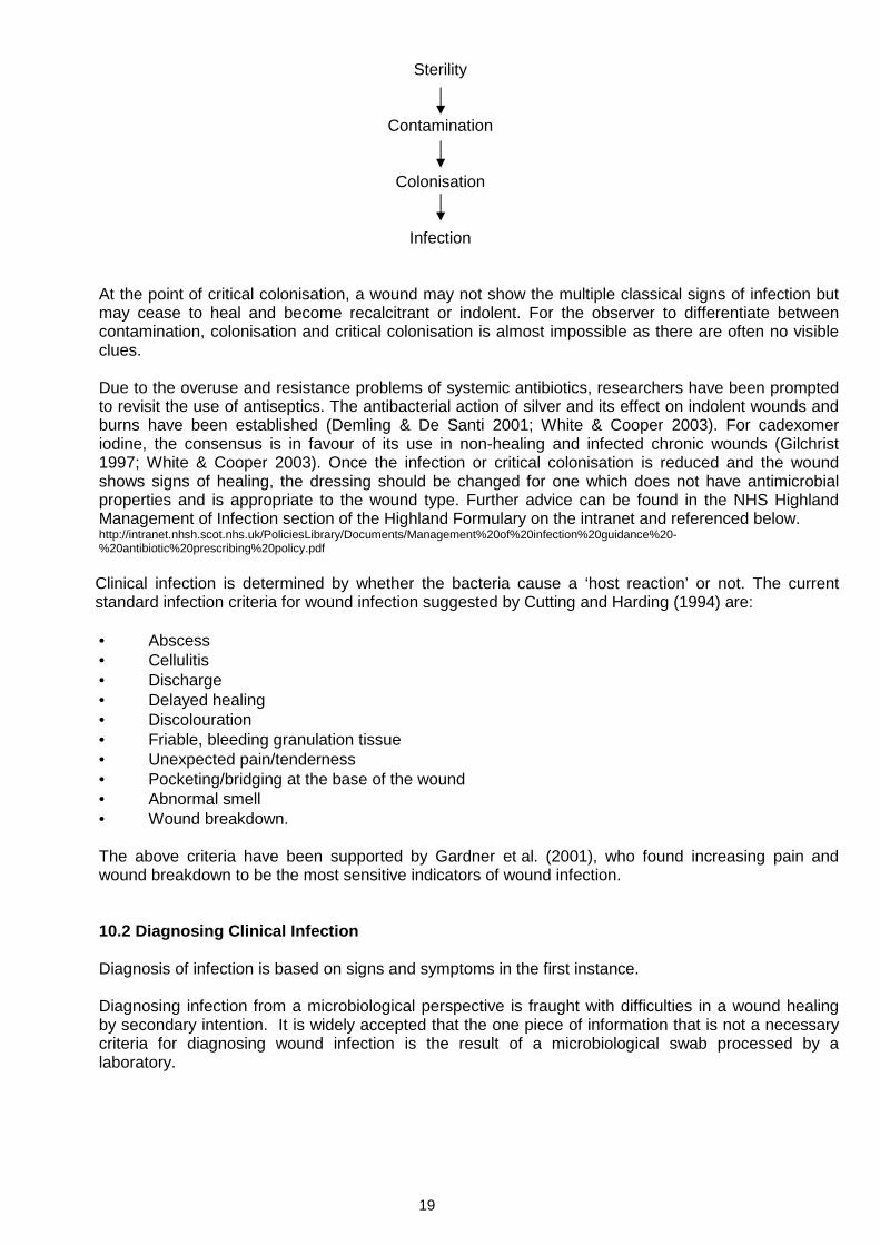
19
Sterility
Contamination
Colonisation
Infection
At the point of critical colonisation, a wound may not show the multiple classical signs of infection but may cease to heal and become recalcitrant or indolent. For the observer to differentiate between contamination, colonisation and critical colonisation is almost impossible as there are often no visible clues. Due to the overuse and resistance problems of systemic antibiotics, researchers have been prompted to revisit the use of antiseptics. The antibacterial action of silver and its effect on indolent wounds and burns have been established (Demling & De Santi 2001; White & Cooper 2003). For cadexomer iodine, the consensus is in favour of its use in non-healing and infected chronic wounds (Gilchrist 1997; White & Cooper 2003). Once the infection or critical colonisation is reduced and the wound shows signs of healing, the dressing should be changed for one which does not have antimicrobial properties and is appropriate to the wound type. Further advice can be found in the NHS Highland Management of Infection section of the Highland Formulary on the intranet and referenced below. http://intranet.nhsh.scot.nhs.uk/PoliciesLibrary/Documents/Management%20of%20infection%20guidance%20-%20antibiotic%20prescribing%20policy.pdf
Clinical infection is determined by whether the bacteria cause a ‘host reaction’ or not. The current standard infection criteria for wound infection suggested by Cutting and Harding (1994) are: • Abscess • Cellulitis • Discharge • Delayed healing • Discolouration • Friable, bleeding granulation tissue • Unexpected pain/tenderness • Pocketing/bridging at the base of the wound • Abnormal smell • Wound breakdown. The above criteria have been supported by Gardner et al. (2001), who found increasing pain and wound breakdown to be the most sensitive indicators of wound infection.
10.2 Diagnosing Clinical Infection Diagnosis of infection is based on signs and symptoms in the first instance. Diagnosing infection from a microbiological perspective is fraught with difficulties in a wound healing by secondary intention. It is widely accepted that the one piece of information that is not a necessary criteria for diagnosing wound infection is the result of a microbiological swab processed by a laboratory.
20
10.3 Swabbing Swabs are usually collected and transported to the laboratory where potential pathogens are normally isolated, cultivated and then characterised. Certain pathogens are fully identified and their antibiotic sensitivities determined, but complete identification of other isolates, such as coliforms or anaerobes, is not routinely attempted. Process for swabbing: • Clean surface exudate from wound prior to taking swab with sterile solution of water or sodium
chloride 0.9% (sterile solution removes risk of contaminating the sample) • Take swab from deep tissue (as close to the wound bed as possible) • Where possible, submit actual tissue samples - these may be sent in universal containers • A specimen of pus is more valuable than a pus swab when sampling abscesses at incision and drainage • When delay in transit is unavoidable, keep at room temperature or refrigerate at 4˚C. For effective wound management, the information obtained is frequently sufficient to make correct clinical decisions. However, Cooper (2002) suggests that ‘difficulties arise if pathology reports are used to make inferences about the impact of microbes on the healing process, because comprehensive analysis of the entire microbial community in the wound is essential to make such judgements’.
REFERENCES Bale, S., Harding, K., Leaper, D. (2000) an Introduction to Wounds. London: Emap Healthcare Ltd. Cooper, R.A. (2002) Wound microbiology: past, present and future. British Journal of Nursing 11, s4-s6. Cutting, K. (2003) A dedicated follower of fashion? Topical medications and wounds. In: White, R.J. & Cooper, R. (eds) The Silver Book. Bath: Quay Books, MA Healthcare Ltd, Bath Press. Cutting, K. & Harding, K. (1994) Criteria for identifying wound infection. Journal of Wound Care 3, 198-201. Demling, R.H. & De Santi, L. (2001) as cited by White, R.J. & Cooper, R. (2003) The use of topical antimicrobials in wound bioburden control. In: The Silver Book. Bath: Quay Books, MA Healthcare Ltd, Bath Press. Gardner, S. et al. (2001) The validity of the clinical signs and symptoms used to identify localised wound infection. Wound Repair Regeneration 9, 178-186. Gilchrist, B. (1997) The use of topical antimicrobials in wound bioburden control. In: White, R.J. & Cooper, R. (eds) The Silver Book. Bath: Quay Books, MA Healthcare Ltd, Bath Press. http://intranet.nhsh.scot.nhs.uk/PoliciesLibrary/Documents/Management%20of%20infection%20guidance%20-%20antibiotic%20prescribing%20policy.pdf Hutchinson, J. (1992) Influence of occlusive dressings on wound microbiology: interim results of a multi-centre clinical trial of an occlusive hydrocolloid dressing. In: Harding, K. et al. (eds) Proceedings of the First European Conference on Advances in Wound Management. London: Macmillan. Kingsley, A. (2001) A proactive approach to wound infection. Nursing Standard 15, 50-58. Rodeheaver,G.T. Wound cleansing, wound irrigation, wound disinfection. In: Krasner, D., Rodeheaver, G.T., Sibbald, R.G. (eds) (2001) Chronic Wound Care, 3rd edn. Wayne: HMP Communications. White, R.J. & Cooper, R. (2003) The use of topical antimicrobials in wound bioburden control. In: White, R.J. & Cooper, R. (eds) The Silver Book. Bath: Quay Books, MA Healthcare Ltd, Bath Press. White, R., Cooper, R., Kingsley, A. (2001) Wound colonisation and infection: the role of topical antimicrobials and guidelines in management. British Journal of Nursing 10, 563-578.
21
11. Pain Control In Wound Management Most wounds cause a certain amount of pain (Casey 1998) but pain management, a key function of all health professionals, is often poorly managed. Sometimes pain can be severe and ongoing, such as with chronic wounds, while at other times it may only occur with initial injury, or during infection or during dressing change. Patients may experience pain as a result of: • Products or techniques used to cleanse wounds • Trauma to the tissues and surrounding skin when products are removed • Skin excoriation from exudates or wound drainage • Lack of empathy • Failure to record patient’s earlier reports of pain • Infection, which can exacerbate existing wound pain • Poor techniques when using compression bandaging. Careful wound assessment is required, as selecting an inappropriate dressing can result in considerable pain and discomfort (Dealey 1999). The correct dressing can ensure comfort and reduce pain, especially during dressing change. Emotional responses can also influence the perception of pain and the distress of having a wound. The way patients detect pain appears to be related to the type of damage causing it (Campbell 1995). Clinically, pain, like wound types, can be classified as acute or chronic, but will be related to: • The type of injury • The location of the wound • Patient perception and previous experience • The healing process and approaches to wound management, e.g. choice of dressing and
provision of analgesia.
11.1 Assessment Of Pain
Pain should be assessed prior to each dressing change and appropriate action taken to address the identified cause. Accurate assessment depends on subjective reporting by the patient. Pain can be assessed effectively during ongoing therapy by asking the patient to rate his/her pain. It is recommended that a simple visual analogue scale is used. The patient should be asked if the pain is worse at any particular time or during a particular activity so that analgesic doses can be timed appropriately. Patients should be closely observed throughout the dressing procedure for reaction to treatment. 11.2 Analgesia
Whatever analgesia is used in wound care, its effectiveness should be evaluated continuously. Failure to achieve pain relief may contribute to the depression and anxiety associated with chronic pain. The type of analgesia to be used depends upon: • The type of wound • Whether the wound is acute or chronic • The level of pain reported by the patient. • Patients individual circumstances e.g. other medicines, co-morbidities Effective doses of analgesics should be given. In chronic pain, treatment should be given often enough and regularly to provide continuous pain relief. This is preferable to giving analgesics only when necessary, ie allowing pain to recur before giving further treatment.
22
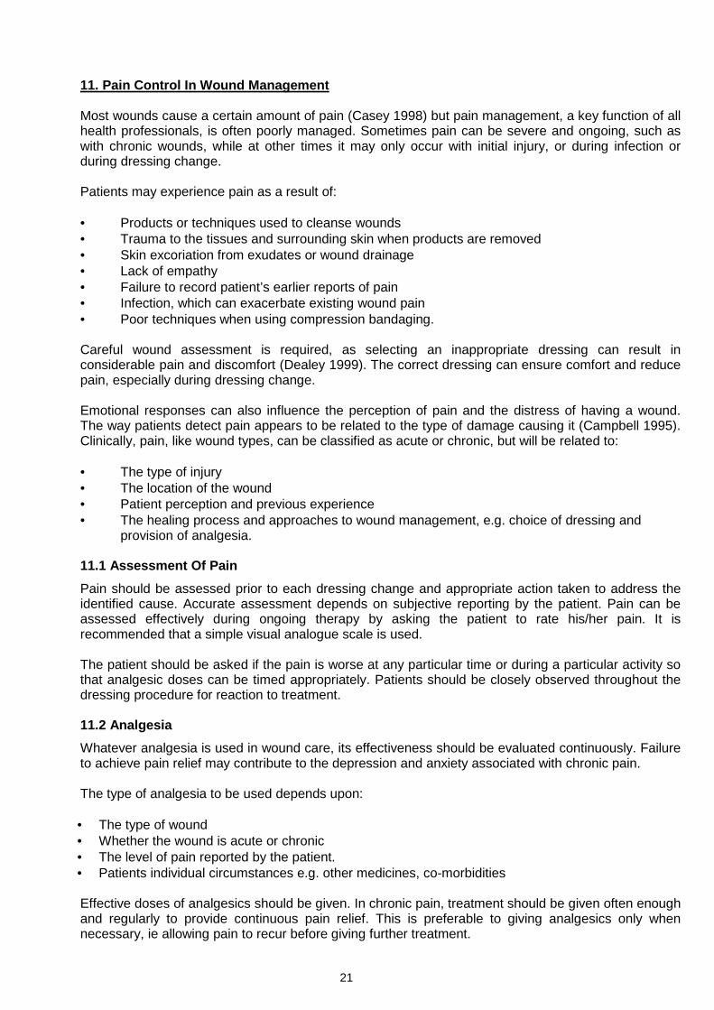
Analgesics should also be given in anticipation of pain, giving careful consideration to any activities which exacerbate pain. In the case of acute pain there is little time to titrate the dose against the patient’s response. Analgesics should be chosen according to assessment of the factors mentioned above. The use of non-steroidal anti-inflammatory drugs (NSAIDs) eg aspirin, ibuprofen, diclofenac etc., is common in treating minor injuries and in long-term inflammatory conditions. This is due to their action of inhibiting the production of prostaglandins (inflammatory mediators). If wound pain is ongoing it may not be appropriate to use an NSAID due to their side effects. The WHO analgesic ladder forms the basis of many approaches to the use of analgesic medicines. There are three essential steps on this ladder.
11.3 The Who Analgesic Ladder
REFERENCES Campbell, J. (1995) Making sense of pain managemen t. Nursing Times 91, 34-35. Casey, G. (1998) The management of pain in wound care. Nursing Standard 13, 49-54. Dealey, C. (2005) The Care of Wounds, 3rd ed. Oxford: Blackwell Science. World Health Organization (1996) WHO Guidelines: Cancer Pain Relief, 2nd ed. Geneva, World Health Organization.
Non-opioid
+/- adjuvant analgesic
STEP 1
STEP 3 Opioid for moderate to
severe pain +/- Non-opioid
+/- adjuvant analgesic
Opioid for mild to moderate pain
+/- non-opioid +/- adjuvant analgesic
STEP 2
Non-opioids, eg Paracetamol
NSAIDs, eg
Ibuprofen Aspirin
Weak opioids, eg Codeine
Dihydrocodeine
Strong opioids, eg
Morphine Diamorphine
Pain persisting or increasing
23
SECTION THREE
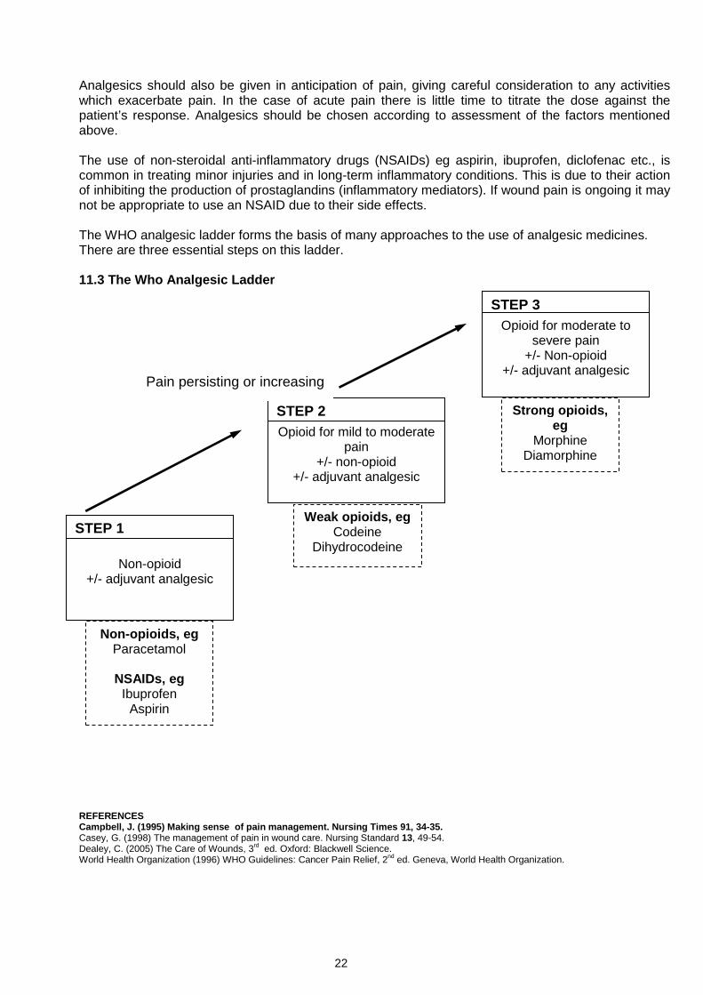
12. Surgical Wound 12.1 Description A surgical wound is the result of a planned procedure, either elective or emergency, where the clinician creates the wound in order to perform a surgical procedure. This wound type is expected in general to follow a rapid, predictable pathway towards healing with minimal scarring and loss of function. The wound may be either incised and closed (this wound heals by primary intention) OR incised and laid open (this wound heals by secondary intention).For guidance see cavity wounds. 12.2 Wound Healing by Primary Intention
Reproduced by permission of NHS Lothian
Treatment Aim • To restore physical integrity and
function without infection and with the minimum of deformity.
• Approximation of wound edges immediately using sutures, clips staples or adhesives, so that each layer (muscle, subcutaneous fat and skin) comes together, thereby expediting haemostasis and the healing mechanism.
Treatment If a dressing is required: • Occlusive dressings should be used post operatively, which may be removed within
48 hours as the wound should be totally sealed, thus preventing the ingress of bacteria (Dealey 2005).
• If there is strike through or leakage, dressing can be replaced or reinforced. 12.3 Wound Healing by Secondary Intention Treatment Aim The wound is left open to heal by granulation, contraction and epithelialisation, for several reasons: • There may be considerable tissue loss, eg radical vulvectomy • The surgical incision is shallow, but has a large surface area, eg donor sites • There may have been infection, eg a ruptured appendix, or an abscess may have been drained, and free drainage of any pus is essential (Dealey 2005). Treatment • Surgical wounds should be dressed according to the wound type. REFERENCE
Dealey, C. (2005 ) General Principles of Wound Management. In: The Care of Wounds, 3rd edn. Oxford: Blackwell Science.
24
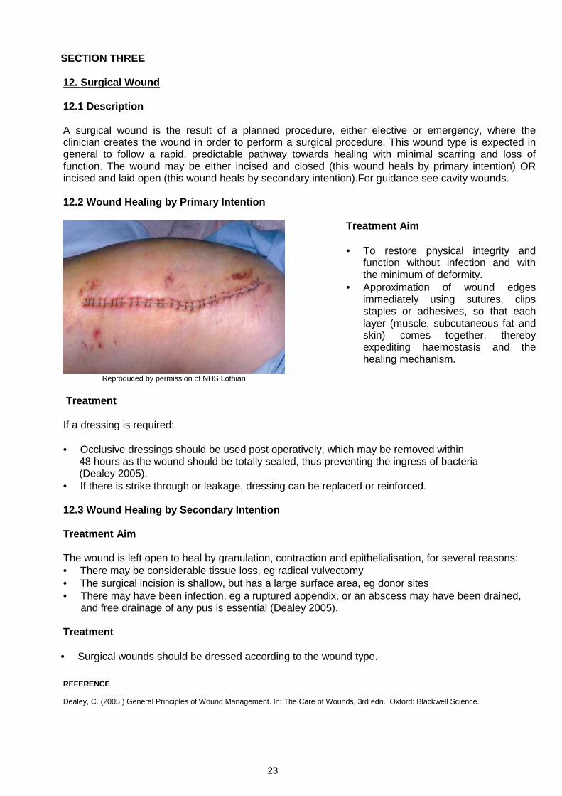
13. Abrasions 13.1 Description These are shearing and friction injuries that result in a scraping or rubbing away of the epidermis or dermis.
13.2 Treatment Aim • To prevent infection and further tissue damage • Abrasions should be cleaned carefully to ensure
that there are no foreign bodies embedded in the wound (Dealey 2005).
13.3 Treatment • Selection of a suitable dressing depends on the extent and depth of the injury. • Exudate levels can vary and, depending on the cause of injury, infection risks can be high. • An occlusive dressing, such as a thin hydrocolloid, which can be left in place for several days,
and also allows the patient to bathe and shower with the dressing in situ. The effect of such a dressing is to prevent the nerve endings drying out. This appears to be the factor which reduces the pain (Dealey 2005).
• Dressings where both the patient and the health professional can see exudate levels and surrounding skin condition can be useful.
REFERENCE Dealey, C. (2005) The management of patients with acute wounds.Oxford: Blackwell Science.
25
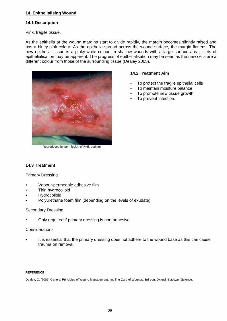
14. Epithelialising Wound 14.1 Description Pink, fragile tissue. As the epithelia at the wound margins start to divide rapidly, the margin becomes slightly raised and has a bluey-pink colour. As the epithelia spread across the wound surface, the margin flattens. The new epithelial tissue is a pinky-white colour. In shallow wounds with a large surface area, islets of epithelialisation may be apparent. The progress of epithelialisation may be seen as the new cells are a different colour from those of the surrounding tissue (Dealey 2005).
14.3 Treatment Primary Dressing • Vapour-permeable adhesive film • Thin hydrocolloid • Hydrocolloid • Polyurethane foam film (depending on the levels of exudate). Secondary Dressing • Only required if primary dressing is non-adhesive Considerations • It is essential that the primary dressing does not adhere to the wound base as this can cause
trauma on removal.
REFERENCE
Dealey, C. (2005) General Principles of Wound Management. In: The Care of Wounds, 3rd edn. Oxford: Blackwell Science.
Reproduced by permission of NHS Lothian
14.2 Treatment Aim • To protect the fragile epithelial cells • To maintain moisture balance • To promote new tissue growth • To prevent infection.
26
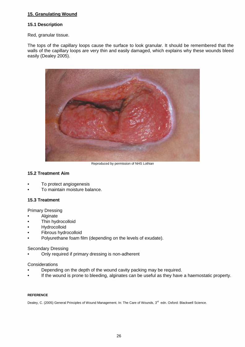
15. Granulating Wound 15.1 Description Red, granular tissue. The tops of the capillary loops cause the surface to look granular. It should be remembered that the walls of the capillary loops are very thin and easily damaged, which explains why these wounds bleed easily (Dealey 2005).
Reproduced by permission of NHS Lothian
15.2 Treatment Aim • To protect angiogenesis • To maintain moisture balance. 15.3 Treatment Primary Dressing • Alginate • Thin hydrocolloid • Hydrocolloid • Fibrous hydrocolloid • Polyurethane foam film (depending on the levels of exudate). Secondary Dressing • Only required if primary dressing is non-adherent Considerations • Depending on the depth of the wound cavity packing may be required. • If the wound is prone to bleeding, alginates can be useful as they have a haemostatic property. REFERENCE Dealey, C. (2005) General Principles of Wound Management. In: The Care of Wounds, 3rd edn. Oxford: Blackwell Science.
27
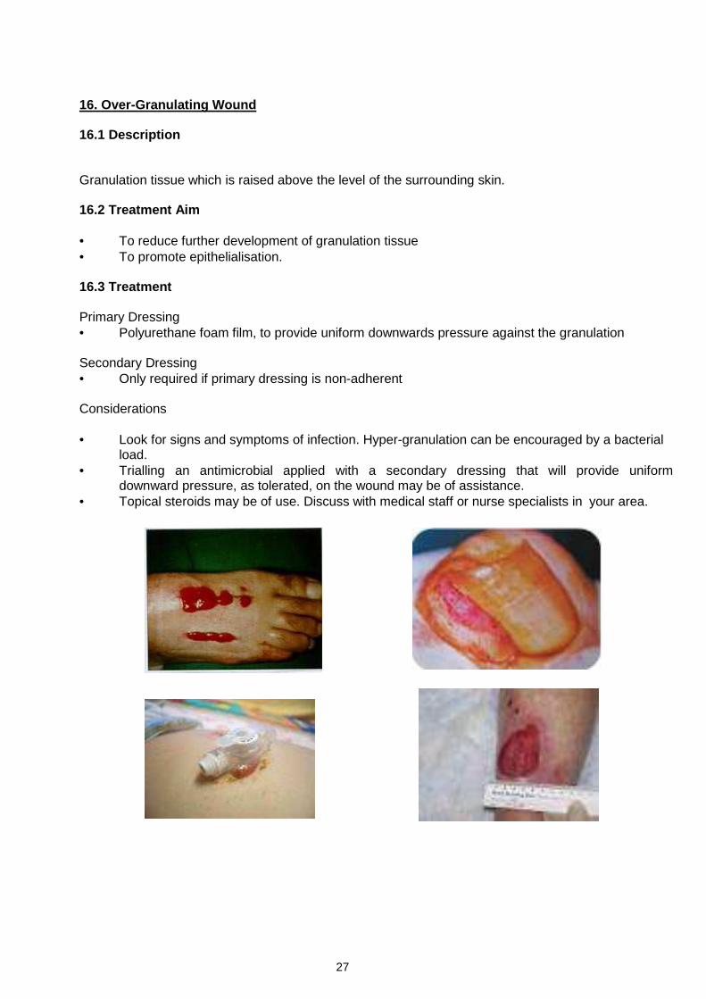
16. Over-Granulating Wound 16.1 Description Granulation tissue which is raised above the level of the surrounding skin. 16.2 Treatment Aim • To reduce further development of granulation tissue • To promote epithelialisation. 16.3 Treatment Primary Dressing • Polyurethane foam film, to provide uniform downwards pressure against the granulation Secondary Dressing • Only required if primary dressing is non-adherent
Considerations • Look for signs and symptoms of infection. Hyper-granulation can be encouraged by a bacterial
load. • Trialling an antimicrobial applied with a secondary dressing that will provide uniform downward pressure, as tolerated, on the wound may be of assistance. • Topical steroids may be of use. Discuss with medical staff or nurse specialists in your area.
28
17. Sloughy Wound
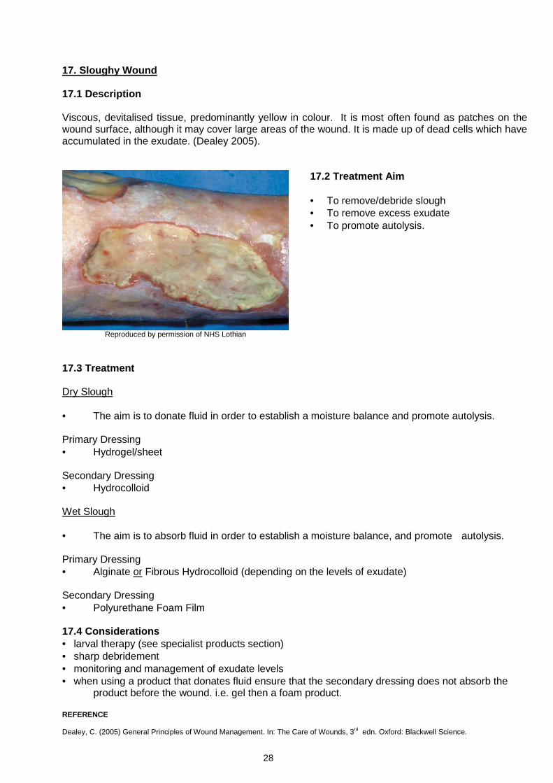
17.1 Description Viscous, devitalised tissue, predominantly yellow in colour. It is most often found as patches on the wound surface, although it may cover large areas of the wound. It is made up of dead cells which have accumulated in the exudate. (Dealey 2005).
Reproduced by permission of NHS Lothian
17.2 Treatment Aim • To remove/debride slough • To remove excess exudate • To promote autolysis.
17.3 Treatment Dry Slough • The aim is to donate fluid in order to establish a moisture balance and promote autolysis. Primary Dressing • Hydrogel/sheet Secondary Dressing • Hydrocolloid Wet Slough • The aim is to absorb fluid in order to establish a moisture balance, and promote autolysis. Primary Dressing • Alginate or Fibrous Hydrocolloid (depending on the levels of exudate) Secondary Dressing • Polyurethane Foam Film
17.4 Considerations • larval therapy (see specialist products section) • sharp debridement • monitoring and management of exudate levels • when using a product that donates fluid ensure that the secondary dressing does not absorb the
product before the wound. i.e. gel then a foam product. REFERENCE Dealey, C. (2005) General Principles of Wound Management. In: The Care of Wounds, 3rd edn. Oxford: Blackwell Science.
29
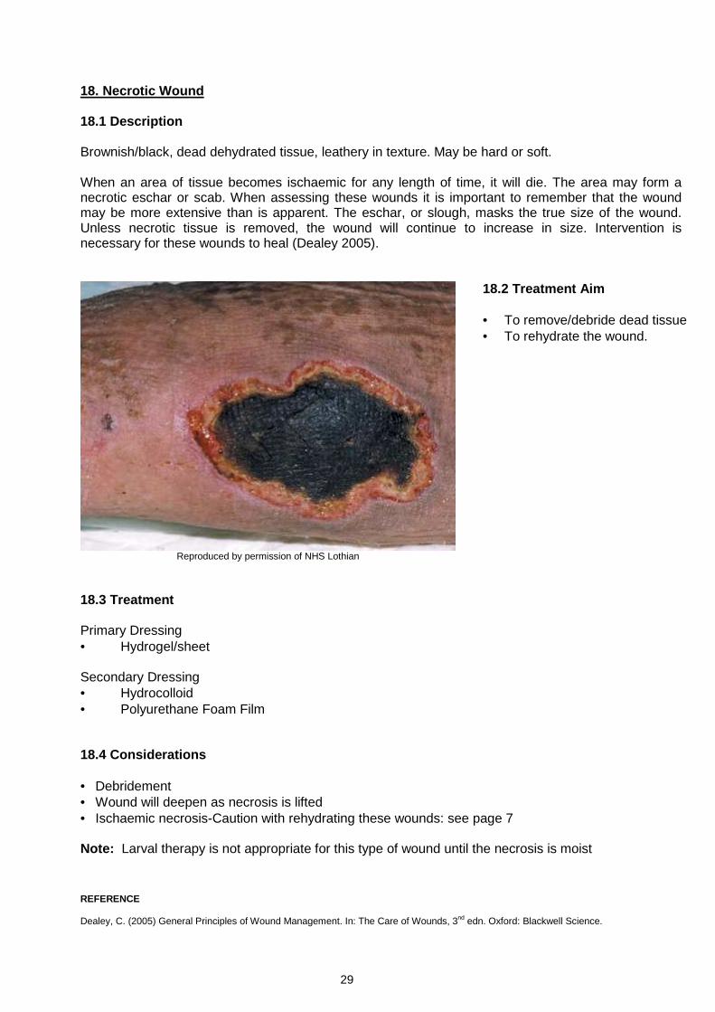
18. Necrotic Wound
18.1 Description Brownish/black, dead dehydrated tissue, leathery in texture. May be hard or soft. When an area of tissue becomes ischaemic for any length of time, it will die. The area may form a necrotic eschar or scab. When assessing these wounds it is important to remember that the wound may be more extensive than is apparent. The eschar, or slough, masks the true size of the wound. Unless necrotic tissue is removed, the wound will continue to increase in size. Intervention is necessary for these wounds to heal (Dealey 2005).
Reproduced by permission of NHS Lothian
18.2 Treatment Aim • To remove/debride dead tissue • To rehydrate the wound.
18.3 Treatment Primary Dressing • Hydrogel/sheet Secondary Dressing • Hydrocolloid • Polyurethane Foam Film 18.4 Considerations • Debridement • Wound will deepen as necrosis is lifted • Ischaemic necrosis-Caution with rehydrating these wounds: see page 7 Note: Larval therapy is not appropriate for this type of wound until the necrosis is moist REFERENCE Dealey, C. (2005) General Principles of Wound Management. In: The Care of Wounds, 3nd edn. Oxford: Blackwell Science.
30
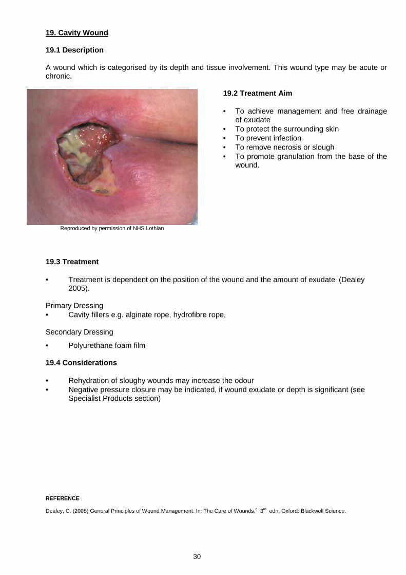
19. Cavity Wound 19.1 Description A wound which is categorised by its depth and tissue involvement. This wound type may be acute or chronic.
Reproduced by permission of NHS Lothian
19.2 Treatment Aim • To achieve management and free drainage
of exudate • To protect the surrounding skin • To prevent infection • To remove necrosis or slough • To promote granulation from the base of the
wound.
19.3 Treatment • Treatment is dependent on the position of the wound and the amount of exudate (Dealey
2005). Primary Dressing • Cavity fillers e.g. alginate rope, hydrofibre rope, Secondary Dressing
• Polyurethane foam film
19.4 Considerations • Rehydration of sloughy wounds may increase the odour • Negative pressure closure may be indicated, if wound exudate or depth is significant (see
Specialist Products section)
REFERENCE Dealey, C. (2005) General Principles of Wound Management. In: The Care of Wounds,d 3rd edn. Oxford: Blackwell Science.
31
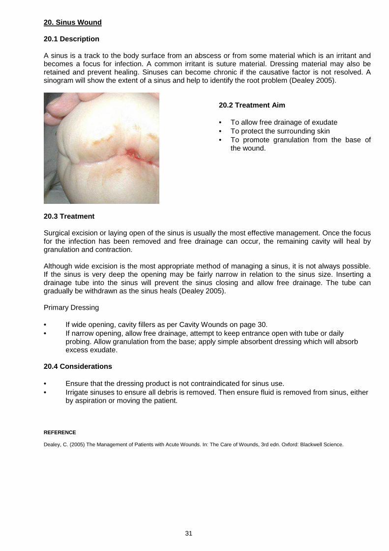
20. Sinus Wound 20.1 Description A sinus is a track to the body surface from an abscess or from some material which is an irritant and becomes a focus for infection. A common irritant is suture material. Dressing material may also be retained and prevent healing. Sinuses can become chronic if the causative factor is not resolved. A sinogram will show the extent of a sinus and help to identify the root problem (Dealey 2005).
20.2 Treatment Aim • To allow free drainage of exudate • To protect the surrounding skin • To promote granulation from the base of
the wound.
20.3 Treatment Surgical excision or laying open of the sinus is usually the most effective management. Once the focus for the infection has been removed and free drainage can occur, the remaining cavity will heal by granulation and contraction. Although wide excision is the most appropriate method of managing a sinus, it is not always possible. If the sinus is very deep the opening may be fairly narrow in relation to the sinus size. Inserting a drainage tube into the sinus will prevent the sinus closing and allow free drainage. The tube can gradually be withdrawn as the sinus heals (Dealey 2005). Primary Dressing • If wide opening, cavity fillers as per Cavity Wounds on page 30. • If narrow opening, allow free drainage, attempt to keep entrance open with tube or daily
probing. Allow granulation from the base; apply simple absorbent dressing which will absorb excess exudate.
20.4 Considerations • Ensure that the dressing product is not contraindicated for sinus use. • Irrigate sinuses to ensure all debris is removed. Then ensure fluid is removed from sinus, either
by aspiration or moving the patient. REFERENCE Dealey, C. (2005) The Management of Patients with Acute Wounds. In: The Care of Wounds, 3rd edn. Oxford: Blackwell Science.
32
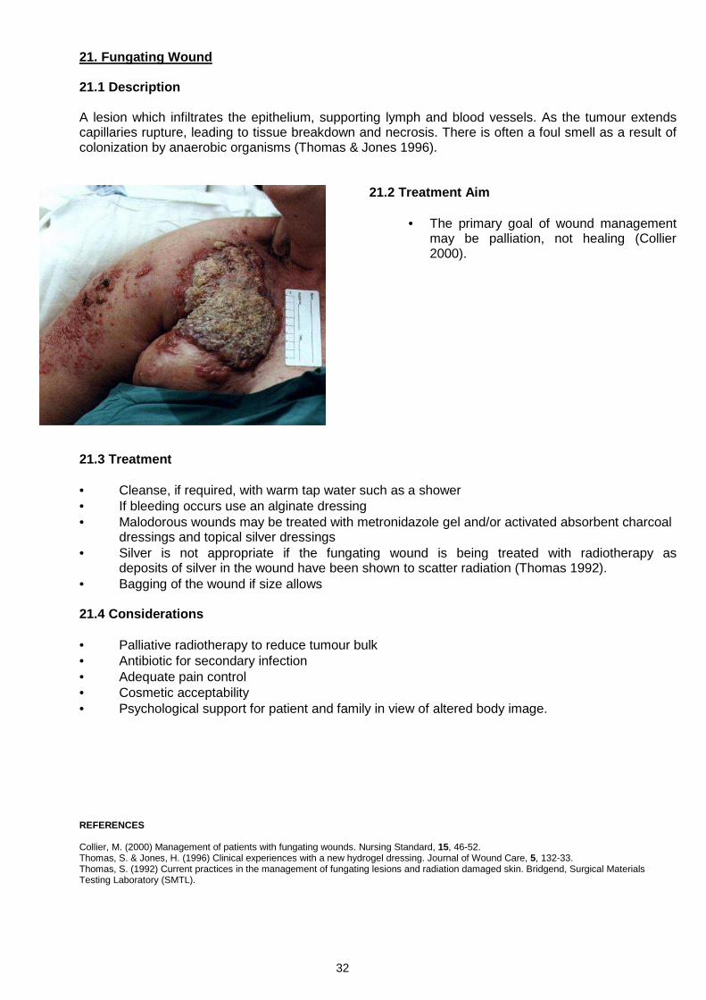
21. Fungating Wound 21.1 Description A lesion which infiltrates the epithelium, supporting lymph and blood vessels. As the tumour extends capillaries rupture, leading to tissue breakdown and necrosis. There is often a foul smell as a result of colonization by anaerobic organisms (Thomas & Jones 1996).
21.2 Treatment Aim
• The primary goal of wound management may be palliation, not healing (Collier 2000).
21.3 Treatment • Cleanse, if required, with warm tap water such as a shower • If bleeding occurs use an alginate dressing • Malodorous wounds may be treated with metronidazole gel and/or activated absorbent charcoal
dressings and topical silver dressings • Silver is not appropriate if the fungating wound is being treated with radiotherapy as deposits of silver in the wound have been shown to scatter radiation (Thomas 1992). • Bagging of the wound if size allows 21.4 Considerations • Palliative radiotherapy to reduce tumour bulk • Antibiotic for secondary infection • Adequate pain control • Cosmetic acceptability • Psychological support for patient and family in view of altered body image.
REFERENCES Collier, M. (2000) Management of patients with fungating wounds. Nursing Standard, 15, 46-52. Thomas, S. & Jones, H. (1996) Clinical experiences with a new hydrogel dressing. Journal of Wound Care, 5, 132-33. Thomas, S. (1992) Current practices in the management of fungating lesions and radiation damaged skin. Bridgend, Surgical Materials Testing Laboratory (SMTL).
33
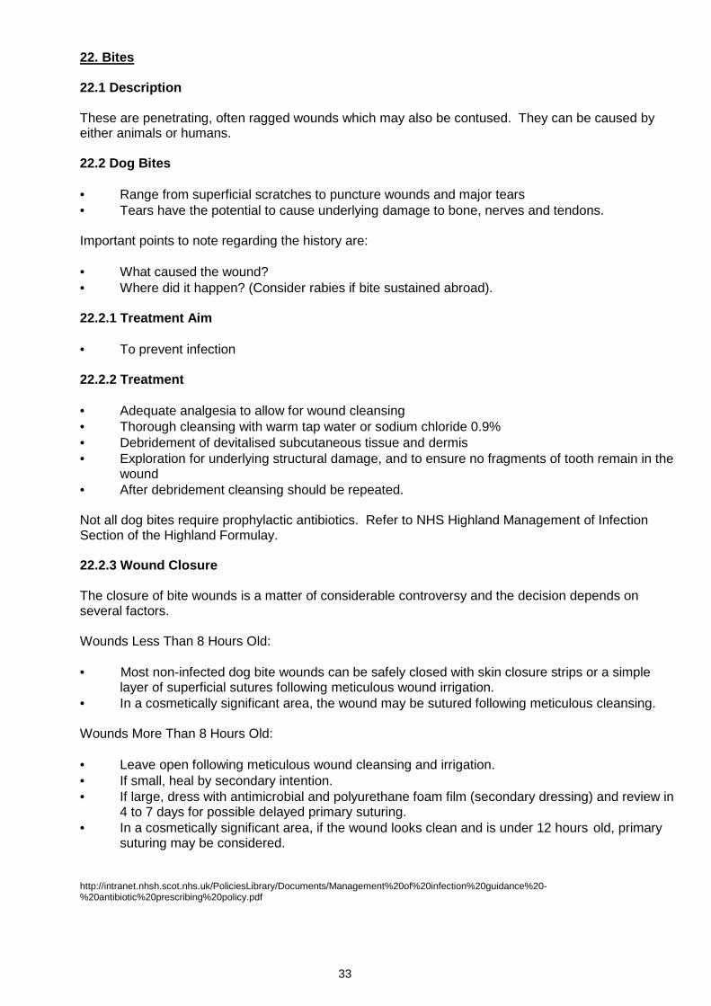
22. Bites 22.1 Description These are penetrating, often ragged wounds which may also be contused. They can be caused by either animals or humans. 22.2 Dog Bites • Range from superficial scratches to puncture wounds and major tears • Tears have the potential to cause underlying damage to bone, nerves and tendons. Important points to note regarding the history are: • What caused the wound? • Where did it happen? (Consider rabies if bite sustained abroad). 22.2.1 Treatment Aim • To prevent infection 22.2.2 Treatment • Adequate analgesia to allow for wound cleansing • Thorough cleansing with warm tap water or sodium chloride 0.9% • Debridement of devitalised subcutaneous tissue and dermis • Exploration for underlying structural damage, and to ensure no fragments of tooth remain in the
wound • After debridement cleansing should be repeated. Not all dog bites require prophylactic antibiotics. Refer to NHS Highland Management of Infection Section of the Highland Formulay. 22.2.3 Wound Closure The closure of bite wounds is a matter of considerable controversy and the decision depends on several factors. Wounds Less Than 8 Hours Old: • Most non-infected dog bite wounds can be safely closed with skin closure strips or a simple
layer of superficial sutures following meticulous wound irrigation. • In a cosmetically significant area, the wound may be sutured following meticulous cleansing.
Wounds More Than 8 Hours Old: • Leave open following meticulous wound cleansing and irrigation. • If small, heal by secondary intention. • If large, dress with antimicrobial and polyurethane foam film (secondary dressing) and review in
4 to 7 days for possible delayed primary suturing. • In a cosmetically significant area, if the wound looks clean and is under 12 hours old, primary
suturing may be considered.
http://intranet.nhsh.scot.nhs.uk/PoliciesLibrary/Documents/Management%20of%20infection%20guidance%20-%20antibiotic%20prescribing%20policy.pdf
34
22.3 Human Bites 22.3.1 High Risk of Infection These are potentially more serious than dog bites and constitute 18% of all bite presentations (Higgins et al. 1997). Multiple organisms are found in the mouth, commonly staphylococcus and streptococcus (Wienert 1999). Other infectious diseases transmitted by human bite include scarlet fever, TB, syphilis, Hepatitis B and C, HIV, and tetanus. Human bites can be separated into actual bites and clenched fist injuries. In clenched fist injuries the lacerated skin retracts and then returns to its original position, carrying dirt into the wound. There is also a much easier access into the joint space for the teeth, creating a high risk of tendon sheath and web space infections. 22.3.2 Treatment • Analgesia if required • Irrigate with warm tap water or sodium chloride 0.9% • Leave open – do not suture or apply skin closure strip • Prophylactic antibiotics must be given for aerobic and anaerobic bacteria. Refer to NHS Highland Management of Infection Section of the Highland Formulay. • Large wounds may be dressed according to amount of exudate and state of wound bed • An alginate (primary dressing) can be packed loosely into puncture wounds to facilitate removal
of exudate (Young 2002). Polyurethane foam film (secondary dressing). 22.4 Cat Bites 22.4.1 High Risk of Infection These have a high potential for infection. Wounds are usually very difficult to cleanse adequately, as cat bites usually puncture the skin rather than tear it. Pasturella multicoda is the commonest infection micro-organism which is highly sensitive to penicillin, however, Capnocytophaga canimorsus is a far more serious infection with a mortality rate of 28-50% (Higgins et al. 1997). 22.4.2 Treatment As for human bites.
REFERENCES http://intranet.nhsh.scot.nhs.uk/PoliciesLibrary/Documents/Management%20of%20infection%20guidance%20-%20antibiotic%20prescribing%20policy.pdf Higgins, M.A.G., Evans, R.C., Evans, R.J. (1997) Managing animal bite wounds. Journal of Wound Care 6, 377-380. Wienert, P., Heiss, J., Rinecker, H., Sing, A. (1999) A Human Bite. The Lancet 354, 572. Young T (2002). Wound care in the accident and emergency department. In: British Journal of Nursing Monograph: Trends in Wound Care. Wiltshire: Mark Allen Publishing Ltd.
35
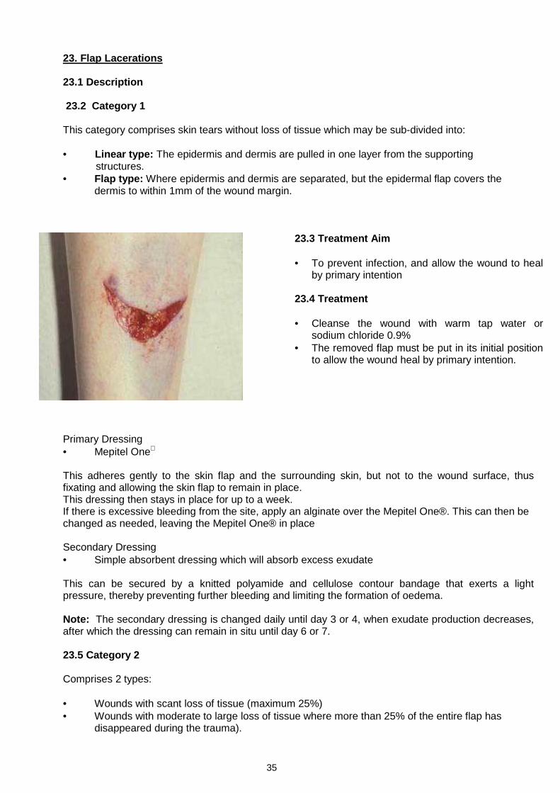
23. Flap Lacerations 23.1 Description 23.2 Category 1 This category comprises skin tears without loss of tissue which may be sub-divided into: • Linear type: The epidermis and dermis are pulled in one layer from the supporting
structures. • Flap type: Where epidermis and dermis are separated, but the epidermal flap covers the
dermis to within 1mm of the wound margin.
23.3 Treatment Aim • To prevent infection, and allow the wound to heal
by primary intention 23.4 Treatment • Cleanse the wound with warm tap water or
sodium chloride 0.9% • The removed flap must be put in its initial position
to allow the wound heal by primary intention.
Primary Dressing • Mepitel One This adheres gently to the skin flap and the surrounding skin, but not to the wound surface, thus fixating and allowing the skin flap to remain in place. This dressing then stays in place for up to a week. If there is excessive bleeding from the site, apply an alginate over the Mepitel One®. This can then be changed as needed, leaving the Mepitel One® in place Secondary Dressing • Simple absorbent dressing which will absorb excess exudate This can be secured by a knitted polyamide and cellulose contour bandage that exerts a light pressure, thereby preventing further bleeding and limiting the formation of oedema. Note: The secondary dressing is changed daily until day 3 or 4, when exudate production decreases, after which the dressing can remain in situ until day 6 or 7.
23.5 Category 2 Comprises 2 types: • Wounds with scant loss of tissue (maximum 25%) • Wounds with moderate to large loss of tissue where more than 25% of the entire flap has
disappeared during the trauma).
36
23.6 Treatment Aim • To prevent infection • For tears with skin loss of more than 25%, the aim is to use what is remaining of the skin flap • As exudate production decreases, desiccation or drying of the wound needs to be prevented. 23.7 Treatment Primary Dressing • A hydrogel under Mepitel One can hydrate the wound. • This dressing then stays in place for up to a week. • If there is excessive bleeding from the site, apply an alginate over the Mepitel One ®. This can then be changed as needed, leaving the Mepitel One® in place. Secondary Dressing • Simple absorbent dressing After 6 or 7 days, when the skin flap has grown into the wound, the treatment continues as for category 1. 23.8 Category 3 This type of skin tear involves the entire loss of tissue. It can be caused by the initial trauma, or necrosis of the skin flap. 23.9 Treatment Aim To prevent infection and reduce pain 23.10 Treatment If skin has been ripped off during the trauma, or if the flap has necrotised, the wound requires a moderately moist environment, and is treated as an abrasion. Primary Dressing • Polyurethane foam film } • Fibrous hydrocolloid } depending on the levels of exudate • Alginate } Secondary Dressing • Only required if primary dressing is non-adherent As the amount of exudate decreases, the wound may be hydrated with a hydrogel. If there are no signs of clinical infection a hydrocolloid or polyurethane foam film can be used until complete epithelialisation. 23.11 Note - deep laceration wounds: If the skin is torn until just above the fascia, check if any crucial nerves, blood vessels or tendons have been damaged. Further tearing and separation can be prevented by securing with Mepitel One. Most deep lacerations are closed with sutures or skin grafts, or heal by secondary intention due to the loss of substance (Meuleneire 2002). See also: Best Practice for the assessment and management of superficial skin tears http://www.wounds-uk.com/pdf/content_9378.pdf REFERENCE Meuleneire, F. (2002) Using a soft silicone coated net dressing to manage skin tears. Journal of Wound Care 11, 365-369.
37
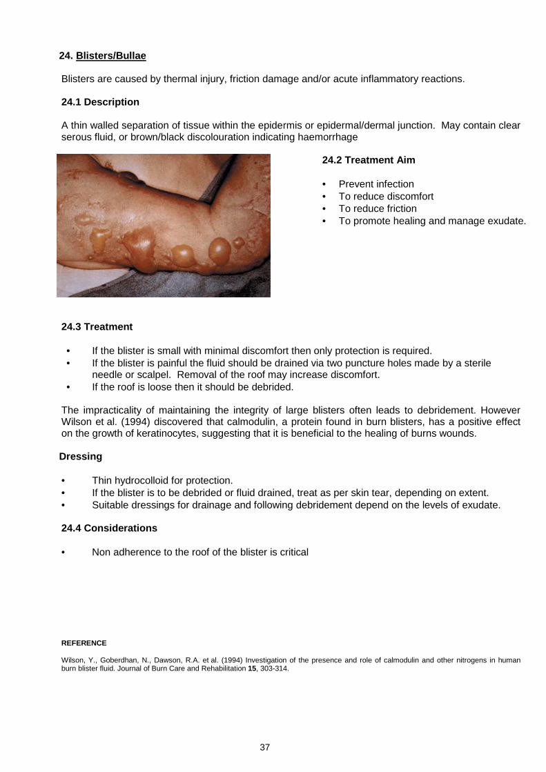
24. Blisters/Bullae Blisters are caused by thermal injury, friction damage and/or acute inflammatory reactions. 24.1 Description A thin walled separation of tissue within the epidermis or epidermal/dermal junction. May contain clear serous fluid, or brown/black discolouration indicating haemorrhage
24.2 Treatment Aim • Prevent infection • To reduce discomfort • To reduce friction • To promote healing and manage exudate.
24.3 Treatment
• If the blister is small with minimal discomfort then only protection is required. • If the blister is painful the fluid should be drained via two puncture holes made by a sterile
needle or scalpel. Removal of the roof may increase discomfort. • If the roof is loose then it should be debrided.
The impracticality of maintaining the integrity of large blisters often leads to debridement. However Wilson et al. (1994) discovered that calmodulin, a protein found in burn blisters, has a positive effect on the growth of keratinocytes, suggesting that it is beneficial to the healing of burns wounds.
Dressing • Thin hydrocolloid for protection. • If the blister is to be debrided or fluid drained, treat as per skin tear, depending on extent. • Suitable dressings for drainage and following debridement depend on the levels of exudate. 24.4 Considerations • Non adherence to the roof of the blister is critical
REFERENCE Wilson, Y., Goberdhan, N., Dawson, R.A. et al. (1994) Investigation of the presence and role of calmodulin and other nitrogens in human burn blister fluid. Journal of Burn Care and Rehabilitation 15, 303-314.
38
25. Thermal Injuries/Burns The British Burns Association has identified the following as requiring referral to a burn unit: • Burns > 10% of total body surface area (TBSA) in adults (a crude calculation may be made,
assuming that the palm of the patient’s hand is equivalent to 1% of total body surface) • Burns > 5% TBSA in children • Burns of special areas, eg face, hands, feet, genitalia and major joints • Full thickness burns > 5% TBSA • Electrical and chemical burns • Burns associated with inhalation injury • Circumferential burns of the limbs or chest • Burns in young children or the elderly • Burn injuries in patients with pre-existing medical disorders which complicate management,
prolong recovery or effect mortality • Suspected ‘non-accidental injury’ (NAI).
25.1 Superficial Burns a) Description Quick capillary return. Red, slightly swollen appearance. No blister formation. Any damaged epithelium may peel off after 5 to 7 days without scarring. b) Treatment Aim • To relieve pain • To protect from infection. c) Treatment • Immediately place the affected part under cold running water (approx 15°C) for at least 20
minutes (Yuan et al 2007). This relieves pain and reduces the temperature of the burning process
• Remove any clothing carefully • Apply dressings as for blisters.
25.2 Superficial, Partial Thickness Skin Loss Burns a) Description Slow capillary return. Epidermis and superficial layers of dermis are destroyed. Hair follicles, sebaceous and sweat glands are intact. This is likely to be a painful burn as the nerve endings have not been damaged. Usually heals in 10 to 14 days, without scarring. 25.3 Deep, Partial Thickness Skin Loss Burns a) Description Slow capillary return. Greater part of the dermis is lost. Sensation is altered. Patient may have no pin-prick sensation.
39
Reproduced by permission of NHS Lothian
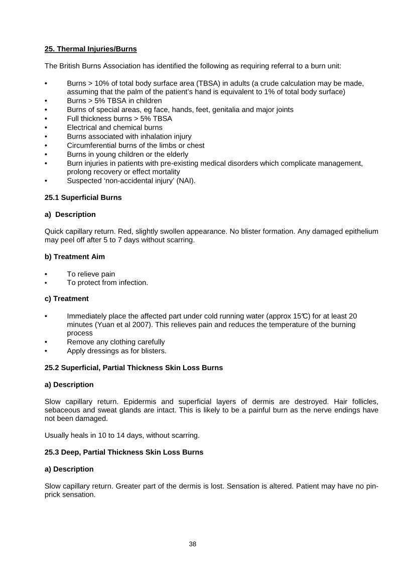
b) Treatment Aims of Both Superficial and Deep Partial Thickness Skin Loss Burns • To relieve pain • To protect from infection • To manage exudate.
c) Treatment • Immediately place the affected part under cold running water (approx 15°C) for at least 10 to 15
minutes • Remove any clothing carefully avoiding any further injury. • Apply non-adherent interface dressing such as Atrauman®. An absorbent
secondary dressing such as polyurethane foam film may be used. The primary dressing may be left in place and the secondary dressing changed as often as necessary, depending on choice of product and exudate levels.
Note: Silver sulfadiazine cream should not be used routinely until after specialist assessment as this will mask the wound bed and make for difficult assessment.
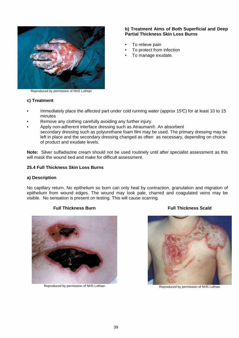
25.4 Full Thickness Skin Loss Burns a) Description No capillary return. No epithelium so burn can only heal by contraction, granulation and migration of epithelium from wound edges. The wound may look pale, charred and coagulated veins may be visible. No sensation is present on testing. This will cause scarring.
Full Thickness Burn
Reproduced by permission of NHS Lothian
Full Thickness Scald
Reproduced by permission of NHS Lothian

40
b) Treatment Aim • To protect from infection • To manage exudate. c) Initial Treatment and Assessment • Immediately place the affected part under cold running water (approx 15°C) for at least 10 to 15
minutes. If greater than 3 hours from time of injury, cold water will have no beneficial effect. • Remove any clothing carefully avoiding any further injury. d) If Patient is to be Transferred to Accident & Em ergency or Burns Unit • Cover all burned areas primarily with cling film (which prevents infection and allows for ease of
assessment) and wrap patient in clean covers to prevent heat loss • If transfer journey is greater than 2 to 3 hours, a secondary surgical absorbent dressing of
gauze and cotton tissue will be necessary to retain exudates, which may be extensive. Note: Silver sulfadiazine cream should not be used until after specialist assessment as this will mask the wound bed and make for difficult assessment.
f) If Patient is not to be transferred • Apply primary non-adherent interface dressing such as Atrauman®. • Secondary dressings should be highly absorbent whilst maintaining a moist wound bed as thick
eschar usually forms. This may be debrided surgically or by autolysis. • Treat as for wound type as it progresses through wound healing stages. • Initially, dressings may need to be carried out daily but this is dependent on the amount of
exudate. • Argyll and Bute CHP patient pathway is to NHS Greater Glasgow & Clyde. There may, therefore, be slight differences in dressings selection.
More information is available from the following website: http://www.cobis.scot.nhs.uk/
REFERENCE Yuan J, Wu C, Holland AJA, Harvey JG, Martin HCO, La Hei ER, Arbuckle S, Godfrey C (2007) Assessment of cooling on an acute scald burn injury in a porcine model. J Burn Care Res 28: 514–20.
41
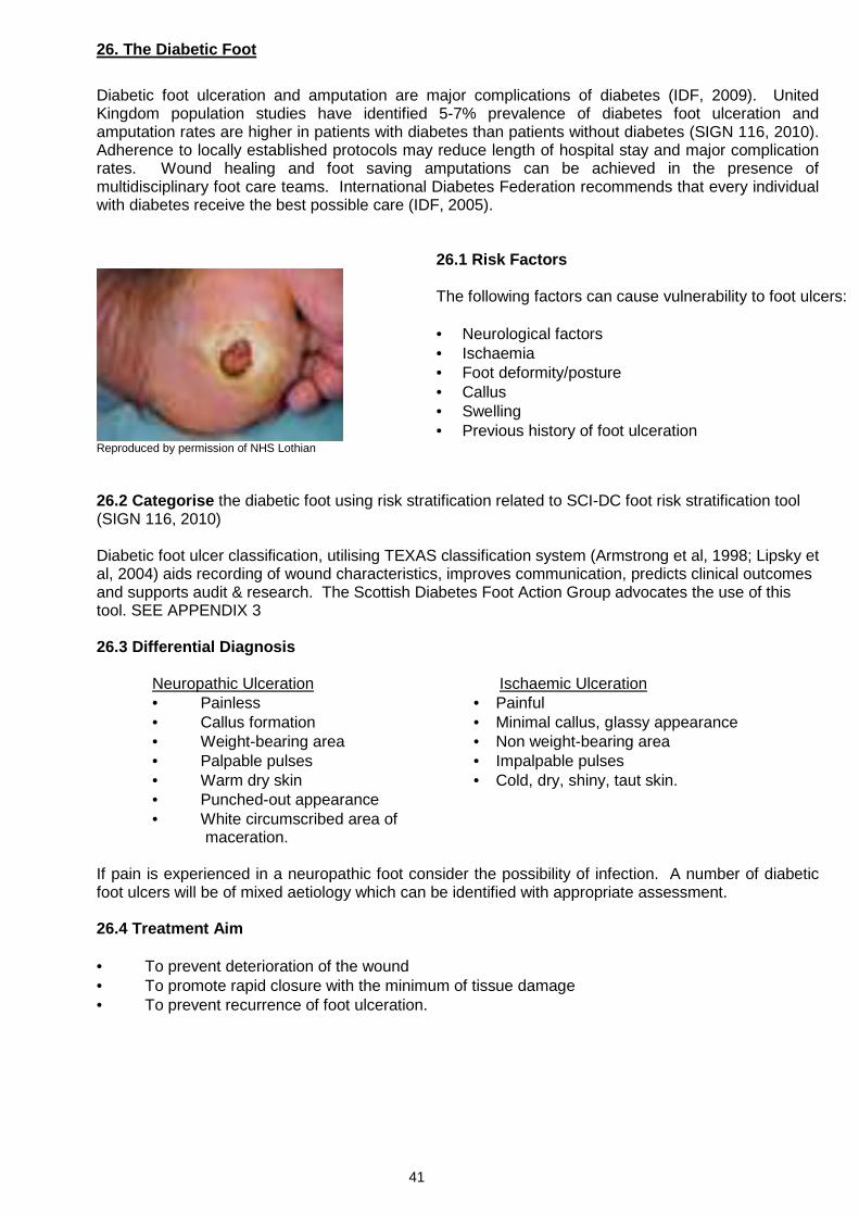
26. The Diabetic Foot
Diabetic foot ulceration and amputation are major complications of diabetes (IDF, 2009). United Kingdom population studies have identified 5-7% prevalence of diabetes foot ulceration and amputation rates are higher in patients with diabetes than patients without diabetes (SIGN 116, 2010). Adherence to locally established protocols may reduce length of hospital stay and major complication rates. Wound healing and foot saving amputations can be achieved in the presence of multidisciplinary foot care teams. International Diabetes Federation recommends that every individual with diabetes receive the best possible care (IDF, 2005).
Reproduced by permission of NHS Lothian
26.1 Risk Factors The following factors can cause vulnerability to foot ulcers: • Neurological factors • Ischaemia • Foot deformity/posture • Callus • Swelling • Previous history of foot ulceration
26.2 Categorise the diabetic foot using risk stratification related to SCI-DC foot risk stratification tool (SIGN 116, 2010) Diabetic foot ulcer classification, utilising TEXAS classification system (Armstrong et al, 1998; Lipsky et al, 2004) aids recording of wound characteristics, improves communication, predicts clinical outcomes and supports audit & research. The Scottish Diabetes Foot Action Group advocates the use of this tool. SEE APPENDIX 3 26.3 Differential Diagnosis
Neuropathic Ulceration Ischaemic Ulceration • Painless • Callus formation • Weight-bearing area • Palpable pulses • Warm dry skin • Punched-out appearance • White circumscribed area of maceration.
• Painful • Minimal callus, glassy appearance • Non weight-bearing area • Impalpable pulses • Cold, dry, shiny, taut skin.
If pain is experienced in a neuropathic foot consider the possibility of infection. A number of diabetic foot ulcers will be of mixed aetiology which can be identified with appropriate assessment.
26.4 Treatment Aim • To prevent deterioration of the wound • To promote rapid closure with the minimum of tissue damage • To prevent recurrence of foot ulceration.
42
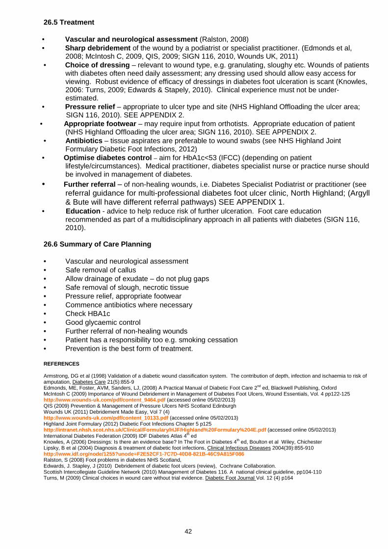
26.5 Treatment • Vascular and neurological assessment (Ralston, 2008) • Sharp debridement of the wound by a podiatrist or specialist practitioner. (Edmonds et al,
2008; McIntosh C, 2009, QIS, 2009; SIGN 116, 2010, Wounds UK, 2011) • Choice of dressing – relevant to wound type, e.g. granulating, sloughy etc. Wounds of patients
with diabetes often need daily assessment; any dressing used should allow easy access for viewing. Robust evidence of efficacy of dressings in diabetes foot ulceration is scant (Knowles, 2006: Turns, 2009; Edwards & Stapely, 2010). Clinical experience must not be under-estimated.
• Pressure relief – appropriate to ulcer type and site (NHS Highland Offloading the ulcer area; SIGN 116, 2010). SEE APPENDIX 2.
• Appropriate footwear – may require input from orthotists. Appropriate education of patient (NHS Highland Offloading the ulcer area; SIGN 116, 2010). SEE APPENDIX 2.
• Antibiotics – tissue aspirates are preferable to wound swabs (see NHS Highland Joint Formulary Diabetic Foot Infections, 2012)
• Optimise diabetes control – aim for HbA1c<53 (IFCC) (depending on patient lifestyle/circumstances). Medical practitioner, diabetes specialist nurse or practice nurse should
be involved in management of diabetes. • Further referral – of non-healing wounds, i.e. Diabetes Specialist Podiatrist or practitioner (see
referral guidance for multi-professional diabetes foot ulcer clinic, North Highland; (Argyll & Bute will have different referral pathways) SEE APPENDIX 1.
• Education - advice to help reduce risk of further ulceration. Foot care education recommended as part of a multidisciplinary approach in all patients with diabetes (SIGN 116,
2010). 26.6 Summary of Care Planning • Vascular and neurological assessment • Safe removal of callus • Allow drainage of exudate – do not plug gaps • Safe removal of slough, necrotic tissue • Pressure relief, appropriate footwear • Commence antibiotics where necessary • Check HBA1c • Good glycaemic control • Further referral of non-healing wounds • Patient has a responsibility too e.g. smoking cessation • Prevention is the best form of treatment.
REFERENCES
Armstrong, DG et al (1998) Validation of a diabetic wound classification system. The contribution of depth, infection and ischaemia to risk of amputation, Diabetes Care 21(5):855-9 Edmonds, ME, Foster, AVM, Sanders, LJ, (2008) A Practical Manual of Diabetic Foot Care 2nd ed, Blackwell Publishing, Oxford McIntosh C (2009) Importance of Wound Debridement in Management of Diabetes Foot Ulcers, Wound Essentials, Vol. 4 pp122-125 http://www.wounds-uk.com/pdf/content_9464.pdf (accessed online 05/02/2013) QIS (2009) Prevention & Management of Pressure Ulcers NHS Scotland Edinburgh Wounds UK (2011) Debridement Made Easy, Vol 7 (4) http://www.wounds-uk.com/pdf/content_10133.pdf (accessed online 05/02/2013) Highland Joint Formulary (2012) Diabetic Foot Infections Chapter 5 p125 http://intranet.nhsh.scot.nhs.uk/Clinical/Formulary /HJF/Highland%20Formulary%204E.pdf (accessed online 05/02/2013) International Diabetes Federation (2009) IDF Diabetes Atlas 4th ed Knowles, A (2006) Dressings: Is there an evidence base? In The Foot in Diabetes 4th ed, Boulton et al Wiley, Chichester Lipsky, B et al (2004) Diagnosis & treatment of diabetic foot infections, Clinical Infectious Diseases 2004(39):855-910 http://www.idf.org/node/1255?unode=F2E52CF1-7C7D-40D8-821B-46C9A815F086 Ralston, S (2008) Foot problems in diabetes NHS Scotland, Edwards, J. Stapley, J (2010) Debridement of diabetic foot ulcers (review), Cochrane Collaboration. Scottish Intercollegiate Guideline Network (2010) Management of Diabetes 116. A national clinical guideline, pp104-110 Turns, M (2009) Clinical choices in wound care without trial evidence. Diabetic Foot Journal Vol. 12 (4) p164
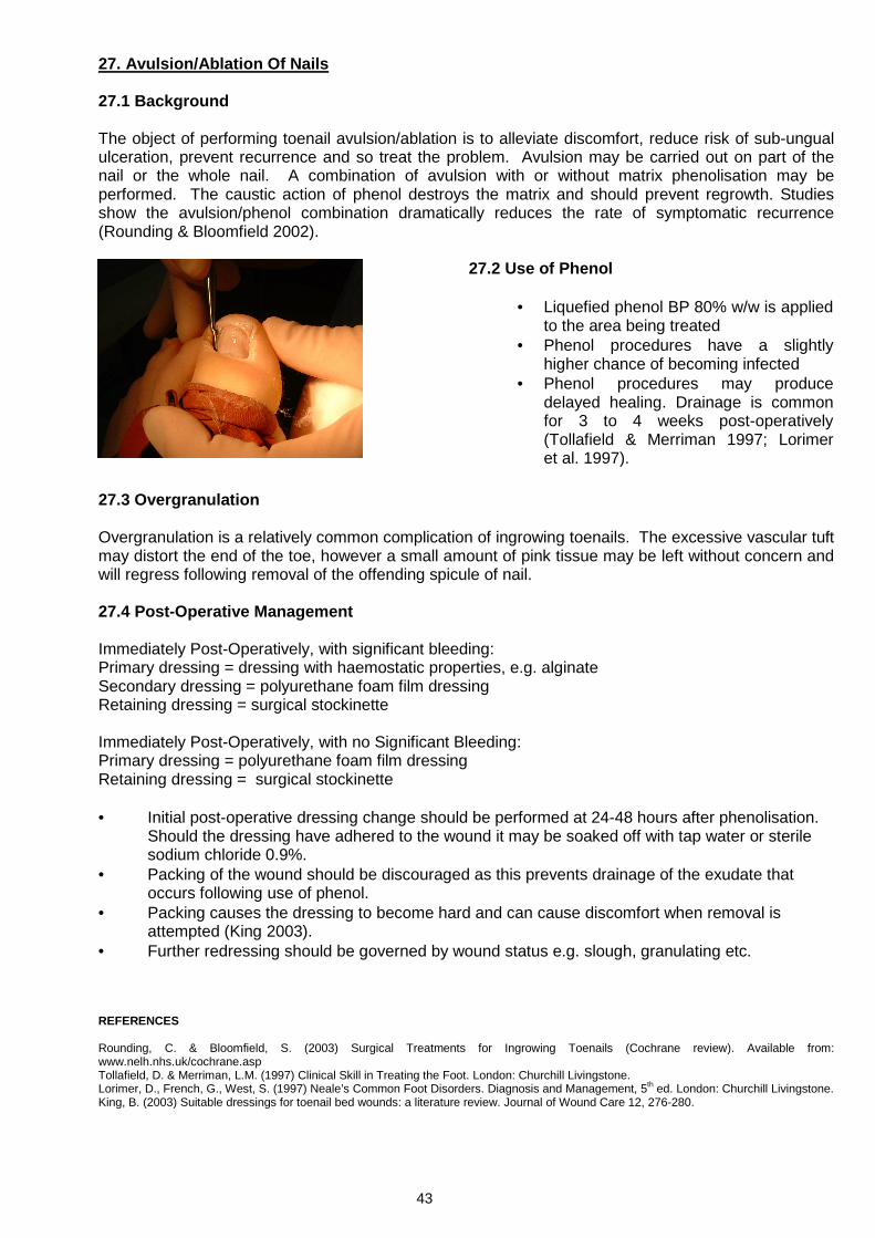
43
27. Avulsion/Ablation Of Nails 27.1 Background
The object of performing toenail avulsion/ablation is to alleviate discomfort, reduce risk of sub-ungual ulceration, prevent recurrence and so treat the problem. Avulsion may be carried out on part of the nail or the whole nail. A combination of avulsion with or without matrix phenolisation may be performed. The caustic action of phenol destroys the matrix and should prevent regrowth. Studies show the avulsion/phenol combination dramatically reduces the rate of symptomatic recurrence (Rounding & Bloomfield 2002).
27.2 Use of Phenol
• Liquefied phenol BP 80% w/w is applied to the area being treated
• Phenol procedures have a slightly higher chance of becoming infected
• Phenol procedures may produce delayed healing. Drainage is common for 3 to 4 weeks post-operatively (Tollafield & Merriman 1997; Lorimer et al. 1997).
27.3 Overgranulation Overgranulation is a relatively common complication of ingrowing toenails. The excessive vascular tuft may distort the end of the toe, however a small amount of pink tissue may be left without concern and will regress following removal of the offending spicule of nail. 27.4 Post-Operative Management Immediately Post-Operatively, with significant bleeding: Primary dressing = dressing with haemostatic properties, e.g. alginate Secondary dressing = polyurethane foam film dressing Retaining dressing = surgical stockinette Immediately Post-Operatively, with no Significant Bleeding: Primary dressing = polyurethane foam film dressing Retaining dressing = surgical stockinette • Initial post-operative dressing change should be performed at 24-48 hours after phenolisation.
Should the dressing have adhered to the wound it may be soaked off with tap water or sterile sodium chloride 0.9%.
• Packing of the wound should be discouraged as this prevents drainage of the exudate that occurs following use of phenol.
• Packing causes the dressing to become hard and can cause discomfort when removal is attempted (King 2003).
• Further redressing should be governed by wound status e.g. slough, granulating etc.
REFERENCES
Rounding, C. & Bloomfield, S. (2003) Surgical Treatments for Ingrowing Toenails (Cochrane review). Available from: www.nelh.nhs.uk/cochrane.asp Tollafield, D. & Merriman, L.M. (1997) Clinical Skill in Treating the Foot. London: Churchill Livingstone. Lorimer, D., French, G., West, S. (1997) Neale’s Common Foot Disorders. Diagnosis and Management, 5th ed. London: Churchill Livingstone. King, B. (2003) Suitable dressings for toenail bed wounds: a literature review. Journal of Wound Care 12, 276-280.
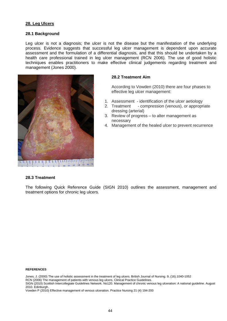
44
28. Leg Ulcers 28.1 Background Leg ulcer is not a diagnosis; the ulcer is not the disease but the manifestation of the underlying process. Evidence suggests that successful leg ulcer management is dependent upon accurate assessment and the formulation of a differential diagnosis, and that this should be undertaken by a health care professional trained in leg ulcer management (RCN 2006). The use of good holistic techniques enables practitioners to make effective clinical judgements regarding treatment and management (Jones 2000).
28.2 Treatment Aim According to Vowden (2010) there are four phases to effective leg ulcer management:
1. Assessment - identification of the ulcer aetiology 2. Treatment - compression (venous), or appropriate
dressing (arterial) 3. Review of progress – to alter management as
necessary 4. Management of the healed ulcer to prevent recurrence
28.3 Treatment The following Quick Reference Guide (SIGN 2010) outlines the assessment, management and treatment options for chronic leg ulcers.
REFERENCES
Jones, J. (2000) The use of holistic assessment in the treatment of leg ulcers. British Journal of Nursing. 9, (16).1040-1052 RCN (2006) The management of patients with venous leg ulcers. Clinical Practice Guidelines. SIGN (2010) Scottish Intercollegiate Guidelines Network. No120. Management of chronic venous leg ulceration: A national guideline. August 2010. Edinburgh. Vowden P (2010) Effective management of venous ulceration. Practice Nursing 21 (4) 194-200
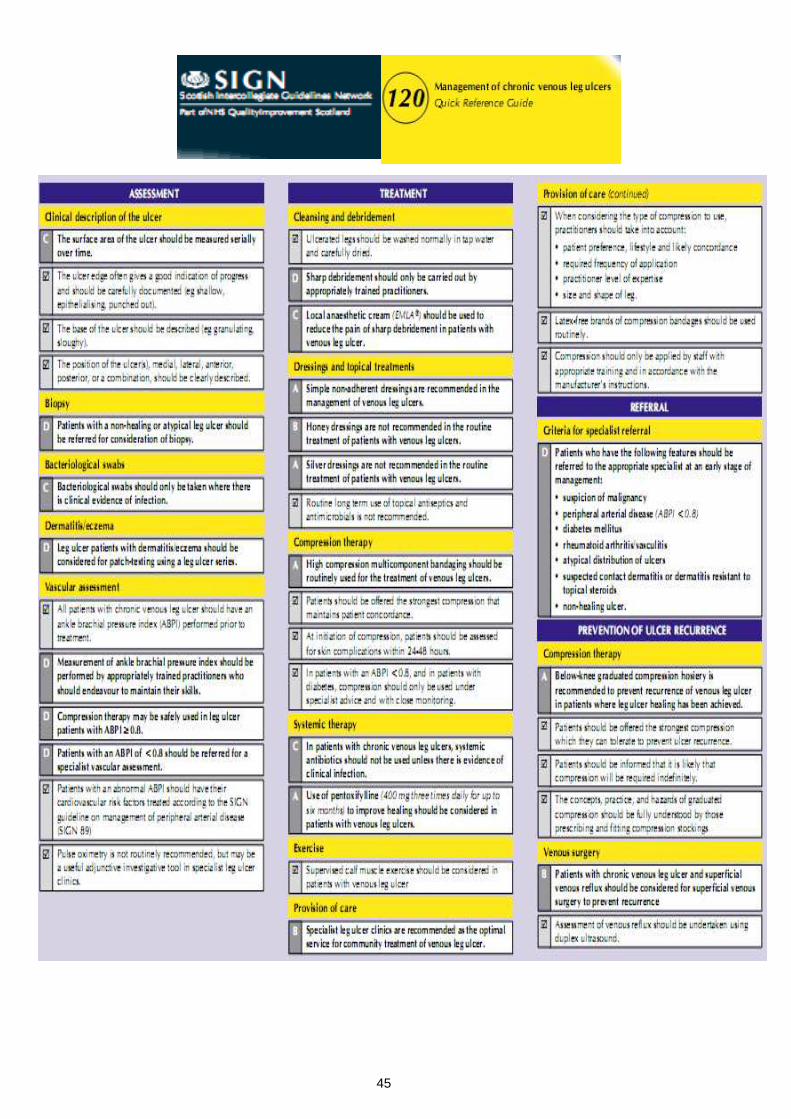
45
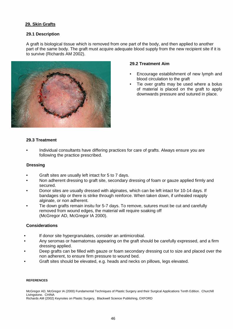
46
29. Skin Grafts 29.1 Description A graft is biological tissue which is removed from one part of the body, and then applied to another part of the same body. The graft must acquire adequate blood supply from the new recipient site if it is to survive (Richards AM 2002).
29.2 Treatment Aim • Encourage establishment of new lymph and
blood circulation to the graft • Tie over grafts may be used where a bolus
of material is placed on the graft to apply downwards pressure and sutured in place.
29.3 Treatment • Individual consultants have differing practices for care of grafts. Always ensure you are
following the practice prescribed. Dressing • Graft sites are usually left intact for 5 to 7 days. • Non adherent dressing to graft site, secondary dressing of foam or gauze applied firmly and
secured. • Donor sites are usually dressed with alginates, which can be left intact for 10-14 days. If bandages slip or there is strike through reinforce. When taken down, if unhealed reapply alginate, or non adherent. • Tie down grafts remain insitu for 5-7 days. To remove, sutures must be cut and carefully
removed from wound edges, the material will require soaking off (McGregor AD, McGregor IA 2000).
Considerations
• If donor site hypergranulates, consider an antimicrobial. • Any seromas or haematomas appearing on the graft should be carefully expressed, and a firm
dressing applied. • Deep grafts can be filled with gauze or foam secondary dressing cut to size and placed over the
non adherent, to ensure firm pressure to wound bed. • Graft sites should be elevated, e.g. heads and necks on pillows, legs elevated. REFERENCES McGregor AD, McGregor IA (2000) Fundamental Techniques of Plastic Surgery and their Surgical Applications Tenth Edition. Churchill Livingstone. CHINA Richards AM (2002) Keynotes on Plastic Surgery, Blackwell Science Publishing, OXFORD
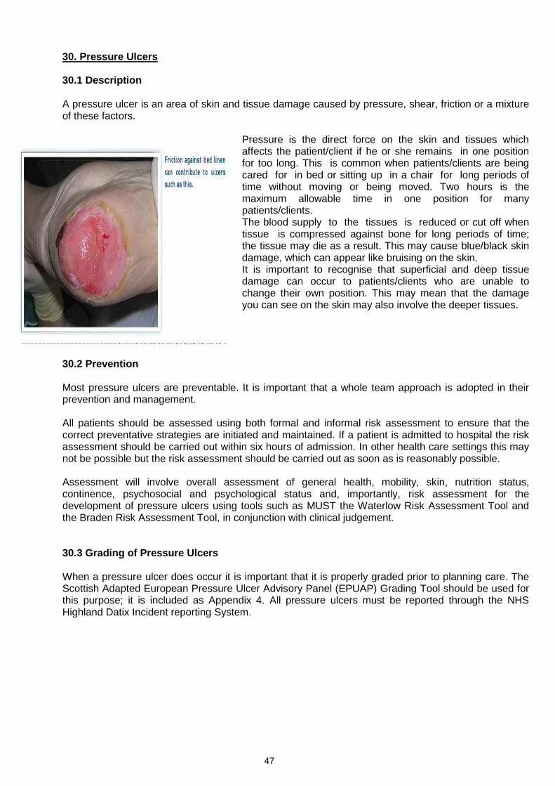
47
30. Pressure Ulcers 30.1 Description A pressure ulcer is an area of skin and tissue damage caused by pressure, shear, friction or a mixture of these factors.
Pressure is the direct force on the skin and tissues which affects the patient/client if he or she remains in one position for too long. This is common when patients/clients are being cared for in bed or sitting up in a chair for long periods of time without moving or being moved. Two hours is the maximum allowable time in one position for many patients/clients. The blood supply to the tissues is reduced or cut off when tissue is compressed against bone for long periods of time; the tissue may die as a result. This may cause blue/black skin damage, which can appear like bruising on the skin. It is important to recognise that superficial and deep tissue damage can occur to patients/clients who are unable to change their own position. This may mean that the damage you can see on the skin may also involve the deeper tissues.
30.2 Prevention Most pressure ulcers are preventable. It is important that a whole team approach is adopted in their prevention and management. All patients should be assessed using both formal and informal risk assessment to ensure that the correct preventative strategies are initiated and maintained. If a patient is admitted to hospital the risk assessment should be carried out within six hours of admission. In other health care settings this may not be possible but the risk assessment should be carried out as soon as is reasonably possible. Assessment will involve overall assessment of general health, mobility, skin, nutrition status, continence, psychosocial and psychological status and, importantly, risk assessment for the development of pressure ulcers using tools such as MUST the Waterlow Risk Assessment Tool and the Braden Risk Assessment Tool, in conjunction with clinical judgement.
30.3 Grading of Pressure Ulcers When a pressure ulcer does occur it is important that it is properly graded prior to planning care. The Scottish Adapted European Pressure Ulcer Advisory Panel (EPUAP) Grading Tool should be used for this purpose; it is included as Appendix 4. All pressure ulcers must be reported through the NHS Highland Datix Incident reporting System.
48
Key Principles of pressure ulcer grading
• Knowing how to grade a pressure ulcer accurately requires knowledge of the skin and its underlying anatomy. You must also be able to recognise different types of tissue and be able to differentiate between healthy tissue and damaged tissue. • Making a visual assessment of a lesion is the most common way to defining whether or not it is a pressure ulcer. Our grading and excoriation tools as well as discussion with colleagues can assist your assessment. Nurse specialists in the field of Tissue Viability are also excellent points of reference. • Once a lesion is classified as a pressure ulcer, it is important that the ulcer is assessed. You can determine its severity by allocating an appropriate grade. • Once a grade is allocated, you should formulate an appropriate plan of care, allocate appropriate resources and implement the plan. Such action(s) should prevent the ulcer from getting worse and prevent further ulcers from developing. • In accordance with good practice, you should always document your actions, and this information should be made accessible to all staff involved in the care of an individual who has developed a pressure ulcer, or who is at risk of doing so. • You must evaluate all plans of care on a regular basis in order to determine if the plan of care is working in the way that it is intended. • A pressure ulcer grading tool acts as a method of communication.
Comprehensive information and educations resources are available through the following link: http://www.healthcareimprovementscotland.org/progra mmes/patient_safety/tissue_v iability.aspx
Source NHS Healthcare Improvement Scotland (NHS HIS)
49
SECTION FOUR PRODUCT INFORMATION
31. Introduction Wound management products include topical agents and dressings. Topical agents are applied directly to the wound whereas dressings cover over the wound, intending to promote healing and provide protection from further harm. There are two types of dressing: • Primary dressing – used in direct contact with damaged tissue • Secondary dressing – superimposed over primary dressings. Selection of the most appropriate dressing material from the wide range now available is an essential step towards wound healing. The type of wound and the stage of tissue repair are important factors to be considered. Key factors include: • Type of wound, i.e. acute or chronic • Wound depth and the tissues involved • Size and shape of the wound • Amount and nature of wound exudate • Position, whether location of wound will make it difficult for the dressing to remain in place • Appearance and stage of wound healing, e.g. necrotic, sloughy, infected, granulating or
epithelialising • Pain and discomfort • Care of skin around the wound, including allergies and sensitivities • Presence of infection • Patient preference. As wounds heal different types of dressings may be required. There have been few clinical trials able to establish a clear advantage for specific products and choice will depend on the above factors as well as product cost-effectiveness.
32. Dressing Descriptions
Characteristics of the Ideal Wound Dressing (Turner 1982) • To maintain high humidity at the wound dressing interface • To remove excess exudate • To allow gaseous exchange • To provide thermal insulation • To be impermeable to bacteria • To be free of particles and toxic wound contaminants • To allow removal without causing trauma to the wound.
REFERENCE
Turner, T.D. (1982) Which dressing and why? Nursing Times 78 (suppl.), 1-3.
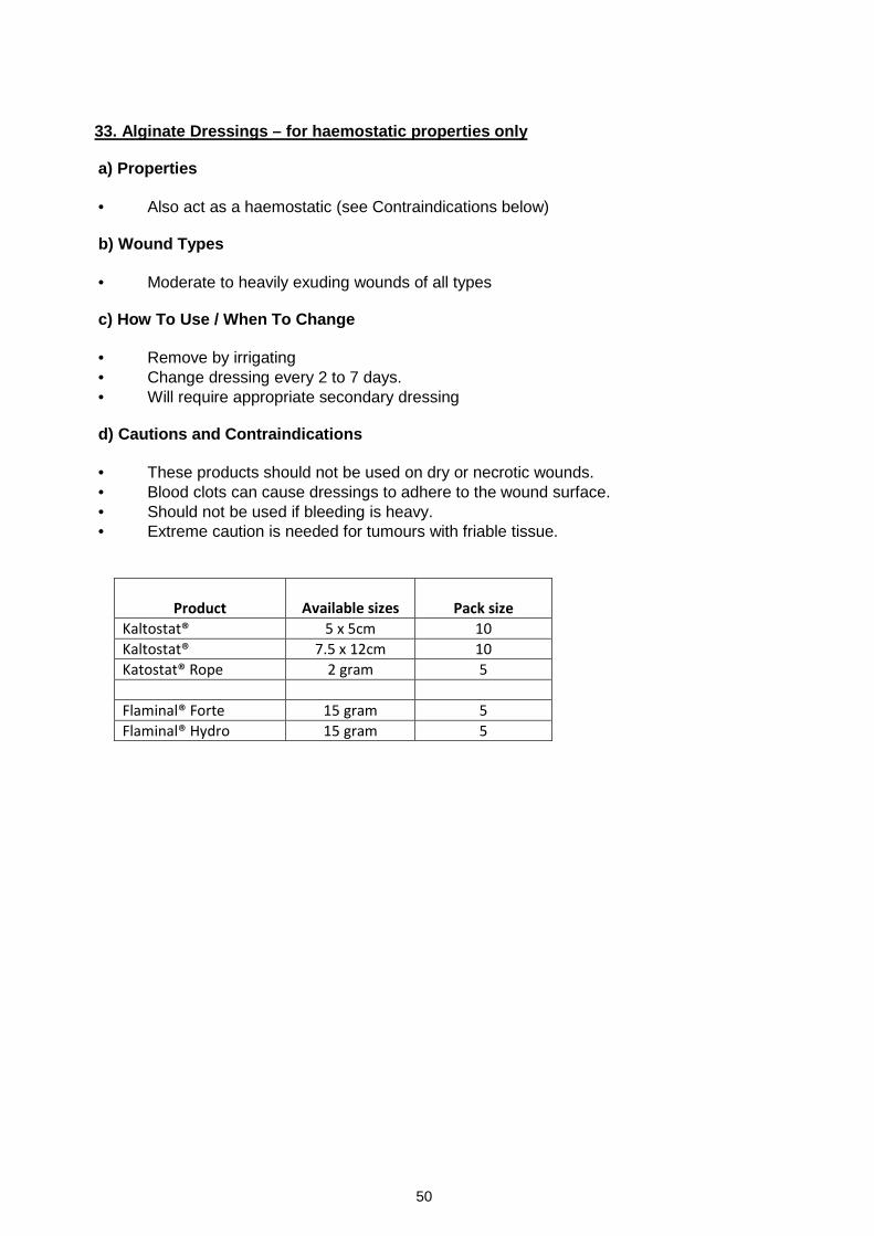
50
33. Alginate Dressings – for haemostatic properties only
a) Properties • Also act as a haemostatic (see Contraindications below) b) Wound Types • Moderate to heavily exuding wounds of all types c) How To Use / When To Change • Remove by irrigating • Change dressing every 2 to 7 days. • Will require appropriate secondary dressing d) Cautions and Contraindications • These products should not be used on dry or necrotic wounds. • Blood clots can cause dressings to adhere to the wound surface. • Should not be used if bleeding is heavy. • Extreme caution is needed for tumours with friable tissue.
Product
Available sizes Pack size
Kaltostat® 5 x 5cm 10
Kaltostat® 7.5 x 12cm 10
Katostat® Rope 2 gram 5
Flaminal® Forte 15 gram 5
Flaminal® Hydro 15 gram 5
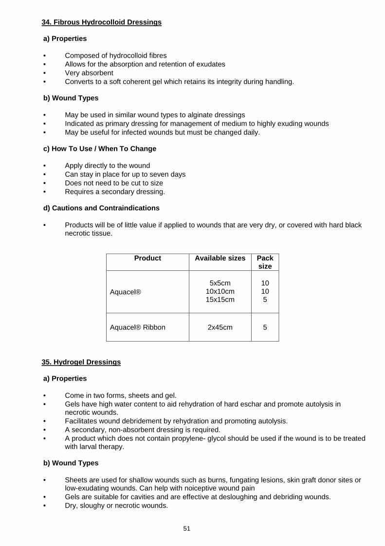
51
34. Fibrous Hydrocolloid Dressings
a) Properties • Composed of hydrocolloid fibres • Allows for the absorption and retention of exudates • Very absorbent • Converts to a soft coherent gel which retains its integrity during handling. b) Wound Types • May be used in similar wound types to alginate dressings • Indicated as primary dressing for management of medium to highly exuding wounds • May be useful for infected wounds but must be changed daily. c) How To Use / When To Change • Apply directly to the wound • Can stay in place for up to seven days • Does not need to be cut to size • Requires a secondary dressing. d) Cautions and Contraindications • Products will be of little value if applied to wounds that are very dry, or covered with hard black
necrotic tissue.
Product Available sizes Pack
size
Aquacel®
5x5cm
10x10cm 15x15cm
10 10 5
Aquacel® Ribbon 2x45cm 5
35. Hydrogel Dressings
a) Properties • Come in two forms, sheets and gel. • Gels have high water content to aid rehydration of hard eschar and promote autolysis in
necrotic wounds. • Facilitates wound debridement by rehydration and promoting autolysis. • A secondary, non-absorbent dressing is required. • A product which does not contain propylene- glycol should be used if the wound is to be treated
with larval therapy. b) Wound Types • Sheets are used for shallow wounds such as burns, fungating lesions, skin graft donor sites or
low-exudating wounds. Can help with noiceptive wound pain • Gels are suitable for cavities and are effective at desloughing and debriding wounds. • Dry, sloughy or necrotic wounds.
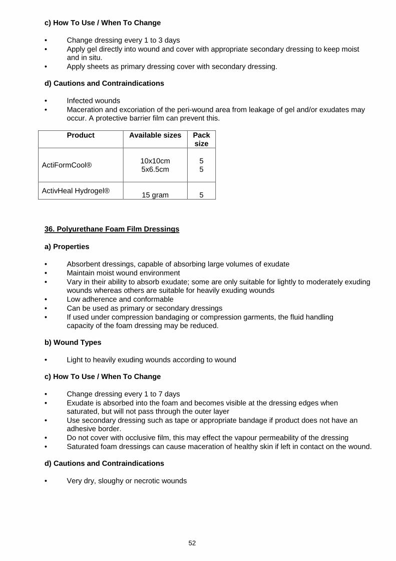
52
c) How To Use / When To Change • Change dressing every 1 to 3 days • Apply gel directly into wound and cover with appropriate secondary dressing to keep moist and in situ. • Apply sheets as primary dressing cover with secondary dressing. d) Cautions and Contraindications • Infected wounds • Maceration and excoriation of the peri-wound area from leakage of gel and/or exudates may
occur. A protective barrier film can prevent this.
Product Available sizes Pack size
ActiFormCool®
10x10cm 5x6.5cm
5 5
ActivHeal Hydrogel® 15 gram
5
36. Polyurethane Foam Film Dressings a) Properties • Absorbent dressings, capable of absorbing large volumes of exudate • Maintain moist wound environment • Vary in their ability to absorb exudate; some are only suitable for lightly to moderately exuding
wounds whereas others are suitable for heavily exuding wounds • Low adherence and conformable • Can be used as primary or secondary dressings • If used under compression bandaging or compression garments, the fluid handling capacity of the foam dressing may be reduced. b) Wound Types • Light to heavily exuding wounds according to wound
c) How To Use / When To Change • Change dressing every 1 to 7 days • Exudate is absorbed into the foam and becomes visible at the dressing edges when saturated, but will not pass through the outer layer • Use secondary dressing such as tape or appropriate bandage if product does not have an
adhesive border. • Do not cover with occlusive film, this may effect the vapour permeability of the dressing • Saturated foam dressings can cause maceration of healthy skin if left in contact on the wound. d) Cautions and Contraindications • Very dry, sloughy or necrotic wounds
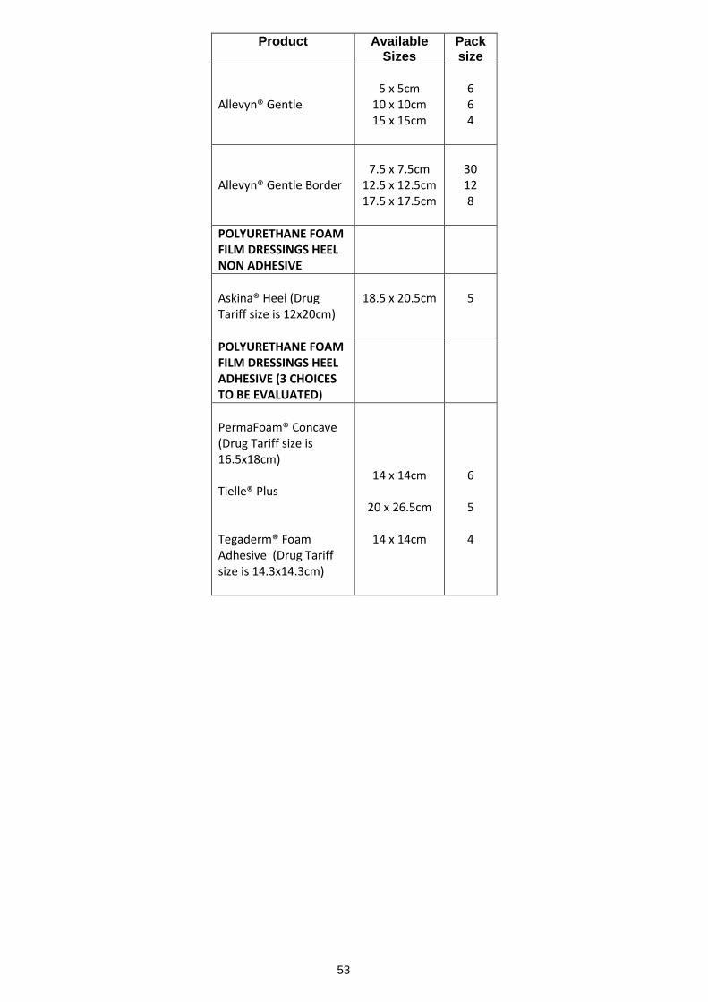
53
Product Available Sizes
Pack size
Allevyn® Gentle
5 x 5cm
10 x 10cm
15 x 15cm
6
6
4
Allevyn® Gentle Border
7.5 x 7.5cm
12.5 x 12.5cm
17.5 x 17.5cm
30
12
8
POLYURETHANE FOAM
FILM DRESSINGS HEEL
NON ADHESIVE
Askina® Heel (Drug
Tariff size is 12x20cm)
18.5 x 20.5cm
5
POLYURETHANE FOAM
FILM DRESSINGS HEEL
ADHESIVE (3 CHOICES
TO BE EVALUATED)
PermaFoam® Concave
(Drug Tariff size is
16.5x18cm)
Tielle® Plus
Tegaderm® Foam
Adhesive (Drug Tariff
size is 14.3x14.3cm)
14 x 14cm
20 x 26.5cm
14 x 14cm
6
5
4
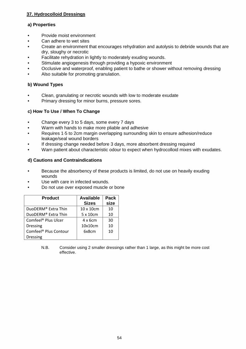
54
37. Hydrocolloid Dressings
a) Properties • Provide moist environment • Can adhere to wet sites • Create an environment that encourages rehydration and autolysis to debride wounds that are
dry, sloughy or necrotic • Facilitate rehydration in lightly to moderately exuding wounds. • Stimulate angiogenesis through providing a hypoxic environment • Occlusive and waterproof, enabling patient to bathe or shower without removing dressing • Also suitable for promoting granulation. b) Wound Types • Clean, granulating or necrotic wounds with low to moderate exudate • Primary dressing for minor burns, pressure sores. c) How To Use / When To Change • Change every 3 to 5 days, some every 7 days • Warm with hands to make more pliable and adhesive • Requires 1·5 to 2cm margin overlapping surrounding skin to ensure adhesion/reduce
leakage/seal wound borders • If dressing change needed before 3 days, more absorbent dressing required • Warn patient about characteristic odour to expect when hydrocolloid mixes with exudates.
d) Cautions and Contraindications • Because the absorbency of these products is limited, do not use on heavily exuding wounds • Use with care in infected wounds. • Do not use over exposed muscle or bone
Product Available
Sizes Pack size
DuoDERM® Extra Thin
DuoDERM® Extra Thin
10 x 10cm
5 x 10cm
10
10
Comfeel® Plus Ulcer
Dressing
Comfeel® Plus Contour
Dressing
4 x 6cm
10x10cm
6x8cm
30
10
10
N.B. Consider using 2 smaller dressings rather than 1 large, as this might be more cost effective.
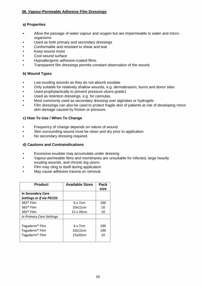
55
38. Vapour-Permeable Adhesive Film Dressings
a) Properties • Allow the passage of water vapour and oxygen but are impermeable to water and micro-
organisms • Used as both primary and secondary dressings • Conformable and resistant to shear and tear • Keep wound moist • Cool wound surface • Hypoallergenic adhesive-coated films. • Transparent film dressings permits constant observation of the wound. b) Wound Types • Low exuding wounds as they do not absorb exudate • Only suitable for relatively shallow wounds, e.g. dermabrasion, burns and donor sites • Used prophylactically to prevent pressure ulcers grade1 • Used as retention dressings, e.g. for cannulas. • Most commonly used as secondary dressing over alginates or hydrogels • Film dressings can also be used to protect fragile skin of patients at risk of developing minor
skin damage caused by friction or pressure. c) How To Use / When To Change • Frequency of change depends on nature of wound • Skin surrounding wound must be clean and dry prior to application • No secondary dressing required. d) Cautions and Contraindications • Excessive exudate may accumulate under dressing • Vapour-permeable films and membranes are unsuitable for infected, large heavily exuding wounds, and chronic leg ulcers • Film may cling to itself during application • May cause adhesive trauma on removal.
Product Available Sizes Pack size
In Secondary Care
Settings or if via PECOS
365® Film
365® Film
365® Film
6 x 7cm
10x12cm
15 x 20cm
100
10
10
In Primary Care Settings
Tegaderm® Film
Tegaderm® Film
Tegaderm® Film
6 x 7cm
10x12cm
15x20cm
100
100
10
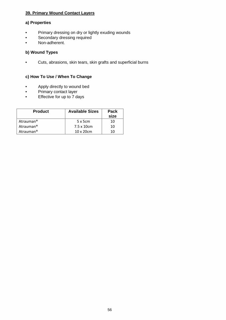
56
39. Primary Wound Contact Layers a) Properties • Primary dressing on dry or lightly exuding wounds • Secondary dressing required • Non-adherent. b) Wound Types
• Cuts, abrasions, skin tears, skin grafts and superficial burns c) How To Use / When To Change • Apply directly to wound bed • Primary contact layer • Effective for up to 7 days
Product Available Sizes Pack
size Atrauman®
Atrauman®
Atrauman®
5 x 5cm
7.5 x 10cm
10 x 20cm
10
10
10
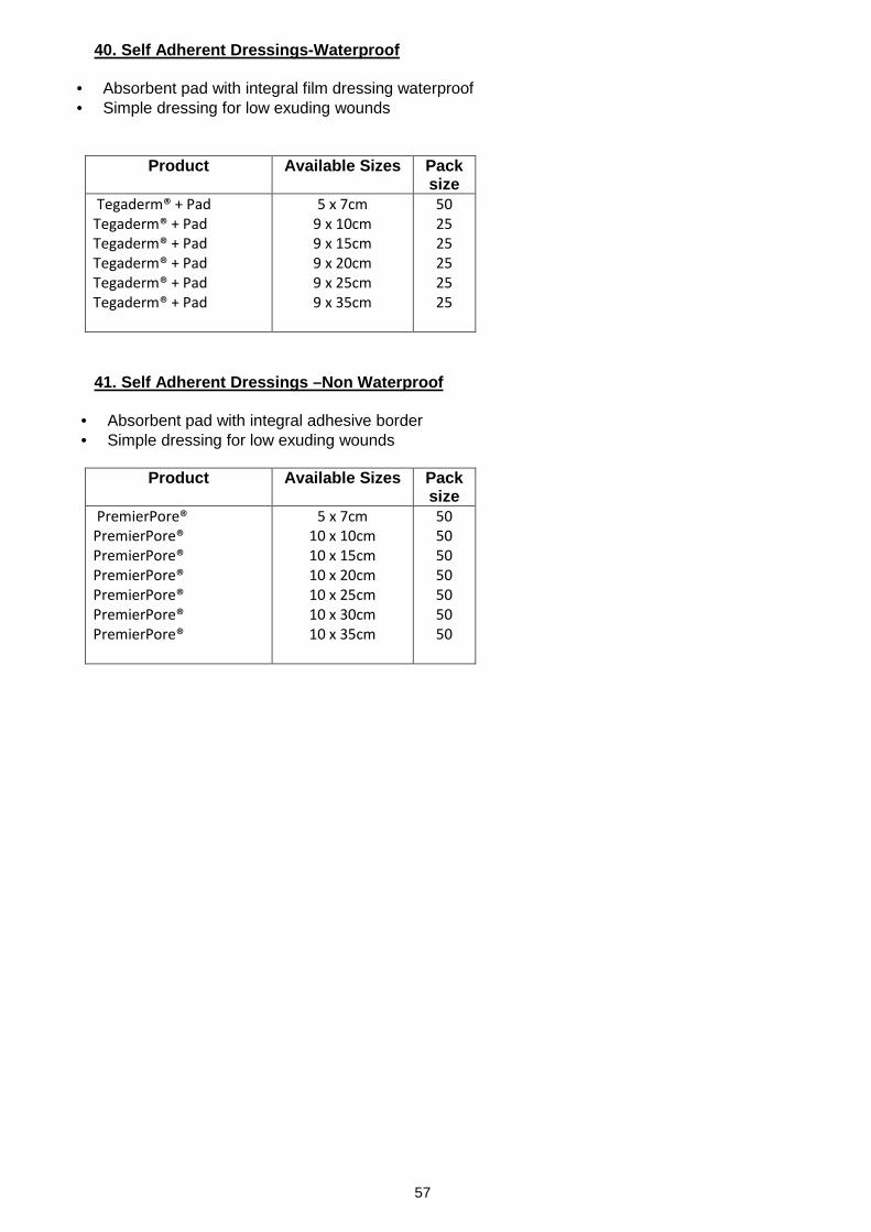
57
40. Self Adherent Dressings-Waterproof
• Absorbent pad with integral film dressing waterproof • Simple dressing for low exuding wounds
Product Available Sizes Pack size
Tegaderm® + Pad
Tegaderm® + Pad
Tegaderm® + Pad
Tegaderm® + Pad
Tegaderm® + Pad
Tegaderm® + Pad
5 x 7cm
9 x 10cm
9 x 15cm
9 x 20cm
9 x 25cm
9 x 35cm
50
25
25
25
25
25
41. Self Adherent Dressings –Non Waterproof
• Absorbent pad with integral adhesive border • Simple dressing for low exuding wounds
Product Available Sizes Pack
size PremierPore®
PremierPore®
PremierPore®
PremierPore®
PremierPore®
PremierPore®
PremierPore®
5 x 7cm
10 x 10cm
10 x 15cm
10 x 20cm
10 x 25cm
10 x 30cm
10 x 35cm
50
50
50
50
50
50
50
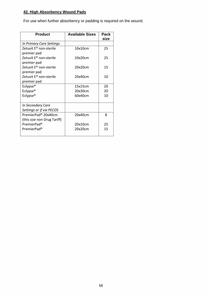
58
42. High Absorbency Wound Pads For use when further absorbency or padding is required on the wound.
Product Available Sizes Pack
size In Primary Care Settings
Zetuvit E® non-sterile
premier pad
Zetuvit E® non-sterile
premier pad
Zetuvit E® non-sterile
premier pad
Zetuvit E® non-sterile
premier pad
10x10cm
10x20cm
20x20cm
20x40cm
25
25
15
10
Eclypse®
Eclypse®
Eclypse®
15x15cm
20x30cm
60x40cm
20
20
10
In Secondary Care
Settings or if via PECOS
PremierPad® 20x40cm
(this size non Drug Tariff)
PremierPad®
PremierPad®
20x40cm
20x10cm
20x20cm
8
25
15
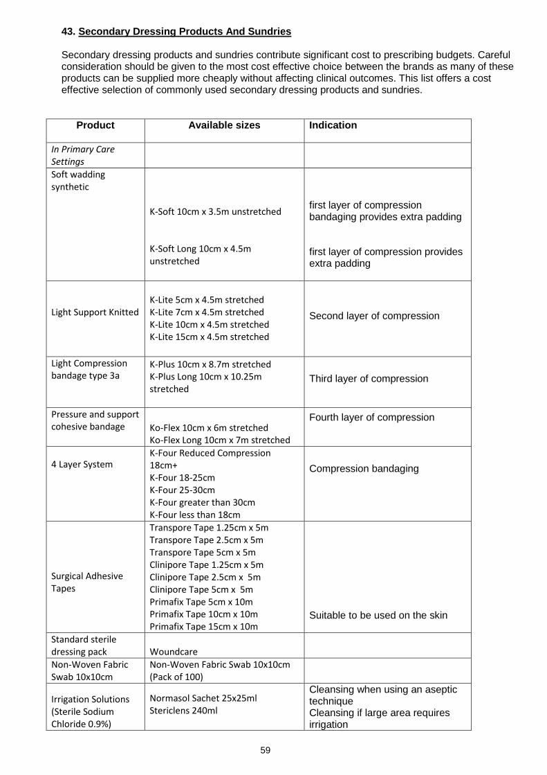
59
43. Secondary Dressing Products And Sundries Secondary dressing products and sundries contribute significant cost to prescribing budgets. Careful consideration should be given to the most cost effective choice between the brands as many of these products can be supplied more cheaply without affecting clinical outcomes. This list offers a cost effective selection of commonly used secondary dressing products and sundries.
Product
Available sizes Indication
In Primary Care
Settings Soft wadding
synthetic
K-Soft 10cm x 3.5m unstretched
K-Soft Long 10cm x 4.5m
unstretched
first layer of compression bandaging provides extra padding first layer of compression provides extra padding
Light Support Knitted
K-Lite 5cm x 4.5m stretched
K-Lite 7cm x 4.5m stretched
K-Lite 10cm x 4.5m stretched
K-Lite 15cm x 4.5m stretched
Second layer of compression
Light Compression
bandage type 3a
K-Plus 10cm x 8.7m stretched
K-Plus Long 10cm x 10.25m
stretched
Third layer of compression
Pressure and support
cohesive bandage
Ko-Flex 10cm x 6m stretched
Ko-Flex Long 10cm x 7m stretched
Fourth layer of compression
4 Layer System
K-Four Reduced Compression
18cm+
K-Four 18-25cm
K-Four 25-30cm
K-Four greater than 30cm
K-Four less than 18cm
Compression bandaging
Surgical Adhesive
Tapes
Transpore Tape 1.25cm x 5m
Transpore Tape 2.5cm x 5m
Transpore Tape 5cm x 5m
Clinipore Tape 1.25cm x 5m
Clinipore Tape 2.5cm x 5m
Clinipore Tape 5cm x 5m
Primafix Tape 5cm x 10m
Primafix Tape 10cm x 10m
Primafix Tape 15cm x 10m Suitable to be used on the skin
Standard sterile
dressing pack Woundcare Non-Woven Fabric
Swab 10x10cm
Non-Woven Fabric Swab 10x10cm
(Pack of 100)
Irrigation Solutions
(Sterile Sodium
Chloride 0.9%)
Normasol Sachet 25x25ml
Stericlens 240ml
Cleansing when using an aseptic technique Cleansing if large area requires irrigation
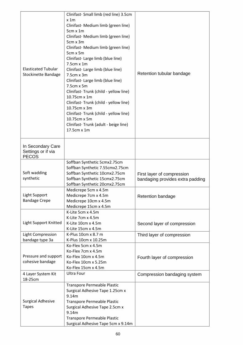
60
Elasticated Tubular
Stockinette Bandage
Clinifast· Small limb (red line) 3.5cm
x 1m
Clinifast· Medium limb (green line)
5cm x 1m
Clinifast· Medium limb (green line)
5cm x 3m
Clinifast· Medium limb (green line)
5cm x 5m
Clinifast· Large limb (blue line)
7.5cm x 1m
Clinifast· Large limb (blue line)
7.5cm x 3m
Clinifast· Large limb (blue line)
7.5cm x 5m
Clinifast· Trunk (child - yellow line)
10.75cm x 1m
Clinifast· Trunk (child - yellow line)
10.75cm x 3m
Clinifast· Trunk (child - yellow line)
10.75cm x 5m
Clinifast· Trunk (adult - beige line)
17.5cm x 1m
Retention tubular bandage
In Secondary Care Settings or if via PECOS
Soft wadding
synthetic
Soffban Synthetic 5cmx2.75cm
Soffban Synthetic 7.55cmx2.75cm
Soffban Synthetic 10cmx2.75cm
Soffban Synthetic 15cmx2.75cm
Soffban Synthetic 20cmx2.75cm
First layer of compression bandaging provides extra padding
Light Support
Bandage Crepe
Medicrepe 5cm x 4.5m
Medicrepe 7cm x 4.5m
Medicrepe 10cm x 4.5m
Medicrepe 15cm x 4.5m
Retention bandage
Light Support Knitted
K-Lite 5cm x 4.5m
K-Lite 7cm x 4.5m
K-Lite 10cm x 4.5m
K-Lite 15cm x 4.5m Second layer of compression
Light Compression
bandage type 3a
K-Plus 10cm x 8.7 m
K-Plus 10cm x 10.25m Third layer of compression
Pressure and support
cohesive bandage
Ko-Flex 5cm x 4.5m
Ko-Flex 7cm x 4.5m
Ko-Flex 10cm x 4.5m
Ko-Flex 10cm x 5.25m
Ko-Flex 15cm x 4.5m
Fourth layer of compression
4 Layer System Kit
18-25cm
Ultra Four
Compression bandaging system
Surgical Adhesive
Tapes
Transpore Permeable Plastic
Surgical Adhesive Tape 1.25cm x
9.14m
Transpore Permeable Plastic
Surgical Adhesive Tape 2.5cm x
9.14m
Transpore Permeable Plastic
Surgical Adhesive Tape 5cm x 9.14m
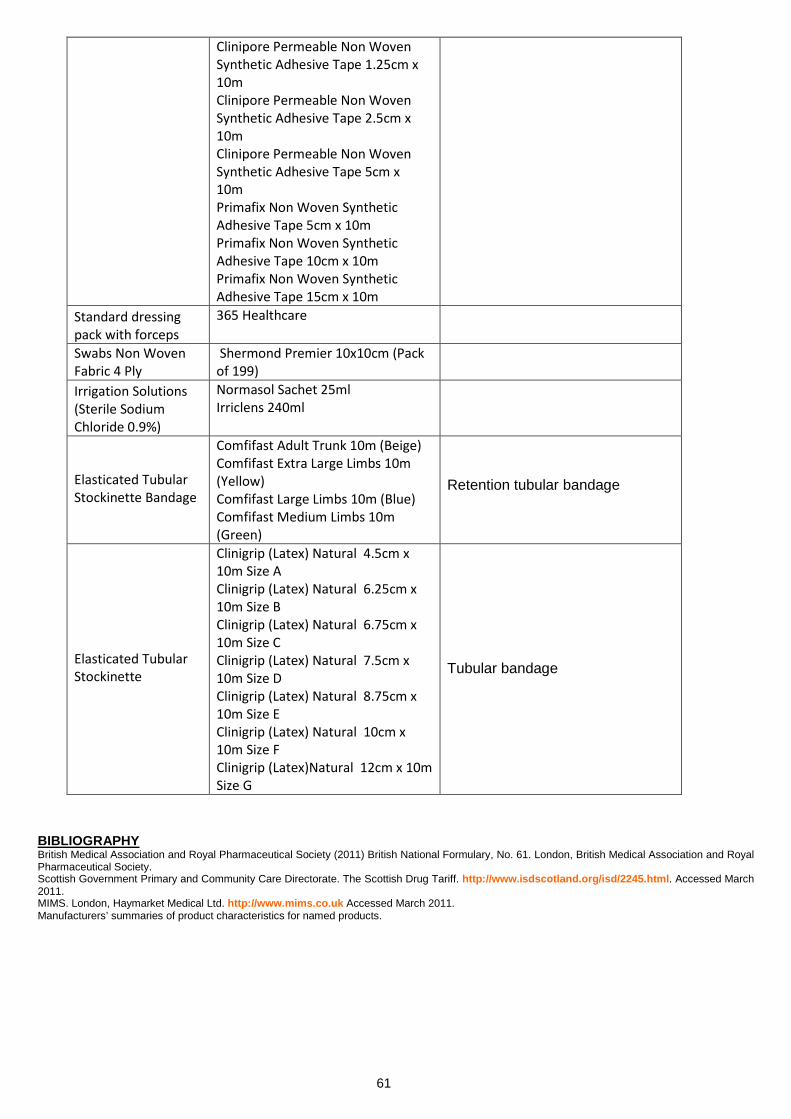
61
Clinipore Permeable Non Woven
Synthetic Adhesive Tape 1.25cm x
10m
Clinipore Permeable Non Woven
Synthetic Adhesive Tape 2.5cm x
10m
Clinipore Permeable Non Woven
Synthetic Adhesive Tape 5cm x
10m
Primafix Non Woven Synthetic
Adhesive Tape 5cm x 10m
Primafix Non Woven Synthetic
Adhesive Tape 10cm x 10m
Primafix Non Woven Synthetic
Adhesive Tape 15cm x 10m Standard dressing
pack with forceps
365 Healthcare
Swabs Non Woven
Fabric 4 Ply
Shermond Premier 10x10cm (Pack
of 199) Irrigation Solutions
(Sterile Sodium
Chloride 0.9%)
Normasol Sachet 25ml
Irriclens 240ml
Elasticated Tubular
Stockinette Bandage
Comfifast Adult Trunk 10m (Beige)
Comfifast Extra Large Limbs 10m
(Yellow)
Comfifast Large Limbs 10m (Blue)
Comfifast Medium Limbs 10m
(Green)
Retention tubular bandage
Elasticated Tubular
Stockinette
Clinigrip (Latex) Natural 4.5cm x
10m Size A
Clinigrip (Latex) Natural 6.25cm x
10m Size B
Clinigrip (Latex) Natural 6.75cm x
10m Size C
Clinigrip (Latex) Natural 7.5cm x
10m Size D
Clinigrip (Latex) Natural 8.75cm x
10m Size E
Clinigrip (Latex) Natural 10cm x
10m Size F
Clinigrip (Latex)Natural 12cm x 10m
Size G
Tubular bandage
BIBLIOGRAPHY British Medical Association and Royal Pharmaceutical Society (2011) British National Formulary, No. 61. London, British Medical Association and Royal Pharmaceutical Society. Scottish Government Primary and Community Care Directorate. The Scottish Drug Tariff. http://www.isdscotland.org/isd/2245.html . Accessed March 2011. MIMS. London, Haymarket Medical Ltd. http://www.mims.co.uk Accessed March 2011. Manufacturers’ summaries of product characteristics for named products.
62
SECTION FIVE
SPECIALIST PRODUCTS
Products available in this section are those which should be used only where none of the choices in the primary and secondary dressings section are appropriate for a particular wound type/clinical condition.
Each operational unit (i.e. North & West Highland, South & Mid Highland, Argyll and Bute CHP and Raigmore Hospital) will develop their own local protocols and guidance as to how use of products from this section are managed. Generally, use of a specialist product will require clinical assessment by a professional with more in-depth knowledge of tissue viability and wound management. In some cases use of a specialist product may also require agreement from a senior member of staff, e.g. team leader/team manager/budget holder. Locally agreed protocols and guidance should be available for all staff outlining the process.
63
Topical Antimicrobial Agents General Note Infection at the wound site which is spreading requires treatment with systemic antibacterials. For specific guidance please refer to Infections Chapter, Highland Formulary. For local wound infection, a topical antimicrobial dressing can be used to reduce the level of bacteria at the wound surface but these will not eliminate a spreading infection. 44 IODINE 44.1 General Notes a) Properties • Active against gram-positive and gram-negative organisms, anaerobes, fungi and yeasts, protozoa, and viruses • Disrupts cell proteins and lipid membranes. b) Wound Types • For use on superficial wounds which are suspected to be critically colonised, but have low
levels of exudate c) Cautions and Contraindications • Should not be used in patients with known or suspected iodine sensitivity. • Should not be used in patients with Hashimoto’s Thyroiditis and in the case of non-toxic nodular
goitre. • Not to be used on children or pregnant or lactating mothers. • Iodine is absorbed systemically and patients with severely impaired renal function or with a past
history of any thyroid disorder are more susceptible to alterations in thyroid metabolism with chronic therapy.
• Should be used with caution on patients who have a history of thyroid disorders, are on lithium therapy, especially when applied to large wounds.
• Where use exceeds four weeks or in extensive wounds, renal function, thyroid function and if appropriate lithium levels should be monitored.
Note: Many of the concerns about iodine are based on the toxicity of older formulations containing elemental iodine, or arise from in vitro studies which may not be relevant to in vivo situations. Newer preparations appear to be safe. Systemic antibiotics will also be required in confirmed cases of infection.
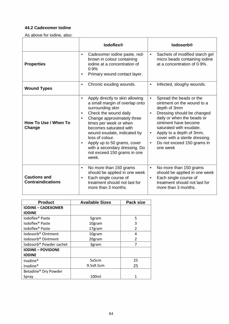
64
44.2 Cadexomer Iodine
As above for iodine, also:
Iodoflex®
Iodosorb®
Properties
• Cadexomer iodine paste, red-brown in colour containing iodine at a concentration of 0·9%
• Primary wound contact layer.
• Sachets of modified starch gel micro beads containing iodine at a concentration of 0·9%.
Wound Types • Chronic exuding wounds. • Infected, sloughy wounds.
How To Use / When To Change
• Apply directly to skin allowing a small margin of overlap onto surrounding skin
• Check the wound daily • Change approximately three
times per week or when becomes saturated with wound exudate, indicated by loss of colour.
• Apply up to 50 grams, cover with a secondary dressing. Do not exceed 150 grams in one week.
• Spread the beads or the ointment on the wound to a depth of 3mm
• Dressing should be changed daily or when the beads or ointment have become saturated with exudate.
• Apply to a depth of 3mm, cover with a sterile dressing.
• Do not exceed 150 grams in one week
Cautions and Contraindications
• No more than 150 grams should be applied in one week
• Each single course of treatment should not last for more than 3 months.
• No more than 150 grams should be applied in one week
• Each single course of treatment should not last for more than 3 months.
Product Available Sizes Pack size
IODINE – CADEXOMER
IODINE
Iodoflex® Paste
Iodoflex® Paste
Iodoflex® Paste
5gram
10gram
17gram
5
3
2
Iodosorb® Ointment
Iodosorb® Ointment
10gram
20gram
4
2
Iodosorb® Powder sachet 3gram 7
IODINE – POVIDONE
IODINE
Inadine®
Inadine®
5x5cm
9.5x9.5cm
25
25
Betadine® Dry Powder
Spray 100ml 1
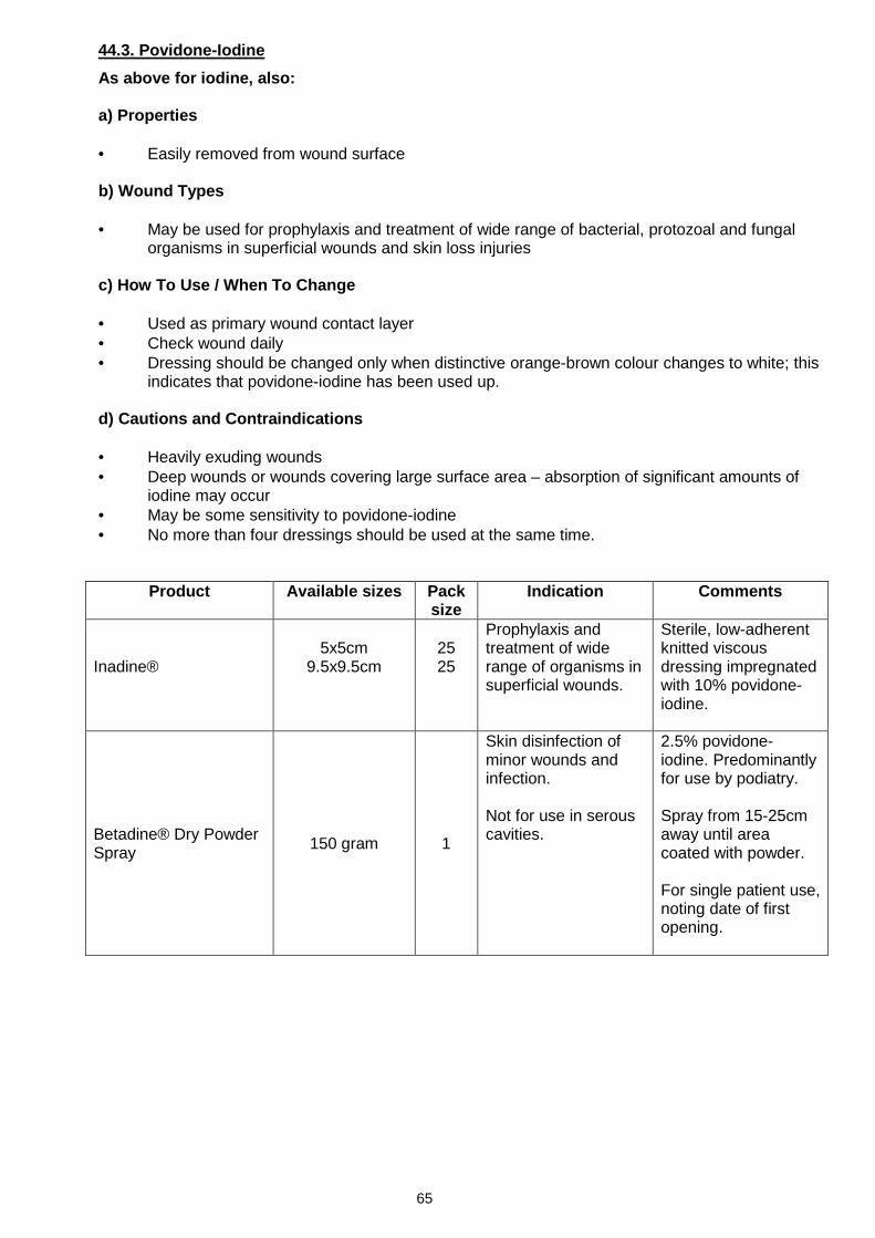
65
44.3. Povidone-Iodine
As above for iodine, also: a) Properties • Easily removed from wound surface b) Wound Types • May be used for prophylaxis and treatment of wide range of bacterial, protozoal and fungal
organisms in superficial wounds and skin loss injuries c) How To Use / When To Change • Used as primary wound contact layer • Check wound daily • Dressing should be changed only when distinctive orange-brown colour changes to white; this
indicates that povidone-iodine has been used up.
d) Cautions and Contraindications • Heavily exuding wounds • Deep wounds or wounds covering large surface area – absorption of significant amounts of
iodine may occur • May be some sensitivity to povidone-iodine • No more than four dressings should be used at the same time.
Product Available sizes Pack size
Indication Comments
Inadine®
5x5cm
9.5x9.5cm
25 25
Prophylaxis and treatment of wide range of organisms in superficial wounds.
Sterile, low-adherent knitted viscous dressing impregnated with 10% povidone-iodine.
Betadine® Dry Powder Spray 150 gram 1
Skin disinfection of minor wounds and infection. Not for use in serous cavities.
2.5% povidone-iodine. Predominantly for use by podiatry. Spray from 15-25cm away until area coated with powder. For single patient use, noting date of first opening.
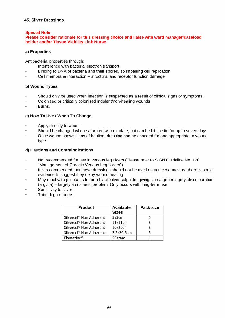
66
45. Silver Dressings
Special Note Please consider rationale for this dressing choice and liaise with ward manager/caseload holder and/or Tissue Viability Link Nurse a) Properties Antibacterial properties through: • Interference with bacterial electron transport • Binding to DNA of bacteria and their spores, so impairing cell replication • Cell membrane interaction – structural and receptor function damage b) Wound Types • Should only be used when infection is suspected as a result of clinical signs or symptoms. • Colonised or critically colonised indolent/non-healing wounds • Burns. c) How To Use / When To Change • Apply directly to wound • Should be changed when saturated with exudate, but can be left in situ for up to seven days • Once wound shows signs of healing, dressing can be changed for one appropriate to wound
type.
d) Cautions and Contraindications • Not recommended for use in venous leg ulcers (Please refer to SIGN Guideline No. 120
“Management of Chronic Venous Leg Ulcers”) • It is recommended that these dressings should not be used on acute wounds as there is some
evidence to suggest they delay wound healing • May react with pollutants to form black silver sulphide, giving skin a general grey discolouration
(argyria) – largely a cosmetic problem. Only occurs with long-term use • Sensitivity to silver. • Third degree burns
Product Available Sizes
Pack size
Silvercel® Non Adherent
Silvercel® Non Adherent
Silvercel® Non Adherent
Silvercel® Non Adherent
5x5cm
11x11cm
10x20cm
2.5x30.5cm
5
5
5
5
Flamazine® 50gram 1
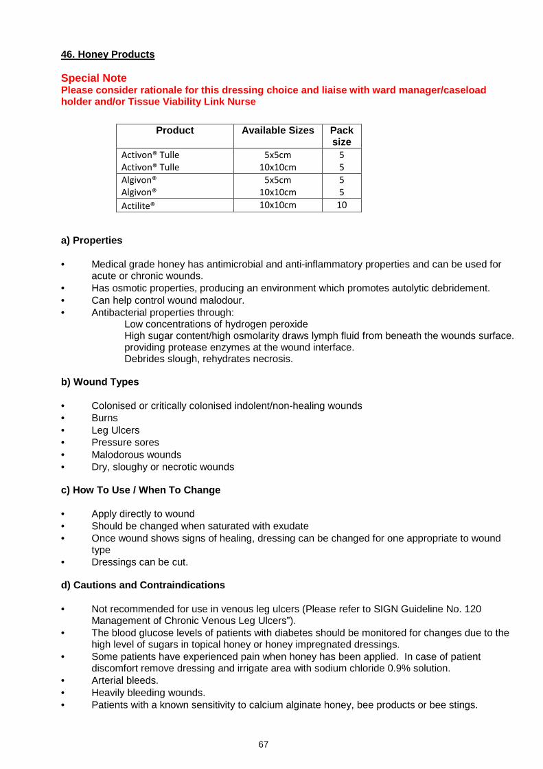
67
46. Honey Products Special Note Please consider rationale for this dressing choice and liaise with ward manager/caseload holder and/or Tissue Viability Link Nurse
a) Properties • Medical grade honey has antimicrobial and anti-inflammatory properties and can be used for
acute or chronic wounds. • Has osmotic properties, producing an environment which promotes autolytic debridement. • Can help control wound malodour. • Antibacterial properties through:
Low concentrations of hydrogen peroxide High sugar content/high osmolarity draws lymph fluid from beneath the wounds surface. providing protease enzymes at the wound interface. Debrides slough, rehydrates necrosis.
b) Wound Types • Colonised or critically colonised indolent/non-healing wounds • Burns • Leg Ulcers • Pressure sores • Malodorous wounds • Dry, sloughy or necrotic wounds
c) How To Use / When To Change • Apply directly to wound • Should be changed when saturated with exudate • Once wound shows signs of healing, dressing can be changed for one appropriate to wound
type • Dressings can be cut. d) Cautions and Contraindications • Not recommended for use in venous leg ulcers (Please refer to SIGN Guideline No. 120 Management of Chronic Venous Leg Ulcers”). • The blood glucose levels of patients with diabetes should be monitored for changes due to the
high level of sugars in topical honey or honey impregnated dressings. • Some patients have experienced pain when honey has been applied. In case of patient
discomfort remove dressing and irrigate area with sodium chloride 0.9% solution. • Arterial bleeds. • Heavily bleeding wounds. • Patients with a known sensitivity to calcium alginate honey, bee products or bee stings.
Product Available Sizes Pack size
Activon® Tulle
Activon® Tulle
5x5cm
10x10cm
5
5
Algivon®
Algivon®
5x5cm
10x10cm
5
5
Actilite® 10x10cm 10
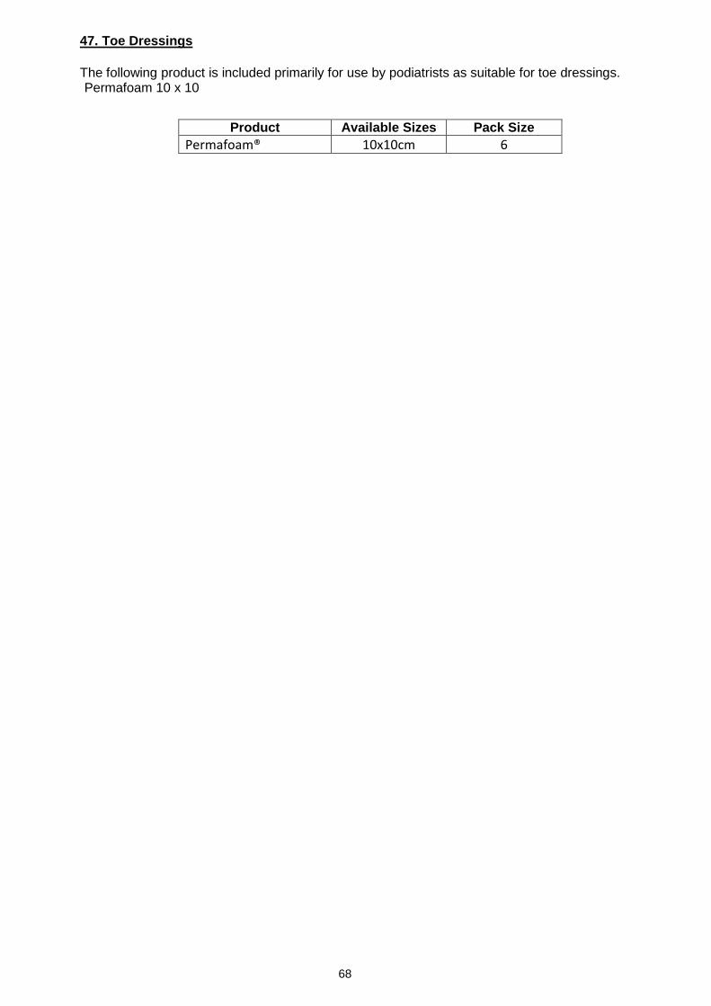
68
47. Toe Dressings
The following product is included primarily for use by podiatrists as suitable for toe dressings. Permafoam 10 x 10
Product Available Sizes Pack Size Permafoam® 10x10cm 6
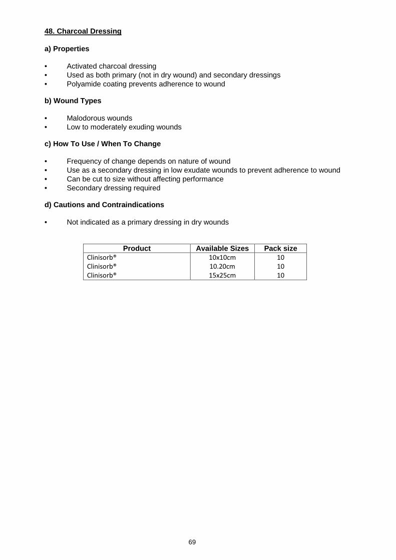
69
48. Charcoal Dressing
a) Properties • Activated charcoal dressing • Used as both primary (not in dry wound) and secondary dressings • Polyamide coating prevents adherence to wound b) Wound Types • Malodorous wounds • Low to moderately exuding wounds c) How To Use / When To Change • Frequency of change depends on nature of wound • Use as a secondary dressing in low exudate wounds to prevent adherence to wound • Can be cut to size without affecting performance • Secondary dressing required d) Cautions and Contraindications • Not indicated as a primary dressing in dry wounds
Product Available Sizes Pack size Clinisorb®
Clinisorb®
Clinisorb®
10x10cm
10.20cm
15x25cm
10
10
10
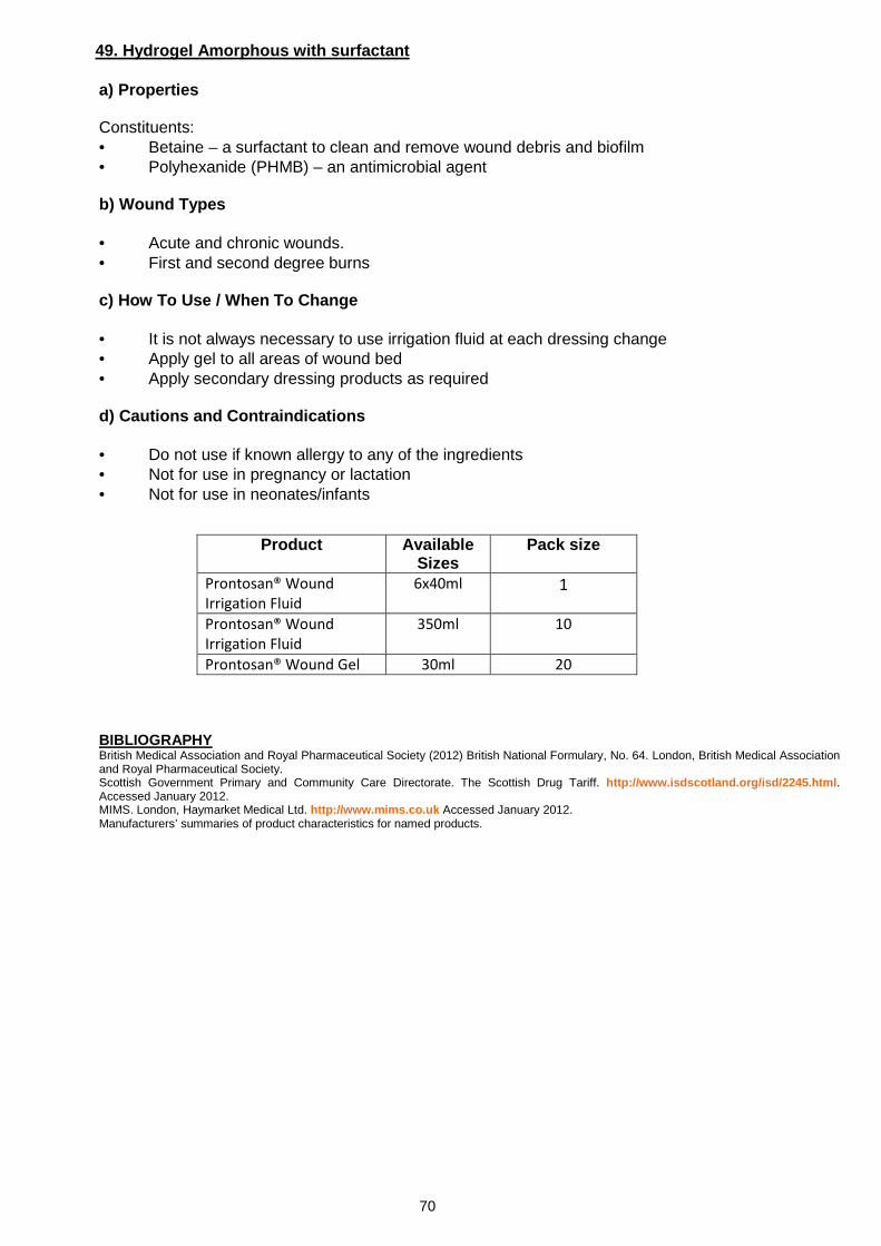
70
49. Hydrogel Amorphous with surfactant a) Properties Constituents: • Betaine – a surfactant to clean and remove wound debris and biofilm • Polyhexanide (PHMB) – an antimicrobial agent b) Wound Types • Acute and chronic wounds. • First and second degree burns c) How To Use / When To Change • It is not always necessary to use irrigation fluid at each dressing change • Apply gel to all areas of wound bed • Apply secondary dressing products as required d) Cautions and Contraindications • Do not use if known allergy to any of the ingredients • Not for use in pregnancy or lactation • Not for use in neonates/infants
BIBLIOGRAPHY British Medical Association and Royal Pharmaceutical Society (2012) British National Formulary, No. 64. London, British Medical Association and Royal Pharmaceutical Society. Scottish Government Primary and Community Care Directorate. The Scottish Drug Tariff. http://www.isdscotland.org/isd/2245.html . Accessed January 2012. MIMS. London, Haymarket Medical Ltd. http://www.mims.co.uk Accessed January 2012. Manufacturers’ summaries of product characteristics for named products.
Product Available Sizes
Pack size
Prontosan® Wound
Irrigation Fluid
6x40ml 1
Prontosan® Wound
Irrigation Fluid
350ml 10
Prontosan® Wound Gel 30ml 20
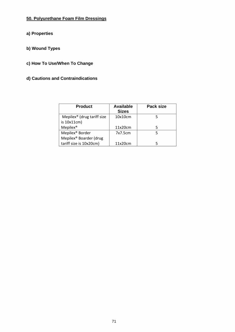
71
50. Polyurethane Foam Film Dressings
a) Properties
b) Wound Types
c) How To Use/When To Change
d) Cautions and Contraindications
Product Available Sizes
Pack size
Mepilex® (drug tariff size
is 10x11cm)
Mepilex®
10x10cm
11x20cm
5
5
Mepilex® Border
Mepilex® Boarder (drug
tariff size is 10x20cm)
7x7.5cm
11x20cm
5
5
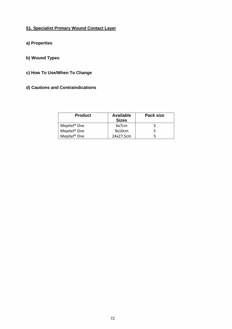
72
51. Specialist Primary Wound Contact Layer
a) Properties
b) Wound Types
c) How To Use/When To Change
d) Cautions and Contraindications
Product Available Sizes
Pack size
Mepitel® One
Mepitel® One
Mepitel® One
6x7cm
9x10cm
24x27.5cm
5
5
5
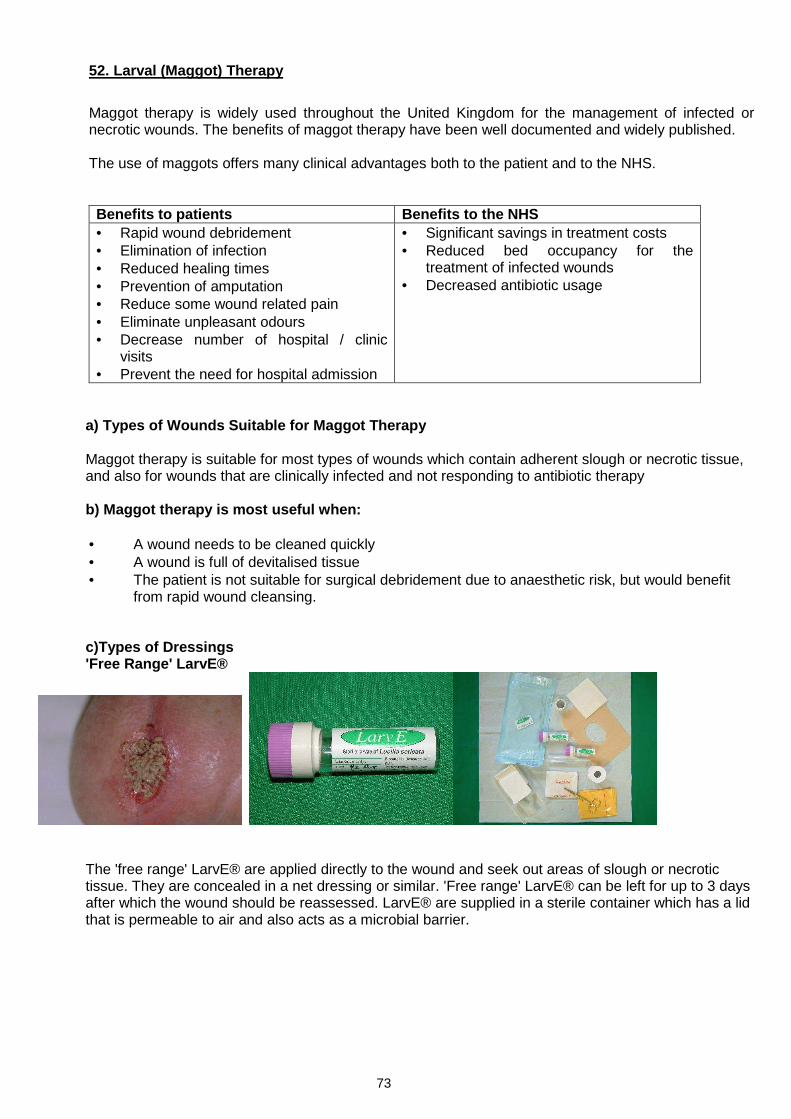
73
52. Larval (Maggot) Therapy
Maggot therapy is widely used throughout the United Kingdom for the management of infected or necrotic wounds. The benefits of maggot therapy have been well documented and widely published. The use of maggots offers many clinical advantages both to the patient and to the NHS. Benefits to patients Benefits to the NHS • Rapid wound debridement • Elimination of infection • Reduced healing times • Prevention of amputation • Reduce some wound related pain • Eliminate unpleasant odours • Decrease number of hospital / clinic
visits • Prevent the need for hospital admission
• Significant savings in treatment costs • Reduced bed occupancy for the
treatment of infected wounds • Decreased antibiotic usage
a) Types of Wounds Suitable for Maggot Therapy Maggot therapy is suitable for most types of wounds which contain adherent slough or necrotic tissue, and also for wounds that are clinically infected and not responding to antibiotic therapy b) Maggot therapy is most useful when:
• A wound needs to be cleaned quickly • A wound is full of devitalised tissue • The patient is not suitable for surgical debridement due to anaesthetic risk, but would benefit
from rapid wound cleansing. c)Types of Dressings 'Free Range' LarvE®
The 'free range' LarvE® are applied directly to the wound and seek out areas of slough or necrotic tissue. They are concealed in a net dressing or similar. 'Free range' LarvE® can be left for up to 3 days after which the wound should be reassessed. LarvE® are supplied in a sterile container which has a lid that is permeable to air and also acts as a microbial barrier.
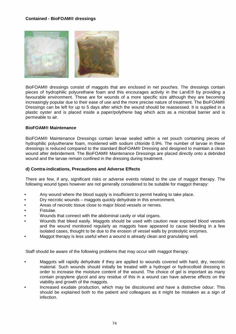
74
Contained - BioFOAM® dressings
BioFOAM® dressings consist of maggots that are enclosed in net pouches. The dressings contain pieces of hydrophilic polyurethane foam and this encourages activity in the LarvE® by providing a favourable environment. These are for wounds of a more specific size although they are becoming increasingly popular due to their ease of use and the more precise nature of treatment. The BioFOAM® Dressings can be left for up to 5 days after which the wound should be reassessed. It is supplied in a plastic oyster and is placed inside a paper/polythene bag which acts as a microbial barrier and is permeable to air.
BioFOAM® Maintenance
BioFOAM® Maintenance Dressings contain larvae sealed within a net pouch containing pieces of hydrophilic polyutherane foam, moistened with sodium chloride 0.9%. The number of larvae in these dressings is reduced compared to the standard BioFOAM® Dressing and designed to maintain a clean wound after debridement. The BioFOAM® Maintenance Dressings are placed directly onto a debrided wound and the larvae remain confined in the dressing during treatment.
d) Contra-indications, Precautions and Adverse Eff ects There are few, if any, significant risks or adverse events related to the use of maggot therapy. The following wound types however are not generally considered to be suitable for maggot therapy:
• Any wound where the blood supply is insufficient to permit healing to take place. • Dry necrotic wounds – maggots quickly dehydrate in this environment. • Areas of necrotic tissue close to major blood vessels or nerves. • Fistulae. • Wounds that connect with the abdominal cavity or vital organs. • Wounds that bleed easily. Maggots should be used with caution near exposed blood vessels
and the wound monitored regularly as maggots have appeared to cause bleeding in a few isolated cases, thought to be due to the erosion of vessel walls by proteolytic enzymes.
• Maggot therapy is less useful when a wound is already clean and granulating well. Staff should be aware of the following problems that may occur with maggot therapy:
• Maggots will rapidly dehydrate if they are applied to wounds covered with hard, dry, necrotic material. Such wounds should initially be treated with a hydrogel or hydrocolloid dressing in order to increase the moisture content of the wound. The choice of gel is important as many contain propylene glycol and any residue of this in a wound can have adverse effects on the viability and growth of the maggots.
• Increased exudate production, which may be discoloured and have a distinctive odour. This should be explained both to the patient and colleagues as it might be mistaken as a sign of infection.
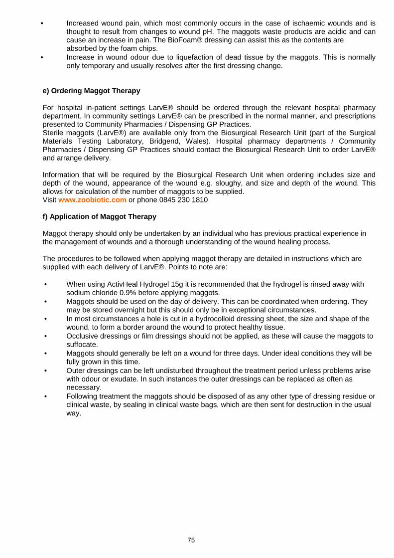
75
• Increased wound pain, which most commonly occurs in the case of ischaemic wounds and is thought to result from changes to wound pH. The maggots waste products are acidic and can cause an increase in pain. The BioFoam® dressing can assist this as the contents are absorbed by the foam chips.
• Increase in wound odour due to liquefaction of dead tissue by the maggots. This is normally only temporary and usually resolves after the first dressing change.
e) Ordering Maggot Therapy For hospital in-patient settings LarvE® should be ordered through the relevant hospital pharmacy department. In community settings LarvE® can be prescribed in the normal manner, and prescriptions presented to Community Pharmacies / Dispensing GP Practices. Sterile maggots (LarvE®) are available only from the Biosurgical Research Unit (part of the Surgical Materials Testing Laboratory, Bridgend, Wales). Hospital pharmacy departments / Community Pharmacies / Dispensing GP Practices should contact the Biosurgical Research Unit to order LarvE® and arrange delivery. Information that will be required by the Biosurgical Research Unit when ordering includes size and depth of the wound, appearance of the wound e.g. sloughy, and size and depth of the wound. This allows for calculation of the number of maggots to be supplied. Visit www.zoobiotic.com or phone 0845 230 1810 f) Application of Maggot Therapy Maggot therapy should only be undertaken by an individual who has previous practical experience in the management of wounds and a thorough understanding of the wound healing process. The procedures to be followed when applying maggot therapy are detailed in instructions which are supplied with each delivery of LarvE®. Points to note are:
• When using ActivHeal Hydrogel 15g it is recommended that the hydrogel is rinsed away with
sodium chloride 0.9% before applying maggots. • Maggots should be used on the day of delivery. This can be coordinated when ordering. They
may be stored overnight but this should only be in exceptional circumstances. • In most circumstances a hole is cut in a hydrocolloid dressing sheet, the size and shape of the
wound, to form a border around the wound to protect healthy tissue. • Occlusive dressings or film dressings should not be applied, as these will cause the maggots to
suffocate. • Maggots should generally be left on a wound for three days. Under ideal conditions they will be fully grown in this time. • Outer dressings can be left undisturbed throughout the treatment period unless problems arise
with odour or exudate. In such instances the outer dressings can be replaced as often as necessary.
• Following treatment the maggots should be disposed of as any other type of dressing residue or clinical waste, by sealing in clinical waste bags, which are then sent for destruction in the usual way.
76
g) Ethical Implications of Maggot Therapy There are no major ethical implications relating to the application of maggot therapy. However, as with any nursing procedure, the nurse is the patient’s advocate and, as such, must ensure that the patient is offered treatment most clinically appropriate for their condition requiring treatment. The nurse or practitioner caring for the patient must ensure that:
• The patient is fully informed regarding maggot therapy, including what is involved with the therapy, the treatment procedure, and what they can expect to happen.
• The patient is given reasonable time to consider this or alternative treatment options. • Informed consent is obtained in all cases where the patient has capacity to give consent to the
treatment. If this is not appropriate consent should be obtained from the patient’s next of kin or • carer. • Maggots are not applied to an individual who is unable to express any concerns / worries
regarding the treatment. • Any cultural or religious objections to the use of maggots have been considered and discussed
with the patient. • If the patient refuses the treatment, for whatever reason, the nurse or practitioner caring for the
patient must ensure that no undue pressure is brought to bear on the patient to accept the treatment, and that an acceptable alternative treatment is offered.
• Even after application of maggot therapy, the maggots will be removed immediately if the patient changes their mind and that an acceptable alternative treatment is offered.
• Interest is not attracted from members of staff who are not directly involved with the patient’s care, which may result in the patient becoming the focus of attention due to the unusual nature of maggot therapy.
REFERENCES
LarvE® SMTL Dressings Datacard. http://www.dressings.org/Dressings/larve.html. Acce ssed 12/03/2007 . Zoobiotic Home Page. http://www.zoobiotic.com . Accessed 12/03/2007.
77
53. Negative Pressure Wound Therapy
a) Modes of action: • Applies controlled, localised pressure to help draw wounds closed • Helps remove interstitial fluid allowing tissue decompression and enhanced blood flow • Promotes granulation formation • Removes infectious material • Provides a closed, moist wound healing environment b) Indications: • Acute and traumatic wounds • Dehisced wounds • Chronic wounds • Meshed grafts • Flaps c) Contraindications: • Fistulae to organs or body cavities • Necrotic tissue with eschar present • Untreated osteomyelitis • Malignancy in the wound • Placing over exposed blood vessels or organs d) Precautions: • Active Bleeding • Patients on anticoagulants • Difficult wound haemostasis • Placing near blood vessels, tendons or bone e) Ordering: • This treatment is very expensive and should be reserved for exceptional use and must be done in conjunction with the Tissue Viability Nurse Specialist.
NHS HIGHLAND POLICY on use of NPWT is currently bei ng developed.
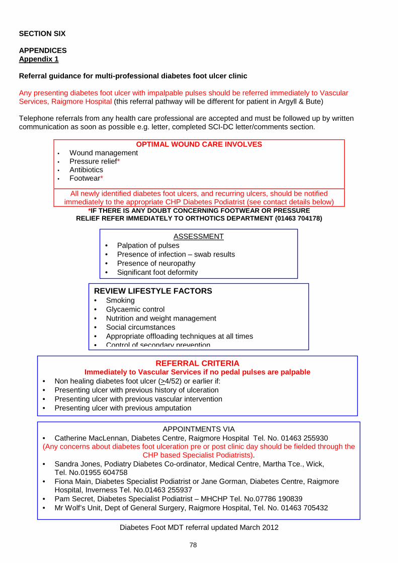
78
SECTION SIX APPENDICES Appendix 1 Referral guidance for multi-professional diabetes f oot ulcer clinic Any presenting diabetes foot ulcer with impalpable pulses should be referred immediately to Vascular Services, Raigmore Hospital (this referral pathway will be different for patient in Argyll & Bute) Telephone referrals from any health care professional are accepted and must be followed up by written communication as soon as possible e.g. letter, completed SCI-DC letter/comments section.
OPTIMAL WOUND CARE INVOLVES • Wound management • Pressure relief* • Antibiotics • Footwear*
All newly identified diabetes foot ulcers, and recurring ulcers, should be notified immediately to the appropriate CHP Diabetes Podiatrist (see contact details below)
*IF THERE IS ANY DOUBT CONCERNING FOOTWEAR OR PRESSU RE RELIEF REFER IMMEDIATELY TO ORTHOTICS DEPARTMENT (0 1463 704178)
Diabetes Foot MDT referral updated March 2012
ASSESSMENT • Palpation of pulses • Presence of infection – swab results • Presence of neuropathy • Significant foot deformity
APPOINTMENTS VIA • Catherine MacLennan, Diabetes Centre, Raigmore Hospital Tel. No. 01463 255930 (Any concerns about diabetes foot ulceration pre or post clinic day should be fielded through the
CHP based Specialist Podiatrists). • Sandra Jones, Podiatry Diabetes Co-ordinator, Medical Centre, Martha Tce., Wick,
Tel. No.01955 604758 • Fiona Main, Diabetes Specialist Podiatrist or Jane Gorman, Diabetes Centre, Raigmore
Hospital, Inverness Tel. No.01463 255937 • Pam Secret, Diabetes Specialist Podiatrist – MHCHP Tel. No.07786 190839 • Mr Wolf’s Unit, Dept of General Surgery, Raigmore Hospital, Tel. No. 01463 705432
REVIEW LIFESTYLE FACTORS • Smoking • Glycaemic control • Nutrition and weight management • Social circumstances • Appropriate offloading techniques at all times • Control of secondary prevention
REFERRAL CRITERIA Immediately to Vascular Services if no pedal pulses are palpable
• Non healing diabetes foot ulcer (>4/52) or earlier if: • Presenting ulcer with previous history of ulceration • Presenting ulcer with previous vascular intervention • Presenting ulcer with previous amputation • Control of secondary prevention
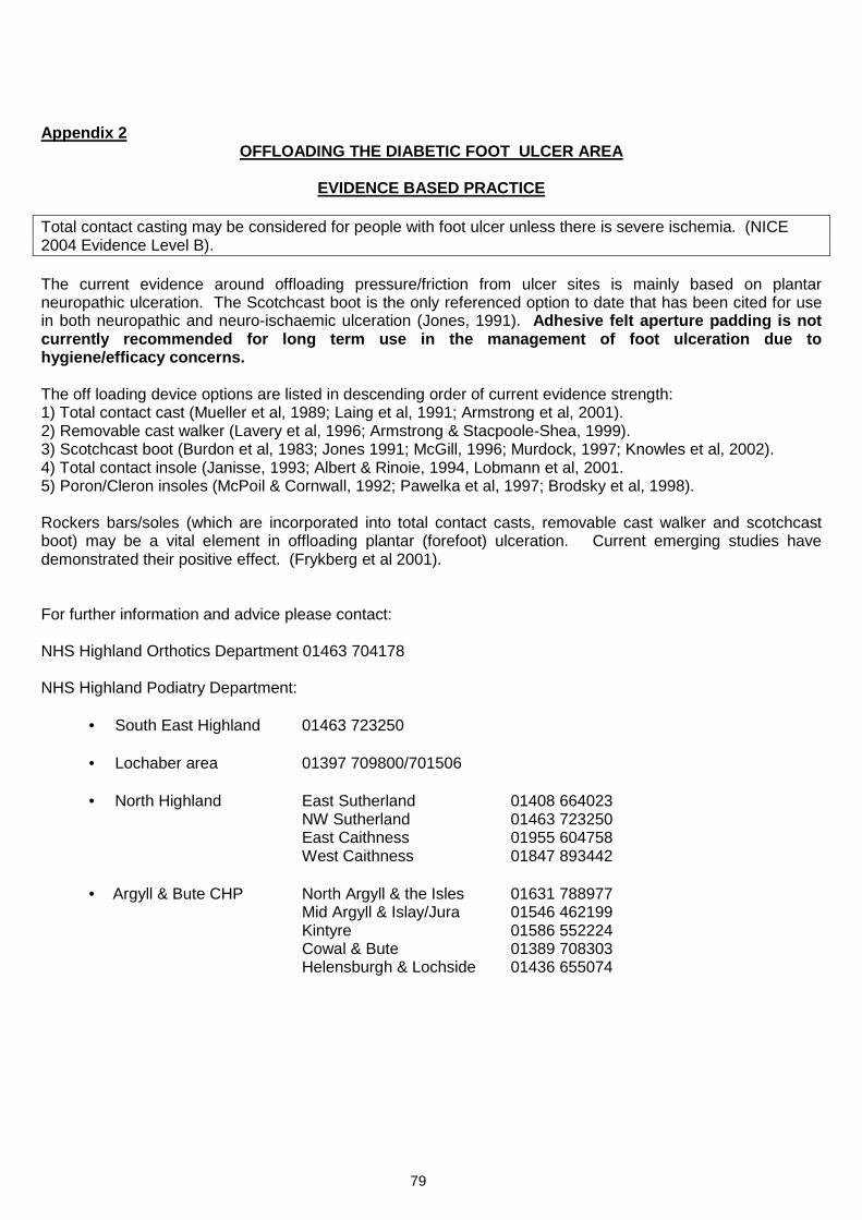
79
Appendix 2
OFFLOADING THE DIABETIC FOOT ULCER AREA
EVIDENCE BASED PRACTICE Total contact casting may be considered for people with foot ulcer unless there is severe ischemia. (NICE 2004 Evidence Level B). The current evidence around offloading pressure/friction from ulcer sites is mainly based on plantar neuropathic ulceration. The Scotchcast boot is the only referenced option to date that has been cited for use in both neuropathic and neuro-ischaemic ulceration (Jones, 1991). Adhesive felt aperture padding is not currently recommended for long term use in the mana gement of foot ulceration due to hygiene/efficacy concerns. The off loading device options are listed in descending order of current evidence strength: 1) Total contact cast (Mueller et al, 1989; Laing et al, 1991; Armstrong et al, 2001). 2) Removable cast walker (Lavery et al, 1996; Armstrong & Stacpoole-Shea, 1999). 3) Scotchcast boot (Burdon et al, 1983; Jones 1991; McGill, 1996; Murdock, 1997; Knowles et al, 2002). 4) Total contact insole (Janisse, 1993; Albert & Rinoie, 1994, Lobmann et al, 2001. 5) Poron/Cleron insoles (McPoil & Cornwall, 1992; Pawelka et al, 1997; Brodsky et al, 1998). Rockers bars/soles (which are incorporated into total contact casts, removable cast walker and scotchcast boot) may be a vital element in offloading plantar (forefoot) ulceration. Current emerging studies have demonstrated their positive effect. (Frykberg et al 2001). For further information and advice please contact: NHS Highland Orthotics Department 01463 704178 NHS Highland Podiatry Department:
• South East Highland 01463 723250
• Lochaber area 01397 709800/701506
• North Highland East Sutherland 01408 664023 NW Sutherland 01463 723250 East Caithness 01955 604758 West Caithness 01847 893442
• Argyll & Bute CHP North Argyll & the Isles 01631 788977 Mid Argyll & Islay/Jura 01546 462199 Kintyre 01586 552224
Cowal & Bute 01389 708303 Helensburgh & Lochside 01436 655074
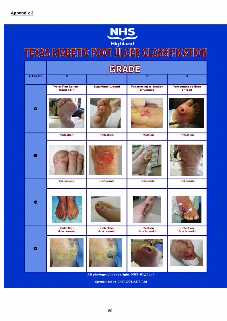
80
Appendix 3
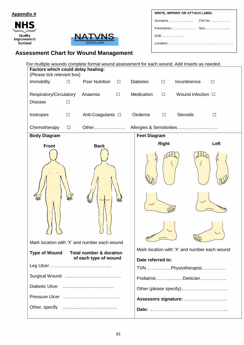
81
Appendix 4
Assessment Chart for Wound Management
For multiple wounds complete formal wound assessment for each wound. Add Inserts as needed. Factors which could delay healing: (Please tick relevant box)
Immobility □ Poor Nutrition □ Diabetes □ Incontinence □
Respiratory/Circulatory Anaemia □ Medication □ Wound Infection □ Disease □
Inotropes □ Anti-Coagulants □ Oedema □ Steroids □
Chemotherapy □ Other………………… Allergies & Sensitivities………………………
Body Diagram Front Back
Mark location with ‘X’ and number each wound Type of Wound Total number & duration of each type of wound
Leg Ulcer ………………………………….. Surgical Wound ………………………………. Diabetic Ulcer ………………………….… Pressure Ulcer ……………………………….. Other, specify ………………………………
Feet Diagram
Right Left
Mark location with ‘X’ and number each wound Date referred to:
TVN …………….Physiotherapist……………. Podiatrist………………Dietician……………... Other (please specify)…………………………. Assessors signature: ……………………….. Date: ………………………..…………………...
WRITE, IMPRINT OR ATTACH LABEL Surname ……………….... CHI No ….………….. Forenames ………………… Sex………………….. DoB ……………….. Location……………………………………………………
82
Formal Wound Assessment
Complete on initial assessment and thereafter compl ete at every dressing/wound review
CHI Number:
Date of Assessment
Number of wound
Analgesia required (Refer to local pain assessment tool)
Yes/No Yes/No Yes/No Yes/No Yes/No Yes/No Yes/No Yes/No
Regular/ongoing analgesia
Pre-dressing only
Wound Dimensions (enter size) Length (cm/mm) Width (cm/mm) Depth (cm/mm) Or trace wound circumference Is wound tracking/undermining Photography Tissue type on wound bed ( enter percentages) Necrotic (Black)
Sloughy (Yellow/Green)
Granulating (Red)
Epithelialising (Pink)
Hypergranulating (Red)
Haematoma
Bone/tendon
Wound exudate levels/ type (tick all relevant boxes ) Low Moderate High * Serous (Straw) Haemoserous (Red/Straw) Purulent (Green/Brown/Yellow)* Peri-wound skin (tick relevant boxes) Macerated (White) Oedematous * Erythema (Red)* Excoriated (Red) Fragile Dry/scaly Healthy/intact Signs of Infection * 1 or more of these signs may i ndicate possible infection Heat * New slough/necrosis(deteriorating wound bed)* Increasing pain* Increasing exudate* Increasing odour* Friable granulation tissue* Treatment objectives (tick relevant box) Debridement Absorption Hydration Protection Palliative / conservative Reduce bacterial load Assessors Print Initials Dressing Renewed (planned or unplanned dressing change)
Re-assessment date
83
Wound Treatment Plan and Evaluation of Care
To be completed when treatment undertaken.
Please write clearly
CHI Number
Date Wound Number
Cleansing Method, Dressing Choice & Rationale
Frequency
Evaluation & Rationale for changing dressing type
Signature
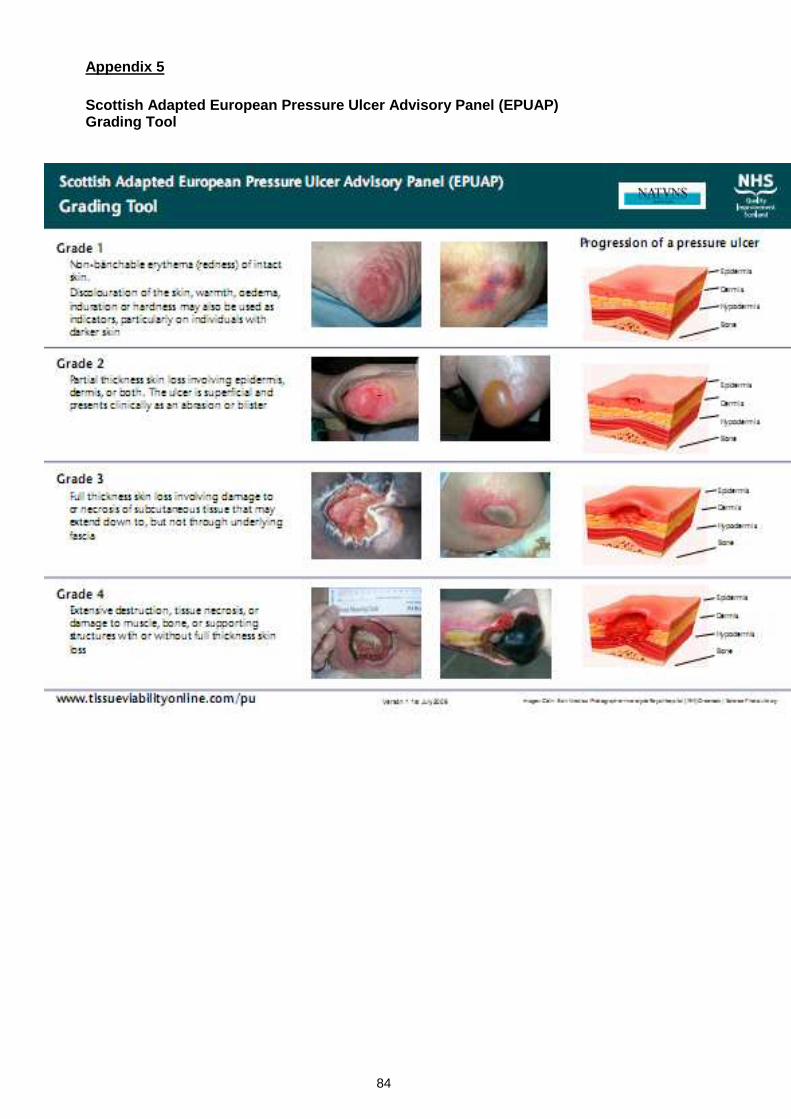
84
Appendix 5 Scottish Adapted European Pressure Ulcer Advisory Panel (EPUAP) Grading Tool
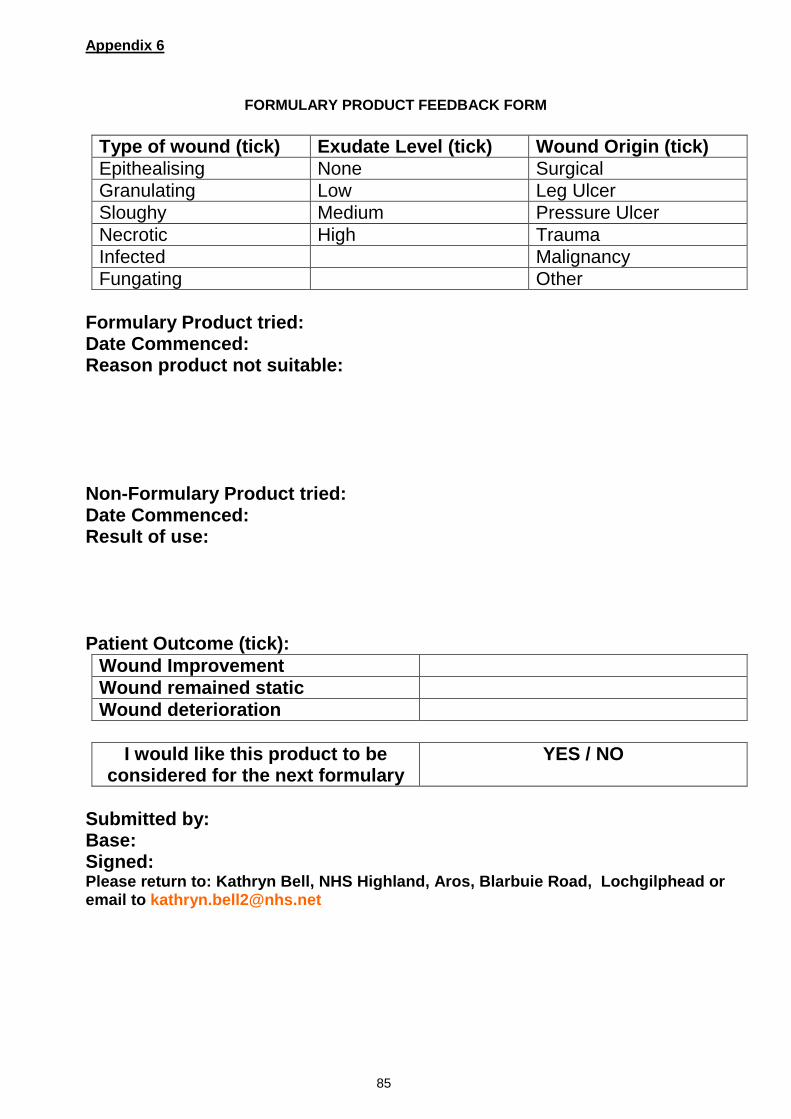
85
Appendix 6
FORMULARY PRODUCT FEEDBACK FORM
Type of wound (tick) Exudate Level (tick) Wound Ori gin (tick) Epithealising None Surgical Granulating Low Leg Ulcer Sloughy Medium Pressure Ulcer Necrotic High Trauma Infected Malignancy Fungating Other
Formulary Product tried: Date Commenced: Reason product not suitable: Non-Formulary Product tried: Date Commenced: Result of use: Patient Outcome (tick):
Wound Improvement Wound remained static Wound deterioration
I would like this product to be
considered for the next formulary YES / NO
Submitted by: Base: Signed: Please return to: Kathryn Bell, NHS Highland, Aros , Blarbuie Road, Lochgilphead or email to [email protected]
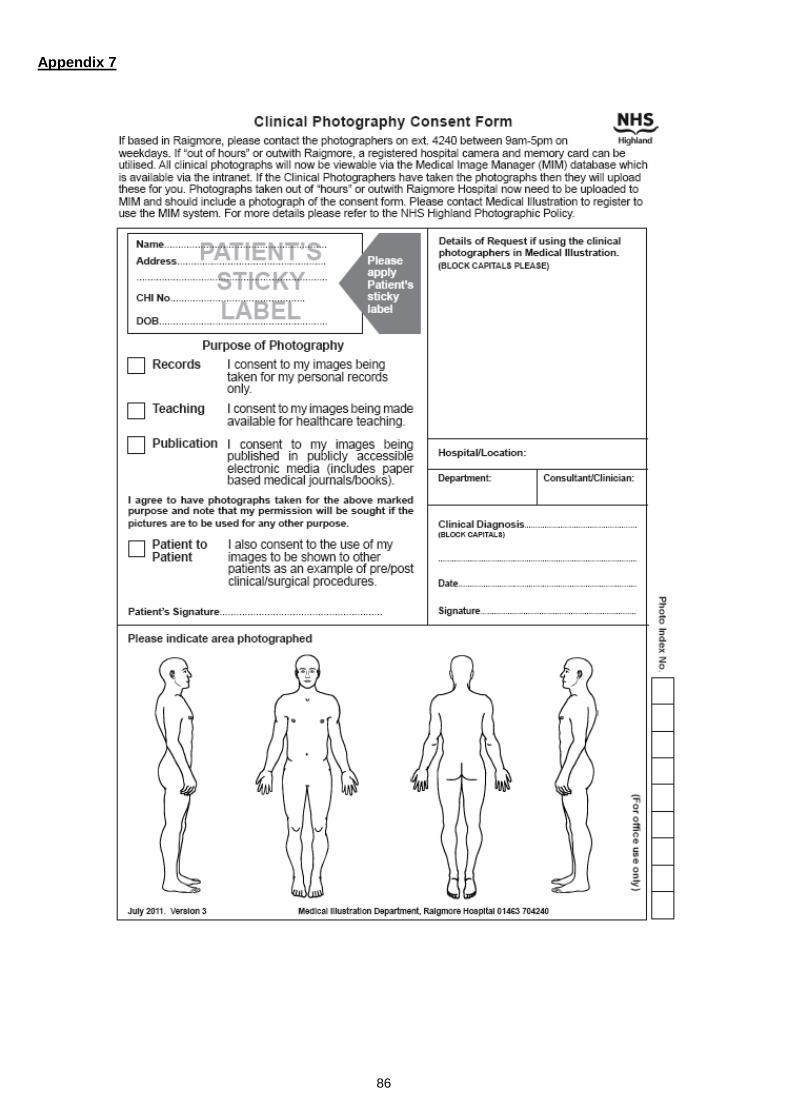
86
Appendix 7
87
SECTION SEVEN GLOSSARY OF TERMS
Ablation: Excision of a part of the body. Abscess: A local collection of necrotic tissue, bacteria and white cells known as pus. This collection of infection is retained within a wall formed of phagocytes and strands of fibrin Aerobic: This term relates to organisms which need oxygen to survive. Anaerobic: This term relates to organisms which derive their oxygen from the media in which they grow. Angiogenesis: The generation of new blood vessels initially seen at the base of a wound. Antibacterial: A substance which is used to inhibit the proliferation of pathogens. Antimicrobial: An agent that kills or inhibits the growth or replication of micro-organisms. Antiseptic: A substance that tends to inhibit the growth and reproduction of micro-organisms. Autolysis: The breakdown of devitalised tissue by leucocytes. Avulsion: The separation, by tearing, of any part of the body from the whole. Biofilm: A community of micro-organisms that adhere to each other on the surface of a wound, dental plaque is a biofilm. Callus: A common, usually painless, thickening of the epidermis at locations of external pressure or friction. Cellulitis: A diffuse, acute infection of the skin and subcutaneous tissue characterised most commonly by local heat, redness, pain and swelling. Debridement: Removal of necrotic or devitalised tissue from the wound surgically, chemically or by autolysis. Decubitus ulcer: A pressure sore. Dermabrasion: A treatment for the removal of superficial scars on the skin. Epithelialisation: The final stage of the proliferative phase in wound healing. Epithelial cells migrate across the surface of the wound, completing repair, but this is impaired if the surface is dry or necrotic. Eschar: The thick hard necrotic scab covering a wound. Granulation: The formation of new tissue filling the defect which takes place during the proliferative phase of healing. The name is derived from the fact that the buds of new tissue take on the appearance of small granules. Haemostasis: Arrest of haemorrhage. Hypoxia: Low oxygen concentrations in tissue. Inflammation: The immediate physiological response of the body to any injury or infection.
88
Ischaemia: Localised deficiency of the blood supply. Isotonic: A solution in which body cells can be bathed without a net flow of water across a semi- permeable cell membrane. Keratinocytes: An epithelial cell. They form 95% of epidermal cells and synthesise keratin. Maceration: A softening or sogginess of the tissue owing to retention of excessive moisture. This is seen on the skin immediately surrounding a wound. Maturation: The final stage of wound healing. The wound becomes less vascularised as there is a reduction in the need to bring cells to the wound site. The collagen fibres are re-organised. The scar tissue present is gradually remodelled and becomes comparable to normal tissue after a long period of time. In healthy individuals this stage begins approximately 20 days after injury and can last for many months or even years in complex wounds. Mitosis: The usual method of multiplication of cells by a specific process of division. Necrosis: Death of a portion of tissue. Neuropathy: Abnormal condition characterised by inflammation and degeneration of the peripheral nerves. Orthotist: A person who provides artificial or mechanical aids, to prevent or assist movement of weak or injured joints or muscles. Proteolytic (Proteolysis): The hydrolysis of proteins into simpler compounds by the action of enzymes. Protozoa: Minute animal of lowest and simplest class. Spicule: A small slender pointed structure. Wound contraction: A function of the healing process in granulating wounds whereby the edges of the wound are drawn towards each other.
Websites with further information: www.tissueviabilityonline.com www.worldwidewounds.com www.nes-hai.info
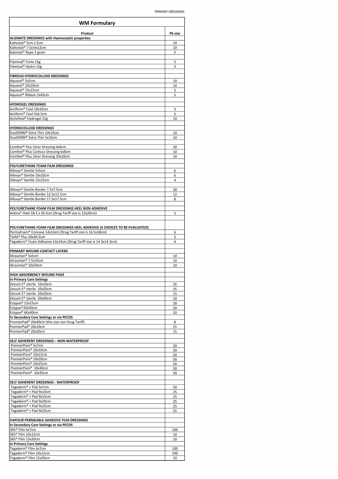
PRIMARY DRESSINGS
Product Pk size
ALGINATE DRESSINGS with Haemostatic properties
Kaltostat® 5cm x 5cm 10
Kaltostat® 7.5cmx12cm 10
Katostat® Rope 2 gram 5
Flaminal® Forte 15g 5
Flaminal® Hydro 15g 5
FIBROUS HYDROCOLLOID DRESSINGS
Aquacel® 5x5cm 10
Aquacel® 10x10cm 10
Aquacel® 15x15cm 5
Aquacel® Ribbon 2x45cm 5
HYDROGEL DRESSINGS
Actiform® Cool 10x10cm 5
Actiform® Cool 5x6.5cm 5
ActivHeal® Hydrogel 15g 10
HYDROCOLLOID DRESSINGS
DuoDERM® Extra Thin 10x10cm 10
DuoDERM® Extra Thin 5x10cm 10
Comfeel® Plus Ulcer Dressing 4x6cm 30
Comfeel® Plus Contour Dressing 6x8cm 10
Comfeel® Plus Ulcer Dressing 10x10cm 10
POLYURETHANE FOAM FILM DRESSINGS
Allevyn® Gentle 5x5cm 6
Allevyn® Gentle 10x10cm 6
Allevyn® Gentle 15x15cm 4
Allevyn® Gentle Border 7.5x7.5cm 30
Allevyn® Gentle Border 12.5x12.5cm 12
Allevyn® Gentle Border 17.5x17.5cm 8
POLYURETHANE FOAM FILM DRESSINGS HEEL NON ADHESIVE
Askina® Heel 18.5 x 20.5cm (Drug Tariff size is 12x20cm) 5
POLYURETHANE FOAM FILM DRESSINGS HEEL ADHESIVE (3 CHOICES TO BE EVALUATED)
PermaFoam® Concave 14x14cm (Drug Tariff size is 16.5x18cm) 6
Tielle® Plus 20x26.5cm 5
Tegaderm® Foam Adhesive 14x14cm (Drug Tariff size is 14.3x14.3cm) 4
PRIMARY WOUND CONTACT LAYERS
Atrauman® 5x5cm 10
Atrauman® 7.5x10cm 10
Atrauman® 10x20cm 10
HIGH ABSORBENCY WOUND PADS
In Primary Care Settings
Zetuvit E® sterile 10x10cm 25
Zetuvit E® sterile 10x20cm 25
Zetuvit E® sterile 20x20cm 15
Zetuvit E® sterile 20x40cm 10
Eclypse® 15x15cm 20
Eclypse®20x30cm 20
Eclypse® 60x40cm 10
In Secondary Care Settings or via PECOS
PremierPad® 20x40cm (this size non Drug Tariff) 8
PremierPad® 20x10cm 25
PremierPad® 20x20cm 15
SELF ADHERENT DRESSINGS – NON-WATERPROOF
PremierPore® 5x7cm 50
PremierPore® 10x10cm 50
PremierPore® 10x15cm 50
PremierPore® 10x20cm 50
PremierPore® 10x25cm 50
PremierPore® 10x30cm 50
PremierPore® 10x35cm 50
SELF ADHERENT DRESSINGS - WATERPROOF
Tegaderm® + Pad 5x7cm 50
Tegaderm® + Pad 9x10cm 25
Tegaderm® + Pad 9x15cm 25
Tegaderm® + Pad 9x20cm 25
Tegaderm® + Pad 9x25cm 25
Tegaderm® + Pad 9x35cm 25
VAPOUR-PERMEABLE ADHESIVE FILM DRESSINGS
In Secondary Care Settings or via PECOS
365® Film 6x7cm 100
365® Film 10x12cm 10
365® Film 15x20cm 10
In Primary Care Settings
Tegaderm® Film 6x7cm 100
Tegaderm® Film 10x12cm 100
Tegaderm® Film 15x20cm 10
WM Formulary
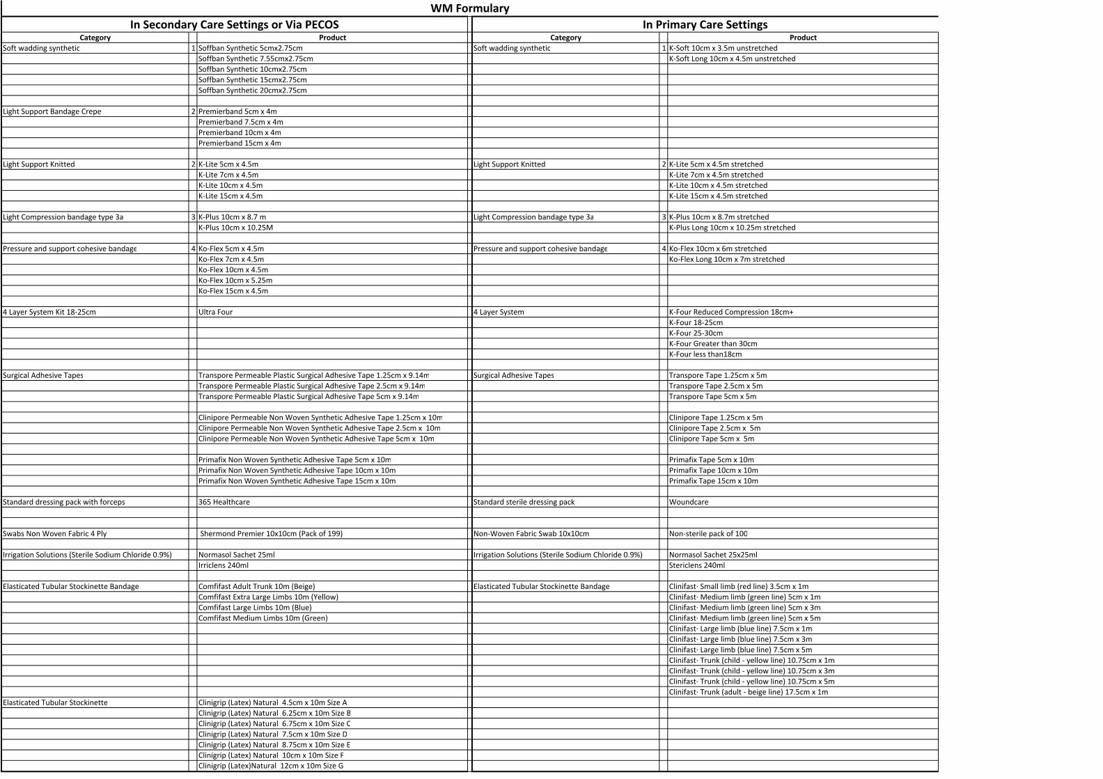
Category Product Category Product
Soft wadding synthetic 1 Soffban Synthetic 5cmx2.75cm Soft wadding synthetic 1 K-Soft 10cm x 3.5m unstretched
Soffban Synthetic 7.55cmx2.75cm K-Soft Long 10cm x 4.5m unstretched
Soffban Synthetic 10cmx2.75cm
Soffban Synthetic 15cmx2.75cm
Soffban Synthetic 20cmx2.75cm
Light Support Bandage Crepe 2 Premierband 5cm x 4m
Premierband 7.5cm x 4m
Premierband 10cm x 4m
Premierband 15cm x 4m
Light Support Knitted 2 K-Lite 5cm x 4.5m Light Support Knitted 2 K-Lite 5cm x 4.5m stretched
K-Lite 7cm x 4.5m K-Lite 7cm x 4.5m stretched
K-Lite 10cm x 4.5m K-Lite 10cm x 4.5m stretched
K-Lite 15cm x 4.5m K-Lite 15cm x 4.5m stretched
Light Compression bandage type 3a 3 K-Plus 10cm x 8.7 m Light Compression bandage type 3a 3 K-Plus 10cm x 8.7m stretched
K-Plus 10cm x 10.25M K-Plus Long 10cm x 10.25m stretched
Pressure and support cohesive bandage 4 Ko-Flex 5cm x 4.5m Pressure and support cohesive bandage 4 Ko-Flex 10cm x 6m stretched
Ko-Flex 7cm x 4.5m Ko-Flex Long 10cm x 7m stretched
Ko-Flex 10cm x 4.5m
Ko-Flex 10cm x 5.25m
Ko-Flex 15cm x 4.5m
4 Layer System Kit 18-25cm Ultra Four 4 Layer System K-Four Reduced Compression 18cm+
K-Four 18-25cm
K-Four 25-30cm
K-Four Greater than 30cm
K-Four less than18cm
Surgical Adhesive Tapes Transpore Permeable Plastic Surgical Adhesive Tape 1.25cm x 9.14m Surgical Adhesive Tapes Transpore Tape 1.25cm x 5m
Transpore Permeable Plastic Surgical Adhesive Tape 2.5cm x 9.14m Transpore Tape 2.5cm x 5m
Transpore Permeable Plastic Surgical Adhesive Tape 5cm x 9.14m Transpore Tape 5cm x 5m
Clinipore Permeable Non Woven Synthetic Adhesive Tape 1.25cm x 10m Clinipore Tape 1.25cm x 5m
Clinipore Permeable Non Woven Synthetic Adhesive Tape 2.5cm x 10m Clinipore Tape 2.5cm x 5m
Clinipore Permeable Non Woven Synthetic Adhesive Tape 5cm x 10m Clinipore Tape 5cm x 5m
Primafix Non Woven Synthetic Adhesive Tape 5cm x 10m Primafix Tape 5cm x 10m
Primafix Non Woven Synthetic Adhesive Tape 10cm x 10m Primafix Tape 10cm x 10m
Primafix Non Woven Synthetic Adhesive Tape 15cm x 10m Primafix Tape 15cm x 10m
Standard dressing pack with forceps 365 Healthcare Standard sterile dressing pack Woundcare
Swabs Non Woven Fabric 4 Ply Shermond Premier 10x10cm (Pack of 199) Non-Woven Fabric Swab 10x10cm Non-sterile pack of 100
Irrigation Solutions (Sterile Sodium Chloride 0.9%) Normasol Sachet 25ml Irrigation Solutions (Sterile Sodium Chloride 0.9%) Normasol Sachet 25x25ml
Irriclens 240ml Stericlens 240ml
Elasticated Tubular Stockinette Bandage Comfifast Adult Trunk 10m (Beige) Elasticated Tubular Stockinette Bandage Clinifast· Small limb (red line) 3.5cm x 1m
Comfifast Extra Large Limbs 10m (Yellow) Clinifast· Medium limb (green line) 5cm x 1m
Comfifast Large Limbs 10m (Blue) Clinifast· Medium limb (green line) 5cm x 3m
Comfifast Medium Limbs 10m (Green) Clinifast· Medium limb (green line) 5cm x 5m
Clinifast· Large limb (blue line) 7.5cm x 1m
Clinifast· Large limb (blue line) 7.5cm x 3m
Clinifast· Large limb (blue line) 7.5cm x 5m
Clinifast· Trunk (child - yellow line) 10.75cm x 1m
Clinifast· Trunk (child - yellow line) 10.75cm x 3m
Clinifast· Trunk (child - yellow line) 10.75cm x 5m
Clinifast· Trunk (adult - beige line) 17.5cm x 1m
Elasticated Tubular Stockinette Clinigrip (Latex) Natural 4.5cm x 10m Size A
Clinigrip (Latex) Natural 6.25cm x 10m Size B
Clinigrip (Latex) Natural 6.75cm x 10m Size C
Clinigrip (Latex) Natural 7.5cm x 10m Size D
Clinigrip (Latex) Natural 8.75cm x 10m Size E
Clinigrip (Latex) Natural 10cm x 10m Size F
Clinigrip (Latex)Natural 12cm x 10m Size G
WM Formulary
In Secondary Care Settings or Via PECOS In Primary Care Settings
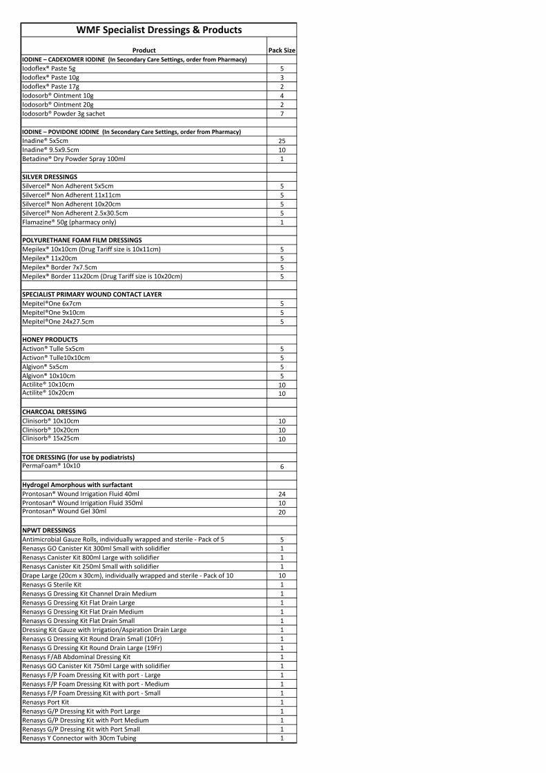
Product Pack Size
IODINE – CADEXOMER IODINE (In Secondary Care Settings, order from Pharmacy)
Iodoflex® Paste 5g 5
Iodoflex® Paste 10g 3
Iodoflex® Paste 17g 2
Iodosorb® Ointment 10g 4
Iodosorb® Ointment 20g 2
Iodosorb® Powder 3g sachet 7
IODINE – POVIDONE IODINE (In Secondary Care Settings, order from Pharmacy)
Inadine® 5x5cm 25
Inadine® 9.5x9.5cm 10
Betadine® Dry Powder Spray 100ml 1
SILVER DRESSINGS
Silvercel® Non Adherent 5x5cm 5
Silvercel® Non Adherent 11x11cm 5
Silvercel® Non Adherent 10x20cm 5
Silvercel® Non Adherent 2.5x30.5cm 5
Flamazine® 50g (pharmacy only) 1
POLYURETHANE FOAM FILM DRESSINGS
Mepilex® 10x10cm (Drug Tariff size is 10x11cm) 5
Mepilex® 11x20cm 5
Mepilex® Border 7x7.5cm 5
Mepilex® Border 11x20cm (Drug Tariff size is 10x20cm) 5
SPECIALIST PRIMARY WOUND CONTACT LAYER
Mepitel®One 6x7cm 5
Mepitel®One 9x10cm 5
Mepitel®One 24x27.5cm 5
HONEY PRODUCTS
Activon® Tulle 5x5cm 5
Activon® Tulle10x10cm 5
Algivon® 5x5cm 5
Algivon® 10x10cm 5
Actilite® 10x10cm 10Actilite® 10x20cm 10
CHARCOAL DRESSING
Clinisorb® 10x10cm 10
Clinisorb® 10x20cm 10
Clinisorb® 15x25cm 10
TOE DRESSING (for use by podiatrists)
PermaFoam® 10x10 6
Hydrogel Amorphous with surfactant
Prontosan® Wound Irrigation Fluid 40ml 24
Prontosan® Wound Irrigation Fluid 350ml 10
Prontosan® Wound Gel 30ml 20
NPWT DRESSINGS
Antimicrobial Gauze Rolls, individually wrapped and sterile - Pack of 5 5
Renasys GO Canister Kit 300ml Small with solidifier 1
Renasys Canister Kit 800ml Large with solidifier 1
Renasys Canister Kit 250ml Small with solidifier 1
Drape Large (20cm x 30cm), individually wrapped and sterile - Pack of 10 10
Renasys G Sterile Kit 1
Renasys G Dressing Kit Channel Drain Medium 1
Renasys G Dressing Kit Flat Drain Large 1
Renasys G Dressing Kit Flat Drain Medium 1
Renasys G Dressing Kit Flat Drain Small 1
Dressing Kit Gauze with Irrigation/Aspiration Drain Large 1
Renasys G Dressing Kit Round Drain Small (10Fr) 1
Renasys G Dressing Kit Round Drain Large (19Fr) 1
Renasys F/AB Abdominal Dressing Kit 1
Renasys GO Canister Kit 750ml Large with solidifier 1
Renasys F/P Foam Dressing Kit with port - Large 1
Renasys F/P Foam Dressing Kit with port - Medium 1
Renasys F/P Foam Dressing Kit with port - Small 1
Renasys Port Kit 1
Renasys G/P Dressing Kit with Port Large 1
Renasys G/P Dressing Kit with Port Medium 1
Renasys G/P Dressing Kit with Port Small 1
Renasys Y Connector with 30cm Tubing 1
WMF Specialist Dressings & Products