Embed Size (px)
Citation preview

t h e s u r g e on 9 ( 2 0 1 1 ) 2 4 5e2 4 8
avai lable at www.sciencedirect .com
The Surgeon, Journal of the Royal Collegesof Surgeons of Edinburgh and Ireland
www.thesurgeon.net
Wound infection after reconstructive arterial surgeryof the lower limbs: Risk factors and consequences
Rachel O’Brien, Nicholas Pocock, Francesco Torella*
Department of Vascular Surgery, University Hospital Aintree, Lower Lane, Liverpool, L9 7AL, United Kingdom
a r t i c l e i n f o
Article history:
Received 8 September 2010
Received in revised form
7 October 2010
Accepted 9 October 2010
Available online 13 November 2010
Keywords:
Wound infection
Peripheral vascular disease
Vein grafts
* Corresponding author. Tel.: þ44 151 529 49E-mail address: [email protected] (F. Tor
1479-666X/$ e see front matter ª 2010 RoyalSurgeons in Ireland. Published by Elsevier Ldoi:10.1016/j.surge.2010.10.005
a b s t r a c t
Objective: To identify predictors, and clinical consequences, of postoperative wound
infection after peripheral vascular surgery.
Materials and methods: Retrospective cohort study. Potential predictors of wound infection
were sought among patient related factors and procedure related factors. Patient outcome
was then analysed according to the incidence of wound infection.
Results: Following 209 procedures, 20 (9.6%) patients suffered a wound infection. On
univariate analysis, infrainguinal surgery, use of vein graft, and tissue loss were associated
with wound infection. On multivariate regression, however, only the association with use
of a vein graft remained (OR ¼ 4.2; 95%CI ¼ 1.2e14.1; P ¼ .022): the incidence of wound
infection was 16/96 (17%; 95%CI ¼ 9.2e24.1) when a vein graft was used and 4/113 (3.5%;
95%CI ¼ .1e7%) when a prosthetic or no graft was used. Wound infection was associated
with increased mortality (4/20 versus 9/189; P ¼ .025) but not limb loss (2/20 versus 7/189;
P ¼ .208). Median (IQR) postoperative hospital stay in patients with wound infection was 22
(15e45) days and 8 (5e15) days in those without wound infection (P ¼ .001).
Conclusions: In lower limb arterial surgery, wound infection is associated with the use of
vein grafts, and results in delayed recovery and increased mortality. Measures to reduce
wound infection should be focussed on such patients.
ª 2010 Royal College of Surgeons of Edinburgh (Scottish charity number SC005317) and
Royal College of Surgeons in Ireland. Published by Elsevier Ltd. All rights reserved.
Introduction used.3 Among patients undergoing lower limb arterial surgery,
Surgical wounds can be classified according to the likelihood
of bacterial contamination and, consequently, of wound
infection (WI). A clean wound implies lack of infection within
the surgical field, no entry into the respiratory, urogenital or
gastrointestinal tracts, and absence of external contamina-
tion: in these circumstances, the risk of WI is very low.1
Surgery on the arterial tree of the lower limbs is an excep-
tion, with a significantly higher incidence of WI than the
average clean wound,2 so much so that routine antibiotic
prophylaxis is indicated even when prosthetic material is not
57; fax: þ44 151 529 6457.ella).College of Surgeons of Ed
td. All rights reserved.
increasedWI risk has been linked with several factors such as
sex, diabetes, presence of tissue loss, previous surgery,
surgery on the lower extremities, use of vein grafts and groin
incision, with little agreement among published studies.4e8
Presently, it is thus very difficult to identify, preoperatively,
high risk patients.
The principal aim of this studywas to identify preoperative
risk factors for WI in patients undergoing reconstructive
arterial surgery of the lower limbs. The secondary aim was to
detect the potential influence of WI on overall patient
outcome, measured by mortality, limb loss and hospital stay.
inburgh (Scottish charity number SC005317) and Royal College of
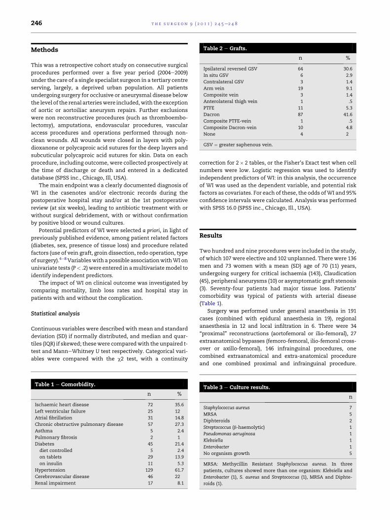
Table 2 e Grafts.
n %
Ipsilateral reversed GSV 64 30.6
In situ GSV 6 2.9
Contralateral GSV 3 1.4
Arm vein 19 9.1
Composite vein 3 1.4
Anterolateral thigh vein 1 .5
PTFE 11 5.3
Dacron 87 41.6
Composite PTFE-vein 1 .5
Composite Dacron-vein 10 4.8
None 4 2
GSV ¼ greater saphenous vein.
t h e s u r g e on 9 ( 2 0 1 1 ) 2 4 5e2 4 8246
Methods
This was a retrospective cohort study on consecutive surgical
procedures performed over a five year period (2004e2009)
under the care of a single specialist surgeon in a tertiary centre
serving, largely, a deprived urban population. All patients
undergoing surgery for occlusive or aneurysmal disease below
the level of the renal arterieswere included,with the exception
of aortic or aortoiliac aneurysm repairs. Further exclusions
were non reconstructive procedures (such as thromboembo-
lectomy), amputations, endovascular procedures, vascular
access procedures and operations performed through non-
clean wounds. All wounds were closed in layers with poly-
dioxanone or polycaproic acid sutures for the deep layers and
subcuticular polycaproic acid sutures for skin. Data on each
procedure, including outcome, were collected prospectively at
the time of discharge or death and entered in a dedicated
database (SPSS inc., Chicago, Ill, USA).
The main endpoint was a clearly documented diagnosis of
WI in the casenotes and/or electronic records during the
postoperative hospital stay and/or at the 1st postoperative
review (at six weeks), leading to antibiotic treatment with or
without surgical debridement, with or without confirmation
by positive blood or wound cultures.
Potential predictors of WI were selected a priori, in light of
previously published evidence, among patient related factors
(diabetes, sex, presence of tissue loss) and procedure related
factors (use of vein graft, groin dissection, redo operation, type
of surgery).4e8 Variableswith a possible associationwithWI on
univariate tests (P< .2) were entered in amultivariatemodel to
identify independent predictors.
The impact of WI on clinical outcome was investigated by
comparing mortality, limb loss rates and hospital stay in
patients with and without the complication.
Statistical analysis
Continuous variables were describedwithmean and standard
deviation (SD) if normally distributed, and median and quar-
tiles (IQR) if skewed; thesewere comparedwith the unpaired t-
test and ManneWhitney U test respectively. Categorical vari-
ables were compared with the c2 test, with a continuity
Table 1 e Comorbidity.
n %
Ischaemic heart disease 72 35.6
Left ventricular failure 25 12
Atrial fibrillation 31 14.8
Chronic obstructive pulmonary disease 57 27.3
Asthma 5 2.4
Pulmonary fibrosis 2 1
Diabetes 45 21.4
diet controlled 5 2.4
on tablets 29 13.9
on insulin 11 5.3
Hypertension 129 61.7
Cerebrovascular disease 46 22
Renal impairment 17 8.1
correction for 2� 2 tables, or the Fisher’s Exact test when cell
numbers were low. Logistic regression was used to identify
independent predictors of WI: in this analysis, the occurrence
of WI was used as the dependent variable, and potential risk
factors as covariates. For each of these, the odds ofWI and 95%
confidence intervals were calculated. Analysis was performed
with SPSS 16.0 (SPSS inc., Chicago, Ill., USA).
Results
Two hundred and nine procedures were included in the study,
of which 107 were elective and 102 unplanned. Therewere 136
men and 73 women with a mean (SD) age of 70 (11) years,
undergoing surgery for critical ischaemia (143), Claudication
(45), peripheral aneurysms (10) or asymptomatic graft stenosis
(3). Seventy-four patients had major tissue loss. Patients’
comorbidity was typical of patients with arterial disease
(Table 1).
Surgery was performed under general anaesthesia in 191
cases (combined with epidural anaesthesia in 19), regional
anaesthesia in 12 and local infiltration in 6. There were 34
“proximal” reconstructions (aortofemoral or ilio-femoral), 27
extraanatomical bypasses (femoro-femoral, ilio-femoral cross-
over or axillo-femoral), 146 infrainguinal procedures, one
combined extraanatomical and extra-anatomical procedure
and one combined proximal and infrainguinal procedure.
Table 3 e Culture results.
n
Staphylococcus aureus 7
MRSA 5
Diphteroids 2
Streptococcus (b-haemolytic) 1
Pseudomonas aeruginosa 1
Klebsiella 1
Enterobacter 1
No organism growth 5
MRSA: Methycillin Resistant Staphylococcus aureus. In three
patients, cultures showed more than one organism: Klebsiella and
Enterobacter (1), S. aureus and Streptococcus (1), MRSA and Diphte-
roids (1).

Table 4 e Factors associated with wound infection onunivariate analysis.
n WI incidencen (%)
P
Patient related factors
Diabetes 45 4 (8.9) 1
Tissue loss 74 11 (14.9) .093
Female sex 73 6 (8.2) .623
Procedure related factors
Groin incision 193 20 (10.3) .374
Redo procedure 42 6 (14.3) .385
Use of vein graft 96 16 (16.7) .003
Infrainguinal surgery 162 19 (11.7) .051
t h e s u r g e on 9 ( 2 0 1 1 ) 2 4 5e2 4 8 247
Seventy patients hadundergone previous vascular surgery, and
42underwent “re-do” procedures, definedas surgery performed
through a previous scar. In 20 cases, patients underwent foot
debridement and/or minor amputation at the same time of the
arterial reconstruction and, in all but 16, surgery involved
a groin incision. Autologous or prosthetic graftswere used in all
but four procedures (Table 2). All patients received antibiotic
prophylaxis with a single dose of a second generation cepha-
losporin andmetronidazole (or with a penicillin plus clavulanic
acid) at induction, repeated in case of prolonged surgery (>3 h)
or large blood loss (>1 L, estimated).
Wound infections and clinical outcome
Twenty patients suffered a WI (9.6%; 95%CI ¼ 5.58e13.56),
which was caused by staphylococci in more than half of the
cases (Table 3). One of these patients also had the only early
graft infection by Staphylococcus aureus (Dacron graft). Univar-
iate analyses suggested that infrainguinal surgery, use of vein
grafts, and tissue loss had a potential association with the
occurrence of WI (Table 4). On multivariate analysis however,
the only variablewith a significant associationwithWIwas the
use of vein grafts (OR ¼ 4.2; 95%CI ¼ 1.2e14.1; P ¼ .022): the
incidence of WI was 16/96 (17%; 95%CI ¼ 9.2e24.1) in proce-
dures where a vein graft was used and 4/113 (3.5%; 95%
CI ¼ .1e7%) in those where prosthetic or no graft was used.
Among those patientswith vein graftswho suffered aWI, nine
had ipsilateral greater saphenous vein grafts (1 in in situ, eight
reversed), three had arm vein grafts, two had composite vein
grafts and one had a composite Dacron-vein graft. Tissue loss
(OR ¼ 1.9; 95%CI ¼ .74e5.1, P ¼ .176) and infrainguinal surgery
(OR ¼ 2.1; 95%CI ¼ .23e19, P ¼ .519) did not predict WI.
Patients who suffered a WI had a significantly higher
mortality (4/20 versus 9/189; P ¼ .025) and their median (IQR)
hospital stay was 22 (15e45) days, much longer than in
patients without this complication, at 8 (5e15) days (P ¼ .001).
However,WI did not appear to increase the risk of limb loss (2/
20 versus 7/189, P ¼ .208).
Discussion and conclusions
Our study confirms that patients undergoing surgery for
peripheral arterial disease are at high risk ofWI; however, this
risk appears to be largely confined to those receiving vein
grafts, as the incidence of WI after reconstructions with pros-
thetic (or no) grafts was compatible with that of other clean
surgical procedures.1 If confirmed, this finding would prove
invaluable to vascular surgeonsandpatients in several areasof
clinical management such as the informed consent process
and the choice of most appropriate treatment modality. It
would also provide a “target” populationwith a high incidence
of this complication. As previously suggested,4 the study also
showed that patients with WI have a higher mortality than
those without the complication, and that they have a signifi-
cantly longer postoperative hospital stay, thus indicating that
a reduction inWI rates would be desirable both on clinical and
financial grounds, to allow reallocation of hospital resources.
In agreement with other authors,2 we found that the
majority of WIs were caused by staphylococci, which are often
carried by thepatient. As carriers of staphylococci have ahigher
risk of wound infection,9 future attempts at decreasing WI
infection rates in arterial surgery should be directed towards
these micro-organisms.
Our findings arenot in complete agreementwithpreviously
published literature. Available evidence, however, is found in
a very heterogeneous group of studies, investigating different
variables, using various definitions of WI, including diverse
groups of patients in different settings.2e8 Furthermore,
several studies included patients treated over a decade ago, at
a time when clinical practice would have been different from
current standards2,5,7: such heterogeneicity makes interpre-
tation of the findings difficult. In particular, it may seem
counterintuitive that WI may occur more frequently when
autologous tissue is used. In general, however, vein harvest
increases the number of wounds, prolongs operative time and
interrupts larger numbers of subcutaneous lymphatic vessels,
all potential causes of increased risk of WI; these possible
associations cannot, obviously, be confirmed by our study.
The strength of our findings is mitigated by several factors,
one being the inclusiveness of the analysis. Our data were not
collected specifically for this study, therefore some variables
with potential associationwithWI (for example obesity7) were
not included. Some postoperative factors (for example, the
occurrence of wound haematoma2) have also been linkedwith
WI, but we chose to investigate only preoperative variables, as
only these can be used preoperatively as predictors. The
retrospective nature of the study and its setting (single
surgeon’s practice, specialist unit, urban hospital) are further
limitations, the latter suggesting that our results may not
necessarily be reproducible elsewhere. Routine preoperative
screening for Staphylococcus aureus and MRSA would also have
added valuable information but, unfortunately, this was not
carried out throughout the study period.
With the exception of prophylactic antibiotic therapy,
there is no single measure proven to decrease WI rates in
peripheral arterial surgery. Several interventions have been
investigated by randomised controlled trials in other settings,
including preoperative eradication of nasal Staphylococci,9,10
high flow oxygen11 and skin preparation with chlorhexidine
in alcohol,12 with mixed results. Further studies in arterial
surgery are obviously necessary, regardless of the type of
grafts used. Our results, however, suggest that the potential

t h e s u r g e on 9 ( 2 0 1 1 ) 2 4 5e2 4 8248
effect of any positive intervention would be easier to
demonstrate in patients receiving vein grafts.
r e f e r e n c e s
1. Culver DH, Horan TC, Gaynes RP, Martone WJ, Jarvis WR,Emori TG, et al. Surgical wound infection rates by woundclass, operative procedure, and patient risk index. Am J Med1991;91:152Se7S. Natl Nosocomial Infections Surveill System.
2. Lee ES, Santilli SM, Olson MM, Kuskowski MA, Lee JT. Woundinfection after infrainguinal bypass operations: multivariateanalysis of putative risk factors. Surg Infect 2000;1:257e63.
3. Stewart AH, Eyers PS, Earnshaw JJ. Prevention of infection inperipheral arterial reconstruction: a systemiatic review andmeta-analysis. J Vasc Surg 2007;46:148e55.
4. Nguyen LL, Brahmanandam S, Bandyk DF, Belkin M,Clowes AW, Moneta GL, et al. Female gender and oralanticoagulants are associated with wound complications inlower extremity vein bypass: Analysis 140 Operations forCritical Limb Ischemia. J Vasc Surg 2007;46:1191e2119.
5. Richet HM, Chidiac C, Prat A, Pol A, David M, Maccario M, et al.Analysis of risk factors for surgical wound infectionsfollowing vascular surgery. Am J Med 1991;91:170Se2S.
6. Ploeg A, Lange C, Lardenoye JW, Breslau P. Nosocomialinfections after peripheral arterial bypass surgery. World JSurg 2007;31:1687e92.
7. Chang JK, Calligaro KD, Ryan S, Runyan D, Dougherty MJ,Stern JJ. Risk factors associated with infection of lowerextremity revascularization: analysis of 365 proceduresperformed at a teaching hospital. Ann Vasc Surg 2003;17:91e6.
8. Bandyk DF. Vascular surgical site infection: risk factors andpreventive measures. Semin Vasc Surg 2008;21:119e23.
9. Perl TM, Cullen JJ, Wenzel RP, Zimmerman MB, Pfaller MA,Sheppard D, et al. Intranasal mupirocin to preventpostoperative Staphylococcus Aureus infections. N Engl J Med2002;346:1871 (MARS study team).
10. Bode LG, Kluytmans JA, Wertheim HF, Bogaers D,Vandenbroucke-Grauls CM, Roosendaal R, et al. Preventingsurgical-site infections in nasal carriers of StaphylococcusAureus. N Engl J Med 2010;362:9e17.
11. Qadan M, Akca O, Mahid SS, Hornung CA, Polk Jr HC.Perioperative supplemental oxygen therapy and surgical siteinfection: a meta-analysis of randomized controlled trials.Arch Surg 2009;144:359e66.
12. Darouiche RO, Wall Jr MJ, Itani KM, Otterson MF, Webb AL,Carrick MM, et al. Chlorhexidine-Alcohol versus Povidone-Iodine for surgical-site antisepsis. N Engl J Med 2010;362:18e26.