Embed Size (px)
Citation preview
Veterinary World, EISSN: 2231-0916 2109
Veterinary World, EISSN: 2231-0916Available at www.veterinaryworld.org/Vol.14/August-2021/17.pdf
RESEARCH ARTICLEOpen Access
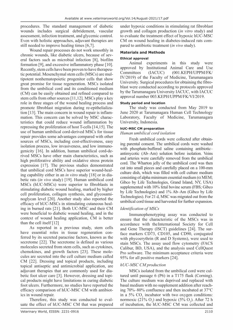
Wound healing potential of human umbilical cord mesenchymal stem cell conditioned medium: An in vitro and in vivo study
in diabetes-induced ratsSiufui Hendrawan1,2 , Yuyus Kusnadi3 , Christine Ayu Lagonda3 , Dilafitria Fauza3 , Jennifer Lheman2 , Erwin Budi2 ,
Brian Saputra Manurung2 , Hans Ulrich Baer4,5 and Sukmawati Tansil Tan6
1. Department of Biochemistry and Molecular Biology, Faculty of Medicine, Tarumanagara University, 11440, Jakarta, Indonesia; 2. Tarumanagara Human Cell Technology Laboratory, Tarumanagara University, 11440, Jakarta, Indonesia; 3. Stem Cell Division, Stem Cell and Cancer Institute, PT. Kalbe Farma, Tbk., 10510, Jakarta, Indonesia; 4. Baermed,
Centre of Abdominal Surgery, Hirslanden Clinic, 2501, Zürich, Switzerland; 5. Department of Visceral and Transplantation Surgery, University of Bern, 3012, Bern, Switzerland; 6. Department of Dermatovenereology, Faculty of Medicine,
Tarumanagara University, 11440, Jakarta, Indonesia.Corresponding author: Siufui Hendrawan, e-mail: [email protected]
Co-authors: YK: [email protected], CAL: [email protected], DF: [email protected], JL: [email protected], EB: [email protected], BSM: [email protected],
HUB: [email protected], STT: [email protected]: 01-03-2021, Accepted: 08-07-2021, Published online: 17-08-2021
doi: www.doi.org/10.14202/vetworld.2021.2109-2117 How to cite this article: Hendrawan S, Kusnadi Y, Lagonda CA, Fauza D, Lheman J, Budi E, Manurung BS, Baer HU, Tan ST (2021) Wound healing potential of human umbilical cord mesenchymal stem cell conditioned medium: An in vitro and in vivo study in diabetes-induced rats, Veterinary World, 14(8): 2109-2117.
AbstractBackground and Aim: Human umbilical cord mesenchymal stem cells (hUC-MSCs) and its conditioned medium (CM) promote wound healing. This study investigated the wound healing potential of hUC-MSC CM in vitro and in vivo using diabetic animal models.
Materials and Methods: The CM from hUC-MSC CM prepared under hypoxic conditions (hypoxic hUC-MSC) was evaluated for stimulating rat fibroblast growth, collagen production (in vitro), and wound healing in animal models (in vivo). An excision wound on the dorsal side of the diabetes-induced rats was established, and the rats were randomly divided into non-treatment, antibiotic, and hypoxic hUC-MSC CM groups. The cell number of fibroblasts and collagen secretion was evaluated and compared among the groups in an in vitro study. By contrast, wound size reduction, width of re-epithelialization, and the collagen formation area were assessed and compared among the groups in an in vivo study.
Results: CM under hypoxic conditions contained a higher concentration of wound healing-related growth factors. Hypoxic hUC-MSC CM could facilitate fibroblast cell growth and collagen synthesis, although not significant compared with the control group. Re-epithelialization and collagen production were higher in the hUC-MSC CM group than in the antibiotic and non-treatment groups.
Conclusion: Hypoxic hUC-MSC CM possessed more positive effects on the wound healing process based on re-epithelialization and collagen formation than antibiotic treatment did.
Keywords: conditioned medium, diabetic induced rat, human umbilical cord mesenchymal stem cells, wound healing.
Introduction
Chronic wounds have become a global public health challenge. Unlike acute wounds, which heal without significant interventions, chronic wounds give major challenges to patients and doctors. There are varying etiologies of chronic wounds, and one of them is diabetes [1]. The prevalence of patients with diabetes worldwide is predicted to rise by 5.4% (300 million) by 2025 [2]. Ulcer in patients with diabe-tes is common, and diabetic foot ulcer in patients is a severe and complex case [3]. Diabetic foot ulcers
significantly influence the quality of life of patients, including limited and reduced mobility, diminished income, loss of job, and spending more to visit a phy-sician or clinic for care [4]. Diabetes influences many aspects of life in patients, such as health, social, and economy. In the economic aspect, the cost of manag-ing patients with diabetes with a lower extremity ulcer has a more economic burden than diabetic patients without ulcers [5]. Alternative solutions are needed to overcome the s evere impacts that arise from diabetes, especially diabetic-related wounds. A diabetic wound is associated with decreased peripheral blood flow. Impaired angiogenesis and neovascularization result in insufficient oxygen and nutrient supply for the cells, leading to further impaired healing. Healing defi-ciency of diabetic wounds can be attributed to other factors, including decreased production of growth fac-tors and reduced revascularization. A diabetic wound is challenging to treat and requires comprehensive
Copyright: Hendrawan, et al. Open Access. This article is distributed under the terms of the Creative Commons Attribution 4.0 International License (http://creativecommons.org/licenses/by/4.0/), which permits unrestricted use, distribution, and reproduction in any medium, provided you give appropriate credit to the original author(s) and the source, provide a link to the Creative Commons license, and indicate if changes were made. The Creative Commons Public Domain Dedication waiver (http://creativecommons.org/publicdomain/zero/1.0/) applies to the data made available in this article, unless otherwise stated.
Veterinary World, EISSN: 2231-0916 2110
Available at www.veterinaryworld.org/Vol.14/August-2021/17.pdf
procedures. The standard management of diabetic wounds includes surgical debridement, vascular assessment, infection treatment, and glycemic control. Even with holistic approaches, adjuvant therapies are still needed to improve healing times [6,7].
Wound repair processes do not work smoothly in chronic wounds, like diabetic ulcers, because of sev-eral factors such as microbial infection [8], biofilm formation [9], and excessive inflammatory phase [10]. Recently, stem cells have been proven to have therapeu-tic potential. Mesenchymal stem cells (MSCs) are mul-tipotent nonhematopoietic progenitor cells that show great promise for tissue regeneration. MSCs isolated from the umbilical cord and its conditioned medium (CM) can be easily obtained and refined compared to stem cells from other sources [11,12]. MSCs play a key role in three stages of the wound healing process and promote fibroblast migration during re-epithelializa-tion [13]. The main concern in wound repair is inflam-mation. This concern can be solved by MSC charac-teristics that could reduce wound inflammation by repressing the proliferation of host T-cells [14,15]. The use of human umbilical cord-derived MSCs for tissue repair provides some advantages compared with other sources of MSCs, including cost-effectiveness, easy isolation process, low invasiveness, and low immuno-genicity [16]. In addition, human umbilical cord-de-rived MSCs have other main characteristics, such as high proliferative ability and oxidative stress protein expression [17]. The previous studies demonstrated that umbilical cord MSCs have superior wound-heal-ing capability either in an in vitro study [18] or in dia-betic rats (in vivo study) [19]. Human umbilical cord MSCs (hUC-MSCs) were superior to fibroblasts in stimulating diabetic wound healing, marked by higher cell proliferation, collagen synthesis, and glycosami-noglycan level [20]. Another study also reported the efficacy of hUC-MSCs in stimulating cutaneous heal-ing in burned rats [21]. Both UC-MSC and their CM were beneficial to diabetic wound healing, and in the context of wound healing application, CM is better than the cell itself [12].
As reported in a previous study, stem cells have essential roles in tissue regeneration con-ferred by its secreted paracrine factors, known as the secretome [22]. The secretome is defined as various molecules secreted from stem cells, such as cytokines, chemokines, and growth factors [23]. These mole-cules are secreted into the cell culture medium called CM [22]. Dressing and topical products, including topical antiseptic and antimicrobial application, are adjuvant therapies that are commonly used for dia-betic foot ulcer care [3]. However, dressing and topi-cal products might have limitations in curing diabetic foot ulcers. Furthermore, no studies have reported the efficacy comparison of hUC-MSC CM with antibiot-ics in wound repair.
Therefore, this study was conducted to eval-uate the effect of hUC-MSC CM that was prepared
under hypoxic conditions in stimulating rat fibroblast growth and collagen production (in vitro study) and to evaluate the treatment effect of hypoxic hUC-MSC CM on wound healing in diabetes-induced rats com-pared to antibiotic treatment (in vivo study).Materials and MethodsEthical approval
Animal experiments in this study were approved by Institutional Animal Care and Use Committees (IACUC) (001.KEPH/UPPM/FK/IV/2019) of the Faculty of Medicine, Tarumanagara University. Surgical procedures for obtaining the fibro-blast were conducted according to protocols approved by the Tarumanagara University IACUC, with IACUC approval number 003.KEPH/UPPM/FK/VI/2019. Study period and location
The study was conducted from May 2019 to June 2020 at Tarumanagara Human Cell Technology Laboratory, Faculty of Medicine, Tarumanagara University, Indonesia.hUC-MSC CM preparationHuman umbilical cord isolation
Fresh umbilical cords were collected after obtain-ing parental consent. The umbilical cords were washed with phosphate-buffered saline containing antibiotic–antimycotic (Ab–Am) solution before isolation. Veins and arteries were carefully removed from the umbilical cord. The Wharton jelly of the umbilical cord was then cut into small pieces and carefully placed on a 100-mm culture dish, which was filled with cell culture medium consisting of alpha minimum essential medium (α MEM; Gibco by Life Technologies, Grand Island, NY, USA) supplemented with 10% fetal bovine serum (FBS; Gibco by Life Technologies) and 1% Ab–Am (Gibco by Life Technologies). For 21 d, MSC was migrated out from the umbilical cord tissue and harvested for further expansion.
Identification of MSCsImmunophenotyping assay was conducted to
ensure that the characteristic of the MSCs was in accordance with the International Society for Cell and Gene Therapy (ISCT) guidelines [24]. The sur-face markers CD73, CD105, and CD90, conjugated with phycoerythrin (R and D Systems), were used to stain MSCs. The assay used flow cytometry (FACS Calibur, BD, USA), and the analysis used CellQuest Pro software. The minimum acceptance criteria were 95% for all positive markers [24].
hUC-MSC CM productionMSCs isolated from the umbilical cord were cul-
tured until passage 6 (P6) in a T175 flask (Corning). The culture medium was deprived and replaced with basal medium with no supplement addition after reach-ing 70%–80% confluence and then incubated at 37°C in a 5% CO2 incubator with two oxygen conditions: normoxic (21% O2) and hypoxic (5% O2). After 72 h of incubation, the hUC-MSC CM was collected and

Veterinary World, EISSN: 2231-0916 2111
Available at www.veterinaryworld.org/Vol.14/August-2021/17.pdf
stored in a deep freezer (−80°C) for long-term storage. hUC-MSCs were isolated and cultured at the Stem Cell and Cancer Institute Laboratory, Jakarta, Indonesia.
Quantification of basic fibroblast growth factor (bFGF), vascular endothelial growth factor (VEGF), and pro-collagen 1 as wound healing-related para-crine factors
Enzyme-linked immunosorbent assay (ELISA) was performed to quantify paracrine factors secreted by MSCs related to wound healing, such as bFGF, VEGF, and pro-collagen 1 (R and D Systems). The procedure was performed according to the manufac-turer’s instructions, and the analysis was performed using four-parameter logistic software.In vitro studyRat fibroblast cell growth
Primary rat dermal fibroblasts were used for the in vitro study. MSCs (passage 5–6) cultured under hypoxic conditions for 72 h were used to pro-duce the hUC-MSC CM. In addition, rat fibroblasts were obtained from donor rats. One male 12-week-old Sprague–Dawley rat (National Agency of Drug and Food Control, Jakarta) was used as a fibroblast donor. Fibroblasts were isolated from the rat’s skin as described previously [25]. Fibroblasts at a density of 5×10⁵ cells/well were seeded into 24-well plates (n=3) and then cultured in Dulbecco’s modified Eagle medium (DMEM; Gibco by Life Technologies) sup-plemented with 10% (v/v) FBS and 1 mL of penicillin/streptomycin (1×). hUC-MSC CM was added to the media culture with two concentrations, namely, 0.5% and 1%. As a control, the same number of fibroblasts was seeded into wells and cultivated in the media without adding the hUC-MSC CM. Cells were incu-bated at 37°C in 5% CO2 for 24 h. The cell number was analyzed using the Cell Counting Kit-8 (CCK-8) viability assay (Sigma Aldrich, USA) and measured in a Multiskan reader at a wavelength (λ) of 450 nm (Multiskan Ex, Thermo Scientific, USA).Analysis of collagen production on rat fibroblasts
Collagen production by fibroblasts was measured using rat type I collagen ELISA kit (Elabscience Biotechnology Inc., China). Fibroblasts (5×10⁵ cells/well) were seeded into 24-well plates, cultivated in Dulbecco’s modified eagle medium (Gibco) supplemented with 10% (v/v) FBS and 1 mL of penicillin/streptomycin (1×), and incubated at 37°C in 5% CO2 for 24 h. The procedure was performed according to the manufacturer’s instructions. The rat fibroblast collagen concentration was obtained using an ELISA reader at a wavelength (λ) of 450 nm.In vivo study
Rat skin-wound healing model and induction of dia-betes in rats
Nine adult male, 14-week-old Sprague–Dawley rats weighing 150-180 g were obtained from the
National Agency of Drug and Food Control, Jakarta, Indonesia. Before the experimental procedures, the animals were acclimatized to the laboratory con-ditions. They were allowed free access to standard laboratory food and water and housed individually in cages in a controlled environment (23°C±3°C, 30-70% humidity, and a 12:12 h light: dark cycle). For diabetes induction, the rats were rendered diabetic 1 week before treatment by a single-dose intraperi-toneal injection of streptozotocin (STZ) (50 mg/kg body weight; Selleck Chemicals). Blood glucose was checked 48 h post-injection, and blood samples were obtained from the tail vein of the animals for glucose assay. The rats were considered diabetic if three days after the STZ injection, the non-fasting blood glucose was more than 200 mg/dL and presented with at least 3 days of persistent hyperglycemia.
The diabetes-induced rats were randomized and divided into the following three treatment groups (n=3): Which were non-treatment (without any medication), antibiotics (topical medication with bactoderm mupirocin ointment 2%, Ikapharmindo Putramas, Indonesia), and hUC-MSC CM groups (intradermal injection with 0.5 mL of undiluted CM on a peripheral wound). For the experimental pro-cedure, the rats were weighed and anesthetized by intraperitoneal administration of ketamine 10% INJ (Kepro B.V, Holland; 40-80 mg/kg body weight) and x ylazine2% (Interchemie werken “De Adelaar” B.V. Metaalweg 8 Venray, Holland; 5-10 mg/kg body weight). Then, the hair on the backside was shaved and disinfected. The skin on the dorsal area of the rats was punctured using an 8-mm skin punch, with each rat having two ulcer wounds and receiving different treatments. After the procedure, wounds were closed with Tegaderm (3M Nexcare) and fixed with a suture at 3-4 points. Hypafix (Essity, Healthcare 21 Group, Ref. 71442-03) was added to secure the Tegaderm. Three wounds per treatment were analyzed in this study. The wound observations, photos, and measure-ments were conducted at three endpoints: 0, 7, and 14 d post-skin puncture. The percentage of wound clo-sure was calculated using the equation: percentage of wound closure (%) = (A0-At)/A0×100%, where A0 is the wound area at Day 0 and At is the wound area at 7 or 14 d post-skin puncture.Histopathological analysis
All Sprague-Dawley rats were euthanized at 14 d post-skin puncture. Skin specimens from the wound site were dissected and collected. Specimens were fixed in 10% formalin and sent to the Pathology and Anatomy Laboratory, Primate Research Center, Bogor Agricultural University, for further examina-tions, including re-epithelialization with hematoxylin and eosin (HE) staining and collagen formation area measurement with Masson’s trichrome staining. HE staining and MT staining were performed accord-ing to Sheehan and Hrapchak [26]. Tissue sections
Veterinary World, EISSN: 2231-0916 2112
Available at www.veterinaryworld.org/Vol.14/August-2021/17.pdf
were observed and captured using a Nikon Eclipse 80i microscope to evaluate re-epithelialization and collagen-deposited areas. Furthermore, the width of re-epithelialization and collagen-deposited areas was analyzed using the ImageJ software.Statistical analysis
All data were presented as mean±SD. The differ-ences between the two independent groups were ana-lyzed by Mann–Whitney and Kruskal–Wallis tests to analyze the differences among the three independent groups. Statistical analysis was conducted using SPSS v.23, with a 95% confidence interval. Values were considered statistically significant at p<0.05.ResultshUC-MSC characteristics
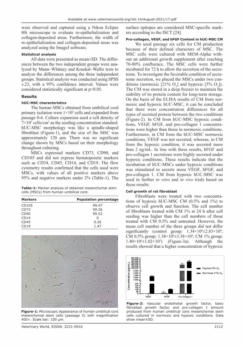
The human MSCs obtained from umbilical cord primary isolation were 1×10⁵ cells and expanded from passage 0-6. Culture expansion used a cell density of 7×10³ cells/cm2 as the seeding concentration standard. hUC-MSC morphology was like a spindle-shaped fibroblast (Figure-1), and the size of the MSC was approximately 120 μm. There was no significant change shown by MSCs based on their morphology throughout culturing.
MSCs expressed markers CD73, CD90, and CD105 and did not express hematopoietic markers such as CD34, CD45, CD14, and CD19. The flow cytometry results confirmed that the cells used were MSCs, with values of all positive markers above 95% and negative markers under 2% (Table-1). The
surface epitopes are considered MSC-specific mark-ers according to the ISCT [24].Pro-collagen, VEGF, and bFGF Content in hUC-MSC CM
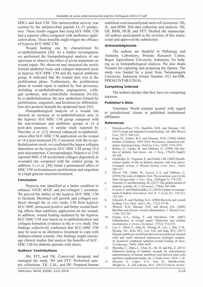
We used passage six cells for CM production because of their defined characters of MSC. The MSC cells were cultured with MEM-Alpha with-out an additional growth supplement after reaching 70-80% confluence. The MSC cells were further incubated for 72 h to allow the secretion of the secre-tome. To investigate the favorable condition of secre-tome secretion, we placed the MSCs under two con-ditions (normoxic [21% O2] and hypoxic [5% O2]). The CM was stored in a deep freezer to maintain the stability of its protein content for long-term storage. On the basis of the ELISA results of CM from nor-moxic and hypoxic hUC-MSC, it can be concluded that there were concentration differences for all types of secreted protein between the two conditions (Figure-2). In CM from hUC-MSC hypoxic condi-tions, VEGF, bFGF, and pro-collagen 1 concentra-tions were higher than those in normoxic conditions. Furthermore, in CM from the hUC-MSC normoxic conditions, VEGF was not secreted, whereas, in CM from the hypoxic condition, it was secreted more than 2 ng/mL. In line with these results, bFGF and pro-collagen 1 secretions were highly secreted under hypoxic conditions. These results indicate that the incubation of hUC-MSCs under hypoxic conditions was stimulated to secrete more VEGF, bFGF, and pro-collagen 1. CM from hypoxic hUC-MSC was used in further in vitro and in vivo trials based on these results.Cell growth of rat fibroblast
Fibroblasts were treated with two concentra-tions of hypoxic hUC-MSC CM (0.5% and 1%) to observe cell growth and function. The cell number of fibroblasts treated with CM 1% at 24 h after cell seeding was higher than the cell numbers of those treated with CM 0.5% and untreated. However, the mean cell number of the three groups did not differ significantly (control group: 1.34×10⁶±2.83×10⁴; CM 0.5% group: 1.38×10⁶±3.38×10⁴; CM 1% group: 1.40×10⁶±1.02×10⁵) (Figure-3a). Although the results showed that a higher concentration of hypoxic
Figure-1: Microscopic Appearance of human umbilical cord mesenchymal stem cells (passage 5) with magnification 400×. Scale bar: 100 μm.
Table-1: Marker analysis of obtained mesenchymal stem cells (MSCs) from human umbilical cord.
Markers Population percentage
CD105 99.47CD73 99.26CD90 99.52CD14 0CD45 0.26CD19 1.47
Figure-2: Vascular endothelial growth factor, basic fibroblast growth factor, and pro-collagen 1 amount produced from human umbilical cord mesenchymal stem cells cultured in normoxic and hypoxic conditions. Data show mean±SD.

Veterinary World, EISSN: 2231-0916 2113
Available at www.veterinaryworld.org/Vol.14/August-2021/17.pdf
hUC-MSC CM might better facilitate fibroblast growth, statistically, there was no significant differ-ence in the cell number of the fibroblast (Figure-3a). These results showed that adding a certain concentra-tion of CM did not significantly affect fibroblast cell growth.Collagen secretion
To examine the effect of adding hypoxic hUC-MSC CM against fibroblast function in produc-ing collagen, we also used two concentrations of CM. Hypoxic hUC-MSC CM (both 0.5% and 1%) showed improved collagen secretion than the control group did. However, the mean collagen concentra-tion was not significantly different between groups (control group: 1.99±0.91 ng/mL; CM 0.5% group: 2.55±0.47 ng/mL; CM 1% group: 1.89±0.23 ng/mL) (Figure-3b). Our study may indicate that collagen pro-duction did not strongly depend on CM, and the lower or higher concentration of CM did not significantly increase collagen production.Wound healing observation in diabetic ulcer
The wound healing process was observed by re-epithelialization and newly synthesized collagen. In addition, different responses were observed between the non-treatment, antibiotic, and hypoxic hUC-MSC CM groups. Our preliminary experiment showed that CM obtained from hypoxic hUC-MSC showed a superior effect in wound healing compared with that from normoxic hUC-MSC. On the basis of these find-ings, we treated the wound with CM of MSCs culti-vated under hypoxic conditions. As hypothesized, the hypoxic hUC-MSC CM demonstrated wound healing potential in diabetic animal models, both in mac-roscopic (Figure-4a) and microscopic observations (Figure-5).
At the macroscopic observation, the hypoxic hUC-MSC CM group demonstrated faster wound closure post-skin puncture. Figure-4b showed the highest wound closure percentage on wounds treated with hypoxic hUC-MSC CM (37.5%±0%) after 7 d post-skin puncture compared with the antibi-otic (20.83%±19.09%) and non-treatment groups (20.83%±19.09%). The hypoxic hUC-MSC CM treated wound showed better and faster wound clo-sure than that treated with an antibiotic and untreated
wound at 7 d post-skin puncture. The hypoxic hUC-MSC CM group had the same wound closure percentage (75%±12.5%) as that with the antibi-otic group (75%±0%) and had a higher wound clo-sure percentage than that with the non-treatment group (62.5%±12.5%) at 14 d post-skin puncture (Figure-4a). The wound closure percentage in wounds treated with hypoxic hUC-MSC CM was the same as that of the antibiotic group at 14 d, but still higher than that of the non-treatment group (Figure-4b).
The hypoxic hUC-MSC CM group exhibited the largest re-epithelialization area for microscopic observation compared with the antibiotic group (Figure-6a). Moreover, there was no re-epithelial-ization area found in the non-treatment group. In addition, the epithelium length (2806±338 μm) of the hypoxic UC-MSC CM group was 2.15-fold lon-ger than the epithelium length of the antibiotic group (1303±940 μm) (Figure-6a). These results were also supported by the width of the collagen forma-tion area data (Figure-6b). Correspondingly, the area of the newly generated collagen tissue in the hUC-MSC CM group (29.03×10⁴±60.99×104 μm2) was five-fold wider than that in the antibiotic group (57.93×10⁴±64.77×104 μm2) (Figure-6b). Altogether, these findings demonstrated that hypoxic hUC-MSC CM treatment showed a distinct effect to facilitate wound repair in our diabetic wound model.Discussion
This study analyzed the efficacy of hypoxic hUC-MSC CM on wound healing of diabetic rats. We also performed antibiotic treatment for comparison. First, we confirmed that our cells were MSCs based on morphology observation and supported by marker analysis results (Figures-1 and 2). To investigate the suitable conditions to produce CM, we observed two MSC-cultivated conditions (normoxic and hypoxic conditions). Interestingly, the production levels of VEGF, bFGF, and pro-collagen 1 were different. The hypoxic condition increased the production of the CM by MSCs. It has been reported that the mRNA level and protein expression of VEGF-A were higher in MSCs prepared under hypoxic conditions com-pared with normoxic conditions [27]. Another study by Page et al. [28] also reported that gene expression
Figure-3: (a) Cell number of fibroblasts treated with two different concentrations of conditioned medium (CM) and (b) collagen concentration secreted from fibroblast post-treated with human umbilical cord mesenchymal stem cells CM 0.5% and 1% concentrations (n=3). Data show mean±SD.
a b

Veterinary World, EISSN: 2231-0916 2114
Available at www.veterinaryworld.org/Vol.14/August-2021/17.pdf
and paracrine secretion are strongly affected by serum and oxygen concentration. Our study observed that there was no VEGF production by the MSCs under normoxic conditions. In contrast to our findings, Chen et al. [29] investigated that VEGF-A was still produced at a lower concentration under normoxic conditions by bone marrow MSCs than under hypoxic conditions. However, our result in bFGF was sup-ported by their findings. Meanwhile, we found that pro-collagen 1 was secreted in these two setup condi-tions, and their concentrations were slightly different. These data suggested that hypoxic conditions were preferable to stimulate the secretion of secretome/paracrine. Similar to our finding, Hsiao et al. [30] also demonstrated that hypoxic conditions favored VEGF-A and ANG production as angiogenic factors. Likewise, a previous study reported that VEGF and bFGF induce angiogenesis in wounded skin [31].
VEGF acts on wound repair through angiogenesis, collagen synthesis, and epithelialization [32].
To explore the effect of hypoxic hUC-MSC CM on fibroblast cell growth and collagen secretion in vitro, we treated fibroblast with hypoxic hUC-MSC CM with two concentrations (0.5% and 1%). Our findings showed that hypoxic hUC-MSC CM increased the number of fibroblasts, particularly in the group treated with a higher concentration (1%). Thus, it suggested that hypoxic hUC-MSC CM addition exerted a positive effect on fibroblast pro-liferation. Kim et al. [33] reported a similar result, and they investigated the highest growth of HDF, cell concentration, and protein level observed in the USC-CM group. They also evaluated the activ-ity of USC-MSC to induce migration of HDF com-pared with HDF and another MSC-CM group. Other studies also proved that the application of MSC-CM
Figure-4: (a) Wound closure measurement on diabetic induced rats (non-treatment, antibiotic and human umbilical cord mesenchymal stem cells conditioned medium groups) at three endpoints (0-, 7-, and 14-days). (b) Percentage of wound closure comparison between groups at 7- and 14-days post-skin puncture.
a
b

Veterinary World, EISSN: 2231-0916 2115
Available at www.veterinaryworld.org/Vol.14/August-2021/17.pdf
facilitated higher fibroblast proliferation than those in the control group [34].
Our findings showed that adding hypoxic hUC-MSC CM enhances collagen secretion, especially in fibroblasts treated with hypoxic hUC-MSC CM 0.5%. It may indicate that the collagen production did not strongly depend on the concentration of used hypoxic hUC-MSC CM. The previous studies by Kim et al. [33] demonstrated that the production of ECM (collagen, elastin, and fibronectin) was also trig-gered by USC-CM. Villanueva et al. [35] examined a lower collagen production from lung fibroblasts found in the control group (without CM addition) compared with adding endothelial cell-derived CM (1:10) at a 6-h incubation. At 24-h incubation, treatment with an increasing concentration of CM elevated the amount of collagen synthesized. As one of the extracellular matrix compositions in the skin, collagen plays a key role in growth and determines skin’s elasticity [36].
To investigate the therapeutic effect of hypoxic hUC-MSC CM in vivo, we provided an antibiotic group for comparison and the non-treatment as a control group. We chose topical antibiotic treatment because of its ability to inhibit or kill microbes on an open wound. Our results showed that the hypoxic hUC-MSC CM group had a beneficial effect on the wound healing process, which was revealed by mac-roscopic and microscopic observation. An expected outcome was a reduction of the wound surface area. Although not statistically significant, the hUC-MSC CM group demonstrated faster wound closure since observation at 7 d post-skin puncture. Non-significant results might be caused by the small sample size and the length of the observation time. The created wound site was reduced at 14 d post-wounding. The hypoxic hUC-MSC CM group had the same wound closure percentage as that of the antibiotic group and had a higher wound closure percentage than that of the non-treatment group at 14 d post-skin puncture. A study by Han et al. [19] showed a wound healing process that was significantly lower in diabetic rats than in normal rats at days 7 and 14. This phenomenon in their study showed that blockade of the Wnt signal-ing pathway slowed the healing of skin wounds in dia-betic rats. A study by Assi et al. [37] in mice showed that wounds treated with topical scaffolds contain-ing MSCs increased proliferation without increasing apoptosis and VEGF-positive cells. Another study by Zhou et al. [38] demonstrated that hUC-MSC CM/hydrogel promoted wound closure and constricted fibrotic and hypertrophic scar tissue formation.
The efficacy of antibiotics to facilitate wound healing was not as high as that triggered by hUC-MSC CM application but was better than the non-treatment group. Reports from Padeta et al. [34] demonstrated a similar finding to our results. They found that the MSC CM showed a prominent effect in promoting wound healing than bioplacenton application as their control group prepared from bovine placenta extract 10%, neomycin sulfate 0.5%, and gel base. Thus, besides the self-renewal capacity, human MSCs and their secre-tome may have antimicrobial activity. Our hypothe-sis was supported by Krasnodembskaya et al. [39], who observed the antimicrobial efficacy of human
Figure-6: (a) Length of epithelium and (b) width of collagen-deposited area comparison between non-treatment, antibiotic, and human umbilical cord mesenchymal stem cells conditioned medium group in diabetic induced rats at 14 days post skin punctures.
a b
Figure-5: Histopathological analysis of (a) re-epithelialization using Hematoxylin and Eosin staining and (b) collagen formation using Masson’s trichrome staining on the wound site of diabetic induced rats. Re-epithelialization and collagen formation areas are indicated with yellow lines. Scale bar 200 µm in all panels. The double-headed arrows point the edge of the scar.
a b
Veterinary World, EISSN: 2231-0916 2116
Available at www.veterinaryworld.org/Vol.14/August-2021/17.pdf
MSCs and their CM. This antimicrobial activity was exerted by the antimicrobial peptide LL-37 produc-tion. These results suggest that using hUC-MSC CM had a superior effect compared with antibiotics appli-cation alone. These benefits might trigger the efficacy of hypoxic hUC-MSC CM.
Wound healing can be characterized by re-epithelialization [40]. As a further investigation, we performed the histopathological analysis of our specimen to observe the effect of given treatments on wound repair. We observed and measured the newly formed epithelial tissue and collagen-deposited areas in hypoxic hUC-MSC CM and the topical antibiotic group. It indicated that the treated skin was at the proliferation phase. Furthermore, the proliferation phase in wound repair is marked by four processes, including re-epithelialization, angiogenesis, colla-gen synthesis, and extracellular formation [41,42]. In re-epithelialization, the new epithelium formed by proliferation, migration, and keratinocyte differentia-tion also protects beneath the epidermal layer [43].
Histopathological analysis of a wound site showed an increase in re-epithelialization area in the hypoxic hUC-MSC CM group compared with the non-treatment and antibiotic groups at 14 d post-skin puncture. A similar study reported by Shrestha et al. [12] showed enhanced re-epitheliali-zation after hUC-MSC CM application on the wound at 14 d post-treatment [9]. Consistent with the re-epi-thelialization result, we confirmed the largest collagen deposition on the hypoxic hUC-MSC CM group 14 d post-skin puncture. Correspondingly, Padeta et al. [34] reported MSC-CM accelerated collagen deposition in wounded rats compared with the control group. In addition, Li et al. [44] found a pronounced effect of MSC CM on keratinocyte proliferation and migration in a high-glucose microenvironment.Conclusion
Hypoxia was identified as a better condition to enhance VEGF, bFGF, and pro-collagen 1 secretion. We proved the ability of the hypoxic hUC-MSC CM to facilitate fibroblast cell growth and collagen syn-thesis through the in vitro study. CM from hypoxic hUC-MSC possessed positive and better wound heal-ing effects than antibiotic application on the wound. In addition, wound healing mediated by the hypoxic hUC-MSC CM was based on re-epithelialization and collagen formation evidence in the in vivo study. Our findings collectively confirmed that hUC-MSC CM may be used as an alternative treatment to cope with diabetes-related wounds. Our findings might encour-age clinical studies that analyze the benefits of hUC-MSC CM for diabetic patients with ulcers.Authors’ Contributions
SH, STT, and YK: Conceived, designed, and managed the study. SH and STT: Performed sam-ple collections. YK, CAL, and DF: Prepared human
umbilical cord mesenchymal stem cell secretome. SH, JL, and BSM: Did data collection and analysis. SH, EB, BSM, HUB, and STT: Drafted the manuscript. All authors participated in the revision of this manu-script and approved the submission. Acknowledgments
The authors are thankful to Pathology and Anatomy Laboratory, Primate Research Center, Bogor Agricultural University, Indonesia, for help-ing us in histopathological analysis. We also thank Nuraeni for capturing and preparing the figures. The study was funded by a grant from Tarumanagara University, Indonesia (Grant Number: 012 Int-DIR.PPKM/UNTAR/I/2018).Competing Interest
The authors declare that they have no competing interests.Publisher’s Note
Veterinary World remains neutral with regard to jurisdictional claims in published institutional affiliation.References1. Demidova-Rice, T.N., Hamblin, M.R. and Herman, I.M.
(2012) Acute and impaired wound healing. Adv. Skin Wound Care, 25(7): 304-314.
2. King, H., Aubert, R.E. and Herman, W.H. (1998) Global burden of diabetes, 1995-2025: Prevalence, numerical esti-mates, and projections. Diabetes Care, 21(9): 1414-1431.
3. Reiber, G., Lipsky, B. and Gibbons, G. (1998) The bur-den of diabetic foot ulcers. Am. J. Surg., 176(2A Suppl): 5S-10S.
4. Goodridge, D., Trepman, E. and Embil, J.M. (2005) Health-related quality of life in diabetic patients with foot ulcers: Literature review. J. Wound Ostomy Cont. Nurs., 32(6): 368-377.
5. Driver, V.R., Fabbi, M., Lavery, L.A. and Gibbons, G. (2010) The costs of diabetic foot: The economic case for the limb salvage team. J. Vasc. Surg., 52(Suppl 3): 17S-22S.
6. Schreml, S. and Berneburg, M. (2017) The global burden of diabetic wounds. Br. J. Dermatol., 176(4): 845-846.
7. Everett, E. and Mathioudakis, N. (2018) Update on manage-ment of diabetic foot ulcers. Ann. N. Y. Acad. Sci., 1411(1): 153-165.
8. Edwards, R. and Harding, K.G. (2004) Bacteria and wound healing. Curr. Opin. Infect. Dis., 17(2): 91-96.
9. Wolcott, R.D., Rhoads, D.D. and Dowd, S.E. (2008) Biofilms and chronic wound inflammation. J. Wound Care, 17(8): 333-341.
10. Eming, S.A., Krieg, T. and Davidson, J.M. (2007) Inflammation in wound repair: Molecular and cellular mechanisms. J. Invest. Dermatol., 127(3): 514-525.
11. Lee, C., Shim, S., Jang, H., Myung, H., Lee, J., Bae, C.H., Myung, J.K., Kim, M.J., Lee, S.B. and Jang, W.S. (2017) Human umbilical cord blood-derived mesenchymal stromal cells and small intestinal submucosa hydrogel compos-ite promotes combined radiation-wound healing of mice. Cytotherapy, 19(9): 1048-1059.
12. Shrestha, C., Zhao, L., Chen, K., He, H. and Mo, Z. (2013) Enhanced healing of diabetic wounds by subcutaneous administration of human umbilical cord derived stem cells and their conditioned media. Int. J. Endocrinol., 2014: 1-10.
13. Maxson, S., Lopez, E.A., Yoo, D., Danilkovitch-Miagkova, A. and LeRoux, M.A. (2012) Concise review:
Veterinary World, EISSN: 2231-0916 2117
Available at www.veterinaryworld.org/Vol.14/August-2021/17.pdf
Role of mesenchymal stem cells in wound repair. Stem Cells Transl. Med., 1(2): 142-149.
14. Nuschke, A. (2014) Activity of mesenchymal stem cells in therapies for chronic skin wound healing. Organogenesis, 10(1): 29-37.
15. Beyth, S., Borovsky, Z., Mevorach, D., Liebergall, M., Gazit, Z., Aslan, H., Galun, E. and Rachmilewitz, J. (2005) Human mesenchymal stem cells alter antigen-presenting cell maturation and induce T-cell unresponsiveness. Blood, 105(5): 2214-2219.
16. Hoffmann, A., Floerkemeier, T., Melzer, C. and Hass, R. (2017) Comparison of in vitro-cultivation of human mes-enchymal stroma/stem cells derived from bone marrow and umbilical cord. J. Tissue Eng. Regen. Med., 11(9): 2565-2581.
17. Zazzeroni, L., Lanzoni, G., Pasquinelli, G. and Ricordi, C. (2017) Considerations on the harvesting site and donor derivation for mesenchymal stem cells-based strategies for diabetes. CellR4 Repair Replace. Regen. Reprogram., 5(5): e2435.
18. You, H.J., Namgoong, S., Han, S.K., Jeong, S.H., Dhong, E.S. and Kim, W.K. (2015) Wound-healing potential of human umbilical cord blood-derived mesenchymal stromal cells in vitro a pilot study. Cytotherapy, 17(11): 1506-1513.
19. Han, Y., Sun, T., Han, Y., Lin, L., Liu, C., Liu, J., Yan, G., and Tao, R. (2019) Human umbilical cord mesenchymal stem cells implantation accelerates cutaneous wound heal-ing in diabetic rats via the Wnt signaling pathway. Eur. J. Med. Res., 24(10): 1-9.
20. Jung, J., Yoon, Y., Lee, H., Kang, S. and Han, S. (2018) Comparison of human umbilical cord blood-derived mesen-chymal stem cells with healthy fibroblasts on wound-heal-ing activity of diabetic fibroblasts. Int. Wound J., 15(1): 133-139.
21. Liu, L., Yu, Y., Hou, Y., Chai, J., Duan, H., Chu, W., Zhang, H., Hu, Q. and Du, J. (2014) Human umbilical cord mesenchymal stem cells transplantation promotes cutane-ous wound healing of severe burned rats. PLoS One, 9(2): e88348.
22. Xu, Y., Guo, S., Wei, C., Li, H., Chen, L., Yin, C. and Zhang, C. (2016) The comparison of adipose stem cell and placental stem cell in secretion characteristics and in facial antiaging. Stem Cells Int., 2016: 7315830.
23. Park, S.R., Kim, J.W., Jun, H.S., Roh, J.Y., Lee, H.Y. and Hong, I.S. (2018) Stem cell secretome and its effect on cel-lular mechanisms relevant to wound healing. Mol. Ther., 26(2): 606-617.
24. Dominici, M., Le Blanc, K., Mueller, I., Slaper-Cortenbach, I., Marini, F.C., Krause, D.S., Deans, R.J., Keating, A., Prockop, D.J. and Horwitz, E.M. (2006) Minimal criteria for defining multipotent mesenchymal stromal cells. The international society for cellular therapy position statement. Cytotherapy, 8(4): 315-317.
25. Hendrawan, S., Bono, E., Hutter, A., Weber, U., Lheman, J. and Baer, H.U. (2020) Evaluation of 3D PLLA scaffolds coated with nano-thick collagen as carrier for hepatocytes. J. Biomed. Mater. Res. B Appl. Biomater., 109(5): 723-732.
26. Sheehan, D.C. and Hrapchak, B.B. (1980) Theory and Practice of Histotechnology. Vol. 2. Mosby, Maryland Heights, Missouri. p101-120.
27. Bartaula-Brevik, S., Bolstad, A.I., Mustafa, K. and Pedersen, T.O. (2017) Secretome of mesenchymal stem cells grown in hypoxia accelerates wound healing and ves-sel formation in vitro. Int. J. Stem. Cell Res. Ther., 4(1): 1-9.
28. Page, P., DeJong, J., Bandstra, A. and Boomsma, R.A. (2014) Effect of serum and oxygen concentration on gene expression and secretion of paracrine factors by
mesenchymal stem cells. Int. J. Cell Biol., 2014: 601063.29. Chen, L., Xu, Y., Zhao, J., Zhang, Z., Yang, R., Xie, J.,
Liu, X. and Qi, S. (2014) Conditioned medium from hypoxic bone marrow-derived mesenchymal stem cells enhances wound healing in mice. PLoS One., 9(4): e96161.
30. Hsiao, S.T., Lokmic, Z., Peshavariya, H., Abberton, K.M., Dusting, G.J., Lim, S.Y. and Dilley, R.J. (2013) Hypoxic conditioning enhances the angiogenic paracrine activity of human adipose-derived stem cells. Stem Cells Dev., 22(10): 1614-1623.
31. Nissen, N.N., Polverini, P., Koch, A.E., Volin, M.V., Gamelli, R.L. and Dipietro, L.A. (1998) Vascular endo-thelial growth factor mediates angiogenic activity during the proliferative phase of wound healing. Am. J. Pathol., 152(6): 1445.
32. Stojadinovic, O. (2007) A novel non-angiogenic mecha-nism of VEGF: Stimulation of keratinocyte and fibroblast migration. Wound Repair Regen., 15(2): A30.
33. Kim, Y.J., Seo, D.H., Lee, S.H., Lee, S.H., An, G.H., Ahn, H.J., Kwon, D., Seo, K.W. and Kang, K.S. (2018) Conditioned media from human umbilical cord blood-de-rived mesenchymal stem cells stimulate rejuvenation func-tion in human skin. Biochem. Biophys. Rep., 16(5): 96-102.
34. Padeta, I., Nugroho, W.S., Kusindarta, D.L., Fibrianto, Y.H. and Budipitojo, T. (2017) Mesenchymal stem cell-condi-tioned medium promote the recovery of skin burn wound. Asian J. Anim. Vet. Adv., 12(3): 132-141.
35. Villanueva, A.G., Farber, H.W., Rounds, S. and Goldstein, R.H. (1991) Stimulation of fibroblast collagen and total protein formation by an endothelial cell-derived factor. Circ. Res., 69(1): 134-141.
36. Fulop, T., Khalil, A. and Larbi, A. (2012) The role of elastin peptides in modulating the immune response in aging and age-related diseases. Pathol. Biol., 60(1): 28-33.
37. Assi, R., Foster, T.R., He, H., Stamati, K., Bai, H., Huang, Y., Hyder, F., Rothman, D., Shu, C. and Homer-Vanniasinkam, S. (2016) Delivery of mesenchymal stem cells in biomimetic engineered scaffolds promotes healing of diabetic ulcers. Regen. Med., 11(3): 245-260.
38. Zhou, P., Li, X., Zhang, B., Shi, Q., Li, D. and Ju, X. (2019) A human umbilical cord mesenchymal stem cell-condi-tioned medium/chitosan/collagen/β-glycerophosphate ther-mosensitive hydrogel promotes burn injury healing in mice. Biomed Res. Int., 2019(2): 5768285.
39. Krasnodembskaya, A., Song, Y., Fang, X., Gupta, N., Serikov, V., Lee, J. and Matthay, M.A. (2010) Antibacterial effect of human mesenchymal stem cells is mediated in part from secretion of the antimicrobial peptide LL-37. Stem Cells, 28(12): 2229-2238.
40. Pastar, I., Stojadinovic, O., Yin, N.C., Ramirez, H., Nusbaum, A.G., Sawaya, A., Patel, S.B., Khalid, L., Isseroff, R.R. and Tomic-Canic, M. (2014) Epithelialization in wound healing: A comprehensive review. Adv. Wound Care, 3(7): 445-464.
41. Guo, S. and Dipietro, L.A. (2010) Factors affecting wound healing. J. Dent. Res., 89(3): 219-229.
42. Mathieu, D., Linke, J.C. and Wattel, F. (2006) Non-healing wounds. In: Handbook on Hyperbaric Medicine. Springer , Dordrecht. p401-428.
43. Krishnaswamy, V.R. and Korrapati, P.S. (2014) Role of der-matopontin in re-epithelialization: Implications on kerati-nocyte migration and proliferation. Sci. Rep., 4(7385): 1-10.
44. Li, M., Zhao, Y., Hao, H., Dai, H., Han, Q., Tong, C., Liu, J., Han, W. and Fu, X. (2015) Mesenchymal stem cell-condi-tioned medium improves the proliferation and migration of keratinocytes in a diabetes-like microenvironment. Int. J. Low. Extrem. Wounds, 14(1): 73-86.
********