Embed Size (px)
Citation preview
Wound HealingOrlando Canizares, MDTulane University Health Science CenterDivision of Plastic & Reconstructive Surgery
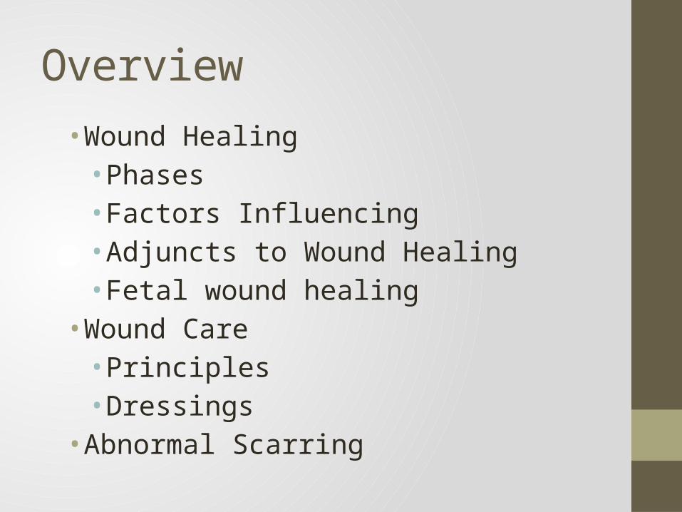
Overview
• Wound Healing• Phases• Factors Influencing• Adjuncts to Wound Healing• Fetal wound healing
• Wound Care• Principles• Dressings
• Abnormal Scarring
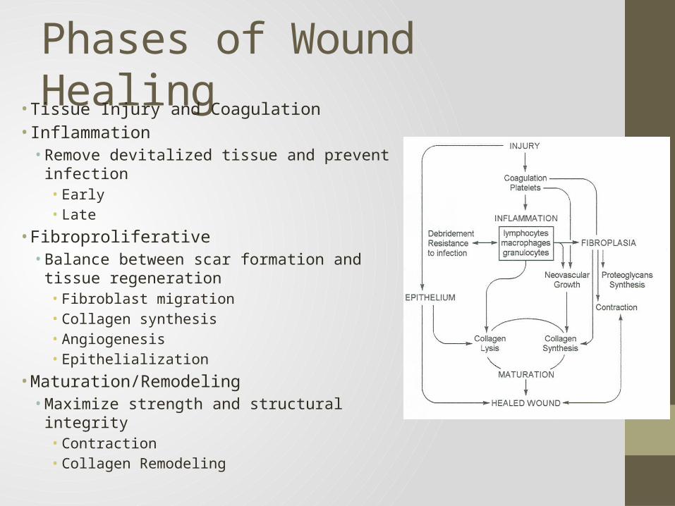
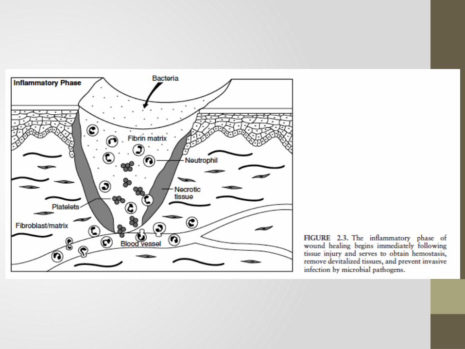
Phases of Wound Healing• Tissue Injury and Coagulation• Inflammation• Remove devitalized tissue and prevent infection
• Early• Late
• Fibroproliferative• Balance between scar formation and tissue
regeneration• Fibroblast migration• Collagen synthesis• Angiogenesis• Epithelialization
• Maturation/Remodeling• Maximize strength and structural integrity
• Contraction• Collagen Remodeling
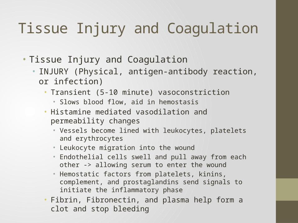
Tissue Injury and Coagulation• Tissue Injury and Coagulation• INJURY (Physical, antigen-antibody reaction, or infection)
• Transient (5-10 minute) vasoconstriction• Slows blood flow, aid in hemostasis
• Histamine mediated vasodilation and permeability changes• Vessels become lined with leukocytes, platelets and erythrocytes• Leukocyte migration into the wound• Endothelial cells swell and pull away from each other -> allowing serum to
enter the wound• Hemostatic factors from platelets, kinins, complement, and prostaglandins
send signals to initiate the inflammatory phase• Fibrin, Fibronectin, and plasma help form a clot and stop bleeding
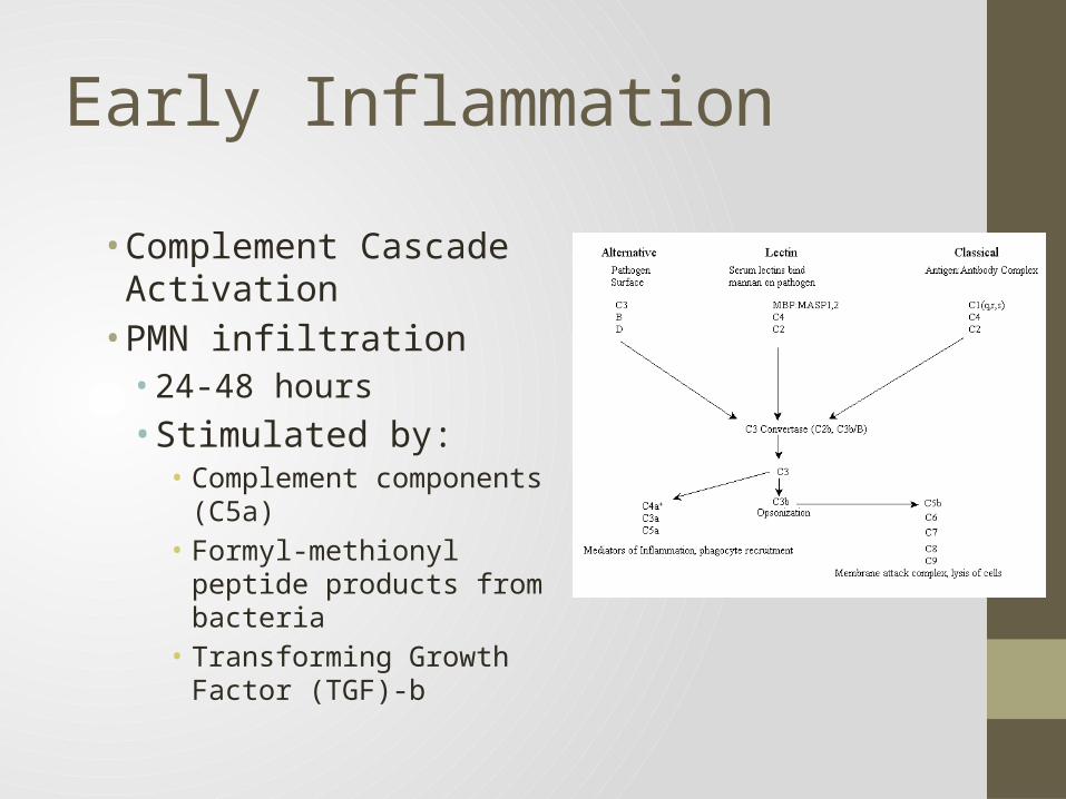
Early Inflammation
• Complement Cascade Activation• PMN infiltration• 24-48 hours• Stimulated by:• Complement components
(C5a)• Formyl-methionyl peptide
products from bacteria• Transforming Growth
Factor (TGF)-b
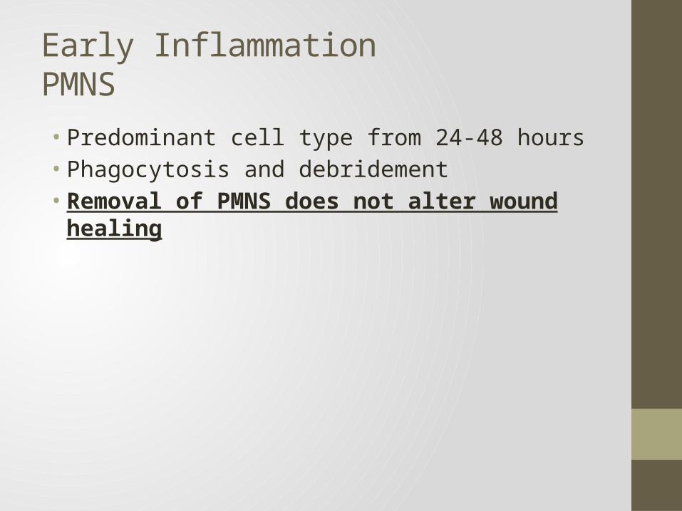
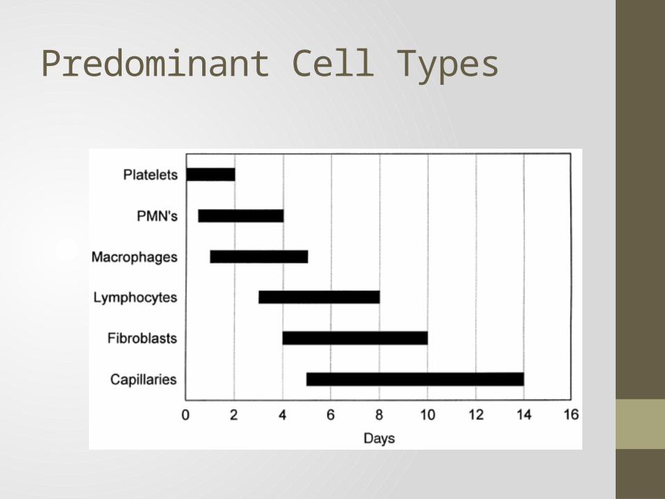
Early InflammationPMNS• Predominant cell type from 24-48 hours• Phagocytosis and debridement• Removal of PMNS does not alter wound healing
Late InflammationMacrophage
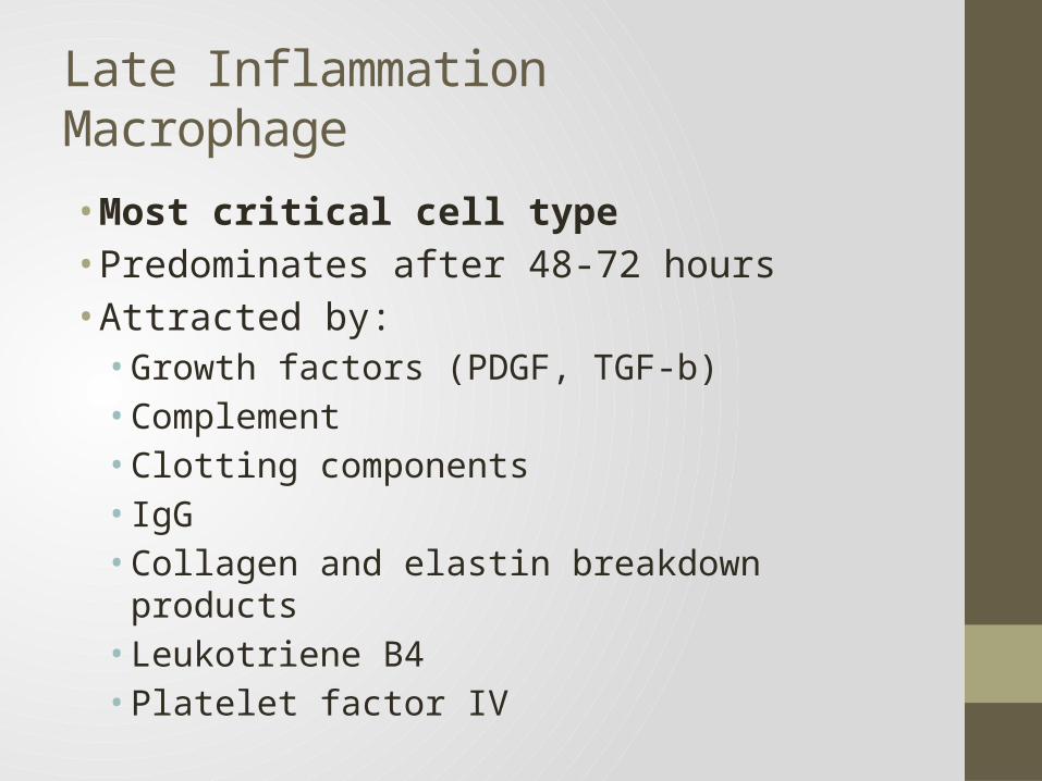
• Most critical cell type• Predominates after 48-72 hours• Attracted by:• Growth factors (PDGF, TGF-b) • Complement• Clotting components• IgG• Collagen and elastin breakdown products• Leukotriene B4• Platelet factor IV
Late InflammationMacrophage Functions
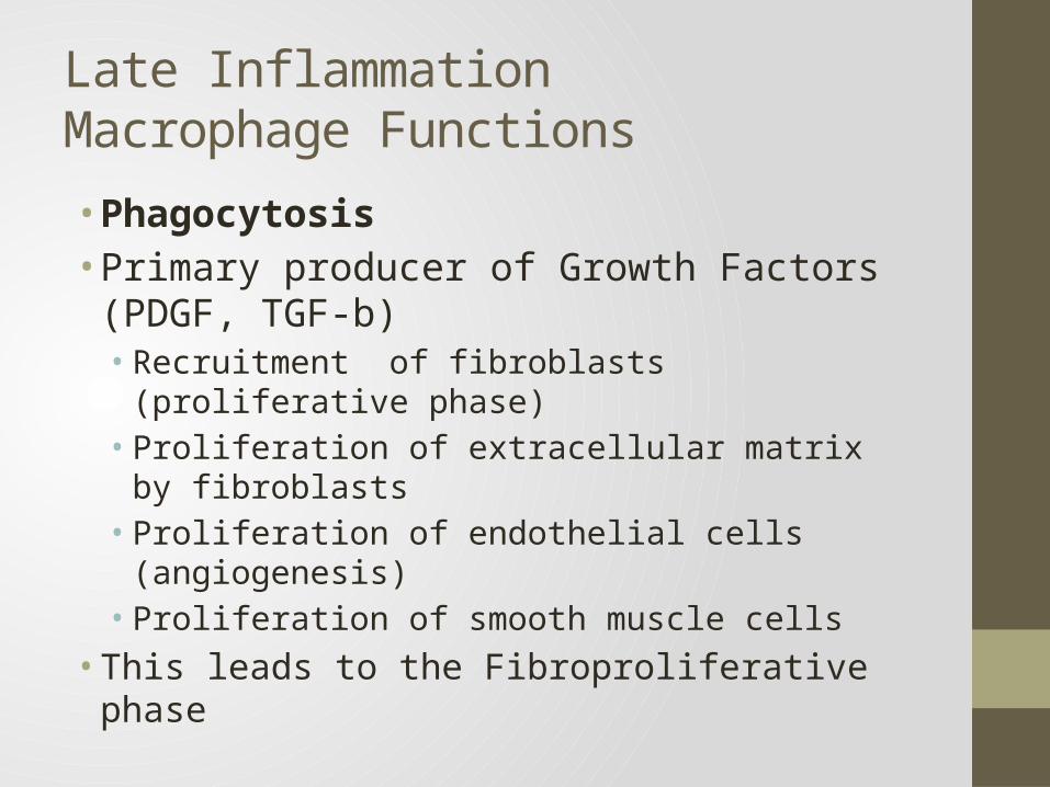
• Phagocytosis• Primary producer of Growth Factors (PDGF, TGF-
b)• Recruitment of fibroblasts (proliferative phase)• Proliferation of extracellular matrix by fibroblasts• Proliferation of endothelial cells (angiogenesis)• Proliferation of smooth muscle cells
• This leads to the Fibroproliferative phase
Late InflammationLymphocyte
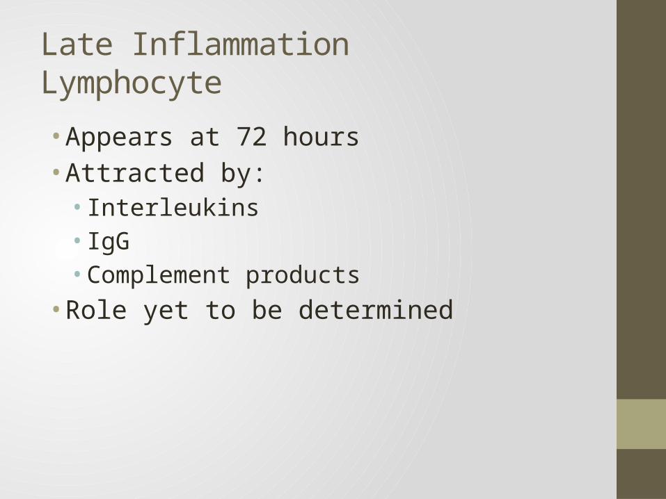
• Appears at 72 hours• Attracted by: • Interleukins• IgG• Complement products
• Role yet to be determined
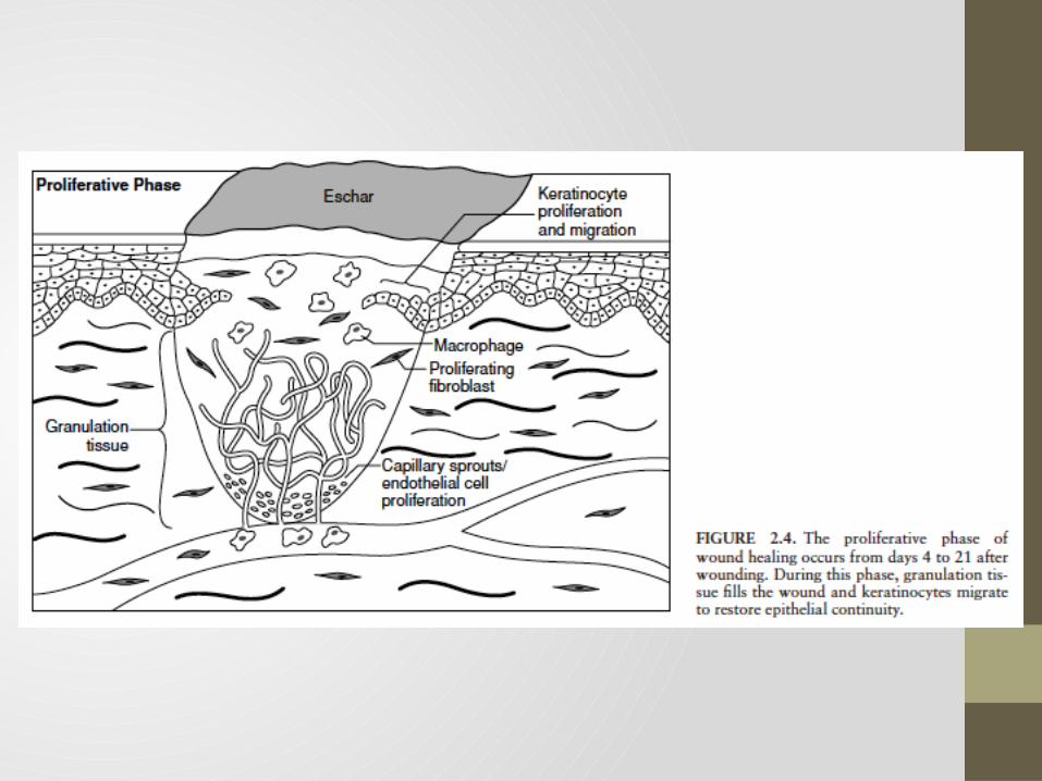
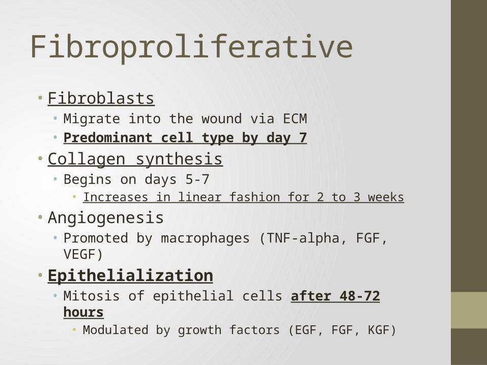
Fibroproliferative
• Fibroblasts• Migrate into the wound via ECM• Predominant cell type by day 7
• Collagen synthesis• Begins on days 5-7
• Increases in linear fashion for 2 to 3 weeks
• Angiogenesis• Promoted by macrophages (TNF-alpha, FGF, VEGF)
• Epithelialization• Mitosis of epithelial cells after 48-72 hours
• Modulated by growth factors (EGF, FGF, KGF)
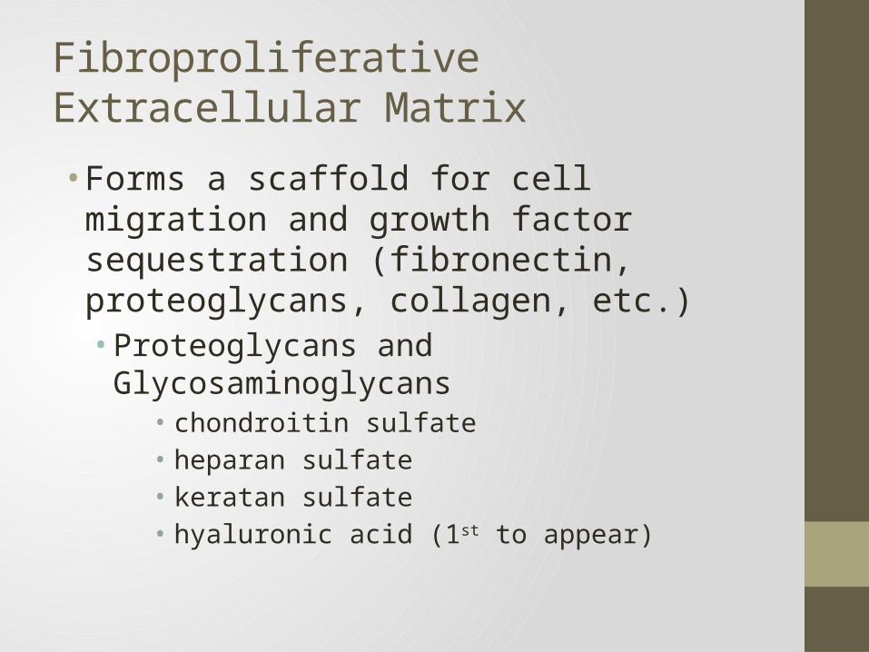
FibroproliferativeExtracellular Matrix
• Forms a scaffold for cell migration and growth factor sequestration (fibronectin, proteoglycans, collagen, etc.)• Proteoglycans and Glycosaminoglycans
• chondroitin sulfate• heparan sulfate• keratan sulfate• hyaluronic acid (1st to appear)
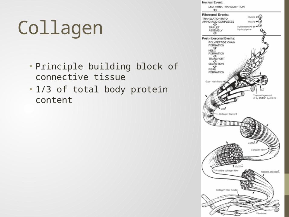
Collagen
• Principle building block of connective tissue
• 1/3 of total body protein content
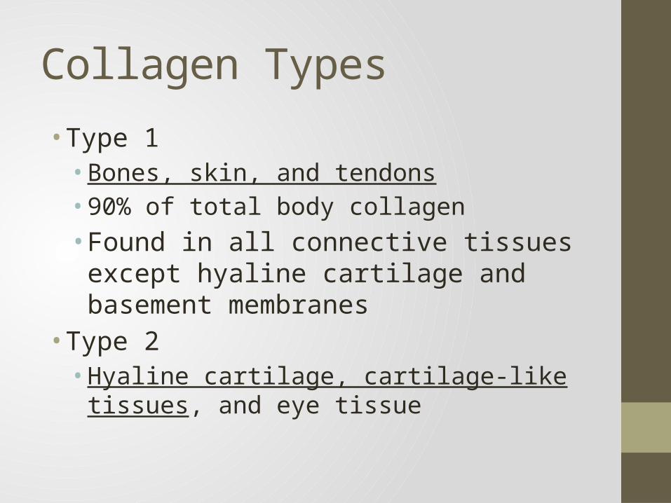
Collagen Types
• Type 1• Bones, skin, and tendons• 90% of total body collagen• Found in all connective tissues except hyaline
cartilage and basement membranes• Type 2• Hyaline cartilage, cartilage-like tissues, and eye
tissue
Collagen Types
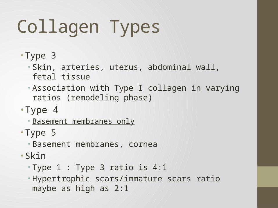
• Type 3• Skin, arteries, uterus, abdominal wall, fetal tissue• Association with Type I collagen in varying ratios
(remodeling phase) • Type 4• Basement membranes only
• Type 5• Basement membranes, cornea
• Skin• Type 1 : Type 3 ratio is 4:1• Hypertrophic scars/immature scars ratio maybe as high
as 2:1
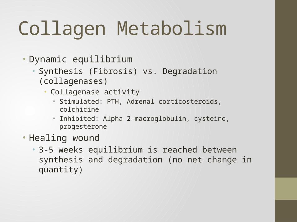
Collagen Metabolism• Dynamic equilibrium• Synthesis (Fibrosis) vs. Degradation (collagenases)
• Collagenase activity• Stimulated: PTH, Adrenal corticosteroids, colchicine• Inhibited: Alpha 2-macroglobulin, cysteine, progesterone
• Healing wound• 3-5 weeks equilibrium is reached between synthesis and
degradation (no net change in quantity)
Angiogenesis
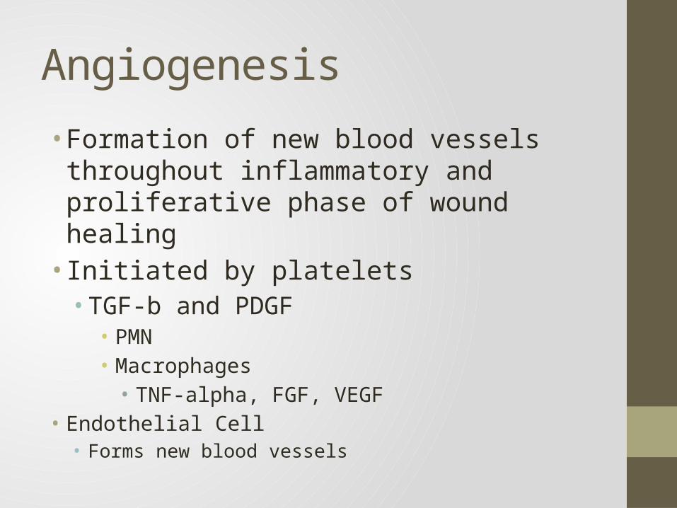
• Formation of new blood vessels throughout inflammatory and proliferative phase of wound healing• Initiated by platelets• TGF-b and PDGF• PMN• Macrophages• TNF-alpha, FGF, VEGF
• Endothelial Cell• Forms new blood vessels
Epithelialization
• Repithelialization begins within hours of injury• Stimulated by• Loss of contact-inhibition• Growth factors• EGF (mitogenesis and chemotaxis)• KGF, FGF (proliferation)
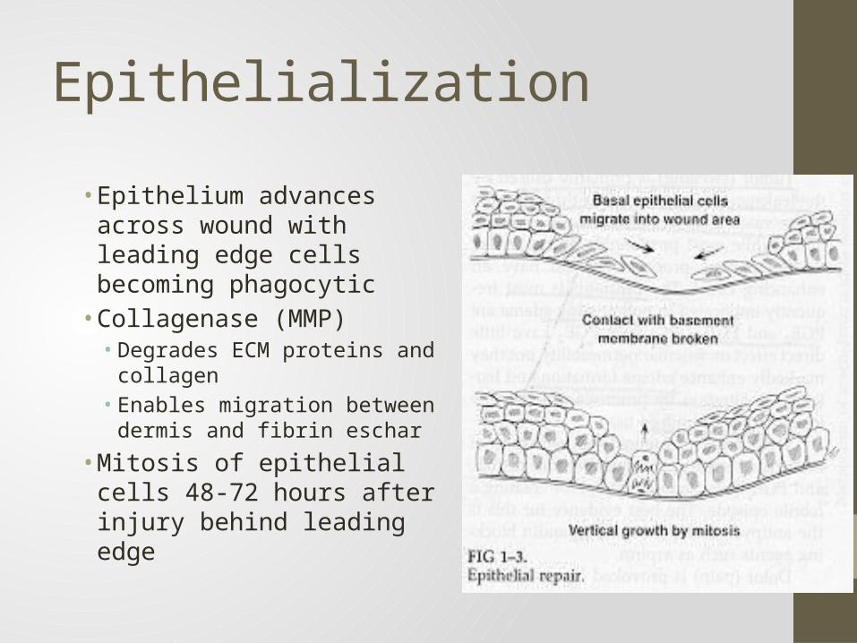
Epithelialization
• Epithelium advances across wound with leading edge cells becoming phagocytic• Collagenase (MMP)• Degrades ECM proteins and
collagen• Enables migration between
dermis and fibrin eschar
• Mitosis of epithelial cells 48-72 hours after injury behind leading edge
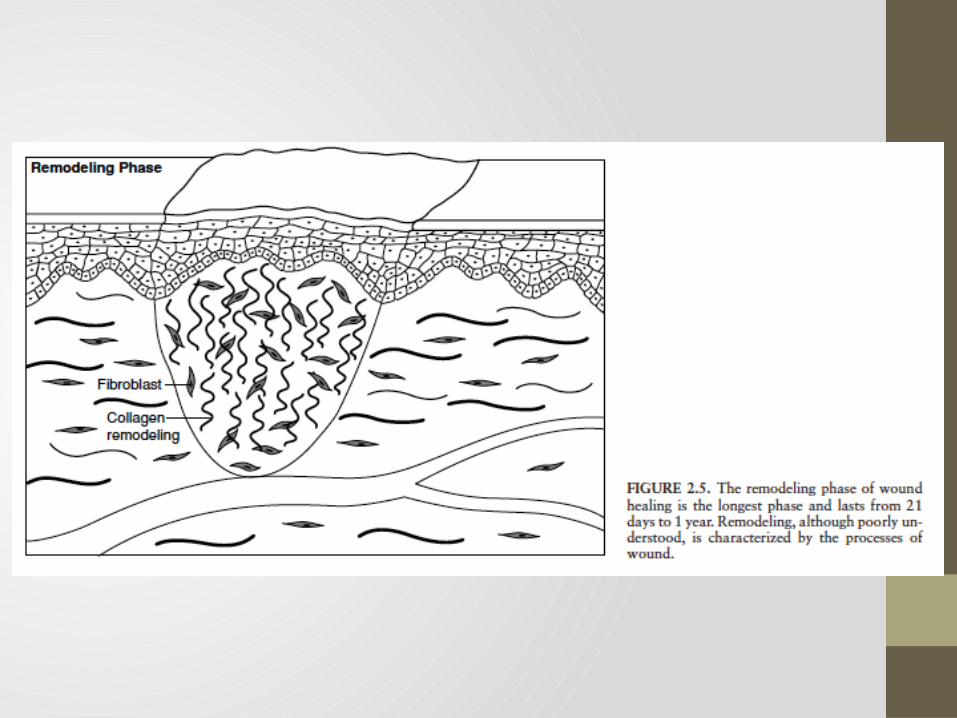
Maturation/Remodeling
• Longest phase: 3 weeks – 1 year• Least understood phase• Wound Contraction and Collagen Remodeling• Wound Contraction
• Myofibroblast• Fibroblasts with intracellular actin microfilaments
Maturation/Remodeling
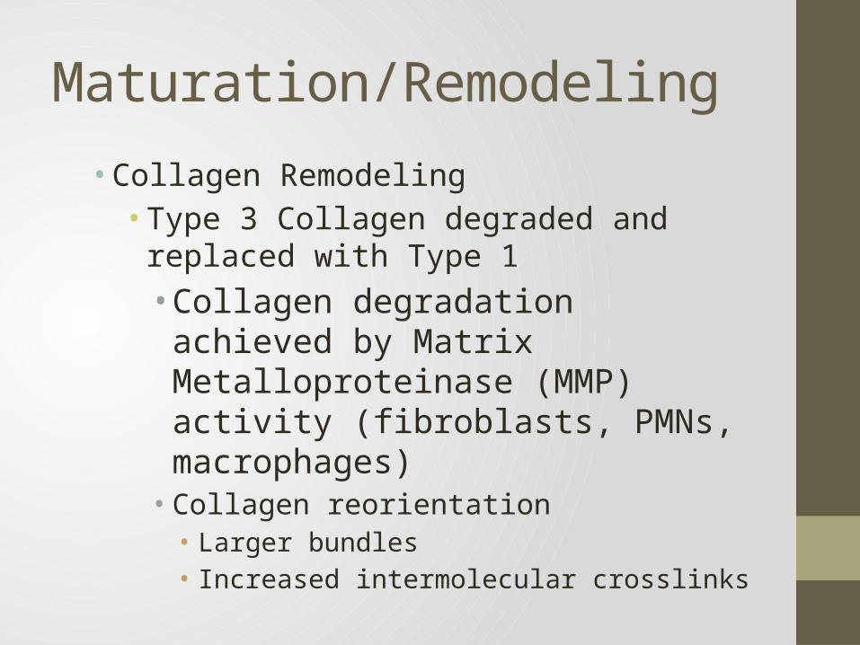
• Collagen Remodeling• Type 3 Collagen degraded and replaced with
Type 1• Collagen degradation achieved by Matrix
Metalloproteinase (MMP) activity (fibroblasts, PMNs, macrophages)• Collagen reorientation• Larger bundles• Increased intermolecular crosslinks
Tensile Strength
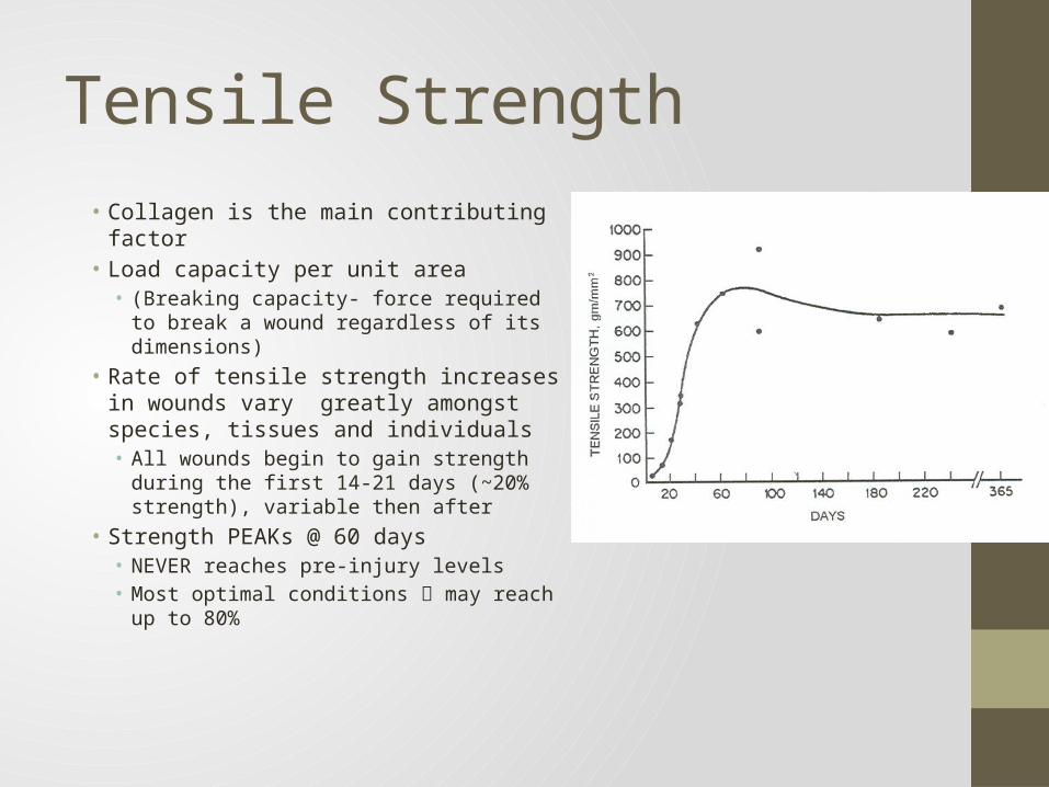
• Collagen is the main contributing factor• Load capacity per unit area• (Breaking capacity- force required to break
a wound regardless of its dimensions)• Rate of tensile strength increases in
wounds vary greatly amongst species, tissues and individuals• All wounds begin to gain strength during
the first 14-21 days (~20% strength), variable then after
• Strength PEAKs @ 60 days• NEVER reaches pre-injury levels• Most optimal conditions may reach up
to 80%
Predominant Cell Types
Special Characteristics of Fetal Wound Healing• Lack of inflammation• Absence of FGF and TGF-b
• Regenerative process with minimal or no scar formation• Collagen deposition is more organized and rapid• Type 3 Collagen (No Type 1)
• High in hyaluronic acid• Area of ongoing research
Factors That Influence Wound Healing
• Oxygen• Fibroblasts are oxygen-sensitive
• Collagen synthesis cannot occur unless the PO2 >40mmHg• Deficiency is the most common cause for wound infection and
breakdown• Hematocrit
• Mild to moderate anemia does not appear to have a negative influence wound healing (given sufficient oxygenation)
• >50% decrease in HCT• some studies report a significant decrease in wound tensile strength• while other studies find no change
Factors That Influence Wound Healing
• Smoking• Multifactorial in limiting wound healing
• Nicotine• Vasoconstrictive -> decreases proliferation of erythrocytes, macrophages,
and fibroblasts• CO• Decreases the oxygen carrying capacity of Hgb
• Hydrogen Cyanide• Inhibits oxidative enzymes
• Increases blood viscosity, decrease collagen deposition and prostacyclin formation
• A single cigarette may cause cutaneous vasoconstriction for up to 90 minutes
Factors That Influence Wound Healing
• Mechanical Stress• Affects the quantity, aggregation, and orientation of collagen
fibers• Abnormal tension -> blanching, necrosis, dermal rupture, and
permanent stretching
• Hydration• Well hydrated wounds epithelialize faster
• Environmental Temperature• Healing is accelerated at temperatures of 30 C• Tensile strength decrease by 20% in 12C environment
Factors That Influence Wound Healing
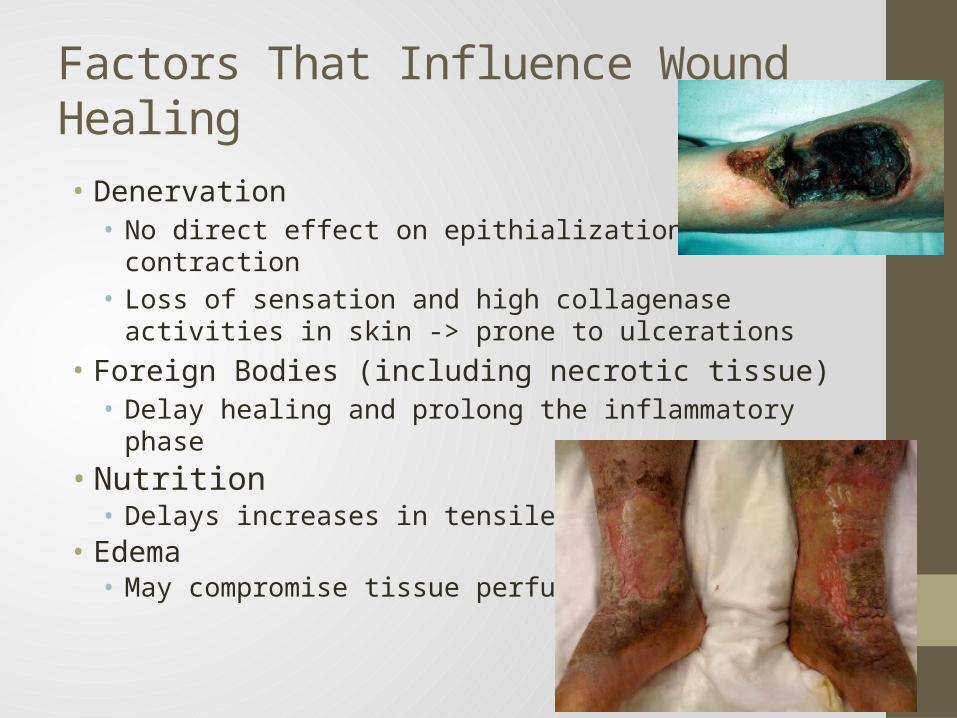
• Denervation• No direct effect on epithialization or contraction• Loss of sensation and high collagenase activities in skin -> prone
to ulcerations• Foreign Bodies (including necrotic tissue)• Delay healing and prolong the inflammatory phase
• Nutrition• Delays increases in tensile strength
• Edema• May compromise tissue perfusion
Factors That Influence Wound Healing
• Oxygen Derived Free Radicals• Degrade Hyaluronic acid and collagen• Destroy cell and organelle membranes• Interfere with enzymatic functions
• Age• Tensile strength and wound closure rates decrease with age
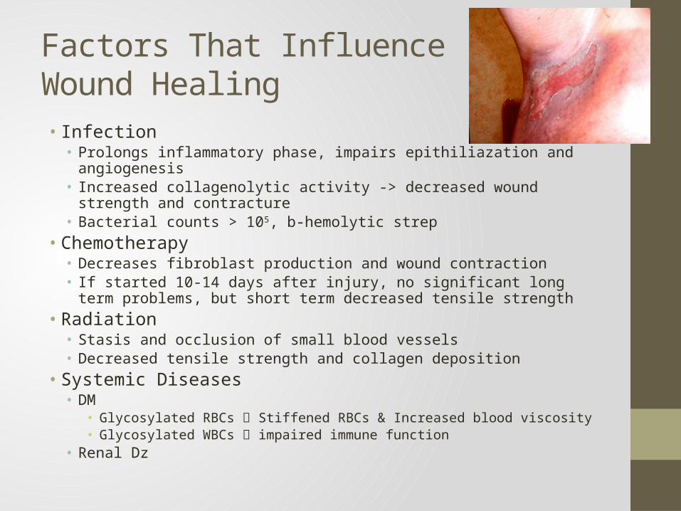
Factors That Influence Wound Healing• Infection• Prolongs inflammatory phase, impairs epithiliazation and angiogenesis• Increased collagenolytic activity -> decreased wound strength and
contracture• Bacterial counts > 105, b-hemolytic strep
• Chemotherapy• Decreases fibroblast production and wound contraction• If started 10-14 days after injury, no significant long term problems, but
short term decreased tensile strength• Radiation• Stasis and occlusion of small blood vessels• Decreased tensile strength and collagen deposition
• Systemic Diseases• DM
• Glycosylated RBCs Stiffened RBCs & Increased blood viscosity• Glycosylated WBCs impaired immune function
• Renal Dz
Factors That Influence Wound Healing
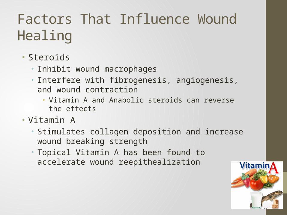
• Steroids• Inhibit wound macrophages• Interfere with fibrogenesis, angiogenesis, and wound contraction
• Vitamin A and Anabolic steroids can reverse the effects
• Vitamin A• Stimulates collagen deposition and increase wound breaking
strength• Topical Vitamin A has been found to accelerate wound
reepithealization
Factors That Influence Wound Healing
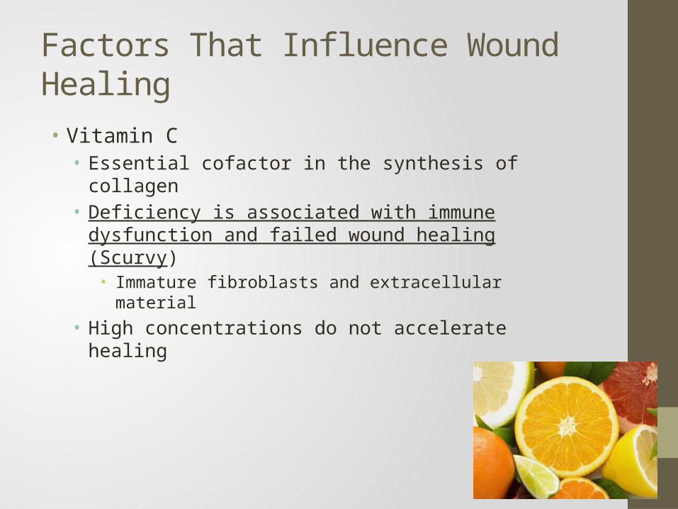
• Vitamin C• Essential cofactor in the synthesis of collagen• Deficiency is associated with immune dysfunction and failed
wound healing (Scurvy)• Immature fibroblasts and extracellular material
• High concentrations do not accelerate healing
Factors That Influence Wound Healing
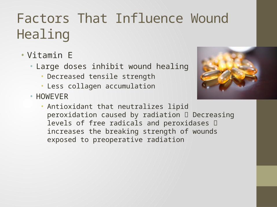
• Vitamin E• Large doses inhibit wound healing
• Decreased tensile strength• Less collagen accumulation
• HOWEVER• Antioxidant that neutralizes lipid peroxidation caused by radiation
Decreasing levels of free radicals and peroxidases increases the breaking strength of wounds exposed to preoperative radiation
Factors That Influence Wound Healing
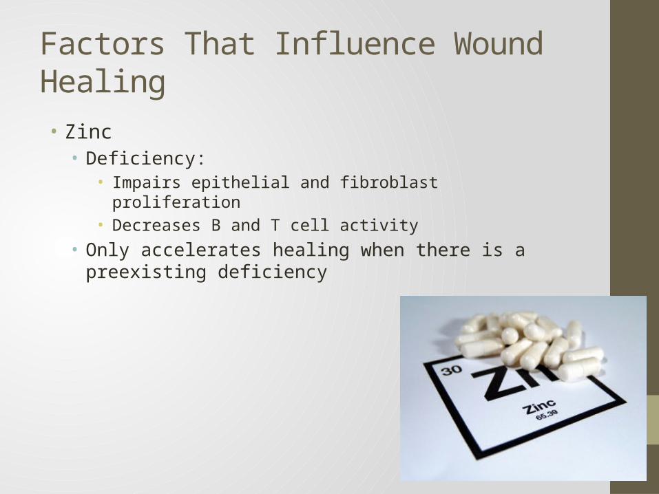
• Zinc• Deficiency:
• Impairs epithelial and fibroblast proliferation• Decreases B and T cell activity
• Only accelerates healing when there is a preexisting deficiency
Factors That Influence Wound Healing
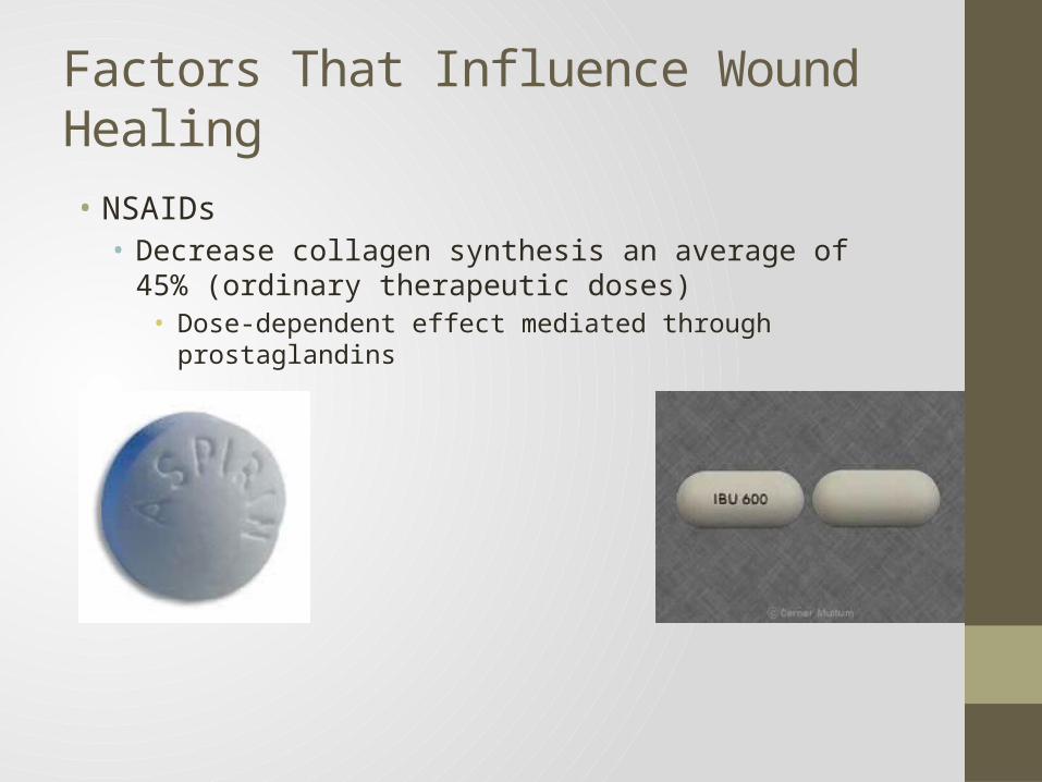
• NSAIDs• Decrease collagen synthesis an average of 45% (ordinary
therapeutic doses)• Dose-dependent effect mediated through prostaglandins
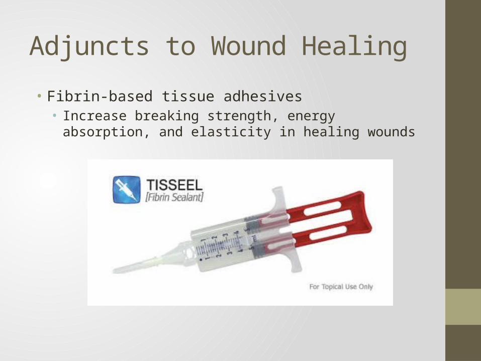
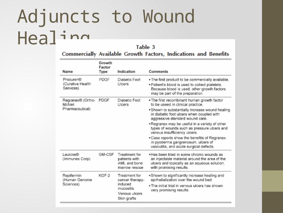
Adjuncts to Wound Healing• Fibrin-based tissue adhesives• Increase breaking strength, energy absorption, and elasticity in
healing wounds
Adjuncts to Wound Healing• Hydrotherapy• Whirlpool• Pulsed Lavage
• Stimulates formation of granulation tissue• Clean non draining wounds with healthy granulation tissue
should NEVER be subjected to hydrotherapy• Water agitation damages fragile cells
Adjuncts to Wound Healing• Hyperbaric Oxygen• Increases levels of O2 and NO to the wound
• Benefit: Amputations, osteoradionecrosis, surgical flaps, skin grafts• None to minimal benefit with necrotizing soft-tissue infections• Wounds require adequate perfusion
• Many off-label uses (Benefit? Financial?)• Acne, Migraines, Lupus, Stroke, MS, and many more
• Medicare Coverage• 14 Covered Areas (next slide)
Medicare Coverage of HBO• (1) Acute carbon
monoxide intoxication • (2) Decompression
illness • (3) Gas embolism • (4) Gas gangrene • (5) Acute traumatic
peripheral ischemia • (6) Crush injuries • (7) Progressive
necrotizing infections
• (8) Acute peripheral arterial insufficiency
• (9) Preparation and preservation of compromised skin grafts
• (10) Chronic refractory osteomyelitis
• (11) Osteoradionecrosis (ORN)
• (12) Soft tissue radionecrosis (STRN)
• (13) Cyanide poisoning • (14) Actinomycosis
Adjuncts to Wound Healing
Wound Care General Principles• Cleaning and Irrigation• Need at least 7psi to flush bacteria out of a wound• High pressure can damage wounds and should be reserved only
for heavily contaminated wounds• Debridement• Most critical step to produce a wound that will heal rapidly
without infection• Non-selective: Dakin solution, Hydrogen Peroxide, etc.• Useful in wounds with heavy contamination• When starts to granulate, start selective
• Selective: sharp, enzymatic, autolytic, or biologic
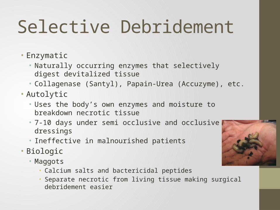
Selective Debridement• Enzymatic• Naturally occurring enzymes that selectively digest devitalized
tissue• Collagenase (Santyl), Papain-Urea (Accuzyme), etc.
• Autolytic• Uses the body’s own enzymes and moisture to breakdown
necrotic tissue• 7-10 days under semi occlusive and occlusive dressings• Ineffective in malnourished patients
• Biologic• Maggots
• Calcium salts and bactericidal peptides• Separate necrotic from living tissue making surgical debridement
easier
Wound Care General Principles• Fundamentals of Surgical Wound Closure• Incision should follow tension lines and natural folds in the skin• Gentle tissue handling• Complete hemostasis• Eliminate tension• Fine sutures and early removal• Evert wound edges• Allow scars to mature before repeat intervention (2 weeks to 2
months scar appearance is the worst)• Scar appearance depends more on type of injury than method of
closure• Technical factors of suture placement and removal are more critical
than type of suture used• Immobilization of wounds to prevent disruptions and excessive
scarring (Adhesive strips after suture removal)
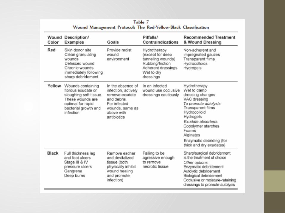
Wound Dressings• Over 2,000 commercially available • Red-Yellow-Black Classification• Created to help choose appropriate dressings in wounds healing
by secondary intention• Treat worse colors first
• Black -> Yellow -> Red
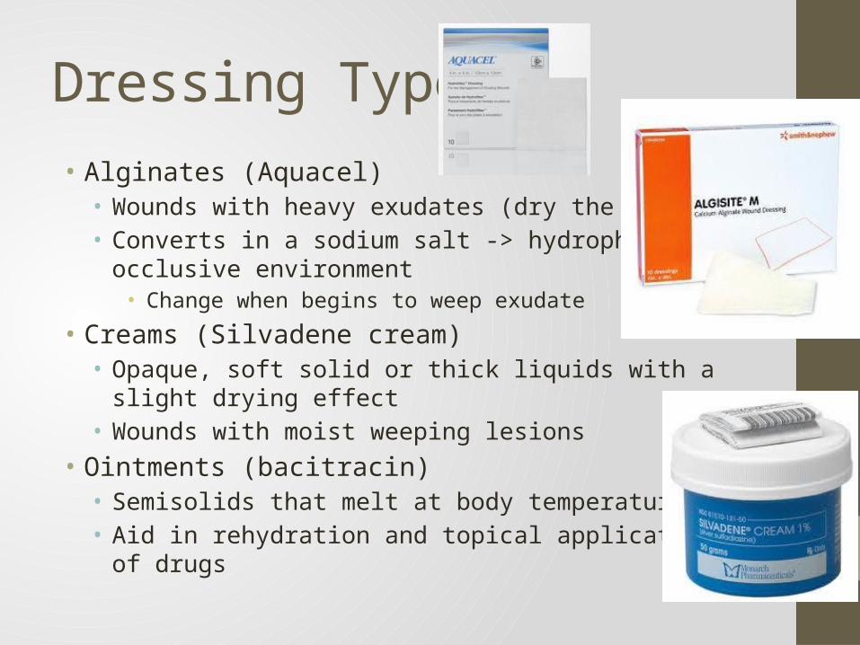
Dressing Types• Alginates (Aquacel)• Wounds with heavy exudates (dry the wound)• Converts in a sodium salt -> hydrophilic gel occlusive environment
• Change when begins to weep exudate
• Creams (Silvadene cream)• Opaque, soft solid or thick liquids with a slight drying effect• Wounds with moist weeping lesions
• Ointments (bacitracin)• Semisolids that melt at body temperature• Aid in rehydration and topical application of drugs
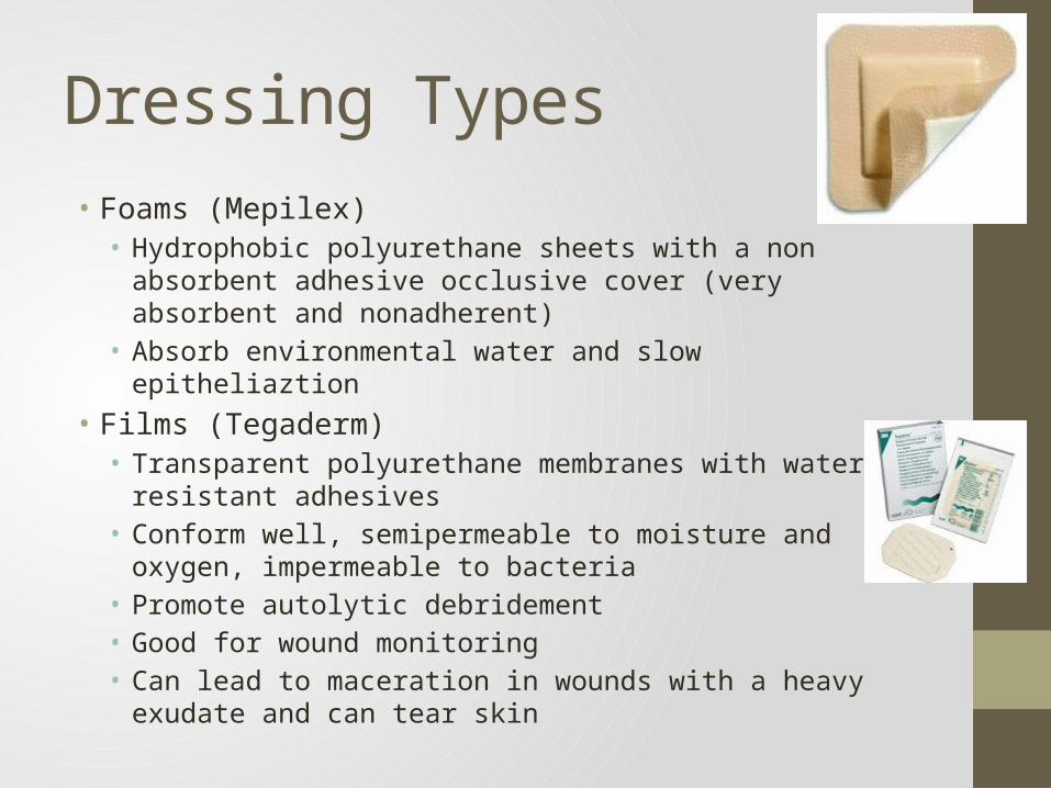
Dressing Types• Foams (Mepilex)• Hydrophobic polyurethane sheets with a non absorbent adhesive
occlusive cover (very absorbent and nonadherent)• Absorb environmental water and slow epitheliaztion
• Films (Tegaderm)• Transparent polyurethane membranes with water-resistant
adhesives• Conform well, semipermeable to moisture and oxygen,
impermeable to bacteria• Promote autolytic debridement• Good for wound monitoring• Can lead to maceration in wounds with a heavy exudate and can
tear skin
Dressing Types
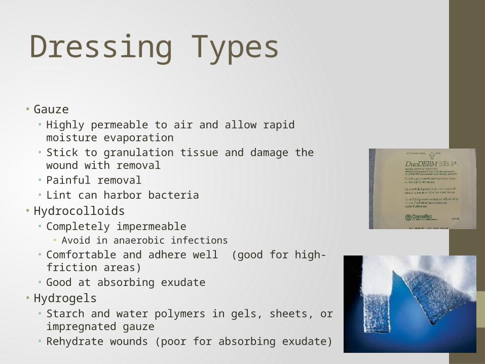
• Gauze• Highly permeable to air and allow rapid moisture
evaporation• Stick to granulation tissue and damage the wound with
removal• Painful removal• Lint can harbor bacteria
• Hydrocolloids• Completely impermeable
• Avoid in anaerobic infections• Comfortable and adhere well (good for high-friction areas)• Good at absorbing exudate
• Hydrogels• Starch and water polymers in gels, sheets, or impregnated
gauze• Rehydrate wounds (poor for absorbing exudate)
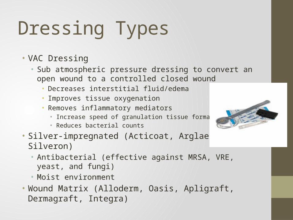
Dressing Types• VAC Dressing• Sub atmospheric pressure dressing to convert an open wound to
a controlled closed wound• Decreases interstitial fluid/edema• Improves tissue oxygenation• Removes inflammatory mediators• Increase speed of granulation tissue formation• Reduces bacterial counts
• Silver-impregnated (Acticoat, Arglaes, Silveron)• Antibacterial (effective against MRSA, VRE, yeast, and fungi)• Moist environment
• Wound Matrix (Alloderm, Oasis, Apligraft, Dermagraft, Integra)
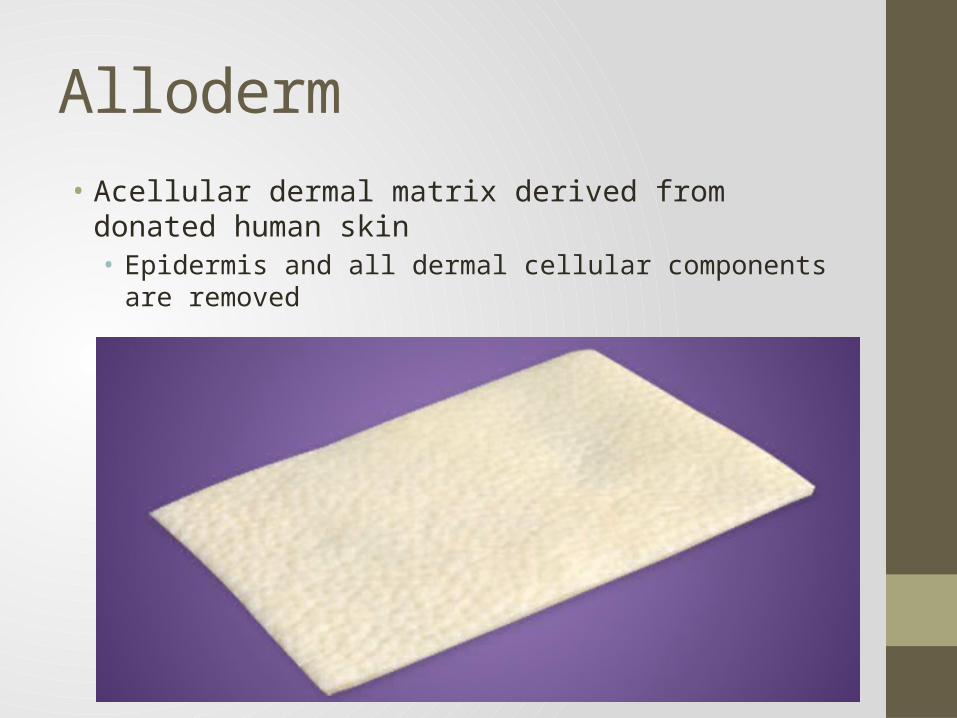
Alloderm• Acellular dermal matrix derived from donated human skin• Epidermis and all dermal cellular components are removed
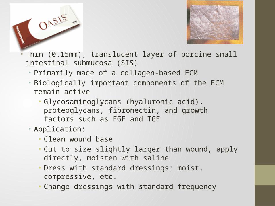
Oasis• Thin (0.15mm), translucent layer of porcine small intestinal
submucosa (SIS)• Primarily made of a collagen-based ECM • Biologically important components of the ECM remain active• Glycosaminoglycans (hyaluronic acid), proteoglycans,
fibronectin, and growth factors such as FGF and TGF• Application:• Clean wound base• Cut to size slightly larger than wound, apply directly,
moisten with saline• Dress with standard dressings: moist, compressive, etc.• Change dressings with standard frequency
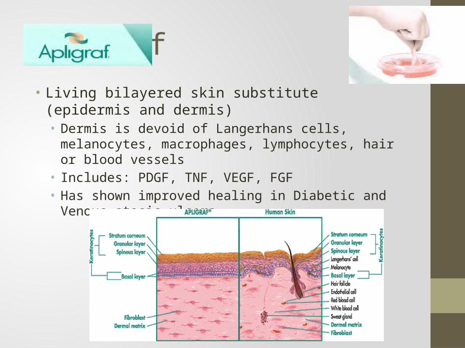
Apligraf• Living bilayered skin substitute (epidermis and dermis)• Dermis is devoid of Langerhans cells, melanocytes, macrophages,
lymphocytes, hair or blood vessels• Includes: PDGF, TNF, VEGF, FGF• Has shown improved healing in Diabetic and Venous stasis ulcers
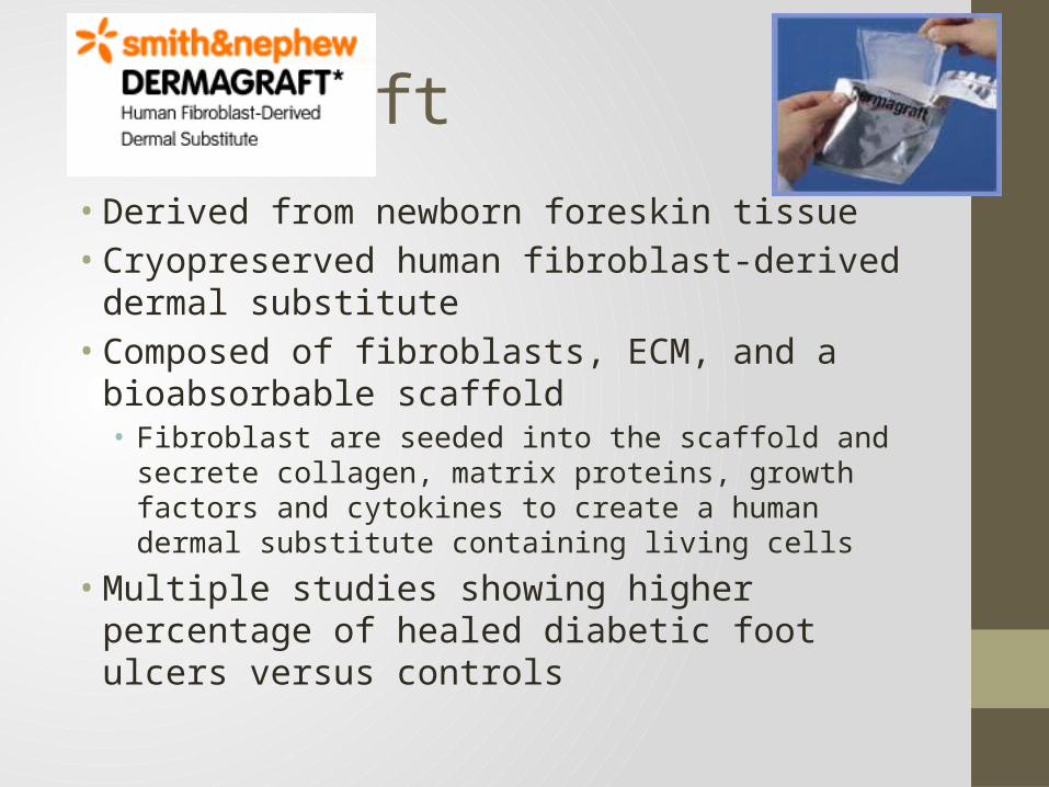
Dermagraft
• Derived from newborn foreskin tissue• Cryopreserved human fibroblast-derived dermal
substitute• Composed of fibroblasts, ECM, and a bioabsorbable
scaffold• Fibroblast are seeded into the scaffold and secrete collagen,
matrix proteins, growth factors and cytokines to create a human dermal substitute containing living cells
• Multiple studies showing higher percentage of healed diabetic foot ulcers versus controls
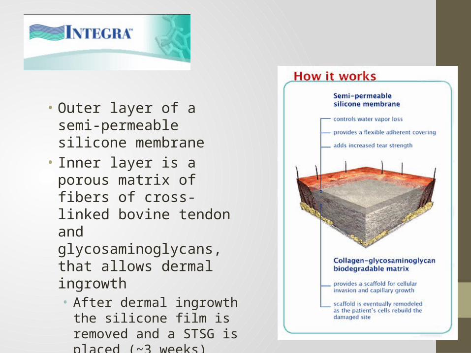
Integra
• Outer layer of a semi-permeable silicone membrane
• Inner layer is a porous matrix of fibers of cross-linked bovine tendon and glycosaminoglycans, that allows dermal ingrowth• After dermal ingrowth the
silicone film is removed and a STSG is placed (~3 weeks)
Abnormal Scarring• Hypertrophic Scars• Keloids• Widespread Scar
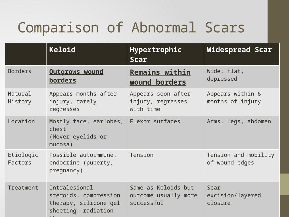
Comparison of Abnormal ScarsKeloid Hypertrophic Scar Widespread Scar
Borders Outgrows wound borders Remains within wound borders
Wide, flat, depressed
Natural History Appears months after injury, rarely regresses
Appears soon after injury, regresses with time
Appears within 6 months of injury
Location Mostly face, earlobes, chest(Never eyelids or mucosa)
Flexor surfaces Arms, legs, abdomen
Etiologic Factors
Possible autoimmune, endocrine (puberty, pregnancy)
Tension Tension and mobility of wound edges
Treatment Intralesional steroids, compression therapy, silicone gel sheeting, radiation therapyOften worse after surgery alone
Same as Keloids but outcome usually more successful
Scar excision/layered closure
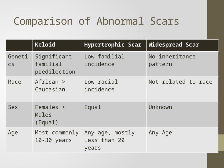
Comparison of Abnormal ScarsKeloid Hypertrophic Scar Widespread Scar
Genetics Significant familial predilection
Low familial incidence No inheritance pattern
Race African > Caucasian Low racial incidence Not related to race
Sex Females > Males(Equal)
Equal Unknown
Age Most commonly 10-30 years
Any age, mostly less than 20 years
Any Age
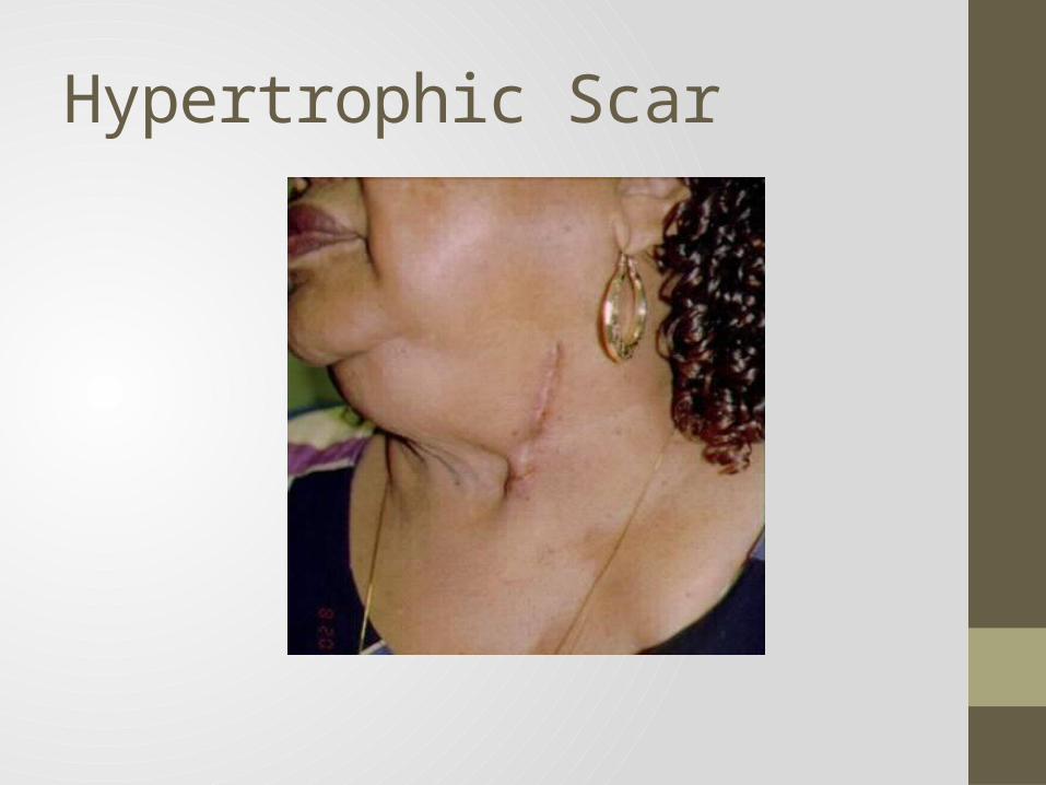
Hypertrophic Scar
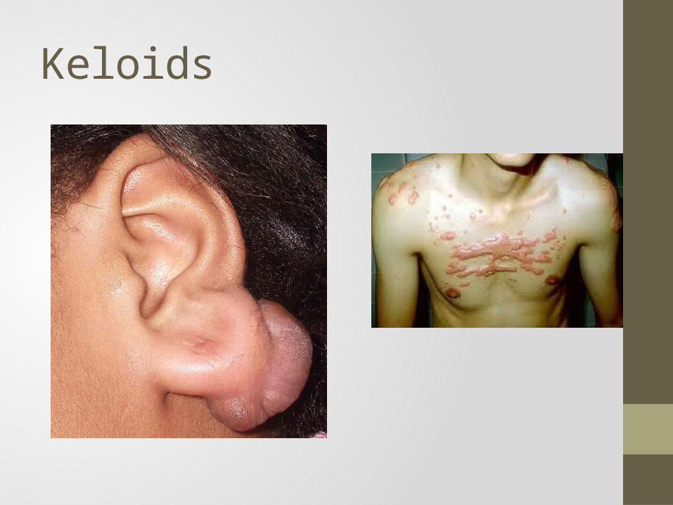
Keloids
Keloid: Treatments• No universally effective treatment, usually a combination of
treatment types• Case by Case basis• Prevention (the best therapy)• Avoid non essential surgery, minimal tension, use cuticular
monofilament synthetic sutures, avoid wound-lengthening techniques, and avoid incisions across joints
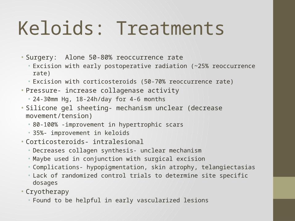
Keloids: Treatments• Surgery: Alone 50-80% reoccurrence rate• Excision with early postoperative radiation (~25% reoccurrence rate)• Excision with corticosteroids (50-70% reoccurrence rate)
• Pressure- increase collagenase activity• 24-30mm Hg, 18-24h/day for 4-6 months
• Silicone gel sheeting- mechanism unclear (decrease movement/tension)• 80-100% -improvement in hypertrophic scars• 35%- improvement in keloids
• Corticosteroids- intralesional• Decreases collagen synthesis- unclear mechanism• Maybe used in conjunction with surgical excision• Complications- hypopigmentation, skin atrophy, telangiectasias• Lack of randomized control trials to determine site specific dosages
• Cryotherapy• Found to be helpful in early vascularized lesions
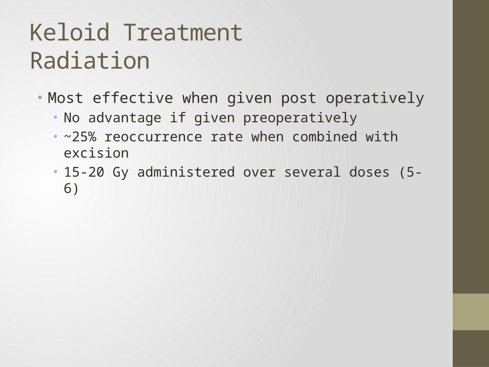
Keloid TreatmentRadiation• Most effective when given post operatively• No advantage if given preoperatively• ~25% reoccurrence rate when combined with excision• 15-20 Gy administered over several doses (5-6)
Keloid TreatmentsAntitumor/Immunosuppressive Agents
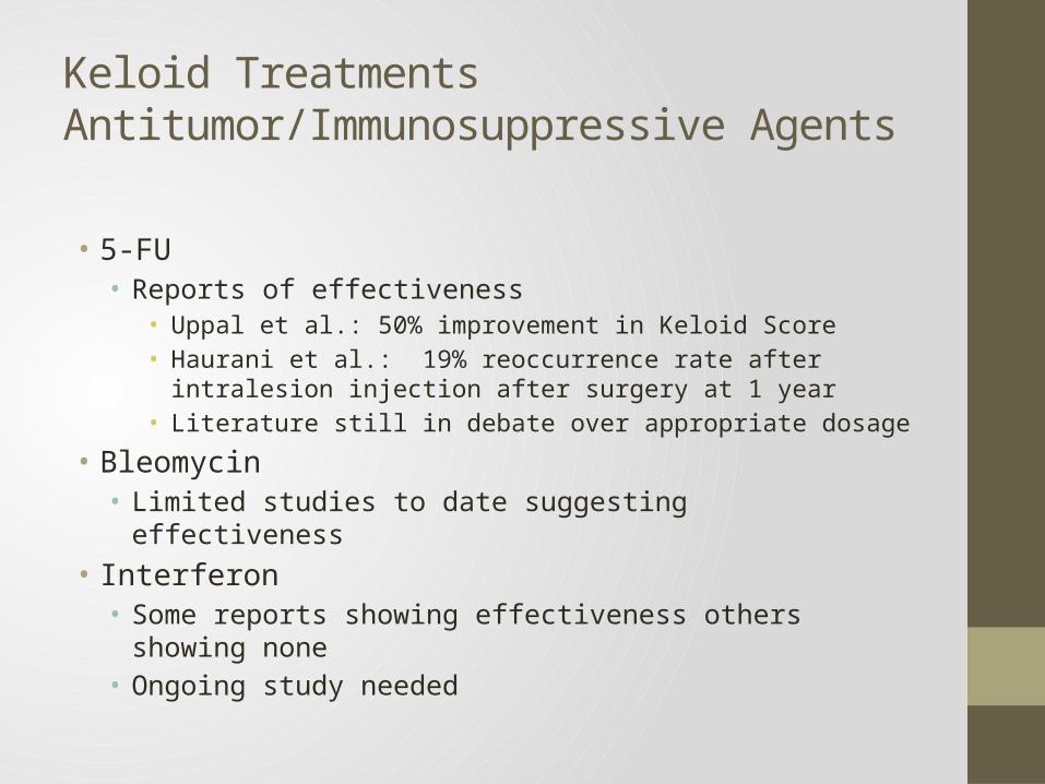
• 5-FU• Reports of effectiveness
• Uppal et al.: 50% improvement in Keloid Score• Haurani et al.: 19% reoccurrence rate after intralesion injection after
surgery at 1 year• Literature still in debate over appropriate dosage
• Bleomycin• Limited studies to date suggesting effectiveness
• Interferon• Some reports showing effectiveness others showing none• Ongoing study needed
Thank You