Embed Size (px)
Citation preview
79���������������� ������� ���������������������� ������������� ��!������"# $
�����������
Chitin, the second most widespread natu-ral polymer after cellulose, can be extrac-ted from seafood waste. It was found andconfirmed in many experiments that chi-tin promotes the ordered healing of tissu-es, activation of macrophages, and worksas a bacteriostatic, antistereoporotic andimmunoadjuvant agent [1-16]. However,due to its high molecular weight and strongcrystalline structure, chitin is insoluble incommon organic solvents, which makesits direct application difficult and expen-sive. A thin chitin fleece-forming dressingmaterial, which is manufactured and mar-keted in Japan, is produced from chitinfibre spun from 3-5% solutions of nativechitin in the presence of huge amounts ofcoagulating and washing media [17-21].
Such a troublesome operation is one re-ason for the high cost of this chitin pro-duct: the price of one 120-cm2 piece ofJapanese chitin dressing material namedBeschitin is ca. 30 USD. However, the re-commended applications of chitin dres-sing materials are very wide, which mayexplain their high prices: Unitika Ltd. (Ja-pan) offers such materials for successfuland fast healing of burns, skin abrasions,postoperative wounds, bed sores, ulcers,and several other injuries. Chitin-basedbioactive dressing materials have not yetbeen produced in Europe.
An original method for synthesising theester derivative of chitin, dibutyrylchitin(DBCH), has been devised in Poland.DBCH is soluble in the common organicsolvents and has both film and fibre for-ming properties [22-27]. The easy solubi-lity of this chitin derivative makes it po-ssible to manufacture a wide assortmentof DBCH materials suitable for medicalapplications. It was also found that the al-kaline treatment of the finished materialsmade from DBCH led to chitin regenera-tion without destroying their macrostruc-ture. Moreover, the regeneration of DBCHto chitin resulted in the improved mecha-nical properties of newly obtained mate-rials, despite the fact that regenerated chi-tin RC had a lower molecular weight thanthe native chitin [28].
The first investigations of biological pro-perties of DBCH and regenerated chitinmaterials were made in vitro and in vivousing their fibrous forms. They includedall tests fulfilling the EN ISO 10993 (Bio-
logical evaluation of medical devices) re-quirements [29-32]. It was found thatDBCH and regenerated chitin fibres metall the basic EN requirements and showedgood biocompatibility when they wereused as implants into the gluteal musclesof rats of the Wistar breed.
The main objective of the present investi-gations was to determine the ability ofDBCH and regenerated chitin to accele-rate wound healing. DBCH and regenera-ted chitin RC were used in the form offilms coating bilaterally commercial po-lypropylene non-woven materials. Thebiological properties of the dressing ma-terials prepared were investigated accor-ding to EN ISO 10993, and their medicalproperties were studied in vivo using arepresentative group of albino rabbits ofthe New Zealand breed.
�� �� ����
Dressing material samples preparation
Krill chitin, a product of the Institute ofSea Fisheries (MIR) in Gdynia, with theintrinsic viscosity value of [η]=13.33dl/g, determined at 25°C in solutions ofdimethylacetamide (DMAc) containing5% of LiCl, was used for the synthesis ofDBCH. The chitin had been previouslyadditionally purified from the remains ofcalcium carbonate. DBCH with the intrin-sic viscosity value of [η]=2.41 dl/g, de-termined at 25°C in DMAc solutions, wasreceived in accordance with the descrip-tion given in Szosland et al. [32].
The commercial non-woven polypropyle-ne material (PP) with a surface mass of
������� �������� � ����������� ��� ��� ����������������������������������
������������ ��������������
����������������� �������������������� ��
����� ���� ����������������� ���!��"�����#��$����%� ���
����������������� ���������������������������������������
������������������������������������ ����������������������������
������������� � �!��������!�
!���������"��#����������$%�&�'���(�����������������������)���������!�������
����"����� �����##���$���%��&'�(���������������������� ����!����)�!�
�*��������������������������������������������������������������!������
����*�������#������##%��������������
AbstractNew textile dressings containing dibutyrylchitin (DBCH) or regenerated chitin (RC) wereprepared in the process of coating a trade polypropylene non-woven material with films ofDBCH or RC. The dry dressing material contained ca. 40% of DBCH and 30% of RC. Thedressings obtained were cut into pieces of 5×5 cm, sterilised by ethylene oxide and thensubjected to biological evaluation required for medical devices. The evaluation includedcytotoxicity effects, levels of cytokines TNF-α and IFNs, synthesis of nitrogen oxides (NO2/NO3), intracutaneous irritation, and the influence of full thickness skin lesions on the heal-ing process.DBCH and RC caused no cytotoxic effects or primary irritation either in vitroor in vivo, nor did the activity of TNF-α, IFNs or the nitrogen oxide levels increase, and bothhad a positive influence on the wound healing process. Both dibutyrylchitin and regeneratedchitin used for coating trade polypropylene non-woven material can be regarded as valu-able dressing materials that accelerate wound healing.
Key words: dressing materials, wound healing, dibutyrylchitin, regenerated chitin, cytotox-icity, intracutaneous irritation, TNF-α, IFNs, NO.
���������������� ������� ��������������������� ������������� ��!������"# $80
ca. 30 g/m2 was washed in distilled waterwith flax shampoo, then rinsed several ti-mes with distilled water, and finally withethanol. Finally, the non-wovens weredried and cut into rectangular 20×30-cmpieces. Each rectangular piece was we-ighed. Clean piece of PP non-woven ma-terial was covered bilaterally with 3%DBCH ethanol solution (3 g of DBCH in100 ml of ethanol) using the polyuretha-ne sponge roll, previously washed in etha-nol and dried. The non-woven materialwas dried and coated for the second timewith DBCH solution prepared as above,but this time containing dehydrated gly-cerin of analytical purity (1ml in 100 mlof solution). All non-woven pieces, do-uble-covered with DBCH, were dried andweighed. The percentage of DBCH in thedry dressing material was ca. 40%.
One part of the DBCH-coated dressingmaterials was treated with 5% NaOH wa-ter solution to restore chitin in the coatinglayer. Alkaline treatment was carried outat 90°C over 15 minutes. The pieces ofdressing materials treated with NaOHwere washed with distilled water to remo-ve any traces of alkali, then with ethanol,and dried. The percentage of regeneratedchitin RC in the obtained dressing mate-rial was ca. 30%. Both dressing materialsused for biomedical investigations werecut into 5×5 cm pieces and sterilised withethylene oxide.
With the purpose of evaluating the chan-ge in RC molecular weight, 10 ml of 5%acetone solution of DBCH was droppedinto 100 ml of 5% NaOH water solutionat 90°C, sediment (regenerated chitin RC)was washed from any traces of alkali, thenwith ethanol, and dried. The intrinsic vi-scosity value of the obtained regeneratedchitin RC determined in DMAc/5% LiClsolutions was 4.4 dl/g. The correlationbetween the intrinsic viscosity value [η]of chitin and its molecular weight Mv isexpressed in the Mark-Houwink equation:[η]=K.....Mv
a, where K=2.1×10⁻⁴ and a=0.88
[33]. The decrease in the intrinsic visco-sity value from 13.33 for initial chitin to4.4 for regenerated chitin RC correspon-ded to a ca. 3.5 times decrease in RC mo-lecular weight.
Biological tests and results
The polypropylene non-wovens coveredbilaterally with the layer of DBCH andregenerated chitin RC were subjected tobiological evaluation in accordance withthe demands determined for the medicaldevices. We evaluated the cytotoxicity ef-fects, the levels of the cytokines TNF-αand IFNs, the synthesis of nitrogen oxi-des (NO2/NO3), intracutaneous irritationand the influence on the healing processof the full thickness skin lesions.
Cytotoxicity effects
The evaluation of cytotoxicity effects wasconducted on the reference cell line ofmouse fibroblast 3T3/Balb, with the di-rect contact method. The cultures of mo-use fibroblasts, with ca. 5×10⁵ cells each,were established on Petri dishes. After 24h the old culture medium was removed andthe new one added. The culture cells werecovered with the samples tested of 2 cmin diameter The changes in the cell cultu-res were recorded after 24, 48 and 72 h.Dyeing with the neutral red dye was em-ployed for the count living cells, while thedead cells were dyed with the trypan bluedye. Both kinds of cells were counted inthe Bürker chamber. The results of ourcytotoxicity assessment are shown in Ta-ble 1. Direct contact of the mouse fibro-blast cultures (3T3/Balb.) with non-wovendressing materials coated with DBCH andregenerated chitin RC did not show anycytotoxicity effect.
Immunological testing
In order to define the inflammatory andimmunomodulation effects of DBCH andRC, an assessment of their influence onthe induction of the cytokines TNF-α andIFNs, and on the synthesis of nitrogenoxides of human leukocytes was conduc-
ted. The leukocytes used for the asses-sment were extracted from the peripheralblood of healthy volunteers, which wascollected for heparin after centrifugationat the gradient of Dextran-Uropolina (i.e.gradient G of the density 1.115 g/ml). Forthe purpose of this assessment, a leuko-cyte suspension in the RPMI medium with2% foetal calf of serum, 100 u/ml of peni-cillin, 100 µg/ml of streptomycin and therequired density of 2×10⁶ cells/ml wasprepared.
On a plate with 24 wells (Costar Ltd.) 1ml of the leukocyte suspension of 2×10⁶
cells/ml in the culture liquid RPMI with2% calf serum was deposited into eachwell. Samples of the tested biomaterialsin the amount of 10 mg of each were ad-ded to prepared cells, which were thenincubated for 24 and 72 h at 37°C and 5%CO2. The activities of TNF-α, IFNs, andnitrogen oxides were assessed in the su-pernatants.
TNF-ααααα levelsThe level of TNF-α was determined bythe biological method on 96-well plates,on which the L929 cell cultures of mousefibroblasts were established with the den-sity of 2×10⁵ /ml and incubated for 24h(37°C and 5% CO2). The proper dilutionsof the supernatant from above leukocytecultures (from 1:2 to 1:256) in the Eaglemedium containing 10% calf foetal of se-rum and actinomycine D (Sigma) (2,5µg/ml) were prepared on a separate plate.Then the liquid from above the L929 cul-tures was removed, and the appropriatepreviously prepared dilutions were depo-sited on these cultures. The degenerationof the cells caused by TNF was observedin the inverted microscope after 24h and72h incubation (at 37°C and 5% CO2). Inparallel, L929 cultures in the Eagle me-dium without actinomycine, with actino-mycine and the standard control of rHuTNF-α were also established for compa-rison. The results of TNF-α level deter-mination are collected in Table 2.
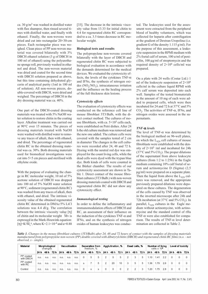
Table 1. Changes in the mouse fibroblast cultures (3T3/Balb) after 24, 48 and 72 hours of contact with the samples of dressing materialsmanufactured from polypropylene non-woven (PP) double covered with dibutyrylchitin (DBCH) and regenerated chitin RC films (n.o. - notobserved, s - single).
�������� �������� �������� �������� �������� ��������� ��������� ��������� ��������� ��������� �����
���� ������ ���� ������ ���� ������ ���� ������ ���� ������ ������������ ������������ ������������ ������������ ��������������������
������������ ������������ ������������ ������������ ������������ �� �������� �� �������� �� �������� �� �������� �� �������� ������������ ������������ ������������ ������������ ��������������� ���� �����
�����������! �����������! �����������! �����������! �����������!
�"# �"# �"# �"# �"# �$" �$" �$" �$" �$" �#% �#% �#% �#% �#% �"# �"# �"# �"# �"# �$" �$" �$" �$" �$" �#% �#% �#% �#% �#% �"# �"# �"# �"# �"# �$" �$" �$" �$" �$" �#% �#% �#% �#% �#% �"# �"# �"# �"# �"# �$" �$" �$" �$" �$" �#% �#% �#% �#% �#% �"# �"# �"# �"# �"# �$" �$" �$" �$" �$" �#% �#% �#% �#% �#% �"# �"# �"# �"# �"# �$" �$" �$" �$" �$" �#% �#% �#% �#% �#% �"# �"# �"# �"# �"# �$" �$" �$" �$" �$" �#% �#% �#% �#% �#%
�����&&'()����*
++� ++� ++� ++� ++� ++� � , , # , , # - , ��+� �"+� #+# � � �
�����&&(.���*
++� ++� ++� ++� ++� ++� % , # �# �� - , , �-+� /%+� �+# � � �
����( ++� ++� ++� ++� ++� ++� � � � � # # - �#+� ,%+� $+# � � �
81���������������� ������� ���������������������� ������������� ��!������"# $
IFNs activityThe interferon level was assessed by themicro method of incubation of the cyto-pathological effect (CPE) of the EMC vi-rus in the A549 cell culture of human lungadenocarcinoma. The interferon titrationwas conducted on plastic 96-well plates.An A549 cell culture of the 2×10⁵/ml den-sity was established on each plate and in-cubated for 24h at 37°C and 5% CO2. Theproper dilutions (i.e. from 1:2 to 1:256)of the tested materials in the Dulbeccomedium with 10% calf foetal of serumwere prepared on the additional plate. Thesupernatant from the above cell cultureswas removed, and the prepared dilutionswere deposited. After 24h of incubation(at 37°C and 5% CO2) the culture was in-fected with the titre 102 TCID50/ml ofEMCV virus. The virus suspension wasprepared in the liquid culture mediumDMEM with 2% calf foetal serum. Thecontrol EMCV virus in the culture, thecontrol-referenced interferon and non-in-fected A549 cell culture were left on theplate. The cytopathological effect wasobserved in the reverse microscope after48 h incubation. The presence of interfe-ron resulted in the protection of the cellsagainst the cytopathological effects of thevirus. The dilution of the interferon, whichprotected 50% of the cells, was adoptedas one unit of IFNs. All results were cor-rected to the standard titre of IFN-s. Theresults obtained for the materials investi-gated are presented in Table 2.
Nitrogen oxides level
The concentration of nitrogen oxide (N02)was measured in the supernatant from theabove-mentioned leukocyte cultures usingthe colorimeter method according to Dinget al. [34]. 100 µl of Griess reagent (i.e.0.1% dihydrochloride naphthylenediami-ne in H2O and 1% sulphanilamide in 5%H3PO4 at the ratio 1:1) was added to100 µl of the tested material for one well.Then, the plate was incubated at room tem-perature for only 12 minutes, and opticaldensity was measured in the Stat Fax 2100counter (Awareness Technology Inc.) atthe wavelength of 540 nm. The concen-tration of NO2 was calculated in relation
to the standard curve of NaNO2 preparedwithin concentrations from 1 µM/ml to100 µM/ml.
The results of the assessments of nitrogenoxide synthesis in the human leukocytecultures are also shown in Table 2. It wasfound that neither the PP non-woven ma-terial coated with DBCH nor that coatedwith regenerated chitin RC stimulatedhuman leukocytes to a greater activity ofTNF-α and IFNs, nor to a greater synthe-sis of nitrogen oxides. The results obta-ined were comparable to those for the con-trol samples. Neither inflammatory norimmunomodulation effects were obse-rved.
Intracutaneous irritation testThis test was conducted using the extractsof the samples of PP non-wovens coatedwith DBCH and RC. The polar and non-polar extracts were prepared using thesaline solution and sesame oil respective-ly, after sinking the tested materials (120cm2 each) in 20 ml of the correspondingliquid. Incubation was carried out at thetemperature of 37°C for 72 h. The salinesolution and pure sesame oil, whichhad no contact with the tested materials,were used as the control samples and wereincubated under the same conditions asabove.
The assessments of the extracts from eachmaterial were conducted on 3 albino rab-bits of the New Zealand breed. On the backof each animal 5 intracutaneous injectionsof tested extract and 5 injections of con-trol solution, each of 0.2 ml, were carriedout.
Observations of skin were made 24, 48,and 72 h after the injections, and no skinchanges were found. The Primary Irrita-tion Index for the polar and non-polarextracts from the polypropylene non-wo-ven materials coated with DBCH and re-generated chitin was equal to 0.00.
Influence on healing of skin defects
The evaluation of the influence of the po-lypropylene non-woven materials coated
with DBCH and regenerated chitin on thehealing process of skin wounds was con-ducted on 16 albino rabbits of the NewZealand breed of near-equal body massof 3.2-3.5 kg.
The surgery was carried out under gene-ral anaesthesia and in fully aseptic condi-tions. Four oval wounds (ca. 12 mm indiameter) across the entire skin thicknesswere incised with the scalpel on the backof each rabbit. The wounds to the left ofthe backbone were covered with asepticswaps as the controls. On the right side,the anterior wound was covered with thepolypropylene non-woven material coatedwith regenerated chitin, while the poste-rior wound was covered with the polypro-pylene non-woven material coated withDBCH. In addition, all those dressingswere protected by a gauze band. The wo-und healing was observed and the dres-sings were changed every 24 h until thewounds were covered with scabs. Later on,the wounds with scabs were protected onlyby a gauze band.
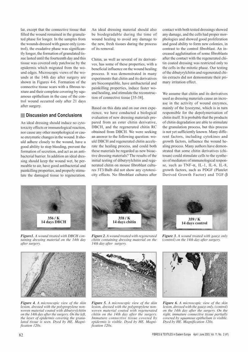
During the macroscopic observations, nosignificant differences were noted in thehealing of full-thickness skin lesions co-vered with dressings containing eitherDBCH or regenerated chitin. The woundedges were flat, and the neighbouring skinshowed no signs of inflammation. All thefull-thickness skin lesions dressed with thetested materials were filled with white-yellowish, elastic tissue and all appearedto be more contracted, wetter and moreelastic as compared to the control woundscovered only with gauze. The edges ofcontrol wounds were thickened and withsignificant areas of redness, while the skinwas congested. By the sixth day after sur-gery, massive scabs covered the skin le-sions. The photographs of the wounds ta-ken on the 14th day after surgery are shownin Figures 1-3.
The microscopic assessment showed thatthe wounds covered with the dressing con-taining DBCH healed fastest. By the tenthday a new, granulated tissue was observedin the site of the lesion, the epithelium al-most totally covered by the squamous epi-thelium. On the fourteenth day, most ofthe wounds were filled with immatureconnective tissue with numerous bloodvessels and collagenous fibres, and theconnective scars were all completely co-vered by the epidermis. The healing ofwounds covered with the dressing conta-ining regenerated chitin was quite simi-
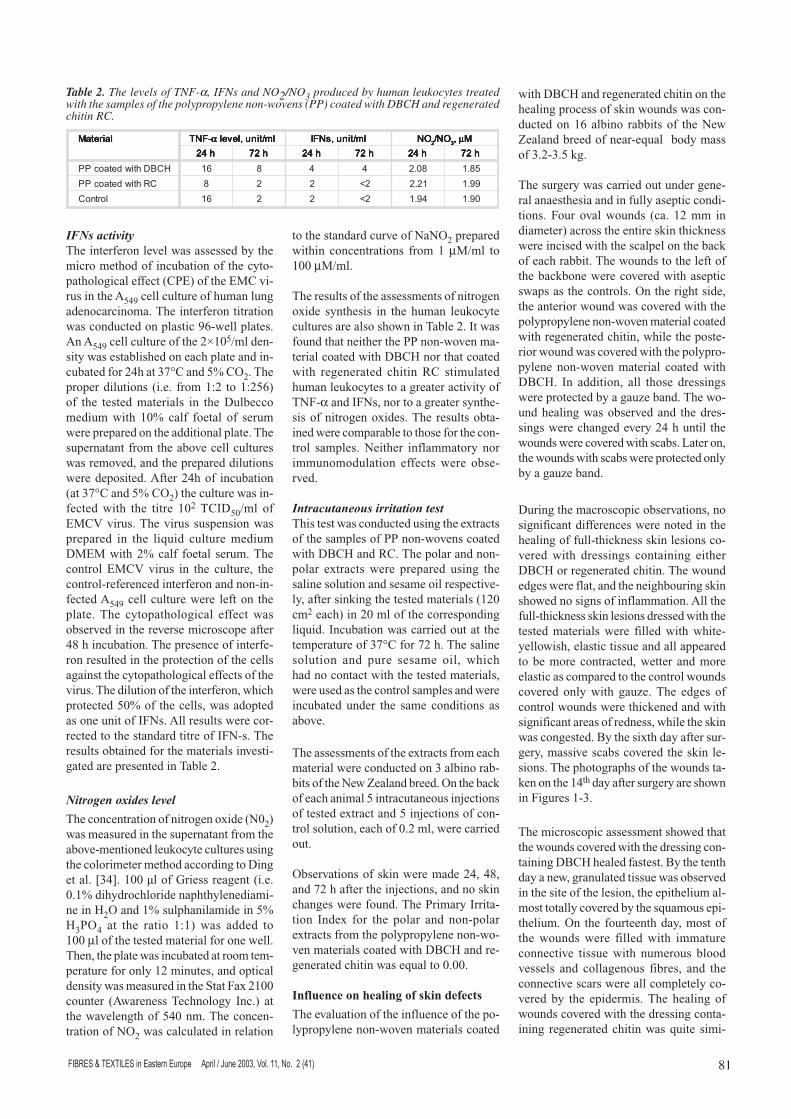
Table 2. The levels of TNF-α, IFNs and NO2/NO3 produced by human leukocytes treatedwith the samples of the polypropylene non-wovens (PP) coated with DBCH and regeneratedchitin RC.
�������� �������� �������� �������� �������� 01�2 01�2 01�2 01�2 01�2 ααααα ��3���������� ��3���������� ��3���������� ��3���������� ��3���������� ��3����� �14 ��3����� �14 ��3����� �14 ��3����� �14 ��3����� �14 5�5�5�5�5�#####
5�3 5�3 5�3 5�3 5�3-----����� µµµµµ�����
�"# �"# �"# �"# �"# �#% �#% �#% �#% �#% �"# �"# �"# �"# �"# �#% �#% �#% �#% �#% �"# �"# �"# �"# �"# �#% �#% �#% �#% �#%
'()����*�����&& �� $ " " $�+# ,$+�
(.���*�����&& $ # # #6 �#+# //+�
����( �� # # #6 "/+� �/+�
���������������� ������� ��������������������� ������������� ��!������"# $82
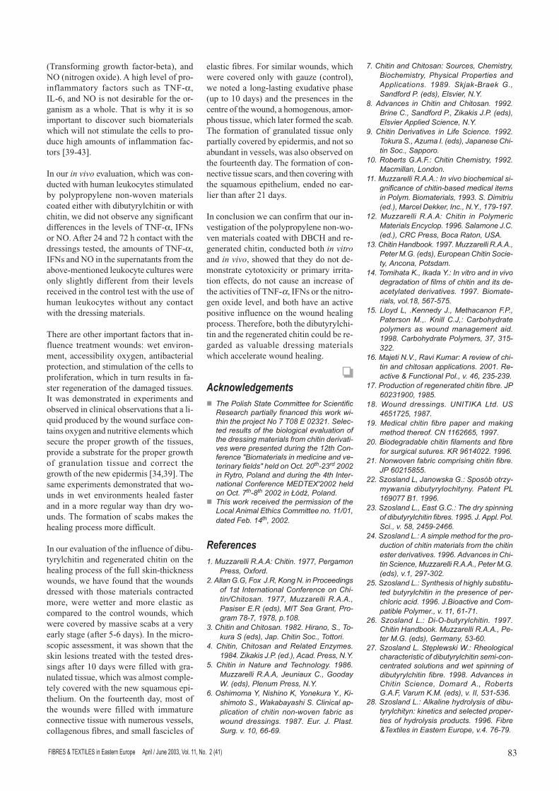
lar, except that the connective tissue thatfilled the wound remained in the granula-ted phase for longer. In the samples fromthe wounds dressed with gauze only (con-trol), the exudative phase was significan-tly longer, the formation of granulated tis-sue lasted until the fourteenth day and thistissue was covered only patchwise by theepidermis which migrated from the wo-und edges. Microscopic views of the wo-unds at the 14th day after surgery areshown in Figures 4-6. Formation of theconnective tissue scars with a fibrous te-xture and their complete covering by squ-amous epithelium in the case of the con-trol wound occurred only after 21 daysafter surgery.
�����������������������
An ideal dressing should induce no cyto-toxicity effects or immunological reaction,nor cause any other morphological or cau-se enzymatic changes in the wound. It sho-uld adhere closely to the wound, have agood ability to stop bleeding, prevent theformation of secretion, and act as an anti-bacterial barrier. In addition an ideal dres-sing should keep the wound wet, be per-meable to air, have good antibacterial andpainkilling properties, and properly stimu-late the damaged tissue to regeneration.
An ideal dressing material should alsobe biodegradable during the time ofwound healing to avoid any damage tothe new, fresh tissues during the processof its removal.
Chitin, as well as several of its derivati-ves, has some of these properties, with abeneficial influence on the wound healingprocess. It was demonstrated in manyexperiments that chitin and its derivativesare biocompatible, have antibacterial andpainkilling properties, induce faster wo-und healing, and stimulate the reconstruc-tion of connective tissue [35-38].
Based on this data and on our own expe-rience, we have conducted a biologicalevaluation of new dressing materials pre-pared from an ester chitin derivative,DBCH, and the regenerated chitin RCobtained from DBCH. We were seekingan answer to the following question: wo-uld DBCH and regenerated chitin accele-rate the healing process, and could boththese materials be regarded as new bioac-tive dressing materials? The results of theinitial testing of dibutyrylchitin and rege-nerated chitin on mouse fibroblast cultu-res 3T3/Balb did not show any cytotoxi-city effects. No fibroblast cultures after
contact with both tested dressings showedany damage, and the cells had proper mor-phologies and showed good proliferationand good ability to form new colonies, incontrast to the control fibroblast. An in-creased agglutination of some fibroblastsafter the contact with the regenerated chi-tin coated dressing was restricted only tothe cells in the mitotic phase. Our testingof the dibutyrylchitin and regenerated chi-tin extracts did not demonstrate their pri-mary irritation effect.
We assume that chitin and its derivativesused as dressing materials cause an incre-ase in the activity of wound enzymes,mainly of the lysozyme, which is in turnresponsible for the depolymerisation ofchitin itself. It is probable that the productsof chitin degradation are able to stimulatethe granulation process, but this processis not yet sufficiently known. Many diffe-rent factors, including cytokines andgrowth factors, influence the wound he-aling process. Many authors have demon-strated that some chitin derivatives (chi-tosan) could stimulate cells to the synthe-sis of mediators of immunological respon-se, such as TNF-α, IL-1, IL-6, IL-8,growth factors, such as PDGF (PlateletDerived Growth Factor) and TGF-β
Figure 4. A microscopic view of the skinlesion, dressed with the polypropylene non-woven material coated with dibutyrylchitinon the 14th day after the surgery. On the left,the layer of epidermis covering the granu-lated tissue is seen. Dyed by HE. Magni-fication 120x.
Figure 5. A microscopic view of the skinlesion, dressed with the polypropylene non-woven material coated with regeneratedchitin on the 14th day after the surgery.Immature connective tissue covered byepidermis is visible. Dyed by HE. Magni-fication 120x.
Figure 6. A microscopic view of the skinlesion, dressed with the gauze only, (control)on the 14th day after the surgery. On theright, immature connective tissue partiallycovered by squamous epithelium is visible.Dyed by HE. Magnification 120x.
Figure1. A wound treated with DBCH con-taining dressing material on the 14th dayafter surgery.
Figure 2. A wound treated with regeneratedchitin containing dressing material on the14th day after surgery.
Figure 3. A wound treated with gauze only(control) on the 14th day after surgery.
356 / K14 days DBCH
358 / K14 days chitin
359 / K14 days control
83���������������� ������� ���������������������� ������������� ��!������"# $
(Transforming growth factor-beta), andNO (nitrogen oxide). A high level of pro-inflammatory factors such as TNF-α,IL-6, and NO is not desirable for the or-ganism as a whole. That is why it is soimportant to discover such biomaterialswhich will not stimulate the cells to pro-duce high amounts of inflammation fac-tors [39-43].
In our in vivo evaluation, which was con-ducted with human leukocytes stimulatedby polypropylene non-woven materialscoated either with dibutyrylchitin or withchitin, we did not observe any significantdifferences in the levels of TNF-α, IFNsor NO. After 24 and 72 h contact with thedressings tested, the amounts of TNF-α,IFNs and NO in the supernatants from theabove-mentioned leukocyte cultures wereonly slightly different from their levelsreceived in the control test with the use ofhuman leukocytes without any contactwith the dressing materials.
There are other important factors that in-fluence treatment wounds: wet environ-ment, accessibility oxygen, antibacterialprotection, and stimulation of the cells toproliferation, which in turn results in fa-ster regeneration of the damaged tissues.It was demonstrated in experiments andobserved in clinical observations that a li-quid produced by the wound surface con-tains oxygen and nutritive elements whichsecure the proper growth of the tissues,provide a substrate for the proper growthof granulation tissue and correct thegrowth of the new epidermis [34,39]. Thesame experiments demonstrated that wo-unds in wet environments healed fasterand in a more regular way than dry wo-unds. The formation of scabs makes thehealing process more difficult.
In our evaluation of the influence of dibu-tyrylchitin and regenerated chitin on thehealing process of the full skin-thicknesswounds, we have found that the woundsdressed with those materials contractedmore, were wetter and more elastic ascompared to the control wounds, whichwere covered by massive scabs at a veryearly stage (after 5-6 days). In the micro-scopic assessment, it was shown that theskin lesions treated with the tested dres-sings after 10 days were filled with gra-nulated tissue, which was almost comple-tely covered with the new squamous epi-thelium. On the fourteenth day, most ofthe wounds were filled with immatureconnective tissue with numerous vessels,collagenous fibres, and small fascicles of
elastic fibres. For similar wounds, whichwere covered only with gauze (control),we noted a long-lasting exudative phase(up to 10 days) and the presences in thecentre of the wound, a homogenous, amor-phous tissue, which later formed the scab.The formation of granulated tissue onlypartially covered by epidermis, and not soabundant in vessels, was also observed onthe fourteenth day. The formation of con-nective tissue scars, and then covering withthe squamous epithelium, ended no ear-lier than after 21 days.
In conclusion we can confirm that our in-vestigation of the polypropylene non-wo-ven materials coated with DBCH and re-generated chitin, conducted both in vitroand in vivo, showed that they do not de-monstrate cytotoxicity or primary irrita-tion effects, do not cause an increase ofthe activities of TNF-α, IFNs or the nitro-gen oxide level, and both have an activepositive influence on the wound healingprocess. Therefore, both the dibutyrylchi-tin and the regenerated chitin could be re-garded as valuable dressing materialswhich accelerate wound healing.
�������������
� ��������������� ������������������������������������������������������������������������������������������ ! "#������������$����������%����&������'��$�����������������&��������������������������'����'�����������������$���&�����" ��� �����������()���������������������������'����������������(���������*��#� ���� !��� �� ��������+�������������$���&�����,���-�������������� ����������.�/��01 �� ��������*��#��������� �� ����23�4+�������#
� ��������������'����������������������5�����6������������ �����������#�""7�"+������8�%#�",��+� �� #
���������
"#�.$99��������#6#6:� �����#�";��+����&�������+�*<����#
#�6�����=#=+�8�<��>#�+�?��&��#�������������&���"�� -������������� ������������� ������7 ������#� ";��+�.$99������� �#6#6#+�������#��@��A+�.-�����=����+�����&��������+�";��+��#"��#
!#� ���������� ������#�";� #�B�����+�#+�����$����@��A+�>��#� ��������#+��������#
,#� �����+� ���������������������9���#";�,#�C������>#�#�@��#A+�6���#����+��#D#
E#� ������ ������$��� ������������&�#� ";�F#.$99��������#6#6+�>�$���$<� #+�=�����G#�@��A+�����$�����+��#D#
F#�*�������D+��������?+�D����$���D#+�?���������#+�G���%������#� ��������������������� ��� ������� ������'��� ��%���� ���$��� �����&#� ";��#��$�#� >#� ����#$�&#�'#�"�+�FF�F;#
�#� ���������� ������:��$���+� �������+)����������+� �������� ���������� ���6����������#� ";�;#� �����)����� =#+���������#�@��A+���'���+��#D#
�#�6�'����� ��� ������ ���� ������#� ";; #)����� #+����������#+�C������>#�#�@��A+��'����6�������������+��#D#
;#� ������/���'���'�� ���5����������#�";; #���$���#+�69$���-#�@��A+�>������� ���������#+�������#
"�#���%����=#6#8#:� ������ �������+�";; #.��������+�5�����#
""#�.$99��������#6#6#:�-��'�'��%�������������&��������������������%������������������������#�)����������+�";;!#�#�/������$@��#A+�.������/�����+�-��#+��#D#+�"�;�";�#
" #�.$99������� �#6#6:� ������ ��� ���������.����������������#�";;F#���������># #@��#A+� � ����+�)���������+�H6#
"!#� ������B���%���#�";;�#�.$99��������#6#6#+������.#=#�@��A+��$������� �������������+�6�����+�������#
",#����������?#+�-�����D#:�-��'������������'�'���&������������������������������������������������ ����'���'�#� ";;�#�)�����������+�'��#"�+�EF��E�E#
"E#�5�����5+� #?�������>#+�.����������8#�#+��������.#+#� ?����� #>+:� ��%���������������� ����$�������&������ ���#";;�#� ��%����������������+�!�+�!"E�! #
"F#�.�������#I#+���'��?$���:�6���'��������������������������������������#� ��"#��������'��J�8$������������#+�'#�,F+� !E� !;#
"�#�����$�����������&������������������%��#�>�F� !";��+�";�E#
"�#�G�$��� �����&#� H�-�-?6� 5��#� H,FE"� E+�";��#
";#�.������� ������� ��%��� ������ ���������&��������������#� ��""F FFE+�";;�#
�#�)����&����%�������������������������%������$�&�����$�$��#�?��;F",� #�";;F#
"#������'�����%������������&����������%��#>��F� "E�EE#
#�9������5+�>�������=#:���3%����9���������� ��%$�������������#� ������� �5"F;����)"#�";;F#
!#�9������5#+�����=# #:���������������&�����%$��������������%��#�";;E#�>#�6���#����#��#+�'#�E�+� ,E;� ,FF#
,#�9������5#:�6���������������������������$�������������������������������������������������'���'�#�";;F#�6�'�������� �������������+�.$99��������#6#6#+�������.#=#@��A+�'#"+� ;��!� #
E#�9������5#:�������������&����$%���$�����%$����������������������������������������������#�";;F#�>#)������'������ �������%����������#+�'#�""+�F"��"#
F#� 9������ 5#:� /��*�%$�����������#� ";;�# ������B���%���#�.$99��������#6#6#+��������.#=#�@��A+�=������+�E!�F�#
�#�9������5#��K�������G#:�������&�����������������������%$��������������������������������$�������������������&�����%$������������ ��%��#� ";;�#�6�'����� �� ������ ������+� /������ 6#+� ��%���=#6#8+�I��$��?#.#�@��A+�'#�--+�E!"�E!F#
�#�9������5#:�6����������������������%$������������:������������������������������������������������$��#�";;F#�8�%��J��<����������������$����+�'#,#��F��;#

���������������� ������� ��������������������� ������������� ��!������"# $84
;#�����9�����?$L�>#+����$���/#+�9�����5#+� ?�M��9���� >#+� ����9�����?$L� >#+9��������9�.#+������5#:�6�%����&������'���&�����������%$��������������%��#���&#���)�����������";;;+�--+����+�E �F�#
!�#����$���/#+��������#+�9������5#+������9�����?$L�>#+�9��������9�.#+�������5#+�N�������)#:�)����&����� ��'���&����������������&������������������%��#���&#���)����������� ���+�" +�"�� #
!"#����$���/#+�9������5#+�����9�����?$L�>#+������5#+�9��������9�.#+�=K�%��������#:�����%����&�������������� ������� ��%��#� �������� ���.������� ���+�000+�I+�!�! #
! #�9������5#+�?�$��O���-#+� �M���#+�����$���/#+�����9�����?$L�>#+������5#+9��������9�.#:�������������%$������������� ���� ������������ ��� ���� ��<��������� �������%$����������������������� ����������������������#�8�%���J���<���������������$����� ��"+�'#;+�@!,A+�E,�E�#
!!#����%���'����.#+ �����6#:�.����$��������&��������������������������������������# ������B���%����";;�#����"�"+�.$99���������#6#6#+�������.#=#�@��A+��$������ ������������+�6�����+�������#
!,#�/��&�6#B#+�������� #�8#+��$����/#� >#:����������������'�������&����������������� ���� ������'�� �<�&��� �������������������$���������������������&�#�>#-��$���#�";��+�","+� ,��� ," #
!E#�-�������.#+����������?#+�*���?#+����.#+�?��$����.#+������D#+�D$����B#+�.��$��#+�B�������B#+�H��������.#+�?$�����6#:�������������%��� ������������������&�������$�������$������������������������������&������#�)����������� ���+ !+��!!��,�#
!F#�*�������D#+�?��������?#+�.��������?#+.��������.#+���&����� -#+�.������#:6���&��������������������������������# ��%����������������� �� +�,;+� ,;� E #
!�#�D$����#5#+�5���5#D#+�?�����#:����������������������������9����������������%���������$��������&�����$���#�>�)�����.������������� ��"+�E,+�"+�E;�F�#
!�#�C������-#+�/��&����#+�G������&�0#:�����������������%�������������'��������P$����������������$����������������#� ���%����������������� ��"+�!!!+�"�F#
!;#�B���&�#.#+� ���� D#+� ����##+� ���># �#:� ������$��������������%�����������<��
�������$������%������'������6G� F,##���������&�#�)������#�)�����#���# ���$�#� ���+� �"+� ;�!!#
,�#�.�����#+�*�$�$���.#+�.��$$���.#+�H���?#+����$���#+�*�������D#+�.������#+8$����&���#Q������������������������������'���'���������������������������������������$�����������%��%�������'����#�)������������";;�+�"�+�;,��E"#
,"#�*��������.#+�I��$��?#.#+������5#+���'���#:� ���������9���������%�����&�������8����$���&��%�����������������������������:� ���� ��'��'������ ��� /",#�I������";;,+�" +�� E�! #
, #�R9����S��#+�R9����=#+�B����S�.#+�6�����������#+���&���8#:� �����<��������������$��&�����������6 B6 B-�-������������������������������������$����������&#�>�)������.������������� ��"+�EF+"+�;!�"��#
,!#�H����B#+������$���8#+�.$�������.#+�*�$��$���.#+�?��������#+�8$����&���#:��'���$���������������������������������<���������$���������<�����$������%����%��%�����������&������ �����������$������%�����������&�#�)����������� ��"+�"E+� " E�!�#
��������������������������������������������