Embed Size (px)
Citation preview

Wound Healing: A New Understanding of the Drama
The Academy of Dental Learning and OSHA Training, LLC, designates this
activity for 2 continuing education credits (2 CEs).
Compilation: Kristine Krafts, MD
Health Science Editor: Megan Wright, RDH, MS
Publication Date: October 2012
Updated Date: May 2017
Expiration Date: June 2020
The Academy of Dental Learning and OSHA Training, LLC is an ADA CERP Recognized
Provider. ADA CERP is a service of the American Dental Association to assist dental
professionals in identifying quality providers of continuing dental education. ADA CERP does not
approve or endorse individual courses or instructors, nor does it imply acceptance of credit hours
by boards of dentistry. Concerns or complaints about a CE provider may be directed to the
provider or to the Commission for Continuing Education Provider Recognition at ADA.org/CERP.
Conflict of Interest Disclosure: ADL does not accept promotional or commercial funding in association with its courses. In order to promote quality and scientific integrity, ADL's evidence-based course content is developed independent of commercial interests. Refund Policy: If you are dissatisfied with the course for any reason, prior to taking the test and receiving your certificate, return the printed materials within 15 days of purchase and we will refund your full tuition. Shipping charges are nonrefundable.
California Registered Provider Number: RP5631

1
Answer Sheet: Wound Healing: A New Understanding of the Drama
1. _______
2. _______
3. _______
4. _______
5. _______
6. _______
7. _______
8. _______
9. _______
10. _______
11. _______
12. _______
13. _______
14. _______
15. _______
16. _______
17. _______
18. _______
19. _______
20. _______
Name: ________________________________________ Profession: _________________________
License State: ____________ License Number: ________________ Expiration Date
Address
City: ____________________________________ State: __________ Zip Code:
Telephone:________________________________ Fax: ____________________________________
E-mail:
If you have downloaded the course and printed the answer sheet from the Internet please enter
payment information below.
Card type: ___________________ Card Number:___________________________________________
Exp. Date: _______________ Name as it appears on card: __________________________________
*To enter your answers online you MUST return to our website www.dentallearning.org.
Return answer sheet:
Via fax: 518.514.1103
Via email: [email protected]
Postal Mail: ADL, PO Box 14585, Albany, NY 12212
***PLEASE PRINT CLEARLY; ILLEGIBLE ANSWER SHEETS WILL NOT BE
PROCESSED.
Notes:

2
Course Evaluation
Please place an X in the box to rate these
statements:
Poor Fair Good Very
Good
Excellent
The content fulfills the overall purpose of the course.
The content fulfills each of the course objectives.
The course subject matter is accurate.
The material presented is understandable.
The teaching/learning method is effective.
The answers to the test questions are appropriately
covered in the course.
How would you rate this course overall?
Time to complete the entire course and the test? Hours: _________ Minutes: _______
Other Search Engine
Friend/Coworker
Other
Do you have any suggestions about how we can improve this course? If so please note them on a
separate sheet of paper and send it in with your answer sheet.
If you studied the course online, did all the links work? If not please note the page and link on a separate
sheet of paper and send it in with your answer sheet so we can fix it.

3
Instructions
1. Review the Objectives: Objectives provide an overview of the entire course.
2. Read the course material.
3. Complete the test:
a. Return to our website: www.dentallearning.org, click on Take the Exam,
enter your answers, register, if you are new customer (existing customers
login), pay for the course, click Grade Test. Your test will be graded
immediately. If required, complete the course evaluation. Your certificate
will display for you to print.
b. If you would rather, you may return your completed answer sheet and
course evaluation to us via the options listed below.
To successfully complete the course you must score 80% or above on the test. If you
do not score 80% you may retake the test one more time free of charge. If you fail a
second time you must purchase a new course and test.
If you’ve downloaded this coursebook off the Internet you can:
Return to our website (www.dentallearning.org) to take the test online (only if you have not purchased the coursebook separately). You will need to provide credit card information at the time you submit your test online for scoring.
Write your answers on the one-page answer sheet included in this book, complete the credit card payment information, and return the form to the address below, fax, or email address below. Or, you may send a check or money order to the address below with your answer sheet.
Academy of Dental Learning and OSHA Training, LLC (ADL)
P.O. Box 14585
Albany, NY 12212
Fax: 518-514-1103
Email: [email protected]
Answer sheets received without payment will not be processed.
We grade all tests in a timely manner; if you do not receive your certificate within five
days, please email ([email protected]) or call us: 518-209-9540.
There is no time limit for return of your answer sheet. Completion dates are taken from
the envelope postmark or the finish date recorded in the computer when you do an
online exam. Tests MUST be completed in the licensing cycle you wish to use the
credits.
If you are dissatisfied with the course for any reason, prior to taking the test and
receiving your certificate, return the printed materials within 15 days of purchase and we
will refund your full tuition. Shipping charges are nonrefundable.

4
If someone else would like to use this material after you are done, he or she may register with us and take advantage of a “sharing discount”. Courses downloaded from the Internet can be shared at the same tuition rate as currently available on our website. Please call us if you need an extra answer sheet or download one from our website. There is no “sharing discount” for online exams. The author and ADL have made every effort to include information in this course that is
factual and conforms to accepted standards of care. This course is not to be used as a
sole reference for treatment decisions. It is your responsibility to understand your legal
obligations and license requirements when treating patients. ADL is not responsible for
the misuse of information presented in this course. The material in this course cannot
be reproduced or transmitted in any way without the written consent of ADL.

5
Table of Contents
Answer Sheet 1
Evaluation 2
Instructions 3
Table of Contents 5
Objectives 6
Introduction 6
Type of Wound Healing 6
The Actors 8
The Cells 8
The Script 10
Growth Factors 10
Cytokines 12
Receptor Mediating Signal 12
The Cell Cycle 13
The Stage 14
Extra-Cellular Matrix 14
The Drama 16
Regeneration 16
Scarring 16
First Intension vs. Second Intention Wounds 17
Act I: Inflammation 17
Act II: Proliferation 18
Act III: Maturation 20
Bad Endings in the Drama 21
Updates in Wound Healing 23
Conclusion 25
References 25
Author’s Note 28
Course Exam 28

6
Objectives
Upon completion of this course, the student will be able to:
• List the main cells and their roles in wound healing.
• List the primary growth factors involved in tissue repair.
• Understand the messages growth factors sent to healing tissues.
• Describe the way cells are able to “hear” instructions using receptor-
mediated signaling.
• Explain why the extra-cellular matrix is critical to wound healing.
• Understand the difference between tissue regeneration and scarring.
• Predict if healing will take place along the regeneration or scarring pathway.
• Know the differences between first-intention and second-intention healing.
• Understand the three phases of the wound healing.
• Describe local and systemic factors that may impede wound healing.
• Name the single most important cause of delayed wound healing.
• Identify two forms of abnormal wound healing.
Introduction
As living beings, we encounter every kind of traumatic event from paper cut to dental
extraction to myocardial infarction. We must possess ways to heal damaged tissues to
survive. While some animals are able to regrow complete body parts following injury
(such as the earthworm who grows a new head following bisection), humans are sadly
incapable of such feats. Our means of recovery following tissue damage consists
largely of repair rather than pure regeneration. Thousands of times in our lives, a
meticulously scripted but unseen wound healing drama is enacted, with cells
serving as actors, extra-cellular matrix as the setting, and growth factors as the means
of communication. This course presents the main features of this fascinating drama as it
is enacted in healthy patients, and describes ways in which the drama ends badly and
healing is not achieved.
Types of Wound Healing
Definitions
The term “repair,” when used in the context of the healing of damaged tissue, is defined
as the restoration of tissue architecture and function after an injury. It encompasses two
separate processes: regeneration and replacement.

7
Regeneration refers to a type of healing in which new growth completely restores
portions of damaged tissue to their normal state. Replacement refers to a type of
healing in which severely damaged or non-regenerable tissues are repaired by the
laying down of connective tissue, a process commonly referred to as scarring.
A few types of tissue injury like minor paper cuts can sometimes be healed in such a
way that no permanent damage remains, yet most of our tissue repair consists of both
regeneration and replacement. Tissue repair may restore some of the original
structures of the damaged tissue such as epithelial layers, but may also result in
structural abnormalities that impair organ function. An example is the scar formed in the
healing of a myocardial infarction.
Types of tissues
The healing of a wound proceeds down the pathway of regeneration or replacement –
or both – depending partly on the type of tissue in which the damage occurs. Certain
tissues of the body are more capable of cellular proliferation and subsequent
regeneration than others.
There are three types of tissues:
• continuously dividing tissues
• quiescent tissues
• non-dividing tissues
Continuously dividing tissues (also known as labile tissues) are comprised of cells
that are constantly proliferating in order to replace dead or sloughed-off cells. Examples
of such tissues include epithelia (such as skin, mucosal membranes, and
gastrointestinal epithelium) and hematopoietic tissues. These tissues contain pools of
stem cells, which have enormous proliferative and self- renewing ability, and which give
rise to more than one type of cell. Replicating asymmetrically, each stem cell gives rise
to one daughter cell which differentiates and matures, and another daughter cell which
remains undifferentiated and capable of beginning another self-renewing cycle.
Some tissues, known as quiescent tissues (or stable tissues) are composed of cells
which normally exist in a non-dividing state but may enter the cell cycle in response to
certain stimuli, such as cell injury. Tissues falling into this category include parenchymal
cells of the liver, kidney, and pancreas, mesenchymal cells such as fibroblasts and
smooth muscle cells, endothelial cells and lymphocytes.
It should be noted that the liver, unlike other quiescent tissues, has a relatively robust
proliferative capacity. When a lobe of the liver is resected for donation, for example, the
remaining liver cells proliferate at such a rate that the liver reaches a size similar to that

8
prior to resection. While this process is commonly described as regeneration, it is more
accurately viewed as compensatory growth, since the original lobe itself does not re-
grow.
A few types of tissue are composed of cells that have left the cell cycle permanently,
and are therefore unable to proliferate. These non-dividing tissues (or permanent
tissues) include cardiac and skeletal muscle. Tissue repair in these tissues always
leaves permanent evidence of injury, such as a scar (Kumar, Abbas, Fausto, Aster,
2010).
The Actors
The Cells
The cast of characters in the tissue repair drama is large and varied. Here we list the
major actors, with a focus on those that are less well-known. Central in the drama are
the tissue’s own lost or damaged cells, which in most cases are terminally differentiated
and incapable of replication. In non-dividing tissues, such as myocardial tissue, lost
cells are simply never replaced. In other tissues, however, replacement is possible.
Continuously dividing tissues are particularly adept at self-renewal, undergoing
innumerable cycles of cell loss and replacement during a normal human lifespan. The
regenerative capacity of these tissues lies not in their parenchymal cells (which are
terminally differentiated and thus unable to replicate), but in stem cells located deep
within the tissue.
Stem cells
Stem cells are unique for two reasons:
1) The ability to self-renew
2) The capacity to generate more than one cell type.
Self-renewal occurs either by symmetric replication, in which a stem cell gives rise to
two daughter stem cells, equally capable of self-renewal, or as asymmetric replication,
in which one daughter cell remains a self-renewing stem cell, and the other daughter
cell differentiates and matures.
The capacity of a stem cell to give rise to multiple lineages of cells is most striking in
embryonic stem cells. These cells, which are denoted as pluripotent, are capable of
generating cells from any of the tissues of the body. Adult (or somatic) stem cells are
designated as multi-potent, and give rise to a more restricted array of cell types. As
expected, somatic stem cells have been found in continuously dividing tissues, such as
skin, gastrointestinal epithelial lining, cornea and hematopoietic tissue. However, they
have also been discovered in certain quiescent tissues, such as liver, pancreas and

9
adipose tissue, in which they do not normally produce differentiated cells. Most
surprising is the recent discovery of stem cells residing in certain parts of the central
nervous system, an organ system whose tissues have long been thought to be
incapable of proliferating (Conover and Notti, 2008).
Most somatic stem cells are located in niches, micro-environments within a tissue
comprised of both stem cells and non-stem cells. Neighboring non-stem cells signal the
stem cell to divide when necessary, a task which the stem cell generally
performs very slowly. In the skin, stem cells located in a niche within the hair follicle
bulge give rise to all the cells comprising the hair follicle and contribute to the production
of new surface epithelial cells after wounding (Tumbar, Guasch, Greco, Blanpain,
Lowry, Rendl, et al.,2004).
Stem cells of the small intestine are located within monoclonal, stem-cell derived crypts
which are completely regenerated every three to five days. The bone marrow contains
not only hematopoietic stem cells, which give rise to all blood cell lineages, but multi-
potent stromal stem cells, which travel to different regions in the body and generate
chondrocytes, osteoblasts, adipocytes, myoblasts, and endothelial cells. These stromal
stem cells participate in cell replenishment after tissue injury, but they do not seem to
have a function in normal tissue homeostasis.
Other cells
Beyond the stem cell, three other types of cells are critical to the process of tissue
repair:
• fibroblasts
• endothelial cells
• macrophages
In most wounds, complete replacement of wounded tissue to its original, unharmed
state is impossible. The wound must therefore be healed using externally obtained
material to reconnect the viable tissue margins. This process, discussed in detail later,
involves the laying down of acellular fibrous tissue to replace the region of lost cells.
The fibrous tissue is laid down by fibroblasts, which migrate to the injured area,
proliferate, and secrete collagen under the influence of numerous growth factors and
cytokines. In the earliest stages of wound healing, fibroblasts are few and far between,
suspended together with tenuous new blood vessels in an edematous pink substance
termed granulation tissue. Initially the new blood vessels are critical in the transport of
nutrients and cells to the new tissue, but after a time, they recede along with the
fibroblasts, leaving a collagenous scar which is remodeled and strengthened over time.
Macrophages are essential directors of this drama, secreting growth factors which
entice and stimulate fibroblasts, endothelial precursor cells and (in skin wounds)

10
keratinocytes. They also oversee the deposition and remodeling of extra-cellular
matrix material.
The Script
The script of the tissue factor drama - the migration and proliferation of cells, the laying
down of extra-cellular matrix, and the remodeling of collagen to form a durable scar – is
carried out by a process known as receptor-mediated signal transduction. Like words
spoken between people, ligands such as growth factors and cytokines float between
cells, carrying directives to perform a certain action. Cells “hear” these words when the
ligands bind to cell-surface receptors, which bring the message into the cell, resulting in
a new action, such as migration, proliferation, or secretion of a substance. Here we will
discuss the words spoken between the actors, the way the actors hear these words,
and the manner in which the message gets to the cell nucleus in order to effect change.
Growth factors and cytokines
Growth factors are specialized polypeptide molecules which bind to receptors on target
cells and deliver messages regarding migration, proliferation, differentiation,
survival, and secretion. The list of growth factors and their attendant functions is so long
that it would tax even the most capable memorizer. Herein we limit our discussion to the
primary growth factors associated with each stage of tissue repair (Table 1).
Table 1. Major steps in wound healing and their associated growth factors
EGF FGF KGF PDGF TGFa TGFb TNF VEGF
Fibroblast migration X X X
Fibroblast proliferation X X X X X
Monocyte migration X X X X
Macrophage activation X
Epithelial migration X X X X
Epithelial proliferation X X X X
Angiogenesis X X X X
Collagen synthesis X X
Collagenase synthesis X X X X X
Wound contraction X X EGF, epidermal growth factor; FGF, fibroblast growth factor; KGF, keratinocyte growth factor; PDGF,
platelet-derived growth factor; TGF, transforming growth factor; TNF, tumor necrosis factor; VEGF,
vascular endothelial growth factor.
Transforming Growth Factor Beta (TGF-β)
One of the most critical growth factors in tissue repair is transforming growth factor beta
(TGF-β). This growth factor belongs to a superfamily which also includes a number of
other factors with wide-ranging functions, such as bone morphogenetic proteins,
activins, inhibins, and Müllerian inhibiting substance (Bessa, Casal, Reis, Bone, 2008).

11
Made by platelets, endothelial cells, lymphocytes and macrophages, TGF-β binds
to cell-surface receptors which have serine/threonine kinase activity, triggering
phosphorylation of cytoplasmic transcription factors called Smads, so- named for the
two drosophilia proteins of which they are homologues: the mothers against
decapentaplegic (MAD) and Caenorhabeitis elegans proteins (Itoh, Asao, Sugamura, et
al., 2001). Smads then enter the nucleus and affect gene transcription.
Although TGF-β has many functions, its most important role in tissue repair is to
promote fibrosis, a feat it accomplishes by:
• Attracting fibroblasts and stimulating them to proliferate
• Triggering fibroblasts to secrete collagen
• Inhibiting extra-cellular matrix degradation by metalloproteinases.
Not surprisingly, TGF-β is a key factor in conditions involving pathologic fibrosis, such
as pulmonary fibrosis, systemic sclerosis, and Marfan syndrome (Horan, Wood, Ona, Li,
Lukashev, Weinreb, et al., 2001). In addition to its fibrogenic effects, TGF-β also
inhibits epithelial cell growth, diminishes inflammation, and promotes invasion and
metastasis in tumors (Attisano, Wrana, 2002).
Platelet-Derived Growth Factor (PDGF)
Another essential, multitasking growth factor is platelet-derived growth factor (PDGF),
so named because it is stored in platelet granules and released upon platelet activation.
Made by a number of cells including macrophages, endothelial cells and smooth muscle
cells, PDGF is involved in nearly every aspect of tissue repair. It calls neutrophils,
macrophages and fibroblasts to the wound area, and subsequently stimulates and
activates them. It also induces angiogenesis, triggers production of matrix
metalloproteinases (MMPs), fibronectin and hyaluronic acid, and aids in wound
contraction (Andrae, Gallini, Betsholtz, 2008).
Fibroblast growth factor (FGF)
Fibroblast growth factor (FGF) calls virtually all of the major players (macrophages,
fibroblasts, and endothelial cells) to the scene of the wound. It initiates the migration of
epithelial cells in from the margins of the wound, mediates wound contraction, and
stimulates angiogenesis (Brakenhielm, Pawliuk, Wariaro, Post, Wahlberg, et al., 2003).
Contrary to what one might infer from its name, however, FGF does not stimulate
collagen synthesis. The more appropriately-named vascular endothelial growth factor
(VEGF) acts primarily on endothelial cells. It is a potent inducer of blood vessel
formation in tissue repair as well as in early fetal development (Holmes, Roberts,
Thomas, Cross,2007). Overexpressed in certain tumors, particularly renal cell
carcinoma, VEGF is a target for the development of new chemotherapy drugs (Escudier

12
B., 2007). Other growth factors include epidermal growth factor (EGF) and hepatocyte
growth factor (HGF). Both stimulate epithelial cell and hepatocyte proliferation and
enhance epithelial cell migration. Once known as the scatter factor, HGF also
promotes cell scattering during embryonic development (Schmidt, Bladt, Goedecke,
Brinkmann, Zschiesche, Sharpe, et al., 1995.)
Cytokines
In addition to growth factors, cytokines also carry important messages between cells
during tissue repair. Small proteins secreted by cells of the immune system, cytokines
are perhaps best known for their role as immunomodulators within both the cellular and
humoral arms of the adaptive immune system. Three cytokines in particular are involved
in tissue repair: tumor necrosis factor (TNF) and interleukin-1 (IL-1) participate in the
wound healing stage of tissue repair, and TNF and interleukin-6 (IL-6) are involved in
liver regeneration (Fedyk, Jones, Critchley, Phipps, Blieden, Springer, 2001).
Receptor-mediated signaling
For humans, words must be seen or heard to have meaning. So it is with the messages
between cells in the tissue repair drama: the growth factor or cytokine message must
have a way to get into the target cell for action to take place. In cells, this process has
been given the somewhat cumbersome term receptor- mediated signal transduction,
and it involves binding of a ligand (a growth receptor or cytokine) to a receptor molecule
on a target cell. This binding triggers events which transduce the signal from the outside
of the cell to the inside. The end result is a change in gene expression, for example,
transcription of genes that normally may be silent, such as those that control cell cycle
entry.
Receptor-mediated signaling often occurs between cells adjacent to one another
(paracrine signaling). For example, macrophages produce growth factors which bind to
receptors on adjacent fibroblasts. However, signaling can also occur between cells
located at some distance from each other (endocrine signaling), as is often the case
with cytokines and their distant targets. Signaling may even occur within the same cell
(autocrine signaling), as when a tumor cell overproduces both a growth factor and its
receptor, which interact and lead to unbridled cell growth.
Whatever the mode of signaling may be, there are four main types of receptors: those
with intrinsic tyrosine kinase activity, those without intrinsic tyrosine kinase activity, G
protein-coupled receptors, and steroid hormone receptors. Receptors with intrinsic
tyrosine kinase activity are common targets for growth factors. When a growth
factor binds to this type of receptor, the receptor dimerizes, causing phosphorylation of
tyrosine residues and activation of the receptor. The activated receptor in turn
phosphorylates other molecules, inducing them to carry the message (or “signal”) into

13
the nucleus, resulting in a change of gene expression. Examples of actions mediated by
this type of receptor include: production of growth factors, production of growth factor
receptors, production of proteins that control entry of the cell into the cell cycle, and
activation of cell proliferation and survival (through inhibition of apoptosis) (Robinson,
Wu, Lin, 2001).
Receptors lacking intrinsic tyrosine kinase activity use a similar phosphorylation-
activation system to transmit messages into the cell; however, since they lack
phosphorylating capability, they must recruit a separate kinase system, called the JAK-
STAT pathway, to complete the job. The receptor transmits the signal from the ligand
(often a cytokine) on the surface of the cell to a Janus kinase (JAK) protein inside the
cell. Once activated, the JAK protein in turn phosphorylates and activates cytoplasmic
transcription factors called STATs (signal transducer and activator of transcription). As
their name implies, STATs transduce the signal from the JAK protein to the nucleus
of the cell, where they activate the transcription of certain genes (Rawlings, Rosler,
Harrison, 2004).
The largest family of signal transduction cell membrane receptors is comprised of G
protein-coupled receptors. Many different types of ligands bind to this type of receptor,
such as chemokines, vasopressin, serotonin, histamine, epinephrine, norepinephrine,
calcitonin, glucagon, parathyroid hormone, corticotropin and rhodopsin.16 When the
receptor receives a signal from a ligand, its seven transmembrane alpha helices change
conformation, allowing it to interact with a G protein. Once activated, the G proteins
transmits the signal to a second messenger molecule, such as 3’, 5’ cyclic adenosine
monophosphate (cAMP), which carries the signal to the nucleus of the cell. Such
cAMP-mediated pathways are involved in vision and olfactory sensing. Steroid hormone
receptors are unique in that they are located inside the cell rather than on the cell
membrane. Their ligands, which include steroid hormones, thyroid hormone, vitamin D
and retinoids, must diffuse through the cell membrane in order to bind to the receptor,
which binds to DNA and alters gene expression.
The Cell Cycle
A crucial element in the tissue repair script is cell proliferation. In order to heal after
injury - whether by regeneration or scarring - cells must enter and progress through the
cell cycle, a tightly-regulated process that consists of two main activities: DNA
replication and mitosis. Continuously proliferating cells are always moving through the
cell cycle, whereas quiescent cells must be called into the cell cycle by growth factors or
cytokines (via receptor-mediated signal transduction) or by ECM components (via
integrins).

14
The cycle consists of four consecutive phases: the G1 (presynthetic) phase, the S (DNA
synthesis) phase, the G2 (premitotic) phase, and the M (mitotic) phase. Cells may begin
their journey through the cycle from G1 or G0 (a resting phase outside the cell cycle). In
order to move from G0 to G1, cells must activate numerous previously-silent genes
including proto-oncogenes, genes required for ribosome synthesis, and genes required
for protein translation. To move into the S phase and begin the process of replication,
cells must pass a checkpoint at the end of G1. Several such checkpoints built into the
cycle operate as quality- control monitors, validating that the cell has accurately
completed one phase of the cell cycle before allowing the cell to progress to the next
phase. The G1 checkpoint assesses the integrity of DNA before allowing cells to pass
through to S phase. It also serves as a decision point, determining whether the cell
should divide immediately, divide at a later point, or enter a resting stage. Another
checkpoint at the end of G2 evaluates DNA after replication to see if the cell can safely
enter mitosis. A separate metaphase checkpoint monitors the alignment of
chromosomes at the mitotic plate, and when alignment is achieved, allows
chromosomes to separate into their sister chromatids. After splitting into two daughter
cells, the cell again enters G1.
Because the correct functioning of the cell cycle is crucial to the integrity of the cell,
there are multiple controls built into the system, including activators, inhibitors, and
sensors that control checkpoints in the cycle. Proteins called cyclins, together with
associated cyclin-dependent protein kinases (CDKs), drive the cell cycle by
phosphorylating proteins that are essential for cell cycle transitions.17 One such protein
is the retinoblastoma (RB) protein, which in its normal, unphosphorylated state stalls the
cell at the G1/S transition by binding and inactivating the transcription factor E2F.18
When RB is phosphorylated by cyclins, it releases (and activates) E2F, allowing
transcription of genes necessary for cell cycle progression. Cyclin proteins are
themselves regulated by CDK inhibitors.
The Stage
Functions of the Extracellular Matrix
The stage upon which the drama unfolds is a special type of tissue known as
extracellular matrix (ECM). ECM exists in two forms: interstitial matrix (a gelatinous,
amorphous intercellular material) and basement membrane (a thin, highly-organized,
plate-like layer upon which epithelial cells rest). Although it may appear inert and static,
ECM has a long list of responsibilities and functions. Beyond its obvious physical
characteristics - imparting resilience to soft tissues and firmness to bone - it also stores
and presents growth factors, acts as a scaffold to which migrating cells can adhere and

15
establish polarity, and facilitates cell growth both in physiologic and tissue repair
settings. ECM is the ever- changing background for regeneration and wound healing,
but it also accompanies other processes such as morphogenesis, chronic fibrotic
processes, and tumor invasion/metastasis.
The many constituents of the ECM can be grouped into three types of molecules,
according to their main functions: structural molecules (such as collagen and elastin),
adhesive molecules (such as integrins, fibronectin and laminin), and resilient
components (such as proteoglycans and hyaluronan). The interstitial matrix is
composed primarily of collagen, elastin, fibronectin, proteoglycans, and hyaluronan,
whereas the basement membrane is composed largely of nonfibrillar collagen, laminin,
and proteoglycans (Kumar, Abbas, Fausto, Aster, 2010). A short description of each
major ECM component follows.
Components of the Extra-cellular Matrix
Collagen, the most common protein in the animal world, consists of three chains which
combine to form a triple helix trimer. Although 27 types of collagen have been identified,
less than half of these have major roles in the human body. Fibrillar collagens (types I,
II, III, V, and IX) are found in many types of hard and soft tissues. Type IV collagen,
which is arranged in long sheets rather than fibrils, is a primary component of basement
membranes. Collagen imparts strength to tissues, but the ability to stretch and snap
back into shape is provided by elastic fibers. Elastic fibers are composed of a central
core of elastin surrounded by fibrillin, a glycoprotein which controls the availability of
TGF-β within the ECM.
Adhesive molecules, such as integrins, fibronectin and laminin, provide connections
between cells, or between cells and ECM components. Integrins are transmembrane
glycoproteins with an extracellular domain that attaches to ECM components, such as
laminin and fibronectin, and an intracellular domain that links to cytoskeletal complexes
(Hynes. Integrins, 2002). They are operative in the earliest stages of wound healing,
helping leukocytes adhere to vascular endothelium in preparation for their
transmigration through the vessel. Fibronectin helps stabilize the initial blood clot filling
the wound by binding to fibrin; it also provides a framework for building the collagen
matrix during wound healing. Laminin provides a connection between cells and the
ECM; it also binds with type IV collagen to form the basement membrane.
The resilient nature of the ECM is provided by proteoglycans and hyaluronan. Both of
these substances are composed of glycosaminoglycans (GAGs), long repeating
polymers of disaccharides. Proteoglycans, which consist of GAGs linked to a
protein core, have many different roles: regulation of ECM permeability, mediation
of inflammatory and immune responses, and control of cell growth. Hyaluronan,

16
comprised of many repeats of a single dissacharide, binds huge amounts of water and
provides strength and turgor to connective tissue.
The Drama
Regeneration
True regeneration - in which new cells replace damaged or dead cells so that the tissue
is restored to its original state - occurs infrequently in humans. Two conditions must be
met for a tissue to undergo pure regeneration (without scarring):
1) the injured cells must be capable of proliferation (as is the case in continuously
dividing and quiescent tissues), and
2) the underlying stromal framework must be intact.
Regeneration occurs in a physiologic manner in continuously dividing tissues.
Sloughed-off gastrointestinal epithelial cells, for example, are replaced by new cells
arising from stem cells in the intestinal crypts. Pathologic examples of pure
regeneration, however, are few and far between. One example occurs when the liver
undergoes acute, toxic injury from acetaminophen overdose. In this type of injury,
hepatocytes are destroyed, but the underlying stromal framework remains intact,
allowing for the possibility of complete regeneration of the injured tissue (Apte, Singh,
Zeng, Cieply, Virji, Wu, et al., 2009).
In a process akin to regeneration, some organs are able to grow in size in response to
resection. If a lobe of the liver is resected, the remaining liver will grow larger in
response. This process, though often termed “regeneration,” is not true regeneration,
but compensatory growth. True regeneration of lost organs is, at this time, a process
relegated to certain animal species and science fiction movies – but perhaps in the not-
so-distant future stem cells will be used for this purpose (Apte, Singh, Zeng, Cieply,
Virji, Wu, et al., 2009).
Scarring
If an injury damages only parenchymal cells in a continuously-dividing or quiescent
tissue, repair by regeneration is possible. However, if an injury is so severe as to
damage not only the parenchymal cells but the underlying stromal framework of the
tissue (as occurs in most injuries), or if the injury occurs in non- dividing tissues,
regeneration is impossible. In these instances, the tissue is repaired by the deposition of
fibrous tissue – a scar – in the defect created by the wound. Regeneration involves
restitution of tissue components; repair involves “patching” rather than restoring. The
amount of regeneration vs. repair that occurs depends on the proliferative capacity of
the cells, the integrity of the stromal framework, and the duration of the injury and

17
inflammatory response (Kumar, Abbas, Fausto, Aster, 2010).
Repair by scarring involves the influx of debris-removing inflammatory cells, formation of
granulation tissue (a substance consisting of fibroblasts and delicate capillaries in a
loose extra-cellular matrix), and conversion of said granulation tissue into fibrous tissue
which is remodeled over time to form a scar. There are five major components in this
process: inflammation, new vessel formation, fibroblast proliferation, collagen synthesis,
and scar remodeling.
First-Intention vs. Second-Intention Wounds
Though the general principles are the same no matter what type of wound is being
healed, the extent of granulation tissue, inflammation and scarring vary depending on
the size and type of wound. In the case of skin wounds, there are two types of wound
healing: first-intention healing and second-intention healing (Broughton, Janis, Attinger,
2006).
Wounds healing by first intention are relatively small, with limited epithelial and
connective tissue damage, such as paper cuts, well-approximated surgical incisions and
superficial stab wounds. Healing in this type of wound is generally rapid, since the area
of the defect is relatively small. Inflammation and granulation tissue are present but
not abundant, and scarring is minimal.
In contrast, wounds healing by second intention are larger wounds with margins that are
not easily approximated, such as extraction sockets, burns, and large excisional skin
wounds. This type of wound necessarily heals more slowly, as the defect to be repaired
is larger. The abundance of fibrin, necrotic debris, and exudate necessitates a more
intense inflammatory response, and consequently the chance of inflammation-mediated
injury is greater than it is in first-intention healing. Granulation tissue is abundant, and
wound contraction and scarring is maximal. Despite these differences between first and
second intention healing, the underlying drama is very similar. We will present the
drama as it occurs in first intention wounds, noting second intention differences where
necessary.
The Drama in Three Acts
Act I: Inflammation
Immediately after the wound is inflicted, the most urgent task is not to repair the
damaged tissue, but to stop the flow of blood from the wound. Within seconds of the
injury, exposed collagen activates coagulation in the region of the injury. Platelets form
an initial plug, coagulation cascade factors interact to form fibrin (which seals and
solidifies the plug), and a blood clot soon appears at the skin surface. Not only does the

18
clot stop the bleeding, but it serves as a scaffold for incoming cells and a repository of
cytokines and growth factors (Broughton, Janis, Attinger, 2006) (Witte, Barbul, 1997).
At the skin surface, all appears static and calm as the clot dries and forms a scab.
Underneath the surface, however, a flurry of activity begins. An army of neutrophils
rushes to the scene through dilated local blood vessels. Within 24 hours, the neutrophils
appear at the edges of the wound incision, using the fibrin scaffolding of the clot to
invade the region of destruction, their primary tasks being to break down debris and kill
bacteria.
Epithelial cells, meanwhile, slowly begin to crawl from the basal layers of the epidermis
into the wounded region, depositing basement membrane components as they migrate.
They are called to begin this process by growth factors secreted by platelets,
macrophages and fibroblasts (Smola, Thiekötter, Fusenig, 1993) (Xia, Zhao, Marcus,
Jiminez, Ruben, Moore, et al., 1999). Fusing in the midline underneath the inert scab,
these epithelial cell pioneers form the first thin, continuous layer of epithelium upon
which the rest of the new epithelium will be built (Lawrence WT, Diegelmann, 1996).
Act II: Proliferation
Following the initial flurry of clotting and neutrophil activity, the work of cell proliferation
begins in earnest. The characteristic feature of this act in the drama is the formation of a
substance called granulation tissue, a pink, soft material so- named for its irregular,
grainy appearance. Consisting of new blood vessels and fibroblasts in an extra-cellular
matrix, granulation tissue is laid down over a period of a few days, beginning with a
few small, tenuous blood vessels and scattered fibroblasts. Initially, the new vessels are
leaky, and the granulation tissue has an edematous, loose appearance (Figure 1). As
the vessels grow stronger, and fibroblasts begin to lay down collagen, it takes on
a more substantial appearance, reaching its maximum vascularity by day five (Kumar,
Abbas, Fausto, Aster, 2010).
Figure 1. Skin ulcer showing early-stage granulation tissue with numerous blood vessels and scattered
fibroblasts in a loose extracellular matrix. Numerous neutrophils are present within the granulation tissue.

19
Macrophages - large, multitasking cells derived from monocytes - now replace the
neutrophils that were so numerous in the immediate aftermath of the wounding. As their
name suggests, a primary function of macrophages is the ingestion of unwanted
materials: bacteria, cell remnants, debris, fibrin, and foreign material. But
macrophages also serve as directors for many other parts of the drama. They call in
and stimulate fibroblasts and keratinocytes, stimulate the formation of new blood
vessels, and direct the laying down and remodeling of extra-cellular matrix (Broughton,
Janis, Attinger, 2006).
The formation of new blood vessels (angiogenesis) begins within the first few days
following injury (WernerS, Grose, 2003). The process is unexpectedly aided by a small
number of bone-marrow-derived endothelial precursor cells which home to the wound
by mysterious mechanisms. Their contribution to the overall angiogenesis effort,
however, is minimal (Bluff, Ferguson, O’Kane, Ireland, 2007). Formation of the correct
blood vessel structure is aided by integrins on the surface of endothelial cells;
surrounding perithelial cells are recruited with the help of angiopoietin.
The end point toward which the entire drama is directed is the formation of a scar, a
strong collagen filler that bridges the gap left by tissue destruction, restoring strength
and integrity to the tissue. This process begins with the entrance of the fibroblast
between days one and three. At first, the collagen secreted by fibroblasts is arrayed
vertically at the margins of the incision. Later, the it combines with fibrin and plasma
fibronectin, forming a provisional scaffolding for further fibroblast and endothelial
cell ingrowth (Figure 2) (Pierce, Mustoe, Altrock, Deuel, Thomason, 2004).
Figure 2. Early granulation tissue with blood vessels in a loose matrix of collagen and fibrin.
By day three to day five, ECM formation is well underway, with granulation tissue
serving as a substrate (Figure 3).

20
Figure 3. Skin ulcer showing later-stage granulation tissue with blood vessels and early collagen
deposition.
Shards of elastic tissue form within the wound site. Plump fibroblasts mature into thin,
spindle-shaped fibrocytes, which become entrapped in the dense, fibrillar collagen they
have secreted (Figure 4). Collagen deposition is a balance between synthesis and
degradation. In these early stages, the balance tips toward collagen synthesis. The
process of ECM deposition ends within two to three weeks.
Figure 4. Early scar formation showing fibrillar collagen, plump fibroblasts and spindle-shaped fibrocytes.
Meanwhile, the epithelium quietly continues to proliferate. It increases in thickness,
eventually regaining its full, stratified architecture and keratinized surface some time
between one and three weeks (or later in second-intention healing) (Werner, Grose,
2003).
Act III: Maturation
During the second week following the incision, leukocytes gradually abandon the wound
area and vessels recede (Figure 5). Type I collagen synthesis increases, due to both
an increased number of fibroblasts and an increased rate of synthesis per
fibroblast.30 Where there was once granulation tissue, there is now a preliminary scar,
pale and firm, composed of dense collagen, fibroblasts, and shards of elastic tissue. In
another two weeks, even the fibroblasts begin to disappear, and eventually an acellular
scar bridges the region of the wound, covered by intact, fully-developed epidermis.
21
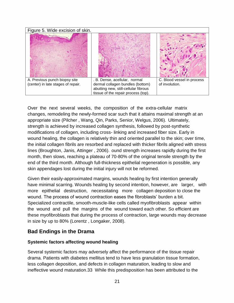
Figure 5. Wide excision of skin.
A. Previous punch biopsy site (center) in late stages of repair.
. B. Dense, acellular, normal dermal collagen bundles (bottom) abutting new, still-cellular fibrous tissue of the repair process (top).
C. Blood vessel in process of involution.
Over the next several weeks, the composition of the extra-cellular matrix
changes, remodeling the newly-formed scar such that it attains maximal strength at an
appropriate size (Pilcher , Wang, Qin, Parks, Senior, Welgus, 2006). Ultimately,
strength is achieved by increased collagen synthesis, followed by post-synthetic
modifications of collagen, including cross- linking and increased fiber size. Early in
wound healing, the collagen is relatively thin and oriented parallel to the skin; over time,
the initial collagen fibrils are resorbed and replaced with thicker fibrils aligned with stress
lines (Broughton, Janis, Attinger , 2006). ound strength increases rapidly during the first
month, then slows, reaching a plateau of 70-80% of the original tensile strength by the
end of the third month. Although full-thickness epithelial regeneration is possible, any
skin appendages lost during the initial injury will not be reformed.
Given their easily-approximated margins, wounds healing by first intention generally
have minimal scarring. Wounds healing by second intention, however, are larger, with
more epithelial destruction, necessitating more collagen deposition to close the
wound. The process of wound contraction eases the fibroblasts’ burden a bit.
Specialized contractile, smooth-muscle-like cells called myofibroblasts appear within
the wound and pull the margins of the wound toward each other. So efficient are
these myofibroblasts that during the process of contraction, large wounds may decrease
in size by up to 80% (Lorentz , Longaker, 2008).
Bad Endings in the Drama
Systemic factors affecting wound healing
Several systemic factors may adversely affect the performance of the tissue repair
drama. Patients with diabetes mellitus tend to have less granulation tissue formation,
less collagen deposition, and defects in collagen maturation, leading to slow and
ineffective wound maturation.33 While this predisposition has been attributed to the

22
microangiopathy inherent in diabetes, recent research indicates that other factors, such
as collagen glycosylation and pericapillary albumin deposition, may play a role
(Broughton, Janis, Attinger, 2006) (Bucalo, Eaglstein, Falanga, 2002). Poor nutrition
also retards the healing process. Vitamin C, in particular, is necessary for collagen
synthesis. Deprivation of this vitamin results in failure of activation of proline and lysine
hydroxylases and formation of unhydroxylated procollagen peptides, resulting in an
unstable, poorly cross-linked collagen helix. Conditions in which blood flow is
compromised - such as cardiac insufficiency or arteriosclerosis - delay the delivery of
necessary cells and factors to the scene of the wound. Certain drugs may also slow
wound healing; glucocorticoids, for example, inhibit inflammation and collagen
formation.
Local factors affecting wound healing
Local factors can also greatly affect the quality of wound healing. The size of the wound
is important (small injuries heal faster than large ones) as is the location (wounds in
richly-vascularized areas of the body, such as the face, heal relatively quickly). The
presence of foreign bodies may prolong healing, as may motion or pressure at the
wound site. However, the single most important cause of delayed healing of wounds is
infection (Kumar, Abbas, Fausto, Aster, 2010). If any beta-hemolytic Streptococcus
organisms are present, or if there are over 105 organisms of any bacterial species per
gram of tissue, the wound will not heal (Robson, 1997). More common in second-
intention healing, infection prolongs the inflammatory phase of wound healing, impeding
epithelialization, collagen deposition, and wound contraction. Bacterial endotoxins
stimulate collagenase secretion, leading to degradation of not only the forming scar, but
of surrounding normal tissue (Broughton, Janis, Attinger, 2006).
Abnormally Exuberant Wound Healing
On the other hand, sometimes acts in the drama are played so well - so exuberantly -
that the end result is an abnormally-healed wound. Occasionally, the balance of
collagen formation and degradation tips in favor of formation, leading to hypertrophic
scars or keloids. Hypertrophic scars are raised scars that remain confined to the region
of the wound. They generally occur within four weeks of injury (often a severe traumatic
or thermal injury to the dermis), and may regress over time (Broughton, Janis, Attinger,
2006) (Dabiri, Tumbarello, Turner, Van De Water, 2008). An aberrant autocrine loop
in which myofibroblasts produce excessive TGF-β, and hence collagen, appears to
contribute to their formation (Dabiri, Tumbarello, Turner, Van De Water, 2008). Keloids
are scars that have overgrown the boundaries of the initial incision, presenting as
nodules or masses of fibrous tissue (Figure 6). While keloids generally appear within
one year of the inciting injury, some may begin growing years later (Broughton, Janis,
Attinger, 2006). For unknown reasons, keloids occur much more frequently in patients

23
with darkly-pigmented skin; certain people seem to have a predisposition towards their
formation.
Figure 6:
Keloid showing dense, haphazardly-arranged collagen bundles.
Formation of granulation tissue may likewise occur in an excessive fashion. In a lesion
known as proud flesh, the process of wound healing is interrupted by masses of
granulation tissue protruding above the skin surface, preventing re- epithelialization and
appropriate scar formation (Figure 7).
Figure 7:
Proud flesh with exuberant granulation tissue.
Updates in Wound Healing
Stem Cell Research
There are two therapies, currently, that utilize stem cell sheets derived from the basal
layer of the skin for skin grafts, according to the Canadian Stem Cell Foundation.
Primarily used to treat large burns, both treatments have been approved in the United
States for clinical use.
Senior citizens with cutaneous injuries may witness a slower progression in the
restoration of their wounds due to reduced skin elasticity, slower collagen replacement
and age-related diseases, especially those that affect blood flow (Advanced Tissue

24
2017).
As a result, doctors frequently search for methods to speed up the healing process, and
stem cell therapy may do the trick. These organisms stimulate the formation of blood
vessels and naturally adjust the amount of inflammation, according to a study published
in the journal Gerontology. While the publication cites the need for additional research
into the identification and isolation of the most therapeutic cells, as well as an effective
delivery method for cell protection for this particular purpose, the results of this analysis
show promise (Advanced Tissue 2017).
As scientists keep studying these miraculous cells, more advancements will be made in
wound healing, and beyond!
Genetic Research
On March 2, 2016, the Biophysical Society publishes that:
“Researchers at Ohio State University have pinpointed a human gene product
that helps to regulate wound healing and may control scarring in people
recovering from severe injuries and damage to certain internal organs.
The protein, MG53, travels throughout the bloodstream and helps the body fix
injuries to the skin, heart, lungs, kidneys and other organs without causing scars.
It's a discovery that could help heal open wounds, decrease recovery time after
surgery and reduce the spread of infections.
"A massive scar on your skin may look bad, but imagine you have a heart attack
and get a scar on your heart--that could be lethal," says Jianjie Ma, a physiologist
at Ohio State and co-author of the presentation.
All animals carry this gene, he said, and it's almost identical no matter which
species. MG53 fixes the cell and tissue damage that occurs during everyday
living. Even simple actions, like walking or typing, will cause injuries to the body.
Usually this isn't a problem because MG53 can make repairs before there's any
serious harm.
Ma and his team genetically engineered mice without the gene that makes MG53
to see what would happen without its healing capabilities. The experiments
showed that the mice lacking MG53 had difficulty recovering from injury, because
of their compromised repair capacity; their heart would not function well under
stress conditions.
MG53 works in tandem with another protein called TGF Beta, a type of "cytokine"
protein that also heals wounds, but the healing process happens so quickly that it

25
causes scars. If you have more TGF Beta in your bloodstream than MG53, you
scar easily.
Ma's goal is to develop a therapy that will inhibit TGF Beta and promote MG53.
Medical professionals can use the therapy during procedures to promote quick,
scarless healing. His next step is to identify a small compound that can do this
and eventually test whether it has the desired effect in human trials.”
Conclusion
Wound healing is a carefully-scripted drama performed countless times throughout a
human lifespan. Though occasionally the drama does not go as planned, in most cases
damage is contained, dead cells and tissue are removed, and a scar restores integrity
to the injured tissue – a triumphant performance indeed.
References
Advanced Tissue. How Stem Cells Benefit Wound Healing. April 20, 2017
https://www.advancedtissue.com/cutting-edge-techniques-for-chronic-wound-healing/
Accessed April 2017.
Andrae J, Gallini R, Betsholtz C. Role of platelet-derived growth factors in physiology
and medicine. Genes Dev 2008; 22(10):1276-1312.
Apte U, Singh S, Zeng G, Cieply B, Virji MA, Wu T, et al. Beta-catenin activation
promotes liver regeneration after acetaminophen-induced injury. Am J Pathol 2009;
175(3):1056-65.
Attisano L, Wrana JL. Signal transduction by the TGF-beta superfamily. Science 2002;
296(5573):1646-1647.
Bessa PC, Casal M, Reis RL. Bone morphogenetic proteins in tissue engineering: the
road from laboratory to the clinic. Part 1 – basic concepts. J Tiss Eng Regen Med 2008;
2(1):1-13.
Biophysical Society. "Gene identified that helps wound healing: New research on gene
that regulates healing and may control scarring,." ScienceDaily, 2 March 2016.
www.sciencedaily.com/releases/2016/03/160302135149.htm Accessed April 2017.
Bluff J, Ferguson M, O’Kane S, Ireland G. Bone marrow-derived endothelial progenitor
cells do not contribute significantly to new vessels during incisional wound healing. Exp
Hematol 2007; 35(3):500-506.
Broughton G, Janis JE, Attinger CE. Wound healing: An overview. Plast Reconstr Surg
2006(suppl); 117:1-32.

26
Bucalo B, Eaglstein WH, Falanga V. Inhibition of cell proliferation by chronic wound
fluid. Wound Rep Regen 2002; 1(3):181-186.
Cao R, Brakenhielm E, Pawliuk R, Wariaro D, Post MJ, Wahlberg E, et al. Angiogenic
synergism, vascular stability and improvement of hind-limb ischemia by a combination
of PDGF- BB and FGF-2. Nat Med 2003; 9(5):604-613.
Cherezov V, Rosenbaum DM, Hanson MA, Rasmussen SG, Thian FS, Kobilka TS, et
al. High-resolution crystal structure of an engineered human b2-adrenergic G protein-
coupled receptor. Science 2007; 318(5854):1258-1265.
Chow LW, Loo WT, Yuen KY, Cheng C. The study of cytokine dynamics at the
operation site after mastectomy.
Conover JC, Notti RQ. The neural stem cell niche. Cell Tissue Res 2008; 331:211-224.
Dabiri G, Tumbarello DA, Turner CE, Van De Water L. Hic-5 promotes the hypertrophic
scar myofibroblast phenotype by regulating the TGF-b-1 autocrine loop. J Invest
Dermatol 2008; 128:2518-2525.
Diegelmann R. Analysis of collagen synthesis. Methods Mol Med 2003; 78:349-358.
Escudier B. Anti-VEGF therapy for renal cell carcinoma. Clin Adv Hematol Oncol 2007;
5(7):530-531.
Fedyk ER, Jones D, Critchley HO, Phipps RP, Blieden TM, Springer TA. Expression of
stromal-derived factor-1 is decreased by IL-1 and TNF and in dermal wound healing. J
Immunol 2001; 166:5749-5754.
Holmes K, Roberts OL, Thomas AM, Cross MJ. Vascular endothelial growth factor
receptor-2: Structure, function, intracellular signaling and therapeutic inhibition. Cell
Signal 2007; 19(10):2003-2012.
Horan GS, Wood S, Ona V, Li DJ, Lukashev ME, Weinreb PH, et al. Partial inhibition of
integrin avb prevents pulmonary fibrosis without exacerbating inflammation. Am J Resp
Crit Care Med 2008; 177:56-65.
Hynes RO. Integrins: bidirectional, allosteric signaling machines. Cell 2002 110(6):673-
687.
Itoh F, Asao H, Sugamura K, Heldin CH, ten Dijke P, Itoh S. Promoting bone
morphogenetic protein signaling through negative regulation of inhibitory Smads. EMBO
J 2001; 20(15):4132-4142.
Kumar V, Abbas AK, Fausto N, Aster JC. Tissue Renewal, Regeneration and Repair. In:

27
Robbins and Cotran Pathologic Basis of Disease. Eighth edition. Philadelphia: Elsevier,
2010:79-110.
Lawrence WT, Diegelmann RF. Growth factors in wound healing. Clin Dermatol 1994;
12(1):157-169.
Lorentz HP, Longaker MT. Wounds: Biology, pathology and management. In: Norton
JA, Barie PS, Bollinger RR, Chang AE, Lowry S, Mulvihill SJ, et al., eds. Surgery: Basic
science and clinical evidence. 2nd edition. New York: Springer, 2008:191-208.
Münger K, Howley PM. Human papillomavirus immortalization and transformation
functions. Virus Res 2002 89(2):213-228.
Nigg EA. Cyclin-dependent protein kinases: key regulators of the eukaryotic cell cycle.
Bioessays 1995; 17(6):471-480.
Pierce GF, Mustoe TA, Altrock BW, Deuel TF, Thomason A. Role of platelet-derived
growth factor in wound healing. J Cell Biochem 2004; 45(4):319-326.
Pilcher BK, Wang M, Qin X, Parks WC, Senior RM, Welgus HG. Role of matrix
metalloproteinases and their inhibition in cutaneous wound healing and allergic contact
hypersensitivity. Ann NY Acad Sci 2006; 878:12-24.
Rafii S, Lyden D. Therapeutic stem and progenitor cell transplantation for organ
vascularization and regeneration. Nat Med 2003; 9:702-712.
Rawlings JS, Rosler KM, Harrison DA. The JAK/STAT signaling pathway. J Cell
Science 2004; 117:1281-1283.
Robinson DR, Wu YM, Lin SF. The protein tyrosine kinase family of the human genome.
Oncogene 2000; 19(49):5548-5557.
Robson MC. Wound infection: A failure of wound healing caused by an imbalance of
bacteria. Surg Clin North Am 1997; 77(3):637-650.
Schmidt C, Bladt F, Goedecke S, Brinkmann V, Zschiesche W, Sharpe M, et al. Scatter
factor/hepatocytes growth factor is essential for liver development. Nature 1995;
373:699-702.
Smola H, Thiekötter, Fusenig NE. Mutual induction of growth factor gene expression by
epidermal-dermal interaction. J Cell Biol 1993; 122(2):417-429.
Tumbar T, Guasch G, Greco V, Blanpain C, Lowry W, Rendl M, et al. Defining the
epithelial stem cell niche in skin. Science 2004; 303(5656):359-363.
Werner S, Grose R. Regulation of wound healing by growth factors and cytokines.
28
Physiol Rev 2003; 83:835-870.
Witte MB, Barbul A. General principles of wound healing. Surg Clin North Am 1997;
77(3):509-528.
Xia YP, Zhao Y, Marcus J, Jiminez PA, Ruben SM, Moore PA, et al. Effects of
keratinocyte growth factor-2 (KGF-2) on wound healing in an ischaemia-impaired rabbit
ear model and on scar formation. J Pathol 1999; 188(4):431-438.
Author’s note
Portions of this course were derived from the author’s article: Krafts K. Tissue Repair:
the Hidden Drama. Organogenesis 2010; 6. The Copyright Revision Act (PL 94-553),
which became effective January 1, 1978, states that the copyright of a work is vested in
the author from the moment of creation. Therefore, all authors who wish to publish in
Cell Cycle must grant an exclusive license to Landes Bioscience. It is understood that
the authors grant Landes Bioscience an exclusive license to publish the work and also
grant rights of reproduction, derivation, distribution, sale and display. Landes Bioscience
has granted back to the author the copyright permission to use portions of the above
article in preparation of this course.
Course Exam: Wound Healing: A New Understanding of the Drama
1. Quiescent tissues include all of the following except:
A. Liver
B. Kidney
C. Pancreas
D. Heart
2. Which of the following cells plays a central role in scar formation?
A. Eosinophil
B. Mast cell
C. Fibroblast
D. Dendritic cell
3. The most important role of transforming growth factor beta (TGF-β) in wound healing
is:
A. Promotion of fibrosis
B. Attraction of Neutrophils
C. Activation of macrophages
D. Promotion of angiogenesis
29
4. The end result of receptor mediated signal transduction is:
A. Cell death
B. A change in gene expression
C. Influx of calcium into the cell
D. Neoplastic transformation
5. In which phase of the cell cycle is DNA synthesized?
A. G1
B. S
C. G2
D. M
6. Which of the following is a function of the extra-cellular matrix?
A. Imparts resilience to soft tissues and bone
B. Stores growth factors
C. Acts as a scaffold for migrating cells
D. Facilitates cell growth
E. All of the above
7. An injury to a continuously dividing tissue (such as oral mucosa) that disrupts the
underlying stromal framework will result in:
A. Regeneration
B. Scarring
8. Which of the following is true of first-intention wounds?
A. Relatively larger than second-intention wounds
B. Healing is generally slow
C. Inflammation is abundant
D. Scarring is minimal
9. What happens first in the immediate aftermath of a wound?
A. Neutrophils flood the damaged tissue
B. Exposed collagen activates coagulation
C. Epithelial cells begin to proliferate at the margins
D. Macrophages enter the damaged tissue
E. Fibroblasts secrete collagen

30
10. When does granulation tissue appear?
A. Within the first few days after a wound occurs
B. At approximately 10 days after a wound occurs
C. Between two and three weeks after a wound occurs
D. When the final scar is being formed
11. A scar has more tensile strength than normal skin.
A. True
B. False
12. A mature scar is composed of:
A. Fibrin
B. Elastin
C. Collagen
D. Smooth muscle
13. An insufficiency of which vitamin causes the formation of unstable collagen?
A. A
B. B
C. C
D. D
E. E
14. What is the single most important cause of delayed wound healing?
A. Diabetes
B. Drug side effects
C. Foreign bodies
D. Size of the wound
E. Infection
15. Keloids contain an excess of:
A. Collagen
B. Fibrin
C. Granulation tissue
D. Blood vessels
E. Inflammation