Embed Size (px)
Citation preview
Last updated: August 28, 2015
Content Creators: Members of the South West Regional Wound Care Program’s Clinical Practice and Knowledge Translation Learning Collaborative
Wound Debridement
2
So
uth
West R
egio
na
l W
ound
Care
Pro
gra
m
1. Develop an understanding of the significance of necrotic tissue
2. Review therapeutic interventions for necrotic tissue including:
1. Mechanical debridement
2. Enzymatic debridement
3. Sharp debridement
4. Autolytic debridement
5. Biologic Debridement
3. Review the outcome measurements of debridement and referral criteria
Learning Objectives
3
So
uth
West R
egio
na
l W
ound
Care
Pro
gra
m
Images/illustrations obtained via Google Images, unless otherwise stated
Photographs and Illustrations
4
So
uth
West R
egio
na
l W
ound
Care
Pro
gra
m
SIGNIFICANCE OF NECROTIC TISSUE
5
So
uth
West R
egio
na
l W
ound
Care
Pro
gra
m
• Necrotic tissue impairs wound healing as it is a physical barrier to:
• Granulation tissue formation
• Wound contraction
• Re-epithelialization
• Necrotic tissue may also harbor bacteria, which could lead to wound infection, thus impairing wound healing
• The more necrotic tissue there is in a wound, the1, 5:
• More severe the damage is
• Longer it will take the close the wound
Necrotic Tissue1-4

6
So
uth
West R
egio
na
l W
ound
Care
Pro
gra
m
• As tissues die they change in:
• Color
• Consistency
• Adherence
Necrotic Tissue1
7
So
uth
West R
egio
na
l W
ound
Care
Pro
gra
m
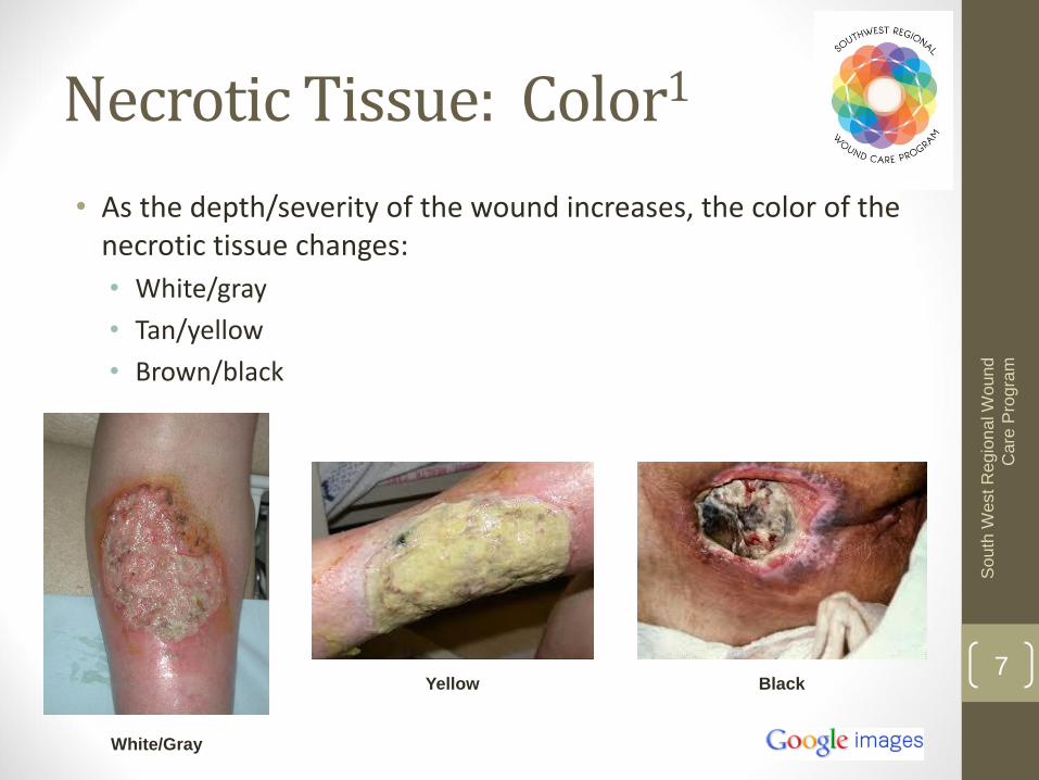
Black Yellow
White/Gray
• As the depth/severity of the wound increases, the color of the necrotic tissue changes:
• White/gray
• Tan/yellow
• Brown/black
Necrotic Tissue: Color1
8
So
uth
West R
egio
na
l W
ound
Care
Pro
gra
m
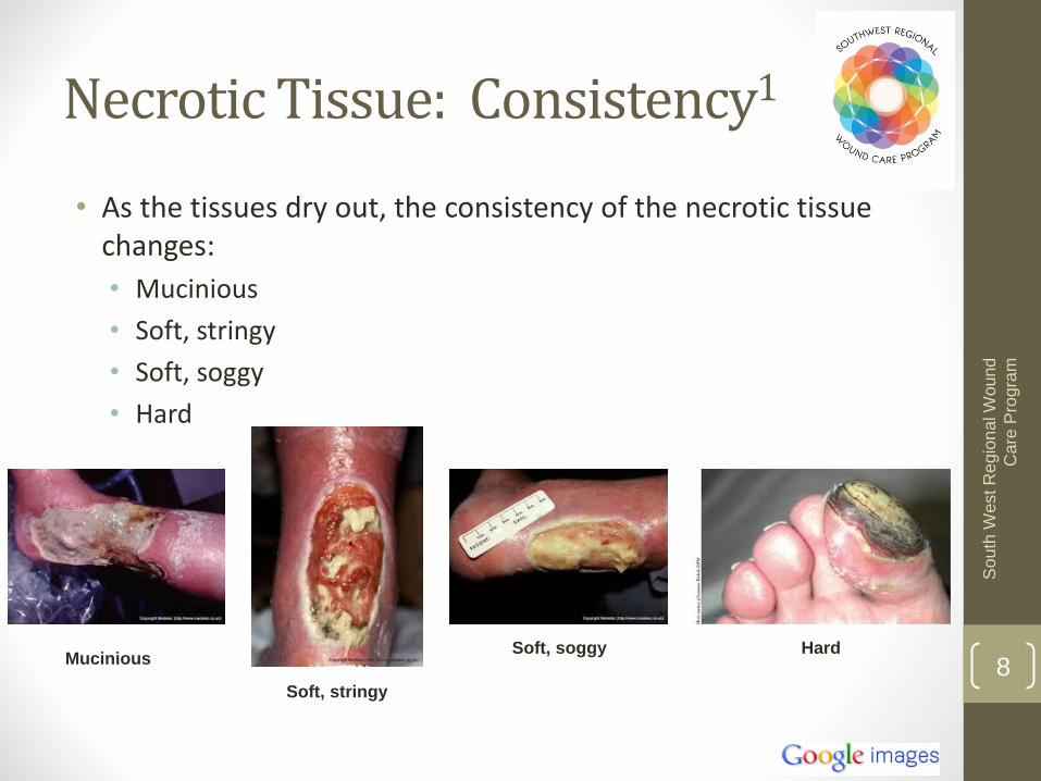
Hard Soft, soggy
Soft, stringy
Mucinious
• As the tissues dry out, the consistency of the necrotic tissue changes:
• Mucinious
• Soft, stringy
• Soft, soggy
• Hard
Necrotic Tissue: Consistency1
9
So
uth
West R
egio
na
l W
ound
Care
Pro
gra
m
• Consistency of necrotic tissue is related to its moisture content and refers to its cohesiveness1
• Consistency also varies as tissue damage worsens/deepens1,5-6:
• Slough: yellow/tan, thin, mucinious or stringy partial thickness damage
• Eschar: brown/black, soft of hard full-thickness damage
Consistency Continued
10
So
uth
West R
egio
na
l W
ound
Care
Pro
gra
m
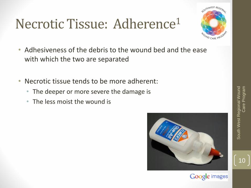
• Adhesiveness of the debris to the wound bed and the ease with which the two are separated
• Necrotic tissue tends to be more adherent:
• The deeper or more severe the damage is
• The less moist the wound is
Necrotic Tissue: Adherence1
11
So
uth
West R
egio
na
l W
ound
Care
Pro
gra
m
Wo
rse
nin
g Ti
ssu
e D
amag
e
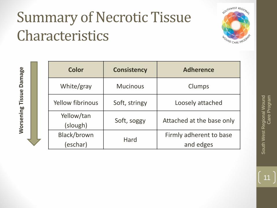
Color Consistency Adherence
White/gray Mucinous Clumps
Yellow fibrinous Soft, stringy Loosely attached
Yellow/tan
(slough) Soft, soggy Attached at the base only
Black/brown
(eschar) Hard
Firmly adherent to base
and edges
Summary of Necrotic Tissue Characteristics
12
So
uth
West R
egio
na
l W
ound
Care
Pro
gra
m
• Predominant types of necrotic tissue include:
• Slough
• Fibrin
• Eschar
• Gangrene
• Hyperkeratosis
Types of Necrotic Tissue
13
So
uth
West R
egio
na
l W
ound
Care
Pro
gra
m
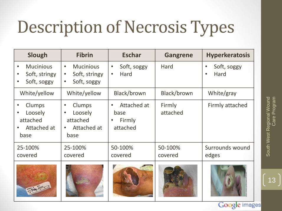
Slough Fibrin Eschar Gangrene Hyperkeratosis
• Mucinious • Soft, stringy • Soft, soggy
• Mucinious • Soft, stringy • Soft, soggy
• Soft, soggy • Hard
Hard • Soft, soggy • Hard
White/yellow White/yellow Black/brown Black/brown White/gray
• Clumps • Loosely attached • Attached at base
• Clumps • Loosely attached • Attached at base
• Attached at base • Firmly attached
Firmly attached
Firmly attached
25-100% covered
25-100% covered
50-100% covered
50-100% covered
Surrounds wound edges
Description of Necrosis Types

14
So
uth
West R
egio
na
l W
ound
Care
Pro
gra
m
• Arterial/ischemic wounds:
• Dry gangrene
• Thick, dry, desiccated black/gray appearance
• Firmly adherent
• May be surrounded by an erythematous halo
• Neurotropic wounds:
• Do not present with necrotic tissue in wound typically
• Have hyperkeratosis surrounding wound
• Venous leg ulcers:
• Eschar or slough
• Usually yellow fibrous material
• Pressure Sores:
• Relates to the depth of the injury
Type of Necrosis By Wound Etiology
15
So
uth
West R
egio
na
l W
ound
Care
Pro
gra
m
DEBRIDEMENT: INTERVENTION FOR NECROTIC TISSUE
16
So
uth
West R
egio
na
l W
ound
Care
Pro
gra
m
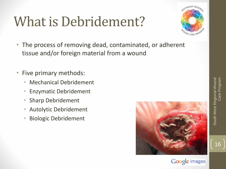
• The process of removing dead, contaminated, or adherent tissue and/or foreign material from a wound
• Five primary methods:
• Mechanical Debridement
• Enzymatic Debridement
• Sharp Debridement
• Autolytic Debridement
• Biologic Debridement
What is Debridement?
17
So
uth
West R
egio
na
l W
ound
Care
Pro
gra
m
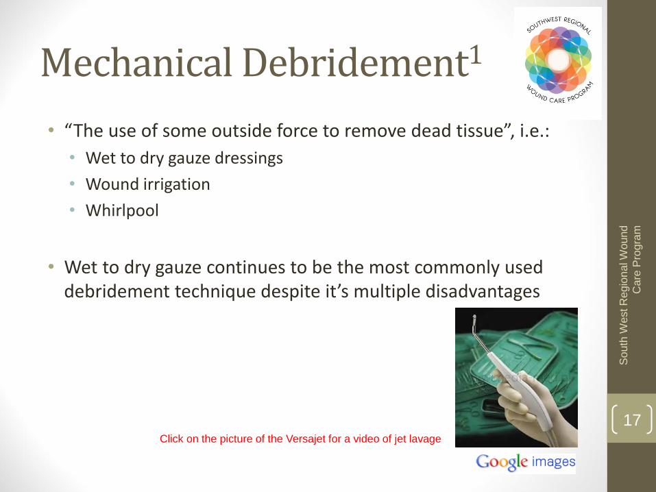
Click on the picture of the Versajet for a video of jet lavage
• “The use of some outside force to remove dead tissue”, i.e.:
• Wet to dry gauze dressings
• Wound irrigation
• Whirlpool
• Wet to dry gauze continues to be the most commonly used debridement technique despite it’s multiple disadvantages
Mechanical Debridement1

18
So
uth
West R
egio
na
l W
ound
Care
Pro
gra
m
• Advantages:
• Familiar to health care providers
• Wound irrigation can reduce bacterial burden
• Whirlpool may soften necrotic debris
• Disadvantages (wet-to-dry gauze):
• Non-selective
• Rarely applied correctly
• Painful
• More costly (labor and supplies)
• May cause maceration
• Releases airborne organisms and causes cross-contamination9
Mechanical Debridement Continued1

19
So
uth
West R
egio
na
l W
ound
Care
Pro
gra
m
“Applying a concentrated, commercially prepared (proteolytic) enzyme to the surface of the necrotic tissue, in the expectation that it will aggressively degrade necrosis by digesting devitalized tissue”
Requires a physician order and must be used according to the manufacturers instructions
Cannot be used on dry wounds … any eschar present must be cross hatched
Enzymatic Debridement1

20
So
uth
West R
egio
na
l W
ound
Care
Pro
gra
m
Advantages: Selective
Effective in combination with other debridement techniques
Disadvantages: Enzymatic use is prolonged more than necessary, increasing
costs
Can be slow – 3-30 days to achieve a completely clean wound bed (it is faster than autolysis however)
Requires a specific pH range (may cause local irritation due to pH changes)
May be inactivated by contact with heavy metals (zinc or silver)
Risk of maceration and infection
Requires frequent dressing changes (1-3 times per day)
Enzymatic Debridement Continued1

21
So
uth
West R
egio
na
l W
ound
Care
Pro
gra
m
• Performed either one time (surgical) or sequentially (conservative)
• Surgical sharp debridement:
• Use of scalpel, scissors, or other sharp instruments
• Removal of viable and non-viable tissue
• Most rapid and effective
• May convert chronic wound into an acute wound
• Requires analgesics and availability of cautery equipment
• Indicated for removal of thick, adherent and/or large amounts of non-viable tissue and when advancing cellulitis or signs of sepsis are present
• Requires a certain level of expertise, education and skill
• Risk of bleeding
Click here for a video of surgical debridement
Sharp Debridement1
22
So
uth
West R
egio
na
l W
ound
Care
Pro
gra
m
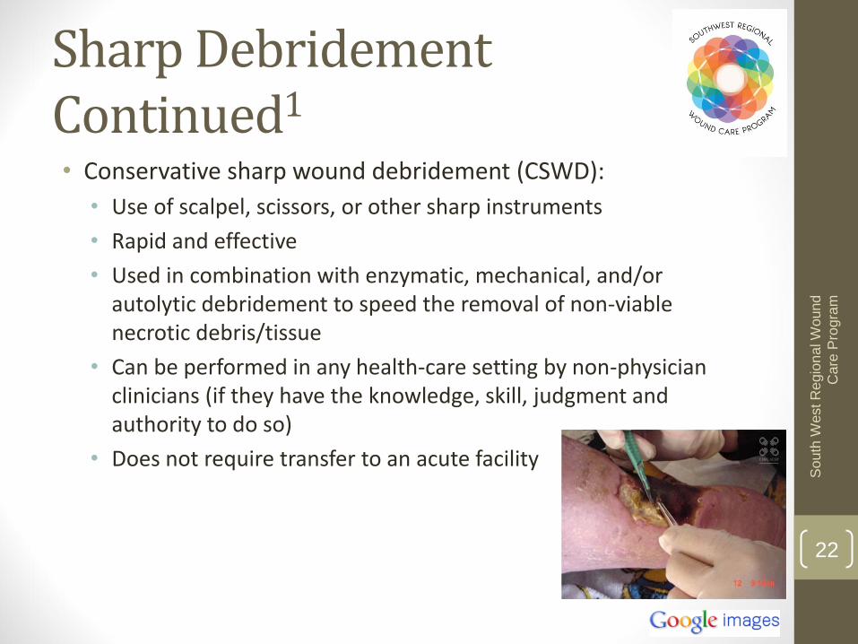
• Conservative sharp wound debridement (CSWD):
• Use of scalpel, scissors, or other sharp instruments
• Rapid and effective
• Used in combination with enzymatic, mechanical, and/or autolytic debridement to speed the removal of non-viable necrotic debris/tissue
• Can be performed in any health-care setting by non-physician clinicians (if they have the knowledge, skill, judgment and authority to do so)
• Does not require transfer to an acute facility
Sharp Debridement Continued1
23
So
uth
West R
egio
na
l W
ound
Care
Pro
gra
m
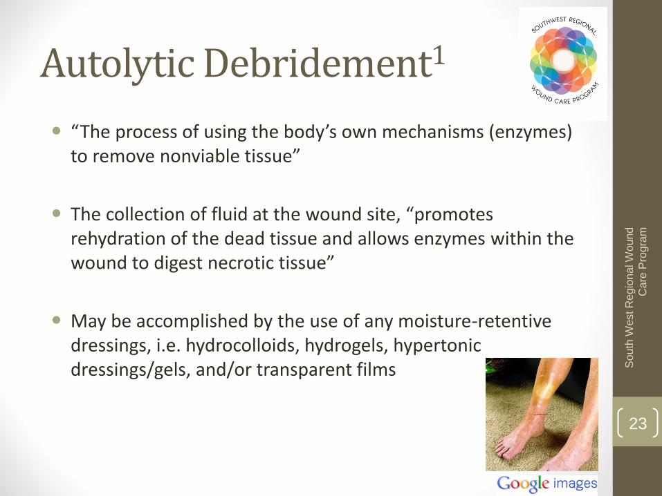
“The process of using the body’s own mechanisms (enzymes) to remove nonviable tissue”
The collection of fluid at the wound site, “promotes rehydration of the dead tissue and allows enzymes within the wound to digest necrotic tissue”
May be accomplished by the use of any moisture-retentive dressings, i.e. hydrocolloids, hydrogels, hypertonic dressings/gels, and/or transparent films
Autolytic Debridement1

24
So
uth
West R
egio
na
l W
ound
Care
Pro
gra
m
Advantages:
Painless in the majority of people with wounds
Effective, versatile, and easy to perform
Selective
Low cost
Can be used in conjunction with other debridement techniques
Disadvantages:
Slow
Caregiver education required for compliance
Autolytic Debridement Continued1

25
So
uth
West R
egio
na
l W
ound
Care
Pro
gra
m
A.k.a. larval/maggot debridement therapy (use of medical grade green bottle fly larvae/maggots)
Controlled “application of disinfected maggots to the wound to remove the nonviable tissue”10
Regulated by the FDA as a prescription only medical device
Maggots are left in the wound for 2-3 days . They secrete “proteolytic enzymes that break down necrotic tissue and then ingest the liquefied tissue”10
The secretions also have antimicrobial properties, promote growth of human fibroblasts and improve granulation tissue formation11-12
Biologic Debridement1
26
So
uth
West R
egio
na
l W
ound
Care
Pro
gra
m
Click on the maggots to see a
short video on this therapy
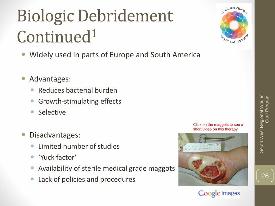
Widely used in parts of Europe and South America
Advantages:
Reduces bacterial burden
Growth-stimulating effects
Selective
Disadvantages:
Limited number of studies
‘Yuck factor’
Availability of sterile medical grade maggots
Lack of policies and procedures
Biologic Debridement Continued1

27
So
uth
West R
egio
na
l W
ound
Care
Pro
gra
m
Debridement
Type Definition Examples
Mechanical Use of an outside force to remove non-viable
tissue
Wet-to-dry gauze, wound
irrigation, whirlpool,
pulsed lavage
Enzymatic Application of a concentrated, commercially
prepared enzyme to digest non-viable tissue Collagenase
Sharp Use of sharp instruments to remove non-
viable tissue Scalpel, scissor, curette use
Autolytic
Use of the body’s own enzymes in wound
fluid along with moisture retentive dressings
to degrade non-viable tissue
Use of hydrocolloids, films,
hydrogels, and/or
hypertonic dressings
Biologic* Application of medical grade maggots to
remove non-viable tissue
Larval debridement
therapy
Review of Types of Debridement
28
So
uth
West R
egio
na
l W
ound
Care
Pro
gra
m
• To remove the physical barrier to epidermal resurfacing, contraction, or granulation
• To reduce bacteria burden by removing necrotic tissue
• To convert a chronic wound to an acute wound by stimulating the healing cascade
• To facilitate earlier coverage of the wound with active dressings or biologicals
Why Debride?
29
So
uth
West R
egio
na
l W
ound
Care
Pro
gra
m
• Under the 1991 Regulated Health Professions Act (Ontario), debridement is within the controlled acts authorized for nursing
• An RN or an RN(EC) who meets certain conditions, i.e. has the knowledge, skill, judgment and authority, can initiate and/or provide an order for an RN or RPN to perform care of wound below the dermis or mucous membrane, which includes cleansing, soaking, irrigating, probing, debriding, packing, dressing8
Who Can Debride?

30
So
uth
West R
egio
na
l W
ound
Care
Pro
gra
m
• The Long Term Care Homes Act and the Public Hospitals Act do not allow a nurse to initiate CSWD in the absence of a physician order
• There is no Act that precludes nurses in the community from performing CSWD in the absence of a physician order, but it is STRONGLY suggested that the nurse communicates her intent to perform CSWD to the primary care physician BEFORE doing so
Who Can Debride: CSWD
31
So
uth
West R
egio
na
l W
ound
Care
Pro
gra
m
• Specialized practice skills such as CSWD are not generally included in the RN’s basic preparation; therefore additional instruction and supervision are necessary to ensure the individual is competent to perform the identified skills or acts
Who Can Debride Continued

32
So
uth
West R
egio
na
l W
ound
Care
Pro
gra
m
• The nurse who performs CSWD is expected to have:
• A good knowledge of relevant anatomy
• The ability to identify viable tissue
• Access to adequate equipment, lighting and assistance
• The capacity to explain the procedure and obtain informed consent
• The ability to manage pain and discomfort prior to, during, and following the procedure
• The skill to deal with complications such as bleeding
• The ability to recognize their skill limitations and those of the technique
• Knowledge of infection control practices
• The ability to utilize secondary debridement techniques if needed
Who Can Debride Wounds
33
So
uth
West R
egio
na
l W
ound
Care
Pro
gra
m
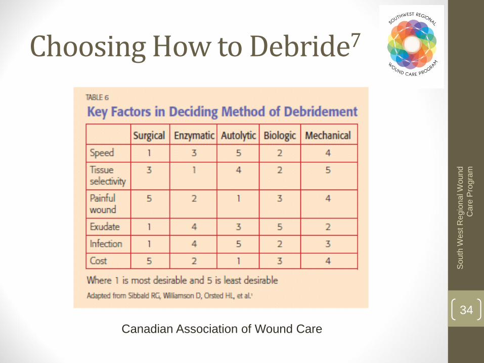
• After a thorough holistic assessment of the person and their wound, and determination that debridement is indicated, you must first choose the most appropriate type(s) of debridement. This is dependent on the:
• Knowledge, skill and authority of the health care practitioner
• Availability of required resources
• Overall condition of the person with the wound, and their ‘healability’
• Characteristics of the wound and wound tissue
• Presence of wound related pain
• Required speed and tissue selectivity of debridement
• Costs associated with available debridement techniques
• Presence of wound infection
• Physical environment
How Do We Debride?
34
So
uth
West R
egio
na
l W
ound
Care
Pro
gra
m
Canadian Association of Wound Care
Choosing How to Debride7
35
So
uth
West R
egio
na
l W
ound
Care
Pro
gra
m
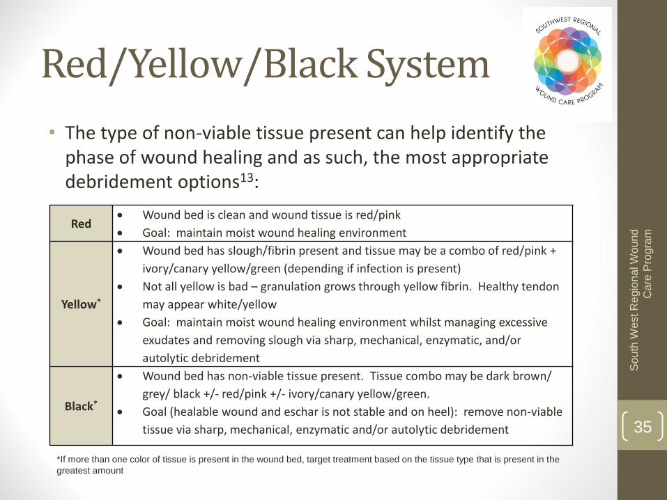
*If more than one color of tissue is present in the wound bed, target treatment based on the tissue type that is present in the
greatest amount
Red Wound bed is clean and wound tissue is red/pink
Goal: maintain moist wound healing environment
Yellow*
Wound bed has slough/fibrin present and tissue may be a combo of red/pink +
ivory/canary yellow/green (depending if infection is present)
Not all yellow is bad – granulation grows through yellow fibrin. Healthy tendon
may appear white/yellow
Goal: maintain moist wound healing environment whilst managing excessive
exudates and removing slough via sharp, mechanical, enzymatic, and/or
autolytic debridement
Black*
Wound bed has non-viable tissue present. Tissue combo may be dark brown/
grey/ black +/- red/pink +/- ivory/canary yellow/green.
Goal (healable wound and eschar is not stable and on heel): remove non-viable
tissue via sharp, mechanical, enzymatic and/or autolytic debridement
• The type of non-viable tissue present can help identify the phase of wound healing and as such, the most appropriate debridement options13:
Red/Yellow/Black System
36
So
uth
West R
egio
na
l W
ound
Care
Pro
gra
m
OUTCOME MEASUREMENTS OF DEBRIDEMENT AND REFERRAL CRITERIA
37
So
uth
West R
egio
na
l W
ound
Care
Pro
gra
m
• Three appropriate characteristics for evaluating the effectiveness of debridement are the:
• Type of necrotic tissue
• Amount of necrotic tissue
• Adherence of the necrotic tissue to the wound
Outcome Measures1
38
So
uth
West R
egio
na
l W
ound
Care
Pro
gra
m
• Amount should diminish progressively if therapy appropriately
• Can be measured:
• Using linear measurements (length x width)
• By determining percentage of wound bed covered
• By photography
• Estimate percentages in the following way:
• <25% wound bed covered
• 25-50% wound covered
• >50 and <75% wound covered
• 75-100% wound covered
Amount of Necrotic Tissue1

39
So
uth
West R
egio
na
l W
ound
Care
Pro
gra
m
• Type of necrotic tissue should change as the wound improves, when conservative methods of debridement are used
• As necrotic tissue rehydrates its appearance will change from dry/black, to soggy/soft/yellow, to mucinous easily dislodged tissue
• Can rate the type of necrotic tissue as:
• White/gray nonviable tissue and/or non-adherent yellow slough
• Loosely adherent yellow slough
• Adherent soft black eschar
• Firmly adherent, hard black eschar
Type of Necrotic Tissue1
40
So
uth
West R
egio
na
l W
ound
Care
Pro
gra
m
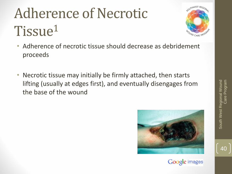
• Adherence of necrotic tissue should decrease as debridement proceeds
• Necrotic tissue may initially be firmly attached, then starts lifting (usually at edges first), and eventually disengages from the base of the wound
Adherence of Necrotic Tissue1

41
So
uth
West R
egio
na
l W
ound
Care
Pro
gra
m
• Dry gangrene or dry ischemic wounds
• Elevated temperature
• No wound improvement
• Evidence of cellulitis or gross infection
• Exposed bone or tendon
• Evidence of abscess
Referral Criteria1
42
So
uth
West R
egio
na
l W
ound
Care
Pro
gra
m
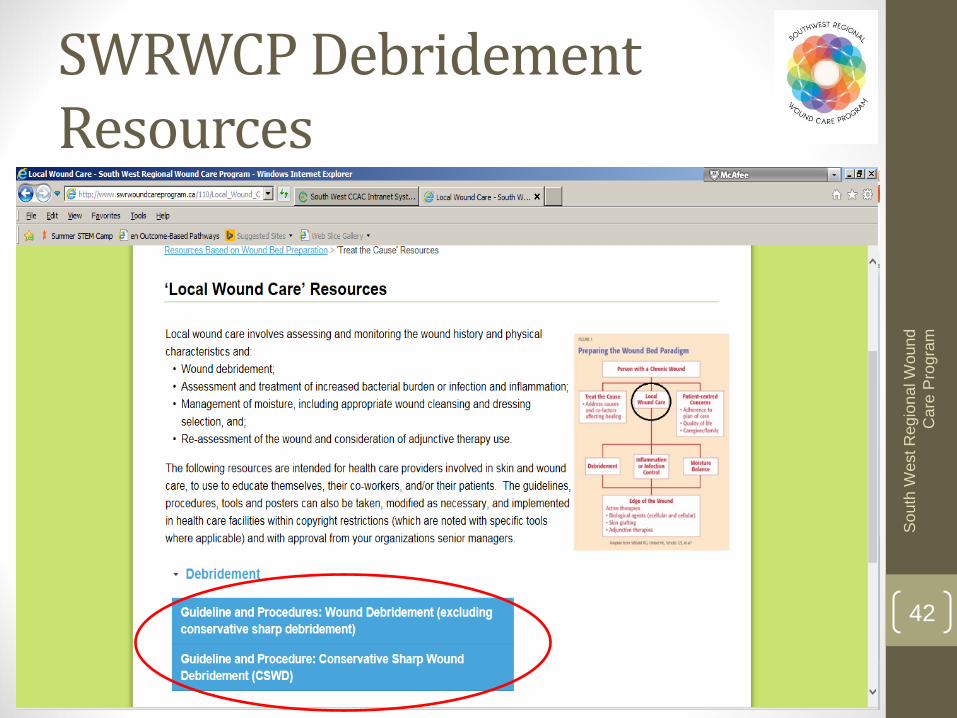
SWRWCP Debridement Resources

43
So
uth
West R
egio
na
l W
ound
Care
Pro
gra
m
1. The significance of necrotic tissue
2. Therapeutic interventions for necrotic tissue including:
1. Mechanical debridement
2. Enzymatic debridement
3. Sharp debridement
4. Autolytic debridement
5. Biologic Debridement
3. Outcome measurements of debridement and referral criteria
Review

44
So
uth
West R
egio
na
l W
ound
Care
Pro
gra
m
For more information visit: swrwoundcareprogram.ca
45
So
uth
West R
egio
na
l W
ound
Care
Pro
gra
m
1. Bates-Jensen BM, Apeles NCR. Management of necrotic tissue. In: Sussman C, Bates-Jensen B., eds. Wound Care: A collaborative practice manual for health professionals. Third Ed. Baltimore: Lippincott Williams & Wilkins, 1997:197-214.
2. Alterescu V, Alterescu K. Etiology and treatment of pressure ulcers. Decubitus. 1988;1:28-35.
3. Winter G. Epidermal regeneration studied in the domestic pig. In: Hung TK, Dunphy JE, eds. Fundamentals of Wound Management. New York: Appleton-Century-Crofts; 1979:71-111.
4. Sapico FL, Ginunas VJ, Thornhill-Hoynes M, et al. Quantitative microbiology of pressure sores in different stages of healing. Diagn Biol Infect Dis. 1986;5:31-38.
5. Shea D. Pressure sores: Classification and management. Clin Orthop. 1975:112:89-100.
6. Witkowski JA, Parish LC. Histopathology of the decubitus ulcer. J Am Acad Dermatol. 1982;6:1014-1021.
7. Sibbald RG, Williamson D, Orsted HL, et al. Preparing the wound bed: Debridement, bacterial balance and moisture balance. Ostomy/Wound Management. 2000;46(11):14-35.
8. College of Nurses of Ontario. Decisions about procedures and authority. Pub. No. 41071. Toronto. Last retrieved October 21, 2014 from: http://www.cno.org/Global/docs/prac/41071_Decisions.pdf
9. Lawrence JC, Lilly HA, Kidson A. Wound dressings and airborne dispersal of bacteria. Lancet. 1992;339(8796):807.
10. Zacur H, Kirsner RS. Debridement: Rationale and therapeutic options. Wounds: Compendium of Clinical Research and Practice. 2002;14(7Suppl E):2E-7E.
11. Prete PE. Growth effects of Phaenicia sericata larval extracts on fibroblasts: Mechanism for wound healing by maggot therapy. Life Sci. 1997;60(8):505-510.
12. Mumcuoglu KY. Clinical applications for maggots in wound care. Am J Clin Dermatol. 2001;2(4):219-227.
13. Krasner D. Wound care: how to use the red-yellow-black system. Am J Nurs. 1995:95(5):44–47.
References