Embed Size (px)
Citation preview

World Journal of Gastrointestinal PathophysiologyWorld J Gastrointest Pathophysiol 2018 February 15; 9(1): 1-36
ISSN 2150-5330 (online)
Published by Baishideng Publishing Group Inc

Contents
February 15, 2018|Volume 9|Issue 1|WJGP|www.wjgnet.com I
Quarterly Volume 9 Number 1 February 15, 2018
MINIREVIEWS1 Acutecholangitis-anupdate
Ahmed M
ORIGINAL ARTICLE Retrospective Cohort Study
8 Emergencyresectionsurgeryforcolorectalcancer:Patternsofrecurrentdiseaseandsurvival
Littlechild J, Junejo M, Simons AM, Curran F, Subar D
Observational Study
18 AbundanceofEnterobacteriaceae inthecolonmucosaindiverticulardisease
Linninge C, Roth B, Erlanson-Albertsson C, Molin G, Toth E, Ohlsson B
28 Livercirrhosis-effectonQTintervalandcardiacautonomicnervoussystemactivity
Tsiompanidis E, Siakavellas SI, Tentolouris A, Eleftheriadou I, Chorepsima S, Manolakis A, Oikonomou K, Tentolouris N

EditorialBoardMemberofWorldJournalofGastrointestinalPathophysiology,RajeevGarg,PhD,AssistantProfessor,DepartmentofPharmaceutics,AsbasjsmCollegeofPharmacy,Punjab140111,India
World Journal of Gastrointestinal Pathophysiology (World J Gastrointest Pathophysiol, WJGP, online ISSN 2150-5330, DOI: 10.4291), is a peer-reviewed open access academic journal that aims to guide clinical practice and improve diagnostic and therapeutic skills of clinicians.
WJGP is to report rapidly the most recent results in basic and clinical research on gastrointestinal pathophysiology, including all aspects of normal or abnormal function of the gastrointestinal tract, hepatobiliary system, and pancreas. WJGP specifically covers growth and development, digestion, secretion, absorption, metabolism and motility relative to the gastrointestinal organs, as well as immune and inflammatory processes, and neural, endocrine and circulatory control mechanisms that affect these organs. This journal will also report new methods and techniques in gastrointestinal pathophysiological research. We encourage authors to submit their manuscripts to WJGP. We will give priority to manuscripts that are supported by major national and international foundations and those that are of great basic and clinical significance.
World Journal of Gastrointestinal Pathophysiology is now indexed in PubMed, PubMed Central.
ContentsWorld Journal of Gastrointestinal Pathophysiology
Volume 9 Number 1 February 15, 2018
EDITORS FOR THIS ISSUE
NAMEOFJOURNALWorld Journal of Gastrointestinal Pathophysiology
ISSNISSN 2150-5330 (online)
LAUNCHDATEApril 15, 2010
FrequencyQuarterly
EDITOR-IN-CHIEFThomas Y Ma, MD, PhD, Professor, Chief, Division of Gastroenterology and Hepatology, University of New Mexico, MSC10 5550, 1 UNM, Albuquerque, NM 87131, United States
EDITORIALBOARDMEMBERSAll editorial board members resources online at http://www.wjgnet.com/2150-5330/editorialboard.htm
EDITORIALOFFICEXiu-Xia Song, DirectorWorld Journal of Gastrointestinal PathophysiologyBaishideng Publishing Group Inc7901 Stoneridge Drive, Suite 501, Pleasanton, CA 94588, USATelephone: +1-925-2238242Fax: +1-925-2238243E-mail: [email protected] Desk: http://www.f6publishing.com/helpdeskhttp://www.wjgnet.com
PUBLISHERBaishideng Publishing Group Inc7901 Stoneridge Drive, Suite 501, Pleasanton, CA 94588, USATelephone: +1-925-2238242Fax: +1-925-2238243E-mail: [email protected] Desk: http://www.f6publishing.com/helpdeskhttp://www.wjgnet.com
PUBLICATIONDATEFebruary 15, 2018
COPYRIGHT© 2018 Baishideng Publishing Group Inc. Articles pub-lished by this Open Access journal are distributed under the terms of the Creative Commons Attribution Non-commercial License, which permits use, distribution, and reproduction in any medium, provided the original work is properly cited, the use is non commercial and is otherwise in compliance with the license.
SPECIALSTATEMENTAll articles published in journals owned by the Baishideng Publishing Group (BPG) represent the views and opinionsof their authors, and not the views, opinions or policies of the BPG, except where other-wise explicitly indicated.
INSTRUCTIONSTOAUTHORShttp://www.wjgnet.com/bpg/gerinfo/204
ONLINESUBMISSIONhttp://www.f6publishing.com
ABOUT COVER
February 15, 2018|Volume 9|Issue 1|WJGP|www.wjgnet.com II
AIM AND SCOPE
INDEXING/ABSTRACTING
Responsible Assistant Editor: Xiang Li Responsible Science Editor: Li-Jun CuiResponsible Electronic Editor: Xiu-Xia Song Proofing Editorial Office Director: Jin-Lei Wang Proofing Editor-in-Chief: Lian-Sheng Ma

of gallstones. Biliary obstruction of any cause is the main predisposing factor. Diagnosis is established by the presence of clinical features, laboratory results and imaging studies. The treatment modalities include administration of intravenous fluid, antibiotics, and drainage of the bile duct. The outcome is good if the treatment is started early, otherwise it could be grave.
Key words: Acute cholangitis; Ascending cholangitis; Biliary infection; Hepatic fever; Infection of the bile duct
© The Author(s) 2018. Published by Baishideng Publishing Group Inc. All rights reserved.
Core tip: Acute cholangitis is a serious medical problem unless treated early. High clinical suspicion is essential to diagnose this condition. The different diagnostic criteria, treatment options, including different modalities of biliary drainage, and prognosis are described in this article.
Ahmed M. Acute cholangitis - an update. World J Gastrointest Pathophysiol 2018; 9(1): 1-7 Available from: URL: http://www.wjgnet.com/2150-5330/full/v9/i1/1.htm DOI: http://dx.doi.org/10.4291/wjgp.v9.i1.1
INTRODUCTIONAcute cholangitis is a clinical entity caused by bacterial infection of the biliary system, most commonly secondary to partial or complete obstruction of the bile duct or hepatic ducts. The diagnosis is established by the characteristic clinical symptoms and signs of infection, abnormal laboratory studies suggestive of infection and biliary obstruction, and abnormal imaging studies suggestive of biliary obstruction[1]. The main importance of this condition is that it is a very treatable condition if treated appropriately, but the mortality
Monjur Ahmed
MINIREVIEWS
� February �5, 20�8|Volume 9|Issue �|WJGP|www.wjgnet.com
Acute cholangitis - an update
Monjur Ahmed, Division of Gastroenterology and Hepatology, Department of Internal Medicine, Thomas Jefferson University, Philadelphia, PA 19107, United States Author contributions: Ahmed M solely contributed to this work.
Conflict-of-interest statement: None to declare.
Open-Access: This article is an open-access article which was selected by an in-house editor and fully peer-reviewed by external reviewers. It is distributed in accordance with the Creative Commons Attribution Non Commercial (CC BY-NC 4.0) license, which permits others to distribute, remix, adapt, build upon this work non-commercially, and license their derivative works on different terms, provided the original work is properly cited and the use is non-commercial. See: http://creativecommons.org/licenses/by-nc/4.0/
Manuscript source: Invited manuscript
Correspondence to: Monjur Ahmed, MD, FRCP, Division of Gastroenterology and Hepatology, Department of Internal Medicine, Thomas Jefferson University, 132 South 10th Street, Suite 468, Main Building, Philadelphia, PA 19107, United States. [email protected]: +1-215-9521493Fax: +1-215-7551850
Received: April 6, 2017Peer-review started: April 10, 2017First decision: May 26, 2017Revised: July 5, 2017Accepted: October 30, 2017Article in press: October 30, 2017Published online: February 15, 2018
AbstractAcute cholangitis is bacterial infection of the extra-hepatic biliary system. As it is caused by gallstones blocking the common bile duct in most of the cases, its prevalence is greater in ethnicities with high prevalence
World J Gastrointest Pathophysiol 20�8 February �5; 9(�): �-7
ISSN 2�50-5330 (online)
Submit a Manuscript: http://www.f6publishing.com
DOI: �0.429�/wjgp.v9.i�.�

2 February �5, 20�8|Volume 9|Issue �|WJGP|www.wjgnet.com
Ahmed M. Acute cholangitis
can be high if there is delay in treatment. There are other varieties of cholangitis, which include primary biliary cholangitis, primary sclerosing cholangitis, IgG4-related autoimmune cholangitis and recurrent pyogenic cholangitis or Oriental cholangiohepatitis[2]. We will be exclusively discussing here acute bacterial cholangitis, also called ascending cholangitis. The term ascending cholangitis comes from the migration of bacteria from the duodenum into the common bile duct. But, rarely, translocation of bacteria from the portal vein into the bile duct can also occur.
ETIOLOGYThe biliary obstruction is most commonly caused by choledocholithiasis. Other causes of obstruction include benign or malignant stricture of the bile duct or hepatic ducts, pancreatic cancer, ampullary adenoma or cancer, porta hepatis tumor or metastasis, biliary stent obstruction (due to microbial biofilm formation, biliary sludge deposition and duodenal reflux of food content), primary sclerosing cholangitis, amyloid deposition in the biliary system[3], Mirizzi syndrome (gallstone impacted in cystic duct or neck of the gall bladder causing compression on common bile duct or common hepatic duct), Lemmel’s syndrome (peri-ampullary diverticulum causing distal biliary obstruction), round worm (Ascaris lumbricoides) or tapeworm (Taenia saginata) infestation of the bile duct[4], acquired immunodeficiency syndrome (commonly known as AIDS) cholangiopathy and strictured bilioenteric anastomoses[5]. Choledochocele and narrow-caliber bile duct are other risk factors for acute cholangitis. Recently, there was an outbreak of cholangitis due to carbapenem-resistant Enterobacteriaceae (CRE) as a result of exposure to contaminated duodenoscope[6]. Post-endoscopic retrograde cholangiopancreatography (ERCP) acute cholangitis can occur in 0.5% to 2.4% cases (Figure 1)[7]. As cholelithiasis is the most important risk factor, the same risk factors may play important roles in the development of acute cholangitis, particularly high fat (triglyceride) intake, sedentary life styles, obesity and rapid weight loss. Heavy alcohol consumption may
lead to cirrhosis of the liver, which is a risk factor for gallstone formation.
EPIDEMIOLOGYThe prevalence of cholelithiasis varies in different ethnicities. Gallstones are found in 10% to 15% of the white population in the United States. It is much more prevalent in native Americans (60%-70%) and Hispanics but less common in Asians and African Americans[8]. Many patients get admitted to the hospital with gallstone disease and 6% to 9% of them are diagnosed with acute cholangitis[9]. Males and females are equally affected. The average age of patients presenting with acute cholangitis is 50 to 60 years. Less than 200000 cases of cholangitis occur per year in the United States.
PATHOPHYSIOLOGYBiliary obstruction is an important factor in the pathogenesis of cholangitis. When bile flow occurs, presence of bacteria in the bile is not that significant because bacterial concentration does not increase and the intraductal pressure does not increase. Normally, there are different defensive mechanisms to prevent cholangitis. The bile salts have bacteriostatic activity and the biliary epithelium secretes IgA and mucous which probably act as anti-adherent factors. Kupffer cells on the biliary epithelium and the tight junction between the cholangiocytes prevent translocation of bacteria from the hepatobiliary system into the portal venous system. Normal bile flow flushes out any bacteria into the duodenum.
The sphincter of Oddi also prevents any migration of bacteria from the duodenum into the biliary system. In case of biliary obstruction, bile becomes stagnant in the biliary system, the intraductal pressure increases, the tight junction between cholangiocytes widen, Kupffer cells malfunction and the production of IgA is decreased[10]. “Choledochal pressure” plays an important role in the pathogenesis of acute cholangitis. The normal biliary ductal pressure is 7 to 14 cm of water (H2O). When the intraductal pressure exceeds 25 cm of H2O, cholangiovenous and cholangiolymphatic reflux can occur, leading to bacteremia and endotoxinemia[11]. Besides this, systemic release of inflammatory mediators like tumor necrosis factor (TNF), soluble TNF receptors, interleukin (IL)-1, IL-6 and IL-10 leads to profound hemodynamic compromise.
The most frequently found pathogens isolated in acute cholangitis are coliform organisms[12,13]. These include Escherechia coli (25%-50%), Klebsiella species (15%-20%), Enterococcus species (10%-20%) and Enterobacter species (5%-10%). Sometimes, anaerobic bacteria like Bacteroids fragilis and Clostridium perfringens can also cause acute cholangitis, particularly in patients with previous biliary surgery and in the elderly population[14]. Parasitic infestation
Figure 1 Pus seen extruding from the ampulla of Vater.

3 February �5, 20�8|Volume 9|Issue �|WJGP|www.wjgnet.com
of the biliary system by the liver flukes Clonorchis sinensis, Opisthorchis viverrini and Opisthorchis felineus and the roundworm Ascaris lumbricoides may lead to cholangitis[15].
CLINICAL PRESENTATIONThe presentation depends on the severity of cholangitis. Classically, patients present with high fever persisting for more than 24 h, abdominal pain and jaundice (Charcot’s triad or hepatic fever). The right upper quadrant abdominal pain is generally mild. When the cholangitis becomes more severe, patients become hypotensive and confused (Reynold’s pentad). Charcot’s triad has low sensitivity (26.4%) and high specificity (95.9%). Although the presence of Charcot’s triad is suggestive of acute cholangitis, it is not diagnostic. Charcot’s triad is present in 26.4% to 72% of patients with acute cholangitis[16].
To improve the sensitivity of Charcot’s triad, TG07 diagnostic criteria for acute cholangitis was made at the International Consensus Meeting held in Tokyo in 2006. TG07 criteria included: A: Clinical: (1) history of biliary disease; (2) fever and/or chills; (3) jaundice; and (4) abdominal pain (RUQ or epigastric); B: Lab data: (5) evidence of inflammatory response; (6) abnormal liver function tests; C: Imaging findings: (7) biliary dilatation or evidence of an etiology (stone, stricture, stent, etc.). Suspected diagnosis: 2 or more items in A. Definite diagnosis: (1) Charcot’s triad (2 + 3 + 4); and (2) two or more items in A plus both items in B plus item C.
The sensitivity and specificity of diagnosing acute cholangitis in TG07 were 82.6% and 79.8% respectively. In 2012, TG13, a new Tokyo guideline for the diagnosis of acute cholangitis was published[17]. The criteria included: (1) Systemic inflammation: A-1: Fever (body temperature > 38 °C and/or shaking chills; A-2: Lab data: Evidence of inflammatory response – white blood cell (WBC) count < 4000/cmm or > 10000/cmm, C-reactive protein (CRP) - ≥ 1 mg/dL; and (2) Cholestasis: B-1: Jaundice-total bilirubin ≥ 2 mg/dL; B-2: Lab data: Abnormal liver function tests. Alkaline phosphatase (IU) > 1.5 × upper limit of normal; Gamma-glutamyl transpeptidase (IU) > 1.5 × upper limit of normal; Aspartate aminotransferase (IU) > 1.5 × upper limit of normal; Alanine aminotransferase (IU) > 1.5 × upper limit of normal. Imaging: C-1: Biliary dilatation; C-2: Evidence of etiology on imaging (stricture, stone stent, etc.).
Suspected diagnosis: One item in A + one item in either B or C. Definite diagnosis: One item in A, one item in B and one item in C. The sensitivity of diagnosing acute cholangitis improved to 91.8% but the specificity remained similar (77.7%) in TG13. The false positive rate of diagnosing acute cholecystitis also decreased to 5.9% in TG13 in comparison to Charcot’s triad (11.9%) and TG07 (15.5%).
Physical examination may show high temperature,
tachycardia, hypotension, jaundice, right upper quadrant or epigastric tenderness and altered mental status.
Severity of acute cholangitis: two clinical factors determine the severity of acute cholangitis: (1) response to initial medical treatment; and (2) organ dysfunction[1].
Grade Ⅰ is mild acute cholangitis. Patients do not have any organ dysfunction and do not meet the criteria of moderate acute cholangitis. They respond to the initial antibiotic treatment.
Grade Ⅱ is moderate acute cholangitis. Patients do not have any organ dysfunction and do not respond to the initial antibiotic treatment. Any two of the five conditions should be present: (1) leukocytosis (WBC > 12000/cmm) or leukopenia (WBC < 4000/cmm); (2) high temperature (≥ 39 °C); (3) elderly (age > 75 years); (4) hyperbilirubinemia (total bilirubin ≥ 5 mg/dL); and (5) hypoalbuminemia (< 0.7 × lower limit of normal).
Grade Ⅲ is severe acute cholangitis. Patients do not respond to initial medical treatment and have organ dysfunction in at least one of the following organs/systems: (1) cardiovascular system: hypotension requiring dopamine infusion ≥ 5 μg/kg per minute, or any dose of norepinephrine; (2) nervous system: disturbance of consciousness; (3) respiratory system: PaO2/FiO2 ratio < 300; (4) renal system: oliguria, serum creatinine > 2 mg/dL; (5) hepatic system: platelet-international normalized ratio (INR) > 1.5; and (6) hematological system: platelet count < 100000/cmm.
Sometimes we make the diagnosis of acute suppurative cholangitis (ASC) when we notice pus extruding from the ampulla of Vater during ERCP (Figures 1 and 2). ASC does not always mean severe acute cholangitis. Sometimes, patients with severe acute cholangitis do not have pus in the bile duct, and sometimes patients with ASC are not that sick[18]. Severe acute cholangitis or toxic cholangitis is present in 5% of all cases of cholangitis[19].
Differential diagnoses of acute cholangitis[20]: (1) acute cholecystitis; (2) cirrhosis of liver; (3) acute hepatitis; (4) liver abscess; (5) septic shock due to any cause; (6) right sided diverticulitis; and (7) righted
Figure 2 Drainage of pus after biliary stenting during endoscopic retrograde cholangiopancreatography.
Ahmed M. Acute cholangitis

4 February �5, 20�8|Volume 9|Issue �|WJGP|www.wjgnet.com
sided pyelonephritis.Recurrent acute cholangitis can occur when pigment
stone is formed in the intrahepatic ducts leading to stricture formation, mainly in the lateral segment of the left lobe or posterior segment of the right lobe[21]. This condition is also called oriental cholangiohepatitis as it occurs almost exclusively in the natives of Southeast Asia. The exact mechanism is not known but related to malnutrition, ascariasis (Ascaris Lumbricoides) and clonorchiasis (Clonorchis sinensis). Transient portal bacteremia allows entrance of bacteria (E. coli, Klebsiella, Pseudomona, Proteus, anaerobes) into the biliary system, initiating a vicious cycle of infection and stone formation[22]. This condition may cause cholangiocarcinoma in 5% of cases.
INVESTIGATIONSLab tests should include complete blood count, erythrocyte sedimentation rate or CRP, complete metabolic profile including renal and hepatic function, prothrombin time and INR. Blood culture should be done as early as possible. TG13 guideline also recommends collection of bile sample during the drainage procedure. Bile culture can be positive in 59% to 93% of acute cholangitis cases.
Imaging studies may include ultrasound of the abdomen, regular or helical computed tomography (CT), magnetic resonance cholangiopancreaticography (MRCP) and endoscopic ultrasound (EUS). CT without contrast is more sensitive than abdominal ultrasound in detecting common bile duct stones[23]. Among these, MRCP (82.2% accuracy in detecting choledocholithiasis) and EUS (96.9% accuracy in detecting choledocholithiasis) are the most sensitive imaging modalities, which can detect the level and cause of biliary obstruction[24]. Transabdominal ultrasound is able to detect choledocholithiasis in 30% of cases, and CT in 42% of cases. Although MRCP is being increasingly used in the setting of acute cholangitis, its sensitivity in detecting less than 6 mm stone is low[11].
MANAGEMENTPatients with cholangitis should be managed at the hospital, as this is considered as an emergent condition.
Patients should be resuscitated first. As cholangitis is due to infection and obstruction of the biliary system, we have to treat both aspects. Intravenous fluid and antibiotics should be started as soon as possible. Fresh frozen plasma or vitamin K may be required for correction of coagulopathy. The choice of antibiotics depends on multiple factors, including the patient’s renal function, hepatic function, drug allergies, comorbidities, hospital-acquired (multiple or resistant organisms like Pseudomonas, CRE, vancomycin-resistant enterococcus or methicillin-resistant Staphylococcus aureus) or community-acquired infection (single agent like E. coli, Klebsiella, or Enterococcus), and also on the severity of cholangitis. The empiric antibiotics should cover both Gram-negative and anaerobic organisms. The initial choice should be piperacillin-tazobactam, ticarcillin-clavulanate, ceftriaxone plus metronidazole or ampicillin-sulbactam. If the patient is sensitive to penicillin, ciprofloxacin plus metronidazole, carbapenems or gentamicin plus metronidazole are good choices[25]. The antibiotics should be further evaluated and adjusted according to the blood culture results. Blood culture is positive in 21% to 71% of cases of acute cholangitis[26]. The dose of the antibiotics should be adjusted according to renal and hepatic functions. Ideally, the antibiotics should be continued for 7 to 10 d[12].
Because of high biliary intraductal pressure, biliary secretion of antibiotics is impaired. So, biliary drainage is the next step. It can be best done by therapeutic ERCP. Depending on the etiology of biliary obstruction, intervention should be done. For example, in case of choledocholithiasis, sphincterotomy and stone extraction (Figures 3-5) should be done with or without transpapillary biliary stent placement. Sometimes, there is an increased risk of bleeding from biliary sphincterotomy if the patient is coagulopathic or on anti-platelet agents. In those cases, biliary stent can be placed temporarily without sphincterotomy. In case of biliary stricture, transpapillary biliary stent placement should give adequate drainage. If there is blockage of the existing stent due to growth of bacterial biofilm and formation of bile sludge, the old stent should be removed and replaced with a new one[27].
Other modalities of biliary drainage include
Figure 3 Biliary sphincterotomy followed by stone extraction.
Ahmed M. Acute cholangitisAhmed M. Acute cholangitis

5 February �5, 20�8|Volume 9|Issue �|WJGP|www.wjgnet.com
endoscopic nasobiliary drainage (ENBD) by nasobiliary catheter (Figure 6), percutaneous transhepatic biliary drainage (PTBD), EUS-guided drainage and open surgical drainage (T-tube drainage after laparotomy).
In clinical practice, ENBD is done much less frequently, as compared to biliary stent placement. ENBD has the advantages that repeat cholangiogram could be done when the location of biliary stricture is not known, thick pus or purulent bile can be drained more effectively, washing can be done if the tube is clogged, biliary aspirate can be cultured and no additional sphincterotomy is required. The disadvantages are that it is uncomfortable to the patient and a confused patient may pull it out[28].
PTBD is generally done in case of failed ERCP or if the patient has multiple comorbidities and is not a good candidate for ERCP. There is no need for intravenous sedation or anesthesia for PTBD. The disadvantages include patient’s discomfort, increased length of hospital stay, risks of biliary peritonitis, intraperitoneal hemorrhage and sepsis[29]. It is contraindicated in patients with ascites, coagulopathies and intrahepatic biliary obstructions.
EUS-guided biliary drainage can be performed when ERCP is unsuccessful due to various reasons like ampullary obstruction, gastric outlet obstruction or surgically altered anatomy (Roux-en-Y surgery, gastric bypass, etc.), and intrahepatic bile ducts are not
dilated[30]. Urgent EUS-guided choledochoduodenostomy with placement of a covered metallic stent is an option in the setting of acute cholangitis[31], mainly in tertiary care centers.
Surgical drainage is reserved when other modalities of biliary drainage are contra-indicated or fail. It is done rarely now-a-days because of high morbidity and mortality of 20% to 60%[32]. To avoid prolonged surgery, choledochotomy with T-tube drainage without choledocholithotomy is recommended[33]. Laparoscopic choledochotomy with stone extraction can be done in case of failed endoscopic extraction of common bile duct stone[34].
In patients with surgically altered anatomy like Roux-en-Y anastomosis or hepaticojejunostomy, balloon enteroscope-assisted ERCP with biliary drainage is done with variable success rate, of 40% to 95%[35].
Timing of biliary drainage: In grade Ⅰ or mild acute cholangitis: Biliary drainage should be done in 24 h to 48 h. In grade Ⅱ or moderate acute cholangitis (i.e., patient has not responded to antibiotics in first 24 h): Early biliary drainage, and in grade Ⅲ or severe acute cholangitis: Urgent biliary drainage should be done. Following endoscopic management of acute cholangitis, laparoscopic cholecystectomy is recommended in patients with gallstone disease[36]. The various techniques of performing laparoscopic cholecystectomy safely have been described over the last few decades[37-39].
Figure 4 Biliary stone extraction followed by stent placement.
Figure 5 Fluoroscopy showing lithotripsy basket-assisted stone extraction. Figure 6 Nasobiliary catheter.
Ahmed M. Acute cholangitis

6 February �5, 20�8|Volume 9|Issue �|WJGP|www.wjgnet.com
Management of recurrent pyogenic cholangitis (Oriental cholangiohepatitis) requires a multidisciplinary team (endoscopist, interventional radiologist and surgeon). Initial treatment includes administration of intravenous fluid and antibiotics, endoscopic treatment with stricture dilation, stone extraction and stent placement for biliary drainage or percutaneous biliary drainage in case of failed ERCP. Segmental hepatic resection should be considered in case of localized disease[40]. Orthotopic liver transplantation has also been reported in case of diffuse disease and end-stage liver disease due to recurrent acute cholangitis[41,42].
PROGNOSISThe prognosis depends on the timing of biliary drainage, administration of antibiotics and comorbidities of the patient. Early biliary drainage leads to rapid clinical improvement. But, if biliary drainage is delayed, patients can deteriorate quickly and die. The overall mortality acute cholangitis is less than 10% after biliary drainage[43]. In the pre-ERCP era, severe acute cholangitis was associated with a mortality of more than 50%[44]. Emergency surgery for severe acute cholangitis also carries a high mortality, of about 30%[45].
Poor prognostic factors in the setting of acute cholangitis include old age, high fever, leukocytosis, hyperbilirubinemia and hypoalbuminemia[11]. Patients with comorbidities like cirrhosis, malignancy, liver abscess and coagulopathy also carry poor prognosis.
Patients with high pre-biliary drainage serum creatinine is also associated with higher mortality[46]. A recent study also suggested that serum IL-7 level of less than 6.0 and serum procalcitonin level of more than 0.5 was associated with higher mortality[47].
REFERENCES1 Wada K, Takada T, Kawarada Y, Nimura Y, Miura F, Yoshida
M, Mayumi T, Strasberg S, Pitt HA, Gadacz TR, Büchler MW, Belghiti J, de Santibanes E, Gouma DJ, Neuhaus H, Dervenis C, Fan ST, Chen MF, Ker CG, Bornman PC, Hilvano SC, Kim SW, Liau KH, Kim MH. Diagnostic criteria and severity assessment of acute cholangitis: Tokyo Guidelines. J Hepatobiliary Pancreat Surg 2007; 14: 52-58 [PMID: 17252297 DOI: 10.1007/s00534-006-1156-7]
2 Lee SP, Roberts JR, Kuver R. The changing faces of cholangitis. F1000Res 2016; 5: pii: F1000 [PMID: 27347393 DOI: 10.12688/f1000research.8745.1]
3 Clough J, Shah R. Primary Amyloidosis Presenting as Common Bile Duct Obstruction With Cholangitis. ACG Case Rep J 2015; 2: 107-109 [PMID: 26157929 DOI: 10.14309/crj.2015.20]
4 Uygur-Bayramiçli O, Ak O, Dabak R, Demirhan G, Ozer S. Taenia saginata a rare cause of acute cholangitis: a case report. Acta Clin Belg 2012; 67: 436-437 [PMID: 23340150 DOI: 10.2143/ACB.67.6.2062709]
5 Mosler P. Management of acute cholangitis. Gastroenterol Hepatol (N Y) 2011; 7: 121-123 [PMID: 21475420]
6 Epstein L, Hunter JC, Arwady MA, Tsai V, Stein L, Gribogiannis M, Frias M, Guh AY, Laufer AS, Black S, Pacilli M, Moulton-Meissner H, Rasheed JK, Avillan JJ, Kitchel B, Limbago BM, MacCannell D, Lonsway D, Noble-Wang J, Conway J, Conover C, Vernon M, Kallen AJ. New Delhi metallo-β-lactamase-producing
carbapenem-resistant Escherichia coli associated with exposure to duodenoscopes. JAMA 2014; 312: 1447-1455 [PMID: 25291580 DOI: 10.1001/jama.2014.12720]
7 Kimura Y, Takada T, Strasberg SM, Pitt HA, Gouma DJ, Garden OJ, Büchler MW, Windsor JA, Mayumi T, Yoshida M, Miura F, Higuchi R, Gabata T, Hata J, Gomi H, Dervenis C, Lau WY, Belli G, Kim MH, Hilvano SC, Yamashita Y. TG13 current terminology, etiology, and epidemiology of acute cholangitis and cholecystitis. J Hepatobiliary Pancreat Sci 2013; 20: 8-23 [PMID: 23307004 DOI: 10.1007/s00534-012-0564-0]
8 Shaffer EA. Gallstone disease: Epidemiology of gallbladder stone disease. Best Pract Res Clin Gastroenterol 2006; 20: 981-996 [PMID: 17127183 DOI: 10.1016/j.bpg.2006.05.004]
9 What if it’s acute cholangitis? Drug Ther Bull 2005; 43: 62-64 [PMID: 16111086 DOI: 10.1136/dtb.2005.43862]
10 Sung JY, Costerton JW, Shaffer EA. Defense system in the biliary tract against bacterial infection. Dig Dis Sci 1992; 37: 689-696 [PMID: 1563308 DOI: 10.1007/BF01296423]
11 Buyukasik K, Toros AB, Bektas H, Ari A, Deniz MM. Diagnostic and therapeutic value of ERCP in acute cholangitis. ISRN Gastroenterol 2013; 2013: 191729 [PMID: 23997958 DOI: 10.1155/2013/191729]
12 van den Hazel SJ, Speelman P, Tytgat GN, Dankert J, van Leeuwen DJ. Role of antibiotics in the treatment and prevention of acute and recurrent cholangitis. Clin Infect Dis 1994; 19: 279-286 [PMID: 7986900 DOI: 10.1093/clinids/19.2.279]
13 Jain MK, Jain R. Acute bacterial cholangitis. Curr Treat Options Gastroenterol 2006; 9: 113-121 [PMID: 16539872 DOI: 10.1007/s11938-006-0030-7]
14 Kinney TP. Management of ascending cholangitis. Gastrointest Endosc Clin N Am 2007; 17: 289-306, vi [PMID: 17556149 DOI: 10.1016/j.giec.2007.03.006]
15 Lim JH. Liver flukes: the malady neglected. Korean J Radiol 2011; 12: 269-279 [PMID: 21603286 DOI: 10.3348/kjr.2011.12.3.269]
16 Kiriyama S, Takada T, Strasberg SM, Solomkin JS, Mayumi T, Pitt HA, Gouma DJ, Garden OJ, Büchler MW, Yokoe M, Kimura Y, Tsuyuguchi T, Itoi T, Yoshida M, Miura F, Yamashita Y, Okamoto K, Gabata T, Hata J, Higuchi R, Windsor JA, Bornman PC, Fan ST, Singh H, de Santibanes E, Gomi H, Kusachi S, Murata A, Chen XP, Jagannath P, Lee S, Padbury R, Chen MF, Dervenis C, Chan AC, Supe AN, Liau KH, Kim MH, Kim SW; Tokyo Guidelines Revision Committee. TG13 guidelines for diagnosis and severity grading of acute cholangitis (with videos). J Hepatobiliary Pancreat Sci 2013; 20: 24-34 [PMID: 23307001 DOI: 10.1007/s00534-012-0561-3]
17 Kiriyama S, Takada T, Strasberg SM, Solomkin JS, Mayumi T, Pitt HA, Gouma DJ, Garden OJ, Büchler MW, Yokoe M, Kimura Y, Tsuyuguchi T, Itoi T, Yoshida M, Miura F, Yamashita Y, Okamoto K, Gabata T, Hata J, Higuchi R, Windsor JA, Bornman PC, Fan ST, Singh H, de Santibanes E, Gomi H, Kusachi S, Murata A, Chen XP, Jagannath P, Lee S, Padbury R, Chen MF; Tokyo Guidelines Revision Committee. New diagnostic criteria and severity assessment of acute cholangitis in revised Tokyo Guidelines. J Hepatobiliary Pancreat Sci 2012; 19: 548-556 [PMID: 22825491 DOI: 10.1007/s00534-012-0537-3]
18 Boey JH, Way LW. Acute cholangitis. Ann Surg 1980; 191: 264-270 [PMID: 7362292 DOI: 10.1097/00000658-198003000-00002]
19 Lipsett PA, Pitt HA. Acute cholangitis. Front Biosci 2003; 8: s1229-s1239 [PMID: 12957832]
20 Scott TM, Rosh AJ. Acute Cholangitis Differential Diagnoses. Medscape. Updated November 21, 2016. Available from: URL: http://www.medscape.com
21 Lim JH. Oriental cholangiohepatitis: pathologic, clinical, and radiologic features. AJR Am J Roentgenol 1991; 157: 1-8 [PMID: 2048504 DOI: 10.2214/ajr.157.1.2048504]
22 Okuno WT, Whitman GJ, Chew FS. Recurrent pyogenic cholangiohepatitis. AJR Am J Roentgenol 1996; 167: 484 [PMID: 8686632 DOI: 10.2214/ajr.167.2.8686632]
23 Gallix BP, Aufort S, Pierredon MA, Garibaldi F, Bruel JM. [Acute cholangitis: imaging diagnosis and management]. J Radiol 2006; 87: 430-440 [PMID: 16691174 DOI: 10.1016/
Ahmed M. Acute cholangitis

7 February �5, 20�8|Volume 9|Issue �|WJGP|www.wjgnet.com
S0221-0363(06)74025-2]24 de Lédinghen V, Lecesne R, Raymond JM, Gense V, Amouretti M,
Drouillard J, Couzigou P, Silvain C. Diagnosis of choledocholithiasis: EUS or magnetic resonance cholangiography? A prospective controlled study. Gastrointest Endosc 1999; 49: 26-31 [DOI: 10.1016/S0016-5107(99)70441-4]
25 Qureshi WA. Approach to the patient who has suspected acute bacterial cholangitis. Gastroenterol Clin North Am 2006; 35: 409-423 [PMID: 16880073 DOI: 10.1016/j.gtc.2006.05.005]
26 Tanaka A, Takada T, Kawarada Y, Nimura Y, Yoshida M, Miura F, Hirota M, Wada K, Mayumi T, Gomi H, Solomkin JS, Strasberg SM, Pitt HA, Belghiti J, de Santibanes E, Padbury R, Chen MF, Belli G, Ker CG, Hilvano SC, Fan ST, Liau KH. Antimicrobial therapy for acute cholangitis: Tokyo Guidelines. J Hepatobiliary Pancreat Surg 2007; 14: 59-67 [PMID: 17252298 DOI: 10.1007/s00534-006-1157-6]
27 Donelli G, Guaglianone E, Di Rosa R, Fiocca F, Basoli A. Plastic biliary stent occlusion: factors involved and possible preventive approaches. Clin Med Res 2007; 5: 53-60 [PMID: 17456835 DOI: 10.3121/cmr.2007.683]
28 Nagino M, Takada T, Kawarada Y, Nimura Y, Yamashita Y, Tsuyuguchi T, Wada K, Mayumi T, Yoshida M, Miura F, Strasberg SM, Pitt HA, Belghiti J, Fan ST, Liau KH, Belli G, Chen XP, Lai EC, Philippi BP, Singh H, Supe A. Methods and timing of biliary drainage for acute cholangitis: Tokyo Guidelines. J Hepatobiliary Pancreat Surg 2007; 14: 68-77 [PMID: 17252299 DOI: 10.1007/s00534-006-1158-5]
29 Clouse ME, Evans D, Costello P, Alday M, Edwards SA, McDermott WV Jr. Percutaneous transhepatic biliary drainage. Complications due to multiple duct obstructions. Ann Surg 1983; 198: 25-29 [PMID: 6859989]
30 Karaliotas C, Sgourakis G, Goumas C, Papaioannou N, Lilis C, Leandros E. Laparoscopic common bile duct exploration after failed endoscopic stone extraction. Surg Endosc 2008; 22: 1826-1831 [PMID: 18071799 DOI: 10.1007/s00464-007-9708-8]
31 Artifon EL, Ferreira FC, Sakai P. Endoscopic ultrasound-guided biliary drainage. Korean J Radiol 2012; 13 Suppl 1: S74-S82 [PMID: 22563291 DOI: 10.3348/kjr.2012.13.S1.S74]
32 Minaga K, Kitano M, Imai H, Yamao K, Kamata K, Miyata T, Omoto S, Kadosaka K, Yoshikawa T, Kudo M. Urgent endoscopic ultrasound-guided choledochoduodenostomy for acute obstructive suppurative cholangitis-induced sepsis. World J Gastroenterol 2016; 22: 4264-4269 [PMID: 27122677 DOI: 10.3748/wjg.v22.i16.4264]
33 Liu CL, Fan ST. Acute cholangitis. In: Surgical Treatment: Evidence-Based and Problem-Oriented. Holzheimer RG, Mannick JA, editors. Munich: Zuckschwerdt, 2001
34 Tsuyuguchi T, Takada T, Kawarada Y, Nimura Y, Wada K, Nagino M, Mayumi T, Yoshida M, Miura F, Tanaka A, Yamashita Y, Hirota M, Hirata K, Yasuda H, Kimura Y, Strasberg S, Pitt H, Büchler MW, Neuhaus H, Belghiti J, de Santibanes E, Fan ST, Liau KH, Sachakul V. Techniques of biliary drainage for acute cholangitis: Tokyo Guidelines. J Hepatobiliary Pancreat Surg 2007; 14: 35-45
[PMID: 17252295 DOI: 10.1007/s00534-006-1154-9]35 Itoi T, Tsuyuguchi T, Takada T, Strasberg SM, Pitt HA, Kim MH,
Belli G, Mayumi T, Yoshida M, Miura F, Büchler MW, Gouma DJ, Garden OJ, Jagannath P, Gomi H, Kimura Y, Higuchi R; Tokyo Guideline Revision Committee. TG13 indications and techniques for biliary drainage in acute cholangitis (with videos). J Hepatobiliary Pancreat Sci 2013; 20: 71-80 [PMID: 23307008 DOI: 10.1007/s00534-012-0569-8]
36 Poon RT, Liu CL, Lo CM, Lam CM, Yuen WK, Yeung C, Fan ST, Wong J. Management of gallstone cholangitis in the era of laparoscopic cholecystectomy. Arch Surg 2001; 136: 11-16 [PMID: 11146767 DOI: 10.1001/archsurg.136.1.11]
37 Strasberg SM, Hertl M, Soper NJ. An analysis of the problem of biliary injury during laparoscopic cholecystectomy. J Am Coll Surg 1995; 180: 101-125 [PMID: 8000648]
38 Callery MP. Avoiding biliary injury during laparoscopic cholecystectomy: technical considerations. Surg Endosc 2006; 20: 1654-1658 [PMID: 17063288 DOI: 10.1007/s00464-006-0488-3]
39 Hori T, Oike F, Furuyama H, Machimoto T, Kadokawa Y, Hata T, Kato S, Yasukawa D, Aisu Y, Sasaki M, Kimura Y, Takamatsu Y, Naito M, Nakauchi M, Tanaka T, Gunji D, Nakamura K, Sato K, Mizuno M, Iida T, Yagi S, Uemoto S, Yoshimura T. Protocol for laparoscopic cholecystectomy: Is it rocket science? World J Gastroenterol 2016; 22: 10287-10303 [PMID: 28058010 DOI: 10.3748/wjg.v22.i47.10287]
40 Cosenza CA, Durazo F, Stain SC, Jabbour N, Selby RR. Current management of recurrent pyogenic cholangitis. Am Surg 1999; 65: 939-943 [PMID: 10515539]
41 Jeyarajah DR. Recurrent Pyogenic Cholangitis. Curr Treat Options Gastroenterol 2004; 7: 91-98 [PMID: 15010022 DOI: 10.1007/s11938-004-0029-x]
42 Strong RW, Chew SP, Wall DR, Fawcett J, Lynch SV. Liver transplantation for hepatolithiasis. Asian J Surg 2002; 25: 180-183 [PMID: 12376243 DOI: 10.1016/S1015-9584(09)60170-6]
43 Zhang WZ, Chen YS, Wang JW, Chen XR. Early diagnosis and treatment of severe acute cholangitis. World J Gastroenterol 2002; 8: 150-152 [PMID: 11833092 DOI: 10.3748/wjg.v8.i1.150]
44 Andrew DJ, Johnson SE. Acute suppurative cholangitis, a medical and surgical emergency. A review of ten years experience emphasizing early recognition. Am J Gastroenterol 1970; 54: 141-154 [PMID: 5458220]
45 Lai EC, Tam PC, Paterson IA, Ng MM, Fan ST, Choi TK, Wong J. Emergency surgery for severe acute cholangitis. The high-risk patients. Ann Surg 1990; 211: 55-59 [PMID: 2294844 DOI: 10.1097/00000658-199001000-00009]
46 Tai DI, Shen FH, Liaw YF. Abnormal pre-drainage serum creatinine as a prognostic indicator in acute cholangitis. Hepatogastroenterology 1992; 39: 47-50 [PMID: 1568708]
47 Suwa Y, Matsuyama R, Goto K, Kadokura T, Sato M, Mori R, Kumamoto T, Taguri M, Miyasho T, Endo I. IL-7 and procalcitonin are useful biomarkers in the comprehensive evaluation of the severity of acute cholangitis. J Hepatobiliary Pancreat Sci 2017; 24: 81-88 [PMID: 28002647 DOI: 10.1002/jhbp.420]
P- Reviewer: Hori H, Sergi CM, Tamori A S- Editor: Qi Y L- Editor: Filipodia E- Editor: Song XX
Ahmed M. Acute cholangitis

Joe Littlechild, Muneer Junejo, Anne-Marie Simons, Finlay Curran, Darren Subar
ORIGINAL ARTICLE
� February 15, 201�|Volume 9|Issue 1|WJGP|www.wjgnet.com
Emergency resection surgery for colorectal cancer: Patterns of recurrent disease and survival
Joe Littlechild, Muneer Junejo, Anne-Marie Simons, Finlay Curran, Department of Surgery, Manchester Royal Infirmary, Manchester M13 9WL, United Kingdom
Darren Subar, Hepatobiliary Surgery Unit, Royal Blackburn Hospital, Blackburn BB2 3HH, United Kingdom
ORCID number: Joe Littlechild (0000-0002-7215-6587); Muneer Junejo (0000-0002-4150-012X); Anne-Marie Simons (0000-0002-1018-3328); Finlay Curran (0000-0002-1273-0428); Daren Subar (0000-0003-4512-9388).
Author contributions: Subar D and Curran F designed the study interpreted findings and prepared manuscript; Littlechild J, Junejo M, Simons AM collected the data, interpreted findings and prepared manuscript.
Institutional review board statement: The study proposal was reviewed, approved and registered by the Audit and Research Department of the Central Manchester Foundation Trust NHS hospital.
Informed consent statement: Patients were not required to give informed consent to the study. Anonymised data was collected and evaluated in a retrospective study following a significant period after primary intervention.
Conflict-of-interest statement: The authors have no conflict of interest.
Data sharing statement: None.
Open-Access: This article is an open-access article which was selected by an in-house editor and fully peer-reviewed by external reviewers. It is distributed in accordance with the Creative Commons Attribution Non Commercial (CC BY-NC 4.0) license, which permits others to distribute, remix, adapt, build upon this work non-commercially, and license their derivative works on different terms, provided the original work is properly cited and the use is non-commercial. See: http://creativecommons.org/licenses/by-nc/4.0/
Manuscript source: Unsolicited manuscript
Correspondence to: Darren Subar, MD, FRCS, Consultant
General and Hepatobiliary Surgeon, Hepatobiliary Surgery Unit, Royal Blackburn Hospital, Heslingdon Road, Blackburn BB2 3HH, United Kingdom. [email protected]: +44-1254-735604
Received: March 23, 2017Peer-review started: March 24, 2017First decision: May 4, 2017 Revised: November 25, 2017 Accepted: December 4, 2017Article in press: December 5, 2017Published online: February 15, 2018
AbstractAIMTo evaluate prognostic pathological factors associated with early metachronous disease and adverse long-term survival in these patients.
METHODSClinical and histological features were analysed retro-spectively over an eight-year period for prognostic impact on recurrent disease and overall survival in patients undergoing curative resection of a primary colorectal cancer.
RESULTSA total of 266 patients underwent curative surgery during the study period. The median age of the study cohort was 68 year (range 26 to 91) with a follow-up of 7.9 years (range 4.6 to 12.6). Resection was undertaken electively in 225 (84.6%) patients and emergency resection in 35 (13.2%). Data on timing of surgery was missing in 6 patients. Recurrence was noted in 67 (25.2%) during the study period and was predominantly early within 3 years (82.1%) and involved hepatic metastasis in 73.1%. Emergency resection (OR = 3.60, P = 0.001), T4 stage (OR = 4.33, P < 0.001) and lymphovascular invasion (LVI)
World J Gastrointest Pathophysiol 201� February 15; 9(1): �-17
ISSN 2150-5330 (online)
Submit a Manuscript: http://www.f6publishing.com
DOI: 10.4291/wjgp.v9.i1.�
Retrospective Cohort Study

9 February 15, 201�|Volume 9|Issue 1|WJGP|www.wjgnet.com
Littlechild J et al . Outcomes in emergency colorectal cancer resection
(OR = 2.37, P = 0.032) were associated with higher risk of recurrent disease. Emergency resection, T4 disease and a high lymph node ratio (LNR) were strong independent predictors of adverse long-term survival.
CONCLUSIONEmergency surgery is associated with adverse disease free and long-term survival. T4 disease, LVI and LNR provide strong independent predictive value of long-term outcome and can inform surveillance strategies to improve outcomes.
Key words: Emergency resection; Colorectal cancer; Metachronous disease; Lymph node ratio; Survival
© The Author(s) 2018. Published by Baishideng Publishing Group Inc. All rights reserved.
Core tip: Despite increasing uptake of national bowel cancer screening programme in the United Kingdom, majority of patients with colorectal cancer are diagnosed following the urgent 2-wk referral or present as an emergency (53%). Emergency resection surgery for colorectal cancer is associated with a high post-operative morbidity and mortality and adverse long-term survival compared to elective surgery. Although immediate survival may be affected by factors associated with provision of emergency surgery and critical care, long-term disease recurrence and survival is dictated by presence of adverse clinical and histological factors which can guide post-operative surveillance for recurrent disease.
Littlechild J, Junejo M, Simons AM, Curran F, Subar D. Emergency resection surgery for colorectal cancer: Patterns of recurrent disease and survival. World J Gastrointest Pathophysiol 2018; 9(1): 8-17 Available from: URL: http://www.wjgnet.com/2150-5330/full/v9/i1/8.htm DOI: http://dx.doi.org/10.4291/wjgp.v9.i1.8
INTRODUCTIONColorectal cancer is the fourth most common cancer in the United Kingdom with over 40000 cases diagnosed each year[1]. At presentation, synchronous hepatic metastases are present in 20%-25% of patients with metachronous hepatic metastases developing in 40%-50%[2,3]. Up to 80% of patients who develop metachronous disease do so within the first 3 years[4,5]. About 10% of patients who have resection of the primary tumour with curative intent will develop metachronous lung metastases[6] with local recurrence having been reported to occur in 4%-10% of patients[7,8]. Recent decades have seen a significant improvement in early post-operative survival in patients undergoing lung or liver resection surgery for colorectal metastasis[9,10]. Intensive post-operative surveillance has shown to improve 5-year survival
with recognition of early, asymptomatic recurrent disease[11]. Five-year survival has further improved with the advent of effective chemotherapy, especially for patients with resectable liver disease, seeing a rise from 35% to 50%[10,12]. Three meta-analyses[13-15] have shown improved, albeit modest, survival with aggressive surveillance, which has been proven to be within the National Health Service’s threshold of cost acceptability[16].
Despite the widespread use of screening programs for detection of colorectal cancer, a large number of cases in England are diagnosed in the acute or urgent setting either as an emergency presentation (26%) or following an urgent 2-wk referral (27%)[17]. Compared to elective resection, emergency surgery is associated with adverse postoperative outcomes (post-operative mortality 4.6% vs 16%), disease-free and overall long-term survival[18-20]. This may represent a multifactorial basis due to altered physiology, immunosuppression, advanced disease and aggressive tumour biology[21,22].
Surveillance after resection of the colorectal primary in the form of colonoscopy and computed tomography (CT) imaging with or without adjuncts such as positron emission tomography (PET/PET-CT) is generally considered the standard of care[4]. There is wide variation in follow-up protocols in randomised trials with no clear consensus about follow-up intensity[23]. The Association of Coloproctologists of Great Britain and Ireland (ACPGBI) recommends a CT scan within the first 2 years after resection of the primary tumour to detect metastases as part of the follow up of these patients[24]. Conversely, the American Society of Clinical Oncology (ASCO) has suggested an annual abdominal/chest CT scan for three years with more frequent scans for higher risk patients (defined as stage III or stage II with multiple high-risk features)[4]. The American National Comprehensive Cancer Network (NCCN) recommends an abdominal/pelvic and chest CT scan annually for up to five years[25]. Furthermore, it is clear that not all patients with colorectal cancer will benefit from intensive surveillance, incurring unnecessary costs. Identifying patients at high risk of developing early recurrence (within the first three years) will help to determine who would benefit from aggressive surveillance.
The aim of this study was to investigate the prognostic effects of histological factors on patterns of recurrent disease and survival in patients undergoing emergency resection surgery for colorectal cancer.
MATERIALS AND METHODSA retrospective audit of patients undergoing surgery for colorectal cancer was carried out after institutional approval. Patients undergoing consecutive curative resection for histologically proven, primary colorectal adenocarcinoma in the absence of synchronous metastatic disease on presentation were included the study covering an eight-year period from January 2001 to December 2008. The study population was identified

10 February 15, 201�|Volume 9|Issue 1|WJGP|www.wjgnet.com
using procedure codes from the hospital database. Demographic data on age, sex and mode of
presentation was recorded. In the emergency surgery setting the presence of bowel obstruction or perforation was recorded. Histological data was retrieved for site of tumour, TNM staging was based on postoperative histological findings along with degree of tumour differentiation, lymphovascular invasion (LVI), perineural invasion (PNI), resection margin status (R). Lymph node ratio (LNR) was calculated as the ratio of disease positive nodes to the total number of lymph nodes retrieved. Clinic letters and correspondences within the computerised hospital database were accessed to gain information on presentation, mode of surgery and neo-adjuvant and adjuvant therapies. Treatment decisions regarding neo-adjuvant and adjuvant therapies were carried out in multi-disciplinary team meetings based on patient fitness, symptoms, synchronous disease and post-operative recovery. Generally, patients with rectal tumour received neo-adjuvant therapy if the tumour was > T3b or in presence of nodal disease on cross-sectional imaging.
Surveillance following curative colorectal cancer resection incorporated 6 monthly clinical assessments for 3 years followed by annual reviews for a total of 5 years for patients without recurrent disease. This assessment was supplemented by 6 monthly CEA levels (Carcinoembroyic Antigen), CT of chest, abdomen and pelvis within 2 years of surgery and 5-yearly surveillance colonoscopy (following completion colonoscopy within a year where indicated) until the age of 75 years (or longer if life expectancy deemed longer than 10 years at this stage). Detection of new symptoms, increased CEA levels, and abnormal CT or colonoscopy findings warranted multidisciplinary team review during follow-up. Patients developing an extra-colonic primary malignancy during the course of follow-up were excluded from the study to minimise bias. Survival data was obtained as all-cause mortality using the Demographics Batch Service (DBS) to access the national electronic database of the United Kingdom NHS.
Primary outcome was development of early re-currence disease within three years of curative resection. This was subdivided into liver, lung and local recurrence. Secondary outcomes were overall recurrence, three-year and overall survival.
Statistical analysis was performed using SPSS®
version 20.0 (IBM, New York, NY, United States) using non-parametric tests (χ 2 test and logistic regression). Continuous data in the text are reported as median (range), unless stated otherwise. P < 0.050 was regarded as significant. Variables with P > 0.100 on univariate analysis were excluded from multiple regression predictive model analyses. Models with multiple variables were assessed for interactions. Receiver operating characteristic (ROC) curves were plotted for continuous variables to estimate threshold values that differentiated groups. Survival analysis was
carried out using the Kaplan-Meier method. Differences in survival curves were assessed using the log rank method.
RESULTSThe database identified 266 patients meeting the inclusion criteria during the study period. Median period of follow-up was 7.9 years (4.6 to 12.6 years). The median age at the time of surgery was 68 years (26 to 91 years) with a male to female ratio of 1.5:1.1. Surgery was undertaken as an elective procedure in 225 patients (84.6%) and as an emergency in 35 (13.2%) patients and remained unclear for 6 (2.2%) patients. The indication for emergency surgery was not identified in 3 patients. Bowel obstruction was the predominant indication for emergency surgery, undertaken in 28 patients compared to 9 patients with a diagnosis of perforation on presentation.
Of the total 266 patients, 151 were diagnosed with tumours situated in the colon and 115 within the rectum. Data on neo-adjuvant therapy was missing in 12 patients (7 colonic and 5 rectal cancers). None of the remaining patients with colon cancer received neo-adjuvant therapy before surgery. Of 115 patients with rectal cancer, 39 (33.91%) received pre-operative radiotherapy, 10 (8.69%) received pre-operative chemo-radiotherapy and 5 (4.31%) had pre-operative chemotherapy alone. Postoperatively, amongst patients with tumour sited in the colon, only 53 (35.10%) received adjuvant therapies (data missing in 6 patients). Amongst patients with rectal cancer, 54 (46.96%) received neo-adjuvant therapy and 14 of these patients proceeded to have further adjuvant therapies post-operatively. Overall, 30 (26.09%) patients with rectal cancer had adjuvant therapies post-operatively (missing data in 3).
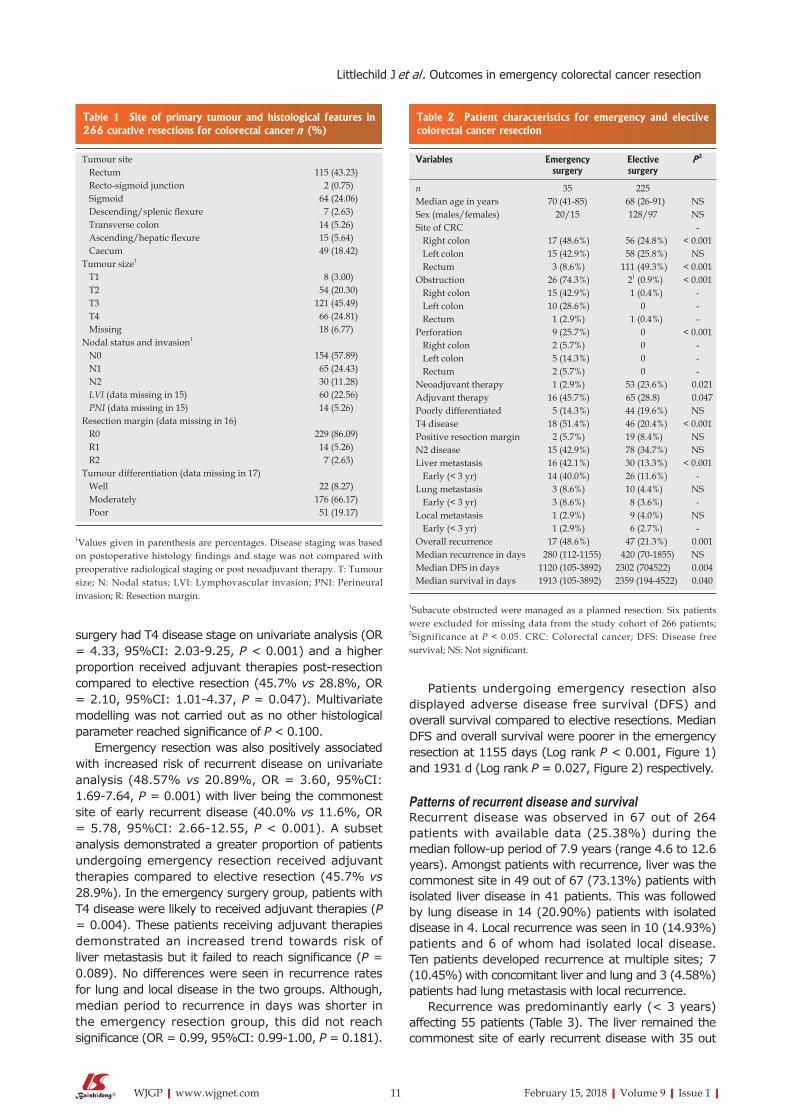
The site and histological features of the primary colorectal tumour in all 266 patients are shown in Table 1. TNM staging is presented from postoperative histological assessment. Pre-operative radiological changes in staging before and after neo-adjuvant therapy were not collected. Although T stage was not recorded for 18 patients positive residual disease was identified in 16 of these patients. Lymph node status was missing in 17 patients.
Emergency surgery for colorectal cancer Complete data was available from 35 patients undergoing emergency colorectal cancer resection. Detailed characteristics of patients undergoing emergency and elective colorectal resection for cancer are shown in Table 2. No significant differences were noted between the two groups in age, sex, tumour grade, nodal disease, LNR, LVI, PNI or resection margin status on univariate regression analysis. Right colon was more commonly the site of primary cancer in the emergency group and associated with higher rate of obstruction. More patients undergoing emergency
Littlechild J et al . Outcomes in emergency colorectal cancer resection
11 February 15, 201�|Volume 9|Issue 1|WJGP|www.wjgnet.com
surgery had T4 disease stage on univariate analysis (OR = 4.33, 95%CI: 2.03-9.25, P < 0.001) and a higher proportion received adjuvant therapies post-resection compared to elective resection (45.7% vs 28.8%, OR = 2.10, 95%CI: 1.01-4.37, P = 0.047). Multivariate modelling was not carried out as no other histological parameter reached significance of P < 0.100.
Emergency resection was also positively associated with increased risk of recurrent disease on univariate analysis (48.57% vs 20.89%, OR = 3.60, 95%CI: 1.69-7.64, P = 0.001) with liver being the commonest site of early recurrent disease (40.0% vs 11.6%, OR = 5.78, 95%CI: 2.66-12.55, P < 0.001). A subset analysis demonstrated a greater proportion of patients undergoing emergency resection received adjuvant therapies compared to elective resection (45.7% vs 28.9%). In the emergency surgery group, patients with T4 disease were likely to received adjuvant therapies (P = 0.004). These patients receiving adjuvant therapies demonstrated an increased trend towards risk of liver metastasis but it failed to reach significance (P = 0.089). No differences were seen in recurrence rates for lung and local disease in the two groups. Although, median period to recurrence in days was shorter in the emergency resection group, this did not reach significance (OR = 0.99, 95%CI: 0.99-1.00, P = 0.181).
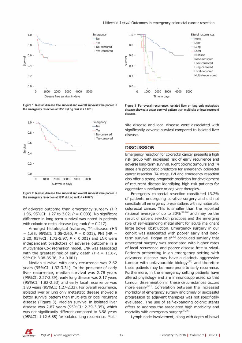
Patients undergoing emergency resection also displayed adverse disease free survival (DFS) and overall survival compared to elective resections. Median DFS and overall survival were poorer in the emergency resection at 1155 days (Log rank P < 0.001, Figure 1) and 1931 d (Log rank P = 0.027, Figure 2) respectively.
Patterns of recurrent disease and survivalRecurrent disease was observed in 67 out of 264 patients with available data (25.38%) during the median follow-up period of 7.9 years (range 4.6 to 12.6 years). Amongst patients with recurrence, liver was the commonest site in 49 out of 67 (73.13%) patients with isolated liver disease in 41 patients. This was followed by lung disease in 14 (20.90%) patients with isolated disease in 4. Local recurrence was seen in 10 (14.93%) patients and 6 of whom had isolated local disease. Ten patients developed recurrence at multiple sites; 7 (10.45%) with concomitant liver and lung and 3 (4.58%) patients had lung metastasis with local recurrence.
Recurrence was predominantly early (< 3 years) affecting 55 patients (Table 3). The liver remained the commonest site of early recurrent disease with 35 out
Tumour site Rectum 115 (43.23) Recto-sigmoid junction 2 (0.75) Sigmoid 64 (24.06) Descending/splenic flexure 7 (2.63) Transverse colon 14 (5.26) Ascending/hepatic flexure 15 (5.64) Caecum 49 (1�.42)Tumour size1
T1 � (3.00) T2 54 (20.30) T3 121 (45.49) T4 66 (24.�1) Missing 1� (6.77)Nodal status and invasion1
N0 154 (57.�9) N1 65 (24.43) N2 30 (11.2�) LVI (data missing in 15) 60 (22.56) PNI (data missing in 15) 14 (5.26)Resection margin (data missing in 16) R0 229 (�6.09) R1 14 (5.26) R2 7 (2.63)Tumour differentiation (data missing in 17) Well 22 (�.27) Moderately 176 (66.17) Poor 51 (19.17)
Table 1 Site of primary tumour and histological features in 266 curative resections for colorectal cancer n (%)
1Values given in parenthesis are percentages. Disease staging was based on postoperative histology findings and stage was not compared with preoperative radiological staging or post neoadjuvant therapy. T: Tumour size; N: Nodal status; LVI: Lymphovascular invasion; PNI: Perineural invasion; R: Resection margin.
Variables Emergency surgery
Elective surgery
P 2
n 35 225Median age in years 70 (41-�5) 6� (26-91) NSSex (males/females) 20/15 12�/97 NSSite of CRC - Right colon 17 (4�.6%) 56 (24.�%) < 0.001 Left colon 15 (42.9%) 5� (25.�%) NS Rectum 3 (�.6%) 111 (49.3%) < 0.001Obstruction 26 (74.3%) 21 (0.9%) < 0.001 Right colon 15 (42.9%) 1 (0.4%) - Left colon 10 (2�.6%) 0 - Rectum 1 (2.9%) 1 (0.4%) -Perforation 9 (25.7%) 0 < 0.001 Right colon 2 (5.7%) 0 - Left colon 5 (14.3%) 0 - Rectum 2 (5.7%) 0 -Neoadjuvant therapy 1 (2.9%) 53 (23.6%) 0.021Adjuvant therapy 16 (45.7%) 65 (2�.�) 0.047Poorly differentiated 5 (14.3%) 44 (19.6%) NST4 disease 1� (51.4%) 46 (20.4%) < 0.001Positive resection margin 2 (5.7%) 19 (�.4%) NSN2 disease 15 (42.9%) 7� (34.7%) NSLiver metastasis 16 (42.1%) 30 (13.3%) < 0.001 Early (< 3 yr) 14 (40.0%) 26 (11.6%) -Lung metastasis 3 (�.6%) 10 (4.4%) NS Early (< 3 yr) 3 (�.6%) � (3.6%) -Local metastasis 1 (2.9%) 9 (4.0%) NS Early (< 3 yr) 1 (2.9%) 6 (2.7%) -Overall recurrence 17 (4�.6%) 47 (21.3%) 0.001Median recurrence in days 2�0 (112-1155) 420 (70-1�55) NSMedian DFS in days 1120 (105-3�92) 2302 (704522) 0.004Median survival in days 1913 (105-3�92) 2359 (194-4522) 0.040
1Subacute obstructed were managed as a planned resection. Six patients were excluded for missing data from the study cohort of 266 patients; 2Significance at P < 0.05. CRC: Colorectal cancer; DFS: Disease free survival; NS: Not significant.
Table 2 Patient characteristics for emergency and elective colorectal cancer resection
Littlechild J et al . Outcomes in emergency colorectal cancer resection

12 February 15, 201�|Volume 9|Issue 1|WJGP|www.wjgnet.com
of 41 patients presenting with isolated liver disease. Early multi-site disease was seen in a small proportion of patients with liver disease (4 patients with liver and lung disease, 2 with liver and lung and local disease). In contrast, isolated lung disease was noted in only 3 out of 11 patients with early disease (4 patients had lung and liver disease, 2 had lung disease with local recurrence, 2 had lung, liver and local recurrence). Local recurrence was seen in 7 patients and was isolated in 3.
Recurrence between 3 to 5 years following curative resection represented 14.92% (10 patients) of overall recurrent disease. The liver remained the predominant site with 5 patients demonstrating isolated liver disease and 1 with liver and lung metastasis. Two patients presented with lung disease (1 isolated and 1 combined lung and liver disease) and 3 patients presented with isolated late local recurrence. Beyond the 5-year follow-up period, 1 patient developed isolated liver disease and 1 had liver and lung metastasis. No local recurrence was noted beyond 5-year follow-up.
Age, sex, tumour location, tumour differentiation and neo-adjuvant therapy did not show correlation with early all-site recurrent disease. Significant histological markers of early recurrence on univariate analyses included T4 stage (OR = 3.61, 95%CI: 1.88-6.96, P < 0.001), N2 nodal status (OR = 4.18, 95%CI: 1.89-9.24, P < 0.001), LVI (OR = 3.40, 95%CI: 1.79-6.46, P < 0.001), PNI (OR = 4.54, 95%CI: 1.51-13.60, P < 0.007) and R1 resection margin (OR = 4.51, 95%CI: 1.50-13.53, P = 0.007). LNR provided a strong correlation for early all-site recurrence on univariate analyses (OR = 25.55, 95%CI: 4.52-144.32, P < 0.001) with an optimal cut-off at 0.015 (52.7% sensitivity and 68.4% specificity). In a multivariate predictive model for early all-site recurrence, emergency surgery with
perforation was the strongest predictor amongst factors including T4 stage and LVI.
For early liver disease, no correlation was noted with age, sex, site of tumour or tumour differentiation on univariate analysis. Emergency surgery was associated with a higher risk of recurrent disease in the liver (OR = 5.13, 95%CI: 2.33-11.30, P < 0.001), with an adverse outcome in the presence of perforation than obstruction alone (OR 4.78 vs 3.74). Histological factors demonstrating significance on univariate analysis included T4 stage (OR = 2.76, 95%CI: 1.32-5.76, P = 0.007), N2 stage (OR = 3.32, 95%CI: 1.42-7.76, P = 0.006), LVI (OR = 3.02, 95%CI: 1.49-6.10, P = 0.002), PNI (OR = 3.71, 95%CI: 1.17-11.80, P = 0.026) and R1 resection margin (OR = 3.70, 95%CI: 1.16-11.74, P = 0.027). LNR (OR = 11.69, 95%CI: 1.94-70.24, P = 0.007) provided AUC of 0.61 (95%CI: 0.51-0.71, P = 0.023) with the optimal cut-off at 0.015 (53.7% sensitivity and 67.3% specificity). A multivariate predictive model of presentation and histological features showed that perforation at the time of surgery was the strongest independent predictor of early liver recurrence amongst other markers of predictive value (Table 4).
No independent predictors were identified for early lung recurrence. Early local recurrence represented a very small number of patients (7) for detailed analyses.
Long-term survival data was available from 262 patients with a median survival of 9.9 years. One-, three- and five-year survival in the study cohort was 96%, 82% and 72% respectively. Emergency surgery was associated with poorer survival (log rank P = 0.027) with 1, 3 and 5 year survival at 90%, 65% and 50% respectively (Figure 2). Multivariate Cox regression demonstrated T4 status to be a stronger predictor
Site of recurrence Early recurrence (n = 55) Colonic cancer (n = 151) Rectal cancer (n = 115) P 1
Liver 41 (74.55%) 25 16 0.554Lung 11 (20.00%) 6 5 0.�79Local 7 (12.73%) 4 3 0.9�4> 1 site � (14.55%) 4 4 0.695
1Significance at P < 0.05, colonic cancer vs rectal cancer. Number in parenthesis is percentage unless stated otherwise.
Table 3 Site of early (< 3 yr) and overall recurrent disease during follow-up
Early liver recurrence B SE OR 95%CI
T4 0.453 0.440 0.303 1.574 0.664-3.729N2 0.706 0.540 0.191 2.025 0.703-5.�31LVI 0.974 0.456 0.033 2.64� 1.0�4-6.46�PNI 0.735 0.700 0.294 2.0�5 0.529-�.214R1 0.954 0.726 0.1�9 2.597 0.626-10.777Obstruction 1.3�5 0.514 0.007 3.995 1.457-10.949Perforation 1.95� 0.7�1 0.012 7.0�6 1.533-32.749
B: Regression coefficient; SE: Standard error; OR: Odds ratio; T: Tumour size; N: Nodal status; LVI: Lymphovascular invasion; PNI: Perineural invasion; R: Resection margin.
Table 4 Prognostic factors for early liver recurrence
Littlechild J et al . Outcomes in emergency colorectal cancer resection
13 February 15, 201�|Volume 9|Issue 1|WJGP|www.wjgnet.com
of adverse outcome than emergency surgery (HR 1.96, 95%CI: 1.27 to 3.02, P = 0.003). No significant difference in long-term survival was noted in patients with colonic or rectal disease (log rank P = 0.217).
Amongst histological features, T4 disease (HR = 1.65, 95%CI: 1.05-2.60, P = 0.031), PNI (HR = 3.20, 95%CI: 1.72-5.97, P < 0.001) and LNR were independent predictors of adverse outcome in a multivariate Cox regression model. LNR was associated with the greatest risk of early death (HR = 11.87, 95%CI: 3.98-35.36, P < 0.001).
Median survival with early recurrence was 2.62 years (95%CI: 1.92-3.31). In the presence of early liver recurrence, median survival was 2.78 years (95%CI: 2.27-3.39); early lung disease was 2.17 years (95%CI: 1.82-2.53) and early local recurrence was 1.80 years (95%CI: 1.27-2.33). For overall recurrence, isolated liver or lung only metastatic disease showed a better survival pattern than multi-site or local recurrent disease (Figure 3). Median survival in isolated liver disease was 2.87 years (95%CI: 2.39-3.35), which was not significantly different compared to 3.98 years (95%CI: 1.12-6.85) for isolated lung recurrence. Multi-
site disease and local disease were associated with significantly adverse survival compared to isolated liver disease.
DISCUSSIONEmergency resection for colorectal cancer presents a high risk group with increased risk of early recurrence and adverse long-term survival. Right colonic tumours and T4 stage are prognostic predictors for emergency colorectal cancer resection. T4 stage, LVI and emergency resection also offer a strong prognostic prediction for development of recurrent disease identifying high-risk patients for aggressive surveillance or adjuvant therapies.
Emergency colorectal resection constituted 13.2% of patients undergoing curative surgery and did not constitute all emergency presentations with symptomatic colorectal cancer. This is smaller than the reported national average of up to 30%[17,26] and may be the result of patient selection practices and the emerging role of self-expanding metal stent for acute malignant large bowel obstruction. Emergency surgery in our cohort was associated with poorer early and long-term survival. Hogan et al[20] concluded similarly that emergent surgery was associated with higher rates of local recurrence and poorer disease-free survival. Patients presenting in an emergency setting with advanced disease may have a distinct, aggressive tumour with unfavourable biology[22] and therefore these patients may be more prone to early recurrence. Furthermore, in the emergency setting patients have altered physiology and are immunosuppressed so that tumour dissemination in these circumstances occurs more easily[21]. Correlation between the increased morbidity of emergency surgery and timely or successful progression to adjuvant therapies was not specifically evaluated. The use of self-expanding colonic stents offers to address the associated high morbidity and mortality with emergency surgery[27,28].
Lymph node involvement, along with depth of bowel
Surv
ival
1.0
0.8
0.6
0.4
0.2
0.0
0 1000 2000 3000 4000 5000
Disease free survival in days
Emergency No Yes No-censored Yes-censored
Figure 1 Median disease free survival and overall survival were poorer in the emergency resection at 1155 d (Log rank P < 0.001).
Surv
ival
1.0
0.8
0.6
0.4
0.2
0.0
0 1000 2000 3000 4000 5000
Survival in days
Emergency No Yes No-censored Yes-censored
Figure 2 Median disease free survival and overall survival were poorer in the emergency resection at 1931 d (Log rank P = 0.027).
Surv
ival
1.0
0.8
0.6
0.4
0.2
0.0
0 1000 2000 3000 4000 5000
Time in days
Site of recurrences None Liver Lung Local Multisite None-censored Liver-censored Lung-censored Local-censored Multisite-censored
Figure 3 For overall recurrence, isolated liver or lung only metastatic disease showed a better survival pattern than multi-site or local recurrent disease.
Littlechild J et al . Outcomes in emergency colorectal cancer resection

14 February 15, 201�|Volume 9|Issue 1|WJGP|www.wjgnet.com
wall invasion, has represented an important prognostic indicator for metachronous metastasis[29]. This is in broad correlation with our study. Nodal status was noted to be an independent predictor in early all-site recurrence, but no independent predictive value was noted for early liver and lung recurrent disease. In the setting of emergency surgery, where adequate lymph node yield may be compromised, LNR may prove to be a more useful prognostic tool since the patient’s physiology might not tolerate prolonged surgery in order to attain a good lymph node yield.
Keum et al[30] concluded that high risk factors for recurrence include rectal cancer, T2 stage and an infiltrative growth pattern. However, this study only looked at patients who underwent resection for stage I colorectal cancer which may explain their finding of T2 stage compared with ours of T4 stage. It has been stated previously that rectal tumours and younger age at presentation have a higher recurrence risk[31]. However, this was not the case in our study for reasons that are not clear to us.
LVI was also found to be an independent predictor of early all-site recurrence in a similar study by Huh et al[32] however they classified early recurrence as less than 1 year after operation. Lim et al[33] found that after attempted curative resection, patients with LVI-positive tumours had a higher rate of all-site recurrence than those without LVI. This is in correlation with our study.
Up to 50% of patients develop hepatic metastases within the first three years after curative resection[3]. In our study, overall recurrence occurred in 25% of the patient population, with 82% of the recurrence diagnosed within three years after resection. This is commensurate with the reported literature[4,34]; Meyerhardt et al[4] reports 80% of recurrences occur within the first three years. The liver was by far the most common site for early recurrence with 74% of cases occurring there. This is due to haematogenous spread via the portal venous system[35]. Pietra et al[36] also reported that 65% of recurrence occurred in the liver, in keeping with our observation.
Median survival of patients with early liver recurrence was 2.78 years. This compares favourably with patients presenting with early local recurrence who had a median survival of 1.8 years. This improved outcome may be a reflection of a more aggressive approach of isolated liver disease with the advent of improved liver parenchyma sparing techniques and advances in surgical techniques and perioperative care. The 5-year survival rate for patients that undergo curative metachronous resection of four or less hepatic lesions is 24%-58%[37-40]. A 5-year survival of 24% has also been reported for curative metachronous resection of > 8 hepatic lesions[41]. Detection of metachronous disease as early as possible is imperative in limiting the extent of resection and improving survival. Renehan et al[15] highlighted the value of intensive follow-up by demonstrating a high detection of early metachronous disease by aid of CEA and computed tomography.
This has delivered an opportunity for early detection and planning of definitive intervention. This may allow for more liver parenchymal-sparing techniques in the form of metastectomies and also improve the success of redo-hepatectomies[42]. The earlier the diagnosis is made, the more likely it is going to be resectable[43].
The lungs are the second most common location for metastatic spread of colorectal cancer[44]. It occurs in 5%-15% of patients and not all of these have concurrent liver metastases[45]. Only 4.1% of patients with synchronous pulmonary metastases are resectable, whereas 14.8% of patients with meta-chronous pulmonary metastases are resectable[46]
with a 36%-40% 5-year survival rate in resected patients[47]. In our study, 14 (5.2%) patients developed lung metastases in the follow-up period with a median survival period of 2.17 years. Nodal status and Dukes stage were predictive of early lung recurrence on univariate analysis but not multivariate analysis. This result should be treated with caution due to the limited number of patients who developed lung metastases. This is in contrast to a similar study by Kim et al[48] that reported a 3 year overall survival rate of 54.6%, although their study included 105 patients. Our study only contained 14 patients who developed lung metastases and it is possible that increased survival may have occurred with more patients in this category.
Negative predictive features when considering a patient for pulmonary metastatectomy follow a similar pattern to metastasis elsewhere and include an unresectable primary tumour, extra-pulmonary metastases, resection margins (R1/2) and mediastinal lymph node disease[49]. Blackmon et al[50] concluded that more than three lung metastases present at the first metastatectomy and a preoperative disease free survival of less than three years predicts recurrence. This suggests that early pickup of metastases means more patients can be considered for a successful resection. However, due to a small number of patients with lung disease in our cohort, no meaningful analyses of multiple clinical and pathological factors could be carried out.
The value of early detection of metastatic disease in offering an absolute reduction in mortality is clear[15]. Predictive tools can aid the clinician in identifying patients at higher risk of early metachronous meta-stasis as not all patients will benefit from aggressive surveillance. Our results suggest that there may be a subgroup of patients who would benefit from more intensive follow-up. As most tumours recur within the first three years after resection[34,51], it is imperative that the focus on follow-up occurs during this time frame. However, there is a lack of specific guidance for patients who may be at increased risk.
Current surveillance protocols consist of a com-bination of CEA testing, CT scans, and colonoscopy. Emergency surgery and the presence of specific histological features can inform the selection of patients
Littlechild J et al . Outcomes in emergency colorectal cancer resection

15 February 15, 201�|Volume 9|Issue 1|WJGP|www.wjgnet.com
at high-risk of early recurrence and should be factored into surveillance strategies. There is a substantial range in intensity of follow-up with the United Kingdom having noticeably less intensive follow-up in comparison to American collaborators[4,21,22]. A recent United Kingdom study of 1202 participants who had undergone curative surgery for primary colorectal cancer found no survival benefit from combined intensive monitoring groups compared to follow-up only if symptoms recurred[52]. This study was a randomised trial and so highlights the need to target the patients that would benefit from an intensive follow-up regimen. This has benefits for all stakeholders; earlier detection of recurrence in high-risk patients and a reduction of unnecessary investigations in the low-risk group. Exciting developments in the availability of other prognostic markers in the future may enhance the efficacy of a risk-adapted follow-up strategy[53]. The overexpression of vascular endothelial growth factor[54] and interleukin-8[55] in colorectal carcinoma cells are two such examples.
Our study is limited, foremost by the retrospective approach of data gathering using the hospital coding process. The weaknesses of such a design are well known[56]. Secondly, relevant data on neoadjuvant and adjuvant therapies and cause of death was not available to afford reliable assessment of correlation. More patients in the emergency resection group received postoperative adjuvant therapies which may have been the result of a higher tumour stage (T4), early recurrence disease or palliative treatment. Indication for post-operative therapies and cause of death outcomes were not collected and survival was calculated on basis of all-cause mortality. The majority of recurrent disease occurred in the liver, leading to more reliable statistical conclusions here. However, this was limited in number for both lung and local recurrent disease reflecting differences in association of histological and clinical predictive features. Furthermore, low event rates in neo-adjuvant therapies and positive resection margins, missing data on total number of lymph nodes harvested, limited reliable evaluation of these factors in overall outcomes.
Although emergency presentation in the form of obstruction continues to represent a significant proportion of patients with initial diagnosis, self-expanding colonic metal stents are likely to play an increasingly important role in improving immediate postoperative outcomes, stoma rates and long-term outcomes without adverse oncological outcomes[28]. Proven survival benefit from primary and redo liver resections in isolated disease and emerging technologies for palliative control of local and distant metastatic disease mean that predictive clinicopathological markers could be used in a more intensive, targeted surveillance strategy to identify more patients with early recurrence. Emergency resection, tumour stage, lymphovascular invasion and lymph node ratio > 0.015 represent a high-risk of recurrent disease and can inform surveillance strategies to enable early
interventions.
ARTICLE HIGHLIGHTS Research backgroundColorectal cancer is the fourth most common cancer in the United Kingdom with over 40000 cases diagnosed each year. Despite the widespread use of screening programs, a large number of cases are diagnosed in the acute or urgent setting with adverse post-operative mortality, disease-free and overall long-term survival.
Research motivationA large proportion of patients with colorectal cancer are diagnosed in the acute setting with an emergency presentation (26%) or following an urgent 2-wk referral (27%). Compared to elective resection, emergency surgery is associated with adverse postoperative outcomes (post-operative mortality 4.6% vs 16%), disease-free and overall long-term survival. The basis for this is multifactorial and may include altered physiology, immune-suppression, adverse tumour biology, advanced disease, peri-operative complications and lower progression to adjuvant therapies.
Research objectivesAdverse predictive factors for survival in colorectal cancer include emergency presentation with obstruction or perforation and histo-pathological features such as T4 disease, advanced nodal disease. Lymph node ratio offers to a new representation of nodal disease. Although it is affected directly but the lymph node yield, its utility as a predictive tool for recurrent disease has not been evaluated. We aimed to identify clinical and histological predictive factors for early recurrence disease and pattern to inform surveillance strategies and aid in early detection.
Research methodsFollowing institutional approval, a retrospective study of clinical and histo-pathological parameters was carried out to study patterns of recurrence and survival in consecutive patients undergoing elective and emergency resection for colorectal cancer over an eight-year study period.
Research resultsOutcomes were evaluated in 266 consecutive patients following curative surgery with a median follow-up of 7.9 years. The proportion of patients undergoing emergency resection was 13.2%. Recurrent disease was detected in 67 patients (25.2%) during follow-up with the majority identified early within 3 years (82.1%). Liver was the predominant site of metastatic disease (73.1%). Emergency resection (OR = 3.60, P = 0.001), T4 stage (OR = 4.33, P < 0.001) and lymphovascular invasion (LVI) (OR = 2.37, P = 0.032) were associated with higher risk of recurrent disease. Emergency resection, T4 disease and a high lymph node ratio (LNR) were strong independent predictors of adverse long-term survival.
Research conclusionsOur study reaffirms the independent predictive potential of histological and clinical features for recurrent disease in patients undergoing emergency resection for colorectal cancer. Furthermore, it introduces the independent utility of lymph node ratio (LNR) alongside T stage and lympho-vascular invasion in identifying patients with high risk of recurrent disease.
Research perspectivesModified surveillance strategies should be evaluated in presence of adverse clinical and histological factors to improve early detection of recurrent disease in high-risk patients to offset adverse disease-free and overall long-term survival.
REFERENCES1 Cancer Research UK. Bowel cancer incidence statistics. [accessed
2014 Jun]. Available from: URL: http://www.cancerresearchuk.
ARTICLE HIGHLIGHTS
Littlechild J et al . Outcomes in emergency colorectal cancer resection

16 February 15, 201�|Volume 9|Issue 1|WJGP|www.wjgnet.com
org/cancer-info/cancerstats/types/bowel/incidence/2 Tan EK, Ooi LL. Colorectal cancer liver metastases - under-
standing the differences in the management of synchronous and metachronous disease. Ann Acad Med Singapore 2010; 39: 719-715 [PMID: 20957308]
3 Garden OJ, Rees M, Poston GJ, Mirza D, Saunders M, Ledermann J, Primrose JN, Parks RW. Guidelines for resection of colorectal cancer liver metastases. Gut 2006; 55 Suppl 3: iii1-iii8 [PMID: 16835351 DOI: 10.1136/gut.2006.098053]
4 Meyerhardt JA, Mangu PB, Flynn PJ, Korde L, Loprinzi CL, Minsky BD, Petrelli NJ, Ryan K, Schrag DH, Wong SL, Benson AB 3rd; American Society of Clinical Oncology. Follow-up care, surveillance protocol, and secondary prevention measures for survivors of colorectal cancer: American Society of Clinical Oncology clinical practice guideline endorsement. J Clin Oncol 2013; 31: 4465-4470 [PMID: 24220554 DOI: 10.1200/JCO.2013.50.7442]
5 Scheele J, Stangl R, Altendorf-Hofmann A. Hepatic metastases from colorectal carcinoma: impact of surgical resection on the natural history. Br J Surg 1990; 77: 1241-1246 [PMID: 2253003 DOI: 10.1002/bjs.1800771115]
6 Nozawa H, Sunami E, Nakajima J, Nagawa H, Kitayama J. Synchronous and metachronous lung metastases in patients with colorectal cancer: A 20-year monocentric experience. Exp Ther Med 2012; 3: 449-456 [PMID: 22969910 DOI: 10.3892/etm.2011.443]
7 Landmann RG, Weiser MR. Surgical management of locally advanced and locally recurrent colon cancer. Clin Colon Rectal Surg 2005; 18: 182-189 [PMID: 20011301 DOI: 10.1055/s-2005-916279]
8 Read TE, Mutch MG, Chang BW, McNevin MS, Fleshman JW, Birnbaum EH, Fry RD, Caushaj PF, Kodner IJ. Locoregional recurrence and survival after curative resection of adenocarcinoma of the colon. J Am Coll Surg 2002; 195: 33-40 [PMID: 12113543 DOI: 10.1016/S1072-7515(02)01224-3]
9 Pastorino U, Buyse M, Friedel G, Ginsberg RJ, Girard P, Goldstraw P, Johnston M, McCormack P, Pass H, Putnam JB Jr; International Registry of Lung Metastases. Long-term results of lung metastasectomy: prognostic analyses based on 5206 cases. J Thorac Cardiovasc Surg 1997; 113: 37-49 [PMID: 9011700 DOI: 10.1016/S0022-5223(97)70397-0]
10 Nordlinger B, Sorbye H, Glimelius B, Poston GJ, Schlag PM, Rougier P, Bechstein WO, Primrose JN, Walpole ET, Finch-Jones M, Jaeck D, Mirza D, Parks RW, Collette L, Praet M, Bethe U, Van Cutsem E, Scheithauer W, Gruenberger T; EORTC Gastro-Intestinal Tract Cancer Group; Cancer Research UK; Arbeitsgruppe Lebermetastasen und-tumoren in der Chirurgischen Arbeitsgemeinschaft Onkologie (ALM-CAO); Australasian Gastro-Intestinal Trials Group (AGITG); Fédération Francophone de Cancérologie Digestive (FFCD). Perioperative chemotherapy with FOLFOX4 and surgery versus surgery alone for resectable liver metastases from colorectal cancer (EORTC Intergroup trial 40983): a randomised controlled trial. Lancet 2008; 371: 1007-1016 [PMID: 18358928 DOI: 10.1016/S0140-6736(08)60455-9]
11 Bruinvels DJ, Stiggelbout AM, Kievit J, van Houwelingen HC, Habbema JD, van de Velde CJ. Follow-up of patients with colorectal cancer. A meta-analysis. Ann Surg 1994; 219: 174-182 [PMID: 8129488 DOI: 10.1097/00000658-199402000-00009]
12 House MG, Ito H, Gönen M, Fong Y, Allen PJ, DeMatteo RP, Brennan MF, Blumgart LH, Jarnagin WR, D’Angelica MI. Survival after hepatic resection for metastatic colorectal cancer: trends in outcomes for 1,600 patients during two decades at a single institution. J Am Coll Surg 2010; 210: 744-752, 752-755 [PMID: 20421043 DOI: 10.1016/j.jamcollsurg.2009.12.040]
13 Jeffery M, Hickey BE, Hider PN. Follow-up strategies for patients treated for non-metastatic colorectal cancer. Cochrane Database Syst Rev 2007; CD002200 [PMID: 17253476 DOI: 10.1002/14651858.CD002200.pub2]
14 Figueredo A, Rumble RB, Maroun J, Earle CC, Cummings B, McLeod R, Zuraw L, Zwaal C; Gastrointestinal Cancer Disease
Site Group of Cancer Care Ontario’s Program in Evidence-based Care. Follow-up of patients with curatively resected colorectal cancer: a practice guideline. BMC Cancer 2003; 3: 26 [PMID: 14529575 DOI: 10.1186/1471-2407-3-26]
15 Renehan AG, Egger M, Saunders MP, O’Dwyer ST. Impact on survival of intensive follow up after curative resection for colorectal cancer: systematic review and meta-analysis of randomised trials. BMJ 2002; 324: 813 [PMID: 11934773 DOI: 10.1136/bmj.324.7341.813]
16 Renehan AG, O’Dwyer ST, Whynes DK. Cost effectiveness analysis of intensive versus conventional follow up after curative resection for colorectal cancer. BMJ 2004; 328: 81 [PMID: 14715603 DOI: 10.1136/bmj.328.7431.81]
17 National Cancer Intelligence Network data briefing. Routes to diagnosis. NCIN, November 2010. [accessed 2015 Mar]. Available from: URL: http://www.ncin.org.uk/databriefings
18 Wong SK, Jalaludin BB, Morgan MJ, Berthelsen AS, Morgan A, Gatenby AH, Fulham SB. Tumor pathology and long-term survival in emergency colorectal cancer. Dis Colon Rectum 2008; 51: 223-230 [PMID: 18097722 DOI: 10.1007/s10350-007-9094-2]
19 Morris EJ, Taylor EF, Thomas JD, Quirke P, Finan PJ, Coleman MP, Rachet B, Forman D. Thirty-day postoperative mortality after colorectal cancer surgery in England. Gut 2011; 60: 806-813 [PMID: 21486939 DOI: 10.1136/gut.2010.232181]
20 Hogan J, Samaha G, Burke J, Chang KH, Condon E, Waldron D, Coffey JC. Emergency presenting colon cancer is an independent predictor of adverse disease-free survival. Int Surg 2015; 100: 77-86 [PMID: 25594643 DOI: 10.9738/INTSURG-D-13-00281.1]
21 Vallejo R, Hord ED, Barna SA, Santiago-Palma J, Ahmed S. Perioperative immunosuppression in cancer patients. J Environ Pathol Toxicol Oncol 2003; 22: 139-146 [PMID: 14533877 DOI: 10.1615/JEnvPathToxOncol.v22.i2.70]
22 Ghazi S, Berg E, Lindblom A, Lindforss U; Low-Risk Colorectal Cancer Study Group. Clinicopathological analysis of colorectal cancer: a comparison between emergency and elective surgical cases. World J Surg Oncol 2013; 11: 133 [PMID: 23758762 DOI: 10.1186/1477-7819-11-133]
23 Baca B, Beart RW Jr, Etzioni DA. Surveillance after colorectal cancer resection: a systematic review. Dis Colon Rectum 2011; 54: 1036-1048 [PMID: 21730795 DOI: 10.1007/DCR.0b013e31820db364]
24 The Association of Coloproctologists of Great Britain and Ireland. Guidelines for the Management of Colorectal Cancer. Third edition, 2007.
25 Benson AB, Arnoletti P, Bekaii-Saab T, Chan E, Chen YJ, Choti MA, Cooper HS, Dilawari RA, Engstrom PF, Enzinger PC, Fleshman JW, Fuchs CS, Grem JL, Knol JA, Leong LA, Lin E, May KS, Mulcahy MF, Murphy K, Rohren E, Ryan DP, Saltz L, Sharma S, Shibata D, Skibber JM, Small W, Sofocleous CT, Venook AP, Willett C. Clinical practice and guidelines in oncology (National Comprehensive Cancer Network Guidelines) Colon Cancer NCCN Version 3, 2014
26 Scott N, Hill J, Smith J, Kelly S, Fearnhead N. National Bowel Cancer Audit Annual Report. Association of Coloproctologist of Great Britain and Ireland. 2013
27 Tan CJ, Dasari BV, Gardiner K. Systematic review and meta-analysis of randomized clinical trials of self-expanding metallic stents as a bridge to surgery versus emergency surgery for malignant left-sided large bowel obstruction. Br J Surg 2012; 99: 469-476 [PMID: 22261931 DOI: 10.1002/bjs.8689]
28 Hill J, Kay C, Morton D, Magill L, Handley K, Gray R. CREST Trial Collaborative Group CREST: Randomised phase III study of stenting as a bridge to surgery in obstructing colorectal cancer-Results of the UK ColoRectal Endoscopic Stenting Trial (CREST). J Clin Oncol 2016; 34 (suppl): Abstr 3507
29 Liang JL, Wan DS, Pan ZZ, Zhou ZW, Chen G, Li LR, Lu ZH, Wu XJ. [Multivariate regression analysis of recurrence following curative surgery for colorectal cancer]. Ai Zheng 2004; 23: 564-567 [PMID: 15142455]
30 Keum MA, Lim SB, Kim SA, Yoon YS, Kim CW, Yu CS, Kim JC. Clinicopathologic factors affecting recurrence after curative surgery
Littlechild J et al . Outcomes in emergency colorectal cancer resection
17 February 15, 201�|Volume 9|Issue 1|WJGP|www.wjgnet.com
for stage I colorectal cancer. J Korean Soc Coloproctol 2012; 28: 49-55 [PMID: 22413082 DOI: 10.3393/jksc.2012.28.1.49]
31 Hellinger MD, Santiago CA. Reoperation for recurrent colorectal cancer. Clin Colon Rectal Surg 2006; 19: 228-236 [PMID: 20011326 DOI: 10.1055/s-2006-956445]
32 Huh JW, Kim CH, Lim SW, Kim HR, Kim YJ. Early recurrence in patients undergoing curative surgery for colorectal cancer: is it a predictor for poor overall survival? Int J Colorectal Dis 2013; 28: 1143-1149 [PMID: 23503665 DOI: 10.1007/s00384-013-1675-z]
33 Lim SB, Yu CS, Jang SJ, Kim TW, Kim JH, Kim JC. Prognostic significance of lymphovascular invasion in sporadic colorectal cancer. Dis Colon Rectum 2010; 53: 377-384 [PMID: 20305435 DOI: 10.1007/DCR.0b013e3181cf8ae5]
34 Sargent DJ, Wieand HS, Haller DG, Gray R, Benedetti JK, Buyse M, Labianca R, Seitz JF, O’Callaghan CJ, Francini G, Grothey A, O’Connell M, Catalano PJ, Blanke CD, Kerr D, Green E, Wolmark N, Andre T, Goldberg RM, De Gramont A. Disease-free survival versus overall survival as a primary end point for adjuvant colon cancer studies: individual patient data from 20,898 patients on 18 randomized trials. J Clin Oncol 2005; 23: 8664-8670 [PMID: 16260700 DOI: 10.1200/JCO.2005.01.6071]
35 Bar-Gil Shitrit A, Shitrit D, Bakal I, Braverman D, Kramer MR. Endobronchial metastases from colon cancer without liver metastases: report of eight cases. Dis Colon Rectum 2007; 50: 1087-1089 [PMID: 17347902 DOI: 10.1007/s10350-006-0880-z]
36 Pietra N, Sarli L, Costi R, Ouchemi C, Grattarola M, Peracchia A. Role of follow-up in management of local recurrences of colorectal cancer: a prospective, randomized study. Dis Colon Rectum 1998; 41: 1127-1133 [PMID: 9749496 DOI: 10.1007/BF02239434]
37 Morris EJ, Forman D, Thomas JD, Quirke P, Taylor EF, Fairley L, Cottier B, Poston G. Surgical management and outcomes of colorectal cancer liver metastases. Br J Surg 2010; 97: 1110-1118 [PMID: 20632280 DOI: 10.1002/bjs.7032]
38 de Jong MC, Pulitano C, Ribero D, Strub J, Mentha G, Schulick RD, Choti MA, Aldrighetti L, Capussotti L, Pawlik TM. Rates and patterns of recurrence following curative intent surgery for colorectal liver metastasis: an international multi-institutional analysis of 1669 patients. Ann Surg 2009; 250: 440-448 [PMID: 19730175 DOI: 10.1097/SLA.0b013e3181b4539b]
39 Fernandez FG, Drebin JA, Linehan DC, Dehdashti F, Siegel BA, Strasberg SM. Five-year survival after resection of hepatic metastases from colorectal cancer in patients screened by positron emission tomography with F-18 fluorodeoxyglucose (FDG-PET). Ann Surg 2004; 240: 438-447; discussion 447-450 [PMID: 15319715 DOI: 10.1097/01.sla.0000138076.72547.b1]
40 Jamison RL, Donohue JH, Nagorney DM, Rosen CB, Harmsen WS, Ilstrup DM. Hepatic resection for metastatic colorectal cancer results in cure for some patients. Arch Surg 1997; 132: 505-510; discussion 511 [PMID: 9161393 DOI: 10.1001/archsurg.1997.01430290051008]
41 Malik HZ, Hamady ZZ, Adair R, Finch R, Al-Mukhtar A, Toogood GJ, Prasad KR, Lodge JP. Prognostic influence of multiple hepatic metastases from colorectal cancer. Eur J Surg Oncol 2007; 33: 468-473 [PMID: 17097260 DOI: 10.1016/j.ejso.2006.09.030]
42 Allen PJ, Jarnagin WR. Current status of hepatic resection. Adv Surg 2003; 37: 29-49 [PMID: 12953626]
43 Adam R, Lucidi V, Bismuth H. Hepatic colorectal metastases:
methods of improving resectability. Surg Clin North Am 2004; 84: 659-671 [PMID: 15062667 DOI: 10.1016/j.suc.2003.12.005]
44 McCormack PM, Attiyeh FF. Resected pulmonary metastases from colorectal cancer. Dis Colon Rectum 1979; 22: 553-556 [PMID: 527448 DOI: 10.1007/BF02587003]
45 Tan KK, Lopes Gde L Jr, Sim R. How uncommon are isolated lung metastases in colorectal cancer? A review from database of 754 patients over 4 years. J Gastrointest Surg 2009; 13: 642-648 [PMID: 19082673 DOI: 10.1007/s11605-008-0757-7]
46 Mitry E, Guiu B, Cosconea S, Jooste V, Faivre J, Bouvier AM. Epidemiology, management and prognosis of colorectal cancer with lung metastases: a 30-year population-based study. Gut 2010; 59: 1383-1388 [PMID: 20732912 DOI: 10.1136/gut.2010.211557]
47 Fiorentino F, Hunt I, Teoh K, Treasure T, Utley M. Pulmonary metastasectomy in colorectal cancer: a systematic review and quantitative synthesis. J R Soc Med 2010; 103: 60-66 [PMID: 20118336 DOI: 10.1258/jrsm.2009.090299]
48 Kim CH, Huh JW, Kim HJ, Lim SW, Song SY, Kim HR, Na KJ, Kim YJ. Factors influencing oncological outcomes in patients who develop pulmonary metastases after curative resection of colorectal cancer. Dis Colon Rectum 2012; 55: 459-464 [PMID: 22426271 DOI: 10.1097/DCR.0b013e318246b08d]
49 Treasure T, Milošević M, Fiorentino F, Macbeth F. Pulmonary metastasectomy: what is the practice and where is the evidence for effectiveness? Thorax 2014; 69: 946-949 [PMID: 24415715 DOI: 10.1136/thoraxjnl-2013-204528]
50 Blackmon SH, Stephens EH, Correa AM, Hofstetter W, Kim MP, Mehran RJ, Rice DC, Roth JA, Swisher SG, Walsh GL, Vaporciyan AA. Predictors of recurrent pulmonary metastases and survival after pulmonary metastasectomy for colorectal cancer. Ann Thorac Surg 2012; 94: 1802-1809 [PMID: 23063195 DOI: 10.1016/j.athoracsur.2012.07.014]
51 Berman JM, Cheung RJ, Weinberg DS. Surveillance after colorectal cancer resection. Lancet 2000; 355: 395-399 [PMID: 10665570 DOI: 10.1016/S0140-6736(99)06552-6]
52 Primrose JN, Perera R, Gray A, Rose P, Fuller A, Corkhill A, George S, Mant D; FACS Trial Investigators. Effect of 3 to 5 years of scheduled CEA and CT follow-up to detect recurrence of colorectal cancer: the FACS randomized clinical trial. JAMA 2014; 311: 263-270 [PMID: 24430319 DOI: 10.1001/jama.2013.285718]
53 Rose J, Augestad KM, Cooper GS. Colorectal cancer surveillance: what’s new and what’s next. World J Gastroenterol 2014; 20: 1887-1897 [PMID: 24587668 DOI: 10.3748/wjg.v20.i8.1887]
54 Toiyama Y, Inoue Y, Saigusa S, Okugawa Y, Yokoe T, Tanaka K, Miki C, Kusunoki M. Gene expression profiles of epidermal growth factor receptor, vascular endothelial growth factor and hypoxia-inducible factor-1 with special reference to local responsiveness to neoadjuvant chemoradiotherapy and disease recurrence after rectal cancer surgery. Clin Oncol (R Coll Radiol) 2010; 22: 272-280 [PMID: 20117921 DOI: 10.1016/j.clon.2010.01.001]
55 Lurje G, Zhang W, Schultheis AM, Yang D, Groshen S, Hendifar AE, Husain H, Gordon MA, Nagashima F, Chang HM, Lenz HJ. Polymorphisms in VEGF and IL-8 predict tumor recurrence in stage III colon cancer. Ann Oncol 2008; 19: 1734-1741 [PMID: 18550579 DOI: 10.1093/annonc/mdn368]
56 Audit commission. PbR Data assurance Framework 2007/2008. Audit Commission London
P- Reviewer: Mayol J, Nishida T, Zhu X S- Editor: Kong JX L- Editor: A E- Editor: Li D
Littlechild J et al . Outcomes in emergency colorectal cancer resection

Caroline Linninge, Bodil Roth, Charlotte Erlanson-Albertsson, Göran Molin, Ervin Toth, Bodil Ohlsson
ORIGINAL ARTICLE
18 February 15, 2018|Volume 9|Issue 1|WJGP|www.wjgnet.com
Abundance of Enterobacteriaceae in the colon mucosa in diverticular disease
Caroline Linninge, Göran Molin, Department of Food Technology, Engineering and Nutrition, Lund University, Lund S-223 63, Sweden
Bodil Roth, Bodil Ohlsson, Department of Internal Medicine, Skåne University Hospital, Lund University, Malmö S-205 02, Sweden
Charlotte Erlanson-Albertsson, Appetite Regulation Unit, Department of Experimental Medical Science, Lund University, Lund S-222 41, Sweden
Ervin Toth, Department of Gastroenterology, Skåne University Hospital, Lund University, Malmö S-205 02, Sweden
ORCID number: Caroline Linninge (0000-0001-8170-6707); Bodil Roth (0000-0001-9745-652X); Charlotte Erlanson-Albe r t s son (0000 -0002 -9752 -1149 ) ; Göran Mol in (0000-0002-0993-3769); Ervin Toth (0000-0002-9314-9239); Bodil Ohlsson (0000-0002-9142-5244).
Author contributions: Roth B, Erlanson-Albertsson C, Molin G, Toth E, and Ohlsson B designed the study; Toth E was responsible for the endoscopic examinations and biopsy sampling; Linninge C and Roth B performed the microbial examinations; Ohlsson B wrote the initial draft of the manuscript; all authors contributed to the intellectual criticism of the manuscript and accepted the final version.
Supported by Development Foundation of Region Skåne (BO), No. F2014/354.
Institutional review board statement: The study was reviewed and approved by the Ethics Review Board of Lund University (2013/903).
Informed consent statement: The subjects provided written, informed consent prior to study enrollment.
Conflict-of-interest statement: The authors have no conflicts of interests to declare.
Data sharing statement: Technical appendix, statistical code, and dataset available from the corresponding author at [email protected]. Consent from participants was not obtained
for data sharing, but the presented data are anonymized and risk of identification is low.
Open-Access: This article is an open-access article which was selected by an in-house editor and fully peer-reviewed by external reviewers. It is distributed in accordance with the Creative Commons Attribution Non Commercial (CC BY-NC 4.0) license, which permits others to distribute, remix, adapt, build upon this work non-commercially, and license their derivative works on different terms, provided the original work is properly cited and the use is non-commercial. See: http://creativecommons.org/licenses/by-nc/4.0/
Manuscript source: Unsolicited manuscript
Correspondence to: Bodil Ohlsson, MD, PhD, Professor, Department of Internal Medicine, Skåne University Hospital, Lund University, Jan Waldenströms Street 15, Malmö S-205 02, Sweden. [email protected]: +46-40-331000Fax: +46-40-336208
Received: August 30, 2017 Peer-review started: September 5, 2017First decision: October 12, 2017Revised: November 10, 2017 Accepted: December 6, 2017Article in press: December 6, 2017Published online: February 15, 2018
AbstractAIMTo compare gut bacterial diversity and amount of Enterobacteriaceae in colonic mucosa between patients with and without diverticular disease (DD).
METHODSPatients in a stable clinical condition with planned elective colonoscopy were included. Blood samples and colon mucosa biopsies were collected at the
World J Gastrointest Pathophysiol 2018 February 15; 9(1): 18-27
ISSN 2150-5330 (online)
Submit a Manuscript: http://www.f6publishing.com
DOI: 10.4291/wjgp.v9.i1.18
Observational Study
colonoscopy. Study questionnaires including questions about gastrointestinal symptoms were completed by the patients and physicians. DNA from mucosa samples was isolated and the amount of Enterobacteriaceae was estimated using PCR assay. Terminal restriction fragment length polymorphism was applied to assess microbial diversity. Diversity was estimated by calculations of richness (number of terminal restriction fragments) and Shannon-Wiener and Simpson’s indices.
RESULTSA total of 51 patients were included, 16 patients with DD [68 (62-76) years] and 35 controls [62 (40-74) years] without any diverticula. Patients with DD had significantly higher levels of Enterobacteriaceae than those without DD (P = 0.043), and there was an inverse relationship between the amount of Enterobacteriaceae and the Simpson’s index (rs = -0.361, P = 0.033) and the Shannon-Wiener index (rs = -0.299, P = 0.081). The Simpson’s index (P = 0.383), Shannon-Wiener index (P = 0.401) or number of restrictions fragments (P = 0.776) did not differ between DD and controls. The majority of patients experienced gastrointestinal symptoms, and 22 patients (43.1%) fulfilled the criteria for irritable bowel syndrome, with no difference between the groups (P = 0.212). Demography, socioeconomic status, lifestyle habits, inflammatory biomarkers, or symptoms were not related to the amount of Enterobacteriaceae or bacterial diversity.
CONCLUSIONPatients with DD had higher amount of Enterobacteriaceae in the colon mucosa compared to patients without diverticula.
Key words: Bacterial diversity; Diverticular disease; Enterobacteriaceae ; Gut microbiota; Irritable bowel syndrome
© The Author(s) 2018. Published by Baishideng Publishing Group Inc. All rights reserved.
Core tip: Colon mucosa biopsies were collected from consecutive patients (n = 51) at the time of elective colonoscopy. Patients were grouped into patients with diverticular disease (DD) (n = 16) and controls without any diverticula (n = 35). The amount of Enterobacteriaceae and bacterial diversity were analyzed. Patients with DD had significantly higher levels of Enterobacteriaceae than controls (P = 0.043). Bacterial diversity did not differ between groups. All but 8 patients exhibited some kind of gastrointestinal symptoms, and 22 patients (43.1%) fulfilled the criteria for irritable bowel syndrome, without difference between groups (P = 0.212). Demography, socioeconomic status, lifestyle habits, inflammatory parameters, or gastrointestinal symptoms did not affect the gut microbiota examined.
Linninge C, Roth B, Erlanson-Albertsson C, Molin G, Toth
19 February 15, 2018|Volume 9|Issue 1|WJGP|www.wjgnet.com
Linninge C et al . Gut microbiota in diverticular disease
E, Ohlsson B. Abundance of Enterobacteriaceae in the colon mucosa in diverticular disease. World J Gastrointest Pathophysiol 2018; 9(1): 18-27 Available from: URL: http://www.wjgnet.com/2150-5330/full/v9/i1/18.htm DOI: http://dx.doi.org/10.4291/wjgp.v9.i1.18
INTRODUCTIONDiverticular disease (DD) is a common gastrointestinal disease of unknown etiology. The symptoms of DD are similar with symptoms of irritable bowel syndrome (IBS)[1], e.g., abdominal pain, bloating and altered bowel habits, and are present in 10%-25% of subjects[2]. About 1.5%-4% of patients with DD develop diverticulitis at some time during their lives[3,4]. An acute attack of diverticulitis may lead to chronic symptoms called post-diverticulitis IBS, in analogy with post-infectious IBS observed after an acute attack of gastroenteritis[5,6]. The hypothesis behind IBS development is that low-grade inflammation and/or altered intestinal gut microbiota in DD may contribute to visceral hypersensitivity and dysmotility with ensuing symptoms[7,8].
The gut microbiota is discussed as important for the etiology and pathophysiology in a wide range of diseases. Bacterial diversity is higher in lean compared to obese individuals, and in healthy states compared to unhealthy states, and some bacterial groups, e.g., Enterobacteriaceae, are associated with over-weight and inflammation[9-11]. The family Enterobacteriaceae is commonly found in the gut ecosystem, where Escherichia coli is the most abundant species of the family[9]. Low bacterial diversity and increased levels of Enterobacteriaceae/Escherichia coli have been linked to inflammatory bowel disease (IBD) in humans[12-14]. The findings of abundance of Enterobacteriaceae/Escherichia coli in experimental animal models of intestinal inflammation[15], and the ability of these bacteria to induce colitis[16], have strengthened the hypothesis that these bacteria are of importance in the etiology of IBD.
Only a few studies have been performed regarding microbial composition in DD. Recently, lower amounts of Enterobacteriaceae were found in the colon mucosa of DD patients compared with healthy controls[17], whereas higher amounts of Akkermansia and no difference in the Escherichia coli subgroup were found in feces in another DD cohort[18].
The primary aim of the present study was to compare the level of the large Gram-negative bacterial family Enterobacteriaceae and gut bacterial diversity in colon mucosa between consecutive patients diagnosed with DD and patients with normal endoscopic findings. Secondary aims were to evaluate the influence of demography, socioeconomic status, lifestyle habits, inflammatory parameters and gastrointestinal symptoms on the gut microbiota.

20 February 15, 2018|Volume 9|Issue 1|WJGP|www.wjgnet.com
MATERIALS AND METHODSStudy population and study designAll consecutive patients referred to elective colonoscopy at the Department of Endoscopy, Skåne University Hospital, Malmö, were invited to participate in the study. All patients were in a stabile clinical condition, and no one suffered from any acute inflammation, such as diverticulitis. The only exclusion criteria were age of ≤ 18 years and inability to understand the Swedish language. The patients were informed in oral and written communications at the arrival to the Department the day of examination. If they agreed to participate, they had to complete a study questionnaire about demography, socioeconomic status, lifestyle habits, family history and medical history, the Visual Analog Scale for Irritable Bowel Syndrome (VAS-IBS), and a nutrition questionnaire to analyze dietary habits. The colonoscopy was performed according to clinical routines. Four different mucosa biopsies were obtained from the mid part of the colon descendens. Samples were stored at -80 °C until the gut microbiota was analyzed by quantitative polymerase chain reaction (qPCR) and terminal restriction fragment length polymorphism (T-RFLP). Blood samples were collected according to clinical routines and analyzed at the Department of Clinical Chemistry. A study protocol was completed by the physician about clinical findings and histopathological diagnoses. The patients were divided into two groups depending on the colonoscopy finding: patients with DD, and patients without any diverticula who served as controls.
Tissue samplingThe patients were examined by colonoscopy according to clinical routines after prior laxation with Laxabon® (potassium chloride and macrogol; BioPhausia, Stockholm, Sweden). At the end of the colonoscopy, when the clinical examination was completed, four different mucosa biopsies were obtained from intact, inter-diverticular mucosa in the mid part of the colon descendens. This location was chosen since the left colon is the region most often affected by diverticula and is more accessible than the right colon. The biopsies were immediately frozen in liquid nitrogen and kept frozen at -80 °C until analysis. Histopathological examination was performed on separate mucosa samples when IBD had to be excluded or verified.
QuestionnairesStudy questionnaire: The questionnaire included questions on age, body mass index (BMI), family history, lifestyle habits, educational achievement, occupation, civil status, circumstances concerning delivery and breast-feeding, place of birth and moving patterns, and medical history. The patients had to answer whether they had been diagnosed with celiac disease, IBD, lactose intolerance, reflux or ulcer. They
were asked whether they experienced gastrointestinal symptoms which fulfilled the Rome IV criteria of functional dyspepsia or IBS[19,20]. This questionnaire was in structure and design similar to questionnaires used by other large current population-based and on-going screening projects in Sweden (i.e., LifeGene, EpiHealth, BIG-3, SCAPIS).
VAS-IBS: The VAS-IBS was used to investigate gastrointestinal complaints in the study groups. VAS-IBS is a validated, self-rating questionnaire for estimation of the most common gastrointestinal complaints experienced during the last 2 wk[21]. This questionnaire has also been validated for estimation of symptoms over time[22]. The five items measured in the VAS-IBS address the symptoms of abdominal pain, diarrhea, constipation, bloating and flatulence, and nausea and vomiting. These items were measured on a scale from 0-100, where 0 represented severe problems and 100 represented a complete lack of problems. Whether the patient suffered from symptoms or not, was defined as a score above the median values in healthy subjects[22].
Food questionnaire: The questionnaire included questions about dietary intake each meal in the form of red meat, fish or vegetables, making it possible to estimate dietary patterns. The number of days per week for intake of juice, coffee/tea, milk, sour milk, muesli, berries and fruit, marmalade, bread, cheese, ham and egg at breakfast, or snack were filled in. The participants were asked whether the lunch and dinner were homemade, or whether the participant had a lunch or dinner at a restaurant or a frozen precooked meal.
Microbial analysesDNA extraction: Three of the four mucosa samples, mean weight 15 ± 0.6 mg, were used for DNA extraction. DNA was isolated and purified by EZ1 Advanced XL (EZ1 DNA Tissue Kit and Bacteria Card; Qiagen, Hilden, Germany)[10,23].
qPCR: The amount of Enterobacteriaceae was estimated using a qPCR assay according to Karlsson et al[10]. Primers used for the qPCR assay have been used and published previously[24,25]. The detection limit was 102 genes/reaction. For standard curves, 10-fold dilution series of the target DNA were made in EB buffer (Qiagen). Number of bacteria was expressed as log10 16S rRNA genes/g feces.
Microbial diversity: T-RFLP was applied to assess the microbial diversity, as previously described[26]. Thresholds for internal standard and terminal restriction fragments (T-RFs) were set to 5 and 15 fluorescence units, respectively.
Calculations: Microbial diversity was estimated by calculation of richness (number of T-RFs) and Shannon-
Linninge C et al . Gut microbiota in diverticular disease

21 February 15, 2018|Volume 9|Issue 1|WJGP|www.wjgnet.com
Wiener and Simpson’s diversity indices as described by Karlsson et al[10], with the exception that T-RFs within 40-580 base pairs were included in the T-RFLP profile analysis and calculation. The diversity indices take into account both richness and evenness when considering the relative abundance of bacterial groups. Both indices are commonly used to assess microbial diversity[27]. Samples below the limit of detection (in qPCR) were replaced by the limit of detection for statistical analysis.
Patient categorizationDepending on presence or absence of diverticula, the included patients were divided into patients with DD or control patients. The control patients either exhibited normal macroscopic endoscopic and microscopic histopathological findings or presence of benign polyps. The group categorization was performed independent of gastrointestinal symptoms. All patients with IBD or malignancy were excluded from the study. The diagnosis of IBD was set when the patients fulfilled the criteria for Crohn’s disease, ulcerative colitis or microscopic colitis, i.e., clinical and endoscopic findings in addition to inflammation at the histopathological examination, in accordance to the diagnoses criteria[28].
Statistical analysisThe statistical calculations were performed using the SPSS software, version 24.0 (Armonk, NY, United States). Non-parametric tests were used because of the low number of participants in each group and the skewed distribution of the values of VAS-IBS. Comparisons of continuous variables between groups were performed by either Mann-Whitney U test or Kruskal-Wallis test. Fisher’s exact test was used for dichotomous variables, and Spearman’s correlation test was used for correlations between parameters. Values are presented in median and interquartile ranges (IQRs) or number and percentage. P < 0.05 was considered statistically significant.
RESULTSPatient characteristicsIn total, 77 patients were invited to participate in the study. Nineteen patients denied to participate and 58 patients were included. Six patients were later excluded since they fulfilled the criteria for IBD, and one because of colon malignancy. Finally, 51 patients were included in the present study, 16 with DD and 35 controls without organic changes visible at the colonoscopy or at the histopathological examination (n = 12), except non-malignant polyps (n = 23). The reasons for referral to colonoscopy were presence of gastrointestinal symptoms which rendered a colonoscopy to exclude IBD, malignancy or DD (n = 17), follow-up after previous resection of polyps (n = 17), rectal bleeding (n = 11), screening for cancer due to heredity (n = 4), or perforation to the urinary tract (n = 2). Only one
subject in the DD group had a history of verified acute diverticulitis.
There was an equal sex distribution in the groups. Subjects without DD were slightly older than controls [68 (62-76) years vs 62 (40-74) years, P = 0.072], which may explain that more DD patients than controls had completed primary school as the highest education level. Age differences may also explain the lower degree of physical activity in the DD group. A few patients in both groups had been treated with antibiotics during the last 6 mo (Table 1). The moving patterns did not differ between groups. Sporadic cases of heart and lung diseases were found in both groups (data not shown).
Gastrointestinal symptomsAltogether, 22 patients (43.1%) fulfilled the Rome IV criteria for IBS. The prevalence of functional dyspepsia, IBS, gastric ulcer, lactose intolerance and reflux was equally distributed between groups. Each symptom item estimated by the VAS-IBS questionnaire was present in about half of all patients examined. Only 4 patients in each group did not have any form of gastrointestinal symptoms (Table 2). There was a wide variety in symptom intensity within each group also. None of the items in VAS-IBS correlated with age (data not shown).
Dietary patternsAll the participants who completed the nutrition questionnaire (n = 42) started the day with a breakfast, which in the vast majority of cases consisted of coffee or tea, together with bread and/or muesli and milk products. Twenty-seven participants had homemade lunch, whereas ten participants had lunch at a restaurant or had precooked meals, and five participants never had any lunch. Thirty-three participants had dinner at home, whereas eight participants had regular dinner at a restaurant or did not have dinner. Those who had homemade lunch suffered from more gastrointestinal symptoms compared with those who did not eat lunch, had lunch at a restaurant or had precooked meals, although bloating and flatulence was the only item that reached statistical significance [52 (25-93) vs 88 (70-100), P = 0.024]. The difference could not be related to any differences in socioeconomic factors or smoking or alcohol habits (data not shown) or in age span [66 (50-76) vs 65 (59-72), P = 0.851]. When the patients were divided into three groups depending on lunch habits [(1) home-maid lunch; (2) lunch at a restaurant or precooked meals; and (3) no lunch], those who had homemade lunch registered the most severe gastrointestinal symptoms on all the VAS scales, although the differences did not reach statistical significance (data not shown).
Microbiota and inflammatory biomarkersPatients with DD had significantly higher levels of Enterobacteriaceae than patients without diverticula (P = 0.043; Table 3). Although patients with DD more
Linninge C et al . Gut microbiota in diverticular disease
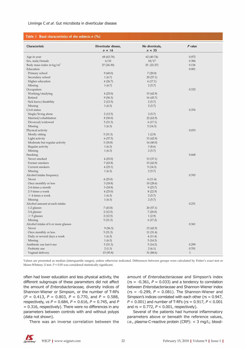
22 February 15, 2018|Volume 9|Issue 1|WJGP|www.wjgnet.com
often had lower education and less physical activity, the different subgroups of these parameters did not affect the amount of Enterobacteriaceae, diversity indices of Shannon-Wiener or Simpson, or the number of T-RFs (P = 0.413, P = 0.803, P = 0.770, and P = 0.588, respectively, vs P = 0.684, P = 0.616, P = 0.745, and P = 0.316, respectively). There were no differences in any parameters between controls with and without polyps (data not shown).
There was an inverse correlation between the
amount of Enterobacteriaceae and Simpson’s index (rs = -0.361, P = 0.033) and a tendency to correlation between Enterobacteriaceae and Shannon-Wiener index (rs = -0.299, P = 0.081). The Shannon-Wiener and Simpson’s indices correlated with each other (rs = 0.947, P < 0.001) and number of T-RFs (rs = 0.917, P < 0.001 and rs = 0.772, P < 0.001, respectively).
Several of the patients had humoral inflammatory parameters above or beneath the reference values, i.e., plasma-C-reactive protein (CRP): < 3 mg/L; blood-
Table 1 Basal characteristics of the subjects n (%)
Characteristic Diverticular disease, No diverticula, P valuen = 16 n = 35
Age in year 68 (62-76) 62 (40-74) 0.072Sex, male/female 6/10 18/17 0.384Body mass index in kg/m2 27 (24-30) 25 (22-27) 0.136Education 0.001 Primary school 9 (60.0) 7 (20.0) Secondary school 1 (6.7) 20 (57.1) Higher education 4 (26.7) 6 (17.1) Missing 1 (6.7) 2 (5.7)Occupation 0.332 Working/studying 4 (25.0) 15 (42.9) Retired 9 (56.3) 16 (45.7) Sick leave/disability 2 (12.5) 2 (5.7) Missing 1 (6.3) 2 (5.7)Civil status 0.376 Single/living alone 2 (12.5) 2 (5.7) Married/cohabitation 8 (50.0) 22 (62.9) Divorced/widowed 5 (31.3) 6 (17.1) Missing 1 (6.3) 5 (14.3)Physical activity 0.033 Mostly sitting 5 (31.3) 1 (2.9) Light activity 6 (37.5) 15 (42.9) Moderate but regular activity 3 (18.8) 14 (40.0) Regular activity 1 (6.3) 3 (8.6) Missing 1 (6.3) 2 (5.7)Smoking 0.668 Never smoked 4 (25.0) 13 (37.1) Former smokers 7 (43.8) 15 (42.9) Current smokers 4 (25.1) 5 (14.3) Missing 1 (6.3) 2 (5.7)Alcohol intake frequency 0.765 Never 4 (25.0) 4 (11.4) Once monthly or less 3 (18.8) 10 (28.6) 2-4 times a month 3 (18.8) 9 (25.7) 2-3 times a week 4 (25.0) 8 (22.9) ≥ 4 times a week 1 (6.3) 2 (5.7) Missing 1 (6.3) 2 (5.7)Alcohol amount at each intake 0.231 1-2 glasses 7 (43.8) 20 (57.1) 3-4 glasses 2 (12.5) 7 (20.0) ≥ 5 glasses 2 (12.5) 1 (2.9) Missing 5 (31.3) 6 (17.2)Alcohol intake of 6 or more glasses 0.361 Never 9 (56.3) 15 (42.9) Once monthly or less 5 (31.3) 11 (31.4) Daily or several days a week 1 (6.3) 4 (11.4) Missing 1 (6.3) 5 (14.3)Antibiotic use last 6 mo 5 (31.3) 5 (14.3) 0.299 Probiotic use 2 (1.3) 2 (6.1) 0.701 Vaginal delivery 15 (93.8) 31 (88.6) 1
Values are presented as median (interquartile ranges), unless otherwise indicated. Differences between groups were calculated by Fisher’s exact test or Mann-Whitney U test. P < 0.05 was considered statistically significant.
Linninge C et al . Gut microbiota in diverticular disease

23 February 15, 2018|Volume 9|Issue 1|WJGP|www.wjgnet.com
leucocytes: 3.5-8.8 × 10⁹/L; blood-thrombocytes 125-340 × 109/L; and plasma-albumin: 36-48 g/L. The level of inflammatory biomarkers did not differ between patients with or without DD (Table 3). Neither did presence nor absence of IBS affect the plasma levels of CRP (P = 0.194) and albumin (P = 0.902), or blood levels of leukocytes (P = 0.912) and thrombocytes (P = 0.509). There was no correlation between any of the inflammatory biomarkers and the level of Enterobacteriaceae or bacterial diversity (data not shown).
Neither the amount of Enterobacteriaceae nor the diversity indices correlated with age, BMI, or any items of the VAS-IBS (data not shown). When calculating differences between patients with and without any of the gastrointestinal symptoms, there were no differences in amount of Enterobacteriaceae or diversity indices (data not shown). Presence of IBS did not affect the amount of Enterobacteriaceae (P = 0.867), Shannon-Wiener index (P = 0.533), Simpson’s index (P = 0.478), or number of T-RFs (P = 0.828).
There were no differences in the amount of
Enterobacteriaceae or the diversity indices between those who had a regular vs irregular breakfast intake of coffee/tea, dairy products, or cereals. The gut microbiota parameters examined were not influenced by intake of homemade lunch or dinner, smoking and alcohol habits, intake of probiotics and antibiotics, or movement patterns (data not shown).
DISCUSSIONIn the present study examining symptomatic patients with elective colonoscopy, patients with DD had higher amount of Enterobacteriaceae compared with patients without diverticula, whereas the presence of gastrointestinal symptoms or IBS did not affect the amount of Enterobacteriaceae. Patients who had homemade lunch showed more symptoms of bloating and flatulence than those who did not have any lunch or had lunch at a restaurant/precooked meal. None of the studied lifestyle and socioeconomic parameters affected the amount of Enterobacteriaceae or bacterial diversity of the gut.
Table 2 Legree of symptoms based on Visual Analog Scale for Irritable Bowel
Diverticular disease, No diverticula, P value Symptom Symptom, P valuen = 16 n = 35 level, median n (%)
VAS-IBS, median (IQR) Abdominal pain 81 (49-100) 84 (48-100) 0.759 95 21 (60)/9 (56) 1 Diarrhea 96 (61-100) 83 (50-100) 0.404 97 23 (66)/8 (50) 0.506 Constipation 95 (52-100) 98 (54-100) 0.613 91 14 (40)/7 (44) 1 Bloating and flatulence 75 (23-100) 61 (40-100) 0.711 85 22 (63)/9 (56) 0.749 Nausea and vomiting 93 (48-100) 97 (80-100) 0.347 98 17 (49)/9 (56) 0.756 Absence of any GI symptom 4 (25) 4 (11.4) 0.236GI comorbidities, n (%) Celiac disease 0 0 Functional dyspepsia 5 (31.3) 8 (22.9) 0.509 IBS 5 (31.3) 17 (48.6) 0.212 Gastric ulcer 5 (31.3) 7 (20.0) 0.476 Lactose intolerance 1 (6.3) 0 0.093 Reflux 5 (31.3) 9 (25.7) 0.738
Data are presented as median [interquartile range (IQR)] or number and percentages. Symptom number is the number in each group presenting with symptoms. The level of VAS-IBS used to differentiate between symptoms or not is defined as a score above the median values in healthy subjects (No 22). Mann-Whitney U test or Fisher’s exact test. P-value < 0.05 was considered statistically significant. GI: Gastrointestinal; IBS: Irritable bowel syndrome; VAS-IBS: Visual Analog Scale for Irritable Bowel Syndrome.
Table 3 Mucosal count of Enterobacteriaceae and gut microbiota diversity and humoral inflammatory biomarkers
Diverticular disease, No diverticula, P valuen = 16 n = 35
Enterobacteriaceae, log10 16S rRNA genes/g 9.27 (7.34-10.04) 7.76 (7.13-8.76) 0.043Shannon-Wiener index 2.02 (1.80-2.36) 2.30 (1.94-2.48) 0.401Simpson’s index 0.80 (0.75-0.86) 0.82 (0.76-0.88) 0.383T-RF, n 17.0 (11.0-21.0) 17.0 (12.5-22.0) 0.776P-CRP, mg/L 4.40 (1.38-5.80) 1.70 (0.60-6.00) 0.346B-leukocytes, 109/L 8.40 (6.38-9.98) 8.10 (5.90-8.85) 0.466B-thrombocytes, 109/L 289 (219-334) 219 (186-266) 0.149P-albumin, g/L 36 (34-42) 36 (34-40) 0.819
Gut microbiota was analyzed in feces, and inflammatory biomarkers in blood or plasma. Data are presented as median (interquartile range). Mann-Whitney U test. P-value < 0.05 was considered statistically significant. B: Blood; P: Plasma; T-RF: Terminal restriction fragments.
Linninge C et al . Gut microbiota in diverticular disease

24 February 15, 2018|Volume 9|Issue 1|WJGP|www.wjgnet.com
The present result of higher levels of Enterobacteriaceae in mucosa of DD is in opposite to the previous result of Barbara et al[17]. The differences may be explained by the different study design and different composition of the control group. The present study enrolled mainly symptomatic patients examined by colonoscopy to exclude organic diseases or patients with heredity for colon cancer. Barbara et al[17] used asymptomatic or symptomatic patients enrolled to colonoscopy in a screening program to exclude malignancy or as follow-up after polyp resections. Thus, the control group in Barbara et al[17] consisted of a smaller cohort (n = 14) of asymptomatic subjects, and a lower percentage of symptomatic DD, with sex and age differences between groups. The microbiota composition differed between mucosal biopsies and feces[17]. We decided not to analyze fecal microbiota in our study, since there are greater differences between fecal and mucosal microbiota than between individual subjects, and it is considered more reliable to measure microbiota composition in mucosa than feces[29]. The general composition estimated by microbial diversity may be more important to health than the levels of individual bacterial strains[9,10,14].
Abundance of Enterobacteriaceae/Escherichia coli is associated with IBD, both in animal models and in humans[12,13,15,16]. The gut microbiota generates biologically active small molecules, e.g., amino acids, short-chain fatty acids, sugars and organic acids, which are presumed to affect the health of the host[30]. Basic microbiome metabolism was altered in IBD, with reduced amino acid synthesis and carbohydrate metabolism and increased nutrient uptake. Furthermore, genes involved in pathogenesis processes such as secretion of enterotoxins, wall-degrading enzymes and cytokine production were over-represented in Crohn’s disease[13]. This would lead to tissue destruction and bacterial overgrowth, with structural and functional dysbiosis.
In the present study of DD, the abundance of Enterobacteriaceae in the colon mucosa at a distance from the diverticula could hypothetically reflect a low-grade inflammation in the bowel wall. The previous publication by Barabra et al[17] suggested chronic low-grade gut mucosa inflammation in DD, through histopathological examination. Such low-grade inflammation was not reflected in the humoral inflammatory system, confirmed by overall normal CRP and blood cells levels, but may be captured in mucosal biopsies[17,31]. A low-grade inflammation may contribute to pain sensitization and visceral hypersensitivity and symptom development[7,8], which contributes to the increased risk of IBS after acute diverticulitis[6].
It remains unclear whether microbial changes are a cause or a consequence of DD. We do not know whether inflammation is a primary event, leading to weakening of the bowel wall and eventually to development of diverticula, or if inflammation is secondary to the presence of DD distant in the bowel with retention of luminal contents and bacterial overgrowth. Even if the
microbial changes are secondary, the dysbiosis may further accelerate the pathologic process and weakening of the bowel wall by mechanisms explained above[13].
Microbial dysbiosis in combination with genetic, environmental, and psychosocial factors are proposed to be involved in the etiology of IBS[20,32]. Escherichia coli was increased in Chinese IBS patients compared with controls, whereas no differences of these feces bacteria were found between IBS patients and healthy controls from other regions[33]. This is in line with our present study, which did not show any correlations between gastrointestinal symptoms or IBS and Enterobacteriaceae.
Gastrointestinal symptoms without visible organic damages are called functional bowel disorders, where IBS is the most common of the disorders, with a prevalence of 10%-15% in the population[20]. A great deal of the present patients suffered from IBS or IBS-like symptoms, whereas some patients experienced gastrointestinal symptoms without fulfilling the Rome IV criteria[20]. Symptomatology is not enough to distinguish between different bowel disorders, as found in the present study. It has previously been shown that patients with IBS have as severe symptoms as those with organic changes, i.e., primary Sjögren´s syndrome and enteric dysmotility[34]. A great symptomatic overlap between DD and IBS is described previously[1], which further underlines that disease classification must be based on organic criteria and not on symptoms solely. Biomarkers for IBS and DD are lacking, but measurements of markers of dysbiosis, inflammatory cells in mucosa, and metabolomes may be able to distinguish IBS from DD in the future. Probiotic therapy is an efficient treatment of IBS[35], whereas the evidence of efficiency in treatment of DD is insufficient.
Since this was a cross-sectional study, we do not know the reason for more symptoms being present in the group with homemade food. The reason may depend on patients with more severe symptoms avoiding visiting a restaurant, to have better control over their food intake.
The strength in the present pilot study is that we have analyzed mucosal biopsies instead of feces. The mucosa microbiota composition is anticipated to be more reliable than the feces composition. To compare another patient group with similar degree of symptoms seems more appropriate than to compare DD with healthy, non-symptomatic subjects. Further, we have considered food intake and other lifestyle habits affecting microbiota composition. The weakness is the small cohort size. Furthermore, since the patients were enrolled consecutively, there was no matching between cases and controls of, e.g., age, sex or lifestyle habits. In a larger study, some of the demographic parameters and lifestyle habits could have shown statistically significant influence on the gut microbiota. We chose to initially perform this as a pilot trial with a limited amount of patients, as the methodology is very expensive. Since it now has been shown that there are differences
Linninge C et al . Gut microbiota in diverticular disease

25 February 15, 2018|Volume 9|Issue 1|WJGP|www.wjgnet.com
in DD according to the gut microbiota, it is important to continue with further studies and more extensive analyses. Since this was a cross-sectional study, we do not know whether the microbial alterations are primary in the development of diverticula or just secondary to DD, with retention of luminal content.
In this pilot study, patients with DD had higher amount of Enterobacteriaceae in the colon mucosa compared to patients without DD. Assessment of gut microbiota may distinguish DD from other patient groups and may be involved in etiology and pathophysiology of the disease. Gastrointestinal symptomatology seems to not be related to the amount of Enterobacteriaceae or to the bacterial diversity.
ARTICLE HIGHLIGHTSResearch backgroundDiverticular disease (DD) is a common gastrointestinal disease of unknown etiology. The symptoms of DD are similar with symptoms of irritable bowel syndrome (IBS). The gut microbiota is discussed as important for the etiology and pathophysiology in a wide range of diseases. Bacterial diversity is higher in lean compared to obese individuals, and in healthy states compared to unhealthy states, and some bacterial groups, e.g., Enterobacteriaceae, are associated with over-weight and inflammation. The family Enterobacteriaceae is commonly found in the gut ecosystem, where Escherichia coli is the most abundant species of the family. Only a few studies have been performed regarding microbial composition in DD. Recently, lower amounts of Enterobacteriaceae were found in the colon mucosa of DD patients compared with healthy controls, whereas higher amounts of Akkermansia and no difference in the Escherichia coli subgroup were found in feces in another DD cohort. Thus, it is hypothesized that gut microbiota is involved in the etiology and pathophysiology of DD, but the few studies performed so far have shown inconclusive results.
Research motivationToday, there is no efficient treatment option for DD, neither to prevent disease development nor to reduce the symptoms when the disease has been established, which renders a lot of suffering to the patients. To find out the etiology is crucial to be able to prevent and efficiently treat the disease. New knowledge within this disease field may point out the direction for future research.
Research objectivesThe primary aim of the present study was to compare the level of the large Gram-negative bacterial family Enterobacteriaceae and gut bacterial diversity in colon mucosa between consecutive patients diagnosed with DD and patients with normal endoscopic findings. Secondary aims were to evaluate the influence of demography, socioeconomic status, lifestyle habits, inflammatory parameters and gastrointestinal symptoms on the gut microbiota. These objectives were possible to realize by the present study design. Further studies according to the same study design, but with larger patient cohorts, are important to perform to confirm the results.
Research methodsAll consecutive patients referred to elective colonoscopy at the Department of Endoscopy, Skåne University Hospital, Malmö, were invited to participate in the study. If the patients agreed to participate, they had to complete a study questionnaire about demography, socioeconomic status, lifestyle habits, family history and medical history, the Visual Analog Scale for Irritable Bowel Syndrome (VAS-IBS), and a nutrition questionnaire to analyze dietary habits. The colonoscopy was performed according to clinical routines. Four different mucosa biopsies were obtained from the mid part of the colon descendens. Samples were stored at -80 °C until the gut microbiota was analyzed by quantitative polymerase chain reaction (qPCR) and terminal restriction fragment length polymorphism (T-RFLP). Blood samples were collected according
to clinical routines and analyzed at the Department of Clinical Chemistry. A study protocol was completed by the physician about clinical findings and histopathological diagnoses. The patients were divided into two groups depending on the colonoscopy finding: patients with DD, and patients without any diverticula who served as controls. Three of the four mucosa samples, mean weight 15 ± 0.6 mg, were used for DNA extraction. DNA was isolated and purified by EZ1 Advanced XL (EZ1 DNA Tissue Kit and Bacteria Card; Qiagen, Hilden, Germany). The amount of Enterobacteriaceae was estimated using a qPCR assay according to Karlsson et al. Primers used for the qPCR assay have been used and published previously. The detection limit was 102 genes/reaction. For standard curves, 10-fold dilution series of the target DNA were made in EB buffer (Qiagen). Number of bacteria was expressed as log10 16S rRNA genes/g feces. T-RFLP was applied to assess the microbial diversity, as previously described. Thresholds for internal standard and terminal restriction fragments (T-RFs) were set to 5 and 15 fluorescence units, respectively. Microbial diversity was estimated by calculation of richness (number of T-RFs) and Shannon-Wiener and Simpson’s diversity indices as described by Karlsson et al, with the exception that T-RFs within 40-580 base pairs were included in the T-RFLP profile analysis and calculation. The diversity indices take into account both richness and evenness when considering the relative abundance of bacterial groups. Both indices are commonly used to assess microbial diversity. Samples below the limit of detection (in qPCR) were replaced by the limit of detection for statistical analysis.
Research resultsFinally, 51 patients were included in the present study, 16 with DD and 35 controls without organic changes visible at the colonoscopy or at the histopathological examination (n = 12), except non-malignant polyps (n = 23). The reasons for referral to colonoscopy were presence of gastrointestinal symptoms which rendered a colonoscopy to exclude inflammatory bowel disease, malignancy or DD (n = 17), follow-up after previous resection of polyps (n = 17), rectal bleeding (n = 11), screening for cancer due to heredity (n = 4), or perforation to the urinary tract (n = 2). Only one subject in the DD group had a history of verified acute diverticulitis. There was an equal sex distribution in the groups. Subjects without DD were slightly older than controls [68 (62-76) years vs 62 (40-74) years, P = 0.072]. Altogether, 22 patients (43.1%) fulfilled the Rome IV criteria for IBS. The prevalence of functional dyspepsia, IBS, gastric ulcer, lactose intolerance and reflux was equally distributed between groups. Each symptom item estimated by the VAS-IBS questionnaire was present in about half of all patients examined. Only 4 patients in each group did not have any form of gastrointestinal symptoms. There was a wide variety in symptom intensity within each group also. None of the items in VAS-IBS correlated with age. Those who had homemade lunch suffered from more gastrointestinal symptoms compared with those who did not eat lunch, had lunch at a restaurant or had precooked meals, although bloating and flatulence was the only item that reached statistical significance [52 (25-93) vs 88 (70-100), P = 0.024]. The difference could not be related to any differences in socioeconomic factors or smoking or alcohol habits or in age span [66 (50-76) vs 65 (59-72), P = 0.851]. Patients with DD had significantly higher levels of Enterobacteriaceae than patients without diverticula (P = 0.043). Although patients with DD more often had lower education and less physical activity, the different subgroups of these parameters did not affect the amount of Enterobacteriaceae, diversity indices of Shannon-Wiener or Simpson, or the number of T-RFs (P = 0.413, P = 0.803, P = 0.770, and P = 0.588, respectively, vs P = 0.684, P = 0.616, P = 0.745, and P = 0.316, respectively). There were no differences in any parameters between controls with and without polyps. There was an inverse correlation between the amount of Enterobacteriaceae and Simpson’s index (rs = -0.361, P = 0.033) and a tendency to correlation between Enterobacteriaceae and Shannon-Wiener index (rs = -0.299, P = 0.081). The Shannon-Wiener and Simpson’s indices correlated with each other (rs = 0.947, P < 0.001) and number of T-RFs (rs = 0.917, P < 0.001 and rs = 0.772, P < 0.001, respectively). Several of the patients had humoral inflammatory parameters above or beneath the reference values, i.e. plasma-C-reactive protein (CRP): < 3 mg/L; blood-leucocytes: 3.5-8.8 × 10⁹/L; blood-thrombocytes 125-340 × 109/L; and plasma-albumin: 36-48 g/L. The level of inflammatory biomarkers did not differ between patients with or without DD. Neither did presence nor absence of IBS affect the plasma levels of CRP (P = 0.194) and albumin (P = 0.902), or blood levels of leukocytes (P = 0.912) and thrombocytes (P = 0.509). There was no correlation between any of the inflammatory biomarkers and the level of Enterobacteriaceae or bacterial
ARTICLE HIGHLIGHTS
Linninge C et al . Gut microbiota in diverticular disease

26 February 15, 2018|Volume 9|Issue 1|WJGP|www.wjgnet.com
diversity. Neither the amount of Enterobacteriaceae nor the diversity indices correlated with age, BMI, or any items of the VAS-IBS. When calculating differences between patients with and without any of the gastrointestinal symptoms, there were no differences in amount of Enterobacteriaceae or diversity indices (data not shown). Presence of IBS did not affect the amount of Enterobacteriaceae (P = 0.867), Shannon-Wiener index (P = 0.533), Simpson’s index (P = 0.478), or number of T-RFs (P = 0.828). There were no differences in the amount of Enterobacteriaceae or the diversity indices between those who had a regular vs irregular breakfast intake of coffee/tea, dairy products, or cereals. The gut microbiota parameters examined were not influenced by intake of homemade lunch or dinner, smoking and alcohol habits, intake of probiotics and antibiotics, or movement patterns. The problems that remain to be solved are whether the difference in gut microbiota composition are primary events in the disease development or secondary to the DD. The causality to DD must still be defined.
Research conclusionsThe new finding of the present study is the abundance of Enterobacteriaceae in colon mucosa in DD, and that this abundance was not related to age, BMI, socioeconomic parameters, gastrointestinal symptoms or lifestyle habits. Microbial diversity was not affected by DD or any other parameters measured. The new theory that this study proposes is that the composition of gut microbiota is involved in DD. The summarization of this study is that gut microbiota may be affected in patients with DD. This study is the first study where a clinical cohort of patients is consecutively enrolled during colonoscopy to analyze gut microbiota in colon mucosa, where the only difference between the groups compared is the presence or absence of colon diverticula. Previous studies have enrolled participants in screening programs or analyzed microbiota composition in feces. The authors also studied socioeconomic features and lifestyle habits in the cohort, to be able to adjust for confounders. The new hypotheses proposed are that gut microbiota is involved in DD and that demography, socioeconomic parameters and dietary habits may be of less importance for the microbiota than the presence or absence of colon diverticula. The new methods proposed are the enrolment of consecutive clinical patients in scientific trials, analyses of gut microbiota in mucosa instead of feces, analysis of microbial diversity to get a general reflection of the gut microbiota, analysis of the amount of Enterobacteriaceae or other bacteria by qPCR, and estimation of gastrointestinal symptoms by the VAS-IBS questionnaire. The new phenomenon found were that presence or absence of colon diverticula are more important for gut microbiota than demography, socioeconomic parameters, gastrointestinal symptoms, or lifestyle habits. Another new phenomenon was that patients with homemade lunch had more gastrointestinal symptoms than patients who did not eat lunch or had lunch at a restaurant. The authors confirmed the hypothesis that the amount of Enterobacteriaceae was affected by DD, but failed to confirm the hypothesis that overall bacterial diversity was influenced by colon diverticula. The authors also failed to confirm the hypotheses that demography, socioeconomic parameters, gastrointestinal symptoms and lifestyle habits were associated with gut microbiota composition. The major implication for clinical practice in the future is to consider dysbiosis in patients with DD. Tests to determine gut microbiota are available for clinical use, and should be considered in the management of these patients.
Research perspectivesThe experience the authors have learnt from this study is that presence or absence of DD is more important for the gut microbiota composition than demography, socioeconomic parameters, gastrointestinal symptoms, and lifestyle habits. The authors have also learnt from this study that homemade food is not always the best for patients in the management of gastrointestinal symptoms. The authors must further study the importance of gut microbiota in DD. The authors should continue to include patients with DD in experiments to analyze gut microbiota composition to get larger cohorts, and to perform clinical trials to evaluate the effect of probiotics in symptom management of DD. The best method is to analyze gut microbiota in colon mucosa instead of feces. The VAS-IBS is also a useful tool to estimate gastrointestinal symptoms.
REFERENCES1 Cuomo R, Barbara G, Andreozzi P, Bassotti G, Casetti T, Grassini
M, Ierardi E, Maconi G, Marchi S, Sarnelli G, Savarino V, Usai
P, Vozzella L, Annibale B. Symptom patterns can distinguish diverticular disease from irritable bowel syndrome. Eur J Clin Invest 2013; 43: 1147-1155 [PMID: 23992370 DOI: 10.1111/eci.12152]
2 Stollman N, Raskin JB. Diverticular disease of the colon. Lancet 2004; 363: 631-639 [PMID: 14987890 DOI: 10.1016/S0140-6736(04)15597-9]
3 Hjern F, Johansson C, Mellgren A, Baxter NN, Hjern A. Diverticular disease and migration--the influence of acculturation to a Western lifestyle on diverticular disease. Aliment Pharmacol Ther 2006; 23: 797-805 [PMID: 16556182 DOI: 10.1111/j.1365-2036.2006.02805.x]
4 Shahedi K, Fuller G, Bolus R, Cohen E, Vu M, Shah R, Agarwal N, Kaneshiro M, Atia M, Sheen V, Kurzbard N, van Oijen MG, Yen L, Hodgkins P, Erder MH, Spiegel B. Long-term risk of acute diverticulitis among patients with incidental diverticulosis found during colonoscopy. Clin Gastroenterol Hepatol 2013; 11: 1609-1613 [PMID: 23856358 DOI: 10.1016/j.cgh.2013.06.020]
5 Humes DJ, Simpson J, Neal KR, Scholefield JH, Spiller RC. Psychological and colonic factors in painful diverticulosis. Br J Surg 2008; 95: 195-198 [PMID: 17939130 DOI: 10.1002/bjs.5962]
6 Cohen E, Fuller G, Bolus R, Modi R, Vu M, Shahedi K, Shah R, Atia M, Kurzbard N, Sheen V, Agarwal N, Kaneshiro M, Yen L, Hodgkins P, Erder MH, Spiegel B. Increased risk for irritable bowel syndrome after acute diverticulitis. Clin Gastroenterol Hepatol 2013; 11: 1614-1619 [PMID: 23524129 DOI: 10.1016/j.cgh.2013.03.007]
7 Simpson J, Scholefield JH, Spiller RC. Origin of symptoms in diverticular disease. Br J Surg 2003; 90: 899-908 [PMID: 12905541 DOI: 10.1002/bjs.4277]
8 Clemens CH, Samsom M, Roelofs J, van Berge Henegouwen GP, Smout AJ. Colorectal visceral perception in diverticular disease. Gut 2004; 53: 717-722 [PMID: 15082591 DOI: 10.1136/gut.2003.018093]
9 Hakansson A, Molin G. Gut microbiota and inflammation. Nutrients 2011; 3: 637-682 [PMID: 22254115 DOI: 10.3390/nu3060637]
10 Karlsson CL, Onnerfält J, Xu J, Molin G, Ahrné S, Thorngren-Jerneck K. The microbiota of the gut in preschool children with normal and excessive body weight. Obesity (Silver Spring) 2012; 20: 2257-2261 [PMID: 22546742 DOI: 10.1038/oby.2012.110]
11 Fåk F, Karlsson CL, Ahrné S, Molin G, Weström B. Effects of a high-fat diet during pregnancy and lactation are modulated by E. coli in rat offspring. Int J Obes (Lond) 2012; 36: 744-751 [PMID: 21730967 DOI: 10.1038/ijo.2011.118]
12 Baumgart M, Dogan B, Rishniw M, Weitzman G, Bosworth B, Yantiss R, Orsi RH, Wiedmann M, McDonough P, Kim SG, Berg D, Schukken Y, Scherl E, Simpson KW. Culture independent analysis of ileal mucosa reveals a selective increase in invasive Escherichia coli of novel phylogeny relative to depletion of Clostridiales in Crohn’s disease involving the ileum. ISME J 2007; 1: 403-418 [PMID: 18043660 DOI: 10.1038/ismej.2007.52]
13 Morgan XC, Tickle TL, Sokol H, Gevers D, Devaney KL, Ward DV, Reyes JA, Shah SA, LeLeiko N, Snapper SB, Bousvaros A, Korzenik J, Sands BE, Xavier RJ, Huttenhower C. Dysfunction of the intestinal microbiome in inflammatory bowel disease and treatment. Genome Biol 2012; 13: R79 [PMID: 23013615 DOI: 10.1186/gb-2012-13-9-r79]
14 Mondot S, Lepage P. The human gut microbiome and its dysfunctions through the meta-omics prism. Ann N Y Acad Sci 2016; 1372: 9-19 [PMID: 26945826 DOI: 10.1111/nyas.13033]
15 Arthur JC, Perez-Chanona E, Mühlbauer M, Tomkovich S, Uronis JM, Fan TJ, Campbell BJ, Abujamel T, Dogan B, Rogers AB, Rhodes JM, Stintzi A, Simpson KW, Hansen JJ, Keku TO, Fodor AA, Jobin C. Intestinal inflammation targets cancer-inducing activity of the microbiota. Science 2012; 338: 120-123 [PMID: 22903521 DOI: 10.1126/science.1224820]
16 Garrett WS, Gallini CA, Yatsunenko T, Michaud M, DuBois A, Delaney ML, Punit S, Karlsson M, Bry L, Glickman JN, Gordon JI, Onderdonk AB, Glimcher LH. Enterobacteriaceae act in concert with the gut microbiota to induce spontaneous and maternally
Linninge C et al . Gut microbiota in diverticular disease

27 February 15, 2018|Volume 9|Issue 1|WJGP|www.wjgnet.com
transmitted colitis. Cell Host Microbe 2010; 8: 292-300 [PMID: 20833380 DOI: 10.1016/j.chom.2010.08.004]
17 Barbara G, Scaioli E, Barbaro MR, Biagi E, Laghi L, Cremon C, Marasco G, Colecchia A, Picone G, Salfi N, Capozzi F, Brigidi P, Festi D. Gut microbiota, metabolome and immune signatures in patients with uncomplicated diverticular disease. Gut 2017; 66: 1252-1261 [PMID: 27618836 DOI: 10.1136/gutjnl-2016-312377]
18 Tursi A, Mastromarino P, Capobianco D, Elisei W, Miccheli A, Capuani G, Tomassini A, Campagna G, Picchio M, Giorgetti G, Fabiocchi F, Brandimarte G. Assessment of Fecal Microbiota and Fecal Metabolome in Symptomatic Uncomplicated Diverticular Disease of the Colon. J Clin Gastroenterol 2016; 50 Suppl 1: S9-S12 [PMID: 27622378 DOI: 10.1097/MCG.0000000000000626]
19 Stanghellini V, Chan FK, Hasler WL, Malagelada JR, Suzuki H, Tack J, Talley NJ. Gastroduodenal Disorders. Gastroenterology 2016; 150: 1380-1392 [PMID: 27147122 DOI: 10.1053/j.gastro.2016.02.011]
20 Mearin F, Lacy BE, Chang L, Chey WD, Lembo AJ, Simren M, Spiller R. Bowel Disorders. Gastroenterology 2016 [PMID: 27144627 DOI: 10.1053/j.gastro.2016.02.031]
21 Bengtsson M, Ohlsson B, Ulander K. Development and psychometric testing of the Visual Analogue Scale for Irritable Bowel Syndrome (VAS-IBS). BMC Gastroenterol 2007; 7: 16 [PMID: 17475020 DOI: 10.1186/1471-230X-7-16]
22 Bengtsson M, Persson J, Sjölund K, Ohlsson B. Further validation of the visual analogue scale for irritable bowel syndrome after use in clinical practice. Gastroenterol Nurs 2013; 36: 188-198 [PMID: 23732784 DOI: 10.1097/SGA.0b013e3182945881]
23 Stenblom EL, Weström B, Linninge C, Bonn P, Farrell M, Rehfeld JF, Montelius C. Dietary green-plant thylakoids decrease gastric emptying and gut transit, promote changes in the gut microbial flora, but does not cause steatorrhea. Nutr Metab (Lond) 2016; 13: 67 [PMID: 27777602 DOI: 10.1186/s12986-016-0128-4]
24 Bartosch S, Fite A, Macfarlane GT, McMurdo ME. Characterization of bacterial communities in feces from healthy elderly volunteers and hospitalized elderly patients by using real-time PCR and effects of antibiotic treatment on the fecal microbiota. Appl Environ Microbiol 2004; 70: 3575-3581 [PMID: 15184159 DOI: 10.1128/AEM.70.6.3575-3581.2004]
25 Castillo M, Martín-Orúe SM, Manzanilla EG, Badiola I, Martín M, Gasa J. Quantification of total bacteria, enterobacteria and lactobacilli populations in pig digesta by real-time PCR. Vet Microbiol 2006; 114: 165-170 [PMID: 16384658 DOI: 10.1016/j.vetmic.2005.11.055]
26 Sand E, Linninge C, Lozinska L, Egecioglu E, Roth B, Molin
G, Weström B, Ekblad E, Ohlsson B. Buserelin treatment to rats causes enteric neurodegeneration with moderate effects on CRF-immunoreactive neurons and Enterobacteriaceae in colon, and in acetylcholine-mediated permeability in ileum. BMC Res Notes 2015; 8: 824 [PMID: 26710832 DOI: 10.1186/s13104-015-1800-x]
27 Magurran AE. An index of diversity. In measuring biological diversity. Oxford: Blackwell Science Ltd, 2004
28 Walsh AJ, Bryant RV, Travis SP. Current best practice for disease activity assessment in IBD. Nat Rev Gastroenterol Hepatol 2016; 13: 567-579 [PMID: 27580684 DOI: 10.1038/nrgastro.2016.128]
29 Tang MS, Poles J, Leung JM, Wolff MJ, Davenport M, Lee SC, Lim YA, Chua KH, Loke P, Cho I. Inferred metagenomic comparison of mucosal and fecal microbiota from individuals undergoing routine screening colonoscopy reveals similar differences observed during active inflammation. Gut Microbes 2015; 6: 48-56 [PMID: 25559083 DOI: 10.1080/19490976.2014.1000080]
30 Martinez KB, Leone V, Chang EB. Microbial metabolites in health and disease: Navigating the unknown in search of function. J Biol Chem 2017; 292: 8553-8559 [PMID: 28389566 DOI: 10.1074/jbc.R116.752899]
31 Humes DJ, Simpson J, Smith J, Sutton P, Zaitoun A, Bush D, Bennett A, Scholefield JH, Spiller RC. Visceral hypersensitivity in symptomatic diverticular disease and the role of neuropeptides and low grade inflammation. Neurogastroenterol Motil 2012; 24 : 318-e163 [PMID: 22276853 DOI: 10.1111/j.1365-2982.2011.01863.x]
32 Distrutti E, Monaldi L, Ricci P, Fiorucci S. Gut microbiota role in irritable bowel syndrome: New therapeutic strategies. World J Gastroenterol 2016; 22: 2219-2241 [PMID: 26900286 DOI: 10.3748/wjg.v22.i7.2219]
33 Zhuang X, Xiong L, Li L, Li M, Chen M. Alterations of gut microbiota in patients with irritable bowel syndrome: A systematic review and meta-analysis. J Gastroenterol Hepatol 2017; 32: 28-38 [PMID: 27300149 DOI: 10.1111/jgh.13471]
34 Bengtsson M, Hammar O, Mandl T, Ohlsson B. Evaluation of gastrointestinal symptoms in different patient groups using the visual analogue scale for irritable bowel syndrome (VAS-IBS). BMC Gastroenterol 2011; 11: 122 [PMID: 22073983 DOI: 10.1186/1471-2393-13-201]
35 Ford AC, Quigley EM, Lacy BE, Lembo AJ, Saito YA, Schiller LR, Soffer EE, Spiegel BM, Moayyedi P. Efficacy of prebiotics, probiotics, and synbiotics in irritable bowel syndrome and chronic idiopathic constipation: systematic review and meta-analysis. Am J Gastroenterol 2014; 109: 1547-1561; quiz 1546, 1562 [PMID: 25070051 DOI: 10.1038/ajg.2014.202]
P- Reviewer: Formica V S- Editor: Ji FF L- Editor: Filipodia E- Editor: Song XX
Linninge C et al . Gut microbiota in diverticular disease

Elias Tsiompanidis, Spyros I Siakavellas, Anastasios Tentolouris, Ioanna Eleftheriadou, Stamatia Chorepsima, Anastasios Manolakis, Konstantinos Oikonomou, Nikolaos Tentolouris
ORIGINAL ARTICLE
28 February 15, 2018|Volume 9|Issue 1|WJGP|www.wjgnet.com
Liver cirrhosis-effect on QT interval and cardiac autonomic nervous system activity
Elias Tsiompanidis, Spyros I Siakavellas, Anastasios Tentolouris, Ioanna Eleftheriadou, Stamatia Chorepsima, Anastasios Manolakis, Konstantinos Oikonomou, Nikolaos Tentolouris, First Department of Propaedeutic Internal Medicine, Medical School, National and Kapodistrian University of Athens, Laiko General Hospital, Athens 11527, Greece
ORCID number: Elias Tsiompanidis (0000-0002-7808-3112); Spyros I Siakavellas (0000-0002-0885-2414); Anastasios Tentolouris (0000-0001-5897-472X); Ioanna Eleftheriadou (0000-0002-0278-1061); Stamatia Chorepsima (0000-0002-6984-2236); Anastasios Manolakis (0000-0002-9324-0411); Konstantinos Oikonomou (0000-0001-7279-9710); Nikolaos Tentolouris (0000-0003-0615-2534).
Author contributions: Tsiompanidis E, Siakavellas SI, Tentolouris A, Eleftheriadou I, Chorepsima S, Manolakis A, Oikonomou K and Tentolouris N were involved in the design of the study, interpretation of results and writing of the manuscript; Tsiompanidis E, Siakavellas SI and Manolakis A participated in data acquisition; Tentolouris A, Eleftheriadou I, Chorepsima S and Tentolouris N conducted the statistical analysis; all authors read and approved the final manuscript.
Institutional review board statement: The experimental protocol was reviewed and approved by the Ethics Committee of Laiko General Hospital (Number of Permission: 2903/10-12-2014).
Informed consent statement: The study was approved by the ethics committee of Laiko General Hospital and written informed consent was obtained from all individuals before participation in the study.
Conflict-of-interest statement: There are no conflicts of interest related to this work.
Open-Access: This article is an open-access article which was selected by an in-house editor and fully peer-reviewed by external reviewers. It is distributed in accordance with the Creative Commons Attribution Non Commercial (CC BY-NC 4.0) license, which permits others to distribute, remix, adapt, build upon this work non-commercially, and license their derivative works on different terms, provided the original work is properly cited and the use is non-commercial. See: http://creativecommons.org/licenses/by-nc/4.0/
Manuscript source: Unsolicited manuscript
Correspondence to: Nikolaos Tentolouris, Associate Professor, First Department of Propaedeutic Internal Medicine, Medical School, National and Kapodistrian University of Athens, Laiko General Hospital, 17 Agiou Thoma Street, Athens 11527, Greece. [email protected]: +30-213-2061061Fax: +30-213-2061794
Received: August 6, 2017 Peer-review started: August 7, 2017 First decision: September 7, 2017Revised: October 4, 2017 Accepted: October 30, 2017Article in press: October 30, 2017Published online: February 15, 2018
AbstractAIMTo examine the impact of liver cirrhosis on QT interval and cardiac autonomic neuropathy (CAN).
METHODSA total of 51 patients with cirrhosis and 51 controls were examined. Standard 12-lead electrocardiogram recordings were obtained and QT as well as corrected QT interval (QTc) and their dispersions (dQT, dQTc) were measured and calculated using a computer-based program. The diagnosis of CAN was based upon the battery of the tests proposed by Ewing and Clarke and the consensus statements of the American Diabetes Association. CAN was diagnosed when two out of the four classical Ewing tests were abnormal.
RESULTSQT, QTc and their dispersions were significantly longer (P < 0.01) in patients with cirrhosis than in controls. No
World J Gastrointest Pathophysiol 2018 February 15; 9(1): 28-36
ISSN 2150-5330 (online)
Submit a Manuscript: http://www.f6publishing.com
DOI: 10.4291/wjgp.v9.i1.28
Observational Study

significant differences in QT interval were found among the subgroups according to the etiology of cirrhosis. Multivariate regression analysis after controlling for age, gender and duration of cirrhosis demonstrated significant association between QT and presence of diabetes mellitus [standardized regression coefficient (beta) = 0.45, P = 0.02] and treatment with diuretics (beta = 0.55, P = 0.03), but not with the Child-Pugh score (P = 0.54). Prevalence of CAN was common (54.9%) among patients with cirrhosis and its severity was associated with the Child-Pugh score (r = 0.33, P = 0.02). Moreover, patients with decompensated cirrhosis had more severe CAN that those with compensated cirrhosis (P = 0.03). No significant association was found between severity of CAN and QT interval duration.
CONCLUSIONPatients with cirrhosis have QT prolongation. Treatment with diuretics is associated with longer QT. CAN is common in patients with cirrhosis and its severity is associated with severity of the disease.
Key words: QT interval; Cardiac autonomic neuropathy; Cirrhotic cardiomyopathy; Child-Pugh score; Model for end-stage liver disease score; Liver cirrhosis
© The Author(s) 2018. Published by Baishideng Publishing Group Inc. All rights reserved.
Core tip: QT interval is significantly prolonged in patients with liver cirrhosis and its duration is associated with the use of diuretics but not with the severity of the disease. More than half of the patients with cirrhosis have cardiac autonomic neuropathy (CAN), while CAN severity is associated strongly with the severity of cirrhosis.
Tsiompanidis E, Siakavellas SI, Tentolouris A, Eleftheriadou I, Chorepsima S, Manolakis A, Oikonomou K, Tentolouris N. Liver cirrhosis-effect on QT interval and cardiac autonomic nervous system activity. World J Gastrointest Pathophysiol 2018; 9(1): 28-36 Available from: URL: http://www.wjgnet.com/2150-5330/full/v9/i1/28.htm DOI: http://dx.doi.org/10.4291/wjgp.v9.i1.28
INTRODUCTIONCirrhosis can affect many organs and systems of the body including cardiovascular and autonomic nervous system (ANS)[1-3]. Among the cardiovascular manifestations often encountered in cirrhotic patients, most common are increased baseline cardiac output, attenuated systolic and diastolic function, blunted ventricular response to stimuli and electrophysiological abnormalities, comprising a group of phenomena, commonly referred to as “cirrhotic cardiomyopathy”[1-3].
As for the involvement of the ANS in the cirrhotic-related manifestations, it has been considered as being the result of toxic, metabolic and immunologic
29 February 15, 2018|Volume 9|Issue 1|WJGP|www.wjgnet.com
Tsiompanidis E et al . QT interval in cirrhosis
disturbances affecting both the sympathetic and parasympathetic constituents of ANS[3,4]. Due to the close interrelation of the two systems-cardiovascular and ANS-an abnormal ANS function in cirrhotic patients has been shown to be reflected in several cardiac- and vascular-related parameters such as QT interval prolongation, heart rate variability (HRV) and arterial pressure changes, all components of the so-called cardiac autonomic neuropathy (CAN)[5,6].
Previous data in patients with diabetes mellitus have shown that CAN is associated with prolongation of QT interval[7]. Both CAN, even subclinical, and QT prolongation have been associated with increased all-cause mortality in patients with diabetes[7-9]. Interestingly, some studies have shown that the prolongation of QT interval in patients with cirrhosis has been associated with the severity and progression of the disease and with poorer survival in cirrhotic patients[10-13]. On the other hand, in other studies, even though prolonged QT was associated with more severe liver dysfunction, this has not been translated to higher mortality[14,15]. Moreover, it is interesting that the prolonged QTc was improved in most patients after liver transplantation, although the extent and degree of improvement is variable, indicating a functional and reversible “nature” of such dysfunction[16]. Similarly, in some studies CAN has been associated with the severity of liver disease[6,17].
In the present cross-sectional study, we examined the association between QT interval-related para-meters with presence and severity of cirrhosis. In addition, we examined the prevalence of CAN and its association with QT interval in patients with cirrhosis.
MATERIALS AND METHODSParticipantsA total of 102 participants were recruited, 51 cirrhotic patients followed consecutively at the outpatient clinic of our hospital and 51 age- and gender-matched healthy controls who were hospital staff and relatives of the patients with cirrhosis. The diagnosis of cirrhosis was established by liver biopsy in 25 subjects. In cases where biopsy was contraindicated (n = 26), the patients had clinical, biochemical and ultrasonographical findings of cirrhosis. The patients were further classified according to the Child-Pugh grading system as having decompensated (Child-Pugh score ≥ 7, n = 29) or compensated (Child-Pugh score < 7, n = 22) cirrhosis. In addition, the model for end-stage liver disease (MELD) score was measured and the histologic activity index was used to stage liver disease in patients who underwent a liver biopsy[18,19]. Diabetes mellitus was diagnosed using the American Diabetes Association criteria[20].
Criteria for exclusion from the study were as follows: (1) any electrolyte disturbance; (2) diseases which may affect ANS activity and QT interval duration such as coronary artery disease, heart failure, atrial fibrillation,

30 February 15, 2018|Volume 9|Issue 1|WJGP|www.wjgnet.com
amyloidosis, hepatocellular carcinoma, episode of infection or gastrointestinal bleeding in the last two months prior to the study; (3) medications which affect ANS activity and QT interval duration like calcium channel blockers, angiotensin converting enzyme inhibitors, angiotensin receptor blockers, digitalis, tricyclic antidepressants, sympathomimetics and anticholinergics; and (4) patients with any degree of hepatic encephalopathy. Patients receiving propranolol or other beta blockers were included in the study after they had stopped the medication for at least ten days prior to examination. In order to minimize the risk of variceal bleeding due to discontinuation of propranolol, only patients at low risk, documented through esophago-gastro-duodenoscopy (varices with a diameter less than 5 mm and without signs of bleeding), were recruited.
The study was approved by the ethics committee of our hospital and informed consent was obtained from all participants according to the principles of the Declaration of Helsinki[21].
ProceduresBlood, urine sampling and all tests were carried out early in the morning after overnight fast of 8-10 h in a room of stable temperature (22 ℃-24 ℃). All individuals refrained from smoking or drinking coffee prior to the examination. Body weight and height was measured in light clothing and body mass index (BMI) was calculated. Blood pressure was measured in the sitting position three consecutive times with 1 min interval in between and the mean value of the second and third measurements was calculated and used in the analysis.
Blood was drawn for determination of hemo-globin (Hb), white blood cell count, platelet count and biochemical measurements. Biochemical determinations were made on an automatic analyzer. Glycosylated hemoglobin (HbA1c) was measured using HPLC. Plasma insulin (Biosure, Brussels, Belgium; coefficient of variation < 5%) was determined by radioimmunoassay. Insulin resistance was calculated by the homeostasis model assessment equation (HOMA-IR)[22].
Assessment of CANThe diagnosis of CAN was based upon the battery of the tests proposed by Ewing and Clarke and the consensus statements of the American Diabetes Association[23,24]. The heart rate response to slow deep breathing (deep breathing test), the Valsalva maneuver and the assumption of upright position (lying-to-standing test) were assessed from electrocardiographic (ECG) recordings of RR intervals automatically using the computer-aided examination system VariaCardio TF4 (Medical Research, Leeds, United Kingdom). The change in systolic blood pressure upon standing, expressed as the
difference between the mean of the last two values obtained in the supine position and the value obtained 60 s after standing up, were recorded. The first three tests were evaluated according to the published age-related heart rate tests, while orthostatic hypotension was diagnosed when a fall in systolic blood pressure ≥ 20 mmHg and/or a fall in diastolic blood pressure ≥ 10 mmHg were observed. Diagnosis of CAN was established when at least two out of four tests were abnormal[23,24]. In order to evaluate the severity of the CAN, each normal test was graded with 0.0, each borderline with 1.0 and each abnormal with 2.0. On the basis of the sum of these scores, we calculated the total score of CAN, which is the sum of the partial scores corresponding to each one of the four individual tests (minimum 0, maximum 8)[23].
Assessment of QT intervalStandard 12-lead ECG recordings at a paper speed of 25 mm/s were obtained. The paper recordings were then scanned to an image at high resolution (300 dpi), edited, and converted to a digital ECG recording, which was analyzed interactively using an ECG analysis program[25]. QT interval was measured from the beginning of the QRS complex to the end of the downslope of the T wave (crossing the isoelectric line). Corrected QT interval for heart rate (QTc) was calculated using Bazett’s formula (QTc = QT/√RR)[26]. QT dispersion (dQT) and QTc dispersion (dQTc) were calculated as the difference between the longest and the shortest QT and QTc intervals, respectively in any of the 12 leads. All measurements were performed by a single experienced investigator who was blind to the participants’ characteristics. The QTc interval was considered prolonged if it was > 440 msec (the upper normal limit commonly used).
Statistical analysisStatistical analysis was performed using programs available in the SPSS statistical package (IBM SPSS software version 22.0 for Windows, Armonk, NY, United States) by four co-authors who have experience in statistical analysis and a biomedical statistician. All variables were tested for normal distribution of the values using the Kolmogorov-Smirnov test. Differences between groups and variables were tested by the Student’s t-test for continuous variables, while the χ2 test was used for categorical variables. Differences in nonparametric variables were compared using the Mann-Whitney test, while bivariate correlations were assessed by Spearman correlation for ordered variables. Multivariate linear regression analysis was performed in the patients with cirrhosis to examine for associations between QT interval parameters and the variables of interest. P values < 0.05 were considered statistically significant.
Tsiompanidis E et al . QT interval in cirrhosis

31 February 15, 2018|Volume 9|Issue 1|WJGP|www.wjgnet.com
RESULTSDemographic and clinical characteristics of the study participantsThe demographic and clinical characteristics of the participants are shown in Table 1 and Table 2. Control subjects and patients with cirrhosis did not differ in terms of age, gender, BMI, arterial blood pressure, white blood cell count or smoking habits. Patients with cirrhosis had significantly higher blood glucose (P = 0.006), fasting insulin (P < 0.001) and HbA1c (P = 0.007), as well as lower Hb and platelet count levels (P < 0.001), compared to control group. A total of 27.5% of the patients received diuretics (combination of furosemide and spironolactone) (Table 1). The main causes of cirrhosis were viral hepatitis (47.1%) and alcohol abuse (33.3%), while 39% of the patients had decompensated cirrhosis (Table 2).
The association between QT-related parameters and presence as well as severity of cirrhosisThe values of all QT interval-related parameters were higher (P < 0.001) in patients with cirrhosis than those in controls (Table 3). None of the controls had a QTc interval longer than 440 msec, while 43.1% of the patients had QTc intervals longer than 440 msec. Considering 60 msec as the highest normal value for dQT, the number of individuals with dQT > 60 msec was higher in patients than in controls [n = 18 (35.3%) vs n = 7 (13.7%), respectively, χ 2 = 6.41, P = 0.011].
In cirrhosis group, QT parameters did not differ significantly between patients with alcoholic and non-alcoholic cirrhosis (QT: 383.8 ± 44.1 msec and 389.9 ± 36.0 msec, respectively, P = 0.98; QTc: 437.1 ± 30.5 msec and 423.7 ± 30.8 msec, P = 0.16; dQT: 65.6 ± 28.6 msec and 54.5 ± 21.2 msec, P = 0.13; dQTc: 74.4 ± 29.5 msec and 60.5 ± 23.1 msec, P = 0.12).
Furthermore, patients with decompensated cirrhosis, in comparison with those with compensated cirrhosis, had longer dQTc (72.2 ± 26.6 msec vs 56.3 ± 22.5 msec, P = 0.03) and tended to have longer QTc (435.3 ± 30.4 msec vs 419.0 ± 30.1 msec, P = 0.070) as well as dQT (64.1 ± 25.4 msec vs 50.8 ± 20.7 msec, P = 0.053). No significant differences were found in QT (P = 0.55) between the two groups. Moreover, no significant correlations were found between the Child-Pugh score and QT (r = 0.11, P = 0.45), dQT (r = 0.20, P = 0.17), QTc (r = 0.22, P = 0.13) or dQTc (r = 0.26, P = 0.08). The same was valid for the MELD score (QT: r = -0.06, P = 0.71; dQT: r = -0.23, P = 0.12; QTc: r= 0.16, P = 0.30; dQTc: r = -0.18, P = 0.21).
Assessment of CANA total of 28 patients (54.9%) had CAN. All indices of cardiac ANS activity were worse and the total score, an index of the severity, of CAN was higher in patients than in controls (Table 4). Prevalence of CAN was not different between patients with compensated and decompensated cirrhosis [n = 9 (40.9%) and n = 19 (65.5%), respectively, χ 2 = 3.06, P = 0.08]. However, the severity of CAN assessed by the total score of CAN was higher in patients with decompensated than in those with compensated cirrhosis [3.0 (0.8-6.0) vs 4.0 (3.0-6.5), P = 0.03]. No significant correlations were found between total score of CAN and QT (r = -0.12, P = 0.40), dQT (r = 0.04, P = 0.78), QTc (r = -0.01, P = 0.98) or dQTc (r = 0.11, P = 0.43). The total score of CAN was significantly correlated with the Child-Pugh score (r = 0.33, P = 0.02) and the MELD score (r = 0.36, P = 0.01).
In addition, mean QT interval duration was not different between patients having both cirrhosis and diabetes (n = 7) and those having cirrhosis without diabetes (n = 44): 395.7 ± 41.2 msec vs 381.9 ±
Controls (n = 51) Patients (n = 51) P value
Male, n (%) 28 (54.9) 32 (62.7) 0.42Age (yr) 53.8 ± 13.9 55.2 ± 14.2 0.60BMI (kg/m2) 26.0 ± 3.5 26.1 ± 4.4 0.95Systolic blood pressure (mmHg) 128.3 ± 18.5 127.4 ± 27.1 0.84Diastolic blood pressure (mmHg) 79.0 ± 10.0 76.0 ± 15.1 0.26Heart rate (beats/min) 77.05 ± 49.82 76.74 ± 15.82 0.96Fasting insulin (μU/mL) 10.6 (7.7-12.4) 13.3 (10.6-24.4) < 0.001HbA1c (%) 4.97 ± 0.50 4.03 ± 0.74 < 0.001HOMA-IR 2.40 (1.59-3.08) 3.39 (2.74-5.50) < 0.001White blood cells (n/μL) × 103 7.4 ± 2.6 5.5 ± 4.6 0.13Hemoglobin (g/dL) 14.8 ± 1.2 11.8 ± 2.1 < 0.001Platelets (n/μL) × 103 225.766 ± 36.24 122.17 ± 94.23 < 0.001Diabetes, n (%) 1 (2.0) 7 (13.7) 0.02Use of diuretics, n (%) 0 14 (27.5) < 0.001Smoking status, n (%) 34.34 Current smokers 16 (31.4) 22 (43.1) Non-smokers 26 (51.0) 23 (45.1) Ex-smokers 9 (17.6) 5 (9.8)
Data presented as mean ± SD or as n (%) or as median value (interquartile range). BMI: Body mass index; HbA1c: Glycated hemoglobin 1c; HOMA-IR: Homeostasis model assessment equation.
Table 1 Demographic and clinical characteristics as well as laboratory results of the study subjects
Tsiompanidis E et al . QT interval in cirrhosis

32 February 15, 2018|Volume 9|Issue 1|WJGP|www.wjgnet.com
38.0 msec, respectively (P = 0.38). Furthermore, the values of the autonomic function tests did not differ significantly between participants having both cirrhosis and diabetes and those having cirrhosis without diabetes; deep breathing test: 1.09 ± 0.05 vs 1.14 ± 0.14, respectively, P = 0.29; Valsalva test: 1.40 ± 0.25 vs 1.32 ± 0.35, respectively, P = 0.48; lying-to-standing test: 1.09 ± 0.08 vs 1.08 ± 0.09, respectively, P = 0.86; orthostatic hypotension: 12.85 ± 9.50 mmHg vs 11.52 ± 9.73 mmHg, respectively, P = 0.73. CAN was present in 3 patients with both cirrhosis and diabetes and in 25 patients with cirrhosis but without diabetes (42.9% vs 56.8%, P = 0.49).
Associations between insulin resistance index (HOMA-IR) with QT-related parametersIn patients with cirrhosis, HOMA-IR values did not correlate significantly with QT (r = -0.09, P = 0.56), QTc (r = 0.18, P = 0.24), dQT (r = -0.03, P = 0.82) or dQTc (r = 0.03, P = 0.84). HOMA-IR was associated significantly with the Child-Pugh score (r = 0.43, P = 0.002) and the MELD score (r = 0.65, P < 0.001).
Multivariate regression analysis on the association between QT interval with the study parametersMultivariate linear regression analysis in patients with cirrhosis with QT interval as dependent variable, after controlling for age, gender and duration of cirrhosis demonstrated significant and independent associations with diagnosed diabetes [standardized regression coefficient (beta) = 0.45, P = 0.02] and use of diuretics (beta = 0.55, P = 0.03). A trend for association with HOMA-IR was observed (beta = 0.40, P = 0.058), while no significant associations were found with the Child-Pugh or the MELD score, the histology activity index, Hb, serum potassium, the total score of CAN, and previous use of beta blockers. The same analysis with either QTc, dQT or dQTc as dependent variables did not show significant associations with the aforementioned parameters.
DISCUSSIONIn the present study, we found that QT and QTc intervals as well as their dispersions were substantially prolonged in patients with cirrhosis in comparison with healthy controls. In addition, we demonstrated that patients with cirrhosis were diagnosed more often with CAN.
The importance of normal liver function on pre-servation of the electrophysiological properties of the heart is supported by several studies that have examined the prolongation of QTc before and after liver transplantation[16]. Although the results are not unanimous, most of the data suggest that liver transplantation improved the prolonged QTc; however, the extend and the degree of the improvement was variable[16]. Thus, our data of prolonged QT interval in patients with liver cirrhosis agree and corroborate these findings.
Previous data showed abnormal QT prolongation in 37% to 84% of patients with cirrhosis of either alcoholic or nonalcoholic etiology[5,10,11,15,27,28]. However, literature data on QT dispersion in cirrhosis are scarce. Dispersion of QT interval is probably a better index of left ventricular dispersion of repolarization than QT or QTc interval and high values of dQT predict cardiovascular mortality in patients with diabetes or
n %
Child-Pugh score 7 (5-9) Child-Pugh Grade A (score: 5-6) 22 43.1 B (score: 7-9) 18 35.3 C (score: 10-15) 11 21.6MELD score 29.8 (14.4-39.6) Decompensated cirrhosis 29 56.9 Alcohol 17 33.3 Viral Hepatitis 24 47.1 Hepatitis Β 10 19.6 Hepatitis C 12 23.5 Hepatitis B + C 2 3.9 Other 10 19.6Systematic use of beta-blockers (yes) 16 31.4Ascites (yes) 23 45.1Esophageal varices (yes) 28 54.9Liver biopsy 25 49.0Histologic activity index 8.6 ± 2.9 Disease duration (yr) 3 (0.8-7) INR 1.41 ± 0.41AST (U/L) 43.0 (34.0-66.0)ALT (U/L) 36.0 (24.0-50.0)ALP (U/L) 262.7 ± 121.8LDH (U/L) 392.4 ± 123.0γ-GT (U/L) 39.0 (28.0-76.0)Cholesterol (mg/dL) 175.9 ± 56.2Triglycerides(mg/dL) 76.0 (50.0-108.0)Total bilirubin (mg/dL) 1.25 (0.68-2.29)Direct bilirubin (mg/dL) 0.75 (0.30-1.13)Total proteins (g/dL) 7.5 ± 0.8Albumin (g/dL) 4.0 ± 0.8Blood potassium (meq/L) 4.2 ± 0.4Blood sodium (meq/L) 138.1 ± 4.6
Data are presented as mean ± SD or as n (%) or as median value (interquartile range). MELD: Model for end-stage liver disease.
Table 2 Clinical characteristics and associated laboratory test results of patients with cirrhosis
Controls Patients P value
Mean QT (msec) 341.6 ± 29.4 383.9 ± 38.4 < 0.001QT max (msec) 358.1 ± 56.6 413.5 ± 46.1 < 0.001QT min (msec) 320.5 ± 28.1 355.4 ± 38.2 < 0.001dQT (msec) 44.8 ± 14.2 65.6 ± 28.6 0.001QTc (msec) 364.0 ± 20.6 428.1 ± 31.0 < 0.001dQTc (msec) 47.6 ± 14.7 65.0 ± 25.9 < 0.001Mean RR (msec) 863.6 ± 177.0 812.9 ± 159.8 0.13
Data are shown as mean values ± SD. QTc: Corrected QT; dQT: QT dispersion; dQTc: QTc dispersion.
Table 3 Comparison of QT-related parameters between patients and controls
Tsiompanidis E et al . QT interval in cirrhosis

33 February 15, 2018|Volume 9|Issue 1|WJGP|www.wjgnet.com
coronary artery disease[25]. In the literature, there are no data on the potential association between dQT and mortality in patients with liver cirrhosis. Herein we found that both dQT and dQTc were more prolonged in patients with cirrhosis. One previous study has shown that QT and dQt is prolonged in patients with cirrhosis[29], while another study shown that QT, but not dQT, is prolonged in patients with alcoholic cirrhosis in comparison with controls[30]. In contrast, in another study, no differences were found in dQT between patients with cirrhosis and controls[31]. Our findings showed that the etiology of cirrhosis was not associated with either QT or dQT prolongation.
One of the mechanisms suggested to play an important role in the pathogenesis of QT prolongation in patients with cirrhosis, is the enhanced sympathetic nervous system activity[5]. This process, which in normal subjects would reduce the QT interval, seems to participate in QT prolongation in cirrhosis. This is further elaborated with the increased circulating levels of noradrenalin, and it is an index of enhanced sympathoadrenal activity, observed in patients with advanced liver disease[10]. One would expect that the heart rate would be affected by this situation, but this is not usually the case, probably due to a downregulation of beta-adrenergic receptors[31]. Likewise, our results did not establish any substantial differences of the RR interval, which represents mean heart rate, between patients and controls. It is possible that the complex physiological changes that occur in chronic liver disease, modulate the cardiac function and may prolong the QT interval-related parameters. Moreover, although it is known that the use of pro-pranolol reduces the risk of gastrointestinal bleeding in patients with cirrhosis[32], there are no data on the potential effect of beta-blockers on cardiovascular mortality in such patients. One systematic review and meta-analysis concluded that the use of non-selective beta-blockers was not associated with a significant increase in all-cause mortality in patients with cirrhosis and ascites or refractory ascites[33].
In our study, no differences between the cirrhotic subgroups (alcoholic vs non-alcoholic cirrhosis) were noticed. Thus, based upon the data originating from current study, no relationship between an increased QT interval and the cause of cirrhosis can be established.
These findings are in agreement with those of previous studies and may imply that QT prolongation is a phenomenon that derives from the pathophysiology of cirrhosis itself and does not reflect abnormalities related to certain causes of cirrhosis[10]. However, in a previous study, patients with alcohol–related liver cirrhosis had a significantly (P = 0.001) higher prevalence of QTc interval prolongation than those with HBV–related liver cirrhosis[14].
Besides, no significant association was found between the values of QT parameters and the severity of cirrhosis, as assessed by the Child-Pugh or the MELD score in either univariate or multivariate analysis. In a previous study with 94 patients with cirrhosis, the Child-Pugh score and plasma norepinephrine were significant and independent determinants of QTc duration[10]. Similarly, in two other studies the prevalence of prolonged QTc increased with the severity of chronic liver disease[5,14]. The discrepancies in the results of these studies may be explained in part by differences in the studied populations. In the present study only patients with low or moderate risk of variceal bleeding were included in order to be able to discontinue safely beta-blockers, medications affecting ANS activity. However, we showed that patients with decompensated cirrhosis had longer dQTc and tended to have longer QTc as well as dQT in comparison to patients with compensated cirrhosis. This finding implies that when liver disease progresses to a point where the human body cannot overcome the cirrhosis effects, one of the clinical features of this process is the exacerbation of the cardiac electrical conductance abnormalities.
According to our findings a substantial percentage of patients have CAN, but interestingly, the severity of CAN was not associated with QT prolongation. These findings are in contrast with those seen in patients with diabetes mellitus[7]. However, our results agree with previous data in patients with cirrhosis[5,11]; thus, a previous study has shown that prolonged QTc is independent of CAN in patients with cirrhosis[5]. Moreover, diabetes was independently associated with QT in multivariate analysis confirming previous reports for association between QT prolongation in subjects with diabetes[7]. The autonomic dysfunction has been shown to correlate with the severity of liver
Controls Patients P value
Deep breathing test (value, N/A) 1.25 ± 0.16 (48/3) 1.13 ± 0.13 (23/28) < 0.001Valsalva test (value, N/A) 1.45 ± 0.24 (45/6) 1.33 ± 0.25 (30/21) 0.01Lying-to-standing test (value, N/A) 1.17 ± 0.24 (45/6) 1.08 ± 0.10 (29/22) 0.01Systolic blood pressure fall to standing (value, N/A) 0 (0-5) (50/1) 10 (0-20) (35/16) < 0.001CAN, n (%) 3 (5.9) 28 (54.9) < 0.001Total score of CAN 1 (0-2) 4 (2-6) < 0.001
Data are shown as mean ± SD or as n (%) or as median value (interquartile range). CAN: Cardiac autonomic neuropathy; N: Number of subjects with normal test; A: Number of subjects with abnormal test.
Table 4 The results of the cardiac autonomic function tests in controls and patients
Tsiompanidis E et al . QT interval in cirrhosis

34 February 15, 2018|Volume 9|Issue 1|WJGP|www.wjgnet.com
disease[5], a finding also observed in our study, as total score of CAN was correlated significantly with the Child-Pugh and the MELD score. Besides, patients with decompensated cirrhosis had more severe CAN than patients with compensated cirrhosis, although the prevalence of CAN was not different between the two groups. Even though experimental and clinical data suggest that ANS influence QT interval[7,34], in the present study no relationships were found between the total score of CAN and the values of QT-related parameters.
Interestingly, insulin resistance was not associated with QT-related parameters in this study, but there was a strong association between HOMA-IR and severity of cirrhosis assessed by the Child-Pugh and the MELD score. This finding implies that insulin resistance per se does not affect QT interval duration and that other mechanisms associated with cirrhosis affect QT interval.
Multivariate analysis demonstrated that use of diuretics was associated with QT prolongation; noteworthy, this effect was seen independently from serum potassium concentrations. This finding emphasizes the need for QT monitoring in patients with cirrhosis who are on treatment with diuretics.
It is known that diabetes is associated with higher prevalence of CAN[24] and with QT prolongation[7]. In our study, we did not find significant differences in these between patients having both diabetes and cirrhosis than those having cirrhosis without diabetes. However, the number of the participants with diabetes was small in our study and we cannot conclude robustly if presence of diabetes burdens further CAN or QT interval in patients with cirrhosis.
The strength of our study is that we examined subjects under controlled conditions and the potential confounding effects of medications, food intake and coffee consumption have been avoided. With regards, to medications, recent data suggested that propranolol administration reduces QT interval in patients with advanced liver cirrhosis waiting for liver transplantation[35]. Thus, discontinuation of beta blockers from our patients eliminated the effect of this medication on QT interval duration and allowed us to examine the net effect of the disease on QT interval duration. However, the number of the participants was not large and the study did not have enough power to support the findings. Furthermore, we did not examine for the presence of cirrhotic cardiomyopathy to look for associations between QT-related parameters and indices of systolic or diastolic function of the heart. Finally, this was a cross-sectional study and a cause and effect relationship cannot be established.
In conclusion, this study has shown that QT interval is prolonged in patients with cirrhosis compared with controls. QT prolongation is independent of the etiology and severity of cirrhosis, as well as of CAN, suggesting that this prolongation probably reflects the liver damage itself or the sympathetic nervous system predominance because of cirrhosis. Therefore, cirrhosis, even in the early stages, affects QT interval. Moreover, patients with
diabetes and those on treatment with diuretics have longer QT interval independently from serum potassium levels, suggesting that they need monitoring for QT prolongation.
ARTICLE HIGHLIGHTS Research backgroundCirrhosis can affect many organs and systems of the body including cardiovascular and autonomic nervous system (ANS). Cirrhotic patients have abnormal ANS function and it is reflected in several cardiac- and vascular-related parameters such as QT interval prolongation, heart rate variability (HRV) and arterial pressure changes, all components of the so-called cardiac autonomic neuropathy (CAN). Both QT prolongation and CAN have been associated with increased cardiovascular and all-cause mortality. The findings of this study show that cirrhotic patients and, in particular those who have at the same time diabetes or who are on treatment with diuretics, have longer QT interval independently from serum electrolyte levels, suggesting that they need monitoring for QT prolongation.
Research motivationThis study has shown that patients with cirrhosis have more often CAN and QT prolongation; however, this is a cross-sectional study and a cause and effect relationship cannot be established. A prospective study is needed to examine whether patients with cirrhosis develop autonomic dysfunction and QT prolongation. Moreover, it would of interest to know the potential impact of treatment with b-blockers on QT interval or cardiac ANS activity. An important finding of this study is that the etiology of cirrhosis does not impact QT prolongation or cardiac autonomic activity.
Research objectives The main aim of this study was to examine the impact of liver cirrhosis on QT-related parameters and on CAN. The authors’ hypothesis was confirmed and implies that cardiac autonomic dysfunction and/or QT prolongation may contribute to the increased mortality in patients with cirrhosis.
Research methodsIn this study, the authors managed to collect complete data related to full blood count and biochemical analyses, while the diagnosis of cirrhosis was confirmed with liver biopsies when it was indicated. The diagnosis of cardiac autonomic dysfunction was based upon robust criteria such as the battery of the tests proposed by Ewing and Clarke by determination of the HRV. QT intervals were measured using a standard 12-lead ECG recordings. Statistical analysis was performed using programs available in the SPSS statistical package by four co-authors who have experience in statistical analysis and a biomedical statistician.
Research resultsIn the present study, the authors found that QT and QTc intervals as well as their dispersions were substantially prolonged in patients with cirrhosis in comparison with healthy controls. In addition, the authors demonstrated that patients with cirrhosis were diagnosed more often with cardiac autonomic dysfunction. Additionally, the authors found that severity of cirrhosis does not impact QT interval but it affects severity of cardiac autonomic dysfunction.
Research conclusionsThe novel finding of this study is that not only QT, but also QT dispersion is prolonged in patient with cirrhosis. Furthermore, CAN or QT prolongation is not associated with the etiology of cirrhosis. Patients with cirrhosis, especially those who have diabetes or an on treatment with diuretics should be screened for cardiac autonomic dysfunction an QT prolongation. Patients with cirrhosis have often CAN and QT prolongation. The original insights of this study are: (1) the authors measured QT dispersion, which is considered as an excellent marker of left ventricular repolarization abnormalities and better than QT prolongation, which has not been studied so far; and (2) the authors found that severity of cirrhosis affects strongly cardiac ANS activity and probably contributes to
ARTICLE HIGHLIGHTS
Tsiompanidis E et al . QT interval in cirrhosis

35 February 15, 2018|Volume 9|Issue 1|WJGP|www.wjgnet.com
the development of the cirrhotic myocardiopathy. The new methods used in this study is the robust methodology for the diagnosis of cardiac autonomic dysfunction and presence as well as severity of cirrhosis.
Research perspectivesThe results of this study suggest that patients with cirrhosis often have QT prolongation and cardiac autonomic dysfunction and therefore, they should be screened for these comorbidities; especially those who have diabetes or an on treatment with diuretics. Future research should be directed to the potential impact of treatment with b-blockers on QT interval or cardiac ANS activity. In addition, a prospective study is needed to examine whether patients with cirrhosis develop autonomic dysfunction and QT prolongation.
REFERENCES1 Møller S, Henriksen JH. Cardiovascular complications of cirrhosis.
Postgrad Med J 2009; 85: 44-54 [PMID: 19240290 DOI: 10.1136/gut.2006.112177]
2 Fede G, Privitera G, Tomaselli T, Spadaro L, Purrello F. Cardiovascular dysfunction in patients with liver cirrhosis. Ann Gastroenterol 2015; 28: 31-40 [PMID: 25608575]
3 Ruiz-del-Árbol L, Serradilla R. Cirrhotic cardiomyopathy. World J Gastroenterol 2015; 21: 11502-11521 [PMID: 26556983 DOI: 10.3748/wjg.v21.i41.11502]
4 Ates F, Topal E, Kosar F, Karincaoglu M, Yildirim B, Aksoy Y, Aladag M, Harputluoglu MM, Demirel U, Alan H, Hilmioglu F. The relationship of heart rate variability with severity and prognosis of cirrhosis. Dig Dis Sci 2006; 51: 1614-1618 [PMID: 16927142 DOI: 10.1007/s10620-006-9073-9]
5 Puthumana L, Chaudhry V, Thuluvath PJ. Prolonged QTc interval and its relationship to autonomic cardiovascular reflexes in patients with cirrhosis. J Hepatol 2001; 35: 733-738 [PMID: 11738100 DOI: 10.1016/S0168-8278(01)00217-3]
6 Dümcke CW, Møller S. Autonomic dysfunction in cirrhosis and portal hypertension. Scand J Clin Lab Invest 2008; 68: 437-447 [PMID: 18609092 DOI: 10.1080/00365510701813096]
7 Tentolouris N, Katsilambros N, Papazachos G, Papadogiannis D, Linos A, Stamboulis E, Papageorgiou K. Corrected QT interval in relation to the severity of diabetic autonomic neuropathy. Eur J Clin Invest 1997; 27: 1049-1054 [PMID: 9466135 DOI: 10.1046/j.1365-2362.1997.2300776.x]
8 Voulgari C, Psallas M, Kokkinos A, Argiana V, Katsilambros N, Tentolouris N. The association between cardiac autonomic neuropathy with metabolic and other factors in subjects with type 1 and type 2 diabetes. J Diabetes Complications 2011; 25: 159-167 [PMID: 20708417 DOI: 10.1016/j.jdiacomp.2010.06.001]
9 Maser RE, Mitchell BD, Vinik AI, Freeman R. The association between cardiovascular autonomic neuropathy and mortality in individuals with diabetes: a meta-analysis. Diabetes Care 2003; 26: 1895-1901 [PMID: 12766130 DOI: 10.2337/diacare.26.6.1895]
10 Bernardi M, Calandra S, Colantoni A, Trevisani F, Raimondo ML, Sica G, Schepis F, Mandini M, Simoni P, Contin M, Raimondo G. Q-T interval prolongation in cirrhosis: prevalence, relationship with severity, and etiology of the disease and possible pathogenetic factors. Hepatology 1998; 27: 28-34 [PMID: 9425913 DOI: 10.1002/hep.510270106]
11 Mohamed R, Forsey PR, Davies MK, Neuberger JM. Effect of liver transplantation on QT interval prolongation and autonomic dysfunction in end-stage liver disease. Hepatology 1996; 23: 1128-1134 [PMID: 8621144 DOI: 10.1002/hep.510230529]
12 Dillon JF, Plevris JN, Nolan J, Ewing DJ, Neilson JM, Bouchier IA, Hayes PC. Autonomic function in cirrhosis assessed by cardiovascular reflex tests and 24-hour heart rate variability. Am J Gastroenterol 1994; 89: 1544-1547 [PMID: 8079935]
13 Kim SM, George B, Alcivar-Franco D, Campbell CL, Charnigo R, Delisle B, Hundley J, Darrat Y, Morales G, Elayi SC, Bailey AL. QT prolongation is associated with increased mortality in end stage liver disease. World J Cardiol 2017; 9: 347-354 [PMID: 28515853 DOI: 10.4330/wjc.v9.i4.347]
14 Zhao J, Qi X, Hou F, Ning Z, Zhang X, Deng H, Peng Y, Li J, Wang X, Li H, Guo X. Prevalence, Risk Factors and In-hospital Outcomes of QTc Interval Prolongation in Liver Cirrhosis. Am J Med Sci 2016; 352: 285-295 [PMID: 27650234 DOI: 10.1016/j.amjms.2016.06.012]
15 Bal JS, Thuluvath PJ. Prolongation of QTc interval: relationship with etiology and severity of liver disease, mortality and liver transplantation. Liver Int 2003; 23: 243-248 [PMID: 12895263 DOI: 10.1034/j.1600-0676.2003.00833.x]
16 Liu H, Jayakumar S, Traboulsi M, Lee SS. Cirrhotic cardio-myopathy: Implications for liver transplantation. Liver Transpl 2017; 23: 826-835 [PMID: 28407402 DOI: 10.1002/lt.24768]
17 Bajaj BK, Agarwal MP, Ram BK. Autonomic neuropathy in patients with hepatic cirrhosis. Postgrad Med J 2003; 79: 408-411 [PMID: 12897221 DOI: 10.1136/pmj.79.933.408]
18 Desmet VJ. Knodell RG, Ishak KG, Black WC, Chen TS, Craig R, Kaplowitz N, Kiernan TW, Wollman J. Formulation and application of a numerical scoring system for assessing histological activity in asymptomatic chronic active hepatitis [Hepatology 1981;1:431-435]. J Hepatol 2003; 38: 382-386 [PMID: 12663226 DOI: 10.1016/S0168-8278(03)00005-9]
19 Kamath PS, Kim WR; Advanced Liver Disease Study Group. The model for end-stage liver disease (MELD). Hepatology 2007; 45: 797-805 [PMID: 17326206 DOI: 10.1002/hep.21563]
20 American Diabetes Association. (2) Classification and diagnosis of diabetes. Diabetes Care 2015; 38 Suppl: S8-S16 [PMID: 25537714 DOI: 10.2337/dc15-S005]
21 World Medical Association. World Medical Association Declaration of Helsinki: ethical principles for medical research involving human subjects. JAMA 2013; 310: 2191-2194 [PMID: 24141714 DOI: 10.1001/jama.2013.281053]
22 Matthews DR, Hosker JP, Rudenski AS, Naylor BA, Treacher DF, Turner RC. Homeostasis model assessment: insulin resistance and beta-cell function from fasting plasma glucose and insulin concentrations in man. Diabetologia 1985; 28: 412-419 [PMID: 3899825 DOI: 10.1007/BF00280883]
23 Ewing DJ, Martyn CN, Young RJ, Clarke BF. The value of cardiovascular autonomic function tests: 10 years experience in diabetes. Diabetes Care 1985; 8: 491-498 [PMID: 4053936 DOI: 10.2337/diacare.8.5.491]
24 Pop-Busui R, Boulton AJ, Feldman EL, Bril V, Freeman R, Malik RA, Sosenko JM, Ziegler D. Diabetic Neuropathy: A Position Statement by the American Diabetes Association. Diabetes Care 2017; 40: 136-154 [PMID: 27999003 DOI: 10.2337/dc16-2042]
25 Psallas M, Tentolouris N, Papadogiannis D, Doulgerakis D, Kokkinos A, Cokkinos DV, Katsilambros N. QT dispersion: comparison between participants with Type 1 and 2 diabetes and association with microalbuminuria in diabetes. J Diabetes Complications 2006; 20: 88-97 [PMID: 16504837 DOI: 10.1016/j.jdiacomp.2005.05.012]
26 Luo S, Michler K, Johnston P, Macfarlane PW. A comparison of commonly used QT correction formulae: the effect of heart rate on the QTc of normal ECGs. J Electrocardiol 2004; 37 Suppl: 81-90 [PMID: 15534815 DOI: 10.1016/j.jelectrocard.2004.08.030]
27 Trevisani F, Merli M, Savelli F, Valeriano V, Zambruni A, Riggio O, Caraceni P, Domenicali M, Bernardi M. QT interval in patients with non-cirrhotic portal hypertension and in cirrhotic patients treated with transjugular intrahepatic porto-systemic shunt. J Hepatol 2003; 38: 461-467 [PMID: 12663238 DOI: 10.1016/S0168-8278(03)00057-6]
28 Cichoż-Lach H, Tomaszewski M, Kowalik A, Lis E, Tomaszewski A, Lach T, Boczkowska S, Celiński K. QT Interval Prolongation and QRS Voltage Reduction in Patients with Liver Cirrhosis. Adv Clin Exp Med 2015; 24: 615-622 [PMID: 26469105 DOI: 10.17219/acem/28681]
29 Tuttolomondo A, Buttà C, Casuccio A, Di Raimondo D, Serio A, D’Aguanno G, Pecoraro R, Renda C, Giarrusso L, Miceli G, Cirrincione A, Pinto A. QT Indexes in Cirrhotic Patients: Relationship with Clinical Variables and Potential Diagnostic Predictive Value. Arch Med Res 2015; 46: 207-213 [PMID:
Tsiompanidis E et al . QT interval in cirrhosis
36 February 15, 2018|Volume 9|Issue 1|WJGP|www.wjgnet.com
25843561 DOI: 10.1016/j.arcmed.2015.03.008]30 Day CP, James OF, Butler TJ, Campbell RW. QT prolongation
and sudden cardiac death in patients with alcoholic liver disease. Lancet 1993; 341: 1423-1428 [PMID: 8099138 DOI: 10.1016/0140-6736(93)90879-L]
31 Hansen S, Møller S, Bendtsen F, Jensen G, Henriksen JH. Diurnal variation and dispersion in QT interval in cirrhosis: relation to haemodynamic changes. J Hepatol 2007; 47: 373-380 [PMID: 17459513 DOI: 10.1016/j.jhep.2007.03.013]
32 Pagliaro L. Lebrec D, Poynard T, Hillon P, Benhamou J-P. Propranolol for prevention of recurrent gastrointestinal bleeding in patients with cirrhosis. A controlled study [N Engl J Med 1981;305:1371-1374]. J Hepatol 2002; 36: 148-150 [PMID: 11830324 DOI: 10.1056/NEJM198112033052302]
33 Chirapongsathorn S, Valentin N, Alahdab F, Krittanawong C, Erwin PJ, Murad MH, Kamath PS. Nonselective β-Blockers and Survival in Patients With Cirrhosis and Ascites: A Systematic Review and Meta-analysis. Clin Gastroenterol Hepatol 2016; 14: 1096-1104.e9 [PMID: 26829026 DOI: 10.1016/j.cgh.2016.01.012]
34 Aytemir K, Aksöyek S, Ozer N, Gürlek A, Oto A. QT dispersion and autonomic nervous system function in patients with type 1 diabetes. Int J Cardiol 1998; 65: 45-50 [PMID: 9699930 DOI: 10.1016/S0167-5273(98)00091-6]
35 Kim YK, Hwang GS, Shin WJ, Bang JY, Cho SK, Han SM. Effect of propranolol on the relationship between QT interval and vagal modulation of heart rate variability in cirrhotic patients awaiting liver transplantation. Transplant Proc 2011; 43: 1654-1659 [PMID: 21693252 DOI: 10.1016/j.transproceed.2011.02.017]
P- Reviewer: Abadi ATB, Liu DY, McMillin MA, Morini S S- Editor: Ji FF L- Editor: A E- Editor: Li D
Tsiompanidis E et al . QT interval in cirrhosis

© 2018 Baishideng Publishing Group Inc. All rights reserved.
Published by Baishideng Publishing Group Inc7901 Stoneridge Drive, Suite 501, Pleasanton, CA 94588, USA
Telephone: +1-925-223-8242Fax: +1-925-223-8243
E-mail: [email protected] Desk: http://www.f6publishing.com/helpdesk
http://www.wjgnet.com