Embed Size (px)
Citation preview

Book of Abstracts
Workshop BIOMETRA 2018 September 24th, Aula Magna – LITA, Segrate

Cover: image by Lorena Benedetti and Maura Francolini
Lateral muscle of zebrafish larva – Second harmonic signal

Workshop BIOMETRA 2018
Organizing and Scientific Committee
Bifari Francesco Borroni Elena
Chiricozzi Elena Crippa Milena Giavazzi Fabio
Riva Marco Rondelli Valeria
Rusconi Francesco

Workshop BIOMETRA 2018 Program
I
Program
9.15 Welcome and introduction: Prof. P. Viani, Director
9.30 Session I: Translational medicine Chairperson: Prof. D. Fornasari
Di Biase E. - The oligosaccharide of GM1 ganglioside as a new neurotrophic agent: evidence on the development of primary neurons in culture
Isailovic N. - Regulatory b and t cell modulation by tofacitinib in rheumatoid arthritis
Oggionni G. - Is autonomic adaptation a component of athlete’s heart? Insight from a novel unitary autonomic index for sport
10.20 Session II: Immunology and Oncology Chairperson: Prof. M. Locati
Oriolo F. - The constitutive expression of nkp46 Identifies a new subset of anti-tumor vδ1 Intraepithelial t cells resident in human intestine
Mazzola M. - Nipbl, a new player with npmc+ in myeloid cells differentiation
Spiombi E. - MeCP2 is required for functional primary cilia
11.10 Coffee break
11.25 Plenary lecture
Prof. Andrea Moro (Scuola Universitaria Superiore di Pavia)
Impossible languages: when linguistics meets the brain
12.15 Lunch and Poster session
14.15 Session III: Methods and technologies Chairperson: Prof. L. Cantù
Todisco M. - Dna nanoruler: a novel biotechnological tool to quantify proximity at high resolution
Edera P. - Delayed creep in Colloidal gels
Mollica L. - A computational view of dsDNA-RNA triplex recognition by l-RNA aptamers
Grilli A. A bioinformatic framework to identify cell subpopulations from bulk gene expression data of cancer samples

Workshop BIOMETRA 2018 Program
II
15.20 Session IV: Molecular mechanisms of diseases Chairperson: Prof. S. Sonnino
Cabitta L. - Identification of the antigen recognized by rHIgM22, A remyelination-promoting human monoclonal antibody
Casati L. - Targeting remodelling bone diseases with nutraceutic compounds: the protective activity of Δ-tocotrienol
Bestetti I. - Identification of novel mechanisms for neurological conditions overlapping smith-magenis syndrome
Forastieri C. - Stress–induced lsd1 and srf modulation and their potential protective role towards neuropsychiatric disorders
Paladini M.S. - Stress-induced modulation of the oxidative balance in the rat brain: effect of the antipsichotic lurasidone
16.40 Aperitif & Best talk and Best poster awards

Workshop BIOMETRA 2018
1

Workshop BIOMETRA 2018
2
Oral presentations

Workshop BIOMETRA 2018 Translational medicine
3
THE OLIGOSACCHARIDE OF GM1 GANGLIOSIDE AS A NEW NEUROTROPHIC AGENT: EVIDENCE ON THE
DEVELOPMENT OF PRIMARY NEURONS IN CULTURE Erika Di Biase*1, Margherita Maggioni1, Giulia Lunghi1, Simona Prioni1, Maura Samarani1, Elena Chiricozzi1
and Sandro Sonnino1
1BIOMETRA, Università degli Studi di Milano, Milan, Italy *email: [email protected] GM1 ganglioside, sialic-acid containing glycosphingolipid, plays a pivotal role during neuronal development and its neurotrophic properties have been largely reported both in vitro and in vivo. In cultured neurons, the GM1 enrichment in plasma membrane microdomains contributes to the activation of neurotrophin receptors belonging to Trk family. This event in turn triggers a specific signaling cascade resulting in actin depolymerization, axon protrusion and elongation. Despite this evidence, the mechanism of action of GM1 is still unknown. Recently, we demonstrated that GM1 oligosaccharide (OligoGM1) directly binds to TrkA receptor triggering the TrkA-MAPK pathway activation which leads to neuroblastoma cells differentiation in neuronal sense. Here, we characterize OligoGM1 effect on the developmental process of mouse primary neurons. Time-lapse recordings of plated neurons showed that exogenously administered OligoGM1 enhances neuron clustering, arborization and networking. Accordingly, treated cells expressed increased level of specific neuronal markers. Moreover, the higher phosphorylation rate of FAK and Src proteins, the intracellular key regulators of neuronal motility, confirms OligoGM1 impact on the migration process. Beside to the observed migratory phenotype, preliminary results suggest that in the presence of OligoGM1, neurons express higher amount of more complex gangliosides and lower level of simpler ones. Concerning its mechanism of action, we found out that OligoGM1 interacted with neuronal surface promoting the early TrkA-MAPK pathway activation. Our data reveal that the specific role of GM1 in neuronal differentiation and maturation is due to its oligosaccharide portion which, by interacting with the cell surface, triggers the activation of intracellular biochemical pathways responsible for neuronal migration, dendrites emission and axon growth. Altogether, our results point out the importance of OligoGM1 as a new promising neurotrophic player. Keywords: gangliosides, GM1 oligosaccharide, neuronal development, TrkA

Workshop BIOMETRA 2018 Translational medicine
4
REGULATORY B AND T CELL MODULATION BY TOFACITINIB IN RHEUMATOID ARTHRITIS
Natasa Isailovic1, Gilberto Cincinelli1,2, Maria De Santis1, Elena Generali1, Angela Ceribelli1, Giacomo
Guidelli1, Carlo Selmi1,3* 1Rheumatology and Clinical Immunology Unit, Humanitas Research Hospital, Rozzano (MI), Italy 2BIOMETRA Department, University of Milan, Milan, Italy *email: [email protected] Introduction: The pathogenesis of rheumatoid arthritis (RA) is based on the dysregulation of both the innate and adaptive immune systems, finally leading to an overboost of inflammatory cytokines which promote systemic symptoms, chronic synovitis, and joint destruction. Tofacitinib is an oral JAK-dependent cytokine signaling inhibitor mainly affecting the signaling of IFN-α, IFN-β, IL-6, IL-7, IL-10, IL-12, IL-15, IL-21, and IL-23, and consequently inhibiting the Th1 and Th17 differentiation central to RA pathogenesis. BCR/CD40-stimulated B cells and CD3-stimulated T cells from healthy subjects show an in vitro reduced production of inflammatory cytokines, but no change has been reported on IL-10 production. However, LPS stimulation in mice leads to an IL-10 overproduction by peripheral mononuclear cells. Moreover, tofacitinib-mediated inhibition of IL-21 signaling may further influence IL-10 production, and IL-21 manifests an important role in the regulation of IL-10 in memory B cells and on the differentiation and maintenance of regulatory type 1 T cells. We hypothesize that tofacitinib induces a response in B and T regulatory cells subsets, to known RA-associated antigens including human collagen epitopes. Methods: We studied 5 RA patients, 4/5 were anti-CCP positive, 1/5 anti-CCP negative and aged matched healthy controls. Synovial fluid mononuclear cells were available of one RA seropositive patient. All patients were analyzed prior to any systemic therapy. We cultured peripheral blood mononuclear cells (PBMCs) for 20 hours in the presence/absence of CD40L/PMA/Iono w/o Tofacitinib at concentration of 100nM, w/o B and T native and modified collagen peptide (native/cit359-369 and native/homo261-273). T and B cells subsets and interleukin production were analyzed by flow cytometry (Fortessa, BD) Results: Treatment with tofacitinib in vitro did not influence the percentage of T and B lymphocytes that express the CD69 marker of activation. The percentage of T lymphocytes that express antigen-related marker of activation, CD40L, was not changed with the treatment but it is noticed decreased expression of CD27, marker of memory B lymphocytes and CD70 ligand that are involved in costimulation with T lymphocytes. Percentages of T and B lymphocytes that produce TNFα and IL-6, respectively with tofacitinib treatment did not change significantly. Furthermore, it did not upregulate production of IL-10 and did not inhibit reduction of IL-10 inducted by collagen epitope stimulation. In vitro tofacitinib treatment did not decrease the percentage of T lymphocytes that produce, IL-17, TNFα, IFNγ while it we noticed a reduction of MFI of IFNγ by 10%. However, synovial fluid lymphocytes stimulated with collagen peptides reduced production of IL-10 and treatment with tofacitinib seems to be able to partially restore the basal production of IL-10. Conclusions: In vitro treament of RA-derived PBMCs with tofacitinib after 20 hours did not induce the regulatory function of T and B lymphocytes while lymphocytes from synovial fluid under the effect of tofacitinib partially acquire regulatory features. Keywords: rheumatoid arthritis, autoimmunity, tofacitinib, regulatory cells
Workshop BIOMETRA 2018 Translational medicine
5
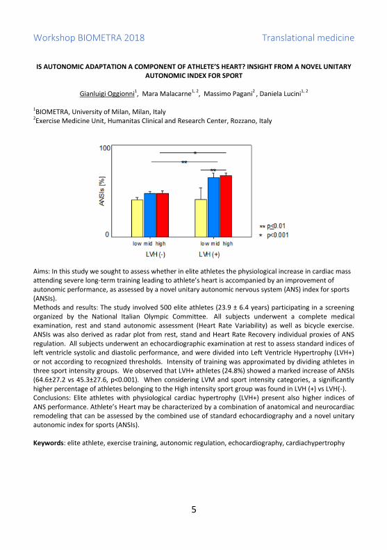
IS AUTONOMIC ADAPTATION A COMPONENT OF ATHLETE’S HEART? INSIGHT FROM A NOVEL UNITARY
AUTONOMIC INDEX FOR SPORT
Gianluigi Oggionni1, Mara Malacarne1, 2, Massimo Pagani2 , Daniela Lucini1, 2
1BIOMETRA, University of Milan, Milan, Italy 2Exercise Medicine Unit, Humanitas Clinical and Research Center, Rozzano, Italy
Aims: In this study we sought to assess whether in elite athletes the physiological increase in cardiac mass attending severe long-term training leading to athlete’s heart is accompanied by an improvement of autonomic performance, as assessed by a novel unitary autonomic nervous system (ANS) index for sports (ANSIs). Methods and results: The study involved 500 elite athletes (23.9 ± 6.4 years) participating in a screening organized by the National Italian Olympic Committee. All subjects underwent a complete medical examination, rest and stand autonomic assessment (Heart Rate Variability) as well as bicycle exercise. ANSIs was also derived as radar plot from rest, stand and Heart Rate Recovery individual proxies of ANS regulation. All subjects underwent an echocardiographic examination at rest to assess standard indices of left ventricle systolic and diastolic performance, and were divided into Left Ventricle Hypertrophy (LVH+) or not according to recognized thresholds. Intensity of training was approximated by dividing athletes in three sport intensity groups. We observed that LVH+ athletes (24.8%) showed a marked increase of ANSIs (64.6±27.2 vs 45.3±27.6, p<0.001). When considering LVM and sport intensity categories, a significantly higher percentage of athletes belonging to the High intensity sport group was found in LVH (+) vs LVH(-). Conclusions: Elite athletes with physiological cardiac hypertrophy (LVH+) present also higher indices of ANS performance. Athlete’s Heart may be characterized by a combination of anatomical and neurocardiac remodeling that can be assessed by the combined use of standard echocardiography and a novel unitary autonomic index for sports (ANSIs). Keywords: elite athlete, exercise training, autonomic regulation, echocardiography, cardiachypertrophy

Workshop BIOMETRA 2018 Immunology and oncology
6
THE CONSTITUTIVE EXPRESSION OF NKp46 IDENTIFIES A NEW SUBSET OF ANTI-TUMOR Vδ1
INTRAEPITHELIAL T CELLS RESIDENT IN HUMAN INTESTINE
Ferdinando Oriolo1,2, Joanna Mikulak1,2, Elena Bruni1,2, Alessandra Roberto3, Federico Simone Colombo4, Anna Villa5, Francesco Dieli6, Michele Maria Carvello7, Silvia Della Bella1,2, Bruno Silva-Santos8, Antonino
Spinelli7 and Domenico Mavilio1,2* 1Unit of Clinical and Experimental Immunology, Humanitas Clinical and Research Center, Rozzano, Milan, Italy 2BIOMETRA Department, University of Milan, Italy 3Laboratory of Translational Immunology, Humanitas Clinical and Research Center, Rozzano, Milan, Italy 4Humanitas Flow Cytometry Core, Humanitas Clinical and Research Center, Rozzano, Milan,Italy 5Telethon Institute for Gene Therapy, Division of Regenerative Medicine, Stem Cells and Gene Therapy, Istituto di Ricovero e Cura a Carattere Scientifico (IRCCS) San Raffaele Scientific Institute, Milan, Italy 6Central Laboratory for Advanced Diagnostic and Biomedical Research, Palermo, Italy 7Colon and Rectal Surgery Unit, Humanitas Clinical and Research Center, Rozzano, Milan, Italy 8Instituto de Medicina Molecular, Faculdade de Medicina, Universidade de Lisboa, Lisboa, Portugal *e-mail: [email protected] γδ T cells are referred to as innate-like lymphocytes that display a broad array of anti-tumor functions. Intestinal intraepithelial lymphocytes (IELs) are particularly enriched of γδ T cells; however, data regarding human γδ IEL subsets are lacking. In this study, specimens from total number of 141 individuals, isolated from intestine, blood and lymph nodes, liver, skin and pediatric thymus of healthy and/or pathologic tissue, mainly obtained from colorectal cancer (CRC) patients, were used for comparative multiparametric flow cytometry analysis in order to perform a compressive quantitative, phenotypical and functional characterization of human γδ T gut-resident populations. We demonstrated that human gut γδ T cells constitutively express Natural Cytotoxic Receptors (NCRs), mostly NKp46 that is restricted to intestinal intraepithelial Vδ1pos subsets. Furthermore, the ontogeny of the NKp46pos/Vδ1 subset depends both on distinctive features of Vδ1 thymic precursors and gut environmental factors and reflects functionally high cytotoxic anti-tumor activity and IFNγ secretion. On the other hand, our results show that elevated prevalence of NKp46pos/Vδ1pos IELs is predictive for CRC progression. In fact, CRC patients with higher frequency of the physiological NKp46pos/Vδ1pos subset have a lower risk of developing advanced/metastatic III/IV stage of disease. However, the intratumoral environment promotes the abolition of this specific subset which drastically decreasing their numbers. Similar to the tumor, inflammatory conditions with the increased risk of CRC such as ulcerative colitis lead to the decreased amount of NKp46pos/Vδ1 cells. In conclusion we identify a novel physiologically sizeable human gut-specific anti- tumor NKp46pos/γδ T subset that has the potential to represent a prognostic marker and a possible cellular/molecular target for novel therapeutic approaches in CRC. Keywords: γδT cells, NKp46, IELs, Tumor, Thymocytes.

Workshop BIOMETRA 2018 Immunology and oncology
7
NIPBL, A NEW PLAYER WITH NPMC+ IN MYELOID CELLS DIFFERENTIATION
Mazzola M.1*, Deflorian G.2, Ferrari L.2, Fazio G.3, Pezzotta A. 1, Bresciani E.4, Saitta C.3, Ferrari L.1, Fumagalli
M.5, Parma M.5, Riva P.1, Cotelli F.6, Biondi A.3, Marozzi A.1, Cazzaniga G.3, Pistocchi A.1
1BIOMETRA, Università degli Studi di Milano, Milan, Italy 2 Istituto FIRC di Oncologia Molecolare, IFOM, Milan, Italy 3 Centro Ricerca Tettamanti, Clinica Pediatrica Università di Milano-Bicocca, Centro Maria Letizia Verga, Monza, Italy 4 Oncogenesis and Development Section, National Human Genome Research Institute, National Institutes of Health, Bethesda, MD, United States 5 Haematology Division and BMT Unit, Ospedale San Gerardo, Monza, Italy 6 Dipartimento di Bioscienze, Università degli Studi di Milano, Milan, Italy *e-mail: [email protected]
Cohesins form a multimeric protein complex (SMC1A, SMC3, RAD21, STAG and additional proteins NIPBL, MAU2, ESCO1, HDAC8) involved in the cohesion of sister chromatids, post-replicative DNA repair and transcriptional regulation. Recently, recurrent somatic mutations and deletions of cohesins have been reported in the 10% of the patients with Acute Myeloid Leukemia (AML) or other myeloid neoplasms. Frequently, mutations in cohesin genes co-occurred with the known AML-associated gene nucleophosmin (NPM1) that, when mutated, aberrantly relocates to the cytoplasm (NPMc+). Forced NPMc+ expression in zebrafish embryos causes an expansion of hematopoietic stem cells (HSCs) according with AML patient features. In our cohort of adult AML patients, we observed a specific and significative reduction of the NIPBL expression in NPMc+ patients. We generated a zebrafish model of nipblb haploinsufficiency to investigate the hematopoietic phenotype and the interactions between NPMc+ and nipblb. In nipblb-loss-of-function zebrafish embryos, we observed an increase in myeloid progenitors, a phenotype resembling the NPMc+ zebrafish model. Therefore, we characterize the functional interaction between NPMc+ and NIPBL in the onset of the aberrant hematopoietic phenotype in zebrafish and showed the involvement of the canonical Wnt pathway in this process. We demonstrate for the first time a role for NIPBL during zebrafish hematopoiesis and that its decreased expression, due to NPM1 mutations, might play a role in leukemia onset. Keywords: cohesins, NIPBL, NPMC+, AML, zebrafish

Workshop BIOMETRA 2018 Immunology and oncology
8
MeCP2 IS REQUIRE FOR FUNCTIONAL PRIMARY CILIA
Eleonora Spiombi2*, Angelisa Frasca2, Anna Bergo3, Barbara Leva3, Michela Palmieri1, Marilena Valente1,
Charlotte Kilstrup-Nielsen3, Ferdinando Di Cunto4 and Nicoletta Landsberger1,2
1Neuroscience Division, San Raffaele Rett Research Center, San Raffaele Scientific Institute, 20132, Milan, Italy 2Department of Medical Biotechnology and Translational Medicine, University of Milan, 20090, Segrate, Italy 3Department of Biotechnology and Life Sciences, University of Insubria, 21052, Busto Arsizio, Italy 4Department of Molecular Biotechnology and Health Sciences, University of Turin, Turin, Italy *e-mail: [email protected] Rett syndrome (RTT; OMIM 312750) is a progressive X- linked neurodevelopmental disorder that, because of its incidence (1:10000 females), represents the most common cause of severe intellectual disability in girls worldwide. RTT is caused by mutations in methyl-CpG-binding protein 2 (MECP2), which encodes the multi- functional protein MeCP2. MeCP2 is mainly localized at nucleus where modulates the expression of several genes. However, we recently demonstrated that MeCP2, in addition to its activity in the nucleus, is functionally associated with centrosomes. Its deficiency causes aberrant spindle geometry, prolonged mitosis and defective nucleation of microtubules. Considering the importance of the centrosome for the assembly of primary cilium, we investigated the involvement of MeCP2 in primary cilium formation and function and we hypothesized that part of the RTT phenotypes are caused by impaired ciliary functions. Here we characterized the ciliary dysfunctions in Mecp2 deficient cells and mouse brains, reporting evidence of reduced ciliated cells and shortened primary cilia, in vitro and in vivo, suggesting that MeCP2 is directly involved in primary cilium formation. We also demonstrated an alteration of the expression and subcellular localization of Shh effectors in the absence of Mecp2, suggesting that the Shh signaling is affected by MeCP2 deficiency. Although the involved molecular mechanisms remain to be elucidated, aberrant ciliogenesis and the observed related phenotypes could be rescued either pharmacologically or by expressing MeCP2. Primary cilia probe and integrate extracellular signals, thereby affecting neuronal migration, dendritic arborization, neural circuits integration, learning and memory. Cilia are associated with a growing list of “ciliopathies” characterized by several features in common with RTT. We demonstrate that functional cilia require MeCP2 and thus propose that at least some of the Rett symptoms can be due to impaired ciliary functions. Keywords: Rett Syndrome, primary cilium, Sonic Hedgehog, neuron

Workshop BIOMETRA 2018 Methods and Technologies
9
DNA NANORULER: A NOVEL BIOTECHNOLOGICAL TOOL TO QUANTIFY PROXIMITY AT HIGH RESOLUTION
Marco Todisco1*, Matteo Marozzi1, Francesca Santini1,2, Marta Busnelli2, Giuliano Zanchetta1, Bice Chini2,
Tommaso Bellini1. 1BIOMETRA, Università degli Studi di Milano, Milan, Italy 2CNR, Institute of Neuroscience, Milan, Italy *e-mail: [email protected]
G-protein-coupled receptors (GPCRs) are the largest group of membrane receptors in eukaryotes and are involved in a variety of critical biological phenomena ranging from sensing the environment (vision, smell, taste) to behavior, immunity and homeostasis. Still, up to this day the characterization of their dimerization dynamics and its role in transmembrane signal transduction is an unsolved conundrum, whose solution is necessary to shed light on the very details of their function.
The emerging field of DNA nanotechnology may provide critical help: a set of DNA strands can be designed to produce structures with nanometric precision and to respond to a variety of stimuli (such as variations in pH, ionic strength or temperature, or binding of DNA, small molecules or proteins) in order to efficiently carry out the desired task.
We designed a novel tool to detect single dimerization events in GPCRs exploiting the potentials of DNA nanotechnology: two DNA hairpins (H1 & H2) are covalently attached to the GPCRs ligands and are designed to stay closed unless the two ligands, bound to the receptors, are in close proximity (d ≤ 15nm), a distance corresponding to dimerization.
The proximity between the two hairpins triggers the opening and hybridization to produce a stable double helix. The newly produced DNA complex, tethering the ligands and their bound receptors, can be visualized: the addition of two more fluorescent DNA sequences (R1 & R2) triggers a hybridization chain reaction (HCR) on the top of the two interacting hairpins and a bright fluorescent spot can be detected by a simple fluorescence microscopy apparatus.
As compared to existing proximity assays like Proximity Ligation Assays, which rely on several steps and layers of antibodies, our proposed approach may provide higher resolution, avoiding spurious proximity events that don’t correspond to dimerization, and ease of use. Keywords: DNA nanotechnology, Receptor dimerization, Proximity, GPCRs

Workshop BIOMETRA 2018 Methods and Technologies
10
DELAYED CREEP IN COLLOIDAL GELS
Paolo Edera1*, Fabio Giavazzi1, Roberto Cerbino1
1BIOMETRA, Università degli Studi di Milano, Milan, Italy *e-mail: [email protected]
Delayed creep is a very general, and still not completely understood phenomenon: solids subjected to a continuous stress can resist for long times before breaking abruptly. This phenomenon is common to a wide class of materials going from the earth crust during earthquakes to biological tissue and hydrogels used in biomedical application. Since the failure occurs without giving any macroscopical sign of the imminent break, signs of the weakening process are searched in the microscopic structure of the tested material. While the microstructure of molecular solids can be explored only by means of complex experimental apparatus (like neutron scattering or x-rays), colloidal systems can be investigated by means simpler techniques such as light scattering or microscopy. Optical techniques, together with the advantage of manageability and availability, are easy to be applied on systems subjected to a mechanical test. Because of these reasons and because of the tunability of their interaction, in the last decades colloidal systems established their relevance as model systems in the understanding of the connection between the macroscopic mechanical properties and the microscopic structure. In this framework I present an experimental work, realized in collaboration with the Foundation Of Research and Technology of Crete (FORTH-IELS), where a rotational rheometer is combined with an optical microscope. An innovative imaging analysis technique (Differential Dynamic Microscopy) is used to obtain quantitative information on the microscopical structure and dynamic of the gels during mechanical tests that bring to the breaking of the material. Keywords: mechanical properties, colloidal gels, microscopy Image: courtesy of Esmaeel Moghimi

Workshop BIOMETRA 2018 Methods and Technologies
11
A COMPUTATIONAL VIEW OF DSDNA-RNA TRIPLEX RECOGNITION BY L-RNA APTAMERS
Luca Mollica1,2*, Valeria Bevilacqua1,2, Serena Curti2, Tanya Fabbris2, Massimiliano Pagani1,2
1BIOMETRA, Università degli Studi di Milano, Via Fratelli Cervi, 93, 20090, Segrate, Italy 2Istituto Nazionale Genetica Molecolare "Romeo ed Enrica Invernizzi", Via F. Sforza 35, Milan, 20122, Italy *e-mail: [email protected] In the last decade, several studies have provided evidence for biological functions of long noncoding RNA molecules (lncRNAs). IncRNAs can form a variety of macromolecular complexes which fulfil their regulatory functions including structures such as RNA-DNA triplexes. These motifs tend to accumulate in the gene-regulatory regions leading to a proposal that triplex formation might play significant regulatory roles in vivo. In order to elucidate the role and the mechanism of triplex formation it is of paramount importance to design molecules that are able to selectively recognize and bind triplexes. The strategy we are adopting in order to design such molecules is based on the synthesis of nucleic acid L-aptamers (Spiegelmers) that are engineered through in vitro selection SELEX methodology (systematic evolution of ligands by exponential enrichment). Aptamers offer advantages over antibodies as they are readily produced by chemical synthesis and elicit little or no immunogenicity in therapeutic applications. Moreover, the chosen chirality allows the synthesis of molecules that are highly resistant to nucleases. An essential step for the rationalization of L-aptamers synthesis is the availability of a reliable atomic level three-dimensional structure of RNA-DNA triplex, which is experimentally rather difficult to access. We present a hybrid computational methodology based on sequence recognition, docking and molecular dynamics simulation for the prediction of the three dimensional atomic level structure of DNA-RNA triplexes from sequences. In particular, the methodology set up, based on the reconstruction of the few available experimentally determined complexes, is presented in detail. Moreover, some preliminary aspects of the aptamer-triplex recognition prediction are illustrated. Keywords: long non-coding RNA, nucleic acids triple helices, RNA-DNA triplex, molecular modeling, structure prediction

Workshop BIOMETRA 2018 Methods and Technologies
12
A BIOINFORMATIC FRAMEWORK TO IDENTIFY CELL SUBPOPULATIONS FROM BULK GENE EXPRESSION
DATA OF CANCER SAMPLES
Andrea Grilli1,2,3*, Silvio Bicciato3, Cristina Battaglia1 1BIOMETRA Department, University of Milan, Via F.lli Cervi 93, 20090, Segrate, Italy 2PhD Program of Molecular and Translational Medicine,University of Milan, 20090 Segrate, Italy 3Department of Life Sciences, Center for Genome Research, University of Modena and Reggio Emilia, Modena, Italy. *e-mail: [email protected] The expression levels of biological samples are affected by the intrinsically heterogeneous cell and tissue composition. Nevertheless, when analyzing transcriptional profiles, each sample is generally evaluated in bulk without considering the presence of multiple subpopulations. This limitation might be extremely critical when analyzing gene expression profiles from cancer samples, where dissecting the mixed cell population could shed light on the intratumoral heterogeneity and on the molecular mechanisms shaping different cancer behaviors. We built on 5 different deconvolution algorithms, Cibersort (Newman, 2016), Epic (Racle, 2017), DeconRNASeq (Gong, 2013) ssGSEA (Charoentong, 2017), ImmQuant (Frishberg, 2016), developed for the identification of hematopoietic subsets in RNA mixtures, to design a framework for the identification of cellular subpopulations from bulk gene expression of cancer samples. Starting from a dataset of breast cancer subtypes based on immunohistochemistry (IHC), we initially created a gene signature of 230 genes; then, we applied the signature with the deconvolution algorithms on 2 datasets of clinically-defined Triple Negative Breast Cancer (TNBC) samples. For each bulk sample, the cellular fraction of each subtype was calculated and the TNBC fraction tested for association with clinical and pathological features. Although clinically triple negative, about two third of samples show a modest or high presence of subtypes other than TNBC in both datasets: in particular, a subset of samples shows an almost complete absence of TNBC-like cells. Noteworthy, association of the TNBC fraction to either clinical response to neo-adjuvant chemotherapy or survival identified a poorer prognosis in samples with lower fraction of TNBC cells. The result is independent from the used tool. In conclusion, we deconvolved the bulk transcriptional profile of cancer samples to assess their tumor heterogeneity and its relationship with their clinical characteristics. Keywords: Deconvolution, Breast Cancer, Tumor Heterogeneity, Triple Negative

Workshop BIOMETRA 2018 Molecular Mechanisms of Diseases
13
IDENTIFICATION OF THE ANTIGEN RECOGNIZED BY rHIgM22, A REMYELINATION-PROMOTING HUMAN
MONOCLONAL ANTIBODY
Livia Cabitta1*, Sara Grassi1, Simona Prioni1, Laura Mauri1, Maria Grazia Ciampa1, Yana Zorina2, Sandro Sonnino1, Alessandro Prinetti1
1BIOMETRA, Università degli Studi di Milano, Milan, Italy 2Acorda Therapeutics, Inc., Ardsley, NY, USA *e-mail: [email protected] Recombinant human IgM22 (rHIgM22) binds to myelin and oligodendrocytes (OLs) and promotes remyelination in mouse models of Multiple Sclerosis. rHIgM22 preferentially reacts with sulfatide positive (O4+) OLs, and binding of rHIgM22 is abolished in CNS tissue slices from Cst (-/-) mice, suggesting that its binding requires the presence of a product of cerebroside sulfotransferase, possibly sulfatide, highly expressed in OLs and myelin. However, the identity of the antigen recognized by this antibody remains to be elucidated. We tested the binding of rHIgM22 to purified lipids and lipid extracts from mouse brain, CNS myelin, mixed glial cells, and O4+ OLs using TLC immunostaining. Our preliminary results show that IgM22 binds to sulfatide in vitro, while it does not bind to other myelin sphingolipids suggesting that sulfatide at the OLs surface might be important for the binding of IgM22 to these cells and to myelin. However, IgM22 does not bind structures expressing sulfatide outside the nervous system, so additional factors are likely relevant for the immunoreactivity of IgM22 in CNS. Indeed, in lipid extracts from different sources we found another lipid antigen selectively recognized by IgM22. To attempt the identification of the antigen, samples were purified using column chromatography and the second IgM22-immunoreactive band enriched fractions were analyzed by ESI Mass Spectrometry. The results obtained led to hypothesize that the unknown antigens could be Phosphatidylinositol (18:0/20:4-PI) and two different Phosphatidylserine species, 18:0/22:6-PS and 18:0/18:1-PS. This lipid is also present in the extracts from Mixed Glial Cultures, which do not contain mature O4+ OLs, suggesting that other glial cells in addition to OLs might be important in the response to IgM22. Keywords: sphingolipids, remyelination

Workshop BIOMETRA 2018 Molecular Mechanisms of Diseases
14
TARGETING REMODELLING BONE DISEASES WITH NUTRACEUTIC COMPOUNDS: THE PROTECTIVE
ACTIVITY OF Δ-TOCOTRIENOL.
Casati Lavinia*1, Pagani Francesca1, Aschedamini Martina1, Montagnani Marelli Marina2, Limonta Patrizia 2,
Sibilia Valeria 1
1BIOMETRA Department 2Department of Pharmacological and Biomolecular Sciences *e-mail: [email protected]
Increasing evidence suggests a key role of the oxidative stress in the bone aging and the potential use of
natural antioxidant compounds in the prevention/treatment of osteoporosis. This study was undertaken
to investigate the ability of δ-tocotrienol, derived from vitamin E, to protect pre-osteoblast cells-like
(MC3T3-E1) and osteocyte cells-like (MLOY-4) against oxidative damage induced by tert-butyl
hydroperoxide (t-BHP) and the molecular pathways involved in its protective action against oxidative
stress.
Pre-treatment with δ-tocotrienol (2,5-20 ug/ml) significantly prevented t-BHP-induced (250 uM or 125 uM
for 3 hours) osteoblastic viability reduction (by MTT assay) and apoptotic induction (by Hoechst Staining),
by decreasing intracellular ROS levels. This effect is probably due to the gene expression induction of the
detoxifying genes mediated by δ-tocotrienol through the reduction of the histone deacetylase activity
induced by t-BHP. To clarify the mechanisms involved in δ-tocotrienol prevention of oxidative damage, we
examined different pathways involved in cell viability under stressful conditions such as sirtuins,
mevalonate, NFR2 and PI3K pathways. We found that δ-tocotrienol protective effects against t-BHP-
induced osteoblastic dysfunction were partially mediated by the PI3K/Akt signalling since they were
partially prevented by LY294002, a PI3K/Akt specific inhibitor. Furthermore, we found that ML385, a
specific inhibitor of the NFR2 pathway, is also able to partially remove the protective effect of δ-
tocotrienol. In agreement we found that δ-tocotrienol increased the Akt phosphorylation level.
These findings indicate that δ-tocotrienol protects bone cells from oxidative damage and suggest the
potential utility of dietary supplements to prevent bone dysfunction in age-related osteoporosis.
Keywords: bone, oxidative stress, epigenetics, δ-tocotrienol, Akt pathway

Workshop BIOMETRA 2018 Molecular Mechanisms of Diseases
15
IDENTIFICATION OF NOVEL MECHANISMS FOR NEUROLOGICAL CONDITIONS OVERLAPPING SMITH-
MAGENIS SYNDROME
Ilaria Bestetti1,2*, Alessandra Sironi1,2, Maria Sciarrillo1,2, Luigia Spaccini3, Maria Francesca Bedeschi4, Lidia Larizza2, Palma Finelli1,2
1 BIOMETRA, Università degli Studi di Milano, Milan, Italy 2 Medical Cytogenetics and Molecular Genetics Laboratory, IRCCS Istituto Auxologico Italiano, Milan, Italy 3 Genetic Service, Department of Obstetrics and Gynecology, "V. Buzzi" Children’s Hospital, University of Milan, Milan, Italy 4 Medical Genetic Unit, IRCCS Ca’Granda Ospedale Maggiore Policlinico, Milano *e-mail: [email protected] Smith Magenis Syndrome (SMS) is a complex heterogeneous disorder principally characterized by developmental delay, behavioural problems and circadian rhythm dysregulation. It is caused by the retinoic acid-induced 1 (RAI1) gene haploinsufficiency for 17p11.2 deletion or gene mutation. Little is known about the molecular mechanism underlying SMS, as RAI1 role and interactors are not well clarified yet. Up to date only 50% of patients with a clinical suspicion of SMS bear the classical molecular defect highlighting that other loci, if disrupted, may cause SMS-like phenotypes. In a cohort of 30 SMS-like patients without either 17p11.2 deletion or RAI1 mutation, here we report two interesting cases that pointed out new putative candidate genes for SMS. In patient 1 a 54 kb maternal deletion at Xq13.3 that involve a predicted conserved regulatory element and maps 29 kb far from ZDHHC15 5’UTR was found. This gene, downregulated in patient, encodes for a palmitoyl-transferase specifically expressed in the brain and linked to XLMR. By molecular and functional studies we evaluated its possible involvement in circadian rhythm. ZDHHC15 silencing led to transcript alteration of several circadian genes, supporting its involvement in sleep disturbances and its possible relation with RAI1. Patient 2 has a t(6;14)dn disrupting STXBP6 gene with a role in regulating SNARE complex formation for the release of neurotransmitters at the neuronal presynaptic membrane. Molecular analysis revealed a truncated isoform of STXBP6 which consist of first four exons and lack the last sequence encoding for the functional domain of the protein. Moreover, RT-qPCR analysis showed a dramatic downregulation of total transcript in the patient indicating that also the wild type allele expression is affected. This approach, taken together our findings, was useful to identify other SMS-like causative loci, providing new insights to deepen the pathomechanism at the basis of SMS and similar phenotypes. Keywords: SMS-like syndrome, RAI1, ZDHHC15, STXBP6

Workshop BIOMETRA 2018 Molecular Mechanisms of Diseases
16
STRESS–INDUCED LSD1 AND SRF MODULATION AND THEIR POTENTIAL PROTECTIVE ROLE TOWARDS
NEUROPSYCHIATRIC DISORDERS
Chiara Forastieri*1, Barbara Grillo1, Alessandra Longaretti1, Lara Monti1, Emanuela Toffolo1, Francesco Rusconi1, Elena Battaglioli1
1BIOMETRA, Università degli Studi di Milano, Milan, Italy *e-mail: [email protected]
In mammals, different forms of stress including psychosocial stress can impact on several aspects of human health, fostering mood and anxiety disorders. Very little is known about the mechanisms underlying brain physiology of stress response, although epigenetic mechanisms have been proposed to play a crucial role. Serum Response Factor (SRF) has been addressed as a stress transducer via promoting inherent experience-induced Immediate Early Genes (IEGs) expression. However, in resting conditions, SRF represents a transcriptional repressor able to assemble the core LSD1/CoREST/HDAC2 corepressor complex, including demethylase and deacetylase activities. Here, we show that a SRF splicing isoform lacking most part of the transactivation domain, namely SRFΔ5, represents a constitutive transcriptional repressor retaining the ability to assemble the same LSD1 corepressor complex. SRFΔ5 is highly expressed in the brain and developmentally regulated, increasing its expression along with brain maturation. We uncover SRFΔ5 as a negative modulator of dendritic spine density, suggesting that its low perinatal expression facilitates SRF-induced synaptogenesis. Upon acute psychosocial stress SRF isoforms are transiently modified at the protein levels. Remarkably, when stress is chronically repeated, SRF protein becomes stably upregulated in vulnerable mice but not in resilient animals, via a splicing dependent mechanism. These data suggest that SRF is a stress response modifier and SRF/SRFΔ5 modulation represents a novel stress vulnerability hallmark possibly representing a new pharmacological target to treat depression. Keywords: Epigenetics, Transcription, Stress susceptibility, Depression

Workshop BIOMETRA 2018 Molecular Mechanisms of Diseases
17
STRESS-INDUCED MODULATION OF THE OXIDATIVE BALANCE IN THE RAT BRAIN: EFFECT OF THE
ANTIPSICHOTIC LURASIDONE
Maria Serena Paladini1, Andrea Carlo Rossetti1, Martina Colombo2, Vittoria Spero1, Federica Orellana1, Mariusz Papp3, Paola Stella Brivio2, Francesca Calabrese2, Marco Andrea Riva2, Raffaella Molteni1
1BIOMETRA, Università degli Studi di Milano, Milan, Italy 2Department of Pharmacological and Biomolecular Sciences, Università degli Studi di Milano, Milan, Italy 3Institute of Pharmacology, Polish Academy of Sciences, Kracow, Poland *e-mail: [email protected] Although the molecular mechanisms underlying the etiology of psychiatric disorders have not been elucidated, they are associated with altered inhibitory neurotransmission that may due to the vulnerability of selective neuronal subtypes to environmental conditions, such as stress. One intriguing aspect for the vulnerability of inhibitory neurons under stress is represented by “redox dysregulation,” an imbalance between oxidative stress and antioxidant defense systems that may lead to oxidative stress-related neuronal damage. Thus, the aim of our study was to investigate the impact of chronic stress (CS) on redox homeostasis in the rat brain and to what extent such changes can be modulated by the treatment with the antipsychotic lurasidone. In the first part of the study, adult rats were exposed to stress for 7 weeks. After 2 weeks of stress, the animals received chronic lurasidone (3 mg/kg/d) for the subsequent 5 weeks. Moreover, to determine if stress-induced molecular changes can still be observed after a recovery phase, in another study chronically-stressed animals treated with lurasidone were left undisturbed for 3 weeks before to be exposed to an acute restraint stress, to examine also differences in their acute responsiveness. We found that chronic stress up-regulates NOX2, a pro-oxidant enzyme, and reduces Nrf-2, a master regulator of antioxidant defense. These alterations were normalized after lurasidone that was also able to reduce Keap-1 that exerts a repressive control over Nrf-2. Moreover, lurasidone was also able to modulate the effect of stress on the acute-responsiveness: acute stress increases Srxn1, one of the antioxidant genes downstream from the transcriptional activity of Nrf-2, an effect that was not observed when the acute challenge was preceded by chronic stress but was partially restored by lurasidone. These effects may promote resilience toward the alterations produced by stress, a major vulnerability factor for psychiatric disorders etiology. Keywords: chronic stress, redox, psychiatric disorders, lurasidone

Workshop BIOMETRA 2018
18
Poster presentations

Workshop BIOMETRA 2018 Poster abstracts
19
P1 SMALL NUCLEAR RNA-BASED CORRECTION OF SPLICING FOR A PERSONALIZED TREATMENT OF
PATHOGENIC MUTATIONS IN CDKL5
Domenico Giorgio1, Matteo Bizzotto1, Dario Balestra2, Mirko Pinotti2, Nicoletta Landsberger1* 1BIOMETRA, Università degli Studi di Milano – sede L.I.T.A., Milan, Italy 2Department of Biochemistry and Molecular Biology, University of Ferrara, Italy *e-mail: [email protected]
Genetic lesions of various kinds in the X-linked CDKL5 gene are responsible for a suite of disorders affecting both genders, which generally share the common feature of early drug-resistant epilepsy, emerging in the first months of life. No cure is currently available for CDKL5 conditions and treatment is usually based on support therapy for comorbidities, and on rehabilitation. Our knowledge on CDKL5 functions and the consequences of its deficiency is still very limited; therefore, the identification of “rationally-designed” therapeutic approaches will probably not occur in the immediate future. We found that almost 15% of pathogenic mutations affect splicing and a large fraction of these might benefit of therapeutic approaches normalizing it. Indeed by acting on pre-mRNA, these approaches circumvent the overexpression problem, the packaging limitations imposed by gene therapy as well as the necessity of transducing, in heterozygous female patients, mainly those cells that express the mutated allele. Through a minigene assay system, we demonstrate that modified U1 snRNAs can restore the aberrant splicing pattern caused by donor site mutations, which reduce the affinity of the pre-mRNA to the 5′ tail of the endogenous U1 snRNA. Since our data indicate a major effect on +5 mutations compared to those affecting the dinucleotide GT (+1, +2 at donor site), we have focused our studies on two human mutations (i.e c.99+5G>A and c.2376+5G>A) affecting two different domains of the protein. In particular, we obtained and characterized the phenotype of a lymphoblastoid cell line (LCL), derived from patient carrying the c.2376+5G>A mutation. These cells display a reduced level of CDKL5 transcript and protein compared to control; in addition, the same cells seem to have a slower growth and defects in cytoskeleton proteins. We’re now testing the capability of modified U1 to rescue the correct amount of protein and the defects previously described. Since no cell line is actually available for the 99+5G>A mutation, we have designed an artificial Splicing Competent (SC) cDNA and expressed it in different human cell lines: this approach allowed us to analyse the CDKL5 pre-mRNA as well as the full protein at a cellular level. Our results confirm that the mutated SC cDNA leads to the skipping of exon 3. The cotransfection of the mutated SC with the compensatory U1 leads to a complete rescue of a full-length catalytically active protein. Keywords: CDKL5, splicing, U1 snRNA

Workshop BIOMETRA 2018 Poster abstracts
20
P2 CONSTITUTIVE UPREGULATION OF TNFSF15-TNFRSF25 AXIS SUSTAINS ENDOTHELIAL DYSFUNCTION IN
PATIENTS WITH UNPROVOKED VENOUS THROMBOEMBOLISM
Francesca Calcaterra1,2, Monica Bacci3, Claudia Carenza1,2, Chiara Pandolfo2, Paola Ferrazzi3, Paolo Uva4, Massimiliano Pagani1,5, Corrado Lodigiani3, Domenico Mavilio1,2, Silvia Della Bella1,2*
1BIOMETRA Department, University of Milan, Milan, Italy 2Unit of Clinical and Experimental Immunology, Humanitas Clinical and Research Center, Rozzano, Milan, Italy 3Thrombosis and Haemorragic Diseases Center, Humanitas Research Hospital, Rozzano, Milan, Italy; 4Center for Advanced Studies, Research and Development in Sardinia (CRS4), Science and Technology Park Polaris, Pula, Italy 5INGM-National Institute of Molecular Genetics "Romeo ed Enrica Invernizzi" Milan, Milan, Italy * e-mail: [email protected] Rationale: The molecular mechanisms involved in the pathogenesis of unprovoked venous thromboembolism (uVTE) are largely unknown, yet their identification would be useful to guide uVTE treatment. Endothelial colony-forming cells (ECFCs) are the best defined population of circulating endothelial progenitors and allow non-invasive endothelial compartment functional assessment. Objective: To investigate whether uVTE patients have impaired endothelial function that may explain their propensity to develop thrombosis, and to identify the molecular pathways involved in endothelial dysfunction. Methods and Results: ECFCs were isolated and cultured from the peripheral blood of 23 uVTE patients and 27 healthy controls. Validation of dysregulated genes identified by gene expression profiling was performed by RT-qPCR, ELISA and functional studies. ECFCs were isolated with similar efficiency from uVTE patients and controls, but ECFCs obtained from uVTE patients underwent earlier senescence and showed lower growth rate than control ECFCs. Microarray profiling showed 2905 genes differentially expressed between patients and controls. Among these genes, the anti-angiogenic cytokine TNFSF15 and its death receptor TNFRSF25 were significantly upregulated in patient ECFCs. These findings were validated by RT-qPCR. And TNFSF15 upregulation was further confirmed at the protein level. After proving that exogenous TNFSF15 exerts pro-apoptotic and anti-proliferative activity on control ECFCs, we demonstrated through inhibition experiments that constitutive upregulation of TNFSF15 is responsible for impaired survival and proliferation of patient ECFCs. Conclusions: Constitutive upregulation of TNFSF15 and TNFRSF25 has a causative role in the pathogenesis of uVTE, as it impairs the ability of ECFCs to maintain endothelial integrity that is needed to prevent thrombosis. These results may encourage novel therapeutic strategies for the prevention of VTE recurrence in uVTE patients. Keywords: Unprovoked venous thromboembolism, endothelial colony-forming cells, gene- expression profiling, TNFSF15, TNFRSF25
Workshop BIOMETRA 2018 Poster abstracts
21
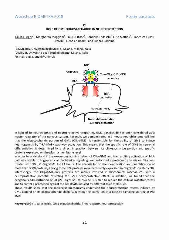
P3 ROLE OF GM1 OLIGOSACCHARIDE IN NEUROPROTECTION
Giulia Lunghi*1, Margherita Maggioni1, Erika Di Biase1, Gabriella Tedeschi2, Elisa Maffioli2, Francesca Grassi
Scalvini2, Elena Chiricozzi1 and Sandro Sonnino1
1BIOMETRA, Università degli Studi di Milano, Milano, Italia 2DiMeVet, Università degli Studi di Milano, Milano, Italia *e-mail: [email protected]
In light of its neurotrophic and neuroprotective properties, GM1 ganglioside has been considered as a master regulator of the nervous system. Recently, we demonstrated in a mouse neuroblastoma cell line that the oligosaccharide portion of GM1 (OligoGM1) is responsible for the ability of GM1 to induce neuritogenesis by TrkA-MAPK pathway activation. This means that the specific role of GM1 in neuronal differentiation is determined by a direct interaction between its oligosaccharide portion and specific proteins expressed on the plasma membrane level. In order to understand if the exogenous administration of OligoGM1 and the resulting activation of TrkA pathway is able to trigger crucial biochemical signaling, we performed a proteomic analysis on N2a cells treated with 50 µM OligoGM1 for 24 hours. The analysis led to the identification and quantification of more than 3500 proteins, among these 324 proteins were exclusively expressed in OligoGM1-treated cells. Interestingly, the OligoGM1-only proteins are mainly involved in biochemical mechanisms with a neuroprotective potential reflecting the GM1 neuroprotective effect. In addition, we found that the exogenous administration of 50 µM OligoGM1 to N2a cells is able to reduce the cellular oxidative stress and to confer a protection against the cell death induced by different toxic molecules. These results show that the molecular mechanisms underlying the neuroprotection effects induced by GM1 depend on its oligosaccharide chain, suggesting the activation of a positive signaling starting at PM level. Keywords: GM1 ganglioside, GM1 oligosaccharide, TrkA receptor, neuroprotection

Workshop BIOMETRA 2018 Poster abstracts
22
P4 PHAGE THERAPY AGAINST PSEUDOMONAS AERUGINOSA INFECTIONS IN A CYSTIC FIBROSIS ZEBRAFISH
MODEL
M. Cafora1, F. Forti2, G. Deflorian3, L. Ferrari3, D. Ghisotti2, F. Briani2, A. Pistocchi1
1BIOMETRA – University of Milan – Italy 2Dept. of Biosciences – University of Milan – Italy 3Istituto FIRC di Oncologia Molecolare – IFOM – Milano – Italy
Cystic fibrosis (CF) is one of the major hereditary disease due to mutations in the CFTR gene, and causes mortality in humans mainly due to infection in the respiratory system. Pseudomonas aeruginosa is the most important pathogen affecting chronic lung infection in 80% of CF patients. In a previous work we isolated and characterized a mix of virulent phages (phage cocktail) able to infect P. aeruginosa and used them as therapeutic agents against in vivo bacterial infections both in mice and in Galleria mellonella larvae. In this work we apply phage therapy to the treatment of P. aeruginosa infections in a zebrafish (Danio rerio) model. The use of this alternative animal model is appealing for better understanding CF patients defects. Indeed, the CFTR gene is evolutionary conserved between fish and mammals and cftr-loss-of-function zebrafish embryos show a phenotype that recapitulates the human CF disease, in particular with destruction of the pancreas. Zebrafish embryos were microinjected with a lethal dose of P. aeruginosa followed by phage injection and the effects of phage therapy on lethality, bacterial burden and immune response were observed. We found that phage therapy is able to reduce lethality caused by P. aeruginosa infection also in a CF zebrafish model. In addition, we found that phage administration relieves the constitutive inflammatory state of cftr-loss of function embryos. Moreover, we showed a synergistic effect of phage therapy and antibiotic treatment against P. aeruginosa infection. Our findings could improve therapeutic treatment of CF patients. Keywords: Cystic fibrosis, bacteriophage, Pseudomonas aeruginosa, zebrafish

Workshop BIOMETRA 2018 Poster abstracts
23
P5 BRACHIAL ARTERY DIAMETER IS AN INDEPENDENT BIOMARKER OF ALTERED HEART GEOMETRY
Daniela Coggi1,2*, Mauro Amato2, Alessio Ravani2, Daniela Sansaro2, Beatrice Frigerio2 and Damiano
Baldassarre2,3 1Dipartimento di Scienze Farmacologiche e Biomolecolari, Università di Milano, Milan, Italy 2Centro Cardiologico Monzino, IRCCS, Milan, Italy 3BIOMETRA, Università degli Studi di Milano, Milan, Italy *e-mail: [email protected]
Arterial diameter in plaque-free areas tend to increase in response to the presence of vascular risk factors (VRFs). Often this aspect is investigated by considering brachial artery diameter (BAD) because this district is rarely affected by atherosclerotic lesions. Cardiac cavities also tend to enlarge in response to VRFs and/or to increased workload. In this study, we have investigated whether BAD enlargement is a biomarker of impaired heart geometry. To this aim, BAD and heart geometry were measured by B-mode ultrasound in 2242 patients (1976 men), who underwent echocardiogram for clinical reasons. The echocardiographic features measured were: heart muscle wall dimensions (interventricular septum and posterior wall thicknesses); cardiac cavities geometry (left atrium and right ventricle geometry); and diameters of aortic-root and ascending aorta. Participant’s clinical/anamnestic characteristics were recorded and 10-year cardiovascular risk was estimated according to the Framingham algorithm. BAD enlargement was significantly associated with the size of right ventricle and left atrium. The relationship between BAD and left atrium size was not linear; indeed, left atrium size remained almost unchanged from the first to the fourth BAD quintiles and increased significantly only in BAD top quintile. BAD also associated with diameter of both aortic-root and initial ascending aorta. All the relationships were independent from VRFs, atherosclerotic profile and arterial enlargement in other vascular districts. No association was found between BAD and the indexes of cardiac muscle wall thickness. We conclude that the enlargement of the cardiac cavities and BAD share at least one etiopathological mechanism and/or common risk factor. From a clinical prospective our data suggest that an echocardiogram, carried out to assess the state of cardiac cavities, would be desirable whenever an altered BAD is found. Keywords: brachial artery diameter, atherosclerosis, cardiac morphology, echocardiogram

Workshop BIOMETRA 2018 Poster abstracts
24
P6 PREVALENCE AND PREDICTIVE VALUE OF RHEUMATOID FACTOR AND ANTI-CYCLIC CITRULLINATED
PEPTIDE ANTIBODIES IN THE GENERAL POPULATION
Elena Generali1, Natasa Isailovic1, Maria De Santis1, Angela Ceribelli1,2, Giacomo Maria Guidelli1, Marta Caprioli1, Marianna Meroni1 and Carlo Selmi1,2*
1Rheumatology and Clinical Immunology Unit, Humanitas Research Hospital, Rozzano (MI), Italy 2BIOMETRA Department, University of Milan, Milan, Italy *e-mail: [email protected] Rheumatoid arthritis (RA) is an autoimmune disease characterized by erosive arthritis, associated with rheumatoid factor (RF) and anti-cyclic citrullinated peptide antibodies (anti-CCP). The diagnostic specificity of RF is relatively low, since it can also occur in other autoimmune diseases but also in chronic infections, including hepatitis C virus (HCV) and hepatitis B virus (HBV). Anti-CCP by contrast are highly specific for RA. We aimed to determine the prevalence of serum RF and anti-CCP and the associated long term risk of developing RA in the general population, as well as their association with HBV and HCV infections. We took advantage of a randomly selected sample of a 1998 general population study (Isola I, including 2828 subjects; 53%women, age 43±13years) from a well-defined Northern Italian area that underwent testing for HBV and HCV. We tested RF and anti-CCP (2012-2016) on the available serum samples. Administrative databases were searched for RA diagnosis using disease copayment exemptions and hospital discharge forms. RF was positive in 430/2196 subjects (19.6%), anti-CCP were detected in 121/2,524 (4.8%). RF and anti-CCP were both positive in 25/2015 (1.2%). RF positive subjects had HBsAg in 29/430(6.7%), anti-HBc in 181/430(42.1%), and anti-HCV in 20/430(4.6%), both anti-HBc and anti-HCV in 16/430(3.7%), while HBsAg and anti-HCV in 1/430(0.23%). Anti-CCP positive subjects had HBsAg in 6/121(5%), anti-HBc in 54/121(44.6%), anti-HCV in 7/121(5.8%), and both anti-HBc and anti-HCV in 5/121(4.1%), none had HBsAg and anti-HCV. From the analysis of the administrative database we identified 12 cases of RA(0.5%), 3(25%) positive for RF and 5(41.7%) positive for anti-CCP antibodies, mostly at medium-high titer(4/5, 80%). Only one(8.3%) case of RA was positive both for RF and anti-CCP antibodies. RF and anti-CCP antibodies are predictive of RA development in an unselected sample of the general population, while being frequently associated with HBV and HCV. Keywords: chronic inflammation, autoimmunity

Workshop BIOMETRA 2018 Poster abstracts
25
P7 THE HUMAN-RESTRICTED DUPLICATED FORM OF THE α7 NICOTINIC RECEPTOR, CHRFAM7A: EXPRESSION
AND TRANSCRIPTIONAL REGULATION IN INFLAMMATORY CELLS
Simona Di Lascio1*, Annalisa Maroli1, Silvia Cardani1, Lorenzo Drufuca1, Massimo Locati1, Roberta Benfante2,1, Diego Fornasari1,2
1BIOMETRA, Università degli Studi di Milano, Milan, Italy 2CNR – Istituto di Neuroscienze, Milan, Italy *e-mail: [email protected]
Introduction: The α7 nicotinic acetylcholine receptor (CHRNA7) plays a role in the modulation of the inflammatory response through the activation of the “cholinergic anti-inflammatory pathway”. In humans, a recombination event involving the exon 5 to 10 of CHRNA7 gene, fused to four novel exons A, B, C and D (FAM7A), gave rise to the CHRFAM7A gene. This hybrid gene, located on chromosome 15q13-q14, 1.6 Mb apart from CHRNA7, is highly expressed in inflammatory cells, where it can regulate the anti-inflammatory effects of α7 activation. Acute treatment of macrophages with LPS down-regulates CHRFAM7A by a mechanism driven by NF-κB, paralleled by CHRNA7 up-regulation. As studies are emerging, which identify CHRFAM7A expression alteration in inflammatory or infective pathologies, the regulation of its expression may become a key step in the modulation of inflammation. However, the region driving the transcriptional regulation of CHRFAM7A gene in human immune tissues is largely unknown. Materials and methods: human monocytic-derived macrophages and THP-1 cell line have been used to characterized the CHRFAM7A regulatory region. Results and conclusions: we provide a detailed analysis of the CHRFAM7A gene regulatory region and its pro-inflammatory stimuli responsiveness. Furthermore, given the anti-inflammatory potential of the acetylcholinesterase inhibitor donepezil, we investigated the CHRFAM7A expression profile in macrophages treated with donepezil, showing an unexpected up-regulation of both CHRFAM7A and CHRNA7 gene, thus highlighting a possible role for CHRFAM7A gene product in the control and modulation of the cholinergic anti-inflammatory pathway, and/or in the modulation of CHRNA7 function. Keywords: CHRNA7, CHRFAM7A, inflammation, cholinergic antinflammatory pathway, donepezil

Workshop BIOMETRA 2018 Poster abstracts
26
P8 MUTATIONS ASSOCIATED WITH CLONAL HEMATOPOIESIS OF INDETERMINATE POTENTIAL ARE FOUND
IN PERIPHERAL BLOOD AND SYNOVIAL FLUID MACROPHAGES FROM PATIENTS WITH RHEUMATOID AND PSORIATIC ARTHRITIS
Maria De Santis1, Matteo Zampini2, Natasa Isailovic1, Elena Generali1, Giacomo Maria Guidelli1, Matteo
Della Porta2 and Carlo Selmi*1,3 1Rheumatology and Clinical Immunology Unit, Humanitas Research Hospital, Rozzano (MI), Italy 2Oncology and Hematology Unit, Humanitas Research Hospital, Humanitas University, Rozzano (MI), Italy 3BIOMETRA Department, University of Milan, Milan, Italy *email: [email protected] Myeloid mutations commonly associated to neoplasia in patients who do not meet the diagnostic criteria for myelodysplastic syndrome are coined as clonal hematopoiesis of indeterminate potential (CHIP). CHIP-related mutations are somatic DNA accumulating with aging, being found in approximately 10% of >65. Chronic inflammation, has been reported to favor the expansion of CHIP-mutant clones, but recent evidence suggests that CHIP-mutated clones contribute to chronic inflammation. We hypothesize that chronic inflammation concurs with age to CHIP-mutation appearance, i.e. they should be more precocious and frequent in rheumatic patients. We investigated the peripheral blood (PB) and synovial fluid (SF) myeloid cells from 4 rheumatoid arthritis(RA), 4 psoriatic arthritis(PsA), and 4 knee osteoarthritis(OA) patients (female 5, median age 73.5 years, range 53-91, median disease duration 10 years, range 7-20). To detect the presence of CHIP mutations we used a high-throughput sequencing platform (Illumina NextSeq) for mutation screening of 78 genes relevant in myelodysplastic diseases. We evaluated only variants known as pathogenic in COSMIC database, with a population frequency (MAF) < 0.1% described in dbSNP and in 1000 Genome Project. We found somatic mutations of JAK2, TP53, and TET2in 2/4 RA patients, GATA2-mutation in 1/4 PsA patients, and a small clone of SF3B1 in 1/4 OA cases. Mutated clones were found both in PB and SF of the patients. The patients with CHIP-mutations did not have a more active disease (DAS28-CRP4.2±0.6vs5.1±1.2), but 1 RA patient with JAK2- mutation and 1 PsA patient had <65years. Our data suggests that CHIP-mutations are frequent in patients with chronic arthritis, also before 65 years. Understanding the mechanisms connecting somatic mutation-driven clonal hematopoiesis and chronic inflammation will be of great interest not only from a pathogenic point of view, but also in considering possible therapeutic options, such as JAK-inhibitors. Keywords: chronic inflammation, autoimmunity

Workshop BIOMETRA 2018 Poster abstracts
27
P9 NKG2A REPRESENTS A TARGETABLE CHECK-POINT TO IMPROVE NK CELLS ALLOREACTIVITY EARLY AFTER
HAPLOIDENTICAL HEMATOPOIETIC STEM CELL TRANSPLANTATION
Elisa Zaghi1, Clara Di Vito1, Arianna Capucetti1, Alessandra Roberto2, Luca Castagna3, Enrico Lugli2,4 and Domenico Mavilio1,5*
1Unit of Clinical and Experimental Immunology, Humanitas Clinical and Research Center, Rozzano, Milan, Italy 2Laboratory of Translational Immunology, Humanitas Clinical and Research Center, Rozzano, Milan, Italy 3Hematology and Bone Marrow Transplant Unit, Humanitas Cancer Center, Rozzano, Milan, Italy 4Humanitas Flow Cytometry Core, Humanitas Clinical and Research Center, Rozzano, Milan, Italy 5BIOMETRA, Università degli Studi di Milano, Milan, Italy. *e-mail: [email protected] Haploidentical hematopoietic stem cell transplantation (h-HSCT) is a therapeutic option to cure hematologic malignancies. Immune-reconstitution (IR) is key in determining a positive or negative clinical outcome of HSCT. Hence, a better understanding of the kinetic and quality of IR following HSCT can better predict and therapeutically target opportunistic infections, tumor-relapse, engraftment and the onset/degree of Graft versus Leukemia effect as wells as Graft versus Host Diseases. Natural Killer (NK) cells are known to play a fundamental role in HSCT as they represent the first lymphocyte subset to arise after few weeks following the engraftment. Here, we demonstrate that NK cells start to reconstitute 2 weeks after h-HSCT prior T and B lymphocytes. In particular, the present study characterizes the predominant and transient expansion, early after h-HSCT, of a donor-derived unconventional subset of CD56dimCD16neg (uCD56dim) NK cells that is poorly represented in healthy donors. Although properly armed to kill tumor cell targets as occurs under homeostatic conditions, this NK cell subset is highly defective in its cytolytic activity in transplanted patients. This impairment is associated, at least in part, with the transient expression of the inhibitory CD94/NKG2A receptor on all uCD56dim NK cells that account for the majority of NK cells within the first weeks after the transplant. Indeed, the blocking of CD94/NKG2A significantly increases the cytolytic capacity of this NK cell subset, thus representing a potential immunotherapeutic target to improve NK cell alloreactivity after h-HSCT. Keywords: Hematologic malignancies; immune-reconstitution; NK cells; alloreactivity; Graft versus Leukemia.
Workshop BIOMETRA 2018 Poster abstracts
28
P10 A BIOCATALYTIC APPROACH TO THE SYNTHESIS OF PHARMACOLOGICALLY ACTIVE COMPOUNDS
S. Ciceri*1, B. Guidi1, S. Reza Elahi1 and P. Ferraboschi1
1BIOMETRA, Università degli Studi di Milano, Via Saldini 50, 20133 Milano, Italy *e-mail: [email protected] Several pharmacologically active compounds present in their structure different functional groups and stereocenters so, for their synthesis, chemo-, regio-, stereoselective transformations are required. This selectivity can be achieved using biocatalysts (enzymes and microorganisms). The aim of our work is the preparation of some pharmacologically active compounds using biocatalytic methodologies which can lead to important improvements compared to traditional approaches, such as better yields and shorter synthetic pathways. Moreover the use of biocatalysts in synthesis is a green approach. For example, in our laboratory through a regioselective transformation catalysed by an enzyme, Alcalase CLEA, we have achieved the synthesis of capecitabine (Xeloda), an antitumor with a nucleosidic scaffold. After the investigation of the activity of different enzymes and microorganisms we have obtained both the enantiomerically pure synthons for the preparation of (S)-pramipexole, a synthetic dopaminergic agonist utilized as anti-Parkinson agent, and (R)-pramipexole, which has been studied for the treatment of amyotrophic lateral sclerosis (ALS). Through a similar biocatalytic approach is under development the synthesis of brivaracetam, a novel anticonvulsant drug. The crucial step of the synthesis of this molecule is the obtainment of the stereocenter bearing the propyl moiety with the proper configuration. This aim was achieved by means of a lipase-catalysed resolution of the suitable precursor of the finale molecule. Keywords: Biocatalysis, Enzymes, Baker’s Yeast, Regioselectivity, Stereoselectivity,

Workshop BIOMETRA 2018 Poster abstracts
29
P11 SYNTHESIS OF A SMALL LIBRARY OF RATIONALLY DESIGNED ANTIGENS RELATED TO STREPTOCOCCUS
PNEUMONIAE 19F AND 19A CAPSULAR POLYSACCHARIDES
Laura Morelli1*, Fabrizio Chiodo2, Luigi Lay3, Federica Compostella1
1BIOMETRA, University of Milan, Milan, Italy. 2Leiden Institute of Chemistry, Leiden, The Netherlands 3Department of Chemistry, University of Milan, Milan, Italy *e-mail: [email protected] Pneumococcal conjugate vaccines (PCVs) have substantially reduced the incidence of invasive pneumococcal disease (IPD) caused by vaccine serotypes; however, replacement disease with non-PCV serotypes remains a concern. Rapid increase in non-vaccine serotypes causing IPD is compromising the benefits of the PCV programme. Therefore, immunologists suggest that polyvalent PCVs should include these rapidly emerging serotypes and they await for new generation vaccine with flexible composition, constantly modified according to different and changing epidemiological conditions. Unfortunately we are still distant from achieving a universal vaccine that is not serotype-dependent, but we can imagine to simplify PCV formulations designing novel saccharide antigens, common to different serotypes . In particular we envisage an antigenic saccharide fragment with a chemical structure shared by different serotypes. Pneumo 19F and 19A repeating units share a common structure, the disaccharide ManNAc-β- (1→4)-Glc, that can be considered the parent compound for the selection of a “common epitope”. In this context, we have designed a small library of compounds corresponding to different combinations of the common disaccharide. Each fragment contains an aminopropyl linker at the reducing end to allow its conjugation to different systems. Keywords: Streptococcus pneumoniae, Pneumococcal vaccine, glycosylations, synthetic polysaccharides, carbohydrate microarrays.

Workshop BIOMETRA 2018 Poster abstracts
30
P12 CHITOSAN-COUPLED SOLID LIPID NANOPARTICLES: TUNING NANOSTRUCTURES AND MUCOADHESION
Emanuela Di Cola1*, Elena Del Favero1, Valeria Rondelli1, Paola Brocca1, Laura Cantù1, Giuseppina Sandri2
1BIOMETRA, Università degli Studi di Milano, Milano, Italy 2Dipartimento di Scienze del Farmaco, Università degli studi di Pavia, Pavia, Italy *e-mail: [email protected]
Solid Lipid Nanoparticles (SLNs) composed of biodegradable physiological lipids have been widely proposed as efficient drug delivery systems, also for ophthalmic administration. Recently, chitosan-associated-SLNs have been developed to further improve the residence time of these colloidal systems in the precorneal area by means of mucoadhesive interaction. In the present study, a one-step preparation protocol was used aiming both at scale-up ease and at stronger coupling between chitosan and SLNs. The resulting particles were chitosan associated-SLNs (CS-SLNs). These nanoparticles were characterized, as compared to both the chitosan-free and the usual chitosan-coated ones, by applying a multi-technique approach: light, small/wide angle x-ray scattering (SAXS and WAXS, respectively), Zeta-potential, AFM, calorimetry (DSC). While keeping the features of nano-size and surface-charge required for an efficient vector, these new nanoparticles display a strong and intimate interaction between chitosan and SLNs, more than the usual simple coverage. Moreover, this one-step preparation method allows obtaining a strong and intimate interaction between chitosan and SLNs, firmer than the usual simple coating. This confers to the CS-SLNs an improved mucoadhesion, opening the way for a high-performing ophthalmic formulation. In addition, their formulations with water-insoluble drug (cyclosporin-A) were recently investigated and structural studies were carried in interaction with model mucus system. Keywords: solid lipid nanoparticles (SLN), muco-adhesion, SAXS, WAXS
(A)
(B)

Workshop BIOMETRA 2018 Poster abstracts
31
P13 STRENGTH AND KINETICS OF DNA HYBRIDIZATION ON A SURFACE MEASURED BY REFLECTIVE PHANTOM
INTERFACE
Luka Vanjur1*, Thomas Carzaniga1, Giuliano Zanchetta1, Matteo Salina2, Tommaso Bellini1, Marco Buscaglia1
1BIOMETRA, Università degli Studi di Milano, Milan, Italy 2Proxentia S.r.l., Milan, Italy. *e-mail: [email protected]
The use of DNA-based molecular probes on a biosensing surface enables unprecedented designs, with finely tuned structures and responsiveness. However, despite the deep knowledge of DNA interactions in bulk solution, the modeling and optimization of DNA-DNA interactions on a surface are still challenging and perceived as strongly system-dependent. This incomplete knowledge hampers the design and the widespread use of more complex functional DNA structures integrated on the surface of biosensors. Here, we exploit the Reflective Phantom Interface (RPI), an optical, multiplexed, label-free technique, to unravel the key parameters controlling the interaction strength and kinetics of DNA-DNA hybridization on a surface. We investigated immobilized DNA probes with various sequences, lengths, structures and attachment chemistry, targeting simple complementary strands or DNA nanostructures. Electrostatic screening, probe-probe cross-interactions and probe-surface adhesion are all found to play a role in the hybridization kinetics, which in turns yields to different affinities. Overall, simple kinetic models accounting for all these effects on surface DNA hybridization are expected to guide the design of more complex functional structures on a biosensing surface. Keywords: DNA surface hybridization, label-free, DNA hybridization kinetics, DNA nanotechnology

Workshop BIOMETRA 2018 Poster abstracts
32
P14 QUANTIFYING SPECIFIC AND NON-SPECIFIC INTERACTIONS BETWEEN PROTEINS AND DNA VIA AN
OPTICAL LABEL-FREE TECHNIQUE BASED ON REFLECTIVITY
T. Carzaniga1*, L. Casiraghi2, G. Zanchetta1, G. Dieci2, M. Buscaglia1, T. Bellini1. 1BIOMETRA, Università degli Studi di Milano, Milan, Italy 2Department of Chemistry, Life sciences and Environmental sustainability, Università degli Studi di Parma, Parma, Italy *e-mail: [email protected]
Protein-DNA interactions regulate a myriad of key biological functions and may even represent a novel tool for DNA nanotechnology. However, understanding the hierarchy and the interplay between sequence-specific interactions and non-specific ones, not to say quantifying them, is typically challenging. Here, we exploit an optical, label-free technique based on reflectivity (Reflective Phantom Interface, RPI), which has been applied to protein-protein [1] and DNA-DNA interactions [2]. We study the DNA-binding properties of two transcription factors, the gene regulator GAL4 [3] and the pre-rRNA-processing FHL1 [4]. We functionalize the surface of the biosensor with DNA double strands of various sequences, lengths and structures, and we measure binding equilibrium constants and kinetic rates for the two proteins. The difference we find between specific and non-specific binding for both proteins is to be mainly attributed to a higher detachment rate koff from non-specific sequences. Moreover, by changing the ionic strength I of the solution, we can modulate the range of the non-specific electrostatic interaction. We find that, at low I, proteins bind more to DNA, but the specificity is weakened, while, at high I, binding is almost suppressed; perhaps unsurprisingly, the most effective compromise between affinity and specificity occurs at physiological I. Overall, our results are consistent with the picture that a purely electrostatic interaction between the residues and the DNA backbone precedes the formation of Hydrogen-bonds in the specific binding mode. References [1] Giavazzi et al. 2013. PNAS. 110:9350-9355 [2] Nava et al. 2016. Phys.Chem.Chem.Phys. 18:13395-13402 [3] Shumaker-Parry et al. 2004. Anal. Chem. 76:2071-2082 [4] Fermi et al. 2016. Nucleic Acids Res. 44:6113-6126 Keywords: label-free biosensor, protein-DNA interactions, GAL4

Workshop BIOMETRA 2018 Poster abstracts
33
P15 INHIBITION OF ATM ACTIVITY TO RESCUE ABNORMAL GABA DEVELOPMENT IN NEURODEVELOPMENTAL
DISORDERS
Lara Pizzamiglio1, Elisa Focchi1, Silvia Ferrara1, Genni Desiato2, Francesco Bifari1, Nicoletta Landsberger1, Michela Matteoli2,3, Elisabetta Menna2,3 and Flavia Antonucci1
1Department of Medical Biotechnology and Translational Medicine (BIOMETRA), University of Milan, Milano, Italy 2IRCCS Humanitas, Italy; 3 IN-CNR, Milano, Italy *e-mail: [email protected]
During the maturation of the nervous system, the trophic action of GABA influences proliferation, migration and maturation of neurons. Alterations in GABA developmental steps may result in pathological conditions such as epilepsy, autism and schizophrenia. Among the mechanisms that influence brain maturation, those associated with DNA double-strand breaks (DSB) machinery have been recently recognized as crucial for the proper development of cognitive abilities. We recently identified Ataxia Telangiectasia Mutated (ATM) as a novel kinase, included in DSB complex, that orchestrates GABA development. In fact, in presence of ATM heterozygosity a higher inhibition is generated as a result of an anticipated GABA development sustained by increased KCC2 expression. Thus, we aimed to test if the pharmacological blockade of ATM kinase activity with a small molecule (KU) was able to promote GABA inhibition. We demonstrated that KU promotes GABA development both in vitro and in vivo. Indeed, neuronal exposure to KU in a specific temporal window during brain development accelerates the inhibitory system maturation (i.e. by the precocious excitatory-to-inhibitory GABA switch), increases inhibitory synaptic transmission, reduces the pharmacologically-induced neuronal hyperexcitability and rescues neuronal dysfunction in Mecp2y/- neurons, the mouse model of Rett syndrome. Coherently, we showed that Mecp2y/- mice display increased levels of ATM. We found that at the basis of these functional effects there is an increased KCC2 transcription, mediated by higher Egr4 activity on KCC2 promoter, which in turn anticipates and sustains GABA development. Accordingly, we demonstrated that intracerebroventricular KU administration is effective in producing higher KCC2 expression also in pups’ brains one day after drug delivery. All together these results provide the basis of a possible KU application for the treatment of neurodevelopmental disorders associated to a delayed GABA maturation such as Rett syndrome, but also autism and epilepsy. Keywords: ATM blockade; inhibition; GABA switch; neurodevelopmental disorders;

Workshop BIOMETRA 2018 Poster abstracts
34
P16 A NEW CANDIDATE GENE INVOLVED IN NERVOUS SYSTEM MYELINATION
Alex Pezzotta1, Stefania Magri2, Daniela Di Bella2, Cinzia Gellera2, Ettore Salsano3, Anna Pistocchi1, Franco
Taroni2 1BIOMETRA, Università degli studi di Milano, Milan, Italy 2Unit of Medical Genetics and Neurogenetics, Fondazione IRCCS Istituto Neurologico Carlo Besta, Milan, Italy 3Department of Clinical Neurosciences, Fondazione IRCCS Istituto Neurologico Carlo Besta, Milan, Italy *e-mail: [email protected] Leukodystrophies (LDs) are a group of hereditary white matter disorders characterized by abnormalities in myelin development with both hypomyelinating and demyelinating forms. The etiology of these diseases is often unknown because of the poor knowledge about genes involved. Our preliminary data obtained from NGS approach identify four genes as possible candidates for LDs insurgence. Due to the conservation of the signaling pathway governing central nervous system development between vertebrates, we decided to use zebrafish as an in vivo model to characterize the role of one of these candidate gene (named gene A) in the formation of myelin. Knocking-down gene A with the injection of subcritical doses of an antisense oligomer called morpholino (MO), we found a defective response in a touch-response motility assay in 2.5 dpf larvae. Moreover, in situ hybridizations (ISH) and RT-qPCR performed on gene A-haploinsufficient larvae at 4 dpf reveal a decreased expression of myelin basic protein (mbp), commonly used as a read-out for the screening of defects in myelin deposition. Moreover, genes involved in oligonderocytes development and maturation, such as oligodendrocyte transcription factor 2 (olig2) and sex-determining region Y-box10 (sox10) had an altered expression in geneA haploinsufficient larvae, as revealed by ISH and RT-qPCR. We hypothesize that the reduced level of mbp in gene A-MO-injected larvae, consequence of impairment in oligodendrocytes development, affects the transmission of nerve impulses and causes the defective motor response. We plan to dissect the defects in myelin formation or deposition by analyzing the ultrastructure of myelin in gene A-haploinsufficient larvae. Our preliminary data point to the role of gene A in myelinating processes, therefore we plan to further analyze the function of other candidate genes with the overall goal to expand the current knowledge about LDs. Moreover, we plan to use the zebrafish as a model system for pharmacological screening, searching for molecules that can ameliorate the phenotype of LDs patients. Keywords: zebrafish, central nervous system, leukodystophies, myelination

Workshop BIOMETRA 2018 Poster abstracts
35
P17 HDAC8 REGULATES CANONICAL WNT PATHWAY TO INDUCE DIFFERENTIATION IN SKELETAL MUSCLE
Luca Ferrari1, Cinzia Bragato2,5, Loredana Brioschi1, Marco Spreafico1*, Simona Esposito1, Alex Pezzotta1,
Fabrizio Pizzetti3, Artal Moreno-Fortuny4,6, Paola Riva1, Flavia Frabetti3, Paola Viani1, Giulio Cossu4, Marina Mora2, Anna Marozzi1, Anna Pistocchi1
1BIOMETRA, Università degli Studi di Milano, Milan, Italy 2Fondazione IRCCS Istituto Neurologico C. Besta, Milan, Italy 3Department of Experimental, Diagnostic and Specialty Medicine, University of Bologna, Bologna, Italy 4Division of Cell Matrix Biology & Regenerative Medicine, FBMH, University of Manchester, Manchester, UK 5PhD program in Neuroscience, University of Milano-Bicocca, Milan, Italy 6Developmental Genetics, Department of Biomedicine, University of Basel, Basel, Switzerland *e-mail: [email protected]
Histone deacetylase 8 (HDAC8) is a class 1 histone deacetylase and a member of the cohesin complex that deacetylates the SMC3 subunit. It was reported that HDAC8 is expressed in smooth muscle but its expression in skeletal muscle has never been described. We showed for the first time a HDAC8 expression in human and zebrafish skeletal muscle. Moreover, using RD/12 and RD/18 rhabdomyosarcoma cells with low and high differentiation capability respectively, we highlighted a specific correlation between HDAC8 expression and an advanced stage of muscle differentiation. To examine the functional role of HDAC8 in skeletal muscle differentiation, we inhibited its action through the specific PCI-34051 inhibitor in differentiating murine C2C12 myoblasts and zebrafish embryos: in both systems skeletal muscle failed to differentiate. We found a positive regulation of HDAC8 on the canonical Wnt signaling in zebrafish muscle and C2C12 myoblasts. HDAC8 inhibition decreased the canonical Wnt signaling and caused a differentiation impairment that was rescued by pharmacological re-activation of the canonical Wnt pathway. These findings suggest a novel mechanism through which HDAC8 expression in a specific time window of skeletal muscle development positively regulates canonical Wnt pathway that, in turns, is necessary for muscle differentiation. Keywords: HDAC8, skeletal muscle, rhabdomyosarcoma, Wnt, zebrafish
Workshop BIOMETRA 2018 Poster abstracts
36
P18 POSSIBLE BENEFICIAL EFFECTS OF FLUOXETINE IN REDUCING GABAERGIC TONE IN ATAXIA
TELANGIECTASIA MUTATED (ATM) HETEROZYGOUS MICE
Elisa Focchi1*, Lara Pizzamiglio1, Flavia Antonucci1. 1BIOMETRA, Università degli Studi di Milano, Milan, Italy *e-mail: [email protected] The capacity to guarantee the proper excitatory/inhibitory balance is one of the most critical step during early development. GABAergic neurons massively guide this process leading to the correct formation of brain circuitry and proper neuronal firing. Alterations in the balance between excitatory/inhibitory transmission are largely addressed in several neurodevelopmental disorders. We already identified ATM, a serine/threonine protein kinase linked to DNA damage response, as a novel kinase orchestrating GABA development. We found that reduced levels of ATM in the hippocampus are associated to a more pronounced inhibitory tone as indicated by an enhanced inhibitory transmission and by a precocious development of the GABAergic system, mediated by higher KCC2 expression. Several practices have been experimented to counteract alterations linked to increased inhibitory tone during development, but one of the most promising consists in the chronic administration of fluoxetine; since it has been demonstrated to reduce GABAergic transmission, modulate cortical plasticity and revert morphological/behavioural defects in pathologies characterized by increased inhibition. By the chronic maternal administration of fluoxetine, we investigated the possible beneficial effects of prenatal fluoxetine in heterozygous (het) mice. Accumulating evidence unveiled the capability of fluoxetine to stimulate learning and memory, so we first investigated exploratory behaviour by Y Maze Spontaneous Alternation test. We already found that untreated het mice are impaired in this hippocampus-associate task compared to wt. Interestingly, prenatal fluoxetine is able to prevent the behavioural defect observed in het mice. Further electrophysiological and biochemical experiments will be necessary to better understand the capability of fluoxetine to counteract the increased inhibitory tone in our model. Indeed, this data open to novel therapeutic strategies for cognitive defects in A-T patients. Keywords: ATM, excitatory/inhibitory balance, fluoxetine, cognitive abilities

Workshop BIOMETRA 2018 Poster abstracts
37
P19 ION CHANNELS AS PUTATIVE DRUG TARGETS IN CONGENITAL CENTRAL HYPOVENTILATION SYNDROME
(CCHS)
Silvia Cardani1*, Simona Di Lascio1, Flavia Antonucci1, Roberta Benfante1,2, Diego Fornasari1,2 1BIOMETRA, University of Milan, Milan (Italy) 2CNR -Institute of Neuroscience, Milan (Italy) *e-mail: [email protected]
Congenital Central Hypoventilation Syndrome (CCHS, MIM 209880) is a rare neonatal disease characterized by abnormal ventilatory response to hypoxia and hypercapnia, owing to failure of autonomic respiratory control. Frameshift mutations (5%) and polyalanine triplet expansions (95%) have been detected in the coding region of the transcription factor PHOX2B, responsible for the proper development and function of the ANS. Consistent with its role as transcriptional regulator, transcriptional dysregulation might be an important mechanism of CCHS pathogenesis. Stemming from the fortuitous observation that progestin Desogestrel can relief some symptoms of the disease, that the progestin-induced down-regulation of the expression of PHOX2B and of its target genes may be part of the molecular mechanism sustaining the clinical effect, and that desogestrel showed the same effect on wild type as well as mutant protein, it has strong proof of concept value in the perspective of a pharmacological intervention in CCHS, at least for ameliorating respiratory symptoms. These observations lead us to identify new PHOX2B target genes, by ChIP-seq analysis, as potential pharmacological targets for alternative molecules without contraceptive effects. Among the newly identified PHOX2B target genes, validated by comparing wild-type and CRISPR-CAS9 Knocked-down PHOX2B expressing IMR32 cells, we focused on genes encoding potassium and sodium channels since they have a central role in neuronal excitability and regulation of respiratory rhythm. Here we show that PHOX2B mutant protein increases the expression of these genes, leading to a possible detrimental effect on the excitability of these cells, opening up the possibility to screen for modulator of their activity or expression in order to restore a quasi-normal chemosensitivity of those neurons affected by PHOX2B mutations in CCHS. Keywords: transcriptional regulation, pharmacology, ChIP-seq, CRISPR-CAS9, ion channels

Workshop BIOMETRA 2018 Poster abstracts
38
P20 VAPB DOWNREGULATION DELAYS NEURITE ELONGATION AND ALTERS PHOSPHOINOSITIDE BALANCE IN
MOTOR NEURON-LIKE NSC34 CELLS: IMPLICATIONS FOR MUTANT VAPB-LINKED ALS
Maria Nicol Colombo1*, Paola Genevini 1,3, Rossella Venditti2, Sara Francesca Colombo1, Maria Antonietta De Matteis2, Nica Borgese1, Francesca Navone1
1CNR Institute of Neuroscience, Milano, Italy; BIOMETRA Dept, University of Milan, Italy 2Telethon Institute of Genetics and Medicine, Pozzuoli (NA), Italy 3Present address: Diagenode, Liège, Belgium *e-mail: [email protected] VAPB, and its homologue VAPA, are conserved and ubiquitously expressed tail-anchored transmembrane proteins of the Endoplasmic Reticulum (ER). The VAPs function as receptors for cytosolic proteins bearing FFAT motifs and play key roles in the genesis of membrane contact sites and in the exchange of lipids between organelles at these sites. A mutant form of VAPB (P56S) is linked to a dominantly inherited form of Amyotrophic Lateral Sclerosis (ALS8). Mutant VAPB, expressed in cultured cells, forms aggregates, which have been thought to cause motor neuron degeneration either by a toxic gain of function and/or by a dominant negative effect, by sequestering VAPB, VAPA, and other interacting proteins into non-functional aggregates. However, we have shown that not only mutant VAPB unstable but, when expressed at moderate levels, it interferes neither with proteostasis nor with secretory pathway function. Thus, we have hypothesized that simple haploinsufficiency may underly the dominant inheritance of the P56S-VAPB allele. To investigate this possibility, we generated motor neuron-like cell lines (NSC34) either partially or nearly completely silenced for VAPB. Although VAPA levels were normal in these cells, the VAPB-depleted cells, upon induction of differentiation, showed a significant reduction in the rate of neurite extension, thus suggesting that selective VAPB downregulation may interfere with motor neuronal viability and contribute to the development of ALS. In the same cells, an increase in phosphatidylinositol-4-phosphate (PI4P) content in the Golgi complex and in LAMP1-positive vesicles was observed. Given the importance of this lipid molecule in both Golgi and endosome function, we are characterizing the dynamics of PI4P positive structures to disclose the relationship between PI4P imbalance and delayed neurite elongation in VAPB-depleted cells, by investigating the effect of the inhibition of the PI4 Kinase III beta on the differentiation of NSC34 cells. Keywords: ALS8, neuritogenesis, phosphoinositides

Workshop BIOMETRA 2018 Poster abstracts
39
P21 GLUCOSYLCERAMIDE ACCUMULATION INDUCES NEURONAL DAMAGE IN BETA-GLUCOCEREBROSIDASE-
DEFICIENT MURINE PRIMARY CEREBELLAR GRANULE NEURONS
Maura Samarani1*, Elena Ferrari1, Giulia Lunghi1, Simona Prioni1, Nicoletta Loberto1, Rosaria Bassi1, Paola Giussani1, Alessandro Prinetti1, Sandro Sonnino1, and Massimo Aureli1
1BIOMETRA, Università degli Studi di Milano, Milan, Italy *e-mail: [email protected] The enzyme β-glucocerebrosidase (GCase), a lysosomal hydrolase coded by the gene GBA, is involved in the catabolism of glucosylceramide (GlcCer). Gaucher disease (GD) is the most common lysosomal storage disease caused by homozygous mutations in GBA. Dysfunctional GCase leads to the lysosomal substrate accumulation in various cell types and different clinical manifestations, including the impairment of the central nervous system. Notably, heterozygous mutations in GBA are also a common genetic risk factor for Parkinson's disease (PD) and other synucleinopathies. Moreover, GCase activity and protein expression levels are reduced in select brain regions of individuals with PD without GBA mutations. In this study, we developed and characterized an in vitro GCase-deficient neuronal cell model to elucidate the cellular mechanisms linking GlcCer accumulation to the onset of neuronal damage in GD and GCase-dependent PD. To induce GlcCer accumulation, primary cultures of murine cerebellar granule neurons were treated with 500 µM Conduritol B epoxide (CBE), an irreversible inhibitor of GCase. Neurons were then analyzed in terms of lipid composition and biochemical markers related to lysosomal function and cell damage. In CBE-treated neurons we found a progressive and time-dependent GlcCer accumulation as well as an augmented content of its deacylated derivative glucosylsphingosine. We also observed an increase of the lysosomal marker Lamp-1, autophagy block as well as axonal and synaptic degeneration. Our results indicate that CBE-treated primary neurons represent a suitable in vitro model of GlcCer accumulation leading to lysosomal impairment and cell damage, thus providing a promising tool to elucidate the molecular mechanisms underlying neurodegeneration in GD and GCase-dependent PD. Keywords: glucocerebrosidase, glucosylceramide, lysosome, neuronal damage

Workshop BIOMETRA 2018 Poster abstracts
40
P22 GANGLIOSIDE GM1 IN ASSOCIATION WITH ORKAMBI® TERAHPY RESTORES PLASMA MEMBRANE
STABILITY AND FUNCTION OF MUTATED CFTR
Giulia Mancini1*, Silvia Munari2, Nicoletta Loberto1, Maria Cristina Dechecchi2, Giuseppe Lippi2, Laura Mauri1, Sandro Sonnino1, Giulio Cabrini2, Anna Tamanini2 and Massimo Aureli1
1BIOMETRA, University of Milano, Milano, Italy 2 Department of Pathology and Diagnostics, University Hospital of Verona, Verona, Italy *e-mail: [email protected]
Cystic fibrosis transmembrane conductance regulator (CFTR) is a chloride channel expressed at the apical surface of epithelial cells. Mutations in CFTR gene cause Cystic Fibrosis (CF), an autosomal recessive disorder characterized by severe lung disease due to the loss of CFTR at plasma membrane (PM) level. Many pharmacological agents have been designed to increase the surface level of mutated CFTR (correctors), as well as its PM activity (potentiators), but for the most common CF-causing mutation F508del, their efficacy seems to be time-limited. Many factors contribute to PM CFTR stability, including its compartmentalization in membrane portions enriched in monosialoganglioside 1 (GM1) and multiprotein complexes involving ezrin and NHERF1. Interestingly, it has been proved that in bronchial epithelial cells the lack of CFTR at PM, is associated with a decreased content of GM1. On the basis of these findings, we investigated the effects of the potentiator (VX-770), the corrector (VX-809) and of GM1 on PM stability and function of CFTR. By the use of a radioactive and photoactivable GM1 derivative, we demonstrated that in bronchial epithelial cells GM1 is closed associated with the wild type form of CFTR. Then, we analysed CFTR expression upon treatment of CF bronchial epithelial cells individually with VX-809, or VX-770 or combination of both drugs, in presence or not of GM1. In CF cells, the treatment with the corrector VX-809 induces an increase in the mature form of CFTR, effect that is reverted by the combined treatment with the potentiator VX-770. Interestingly, when GM1 is exogenously administered to these cells, the content of the mature form of CFTR remains high even after the combined treatment with corrector and potentiator. Consequently, we analysed the effect on CFTR scaffolding proteins NHERF1 and ezrin. We found that the expression of both proteins has increased in cells treated with GM1 and subjected to double pharmacological treatment. Finally, we analysed the effect of treatment with GM1 on CFTR function by performing functional fluorescence assays using live microscopy imaging. We observed that treatment with GM1, in combination with VX-770 and VX-809, increases CFTR function with respect to its control. In conclusion, these results support the role of GM1 in the stabilization and function of CFTR at PM level, suggesting new therapeutic strategy for the treatment of CF. Supported by the Italian Cystic Fibrosis Research Foundation grant FFC # 09/2015 to AT and MA. Keywords: GM1, biochemistry, sphingolipids, cystic fibrosis, therapy

Workshop BIOMETRA 2018 Poster abstracts
41
P23 ROLE OF rHIgM22, A REMYELINATION-PROMOTING ANTIBODY, IN THE REGULATION OF ACID
SPHINGOMYELINASE ACTIVITY IN GLIAL CELLS
Sara Grassi1*, Simona Prioni1, Yana Zorina2, Livia Cabitta1, Sandro Sonnino1, Alessandro Prinetti1 1Department of Medical Biotechnology and Translational Medicine, University of Milano, Via Fratelli Cervi 93, 20090, Segrate (MI), Italy 2Acorda Therapeutics, Inc., Ardsley, NY, USA *e-mail: [email protected] Recombinant human IgM22 (rHIgM22) binds to myelin and oligodendrocytes (OLs), and promotes remyelination in mouse models of multiple sclerosis. The identity of its antigen is still under investigation but we have shown that different sphingolipids might be involved in its ability to bind at the cell surface. Literature suggests that rHIgM22 biological activity is mediated by the reorganization of Lyn, integrin αvβ3 and PDGFRα at the cell surface to form a signaling complex triggering Lyn activation which, in turn, promotes oligodendrocyte precursor cells (OPCs) survival and proliferation. We assessed the effect of a single dose treatment with rHIgM22 on OPC, OL and mixed glial cells (MGC). No significant difference in the lipid pattern of MGC treated cells was observed, while in OPC and OL treated with rHIgM22 there is an increase in GD3 and GM3, supporting the hypothesis that the binding of rHIgM22 on the surface of OL could elicit biological responses mediated by alterations of lipid-dependent membrane organization and/or signaling. We also observed an increased expression of several proteins, including PDGFRα, Lyn, activated Lyn, in both OPC and OL. Previous studies in OL showed that the anti-apoptotic effect of Lyn activation might be due to reduced activity of acid sphingomyelinase(ASMase) and consequent reduced ceramide(Cer) generation. Moreover, ASMase and Cer, are expressed at higher levels in human MS lesions and, deficiency/blockade of the ASMase/Cer system improves myelin repair after demyelination. ASMase activity was assessed in MGC and in OL, following a single dose treatment with rHIgM22, and, in both cells, we observed a decrease in the total activity. rHIgM22-mediated increased Lyn expression and activation could result in a decrease in ASMase activity and in Cer generation, thus inhibiting pro-apoptotic signaling and/or the organization of sphingolipid-dependent signaling platforms. This work was supported by Acorda Therapeutics, Inc. Keywords: Multiple Sclerosis, Demyelination, Glycosphingolipds, ASMase, Lyn

Workshop BIOMETRA 2018 Author index
42
Author index B Bestetti, I. - p. 15 Bizzotto, M. - p. 19 C Cabitta, L. - p. 13 Cafora, M. - p. 22 Calcaterra, F. - p. 20 Cardani, S. - p. 37 Carzaniga, T. - p. 32 Casati, L. - p. 14 Ciceri, S. - p. 28 Coggi, D. - p. 23 Colombo, N.L. - p. 38 D De Santis, M. - p. 26 Di Biase, E. - p. 3 Di Cola, E. - p. 30 Di Lascio, S. - p. 25 E Edera, P. - p. 10 F Focchi, E. - p. 36 Forastieri, C. - p. 16 G Generali, E. -24 Grassi, S. - p. 41 Grilli, A. -12
I Isailovic, N. - p. 4 L Lunghi, G. - p. 21 M Mancini, G. - p. 40 Mazzola, M. - p. 7 Mollica, L. - p. 11 Morelli, L. - p. 29
O Oggionni, G. - p. 5 Oriolo, F. - p. 6 P Paladini, M.S. - p. 17 Pezzotta, A. - p. 34 Pizzamiglio, L. - p.33 S Samarani, M. - p. 39 Spiombi, E. - p. 8 Spreafico, M. - p. 35 T Todisco, M. - p. 9 V Vanjur, L. - p. 31
Z Zaghi, E. - p. 27


![Neurologic and Developmental Features of the Smith-Magenis ... · all ethnic groups with an overall frequency estimated to be 1/25,000 [3]. The diagnosis of Smith-Magenis syndrome](https://img.pdfslide.us/doc/110x75/5fcc3a72c2be4d5607220b35/neurologic-and-developmental-features-of-the-smith-magenis-all-ethnic-groups.jpg)