Embed Size (px)
Citation preview
Journal ournal of Cochin
Periodontists society
J
A PROFESSIONAL SOCIETY JOURNAL (National Publication of Cochin Periodontists Society)
JCOPSVol 1, Issue 2, Ocotber 2016
jcops.copsonweb.org
www.copsonweb.org
JOURNAL OF COCHIN PERIODONTISTS SOCIETY (Jcops)
(Vol 1, Issue 2, October 2016)
EDITORIAL BOARD
Editor-in-Chief : DR. BINIRAJ K. R Editorial Office: Department of Clinical Periodontology& Oral Implantology, Royal Dental College, Chalissery, Palakkad, Kerala, 679536;[email protected]; [email protected]
Conflict of Interest Statement : Dr. Sanil P George
Periodontology articles : Dr. Jayan Jacob Mathew
Non Periodontology articles : Dr. Rishi Emmatty
Article priority, Vol 1, Issue 1 : Dr. Angel Jacob
Article forward, Vol 1, Issue 2 : Dr. Noorudeen A. M.
Statistical Adviser/ Analyst : Dr. Vivek Narayan
SECTION EDITORS
Review : Dr. Jose Paul & Dr. Bijoy John
Case Reports / Case series with discussions : Dr. Mahesh Narayanan & Dr. Sanjeev Ravindran
Original Research : Dr. Majo Ambooken & Dr. Jayachandran P.
Short Communications : Dr. Siby T Chennankara & Dr. Rajesh Vyloppillil
Basic science short research reports : Dr. Tony P Paul & Dr. Plato Palathingal
EXECUTIVE EDITORS
Dr. Devisree R.V.
Dr. Ambili R
Dr. Teenu Abraham
ONLINE PUBLICATION (website)
Dr. Rajeev Simon K
ADVISORY BOARD
Dr. Raju Kurien Ninan
Dr. Priya JoseDr. Divya Bala krishnanDr. Aslam A.R.
Dr. Sajil John
JCOPS the National publication of Cochin Periodontists Society is available online. Need to access JCOPS quickly while on the move? Log on to jcops.copsonweb.org
JCOPS - ONLINE
ASSOCIATE EDITORS – Panel Heads
COCHIN PERIODONTISTS SOCIETY (COPS)
Born in an informal meeting of 11 Periodontists of IDA Cochin branch on 3rd August 2004 COPS has today grown to one of the best regional professional societies in the field of dentistry in the state of Kerala. Over this period, COPS has served as a platform for more than 60 Professional Enrichment Programs including several state level conferences. COPS played an integral role in hosting the national conference of Indian Society of Periodontology in the year 2013. Having majority of its members as active academicians serving across the state, it was a dream of the society to have a scientific journal of its own, which is realized through Jcops, the official publication of Cochin Periodontists Society.
Journal of Cochin Periodontists Society (Jcops) is the official publication of Cochin Periodontists Society. It is a semi-annual peer-reviewed national journal publishing high quality articles in the field of Dentistry. The journal's full text is available online at jcops.copsonweb.org. The journal allows free access to its contents and permits authors to self-archive final accepted version of the articles.
Scope of the journal: Journal of Cochin Periodontists Society is completely devoted to advancing the knowledge and practice in the subject of Periodontology and interrelated specialities in the field of dental and medical sciences. Its goal is to publish the latest information in the field of contemporary dentistry. The Journal publishes original contributions of high scientific merit in every aspect of dentistry and related sciences, with special affinity to the subject of Periodontology under the broad categories of reviews, original researches, case reports, case series with discussions, short communications & basic science short research reports.
Editorial
Today’s article - Tomorrow’s reference!!!
Biniraj K.R
ORIGINAL RESEARCH
1. Root dentine hypersensitivity following hand Vs ultrasonic instrumentation: A randomized clinical trial
M. Maria Subash Aaron, J. Sri Hari, Koshy Chithresan, Arun Maradi, M. Praveen Krishna
2. Morphological variations of mandibular first premolar in Kerala population using cone beam computed tomography:
An in –vitro study
Mohammed Sagir, Nasarudheen C, Thaju Raj P.K, Biju P Babu, Hisham Hameed, Kennet Chirayath
CASE REPORTS
3. Indirect sinus lift – an approach for placement of implants in deficient maxillary ridges: A case report
Angel Fenol, Ashitha Mohandas, Jayachandran, Susan Jebi
4. Platelet-Rich Fibrin–reinforced Vestibular Incision Subperiosteal Tunnel Access (VISTA) Technique for multiple root
coverage : A Case Report
Bittu Saira Koshy, Jaideep Mahendra, R. Vijayalakshmi
5. Surgical management of a periimplant abscess: A case report
Sruthy Purushothaman, Elizabeth Kuruvilla, Biniraj K R, Rishi Emmatty, Tony P Paul, Aslam A R.
6. Peripheral Cementifying Fibroma:A Case Report
Milly Trivedi, Shalini Gupta, Vasumati Patel, Hiral Purani, Krishnan Saraiya, Ajesh Fadadu
7. Oral soft tissue chondroma: A case report with discussion
Mridula Mohan, Rakesh Suresh, Mahija Janardhanan, Vindhya Savithri, Thara Aravind
8. Single Rooted Permanent Maxillary Molars with Vertucci’s Type I canal configuration: 3 Rare Case Reports
Krishna Prasada Lashkari, Rajana Raghunath
9. Langerhans cell histiocytosis in a 7 month old baby: A case report
Sunil M. M., Ratheesh M. S, Sherryl Mathew, VIjesh R. Dev.
10. Guided Bone Regeneration in Apicoectomy surgeries: Case series with discussion
Lekshmy S, Swetha Valsan, Biniraj K. R, Rishi Emmatty, Tony P Paul, Aslam A R, Priya Jose
REVIEW
11. 3D- printed scaffold – future of periodontal tissue engineering Nikhil Das C,Deepa A G,Elizabeth Koshi,Arun Sadasivan
12. A brief insight on salivaomics
Deepthy M, Nandakumar K, Padmakumar T P, Raju Kurien Ninan, Devisree Naveen, Teenu Abraham
13. Correlation of implant protective occlusion with implant failures
Ranjith Kumar P, Rohit Raghavan, Jishnu S
14. Advances in dental local anesthesia techniques and devices
Krishna V. Vijay, Maya George
Contents
103
104
109
113
116
120
123
127
130
134
137
140
144
148
152
Registration with Registrar of News Papers applied
(Journal of Cochin Periodontists Society; Volume 1, Issue 2, October 2016)
Free for members of COPS, (Notional cost Rs:20)
15. Biologic width – the Prosthodontic perspective
Shajahan P A, Rohit Raghavan, Monisha V S
16. Intricacies in Osseoperception : A changing scenario from proprioception
Nisha .S. Rajan, Ambili. R, Seba Abraham, Presanthila Janam, P.S. Thaha
17. Infection control practices in dentistry
Pallavi Menon, Jayachandran P
18. Role of occlusion in restorative dentistry
Pranitha Prabhakaran, Annapoorna BM
19. Oral pyogenic granuloma: A Misnomer
Sajith Abraham, Sheethal Joy, Subair K, Jeena Sebastian, Melwin Mathew
20. Alveolar ridge augmentation in implant dentistry- Rebuilding a strong foundation
Saurabh Kishore P G, Nandakumar K, Padmakumar T P, Raju Kurien Ninan, Devisree Naveen, Teenu Abraham
21. Newer periodontal pathogens and their potential role in Periodontitis
Bhavya B, Ashwini S, Vineeta Shaji
BASIC RESEARCH
22. Comparative evaluation of the effect of diode LASER and arginine containing desensitizing agent:
An in vitro SEM pilot study
Arun Narayanan, Ajay Bhat, Shabeer Ali K
23. Analysis of mineral contents of dental calculus & assessment of its similarity with saliva
Suji A S, Anila Joseph, M K Saleena, Chaithra P, SwethaValsan, Elizabeth Kuruvilla
EDITORIAL
Access this article online
Website :
jcops.copsonweb.org
Quick Response Code
There is only a relative demarcation
between past, present and future. Unless
found worthy, no information gathered
in past is carried to future. Authentic
literature in medical science was a feeble
database till recently, with few standard
text books and journals monopolizing
the review of literature world. Having a
direct access to numerous search
engines & online libraries, today anyone
can publish their interests, whether it is
aimed at learning, training or guidance.
Having free access in publishing any
information for everyone may be a
serious threat to medical literature,
which may lose its credibility. Here is
where the genuine readers rely on peer
reviewed and indexed journals that have
an online access also. Articles published
in such journals definitely reach a larger
crowd across the world that recognizes
the work as well as the effort of authors
behind it. A better reason for having an
article published in such journals would
be to ensure such works become a
reference for someone who would
continue the kind of work you did or
follow up your work. Wish each of the
articles published in this journal become
a reference for someone tomorrow!
Warm wishes to all readers of Jcops
Biniraj K R
(Editor in Chief - Jcops)
Professor & HOD -
Department of Clinical Periodontology
& Oral Implantology
Royal Dental College,
Palakkad, Kerala, India
E mail: [email protected]
103
Contents Contd.....
How to cite this article: Log on to jcops.copsonweb.org. Biniraj K R. Today’s
article - Tomorrow’s reference!!!. Journal of Cochin Periodontists Society
2016;1: 103
Today's article - Tomorrow's reference!!!
Journal of Cochin Periodontists Society-Vol 1, Issue 2, October 2016
156
160
166
175
180
186
193
197
ABSTRACT:
Background: The study aimed at comparing the degree of root dentine
hypersensitivity following Scaling and Root planing using Hand vs Ultrasonic
instruments.
Materials and Methods: 15 out-patients with mild to moderate chronic periodontitis
were included in this clinical trial. After probing depth evaluation, two quadrants
were randomly allotted for either hand or ultrasonic instrumentation. Root dentine
hypersensitivity was recorded using an air-blast derived from a 3 way syringe.
Baseline Visual Analogue Scale (VAS) and plaque scores were recorded. One
quadrant was instrumented with Gracey curettes and other with ultrasonic scalers. st nd rdPatients were recalled after 1 , 2 and 3 week. At every visit VAS and plaque scores
were recorded.
Results: No statistically significant difference in mean total VAS scores between teeth
instrumented with hand instruments and ultrasonic instruments in all visits (p > nd0.05).The mean total VAS scores peaks from baseline to 2 week and gradually falls
rdby 3 week in both the groups.
Conclusion:
No statistically significant difference in root dentine hypersensitivity between teeth
instrumented with ultrasonic and hand instruments after scaling and root planing.
Key words: Root dentine hypersensitivity, ultrasonic instruments, Gracey curettes.
M. Maria Subash Aaron, J. Sri Hari, Koshy Chithresan,
M. Praveen Krishna
Arun Maradi,
ORIGINAL RESEARCH
How to cite this article: Log on to jcops.copsonweb.org. M. Maria Subash Aaron,
J. Sri Hari, Koshy Chithresan, Arun Maradi, M. Praveen Krishna. Root dentine
hypersensitivity following hand Vs ultrasonic instrumentation: A randomized
clinical trial. Journal of Cochin Periodontists Society 2016;1:104-108
Conflict of Interest: None declared Source of Support: Nil
Journal of Cochin Periodontists Society-Vol 1, Issue 2, October 2016
Aaron, et al.: Hypersensitivity following hand Vs ultrasonic instrumentation
Journal of Cochin Periodontists Society-Vol 1, Issue 2, October 2016 105104
Access this article online
Website :
jcops.copsonweb.org
Quick Response Code
Department of Periodontics
and Implantology,
Sri Ramakrishna Dental College,
Coimbatore, Tamil Nadu,
India – 641006.
Address for Correspondence:
Maria Subash Aaron M,
Sri Ramakrishna Dental College,
SNR College Road Coimbatore,
Tamilnadu – 641006.
E-mail: [email protected]
Date of Submission:1 4- 07 -2016Date of acceptance: 29-08-2016
Root dentine hypersensitivity following hand Vs ultrasonic instrumentation: A randomized clinical trial
INTRODUCTION:
Chronic Periodontitis is an inflammatory
disease of periodontium characterised by
inflammation of the gingiva and adjacent
attachment apparatus, illustrated by loss
of clinical attachment due to destruction
of periodontal ligament and loss of
[1] alveolar bone. Periodontitis is a
bacterial plaque induced multifactorial
disease. The successful treatment of
periodontitis depends on the effective
removal of bacterial deposits from the [ 2 ]t o o t h s u r f a c e s . T h i s c a n b e
accomplished through oral hygiene
[3] measures by the patient and by professionally performed [4]mechanical debridement every six months.
Professionally performed mechanical debridement is done
commonly with hand instruments or with ultrasonic
instruments. Clinical findings indicate that similar treatment
outcomes were obtained with hand and sonic or ultrasonic [5,6]instruments. The iatrogenic exposure of root dentine due to
removal of the cementum layer is one of the complications due
to scaling and root planing which leads to exposure of dentinal [7]tubules.
As a result, the patient may experience increased sensitivity of
the exposed root surfaces to thermal, tactile, evaporative and
osmotic stimuli. This pain condition, when severe, has been
termed in the literature as dentine hypersensitivity (DH), which [8]is a well-known ailment to the clinician.
Although, studies had been done to evaluate root dentine
hypersensitivity following non surgical therapy and studies
comparing root dentine hypersensitivity following non surgical [9,10] [11,12]therapy and surgical therapy had been done, very few
studies compared the root dentine hypersensitivity following
ultrasonic and hand instruments.
In this randomized clinical trial, we had compared the degree of
root dentine hypersensitivity developed following scaling and
root planing using hand instruments vs ultrasonic instruments.
MATERIALS AND METHODS:
This randomized clinical trial was approved by Institutional
ethical committee review board of Sri Ramakrishna Dental
College and Hospital, Coimbatore on August 2015. The study
was conducted during September 2015 to January 2016. 15
patients with mild to moderate chronic periodontitis from out-
patients who had visited Department of Periodontology, Sri
Ramakrishna Dental College and Hospital, Coimbatore, were
included in this clinical trial. Inclusion criteria for participation
were the need for periodontal treatment in at least 2 quadrants
comprising a minimum of 4 teeth with vital pulps, but no open
carious lesions. Patients with history of dental treatment in the
last 3 months and patients with ongoing treatment for root
dentine hypersensitivity were excluded from the study.
Crowned teeth and the teeth used as abutment for removable
prostheses were excluded. Informed consent was obtained
from the patients participated in the study. Plaque scores were
obtained in all quadrants before scaling and root planing.
Recording of clinical parameters and, scaling and root planing
are performed by single operator.
After evaluating the probing depth, two quadrants with mild to
moderate periodontitis were selected for instrumentation. Root
dentine hypersensitivity was recorded using an air-blast (60
psi) derived from a dental syringe which was directed
perpendicular to the root surface for one second. During testing
the dentist’s gloved fingers shielded the neighbouring teeth.
After this stimulation the patient was asked again to score the
discomfort. The perceived discomfort for each tooth was
graded for each of the two stimuli by using a 100 mm VAS,
labelled at the two extremes with ‘‘no pain’’ at the zero extreme
and with ‘‘unbearable pain’’ at the 100 mm extreme. Baseline [13] [14]Visual Analogue Scale (VAS) scores and plaque scores
were recorded. The quadrants were randomly allotted for either
hand or ultrasonic instrumentation by tossing a coin. One
quadrant was instrumented with Gracey curettes and other
quadrant with ultrasonic scalers (EMS piezoelectric Scaler)
under local anaesthesia. Patients were instructed to follow
Modified Bass brushing technique and interdental cleaning
aids were also prescribed when needed. Patients were recalled st nd rdafter 1 week, 2 week and 3 week. At every visit VAS scores
and plaque scores were recorded. All the patients were
prescribed with Chlorhexidine mouth wash 0.2% after scaling
and root planing. None of them were prescribed with
analgesics.
STATISTICAL ANALYSIS:2Sample size was determined as 30 by using the formula n = t x
2p(1-p)/ m , where n is the required sample size, t is the
confidence interval and it was set as 95%, p is the expected
frequency of factor under study and it was set as 2%, m is the
margin of error and it was set as 5%. The data were reported as
the mean +/- SD or the median, depending on their distribution.
The differences in quantitative variables between groups were
assessed by means of the unpaired t test. Comparison between
groups was made by the Non parametric Mann - Whitney test.
ANOVA was used to assess the quantitative variable. Scheffe
Post hoc test was performed. A p value of < 0.05 using a two-
tailed test was taken as being of significance for all statistical
tests. All data were analysed with a statistical software
package. (SPSS, version 16.0 for windows)
RESULTS:
There was a statistically significant reduction in mean total rdplaque scores from baseline (4.72) to 3 week (3.07) in all the
patients (Figure 1, 2 and Table 1). The mean total visual
analogue score was high in teeth instrumented with hand
instruments when compared to ultrasonic instruments in all
visits but not statistically significant (p > 0.05) (Table 2). The ndmean total Visual Analogue Score peaks from baseline to 2
rdweek and gradually falls by 3 week in both the groups (Figure
3). Correlation between total mean plaque score and total mean
analogue score was also analysed (Table 3). Positive
correlations present between total mean plaque score and total rdmean analogue score during baseline and during 3 visit.
Negative correlation present between total mean plaque score
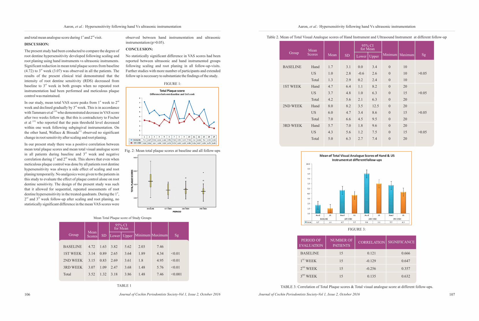
Table 2. Mean of Total Visual Analogue scores of Hand Instrument and Ultrasound Instrument at different follow-up
GroupMeanScores
FIGURE 1:
Journal of Cochin Periodontists Society-Vol 1, Issue 2, October 2016
Aaron, et al.: Hypersensitivity following hand Vs ultrasonic instrumentation
Journal of Cochin Periodontists Society-Vol 1, Issue 2, October 2016 107106
Aaron, et al.: Hypersensitivity following hand Vs ultrasonic instrumentation
st nd and total mean analogue score during 1 and 2 visit.
DISCUSSION:
The present study had been conducted to compare the degree of
root dentine hypersensitivity developed following scaling and
root planing using hand instruments vs ultrasonic instruments.
Significant reduction in mean total plaque scores from baseline rd(4.72) to 3 week (3.07) was observed in all the patients. The
results of the present clinical trial demonstrated that the
intensity of root dentine sensitivity (RDS) decreased from rdbaseline to 3 week in both groups when no repeated root
instrumentation had been performed and meticulous plaque
control was maintained. st ndIn our study, mean total VAS score peaks from 1 week to 2
rdweek and declined gradually by 3 week. This is in accordance [10]with Tammaro et al who demonstrated decrease in VAS score
after two weeks follow up. But this is contradictory to Fischer [15]et al who reported that the pain threshold level decreased
within one week following subgingival instrumentation. On [11]the other hand, Wallace & Bissada observed no significant
change in root sensitivity after scaling and root planing.
In our present study there was a positive correlation between
mean total plaque scores and mean total visual analogue score rdin all patients during baseline and 3 week and negative
st ndcorrelation during 1 and 2 week. This shows that even when
meticulous plaque control was done by all patients root dentine
hypersensitivity was always a side effect of scaling and root
planing temporarily. No analgesics were given to the patients in
this study to evaluate the effect of plaque control alone on root
dentine sensitivity. The design of the present study was such
that it allowed for sequential, repeated assessments of root stdentine hypersensitivity in the treated quadrants. During the 1 ,
nd rd2 and 3 week follow-up after scaling and root planing, no
statistically significant difference in the mean VAS scores were
BASELINE 4.72 1.63 3.82 5.62 2.03 7.46
1ST WEEK 3.14 0.89 2.65 3.64 1.89 4.34 <0.01
2ND WEEK 3.15 0.83 2.69 3.61 1.8 4.95 <0.01
3RD WEEK 3.07 1.09 2.47 3.68 1.48 5.76 <0.01
Total 3.52 1.32 3.18 3.86 1.48 7.46 <0.001
Mean Total Plaque score of Study Groups
GroupMeanScores SD
95% CI for Mean
Lower Upper MaximumMinimum Sg
observed between hand instrumentation and ultrasonic
instrumentation (p>0.05).
CONCLUSION:
No statistically significant difference in VAS scores had been
reported between ultrasonic and hand instrumented groups
following scaling and root planing in all follow-up-visits.
Further studies with more number of participants and extended
follow up is necessary to substantiate the findings of the study.
TABLE 1
Fig. 2: Mean total plaque scores at baseline and all follow-ups
BASELINE Hand 1.7 3.1 0.0 3.4 0 10
US 1.0 2.8 -0.6 2.6 0 10 >0.05
Total 1.3 2.9 0.2 2.4 0 10
1ST WEEK Hand 4.7 6.4 1.1 8.2 0 20
US 3.7 4.8 1.0 6.3 0 15 >0.05
Total 4.2 5.6 2.1 6.3 0 20
2ND WEEK Hand 8.0 8.2 3.5 12.5 0 20
US 6.0 4.7 3.4 8.6 0 15 >0.05
Total 7.0 6.6 4.5 9.5 0 20
3RD WEEK Hand 5.7 7.0 1.8 9.6 0 20
US 4.3 5.6 1.2 7.5 0 15 >0.05
Total 5.0 6.3 2.7 7.4 0 20
95% CI for Mean
Lower Upper MaximumMinimum SgMean SD
FIGURE 3:
BASELINE 15 0.121 0.666
ST1 WEEK 15 -0.129 0.647
ND2 WEEK 15 -0.256 0.357
RD3 WEEK 15 0.135 0.632
PERIOD OF
EVALUATION
NUMBER OF
PATIENTSCORRELATION SIGNIFICANCE
TABLE 3: Correlation of Total Plaque scores & Total visual analogue score at different follow-ups.
109
ABSTRACT:
stAim: To assess the variations in root canal morphology of mandibular 1 premolar
teeth in Kerala population using CBCT .
stMaterial and methodology: 40 extracted mandibular 1 premolar teeth for orthodontic
treatment at different dental clinics, private hospitals and dental colleges of Kerala
population were collected and stored in formalin solution. Teeth samples arranged in
occlusal rims and scanned by CBCTscanning (SIRONA ORTHOPHOS XG
SCANNING machine) and analyzed using Galilio software.
Results: Out of 40 samples studied 17 teeth showed bifurcation of canals and 23 teeth
had no bifurcation. Canals with bifurcation were 42.5% compared with teeth with
single canal (57.5%).
Conclusion: Observations of the current study confirmed the presence of ethnic
differences among various races and provided some information about the internal
anatomy of the mandibular first premolar in Kerala population. Endodontists and
dental clinicians can use such information to achieve a better prognosis for root canal
treatment.
Keywords: Bifurcation, mandibular, CBCT scanning, root canal, occlusal
Mohammed Sagir , Nasarudheen C, Thaju Raj P.K, .Biju P Babu,
Hisham Hameed, Kennet Chirayath
Access this article online
Website :
jcops.copsonweb.org
Quick Response Code
How to cite this article: Log on to jcops.copsonweb.org. Mohammed Sagir ,
Nasarudheen C, Thaju Raj P.K, .Biju P Babu, Hisham Hameed, Kennet
Chirayath. Morphological variations of mandibular first premolar in Kerala
population using cone beam computed tomography: An in –vitro study. Journal
of Cochin Periodontists Society 2016;1: 109-112
Conflict of Interest: None declared Source of Support: Nil
ORIGINAL RESEARCH
Journal of Cochin Periodontists Society-Vol 1, Issue 2, October 2016108
Aaron, et al.: Hypersensitivity following hand Vs ultrasonic instrumentation
REFERENCES:
1) Van dersall DC In: Concise Encyclopedia of
Periodontology. Iowa: Blackwell Munksgaard; 2007. p.
107-27.
2) Lang NP, Loe H. Clinical management of periodontal
disease. Periodontology 2000 1993;2:128–39.
3) Axelsson P, Lindhe J, Nystrom B. On the prevention of
caries and periodontal disease. Results of a 15-year-
longitudinal study in adults. J Clin Periodontol 1991;13:
182–9.
4) Badersten A, Nilveus R, Egelberg J. Effect of non-surgical
periodontal therapy (II). Severely advanced periodontitis.
J Clin Periodontol 1984;11:63–76.
5) Torfason T, Kiger R, Selvig KA, Egelberg J. Clinical
improvement of gingival conditions following ultrasonic
versus hand instrumentation of periodontal pockets. J Clin
Periodontol 1979;6:165-76.
6) Breininger D, O’Leary TJ, Blumenstein R. Comparative
effectiveness of ultrasonic and hand scaling for the
removal of subgingival plaque and calculus. J Periodontol
1987;58:9-18.
7) Bergenholtz G, Lindhe J. Effect of experimentally induced
marginal periodontitis and periodontal scaling on the
dental pulp. Journal of Clinical Periodontology
1978;5:59–73.
8) Bissada NF. Symptomatology and clinical features of
hypersensitive teeth. Archives of Oral Biology
1994;39:31S–32S.
10)
13) Langley GB, Sheppeard H. The visual analogueue scale:
its use in pain measurement. Rheumatol Int 1985;5:145-8.
14) Turesky S, Gilmore ND, Glickman I. Reduced plaque
formation by the chloromethyl analogueue of Victamine C.
J Periodontol 1970;41:41-3.
9) Pihlstrom BL, Hargreaves KM, Bouwsma OJ, Myers WR,
Goodale MB, Doyle MJ. Pain after periodontal scaling and
root planing. J Am Dent Assoc 1999;130:801-7.
Tammaro S, Wennstrom JL, Bergenholtz G. Root-dentin
sensitivity following nonsurgical periodontal treatment. J
Clin Periodontol 2000;27:690-7.
11) Wallace JA, Bissada NF. Pulpal and root sensitivity rated
to periodontal therapy. Oral Surg Oral Med Oral Pathol
1990;69:743-7.
12) Canakci CF, Canakci V. Pain experienced by patients
undergoing different periodontal therapies. J Am Dent
Assoc 2007;138:1563-73.
15) Fischer C, Fischer RG, Wennberg A. Prevalence and
distribution of cervical dentine hypersensitivity in a
population in Rio de Janeiro, Brazil. J Dent 1992;20:272-
76.
10)
13) Langley GB, Sheppeard H. The visual analogueue scale:
its use in pain measurement. Rheumatol Int 1985;5:145-8.
14) Turesky S, Gilmore ND, Glickman I. Reduced plaque
formation by the chloromethyl analogueue of Victamine C.
J Periodontol 1970;41:41-3.
Morphological variations of mandibular first premolar in Kerala population using cone beam computed tomography: An in –vitro study
Department of Conservative
Dentistry and Endodontics,
Royal Dental College, Chalissery,
Palakkad, Kerala, India
INTRODUCTION
Successful endodontic treatment
depends on complete canal cleaning and
shaping and 3-dimensional obturation.
The lack of proper knowledge about the
anatomy of root canals is one of the main
reasons for endodontic treatment
failures. So the main step in achieving
successful endodontic outcome is an
exact diagnosis of root canal system and
its anatomical variations. Any attempt to
perform endodontic therapy must be
p r o c e e d e d w i t h a t h o r o u g h
understanding of the anatomy of both the
Journal of Cochin Periodontists Society-Vol 1, Issue 2, October 2016
Journal of Cochin Periodontists Society-Vol 1, Issue 2, October 2016 111110
Sagir, et al.: Root canal pattern variations of mandibular first premolarSagir, et al.: Root canal pattern variations of mandibular first premolar
[1]pulp chamber and root canal system.st Mandibular 1 premolar shows a considerable variation in root
stcanal morphology. The internal anatomy of mandibular 1
premolar is particularly complex due to their variation in the
number of roots and canal configuration.
Studies on root canal anatomy are usually done by radiography,
clearing technique, plastic resin injection technique, and direct
observation with microscope and macroscopic sections.
Conventional radiography has the problem of superimposition
and moreover it is a two dimensional representation of a three
dimensional object. The difficulties associated with other
methods of study include disturbance of pulp space and its
surrounding structures during preparation of the teeth.
All races and ethnic groups have some degree of dental
anatomic variations. Asian populations shows one of the widest
variations in coronal shape, external root form and canal
morphology (Thews et al.1979, Harris 1980, Ross&Evanchik
1981, cecic et al.1982, Yang et al. 1988). Aim of this invitro
study was to assess the variations in root canal morphology of stmandibular 1 premolar teeth in Kerala population using
CBCT .
MATERIALS AND METHODSstIn this invitro study 40 extracted mandibular 1 premolar teeth
for orthodontic treatment at different dental clinics, private
hospitals and dental colleges of Kerala population were
collected and stored in formalin solution.
§INCLUSION CRITERIA: Teeth with mature root
apex irrespective of age and sex
§EXCLUSION CRITERIA: Teeth with open apex,
complicated fracture, external root resorption
communicating with root canal or grossly damaged
teeth were discarded.
All attached soft tissues and calculus are removed by using an
ultrasonic scaler.Modelling wax were used to make occlusal
rims and the teeth were arranged after determining the various
aspects of the tooth i.e., Buccal, lingual, mesial and distal, so as
to maintain uniformity in the samples.
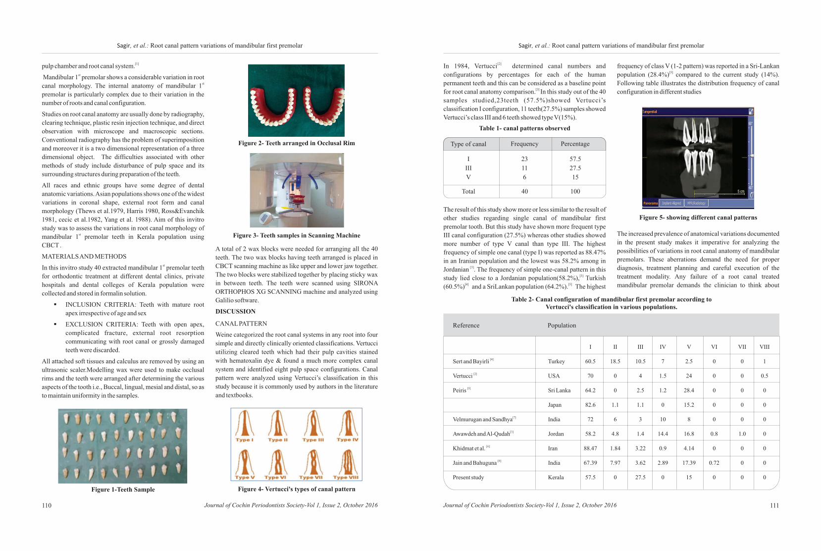
Figure 1-Teeth Sample
Figure 2- Teeth arranged in Occlusal Rim
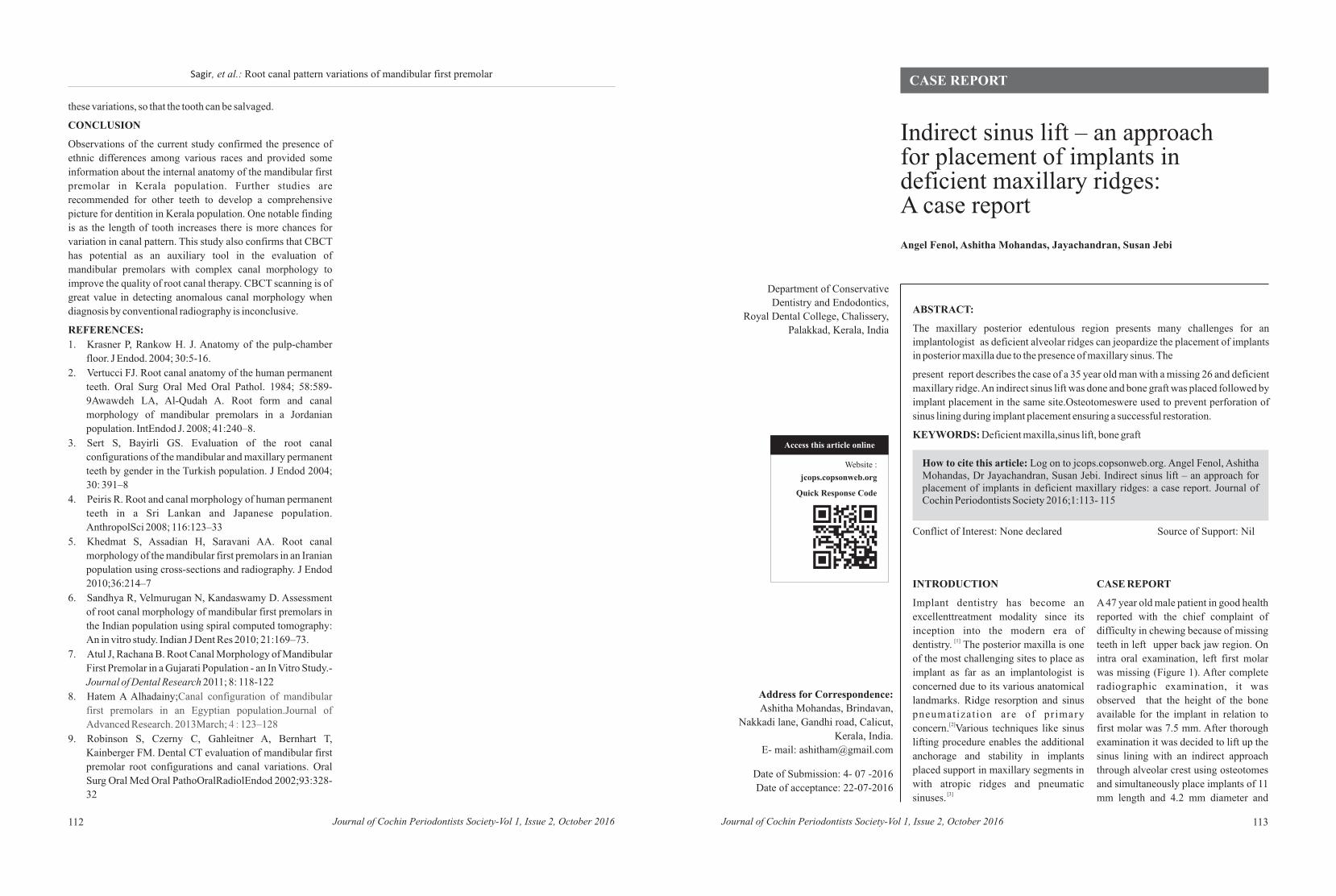
Figure 3- Teeth samples in Scanning Machine
A total of 2 wax blocks were needed for arranging all the 40
teeth. The two wax blocks having teeth arranged is placed in
CBCT scanning machine as like upper and lower jaw together.
The two blocks were stabilized together by placing sticky wax
in between teeth. The teeth were scanned using SIRONA
ORTHOPHOS XG SCANNING machine and analyzed using
Galilio software.
DISCUSSION
CANAL PATTERN
Weine categorized the root canal systems in any root into four
simple and directly clinically oriented classifications. Vertucci
utilizing cleared teeth which had their pulp cavities stained
with hematoxalin dye & found a much more complex canal
system and identified eight pulp space configurations. Canal
pattern were analyzed using Vertucci’s classification in this
study because it is commonly used by authors in the literature
and textbooks.
Figure 4- Vertucci's types of canal pattern
[2]In 1984, Vertucci determined canal numbers and
configurations by percentages for each of the human
permanent teeth and this can be considered as a baseline point [3] for root canal anatomy comparison. In this study out of the 40
samples studied,23teeth (57.5%)showed Vertucci’s
classification I configuration, 11 teeth(27.5%) samples showed
Vertucci’s class III and 6 teeth showed type V(15%).
Type of canal Frequency Percentage
Total 40 100
I
III
V
23
11
6
57.5
27.5
15
Table 1- canal patterns observed
The result of this study show more or less similar to the result of
other studies regarding single canal of mandibular first
premolar tooth. But this study have shown more frequent type
III canal configuration (27.5%) whereas other studies showed
more number of type V canal than type III. The highest
frequency of simple one canal (type I) was reported as 88.47%
in an Iranian population and the lowest was 58.2% among in [3]Jordanian . The frequency of simple one-canal pattern in this
[3]study lied close to a Jordanian population(58.2%), Turkish [4] [5](60.5%) and a SriLankan population (64.2%). The highest
I II III IV V VI VII VIII
[4]Sert and Bayirli Turkey 60.5 18.5 10.5 7 2.5 0 0 1
[2]Vertucci USA 70 0 4 1.5 24 0 0 0.5
[5]Peiris Sri Lanka 64.2 0 2.5 1.2 28.4 0 0 0
Japan 82.6 1.1 1.1 0 15.2 0 0 0
[7]Velmurugan and Sandhya India 72 6 3 10 8 0 0 0
[3]Awawdeh and Al-Qudah Jordan 58.2 4.8 1.4 14.4 16.8 0.8 1.0 0
[6]Khidmat et al. Iran 88.47 1.84 3.22 0.9 4.14 0 0 0
[8]Jain and Bahuguna India 67.39 7.97 3.62 2.89 17.39 0.72 0 0
Present study Kerala 57.5 0 27.5 0 15 0 0 0
Reference Population
Table 2- Canal configuration of mandibular first premolar according to Vertucci's classification in various populations.
frequency of class V (1-2 pattern) was reported in a Sri-Lankan [5]population (28.4%) compared to the current study (14%).
Following table illustrates the distribution frequency of canal
configuration in different studies
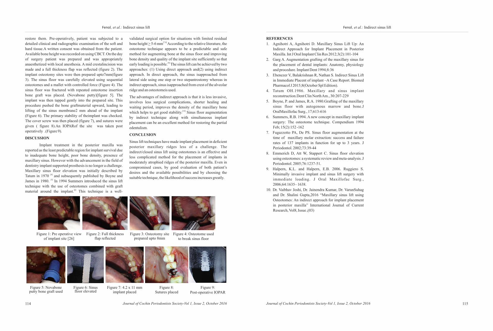
Figure 5- showing different canal patterns
The increased prevalence of anatomical variations documented
in the present study makes it imperative for analyzing the
possibilities of variations in root canal anatomy of mandibular
premolars. These aberrations demand the need for proper
diagnosis, treatment planning and careful execution of the
treatment modality. Any failure of a root canal treated
mandibular premolar demands the clinician to think about
Journal of Cochin Periodontists Society-Vol 1, Issue 2, October 2016
Journal of Cochin Periodontists Society-Vol 1, Issue 2, October 2016112
Sagir, et al.: Root canal pattern variations of mandibular first premolar
these variations, so that the tooth can be salvaged.
CONCLUSION
Observations of the current study confirmed the presence of
ethnic differences among various races and provided some
information about the internal anatomy of the mandibular first
premolar in Kerala population. Further studies are
recommended for other teeth to develop a comprehensive
picture for dentition in Kerala population. One notable finding
is as the length of tooth increases there is more chances for
variation in canal pattern. This study also confirms that CBCT
has potential as an auxiliary tool in the evaluation of
mandibular premolars with complex canal morphology to
improve the quality of root canal therapy. CBCT scanning is of
great value in detecting anomalous canal morphology when
diagnosis by conventional radiography is inconclusive.
REFERENCES:
1. Krasner P, Rankow H. J. Anatomy of the pulp-chamber
floor. J Endod. 2004; 30:5-16.
2. Vertucci FJ. Root canal anatomy of the human permanent
teeth. Oral Surg Oral Med Oral Pathol. 1984; 58:589-
9Awawdeh LA, Al-Qudah A. Root form and canal
morphology of mandibular premolars in a Jordanian
population. IntEndod J. 2008; 41:240–8.
3. Sert S, Bayirli GS. Evaluation of the root canal
configurations of the mandibular and maxillary permanent
teeth by gender in the Turkish population. J Endod 2004;
30: 391–8
4. Peiris R. Root and canal morphology of human permanent
teeth in a Sri Lankan and Japanese population.
AnthropolSci 2008; 116:123–33
5. Khedmat S, Assadian H, Saravani AA. Root canal
morphology of the mandibular first premolars in an Iranian
population using cross-sections and radiography. J Endod
2010;36:214–7
6. Sandhya R, Velmurugan N, Kandaswamy D. Assessment
of root canal morphology of mandibular first premolars in
the Indian population using spiral computed tomography:
An in vitro study. Indian J Dent Res 2010; 21:169–73.
7. Atul J, Rachana B. Root Canal Morphology of Mandibular
First Premolar in a Gujarati Population - an In Vitro Study.-
Journal of Dental Research 2011; 8: 118-122
8. Hatem A Alhadainy;
9. Robinson S, Czerny C, Gahleitner A, Bernhart T,
Kainberger FM. Dental CT evaluation of mandibular first
premolar root configurations and canal variations. Oral
Surg Oral Med Oral PathoOralRadiolEndod 2002;93:328-
32
Canal configuration of mandibular
first premolars in an Egyptian population.Journal of
Advanced Research. 2013March; 123–128 4 :
113
ABSTRACT:
The maxillary posterior edentulous region presents many challenges for an
implantologist as deficient alveolar ridges can jeopardize the placement of implants
in posterior maxilla due to the presence of maxillary sinus. The
present report describes the case of a 35 year old man with a missing 26 and deficient
maxillary ridge. An indirect sinus lift was done and bone graft was placed followed by
implant placement in the same site.Osteotomeswere used to prevent perforation of
sinus lining during implant placement ensuring a successful restoration.
KEYWORDS: Deficient maxilla,sinus lift, bone graft
Angel Fenol, Ashitha Mohandas, Jayachandran, Susan Jebi
Access this article online
Website :
jcops.copsonweb.org
Quick Response Code
How to cite this article: Log on to jcops.copsonweb.org. Angel Fenol, Ashitha Mohandas, Dr Jayachandran, Susan Jebi. Indirect sinus lift – an approach for placement of implants in deficient maxillary ridges: a case report. Journal of Cochin Periodontists Society 2016;1:113- 115
Conflict of Interest: None declared Source of Support: Nil
CASE REPORT
Department of Conservative
Dentistry and Endodontics,
Royal Dental College, Chalissery,
Palakkad, Kerala, India
Indirect sinus lift – an approach for placement of implants in deficient maxillary ridges: A case report
Address for Correspondence:
Ashitha Mohandas, Brindavan,
Nakkadi lane, Gandhi road, Calicut,
Kerala, India.
E- mail: [email protected]
Date of Submission: 4- 07 -2016
Date of acceptance: 22-07-2016
Journal of Cochin Periodontists Society-Vol 1, Issue 2, October 2016
INTRODUCTION
Implant dentistry has become an
excellenttreatment modality since its
inception into the modern era of [1]dentistry. The posterior maxilla is one
of the most challenging sites to place as
implant as far as an implantologist is
concerned due to its various anatomical
landmarks. Ridge resorption and sinus
pneumatizat ion are of primary [2]concern. Various techniques like sinus
lifting procedure enables the additional
anchorage and stability in implants
placed support in maxillary segments in
with atropic ridges and pneumatic [3]sinuses.
CASE REPORT
A 47 year old male patient in good health
reported with the chief complaint of
difficulty in chewing because of missing
teeth in left upper back jaw region. On
intra oral examination, left first molar
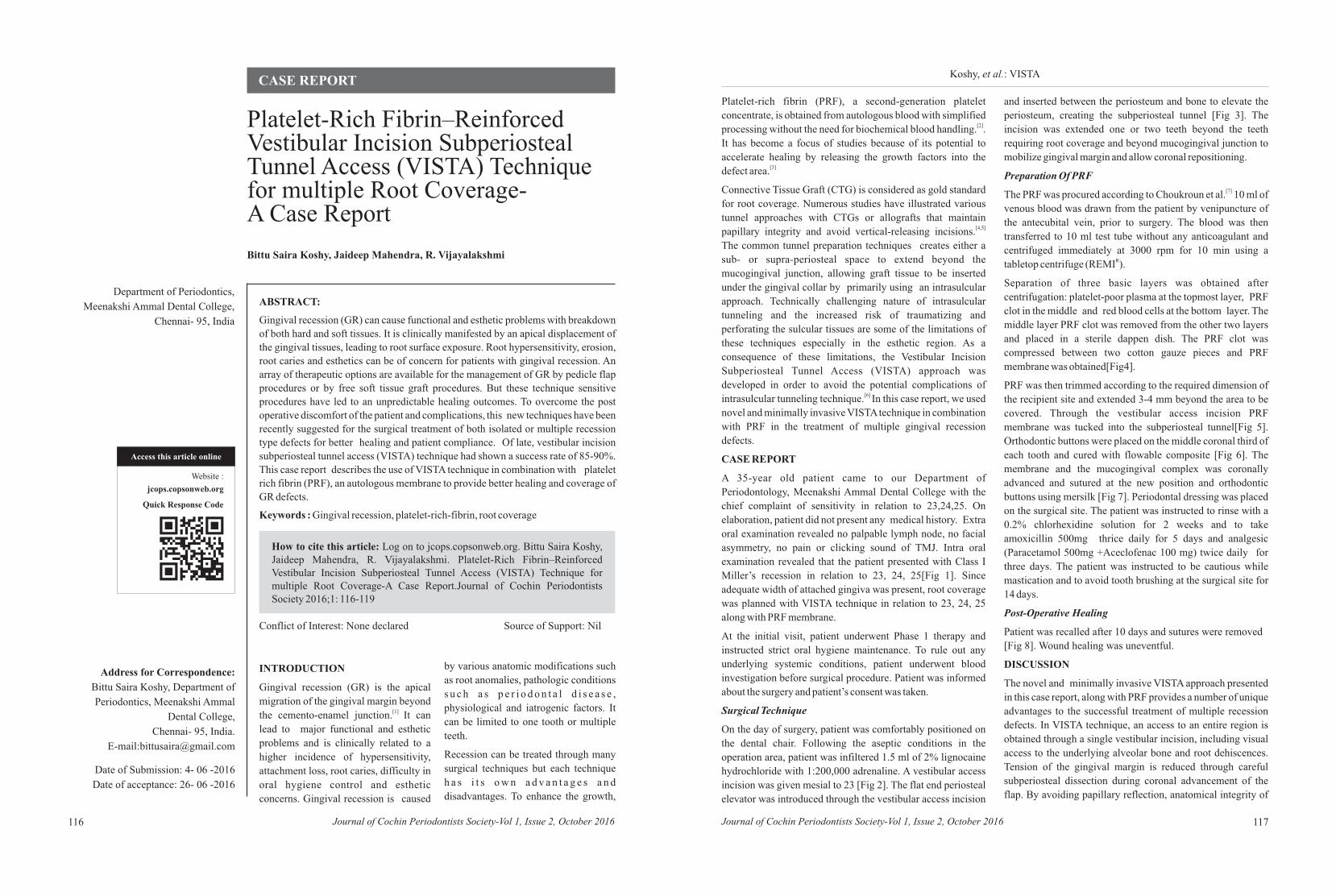
was missing (Figure 1). After complete
radiographic examination, it was
observed that the height of the bone
available for the implant in relation to
first molar was 7.5 mm. After thorough
examination it was decided to lift up the
sinus lining with an indirect approach
through alveolar crest using osteotomes
and simultaneously place implants of 11
mm length and 4.2 mm diameter and
Journal of Cochin Periodontists Society-Vol 1, Issue 2, October 2016114
restore them. Pre-operatively, patient was subjected to a
detailed clinical and radiographic examination of the soft and
hard tissue.A written consent was obtained from the patient.
Available bone height was recorded on using CBCT. On the day
of surgery patient was prepared and was appropriately
anaesthetized with local anesthesia. A mid crestalincision was
made and a full thickness flap was reflected (figure 2). The
implant osteotomy sites were then prepared upto7mm(figure
3). The sinus floor was carefully elevated using sequential
osteotomes and a mallet with controlled force (Figure 4). The
sinus floor was fractured with repeated osteotome insertion
bone graft was placed. (Novabone putty)[figure 5]. The
implant was then tapped gently into the prepared site. This
procedure pushed the bone graftmaterial upward, leading to
lifting of the sinus membrane2 mm ahead of the implant
(Figure 6). The primary stability of theimplant was checked.
The cover screw was then placed (figure 7), and sutures were
given ( figure 8).An IOPARof the site was taken post
operatively .(Figure 9).
DISCUSSION
Implant treatment in the posterior maxilla was
reported as the least predictable region for implant survival due
to inadequate bone height, poor bone density, presence of
maxillary sinus. However with the advancement in the field of
dentistry implant supported prosthesis is no longer a challenge.
Maxillary sinus floor elevation was initially described by [4]Tatum in 1970 and subsequently published by Boyne and [5]James in 1980. In 1994 Summers introduced the sinus lift
technique with the use of osteotomes combined with graft [6]material around the implant. This technique is a well-
Figure 1: Pre operative view of implant site [26]
Figure 2: Full thickness flap reflected
Figure 3: Osteotomy site prepared upto 8mm
Figure 4: Osteotome used to break sinus floor
Figure 5: Novabone putty bone graft used
Figure 6: Sinus floor elevated
Figure 7: 4.2 x 11 mm implant placed
Figure 8: Sutures placed
Figure 9: Post-operative IOPAR
validated surgical option for situations with limited residual [7,8]bone height > 5-6 mm According to the relative literature, the
osteotome technique appears to be a predictable and safe
method for augmenting bone at the sinus floor and improving
bone density and quality of the implant site sufficiently so that [9]early loading is possible. The sinus lift can be achieved by two
approaches: (1) Using direct approach and(2) using indirect
approach. In direct approach, the sinus isapproached from
lateral side using one step or two stepantrostomy whereas in
indirect approach, sinus isapproached from crest of the alveolar
ridge and an osteotomeis used.
The advantages of indirect approach is that it is less invasive,
involves less surgical complications, shorter healing and
waiting period, improves the density of the maxillary bone [10]which helps to get good stability. Sinus floor augmentation
by indirect technique along with simultaneous implant
placement can be an excellent method for restoring the partial
edentulism.
CONCLUSION
Sinus lift techniques have made implant placement in deficient
posterior maxillary ridges less of a challenge. The
indirect/closed sinus lift using osteotomes is an effective and
less complicated method for the placement of implants in
moderately atrophied ridges of the posterior maxilla. Even in
compromised cases, by good evaluation of both patient’s
desires and the available possibilities and by choosing the
suitable technique, the likelihood of success increases greatly.
115Journal of Cochin Periodontists Society-Vol 1, Issue 2, October 2016
Fenol, et al.: Indirect sinus liftFenol, et al.: Indirect sinus lift
REFERENCES
1. Agnihotri A, Agnihotri D. Maxillary Sinus Lift Up: An
Indirect Approach for Implant Placement in Posterior
Maxilla. Int J Oral Implant Clin Res 2012;3(2):101-104
2. Garg A. Augmentation grafting of the maxillary sinus for
the placement of dental implants: Anatomy, physiology
and procedure. Implant Dent 1994;8:36
3. Ebenezer V, Balakrishnan R, Nathan S. Indirect Sinus Lift
in Immediate Placent of implant –A Case Report. Biomed
Pharmacol J 2015;8(October Spl Edition).
4. Tatum OH.1986. Maxillary and sinus implant
reconstruction.Dent Clin North Am., 30:207-229
5. Boyne, P. and James, R.A. 1980.Grafting of the maxillary
sinus floor with autogenous marrow and bone.J
OralMaxillofac Surg., 17;613-616
6. Summers, R.B. 1994. A new concept in maxillary implant
surgery: The osteotome technique. Compendium 1994
Feb; 15(2):152 -162
7. Fugazzotto PA, De PS. Sinus floor augmentation at the
time of maxillary molar extraction: success and failure
rates of 137 implants in function for up to 3 years. J
Periodontol. 2002;73:39-44
8. Emmerich D, Att W, Stappert C. Sinus floor elevation
using osteotomes: a systematic review and meta-analysis. J
Periodontol. 2005;76:1237-51.
9. Halpern, K.L. and Halpern, E.B. 2006. Ruggiero S.
Minimally invasive implant and sinus lift surgery with
immediate loading. J Oral Maxillofac Surg.,
2006;64:1635– 1638.
10. Dr. Vaibhav Joshi, Dr. Jainendra Kumar, Dr. VarunSuhag
and Dr. Shalini Gupta,2016 “Maxillary sinus lift using
Osteotomes: An indirect approach for implant placement
in posterior maxilla” International Journal of Current
Research, Vol8, Issue ,(03)
ABSTRACT:
Gingival recession (GR) can cause functional and esthetic problems with breakdown
of both hard and soft tissues. It is clinically manifested by an apical displacement of
the gingival tissues, leading to root surface exposure. Root hypersensitivity, erosion,
root caries and esthetics can be of concern for patients with gingival recession. An
array of therapeutic options are available for the management of GR by pedicle flap
procedures or by free soft tissue graft procedures. But these technique sensitive
procedures have led to an unpredictable healing outcomes. To overcome the post
operative discomfort of the patient and complications, this new techniques have been
recently suggested for the surgical treatment of both isolated or multiple recession
type defects for better healing and patient compliance. Of late, vestibular incision
subperiosteal tunnel access (VISTA) technique had shown a success rate of 85-90%.
This case report describes the use of VISTA technique in combination with platelet
rich fibrin (PRF), an autologous membrane to provide better healing and coverage of
GR defects.
Keywords : Gingival recession, platelet-rich-fibrin, root coverage
Bittu Saira Koshy, Jaideep Mahendra, R. Vijayalakshmi
How to cite this article: Log on to jcops.copsonweb.org. Bittu Saira Koshy, Jaideep Mahendra, R. Vijayalakshmi. Platelet-Rich Fibrin–Reinforced Vestibular Incision Subperiosteal Tunnel Access (VISTA) Technique for multiple Root Coverage-A Case Report.Journal of Cochin Periodontists Society 2016;1: 116-119
Conflict of Interest: None declared Source of Support: Nil
Journal of Cochin Periodontists Society-Vol 1, Issue 2, October 2016 Journal of Cochin Periodontists Society-Vol 1, Issue 2, October 2016 117116
Access this article online
Website :
jcops.copsonweb.org
Quick Response Code
Department of Periodontics,
Meenakshi Ammal Dental College,
Chennai- 95, India
Address for Correspondence:
Bittu Saira Koshy, Department of
Periodontics, Meenakshi Ammal
Dental College,
Chennai- 95, India.
E-mail:[email protected]
Date of Submission: 4- 06 -2016
Date of acceptance: 26- 06 -2016
Platelet-Rich Fibrin–Reinforced Vestibular Incision Subperiosteal Tunnel Access (VISTA) Technique for multiple Root Coverage-A Case Report
CASE REPORTKoshy, et al.: VISTA
INTRODUCTION
Gingival recession (GR) is the apical
migration of the gingival margin beyond [1] the cemento-enamel junction. It can
lead to major functional and esthetic
problems and is clinically related to a
higher incidence of hypersensitivity,
attachment loss, root caries, difficulty in
oral hygiene control and esthetic
concerns. Gingival recession is caused
by various anatomic modifications such
as root anomalies, pathologic conditions
s u c h a s p e r i o d o n t a l d i s e a s e ,
physiological and iatrogenic factors. It
can be limited to one tooth or multiple
teeth.
Recession can be treated through many
surgical techniques but each technique
h a s i t s o w n a d v a n t a g e s a n d
disadvantages. To enhance the growth,
Platelet-rich fibrin (PRF), a second-generation platelet
concentrate, is obtained from autologous blood with simplified [2] processing without the need for biochemical blood handling. .
It has become a focus of studies because of its potential to
accelerate healing by releasing the growth factors into the [3]defect area.
Connective Tissue Graft (CTG) is considered as gold standard
for root coverage. Numerous studies have illustrated various
tunnel approaches with CTGs or allografts that maintain [4,5]papillary integrity and avoid vertical-releasing incisions.
The common tunnel preparation techniques creates either a
sub- or supra-periosteal space to extend beyond the
mucogingival junction, allowing graft tissue to be inserted
under the gingival collar by primarily using an intrasulcular
approach. Technically challenging nature of intrasulcular
tunneling and the increased risk of traumatizing and
perforating the sulcular tissues are some of the limitations of
these techniques especially in the esthetic region. As a
consequence of these limitations, the Vestibular Incision
Subperiosteal Tunnel Access (VISTA) approach was
developed in order to avoid the potential complications of [6] intrasulcular tunneling technique. In this case report, we used
novel and minimally invasive VISTA technique in combination
with PRF in the treatment of multiple gingival recession
defects.
CASE REPORT
A 35-year old patient came to our Department of
Periodontology, Meenakshi Ammal Dental College with the
chief complaint of sensitivity in relation to 23,24,25. On
elaboration, patient did not present any medical history. Extra
oral examination revealed no palpable lymph node, no facial
asymmetry, no pain or clicking sound of TMJ. Intra oral
examination revealed that the patient presented with Class I
Miller’s recession in relation to 23, 24, 25[Fig 1]. Since
adequate width of attached gingiva was present, root coverage
was planned with VISTA technique in relation to 23, 24, 25
along with PRF membrane.
At the initial visit, patient underwent Phase 1 therapy and
instructed strict oral hygiene maintenance. To rule out any
underlying systemic conditions, patient underwent blood
investigation before surgical procedure. Patient was informed
about the surgery and patient’s consent was taken.
Surgical Technique
On the day of surgery, patient was comfortably positioned on
the dental chair. Following the aseptic conditions in the
operation area, patient was infiltered 1.5 ml of 2% lignocaine
hydrochloride with 1:200,000 adrenaline. A vestibular access
incision was given mesial to 23 [Fig 2]. The flat end periosteal
elevator was introduced through the vestibular access incision
and inserted between the periosteum and bone to elevate the
periosteum, creating the subperiosteal tunnel [Fig 3]. The
incision was extended one or two teeth beyond the teeth
requiring root coverage and beyond mucogingival junction to
mobilize gingival margin and allow coronal repositioning.
Preparation Of PRF[7]The PRF was procured according to Choukroun et al. 10 ml of
venous blood was drawn from the patient by venipuncture of
the antecubital vein, prior to surgery. The blood was then
transferred to 10 ml test tube without any anticoagulant and
centrifuged immediately at 3000 rpm for 10 min using a Rtabletop centrifuge (REMI ).
Separation of three basic layers was obtained after
centrifugation: platelet-poor plasma at the topmost layer, PRF
clot in the middle and red blood cells at the bottom layer. The
middle layer PRF clot was removed from the other two layers
and placed in a sterile dappen dish. The PRF clot was
compressed between two cotton gauze pieces and PRF
membrane was obtained[Fig4].
PRF was then trimmed according to the required dimension of
the recipient site and extended 3-4 mm beyond the area to be
covered. Through the vestibular access incision PRF
membrane was tucked into the subperiosteal tunnel[Fig 5].
Orthodontic buttons were placed on the middle coronal third of
each tooth and cured with flowable composite [Fig 6]. The
membrane and the mucogingival complex was coronally
advanced and sutured at the new position and orthodontic
buttons using mersilk [Fig 7]. Periodontal dressing was placed
on the surgical site. The patient was instructed to rinse with a
0.2% chlorhexidine solution for 2 weeks and to take
amoxicillin 500mg thrice daily for 5 days and analgesic
(Paracetamol 500mg +Aceclofenac 100 mg) twice daily for
three days. The patient was instructed to be cautious while
mastication and to avoid tooth brushing at the surgical site for
14 days.
Post-Operative Healing
Patient was recalled after 10 days and sutures were removed
[Fig 8]. Wound healing was uneventful.
DISCUSSION
The novel and minimally invasive VISTA approach presented
in this case report, along with PRF provides a number of unique
advantages to the successful treatment of multiple recession
defects. In VISTA technique, an access to an entire region is
obtained through a single vestibular incision, including visual
access to the underlying alveolar bone and root dehiscences.
Tension of the gingival margin is reduced through careful
subperiosteal dissection during coronal advancement of the
flap. By avoiding papillary reflection, anatomical integrity of
Journal of Cochin Periodontists Society-Vol 1, Issue 2, October 2016 119
Koshy, et al.: VISTAKoshy, et al.: VISTA
Journal of Cochin Periodontists Society-Vol 1, Issue 2, October 2016118
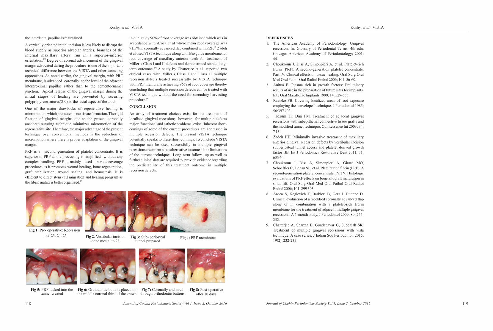
the interdental papillae is maintained.
A vertically oriented initial incision is less likely to disrupt the
blood supply as superior alveolar arteries, branches of the
internal maxillary artery, run in a superior-inferior [6]orientation. Degree of coronal advancement of the gingival
margin advocated during the procedure is one of the important
technical difference between the VISTA and other tunneling
approaches. As noted earlier, the gingival margin, with PRF
membrane, is advanced coronally to the level of the adjacent
interproximal papillae rather than to the cementoenamel
junction. Apical relapse of the gingival margin during the
initial stages of healing are prevented by securing
polypropylene sutures(3-0) to the facial aspect of the tooth.
One of the major drawbacks of regenerative healing is
micromotion, which promotes scar tissue formation. The rigid
fixation of gingival margins due to the present coronally
anchored suturing technique minimizes micromotion of the
regenerative site. Therefore, the major advantage of the present
technique over conventional methods is the reduction of
micromotion where there is proper adaptation of the gingival
margin.
PRF is a second generation of platelet concentrate. It is
superior to PRP as the processing is simplified without any
complex handling. PRF is mainly used in root coverage
procedures as it promotes wound healing, bone regeneration,
graft stabilization, wound sealing, and hemostasis. It is
efficient to direct stem cell migration and healing program as [7]the fibrin matrix is better organized.
Fig 1: Pre- operative: Recession
i.r.t 23, 24, 25 Fig 2: Vestibular incision done mesial to 23
Fig 3: Sub- periosteal tunnel prepared
Fig 4: PRF membrane
Fig 5: PRF tucked into the tunnel created
Fig 6: Orthodontic buttons placed on the middle coronal third of the crown
Fig 7: Coronally anchored through orthodontic buttons
Fig 8: Post-operative after 10 days
In our study 90% of root coverage was obtained which was in
accordance with Aroca et al where mean root coverage was [8]91.5% in coronally advanced flap combined with PRF. Zadeh
et al used VISTA technique along with Bio guide membrane for
root coverage of maxillary anterior teeth for treatment of
Miller’s Class I and II defects and demonstrated stable, long-[6] term outcomes. A study by Chatterjee et al reported two
clinical cases with Miller’s Class I and Class II multiple
recession defects treated successfully by VISTA technique
with PRF membrane achieving 96% of root coverage thereby
concluding that multiple recession defects can be treated with
VISTA technique without the need for secondary harvesting [9]procedure.
CONCLUSION
An array of treatment choices exist for the treatment of
localised gingival recession; however for multiple defects
major functional and esthetic problems exist. Inherent short-
comings of some of the current procedures are addressed in
multiple recession defects. The present VISTA technique potentially speaks to these short-comings. To conclude VISTA
technique can be used successfully in multiple gingival
recessions treatment as an alternative to some of the limitations
of the current techniques. Long term follow- up as well as
further clinical data are required to provide evidence regarding
the predictability of this treatment outcome in multiple
recession defects.
REFERENCES
1. The American Academy of Periodontology. Gingival
recession. In: Glossary of Periodontal Terms, 4th edn.
Chicago: American Academy of Periodontology; 2001:
44.
2. Choukroun J, Diss A, Simonpieri A, et al. Platelet-rich
fibrin (PRF): A second-generation platelet concentrate.
Part IV: Clinical effects on tissue healing. Oral Surg Oral
Med Oral Pathol Oral Radiol Endod 2006; 101: 56-60.
3. Anitua E. Plasma rich in growth factors: Preliminary
results of use in the preparation of future sites for implants.
Int J Oral Maxillofac Implants 1999; 14: 529-535
4. Raetzke PB. Covering localized areas of root exposure
employing the “envelope” technique. J Periodontol 1985;
56:397 402.
5. Tözüm TF, Dini FM. Treatment of adjacent gingival
recessions with subepithelial connective tissue grafts and
the modified tunnel technique. Quintessence Int 2003; 34:
7 13.
6. Zadeh HH. Minimally invasive treatment of maxillary
anterior gingival recession defects by vestibular incision
subperiosteal tunnel access and platelet derived growth
factor BB. Int J Periodontics Restorative Dent 2011; 31:
653 60.
7. Choukroun J, Diss A, Simonpieri A, Girard MO,
Schoeffler C, Dohan SL, et al. Platelet rich fibrin (PRF): A
second-generation platelet concentrate. Part V: Histologic
evaluations of PRF effects on bone allograft maturation in
sinus lift. Oral Surg Oral Med Oral Pathol Oral Radiol
Endod 2006; 101: 299 303.
8. Aroca S, Keglevich T, Barbieri B, Gera I, Etienne D.
Clinical evaluation of a modified coronally advanced flap
alone or in combination with a platelet-rich fibrin
membrane for the treatment of adjacent multiple gingival
recessions: A 6-month study. J Periodontol 2009; 80: 244-
252.
9. Chatterjee A, Sharma E, Gundanavar G, Subbaiah SK.
Treatment of multiple gingival recessions with vista
technique: A case series. J Indian Soc Periodontol. 2015;
19(2): 232-235.
ABSTRACT:
This case report discuss a case of periimplant abscess, surgically managed by bone
regenerative approach. A 22 year old male patient reported with a complaint of
fluctuating swelling that subside and recur over the edentulous ridge area. The
radiograph of that region confirmed it to be a peri implant abscess around the crestal
third of an immediate implant placed an year back.The lesion was surgically TMapproached and the defect was filled with bone graft material (Osseograft ) and
secured with a connective tissue graft over it to secure graft material. The third month
follow-up showed no clinical signs or symptoms of the previous lesion and
significant bone regeneration was observed radiographically in the bone defect.
Key words : Bone Regeneration, Periimplant abscess , immediate implants
Sruthy Purushothaman, Elizabeth Kuruvilla, Biniraj K R,
Tony P Paul, Aslam A R
Rishi Emmatty
How to cite this article: Log on to jcops.copsonweb.org. Sruthy
Purushothaman, Elizabeth Kuruvilla, Biniraj K R, Rishi Emmatty, Tony P Paul,
Aslam A R . Surgical management of a periimplant abscess: A case report.
Journal of Cochin Periodontists Society 2016;1: 120-122
Conflict of Interest: None declared Source of Support: Nil
Journal of Cochin Periodontists Society-Vol 1, Issue 2, October 2016 Journal of Cochin Periodontists Society-Vol 1, Issue 2, October 2016 121120
Access this article online
Website :
jcops.copsonweb.org
Quick Response Code
Department of Clinical
Periodontology and Oral
Implantology, Royal Dental
College, Palakkad,
Kerala, 679536, India.
Address for Correspondence:
Sruthy Purushothaman,
Department of Clinical
Periodontology and Oral
Implantology, Royal Dental
College, Palakkad, Kerala,
679536, India:
E-mail: [email protected]
Date of Submission: 4- 08 -2016
Date of acceptance: 22-08-2016
CASE REPORTPurushothaman, et al.: Periimplant abscess
Surgical management of a periimplant abscess: A case report
INTRODUCTION
A 22 year old systemically healthy male
patient was referred to our department of
clinical periodontology and oral
implantology, with a fluctuant swelling
on the maxillary right posterior
edentulous ridge area. He revealed a
history of 2 immediate implants being
placed in this region 1 year ago. The
swelling was noticed since 6 months
which was painless and kept occurring
intermittently and disappeared following
purulent discharge through a sinus
opening. The immediate implants were
placed following extraction of root
stumps of 15 and 16 resulting from
endodontic treatment failure. According
to his old radiograph, all the extracted
teeth had peri apical radiolucency
confirmed to be peri apical granuloma at
the time of extractions.
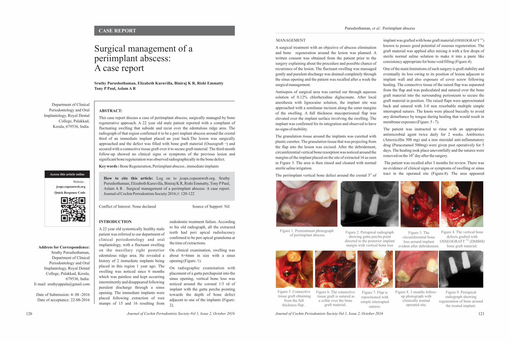
On clinical examination, swelling was
about 6×6mm in size with a sinus
opening (Figure: 1).
On radiographic examination with
placement of a gutta perchapoint into the
sinus opening, vertical bone loss was
noticed around the coronal 1/3 rd of
implant with the gutta percha pointing
towards the depth of bone defect
adjacent to one of the implants (Figure:
2).
MANAGEMENT
A surgical treatment with an objective of abscess elimination
and bone regeneration around the lesion was planned. A
written consent was obtained from the patient prior to the
surgery explaining about the procedure and possible chance of
recurrence of the lesion. The fluctuant swelling was massaged
gently and purulent discharge was drained completely through
the sinus opening and the patient was recalled after a week the
surgical management.
Antisepsis of surgical area was carried out through aqueous
solution of 0.12% chlorhexidine digluconate. After local
anesthesia with lignocaine solution, the implant site was
approached with a semilunar incision along the outer margins
of the swelling. A full thickness mucoperiosteal flap was
elevated over the implant surface involving the swelling. The
implant was confirmed for its integration and observed to have
no signs of mobility.
The granulation tissue around the implants was curetted with
plastic curettes. The granulation tissue that was projecting from
the flap into the lesion was excised. After the debridement,
circumferential vertical bone resorption was noticed around the
margins of the implant placed on the site of extracted 16 as seen
in Figure 3. The area is then rinsed and cleaned with normal
sterile saline irrigation.rdThe periimplant vertical bone defect around the crestal 3 of
TM implant was grafted with bone graft material (OSSEOGRAFT )
known to posses good potential of osseous regeneration. The
graft material was applied after mixing it with a few drops of
sterile normal saline solution to make it into a paste like
consistency appropriate for bone void filling (Figure:4).
One of the main limitations of such surgery is graft stability and
eventually its loss owing to its position of lesion adjacent to
implant wall and also exposure of cover screw following
healing. The connective tissue of the raised flap was separated
from the flap and was pediculated and sutured over the bone
graft material into the surrounding periosteum to secure the
graft material in position. The raised flaps were approximated
back and sutured with 3-0 non resorbable multiple simple
interrupted sutures. The knots were placed buccally to avoid
any disturbance by tongue during healing that would result in
membrane exposure (Figure. 5 - 7).
The patient was instructed to rinse with an appropriate
antimicrobial agent twice daily for 2 weeks. Antibiotics
(Amoxicillin 500 mg) and a non steroidal anti-inflammatory
drug (Paracetamol 500mg) were given post operatively for 5
days. The healing took place uneventfully and the sutures were thremoved on the 10 day after the surgery.
The patient was recalled after 3 months for review. There was
no evidence of clinical signs or symptoms of swelling or sinus
tract in the operated site (Figure.8). The area appeared
Figure 1. Pretreatment photograph of periimplant abscess
Figure 2. Periapical radiograph showing gutta percha point
directed to the posterior implant margin with vertical bone loss
Figure 3. The circumferential bone loss around implant
evident after debridement.
Figure 4. The vertical bone defects grafted with
TMOSSEOGRAFT bone graft material.
(DMBM)
Figure 5. Connective tissue graft obtaining
from the full thickness flap .
Figure 6. The connective tissue graft is sutured as
a collar over the bone graft material.
Figure 7. Flap is repositioned with simple interrupted
sutures.
Figure 8. 3 months follow-up photograph with cliinically normal
operated site.
Figure 9. Periapical radiograph showing
regeneration of bone around the treated implant.
Journal of Cochin Periodontists Society-Vol 1, Issue 2, October 2016122
clinically normal. On radiographic examination, there was
clear evidence of complete bone fill in the defect area around
the implant (Figure .9).
CONCLUSION
The present case described a variant technique of guided bone
regeneration to manage periimplant abscess. This procedure
could achieve complete restoration of perimplant mucosa with
radiographically evident complete bone regeneration in a 3
month review. There are several studies which support the
present case report wherein periimplant bone loss is
successfully treated by different regenerative approaches with [1,2,3]the use of bone graft materials. The quality of the bone
which is regenerated in the present case report is not evaluated
by histological studies or by reentry surgeries. The long term
success rate of such treatment can only be evaluated by regular
follow ups and maintenance.
REFERENCES
1. Fabrizio Bassi et al; Surgical Treatment of Peri-
Implantitis: A 17-Year Follow-Up Clinical Case Report;
Hindawi Publishing Corporation Case Reports in
Dentistry Volume 2015, Article ID 574676
2. Po-Sung Fu et al; Surgical Management of Severe Peri-
lmplantitis in the Esthetic Zone: A Case Report With a 6-
Year Follow-Up; Journal of Oral Implantology Vol.
XLII/No. One/2016:87-92
3. Ali Saad Thafeed AlGhamdi; Successful Treatment of
Early Implant Failure:A Case Series; Clinical Implant
Dentistry and Related Research, Volume 14, Number 3,
2012; 380-87
123
ABSTRACT:
Peripheral cementifying fibroma is one of the inflammatory reactive hyperplasia of
gingiva rather than neoplastic in nature. There are numerous histologically different
types of focal overgrowths which may occur in gingiva. A clinical case report of 15
years old male patient with gingival overgrowth suggesting gradually increase in size
of lesion, involving buccal interdental papilla and attached gingiva in relation to
upper left and right central incisor which is pink in color and firm in consistency is
presented. Histological reports suggested presences of cementicles along with large
number of inflammatory cells.
Key words: Gingival overgrowth, ossifying fibroma, cementicles.
Access this article online
Website :
jcops.copsonweb.org
Quick Response Code
How to cite this article: Log on to jcops.copsonweb.org. Milly Trivedi, Shalini
Gupta, Vasumati Patel, Hiral Purani, Krishnan Saraiya, Ajesh Fadadu.
Peripheral Cementifying Fibroma:A Case Report. Journal of Cochin
Periodontists Society 2016;1:123-126
Conflict of Interest: None declared Source of Support: Nil
CASE REPORT
Department of Periodontics & Oral
Implantology, Faculty of Dental
Sciences, Dharamsinh Desai
University, Nadiad,
Gujarat, India
Address for Correspondence:
Milly Trivedi, 101- Shaligram
Appartment, Behind Fire brigade,
Nirmala convent road, Rajkot-360007,
Gujarat, India.
E-mail: [email protected]
Date of Submission: 4- 08 -2016Date of acceptance: 22-09-2016
Journal of Cochin Periodontists Society-Vol 1, Issue 2, October 2016
Purushothaman, et al.: Periimplant abscess
Peripheral Cementifying Fibroma:A Case Report
Milly Trivedi, Shalini Gupta, Vasumati Patel,
Ajesh Fadadu
Hiral Purani, Krishnan Saraiya,
INTRODUCTION
Peripheral cementifying fibroma is a non
neoplastic entity, which occurs on the
gingiva in response to trauma or
irritation, plaque, calculus, restorations [1] and dental appliances. Considerable
confusion has prevailed in the
nomenclature of peripheral ossifying
fibroma with various synonyms being
used, such as peripheral cementifying
fibroma, ossifying fibroepithelial polyp,
peripheral fibroma with osteogenesis,
p e r i p h e r a l f i b r o m a w i t h
cementogenesis, peripheral fibroma
with calcification, calcifying or
ossifying fibrous epulis and calcifying [2] fibroblastic granuloma. It occurs in the
younger age group with a female
preponderance with a predilection for
maxillary arch. Most of them occur in the
incisor cuspid region. It is usually
present as a painless mass on gingiva or
alveolar mucosa with measurements not
exceeding 3cm. It can be either be
pedunculated or sessile with its
attachment to base. Clinically earlier
lesions appear irregular and red and older
lesions have a smooth pink surface with [3]ulceration.
CASE REPORT
A 15 years old male patient reported to
the department of periodontology with a
chief complaint of growth of gums in
upper front teeth since one month.
Journal of Cochin Periodontists Society-Vol 1, Issue 2, October 2016124 Journal of Cochin Periodontists Society-Vol 1, Issue 2, October 2016 125
Trivedi, et al.: Peripheral Cementifying FibromaTrivedi, et al.: Peripheral Cementifying Fibroma
Patient had no systemic illness and was not on any
medication.The lesion had gradually increased in size during
previous few days.
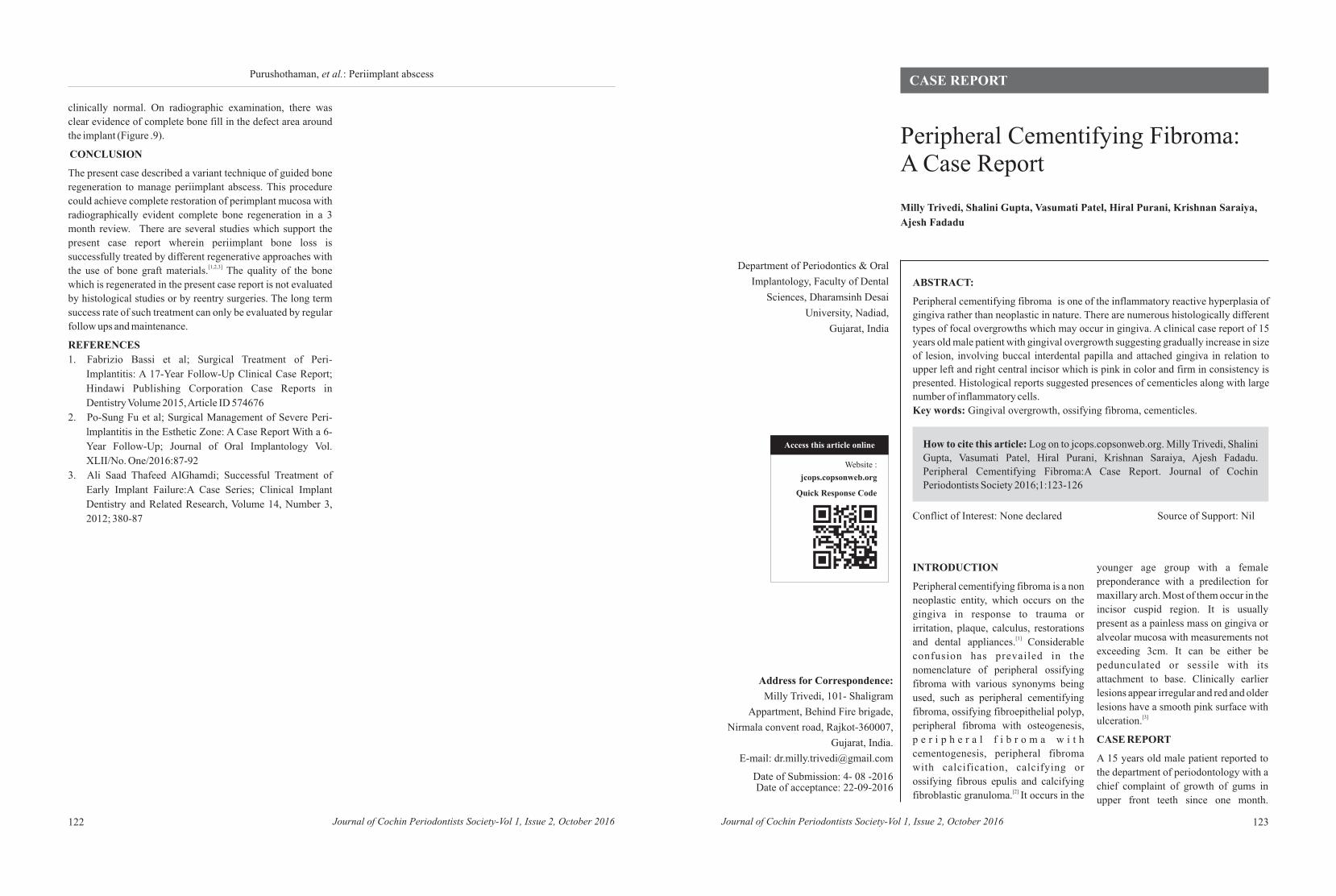
On intra oral examination a solitary, pedunculated mass
involving buccal interdental papilla and attached gingiva in
relation to upper left and right central incisor was seen(figure
1). Intra oral periapical radiograph showed no significant bone
loss(figure 2). Mass was pink in color with a smooth surface,
measuring approximately 1.5 cm × 2 cm and extending from
mesial interdental papilla of right central incisor to distal
surface of left central incisor. No surface ulceration was noted.
On palpation, it was non tender and firm in consistency.
The provisional diagnosis of peripheral ossifying fibroma was
made. The differential diagnosis included traumatic fibroma,
pyogenic granuloma and peripheral giant cell granuloma.
Treatment included thorough scaling and root planning to
eliminate the irritating factors (figure 3). After a week,
complete surgical excision of the lesion was performed under
local anesthesia. Complete removal of the lesion and gingival
curettage was ensured(figure 4). Application of periodontal
dressing (coe-pack) (figure 5) and Oral hygiene maintenance
instructions were given to the patient. Healing was uneventful.
Patient was recalled after 7 days (figure 6) and follow up visit
after 3 months respectively (figure 7).
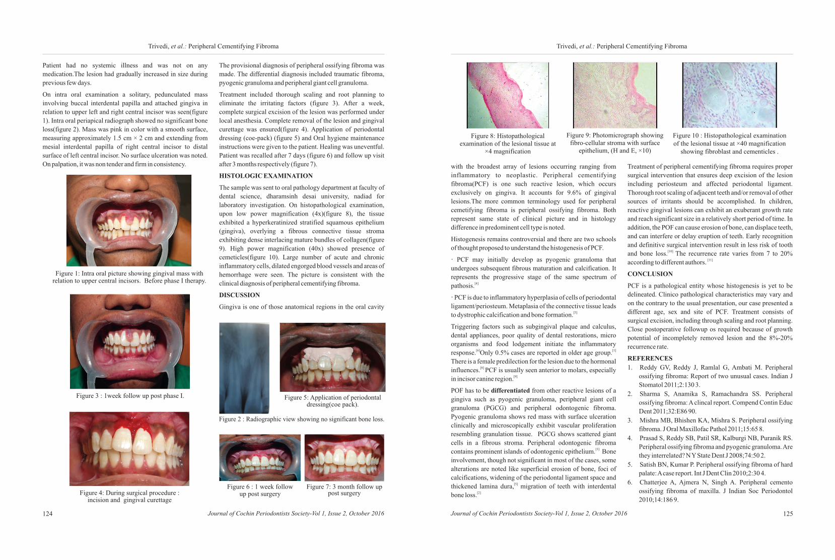
HISTOLOGIC EXAMINATION
The sample was sent to oral pathology department at faculty of
dental science, dharamsinh desai university, nadiad for
laboratory investigation. On histopathological examination,
upon low power magnification (4x)(figure 8), the tissue
exhibited a hyperkeratinized stratified squamous epithelium
(gingiva), overlying a fibrous connective tissue stroma
exhibiting dense interlacing mature bundles of collagen(figure
9). High power magnification (40x) showed presence of
cemeticles(figure 10). Large number of acute and chronic
inflammatory cells, dilated engorged blood vessels and areas of
hemorrhage were seen. The picture is consistent with the
clinical diagnosis of peripheral cementifying fibroma.
DISCUSSION
Gingiva is one of those anatomical regions in the oral cavity
Figure 1: Intra oral picture showing gingival mass with relation to upper central incisors. Before phase I therapy.
Figure 2 : Radiographic view showing no significant bone loss.
Figure 3 : 1week follow up post phase I.
Figure 4: During surgical procedure : incision and gingival curettage
Figure 5: Application of periodontal dressing(coe pack).
Figure 6 : 1 week follow up post surgery
Figure 7: 3 month follow up post surgery
with the broadest array of lesions occurring ranging from
inflammatory to neoplastic. Peripheral cementifying
fibroma(PCF) is one such reactive lesion, which occurs
exclusively on gingiva. It accounts for 9.6% of gingival
lesions.The more common terminology used for peripheral
cemetifying fibroma is peripheral ossifying fibroma. Both
represent same state of clinical picture and in histology
difference in predominent cell type is noted.
Histogenesis remains controversial and there are two schools
of thought proposed to understand the histogenesis of PCF.
· PCF may initially develop as pyogenic granuloma that
undergoes subsequent fibrous maturation and calcification. It
represents the progressive stage of the same spectrum of [4]pathosis.
· PCF is due to inflammatory hyperplasia of cells of periodontal
ligament/periosteum. Metaplasia of the connective tissue leads [5] to dystrophic calcification and bone formation.
Triggering factors such as subgingival plaque and calculus,
dental appliances, poor quality of dental restorations, micro
organisms and food lodgement initiate the inflammatory [6] [1] response. Only 0.5% cases are reported in older age group.
There is a female predilection for the lesion due to the hormonal [8] influences. PCF is usually seen anterior to molars, especially
[9] in incisor canine region.
POF has to be differentiated from other reactive lesions of a
gingiva such as pyogenic granuloma, peripheral giant cell
granuloma (PGCG) and peripheral odontogenic fibroma.
Pyogenic granuloma shows red mass with surface ulceration
clinically and microscopically exhibit vascular proliferation
resembling granulation tissue. PGCG shows scattered giant
cells in a fibrous stroma. Peripheral odontogenic fibroma [5] contains prominent islands of odontogenic epithelium. Bone
involvement, though not significant in most of the cases, some
alterations are noted like superficial erosion of bone, foci of
calcifications, widening of the periodontal ligament space and [5]thickened lamina dura, migration of teeth with interdental
[2] bone loss.
Treatment of peripheral cementifying fibroma requires proper
surgical intervention that ensures deep excision of the lesion
including periosteum and affected periodontal ligament.
Thorough root scaling of adjacent teeth and/or removal of other
sources of irritants should be accomplished. In children,
reactive gingival lesions can exhibit an exuberant growth rate
and reach significant size in a relatively short period of time. In
addition, the POF can cause erosion of bone, can displace teeth,
and can interfere or delay eruption of teeth. Early recognition
and definitive surgical intervention result in less risk of tooth [10] and bone loss. The recurrence rate varies from 7 to 20%
[11]according to different authors.
CONCLUSION
PCF is a pathological entity whose histogenesis is yet to be
delineated. Clinico pathological characteristics may vary and
on the contrary to the usual presentation, our case presented a
different age, sex and site of PCF. Treatment consists of
surgical excision, including through scaling and root planning.
Close postoperative followup os required because of growth
potential of incompletely removed lesion and the 8%-20%
recurrence rate.
REFERENCES
1. Reddy GV, Reddy J, Ramlal G, Ambati M. Peripheral
ossifying fibroma: Report of two unusual cases. Indian J
Stomatol 2011;2:130 3.
2. Sharma S, Anamika S, Ramachandra SS. Peripheral
ossifying fibroma: A clincal report. Compend Contin Educ
Dent 2011;32:E86 90.
3. Mishra MB, Bhishen KA, Mishra S. Peripheral ossifying
fibroma. J Oral Maxillofac Pathol 2011;15:65 8.
4. Prasad S, Reddy SB, Patil SR, Kalburgi NB, Puranik RS.
Peripheral ossifying fibroma and pyogenic granuloma. Are
they interrelated? N Y State Dent J 2008;74:50 2.
5. Satish BN, Kumar P. Peripheral ossifying fibroma of hard
palate: A case report. Int J Dent Clin 2010;2:30 4.
6. Chatterjee A, Ajmera N, Singh A. Peripheral cemento
ossifying fibroma of maxilla. J Indian Soc Periodontol
2010;14:186 9.
Figure 8: Histopathological examination of the lesional tissue at
×4 magnification
Figure 9: Photomicrograph showing fibro-cellular stroma with surface
epithelium, (H and E, ×10)
Figure 10 : Histopathological examination of the lesional tissue at ×40 magnification
showing fibroblast and cementicles .
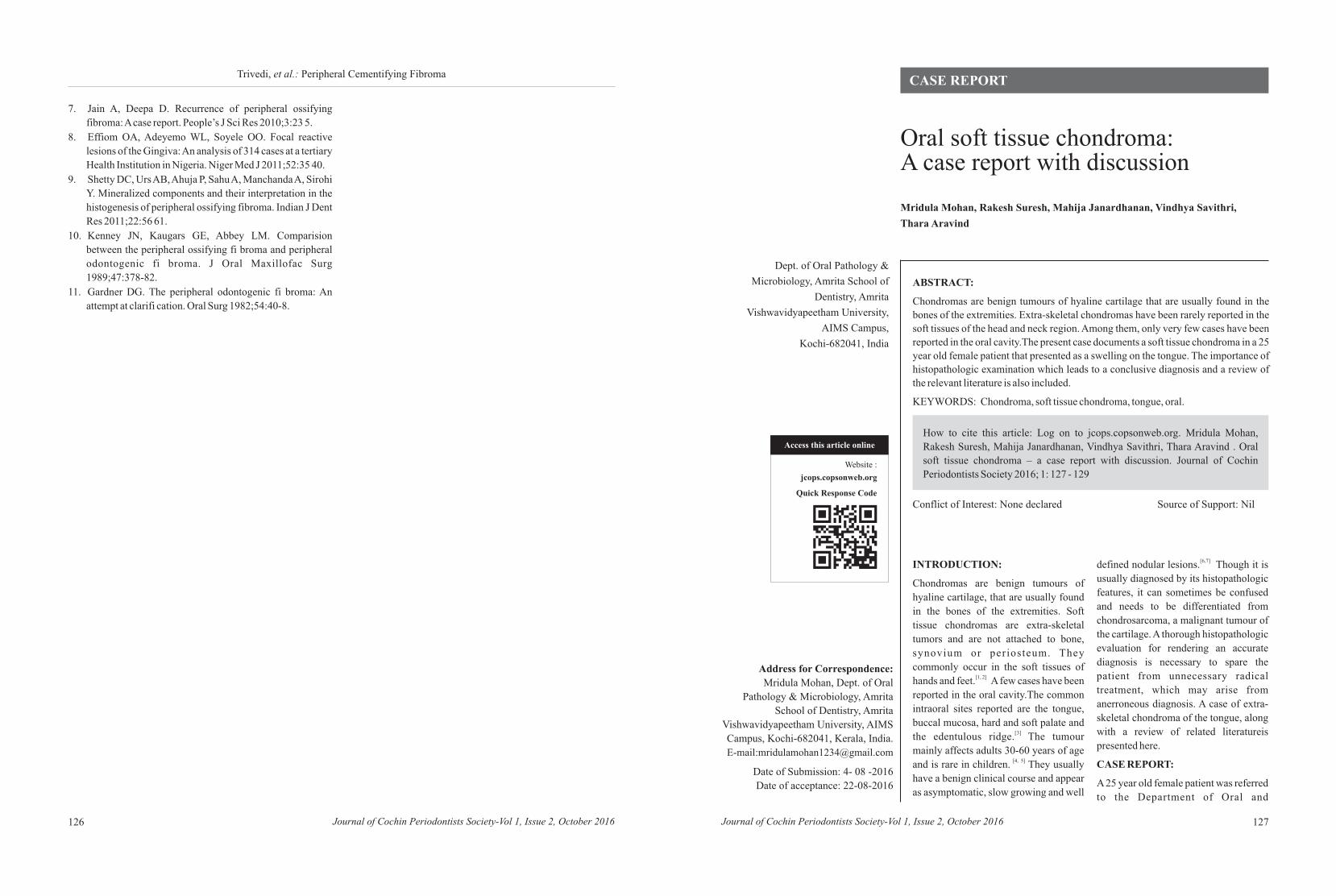
Journal of Cochin Periodontists Society-Vol 1, Issue 2, October 2016126
Trivedi, et al.: Peripheral Cementifying Fibroma
7. Jain A, Deepa D. Recurrence of peripheral ossifying
fibroma: A case report. People’s J Sci Res 2010;3:23 5.
8. Effiom OA, Adeyemo WL, Soyele OO. Focal reactive
lesions of the Gingiva: An analysis of 314 cases at a tertiary
Health Institution in Nigeria. Niger Med J 2011;52:35 40.
9. Shetty DC, Urs AB, Ahuja P, Sahu A, Manchanda A, Sirohi
Y. Mineralized components and their interpretation in the
histogenesis of peripheral ossifying fibroma. Indian J Dent
Res 2011;22:56 61.
10. Kenney JN, Kaugars GE, Abbey LM. Comparision
between the peripheral ossifying fi broma and peripheral
odontogenic fi broma. J Oral Maxillofac Surg
1989;47:378-82.
11. Gardner DG. The peripheral odontogenic fi broma: An
attempt at clarifi cation. Oral Surg 1982;54:40-8.
127
ABSTRACT:
Chondromas are benign tumours of hyaline cartilage that are usually found in the
bones of the extremities. Extra-skeletal chondromas have been rarely reported in the
soft tissues of the head and neck region. Among them, only very few cases have been
reported in the oral cavity.The present case documents a soft tissue chondroma in a 25
year old female patient that presented as a swelling on the tongue. The importance of
histopathologic examination which leads to a conclusive diagnosis and a review of
the relevant literature is also included.
KEYWORDS: Chondroma, soft tissue chondroma, tongue, oral.
Access this article online
Website :
jcops.copsonweb.org
Quick Response Code
How to cite this article: Log on to jcops.copsonweb.org. Mridula Mohan,
Rakesh Suresh, Mahija Janardhanan, Vindhya Savithri, Thara Aravind . Oral
soft tissue chondroma – a case report with discussion. Journal of Cochin
Periodontists Society 2016; 1: 127 - 129
Conflict of Interest: None declared Source of Support: Nil
CASE REPORT
Dept. of Oral Pathology &
Microbiology, Amrita School of
Dentistry, Amrita
Vishwavidyapeetham University,
AIMS Campus,
Kochi-682041, India
Address for Correspondence:
Mridula Mohan, Dept. of Oral
Pathology & Microbiology, Amrita
School of Dentistry, Amrita
Vishwavidyapeetham University, AIMS
Campus, Kochi-682041, Kerala, India.
E-mail:[email protected]
Date of Submission: 4- 08 -2016
Date of acceptance: 22-08-2016
Journal of Cochin Periodontists Society-Vol 1, Issue 2, October 2016
Mridula Mohan, Rakesh Suresh, Mahija Janardhanan,
Thara Aravind
Vindhya Savithri,
Oral soft tissue chondroma: A case report with discussion
INTRODUCTION:
Chondromas are benign tumours of
hyaline cartilage, that are usually found
in the bones of the extremities. Soft
tissue chondromas are extra-skeletal
tumors and are not attached to bone,
synovium or periosteum. They
commonly occur in the soft tissues of [1, 2]hands and feet. A few cases have been
reported in the oral cavity.The common
intraoral sites reported are the tongue,
buccal mucosa, hard and soft palate and [3] the edentulous ridge. The tumour
mainly affects adults 30-60 years of age [4, 5]and is rare in children. They usually
have a benign clinical course and appear
as asymptomatic, slow growing and well
[6,7] defined nodular lesions. Though it is
usually diagnosed by its histopathologic
features, it can sometimes be confused
and needs to be differentiated from
chondrosarcoma, a malignant tumour of
the cartilage. A thorough histopathologic
evaluation for rendering an accurate
diagnosis is necessary to spare the
patient from unnecessary radical
treatment, which may arise from
anerroneous diagnosis. A case of extra-
skeletal chondroma of the tongue, along
with a review of related literatureis
presented here.
CASE REPORT:
A 25 year old female patient was referred
to the Department of Oral and
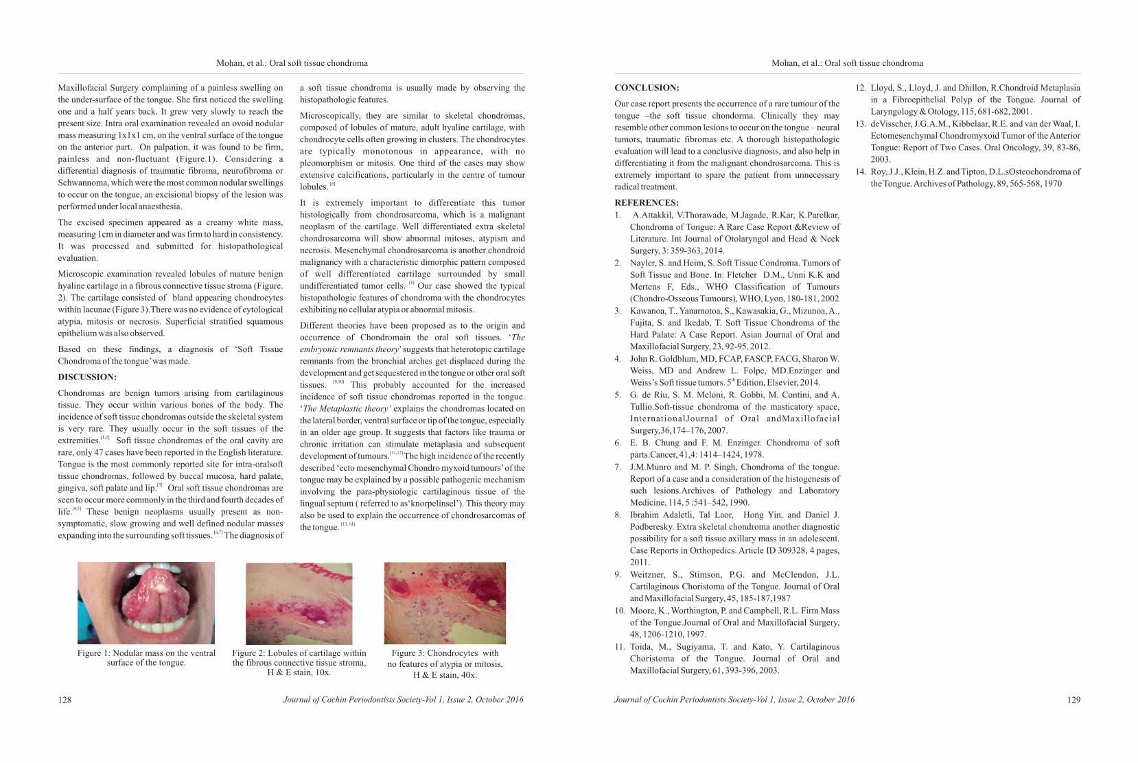
Journal of Cochin Periodontists Society-Vol 1, Issue 2, October 2016128 129Journal of Cochin Periodontists Society-Vol 1, Issue 2, October 2016
Maxillofacial Surgery complaining of a painless swelling on
the under-surface of the tongue. She first noticed the swelling
one and a half years back. It grew very slowly to reach the
present size. Intra oral examination revealed an ovoid nodular
mass measuring 1x1x1 cm, on the ventral surface of the tongue
on the anterior part. On palpation, it was found to be firm,
painless and non-fluctuant (Figure.1). Considering a
differential diagnosis of traumatic fibroma, neurofibroma or
Schwannoma, which were the most common nodular swellings
to occur on the tongue, an excisional biopsy of the lesion was
performed under local anaesthesia.
The excised specimen appeared as a creamy white mass,
measuring 1cm in diameter and was firm to hard in consistency.
It was processed and submitted for histopathological
evaluation.
Microscopic examination revealed lobules of mature benign
hyaline cartilage in a fibrous connective tissue stroma (Figure.
2). The cartilage consisted of bland appearing chondrocytes
within lacunae (Figure 3).There was no evidence of cytological
atypia, mitosis or necrosis. Superficial stratified squamous
epithelium was also observed.
Based on these findings, a diagnosis of ‘Soft Tissue
Chondroma of the tongue’ was made.
DISCUSSION:
Chondromas are benign tumors arising from cartilaginous
tissue. They occur within various bones of the body. The
incidence of soft tissue chondromas outside the skeletal system
is very rare. They usually occur in the soft tissues of the [1,2]extremities. Soft tissue chondromas of the oral cavity are
rare, only 47 cases have been reported in the English literature.
Tongue is the most commonly reported site for intra-oralsoft
tissue chondromas, followed by buccal mucosa, hard palate, [3]gingiva, soft palate and lip. Oral soft tissue chondromas are
seen to occur more commonly in the third and fourth decades of [4,5]life. These benign neoplasms usually present as non-
symptomatic, slow growing and well defined nodular masses [6,7]expanding into the surrounding soft tissues. The diagnosis of
Figure 1: Nodular mass on the ventral surface of the tongue.
Figure 2: Lobules of cartilage within the fibrous connective tissue stroma,
H & E stain, 10x.
Figure 3: Chondrocytes with no features of atypia or mitosis,
H & E stain, 40x.
a soft tissue chondroma is usually made by observing the
histopathologic features.
Microscopically, they are similar to skeletal chondromas,
composed of lobules of mature, adult hyaline cartilage, with
chondrocyte cells often growing in clusters. The chondrocytes
are typically monotonous in appearance, with no
pleomorphism or mitosis. One third of the cases may show
extensive calcifications, particularly in the centre of tumour [6]lobules.
It is extremely important to differentiate this tumor
histologically from chondrosarcoma, which is a malignant
neoplasm of the cartilage. Well differentiated extra skeletal
chondrosarcoma will show abnormal mitoses, atypism and
necrosis. Mesenchymal chondrosarcoma is another chondroid
malignancy with a characteristic dimorphic pattern composed
of well differentiated cartilage surrounded by small [8]undifferentiated tumor cells. Our case showed the typical
histopathologic features of chondroma with the chondrocytes
exhibiting no cellular atypia or abnormal mitosis.
Different theories have been proposed as to the origin and
occurrence of Chondromain the oral soft tissues. ‘The
embryonic remnants theory’ suggests that heterotopic cartilage
remnants from the bronchial arches get displaced during the
development and get sequestered in the tongue or other oral soft [9,10]tissues. This probably accounted for the increased
incidence of soft tissue chondromas reported in the tongue.
‘The Metaplastic theory’ explains the chondromas located on
the lateral border, ventral surface or tip of the tongue, especially
in an older age group. It suggests that factors like trauma or
chronic irritation can stimulate metaplasia and subsequent [11,12] development of tumours. The high incidence of the recently
described ‘ecto mesenchymal Chondro myxoid tumours’ of the
tongue may be explained by a possible pathogenic mechanism
involving the para-physiologic cartilaginous tissue of the
lingual septum ( referred to as‘knorpelinsel’). This theory may
also be used to explain the occurrence of chondrosarcomas of [13, 14]the tongue.
Mohan, et al.: Oral soft tissue chondroma Mohan, et al.: Oral soft tissue chondroma
CONCLUSION:
Our case report presents the occurrence of a rare tumour of the
tongue –the soft tissue chondorma. Clinically they may
resemble other common lesions to occur on the tongue – neural
tumors, traumatic fibromas etc. A thorough histopathologic
evaluation will lead to a conclusive diagnosis, and also help in
differentiating it from the malignant chondrosarcoma. This is
extremely important to spare the patient from unnecessary
radical treatment.
REFERENCES:
1. A.Attakkil, V.Thorawade, M.Jagade, R.Kar, K.Parelkar,
Chondroma of Tongue: A Rare Case Report &Review of
Literature. Int Journal of Otolaryngol and Head & Neck
Surgery, 3: 359-363, 2014.
2. Nayler, S. and Heim, S. Soft Tissue Condroma. Tumors of
Soft Tissue and Bone. In: Fletcher D.M., Unni K.K and
Mertens F, Eds., WHO Classification of Tumours
(Chondro-Osseous Tumours), WHO, Lyon, 180-181, 2002
3. Kawanoa, T., Yanamotoa, S., Kawasakia, G., Mizunoa, A.,
Fujita, S. and Ikedab, T. Soft Tissue Chondroma of the
Hard Palate: A Case Report. Asian Journal of Oral and
Maxillofacial Surgery, 23, 92-95, 2012.
4. John R. Goldblum, MD, FCAP, FASCP, FACG, Sharon W.
Weiss, MD and Andrew L. Folpe, MD.Enzinger and thWeiss’s Soft tissue tumors. 5 Edition, Elsevier, 2014.
5. G. de Riu, S. M. Meloni, R. Gobbi, M. Contini, and A.
Tullio.Soft-tissue chondroma of the masticatory space,
InternationalJournal of Oral andMaxillofacial
Surgery,36,174–176, 2007.
6. E. B. Chung and F. M. Enzinger. Chondroma of soft
parts.Cancer, 41,4: 1414–1424, 1978.
7. J.M.Munro and M. P. Singh, Chondroma of the tongue.
Report of a case and a consideration of the histogenesis of
such lesions.Archives of Pathology and Laboratory
Medicine, 114, 5 :541–542, 1990.
8. Ibrahim Adaletli, Tal Laor, Hong Yin, and Daniel J.
Podberesky. Extra skeletal chondroma another diagnostic
possibility for a soft tissue axillary mass in an adolescent.
Case Reports in Orthopedics. Article ID 309328, 4 pages,
2011.
9. Weitzner, S., Stimson, P.G. and McClendon, J.L.
Cartilaginous Choristoma of the Tongue. Journal of Oral
and Maxillofacial Surgery, 45, 185-187,1987
10. Moore, K., Worthington, P. and Campbell, R.L. Firm Mass
of the Tongue.Journal of Oral and Maxillofacial Surgery,
48, 1206-1210, 1997.
11. Toida, M., Sugiyama, T. and Kato, Y. Cartilaginous
Choristoma of the Tongue. Journal of Oral and
Maxillofacial Surgery, 61, 393-396, 2003.
12. Lloyd, S., Lloyd, J. and Dhillon, R.Chondroid Metaplasia
in a Fibroepithelial Polyp of the Tongue. Journal of
Laryngology & Otology, 115, 681-682, 2001.
13. deVisscher, J.G.A.M., Kibbelaar, R.E. and van der Waal, I.
Ectomesenchymal Chondromyxoid Tumor of the Anterior
Tongue: Report of Two Cases. Oral Oncology, 39, 83-86,
2003.
14. Roy, J.J., Klein, H.Z. and Tipton, D.L.sOsteochondroma of
the Tongue. Archives of Pathology, 89, 565-568, 1970
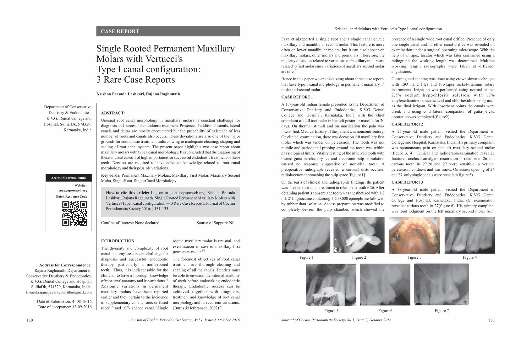
ABSTRACT:
Unusual root canal morphology in maxillary molars is constant challenge for
diagnosis and successful endodontic treatment. Presence of additional canals, lateral
canals and deltas are mostly encountered but the probability of existence of less
number of roots and canals also occurs. These deviations are also one of the major
grounds for endodontic treatment failure owing to inadequate cleaning, shaping and
sealing of root canal system. The present paper highlights two case report about
maxillary molars with type I canal morphology. It is concluded that the diagnosing of
these unusual cases is of high importance for successful endodontic treatment of these
teeth. Dentists are required to have adequate knowledge related to root canal
morphology and their possible variations.
Keywords: Permanent Maxillary Molars, Maxillary First Molar, Maxillary Second
Molar, Single Root, Single Canal Morphology
Krishna Prasada Lashkari, Rajana Raghunath
How to cite this article: Log on to jcops.copsonweb.org. Krishna Prasada
Lashkari, Rajana Raghunath. Single Rooted Permanent Maxillary Molars with
Vertucci'sType I canal configuration — 3 Rare Case Reports. Journal of Cochin
Periodontists Society 2016;1:131-133
Conflict of Interest: None declared Source of Support: Nil
Journal of Cochin Periodontists Society-Vol 1, Issue 2, October 2016 Journal of Cochin Periodontists Society-Vol 1, Issue 2, October 2016 131130
Access this article online
Website :
jcops.copsonweb.org
Quick Response Code
Department of Conservative
Dentistry & Endodontics,
K.V.G. Dental College and
Hospital, Sullia Dk, 574329,
Karnataka, India
Address for Correspondence:
Rajana Raghunath, Department of
Conservative Dentistry & Endodontics,
K.V.G. Dental College and Hospital,
SulliaDk, 574329, Karnataka, India.
E-mail:[email protected]
Date of Submission: 4- 08 -2016
Date of acceptance: 12-09-2016
CASE REPORTKrishna, et al.:Molars with Vertucci's Type I canal configuration
Single Rooted Permanent Maxillary Molars with Vertucci's Type I canal configuration: 3 Rare Case Reports
INTRODUCTION
The diversity and complexity of root
canal anatomy are constant challenge for
diagnosis and successful endodontic
therapy, particularly in multi-rooted
teeth. Thus, it is indispensable for the
clinician to have a thorough knowledge . [1] of root canal anatomy and its variations
Anatomic variations in permanent
maxillary molars have been reported
earlier and they pertain to the incidence
of supplementary canals, roots or fused [2,3] [4]roots and “C”- shaped canal. Single
rooted maxillary molar is unusual, and
even scarcer in case of maxillary first [5]permanent molar.
The foremost objectives of root canal
treatment are thorough cleaning and
shaping of all the canals. Dentists must
be able to envision the internal anatomy
of teeth before undertaking endodontic
therapy. Endodontic success can be
achieved together with diagnosis,
treatment and knowledge of root canal
morphology and its recurrent variations. [6](Burns &Herbranson, 2002)
Fava et al.reported a single root and a single canal on the
maxillary and mandibular second molar. This feature is more
often on lower mandibular molars, but it can also appear on
maxillary molars, other molars and premolars. Therefore, the
majority of studies related to variations of maxillary molars are
related to first molar since variations of maxillary second molar [7]are rare.
Hence in this paper we are discussing about three case reports stthat have type 1 canal morphology in permanent maxillary 1
molar and second molar
CASE REPORT 1
A 17-year-old Indian female presented to the Department of
Conservative Dentistry and Endodontics, K.V.G Dental
College and Hospital, Karnataka, India with the chief
complaint of dull toothache in her left posterior maxilla for 20
days. On thermal stimuli and on mastication the pain was
intensified. Medical history of the patient was noncontributory.
On clinical examination, there was decay on left maxillary first
molar which was tender on percussion. The tooth was not
mobile and periodontal probing around the tooth was within
physiological limits. Vitality testing of the involved tooth with
heated gutta-percha, dry ice and electronic pulp stimulation
caused no response suggestive of non-vital tooth. A
preoperative radiograph revealed a coronal disto-occlusal
radiolucency approaching the pulp space [Figure 1].
On the basis of clinical and radiographic findings, the patient
was advised root canal treatment in relation to tooth # 26. After
obtaining patient’s consent, the tooth was anesthetized with 1.8
mL 2% lignocaine containing 1:200,000 epinephrine followed
by rubber dam isolation. Access preparation was modified to
completely de-roof the pulp chamber, which showed the
presence of a single wide root canal orifice. Presence of only
one single canal and no other canal orifice was revealed on
examination under a surgical operating microscope. With the
help of an apex locator which was later confirmed using a
radiograph the working length was determined. Multiple
working length radiographs were taken at different
angulations.
Cleaning and shaping was done using crown-down technique
with ISO hand files and ProTaper nickel-titanium rotary
instruments. Irrigation was performed using normal saline,
2 .5% sodium hypochlor i te solut ion, wi th 17%
ethylenediamine tetracetic acid and chlorhexidine being used
as the final irrigant. With absorbent points the canals were
dried, and using cold lateral compaction of gutta-percha
obturation was completed (figure2).
CASE REPORT 2
A 25-year-old male patient visited the Department of
Conservative Dentistry and Endodontics, K.V.G Dental
College and Hospital, Karnataka, India. His primary complaint
was spontaneous pain on the left maxillary second molar
(figure 3, 4). Clinical and radiographexamination revealed
fractured occlusal amalgam restoration in relation to 26 and
carious tooth irt 27.26 and 27 were sensitive in vertical
percussion, coldness and warmness. On access opening of 26
and 27, only single canals were revealed (figure 5).
CASE REPORT 3
A 38-year-old male patient visited the Department of
Conservative Dentistry and Endodontics, K.V.G Dental
College and Hospital, Karnataka, India. On examination
revealed carious tooth irt 27(figure 6). His primary complain,
was food lodgment on the left maxillary second molar from
past 6 months and also dull throbbing pain at night. Pain
Figure 1 Figure 2 Figure 3 Figure 4
Figure 7Figure 6Figure 5
Journal of Cochin Periodontists Society-Vol 1, Issue 2, October 2016132
Krishna, et al.:Molars with Vertucci's Type I canal configuration
appeared more frequently during the night lasting 2 to 3 hours
with brief calming periods. The tooth was sensitive in vertical
percussion, coldness and warmness. On access opening of 27
only single canal was discovered. (Figure 7)
DISCUSSION
Dental abnormalities which are formative defects may occur
during any of the developmental stages of the tooth, and they
are revealed clinically in later life once the tooth is fully [8]formed.
Incidence of additional canal is more recurrent rather than the
presence of fused/less number of canals, particularly in the
cases of permanent maxillary first molars. Immense
disproportion in the root/root canal morphology of permanent
maxillary molars of Indian origin exists as compared with
Caucasian and Mongoloid traits, as reported in a computed
tomographic research. The dissimilarities reported in the
population were the presence of single root and canal, two
separate roots, four separate roots and two fused roots, three [9]fused roots.
There are few case reports that were found to have reported
dental anomalies such as fused single rooted maxillary molar: [10] [11] Ackerman et al., Holan and Chosack, Gopikrishna et al.,
[12] [13]Metgud et al. There appears to be a difference in opinion
regarding the term used to describe such a tooth, this includes
“fused.” “Pyramidal,” and “conical” but all investigators
believe that the failure of invagination of Hertwig epithelial
root sheath is accountable for this anomaly.
Maxillary second molars with triple roots and triple canals
compared to maxillary second molar that could have a single
root and a single canalis more often described in endodontic [14]literature . Hartwell & Bellizi concluded that only 0.6% of
cases there is presence of maxillary second molar with a single [15]root and a single canal. . Libfeld and Rotstein assessed 200
radiographies of patients treated in an endodontic therapy in
maxillary second molar and reported that this feature was [16]evident in 0.5% of cases. According to Hua XI et al. the
frequency of maxillary second molar with single root and [17]single canal is very unusual.
This paper highlights 3 different case reports where Vertucci’s
Type 1 canal configuration is seen in maxillary permanent
molars. Since the incidence of single root and single canal is not
high, it is important to confirm the canal configuration before
finishing root canal treatment in order to ensure success.
Presence of single canal in the 3 cases was evaluated through
radiographic assessment and application of operative
microscope and a loop. These teeth have good endodontic
prognosis, because of wide and accessible canal.
CONCLUSION
Anatomical variation continues to be the most challenging
aspect of performing successful endodontic therapy.
Quitemore importance is given to extra canals, merging and
demerging canals, apical deltas, and lateral canals but the
clinician should also focus on the presence of fewer canals. The
present case reports highlight the necessity to develop excellent
observation skills on the part of the dentist to identify any
aberrations from the normal within the tooth, especially in the
presence of anomalies in dentition.
REFERENCES
1. Ahuja P, Ballal S, Velmurugan N. Endodontic
management of maxillary second molar with a single root
and a single canal diagnosed with cone-beam computed
tomography scanning. Saudi Endod J 2012;2:100-3
2. Shin SJ, Park JW, Lee JK, Hwang SW. Unusual root canal
anatomy in maxillary second molars: Two case reports.
Oral Surg Oral Med Oral Pathol Oral RadiolEndod.
2007;104:e61–5.
3. Gopikrishna V, Reuben J, Kandaswamy D. Endodontic
management of a maxillary first molar with two palatal
roots and a single fused buccal root diagnosed with spiral
computed tomography: A case report. Oral Surg Oral Med
Oral Pathol Oral RadiolEndod. 2008;105:e74–8.
4. Kottoor J, Velmurugan N, Ballal S, Roy A. Four-rooted
maxillary first molar having C-shaped palatal root canal
morphology evaluated using cone-beam computerized
tomography: A case report. Oral Surg Oral Med Oral
Pathol Oral RadiolEndod. 2011;111:e41–5.
5. Gopikrishna V, Bhargavi N, Kandaswamy D. Endodontic
management of a maxillary first molar with a single root
and a single canal diagnosed with the aid of spiral CT: A
case report. J Endod. 2006;32:687–91.
6. Single Rooted Maxillary First Molar: A Rare CaseChikoy
Wang, Krishna PrasadPeople’s Journal of Scientific
Research 68 Vol. 4(1), Jan. 2011
7. Fava, L.R.G., Wienfeld, I., Fabri, F.P. and Pais, C.R.
(2000) Four Second Molars with Single Root and Single
Canals in the Same Patient. International Endodontic
Journal, 33, 138-142.
8. Robbins IM, Keene HJ. Multiple morphologic dental
anomalies. Report of a case. Oral Surg Oral Med Oral
Pathol 1964;17:683-90.
9. Neelakantan P, Subbarao C, Ahuja R, Subbarao CV,
Gutmann JL. Cone-beam computed tomography study of
root and canal morphology of maxillary first and second
molars in an Indian population. J Endod. 2010;36:1622–7
10. Ackerman JL, Ackerman AL, Ackerman AB. Taurodont,
pyramidal and fused molar roots associated with other
Journal of Cochin Periodontists Society-Vol 1, Issue 2, October 2016 133
Krishna, et al.:Molars with Vertucci's Type I canal configuration
anomalies in a kindred. Am J Phys Anthropol 1973;
38:681-94.
11. Holan G, Chosack A. Single-rooted molars in the primary
and permanent dentition in two siblings: Case report.
Pediatr Dent 1991;13:367-9. Back to cited text no. 7
12. Gopikrishna V, Bhargavi N, Kandaswamy D. Endodontic
management of a maxillary first molar with a single root
and a single canal diagnosed with the aid of spiral CT: A
case report. J Endod 2006;32:687-91
13. Metgud S, Metgud R, Rani K. Management of a patient
with a taurodont, single-rooted molars associated with
multiple dental anomalies: A spiral computerized
tomography evaluation. Oral Surg Oral Med Oral Pathol
Oral RadiolEndod 2009;108:e81-6.
14. Ajet i N, Vula V, Apostolska S, Pust ina T
KelmendiT,Maxillary Second Molar with Single Root and
Single Canal—Case Report.Open Journal of Stomatology,
2015, 5, 47-52
15. Hartwell, G. and Bellizi, R. (1982) Clinical Investigation
of in Vivo Endodontically Treated Mandibular and
Maxillary Molars. Journal of Endodontics, 8, 555-557
16. Liebfield, H. and Rotstein, I. (1989) Incidence of Four-
Rooted Second Molars: Literature Review and
Radiographic Survey of 1,200 Teeth. Journal of
Endodontics, 15, 129-131.
17. Wang, Y., Hui, X. and Huang, D.M. (2011) Maxillary and
Second Molar with Curved Single Root and Single Canal:
A Case Report. Hua Xi Kou Qiang Yi XueZaZhi, 29, 104-
105.
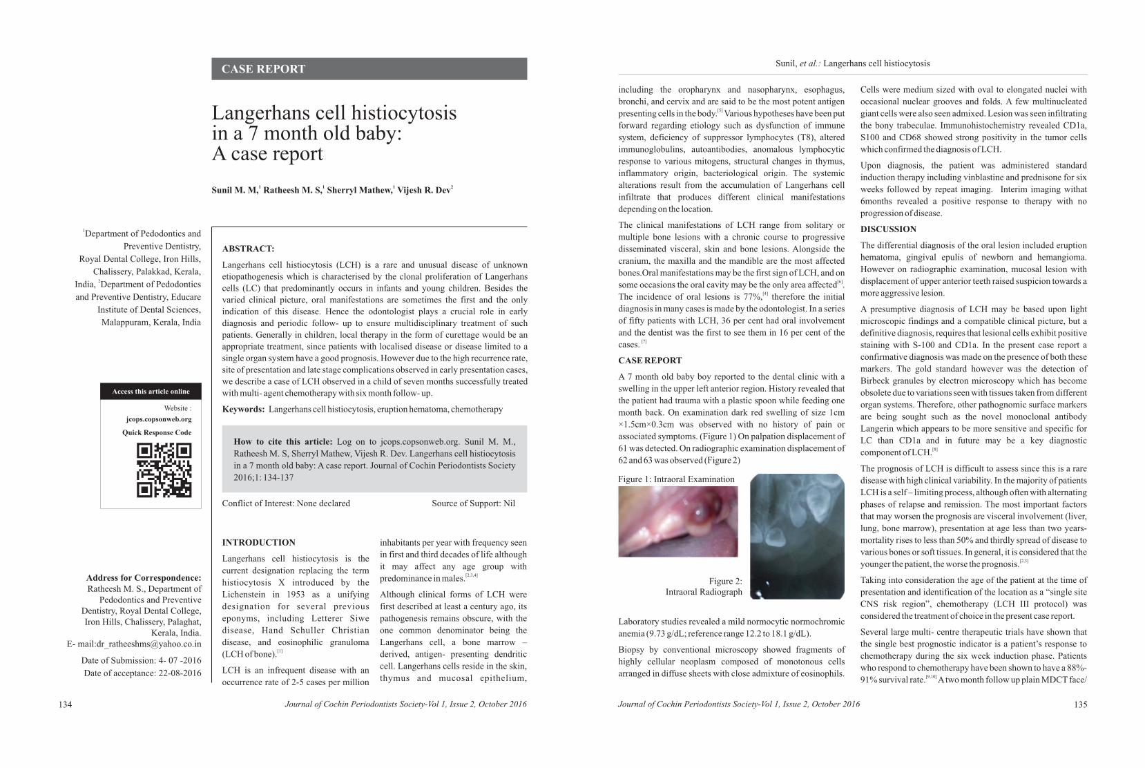
ABSTRACT:
Langerhans cell histiocytosis (LCH) is a rare and unusual disease of unknown
etiopathogenesis which is characterised by the clonal proliferation of Langerhans
cells (LC) that predominantly occurs in infants and young children. Besides the
varied clinical picture, oral manifestations are sometimes the first and the only
indication of this disease. Hence the odontologist plays a crucial role in early
diagnosis and periodic follow- up to ensure multidisciplinary treatment of such
patients. Generally in children, local therapy in the form of curettage would be an
appropriate treatment, since patients with localised disease or disease limited to a
single organ system have a good prognosis. However due to the high recurrence rate,
site of presentation and late stage complications observed in early presentation cases,
we describe a case of LCH observed in a child of seven months successfully treated
with multi- agent chemotherapy with six month follow- up.
Keywords: Langerhans cell histiocytosis, eruption hematoma, chemotherapy
1 1 1 2Sunil M. M, Ratheesh M. S, Sherryl Mathew, Vijesh R. Dev
How to cite this article: Log on to jcops.copsonweb.org. Sunil M. M.,
Ratheesh M. S, Sherryl Mathew, Vijesh R. Dev. Langerhans cell histiocytosis
in a 7 month old baby: A case report. Journal of Cochin Periodontists Society
2016;1: 134-137
Conflict of Interest: None declared Source of Support: Nil
Journal of Cochin Periodontists Society-Vol 1, Issue 2, October 2016 Journal of Cochin Periodontists Society-Vol 1, Issue 2, October 2016 135134
Access this article online
Website :
jcops.copsonweb.org
Quick Response Code
1Department of Pedodontics and
Preventive Dentistry,
Royal Dental College, Iron Hills,
Chalissery, Palakkad, Kerala, 2India, Department of Pedodontics
and Preventive Dentistry, Educare
Institute of Dental Sciences,
Malappuram, Kerala, India
Address for Correspondence: Ratheesh M. S., Department of
Pedodontics and Preventive Dentistry, Royal Dental College, Iron Hills, Chalissery, Palaghat,
Kerala, India. E- mail:[email protected]
Date of Submission: 4- 07 -2016
Date of acceptance: 22-08-2016
CASE REPORTSunil, et al.: Langerhans cell histiocytosis
Langerhans cell histiocytosis in a 7 month old baby: A case report
INTRODUCTION
Langerhans cell histiocytosis is the
current designation replacing the term
histiocytosis X introduced by the
Lichenstein in 1953 as a unifying
designation for several previous
eponyms, including Letterer Siwe
disease, Hand Schuller Christian
disease, and eosinophilic granuloma [1](LCH of bone).
LCH is an infrequent disease with an
occurrence rate of 2-5 cases per million
inhabitants per year with frequency seen
in first and third decades of life although
it may affect any age group with [2,3,4]predominance in males.
Although clinical forms of LCH were
first described at least a century ago, its
pathogenesis remains obscure, with the
one common denominator being the
Langerhans cell, a bone marrow –
derived, antigen- presenting dendritic
cell. Langerhans cells reside in the skin,
thymus and mucosal epithelium,
including the oropharynx and nasopharynx, esophagus,
bronchi, and cervix and are said to be the most potent antigen [5]presenting cells in the body. Various hypotheses have been put
forward regarding etiology such as dysfunction of immune
system, deficiency of suppressor lymphocytes (T8), altered
immunoglobulins, autoantibodies, anomalous lymphocytic
response to various mitogens, structural changes in thymus,
inflammatory origin, bacteriological origin. The systemic
alterations result from the accumulation of Langerhans cell
infiltrate that produces different clinical manifestations
depending on the location.
The clinical manifestations of LCH range from solitary or
multiple bone lesions with a chronic course to progressive
disseminated visceral, skin and bone lesions. Alongside the
cranium, the maxilla and the mandible are the most affected
bones.Oral manifestations may be the first sign of LCH, and on [6]some occasions the oral cavity may be the only area affected .
[4]The incidence of oral lesions is 77%, therefore the initial
diagnosis in many cases is made by the odontologist. In a series
of fifty patients with LCH, 36 per cent had oral involvement
and the dentist was the first to see them in 16 per cent of the [7]cases.
CASE REPORT
A 7 month old baby boy reported to the dental clinic with a
swelling in the upper left anterior region. History revealed that
the patient had trauma with a plastic spoon while feeding one
month back. On examination dark red swelling of size 1cm
×1.5cm×0.3cm was observed with no history of pain or
associated symptoms. (Figure 1) On palpation displacement of
61 was detected. On radiographic examination displacement of
62 and 63 was observed (Figure 2)
Figure 1: Intraoral Examination
Figure 2: Intraoral Radiograph
Laboratory studies revealed a mild normocytic normochromic
anemia (9.73 g/dL; reference range 12.2 to 18.1 g/dL).
Biopsy by conventional microscopy showed fragments of
highly cellular neoplasm composed of monotonous cells
arranged in diffuse sheets with close admixture of eosinophils.
Cells were medium sized with oval to elongated nuclei with
occasional nuclear grooves and folds. A few multinucleated
giant cells were also seen admixed. Lesion was seen infiltrating
the bony trabeculae. Immunohistochemistry revealed CD1a,
S100 and CD68 showed strong positivity in the tumor cells
which confirmed the diagnosis of LCH.
Upon diagnosis, the patient was administered standard
induction therapy including vinblastine and prednisone for six
weeks followed by repeat imaging. Interim imaging withat
6months revealed a positive response to therapy with no
progression of disease.
DISCUSSION
The differential diagnosis of the oral lesion included eruption
hematoma, gingival epulis of newborn and hemangioma.
However on radiographic examination, mucosal lesion with
displacement of upper anterior teeth raised suspicion towards a
more aggressive lesion.
A presumptive diagnosis of LCH may be based upon light
microscopic findings and a compatible clinical picture, but a
definitive diagnosis, requires that lesional cells exhibit positive
staining with S-100 and CD1a. In the present case report a
confirmative diagnosis was made on the presence of both these
markers. The gold standard however was the detection of
Birbeck granules by electron microscopy which has become
obsolete due to variations seen with tissues taken from different
organ systems. Therefore, other pathognomic surface markers
are being sought such as the novel monoclonal antibody
Langerin which appears to be more sensitive and specific for
LC than CD1a and in future may be a key diagnostic [8]component of LCH.
The prognosis of LCH is difficult to assess since this is a rare
disease with high clinical variability. In the majority of patients
LCH is a self – limiting process, although often with alternating
phases of relapse and remission. The most important factors
that may worsen the prognosis are visceral involvement (liver,
lung, bone marrow), presentation at age less than two years-
mortality rises to less than 50% and thirdly spread of disease to
various bones or soft tissues. In general, it is considered that the [2,3]younger the patient, the worse the prognosis.
Taking into consideration the age of the patient at the time of
presentation and identification of the location as a “single site
CNS risk region”, chemotherapy (LCH III protocol) was
considered the treatment of choice in the present case report.
Several large multi- centre therapeutic trials have shown that
the single best prognostic indicator is a patient’s response to
chemotherapy during the six week induction phase. Patients
who respond to chemotherapy have been shown to have a 88%-[9,10]91% survival rate. A two month follow up plain MDCT face/
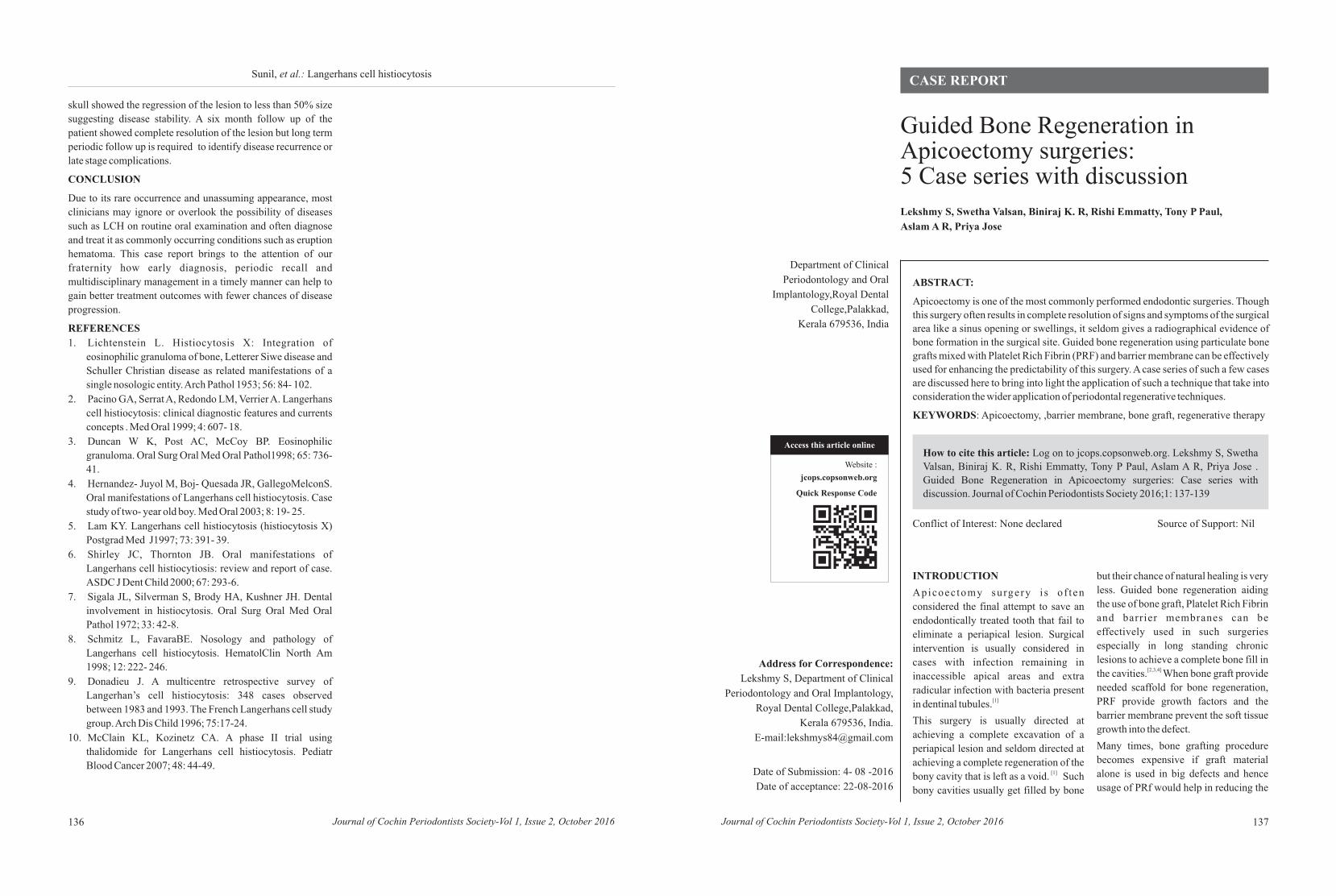
Journal of Cochin Periodontists Society-Vol 1, Issue 2, October 2016136
Sunil, et al.: Langerhans cell histiocytosis
skull showed the regression of the lesion to less than 50% size
suggesting disease stability. A six month follow up of the
patient showed complete resolution of the lesion but long term
periodic follow up is required to identify disease recurrence or
late stage complications.
CONCLUSION
Due to its rare occurrence and unassuming appearance, most
clinicians may ignore or overlook the possibility of diseases
such as LCH on routine oral examination and often diagnose
and treat it as commonly occurring conditions such as eruption
hematoma. This case report brings to the attention of our
fraternity how early diagnosis, periodic recall and
multidisciplinary management in a timely manner can help to
gain better treatment outcomes with fewer chances of disease
progression.
REFERENCES
1. Lichtenstein L. Histiocytosis X: Integration of
eosinophilic granuloma of bone, Letterer Siwe disease and
Schuller Christian disease as related manifestations of a
single nosologic entity. Arch Pathol 1953; 56: 84- 102.
2. Pacino GA, Serrat A, Redondo LM, Verrier A. Langerhans
cell histiocytosis: clinical diagnostic features and currents
concepts . Med Oral 1999; 4: 607- 18.
3. Duncan W K, Post AC, McCoy BP. Eosinophilic
granuloma. Oral Surg Oral Med Oral Pathol1998; 65: 736-
41.
4. Hernandez- Juyol M, Boj- Quesada JR, GallegoMelconS.
Oral manifestations of Langerhans cell histiocytosis. Case
study of two- year old boy. Med Oral 2003; 8: 19- 25.
5. Lam KY. Langerhans cell histiocytosis (histiocytosis X)
Postgrad Med J1997; 73: 391- 39.
6. Shirley JC, Thornton JB. Oral manifestations of
Langerhans cell histiocytiosis: review and report of case.
ASDC J Dent Child 2000; 67: 293-6.
7. Sigala JL, Silverman S, Brody HA, Kushner JH. Dental
involvement in histiocytosis. Oral Surg Oral Med Oral
Pathol 1972; 33: 42-8.
8. Schmitz L, FavaraBE. Nosology and pathology of
Langerhans cell histiocytosis. HematolClin North Am
1998; 12: 222- 246.
9. Donadieu J. A multicentre retrospective survey of
Langerhan’s cell histiocytosis: 348 cases observed
between 1983 and 1993. The French Langerhans cell study
group. Arch Dis Child 1996; 75:17-24.
10. McClain KL, Kozinetz CA. A phase II trial using
thalidomide for Langerhans cell histiocytosis. Pediatr
Blood Cancer 2007; 48: 44-49.
137
ABSTRACT:
Apicoectomy is one of the most commonly performed endodontic surgeries. Though
this surgery often results in complete resolution of signs and symptoms of the surgical
area like a sinus opening or swellings, it seldom gives a radiographical evidence of
bone formation in the surgical site. Guided bone regeneration using particulate bone
grafts mixed with Platelet Rich Fibrin (PRF) and barrier membrane can be effectively
used for enhancing the predictability of this surgery. A case series of such a few cases
are discussed here to bring into light the application of such a technique that take into
consideration the wider application of periodontal regenerative techniques.
KEYWORDS: Apicoectomy, ,barrier membrane, bone graft, regenerative therapy
Access this article online
Website :
jcops.copsonweb.org
Quick Response Code
How to cite this article: Log on to jcops.copsonweb.org. Lekshmy S, Swetha
Valsan, Biniraj K. R, Rishi Emmatty, Tony P Paul, Aslam A R, Priya Jose .
Guided Bone Regeneration in Apicoectomy surgeries: Case series with
discussion. Journal of Cochin Periodontists Society 2016;1: 137-139
Conflict of Interest: None declared Source of Support: Nil
CASE REPORT
Department of Clinical
Periodontology and Oral
Implantology,Royal Dental
College,Palakkad,
Kerala 679536, India
Address for Correspondence:
Lekshmy S, Department of Clinical
Periodontology and Oral Implantology,
Royal Dental College,Palakkad,
Kerala 679536, India.
E-mail:[email protected]
Date of Submission: 4- 08 -2016
Date of acceptance: 22-08-2016
Journal of Cochin Periodontists Society-Vol 1, Issue 2, October 2016
Lekshmy S, Swetha Valsan, Biniraj K. R, Rishi Emmatty,
Aslam A R, Priya Jose
Tony P Paul,
Guided Bone Regeneration in Apicoectomy surgeries: 5 Case series with discussion
INTRODUCTION
Apicoec tomy surgery i s o f ten
considered the final attempt to save an
endodontically treated tooth that fail to
eliminate a periapical lesion. Surgical
intervention is usually considered in
cases with infection remaining in
inaccessible apical areas and extra
radicular infection with bacteria present [1] in dentinal tubules.
This surgery is usually directed at
achieving a complete excavation of a
periapical lesion and seldom directed at
achieving a complete regeneration of the [1] bony cavity that is left as a void. Such
bony cavities usually get filled by bone
but their chance of natural healing is very
less. Guided bone regeneration aiding
the use of bone graft, Platelet Rich Fibrin
and barrier membranes can be
effectively used in such surgeries
especially in long standing chronic
lesions to achieve a complete bone fill in [2,3,4] the cavities. When bone graft provide
needed scaffold for bone regeneration,
PRF provide growth factors and the
barrier membrane prevent the soft tissue
growth into the defect.
Many times, bone grafting procedure
becomes expensive if graft material
alone is used in big defects and hence
usage of PRf would help in reducing the
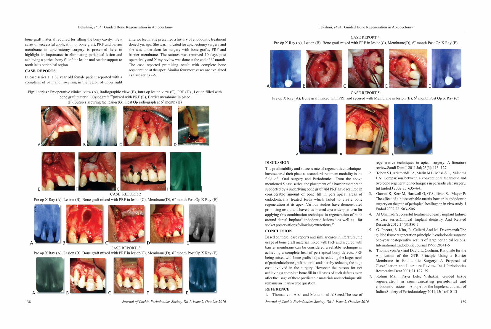
Journal of Cochin Periodontists Society-Vol 1, Issue 2, October 2016138 139Journal of Cochin Periodontists Society-Vol 1, Issue 2, October 2016
Lekshmi, et al.: Guided Bone Regeneration in ApicoectomyLekshmi, et al.: Guided Bone Regeneration in Apicoectomy
bone graft material required for filling the bony cavity. Few
cases of successful application of bone graft, PRF and barrier
membrane in apicoectomy surgery is presented here to
highlight its importance in eliminating periapical lesion and
achieving a perfect bony fill of the lesion and render support to
tooth in its periapical region.
CASE REPORTS
In case series 1, a 37 year old female patient reported with a
complaint of pain and swelling in the region of upper right
CASE REPORT: 2 thPre op X Ray (A), Lesion (B), Bone graft mixed with PRF in lesion(C), Membrane(D), 6 month Post Op X Ray (E)
CASE REPORT :3 thPre op X Ray (A), Lesion (B), Bone graft mixed with PRF in lesion(C), Membrane(D), 6 month Post Op X Ray (E)
anterior teeth. She presented a history of endodontic treatment
done 5 yrs ago. She was indicated for apicoectomy surgery and
she was undertaken for surgery with bone grafts, PRF and
barrier membrane. The sutures was removed 10 days post thoperatively and X ray review was done at the end of 6 month.
The case reported promising result with complete bone
regeneration at the apex. Similar four more cases are explained
as Case series 2-5.
Fig: 1 series : Preoperative clinical view (A), Radiographic view (B), Intra op lesion view (C), PRF (D) , Lesion filled with TMbone graft material (Osseograft )mixed with PRF (E), Barrier membrane in place
th(F), Sutures securing the lesion (G), Post Op radiograph at 6 month (H)
DISCUSSION
The predictability and success rate of regenerative techniques
have secured their place as a standard treatment modality in the
field of Oral surgery and Periodontics. From the above
mentioned 5 case series, the placement of a barrier membrane
supported by a underlying bone graft and PRF have resulted in
considerable amount of bone fill in peri apical areas of
endodontically treated teeth which failed to create bone
regeneration at its apex. Various studies have demonstrated
promising results and have thus opened up a wider platform for
applying this combination technique in regeneration of bone [4], [5]around dental implant endodontic lesions as well as for
[5]socket preservations following extractions.
CONCLUSION
Based on these case reports and similar cases in literature, the
usage of bone graft material mixed with PRF and secured with
barrier membrane can be considered a reliable technique in
achieving a complete heal of peri apical bony defects. PRF
being mixed with bone grafts helps in reducing the larger need
of particulate bone graft material and thereby reducing the huge
cost involved in the surgery. However the reason for not
achieving a complete bone fill in all cases of such defects even
after the usage of these predictable materials and technique still
remains an unanswered question.
REFERENCE
1. Thomas von Arx and Mohammed AlSaeed.The use of
CASE REPORT 4:thPre op X Ray (A), Lesion (B), Bone graft mixed with PRF in lesion(C), Membrane(D), 6 month Post Op X Ray (E)
CASE REPORT 5:thPre op X Ray (A), Bone graft mixed with PRF and secured with Membrane in lesion (B), 6 month Post Op X Ray (C)
regenerative techniques in apical surgery: A literature
review.Saudi Dent J. 2011 Jul; 23(3): 113–127.
2. Tobon S I, Arismendi J A, Marin M L, Mesa A L, Valencia
J A: Comparison between a conventional technique and
two bone regeneration techniques in periradicular surgery.
Int Endod J 2002.35: 635–641
3. Garrett K, Kerr M, Hartwell G, O’Sullivan S, Mayer P:
The effect of a bioresorbable matrix barrier in endodontic
surgery on the rate of periapical healing: an in vivo study. J
Endod 2002.28: 503–506
4. Al Ghamadi.Successful treatment of early implant failure:
A case series.Clinical Implant dentistry And Related
Research 2012;14(3):380-7
5. G. Pecora, S. Kim, R. Celletti And M. Davarpanah.The
guided tissue regeneration principle in endodontic surgery:
one-year postoperative results of large periapical lesions.
International Endodontic Journal 1995; 28: 41-6
6. Thomas von Arx and David L. Cochran. Rationale for the
Application of the GTR Principle Using a Barrier
Membrane in Endodontic Surgery: A Proposal of
Classification and Literature Review. Int J Periodontics
Restorative Dent 2001;21:127–39.
7. Rohini Mali, Priya Lele, Vishakha. Guided tissue
regeneration in communicating periodontal and
endodontic lesions – A hope for the hopeless. Journal of
Indian Society of Periodontology 2011;15(4):410-13
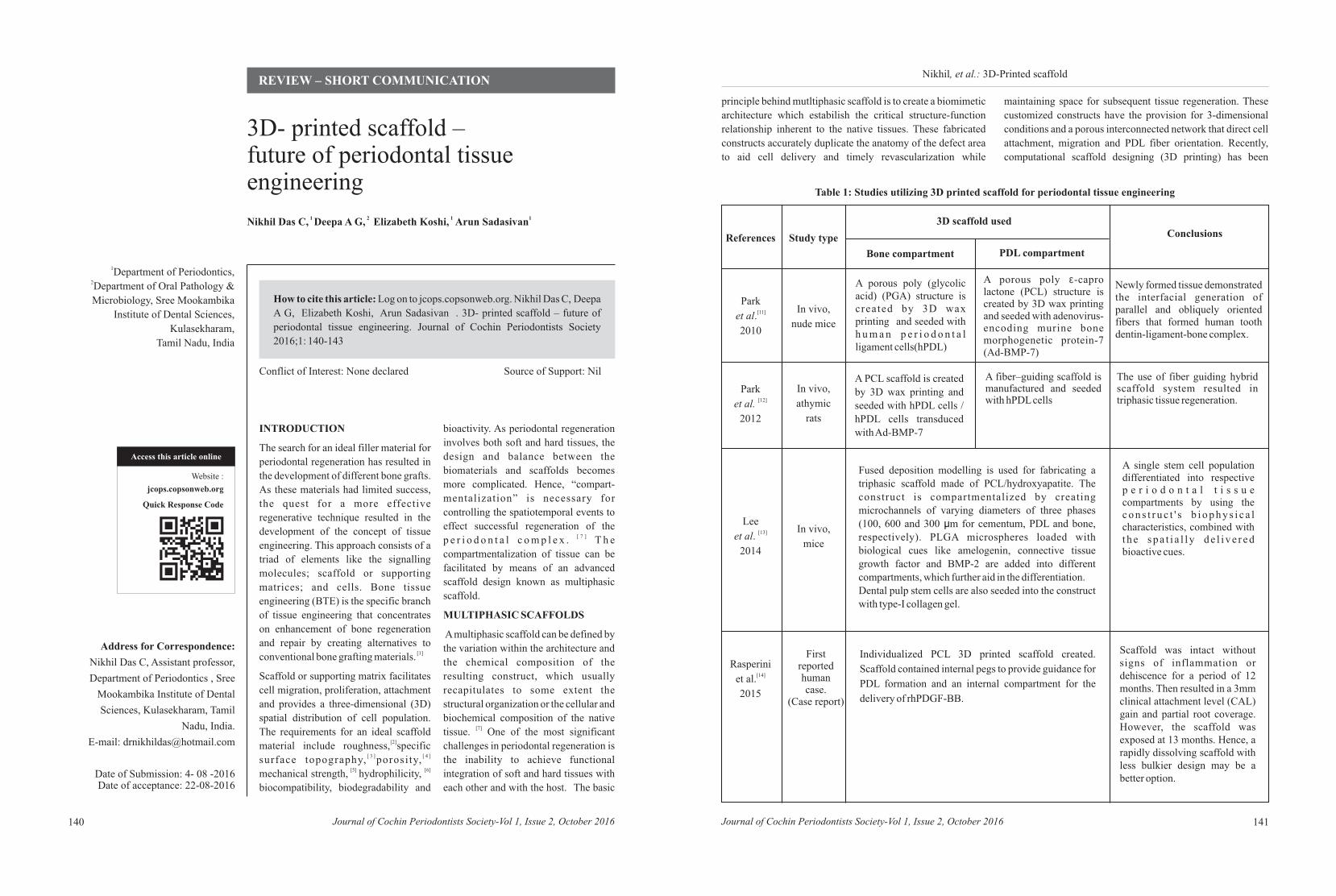
1 2 1 1 Nikhil Das C, Deepa A G, Elizabeth Koshi, Arun Sadasivan
How to cite this article: Log on to jcops.copsonweb.org. Nikhil Das C, Deepa A G, Elizabeth Koshi, Arun Sadasivan . 3D- printed scaffold – future of
periodontal tissue engineering. Journal of Cochin Periodontists Society
2016;1: 140-143
Conflict of Interest: None declared Source of Support: Nil
Journal of Cochin Periodontists Society-Vol 1, Issue 2, October 2016 Journal of Cochin Periodontists Society-Vol 1, Issue 2, October 2016 141140
Access this article online
Website :
jcops.copsonweb.org
Quick Response Code
1Department of Periodontics, 2Department of Oral Pathology &
Microbiology, Sree Mookambika
Institute of Dental Sciences,
Kulasekharam,
Tamil Nadu, India
Address for Correspondence:
Nikhil Das C, Assistant professor,
Department of Periodontics , Sree
Mookambika Institute of Dental
Sciences, Kulasekharam, Tamil
Nadu, India.
E-mail: [email protected]
Date of Submission: 4- 08 -2016Date of acceptance: 22-08-2016
REVIEW – SHORT COMMUNICATIONNikhil, et al.: 3D-Printed scaffold
3D- printed scaffold – future of periodontal tissue engineering
INTRODUCTION
The search for an ideal filler material for
periodontal regeneration has resulted in
the development of different bone grafts.
As these materials had limited success,
the quest for a more effective
regenerative technique resulted in the
development of the concept of tissue
engineering. This approach consists of a
triad of elements like the signalling
molecules; scaffold or supporting
matrices; and cells. Bone tissue
engineering (BTE) is the specific branch
of tissue engineering that concentrates
on enhancement of bone regeneration
and repair by creating alternatives to [1]conventional bone grafting materials.
Scaffold or supporting matrix facilitates
cell migration, proliferation, attachment
and provides a three-dimensional (3D)
spatial distribution of cell population.
The requirements for an ideal scaffold [2]material include roughness, specific
[ 3 ] [ 4 ]surface topography, porosity, [5] [6] mechanical strength, hydrophilicity,
biocompatibility, biodegradability and
bioactivity. As periodontal regeneration
involves both soft and hard tissues, the
design and balance between the
biomaterials and scaffolds becomes
more complicated. Hence, “compart-
mentalization” is necessary for
controlling the spatiotemporal events to
effect successful regeneration of the [ 7 ]p e r i o d o n t a l c o m p l e x . T h e
compartmentalization of tissue can be
facilitated by means of an advanced
scaffold design known as multiphasic
scaffold.
MULTIPHASIC SCAFFOLDS
A multiphasic scaffold can be defined by
the variation within the architecture and
the chemical composition of the
resulting construct, which usually
recapitulates to some extent the
structural organization or the cellular and
biochemical composition of the native [7]tissue. One of the most significant
challenges in periodontal regeneration is
the inability to achieve functional
integration of soft and hard tissues with
each other and with the host. The basic
principle behind mutltiphasic scaffold is to create a biomimetic
architecture which estabilish the critical structure-function
relationship inherent to the native tissues. These fabricated
constructs accurately duplicate the anatomy of the defect area
to aid cell delivery and timely revascularization while
maintaining space for subsequent tissue regeneration. These
customized constructs have the provision for 3-dimensional
conditions and a porous interconnected network that direct cell
attachment, migration and PDL fiber orientation. Recently,
computational scaffold designing (3D printing) has been
References Study type
3D scaffold used
Bone compartment PDL compartment
Conclusions
Park
et al.
2010
[11] In vivo,
nude mice
A porous poly (glycolic acid) (PGA) structure is created by 3D wax printing and seeded with h u m a n p e r i o d o n t a l ligament cells(hPDL)
A porous poly -capro lactone (PCL) structure is created by 3D wax printing and seeded with adenovirus- encoding murine bone morphogenetic protein-7 (Ad-BMP-7)
å Newly formed tissue demonstrated the interfacial generation of parallel and obliquely oriented fibers that formed human tooth dentin-ligament-bone complex.
Park
et al.
2012
[12]
In vivo,
athymic
rats
A PCL scaffold is created
by 3D wax printing and
seeded with hPDL cells /
hPDL cells transduced
with Ad-BMP-7
A fiber–guiding scaffold is manufactured and seeded with hPDL cells
The use of fiber guiding hybrid scaffold system resulted in triphasic tissue regeneration.
Lee[13]et al.
2014
In vivo,
mice
Fused deposition modelling is used for fabricating a triphasic scaffold made of PCL/hydroxyapatite. The construct is compartmentalized by creating microchannels of varying diameters of three phases (100, 600 and 300 ìm for cementum, PDL and bone, respectively). PLGA microspheres loaded with biological cues like amelogenin, connective tissue growth factor and BMP-2 are added into different compartments, which further aid in the differentiation.Dental pulp stem cells are also seeded into the construct with type-I collagen gel.
A single stem cell population differentiated into respective p e r i o d o n t a l t i s s u e compartments by using the c o n s t r u c t ' s b i o p h y s i c a l characteristics, combined with t he s p a t i a l l y de l i ve r ed bioactive cues.
Rasperini [14]et al.
2015
First reported human case.
(Case report)
Individualized PCL 3D printed scaffold created.
Scaffold contained internal pegs to provide guidance for
PDL formation and an internal compartment for the
delivery of rhPDGF-BB.
Scaffold was intact without signs of inflammation or dehiscence for a period of 12 months. Then resulted in a 3mm clinical attachment level (CAL) gain and partial root coverage. However, the scaffold was exposed at 13 months. Hence, a rapidly dissolving scaffold with less bulkier design may be a better option.
Table 1: Studies utilizing 3D printed scaffold for periodontal tissue engineering
Journal of Cochin Periodontists Society-Vol 1, Issue 2, October 2016 Journal of Cochin Periodontists Society-Vol 1, Issue 2, October 2016 143142
Nikhil, et al.: 3D-Printed scaffoldNikhil, et al.: 3D-Printed scaffold
introduced for the production of the multicompartmental
scaffold.
3D PRINTED SCAFFOLDS
3D printing technologies offer a promising platform for
periodontal tissue regeneration, which provides customization
to match the periodontal defect shape, size and topography.
Conventional techniques utilized in 3D fabrication of scaffold
are gas foaming, freeze drying, particle leaching, melt molding, [8]phase separation, and fiber bonding. But disadvantages like
inconsistency in porosity, configuration and interconnectivity [9]made the researchers to focus on more precise technologies.
Some of these newly introduced 3D printing technologies are
solid freeform fabrication (SFF), inkjet printing, selective
laser sintering (SLS), stereolithography (STL), and extrusion [10]printing (e.g., fused deposition modelling-FDM).
3D PRINTED SCAFFOLD FABRICATION
Good quality images of the periodontal defect are generated
with the help of computed tomography scanning after which a
3D model of the site created using CAD program. The 3D
generated image will be converted into a stereolithography
(STL) format to produce the custom-fit substitute for the
defect. STL is a technique that converts CAD design into a solid
object (bone borders and PDL interface) by means of a
combination of laser photochemistry and software technology.
After capturing the periodontal defect image dataset, the
exterior borders of the bone region and PDL interface will then
be created using periodontal defect topography in the STL
surface image. Designed architectures were then subtracted
from STL- formatted, 3-D surface topological defect to
generate an anatomically fit scaffold (eg: Magics 15,
Materialise NV). The final designed scaffold is then assessed
for adaptation to the defect with the help of computed
tomography in a 3D prototyped model.
3D PRINTED SCAFFOLD IN PERIODONTAL
REGENERATION
Applications of 3D printed scaffolds have been recently
studied in various periodontal applications. Studies utilizing
3D printed scaffold for periodontal tissue engineering are
summarized in Table 1.
LIMITATIONS AND FUTURE DIRECTIONS
Main limitations of these multicompartmental scaffolds
include increased stiffness, which make it difficult to precisely
adapt to the defect configuration, slower scaffold resorption
profile that may impede bone regeneration, lack of clarity in
perpendicular periodontal fiber formation and its insertion into
the newly formed cementum, technique sensitivity and cost
effectiveness.
Periodontal literature on 3D printed scaffolds and related
biomaterials are limited. So long term multicenter randomized,
clinical trials with adequate number of patients are needed to
validate the clinical use of these scaffolds. Future researches
should focus on developing thinner, easily adaptable and faster
degrading scaffold.
CONCLUSION
The concept of multiphasic constructs for periodontal tissue
engineering is in the preliminary stages of development.
Translation of the favourable preclinical results into clinical
realities may be a major breakthrough in the field of
regenerative periodontal therapy.
REFERENCES
1. O Keefe RJ, Mao J. Bone tissue engineering and
regeneration: from discovery to the clinic-an overview.
Tissue Eng Part B Rev 2011;17:389-92.
2. Hoffmann W, Bormann T, Rossi A et al. Rapid prototyped
porous nickel–titanium scaffolds as bone substitutes. J
Tissue Eng. 2014;5:1-14.
3. Cheng K, Kisaalita WS. Exploring cellular adhesion and
differentiation in a micro-/nano-hybrid polymer scaffold.
Biotehnol Prog 2010;26(3): 838–46.
4. Costa PF, Vaquette C, Zhang Q, Reis RL, Ivanovski S,
Hutmacher DW. Advanced tissue engineering scaffold
design for regeneration of the complex hierarchical
periodontal structure. J Clin Periodontol 2014;41:283-94.
5. O’Brien FJ. Biomaterials & scaffolds for tissue
engineering. Materials Today 2011;14:88–95.
6. Goddard JM, Hotchkiss JH. Polymer surface modification
for the attachment of bioactive compounds. Prog Polym
Sci 2007;32:698-725.
7. Ivanovski S, Vaquette C, Gronthos S, Hutmacher DW,
Bartold PM. Multiphasic scaffolds for periodontal tissue
engineering. J Dent Res 2014; 93:1212–21.
8. Kinoshita Y, Maed H. Recent developments of functional
scaffolds for cranio maxillofacial bone tissue engineering
a p p l i c a t i o n s .
, , , ,
. Initial cell pre-cultivation can maximize ECM
mineralization by human mesenchymal stem cells on silk
fibroin scaffolds. Acta Biomater 2011;7:2218-28.
10. Obregon F, Vaquette C, Ivanovski S, Hutmacher DW,
Bertassoni LE. Three-dimensional bioprinting for
regenerative dentistry and craniofacial tissue engineering.
J Dent Res. 2015;94(9 Suppl): 143S–152S.
11. Park CH, Rios HF, Jin Q, Bland ME, Flanagan CL,
Hollister SJ et al. Biomimetic hybrid scaffolds for
T h e S c i e n t i f i c Wo r l d J o u r n a l
2013;2013:863157.
9.
Thimm BW Wust S Hofmann S Hagenmuller H Muller
R
engineering human tooth-ligament interfaces.
Biomaterials 2010;31:5945-52.
12. Park CH, Rios HF, Jin Q, Sugai JV, Padial-Molina M, Taut
AD et al. Tissue engineering bone-ligament complexes
using fiber-guiding scaffolds. Biomaterials 2012; 33:137-
145.
13. Lee CH, Hajibandeh J, Suzuki T, Fan A, Shang P, Mao JJ.
Three-dimensional printed multiphase scaffolds for
regeneration of periodontium complex. Tissue Eng Part A
2014;20:1342-51.
14. Rasperini G, Pilipchuk SP, Flanagan CL, Park CH, Pagni
G, Hollister SJ et al. 3D printed bioresorbable scaffold for
periodontal repair. J Dent Res 2015;94:153S-7S.
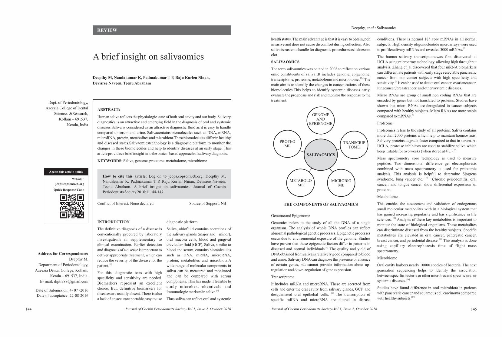
Deepthy M, Nandakumar K, Padmakumar T P,
Devisree Naveen, Teenu Abraham
Raju Kurien Ninan,
How to cite this article: Log on to jcops.copsonweb.org. Deepthy M,
Nandakumar K, Padmakumar T P, Raju Kurian Ninan, Devisree Naveen,
Teenu Abraham. A brief insight on salivaomics. Journal of Cochin
Periodontists Society 2016;1: 144-147
Conflict of Interest: None declared Source of Support: Nil
Journal of Cochin Periodontists Society-Vol 1, Issue 2, October 2016 Journal of Cochin Periodontists Society-Vol 1, Issue 2, October 2016144
Access this article online
Website :
jcops.copsonweb.org
Quick Response Code
Dept. of Periodontology,
Azeezia College of Dental
Sciences &Research,
Kollam – 691537,
Kerala, India
Address for Correspondence:
Deepthy M,
Department of Periodontology,
Azeezia Dental College, Kollam,
Kerala – 691537, India.
E- mail: [email protected]
Date of Submission: 4- 07 -2016
Date of acceptance: 22-08-2016
REVIEWDeepthy, et al.: Salivaomics
A brief insight on salivaomics
ABSTRACT:
Human saliva reflects the physiologic state of both oral cavity and our body. Salivary
diagnostics is an attractive and emerging field in the diagnosis of oral and systemic
diseases.Saliva is considered as an attractive diagnostic fluid as it is easy to handle
compared to serum and urine. Salivacontains biomolecules such as DNA, mRNA,
microRNA, protein, metabolites and microbiota.Thesebiomolecules differ in healthy
and diseased states.Salivaomicstechnology is a diagnostic platform to monitor the
changes in these biomolecules and help to identify diseases at an early stage. This
article provides a brief insight in to the omics- based approach of salivary diagnosis.
KEYWORDS: Saliva, genome, proteome, metabolome, microbiome
INTRODUCTION
The definitive diagnosis of a disease is
conventionally procured by laboratory
investigations in supplementary to
clinical examination. Earlier detection
and diagnosis of a disease is important to
deliver appropriate treatment, which can
reduce the severity of the disease for the [1]patient.
For this, diagnostic tests with high
specificity and sensitivity are needed.
Biomarkers represent an excellent
choice. But, definitive biomarkers for
diseases are usually absent. There is also
a lack of an accurate portable easy to use
diagnostic platform.
Saliva, abiofluid contains secretions of
the salivary glands (major and minor),
oral mucosa cells, blood and gingival
crevicular fluid (GCF). Saliva, similar to
blood and serum, contains biomolecules
such as DNA, mRNA, microRNA,
protein, metabolites and microbiota.A
wide range of molecular components in
saliva can be measured and monitored
and can be compared with serum
components. This has made it feasible to
study microbes, chemicals and [2]immunologic markers in saliva.
Thus saliva can reflect oral and systemic
health status. The main advantage is that it is easy to obtain, non
invasive and does not cause discomfort during collection. Also
saliva is easier to handle for diagnostic procedures as it does not
clot.
SALIVAOMICS
The term salivaomics was coined in 2008 to reflect on various
omic constituents of saliva .It includes genome, epigenome, [3,4]transcriptome, proteome, metabolome and microbiome . The
main aim is to identify the changes in concentrations of these
biomolecules.This helps to identify systemic diseases early,
evaluate the prognosis and risk and monitor the response to the
treatment.
145
THE COMPONENTS OF SALIVAOMICS
GENOME AND
EPIGENOME
TRANSCRIPTOME
MICROBIOME
METABOLOME
PROTEOME
SALIVAOMICS
Genome and Epigenome
Genomics refers to the study of all the DNA of a single
organism. The analysis of whole DNA profiles can reflect
abnormal pathological genetic processes. Epigenetic processes
occur due to environmental exposure of the genome. Studies
have proven that these epigenetic factors differ in patterns in [5]diseased and normal individuals. The quality and yield of
DNA obtained from saliva is relatively good compared to blood
and urine. Salivary DNA can diagnose the presence or absence
of certain genes, but cannot provide information about up-
regulation and down-regulation of gene expression.
Transcriptome
It includes mRNA and microRNA. These are secreted from
cells and enter the oral cavity from salivary glands, GCF, and [6] desquamated oral epithelial cells. The transcription of
specific mRNA and microRNA are altered in disease
conditions. There is normal 185 core mRNAs in all normal
subjects. High density oligonucleotide microarrays were used [7]to profile salivary mRNAs and revealed 3000 mRNAs.
The human salivary transcriptomewas first discovered at
UCLA using microarray technology, allowing high throughput
analysis. Zhang et_al discovered that four mRNA biomarkers
can differentiate patients with early stage resectable pancreatic
cancer from non-cancer subjects with high specificity and [8]sensitivity. It can be used to detect oral cancer, ovariancancer,
lungcancer, breastcancer, and other systemic diseases.
Micro RNAs are group of small non coding RNAs that are
encoded by genes but not translated to proteins. Studies have
shown that micro RNAs are deregulated in cancer subjects
compared with healthy subjects. Micro RNAs are more stable [8]compared to mRNAs.
Proteome
Proteomics refers to the study of all proteins. Saliva contains
more than 2000 proteins which help to maintain homeostasis.
Salivary proteins degrade faster compared to that in serum. At
UCLA, protease inhibitors are used to stabilize saliva which o [9]keep it stable for two weeks (when stored at 4 C).
Mass spectrometry core technology is used to measure
peptides. Two dimensional difference gel electrophoresis
combined with mass spectrometry is used for proteomic
analysis. This analysis is helpful to determine Sjogrens [10, 11]syndrome, lung cancer etc. Chronic periodontitis, oral
cancer, and tongue cancer show differential expression of
proteins.
Metabolome
This enables the assessment and validation of endogenous
small molecular metabolites with in a biological system that
has gained increasing popularity and has significance in life [12]sciences. Analysis of these key metabolites is important to
monitor the state of biological organisms. These metabolites
can discriminate diseased from the healthy subjects. Specific
metabolites are elevated in oral cancer, pancreatic cancer, [13]breast cancer, and periodontal disease. This analysis is done
using capillary electrophoresis time of flight mass
spectrometry.
Microbiome
Oral cavity harbors nearly 10000 species of bacteria. The next
generation sequencing helps to identify the association
between specific bacteria or other microbes and specific oral or [14]systemic diseases.
Studies have found difference in oral microbiota in patients
with pancreatic cancer and squamous cell carcinoma compared [14]with healthy subjects.
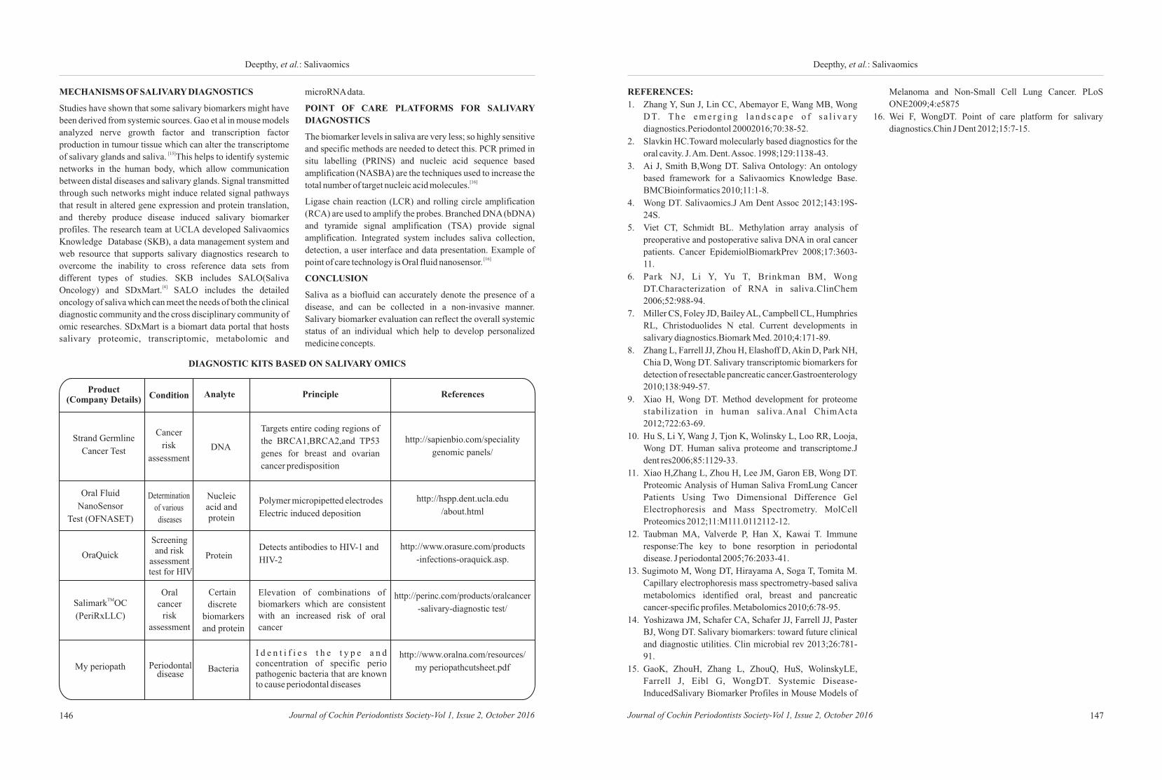
Journal of Cochin Periodontists Society-Vol 1, Issue 2, October 2016 Journal of Cochin Periodontists Society-Vol 1, Issue 2, October 2016146
Deepthy, et al.: Salivaomics
147
Deepthy, et al.: Salivaomics
MECHANISMS OF SALIVARY DIAGNOSTICS
Studies have shown that some salivary biomarkers might have
been derived from systemic sources. Gao et al in mouse models
analyzed nerve growth factor and transcription factor
production in tumour tissue which can alter the transcriptome [15]of salivary glands and saliva. This helps to identify systemic
networks in the human body, which allow communication
between distal diseases and salivary glands. Signal transmitted
through such networks might induce related signal pathways
that result in altered gene expression and protein translation,
and thereby produce disease induced salivary biomarker
profiles. The research team at UCLA developed Salivaomics
Knowledge Database (SKB), a data management system and
web resource that supports salivary diagnostics research to
overcome the inability to cross reference data sets from
different types of studies. SKB includes SALO(Saliva [4]Oncology) and SDxMart. SALO includes the detailed
oncology of saliva which can meet the needs of both the clinical
diagnostic community and the cross disciplinary community of
omic researches. SDxMart is a biomart data portal that hosts
salivary proteomic, transcriptomic, metabolomic and
DIAGNOSTIC KITS BASED ON SALIVARY OMICS
Product (Company Details) Condition Analyte Principle References
Strand Germline
Cancer Test
Cancer
risk
assessmentDNA
Targets entire coding regions of
the BRCA1,BRCA2,and TP53
genes for breast and ovarian
cancer predisposition
http://sapienbio.com/speciality
genomic panels/
http://hspp.dent.ucla.edu
/about.html
http://www.orasure.com/products
-infections-oraquick.asp.
http://perinc.com/products/oralcancer
-salivary-diagnostic test/
http://www.oralna.com/resources/
my periopathcutsheet.pdf
I d e n t i f i e s t h e t y p e a n d concentration of specific perio pathogenic bacteria that are known to cause periodontal diseases
Elevation of combinations of biomarkers which are consistent with an increased risk of oral cancer
Detects antibodies to HIV-1 and
HIV-2
Polymer micropipetted electrodes
Electric induced deposition
Nucleic acid and protein
Protein
Certain
discrete
biomarkers
and protein
BacteriaPeriodontal disease
Oral cancer
risk assessment
Screening and risk
assessment test for HIV
Determination
of various
diseases
Oral Fluid
NanoSensor
Test (OFNASET)
OraQuick
TMSalimark OC
(PeriRxLLC)
My periopath
microRNA data.
POINT OF CARE PLATFORMS FOR SALIVARY
DIAGNOSTICS
The biomarker levels in saliva are very less; so highly sensitive
and specific methods are needed to detect this. PCR primed in
situ labelling (PRINS) and nucleic acid sequence based
amplification (NASBA) are the techniques used to increase the [16]total number of target nucleic acid molecules.
Ligase chain reaction (LCR) and rolling circle amplification
(RCA) are used to amplify the probes. Branched DNA (bDNA)
and tyramide signal amplification (TSA) provide signal
amplification. Integrated system includes saliva collection,
detection, a user interface and data presentation. Example of [16]point of care technology is Oral fluid nanosensor.
CONCLUSION
Saliva as a biofluid can accurately denote the presence of a
disease, and can be collected in a non-invasive manner.
Salivary biomarker evaluation can reflect the overall systemic
status of an individual which help to develop personalized
medicine concepts.
REFERENCES:
1. Zhang Y, Sun J, Lin CC, Abemayor E, Wang MB, Wong
D T. T h e e m e rg i n g l a n d s c a p e o f s a l i v a r y
diagnostics.Periodontol 20002016;70:38-52.
2. Slavkin HC.Toward molecularly based diagnostics for the
oral cavity. J. Am. Dent. Assoc. 1998;129:1138-43.
3. Ai J, Smith B,Wong DT. Saliva Ontology: An ontology
based framework for a Salivaomics Knowledge Base.
BMCBioinformatics 2010;11:1-8.
4. Wong DT. Salivaomics.J Am Dent Assoc 2012;143:19S-
24S.
5. Viet CT, Schmidt BL. Methylation array analysis of
preoperative and postoperative saliva DNA in oral cancer
patients. Cancer EpidemiolBiomarkPrev 2008;17:3603-
11.
6. Park NJ, Li Y, Yu T, Brinkman BM, Wong
DT.Characterization of RNA in saliva.ClinChem
2006;52:988-94.
7. Miller CS, Foley JD, Bailey AL, Campbell CL, Humphries
RL, Christoduolides N etal. Current developments in
salivary diagnostics.Biomark Med. 2010;4:171-89.
8. Zhang L, Farrell JJ, Zhou H, Elashoff D, Akin D, Park NH,
Chia D, Wong DT. Salivary transcriptomic biomarkers for
detection of resectable pancreatic cancer.Gastroenterology
2010;138:949-57.
9. Xiao H, Wong DT. Method development for proteome
stabilization in human saliva.Anal ChimActa
2012;722:63-69.
10. Hu S, Li Y, Wang J, Tjon K, Wolinsky L, Loo RR, Looja,
Wong DT. Human saliva proteome and transcriptome.J
dent res2006;85:1129-33.
11. Xiao H,Zhang L, Zhou H, Lee JM, Garon EB, Wong DT.
Proteomic Analysis of Human Saliva FromLung Cancer
Patients Using Two Dimensional Difference Gel
Electrophoresis and Mass Spectrometry. MolCell
Proteomics 2012;11:M111.0112112-12.
12. Taubman MA, Valverde P, Han X, Kawai T. Immune
response:The key to bone resorption in periodontal
disease. J periodontal 2005;76:2033-41.
13. Sugimoto M, Wong DT, Hirayama A, Soga T, Tomita M.
Capillary electrophoresis mass spectrometry-based saliva
metabolomics identified oral, breast and pancreatic
cancer-specific profiles. Metabolomics 2010;6:78-95.
14. Yoshizawa JM, Schafer CA, Schafer JJ, Farrell JJ, Paster
BJ, Wong DT. Salivary biomarkers: toward future clinical
and diagnostic utilities. Clin microbial rev 2013;26:781-
91.
15. GaoK, ZhouH, Zhang L, ZhouQ, HuS, WolinskyLE,
Farrell J, Eibl G, WongDT. Systemic Disease-
InducedSalivary Biomarker Profiles in Mouse Models of
Melanoma and Non-Small Cell Lung Cancer. PLoS
ONE2009;4:e5875
16. Wei F, WongDT. Point of care platform for salivary
diagnostics.Chin J Dent 2012;15:7-15.
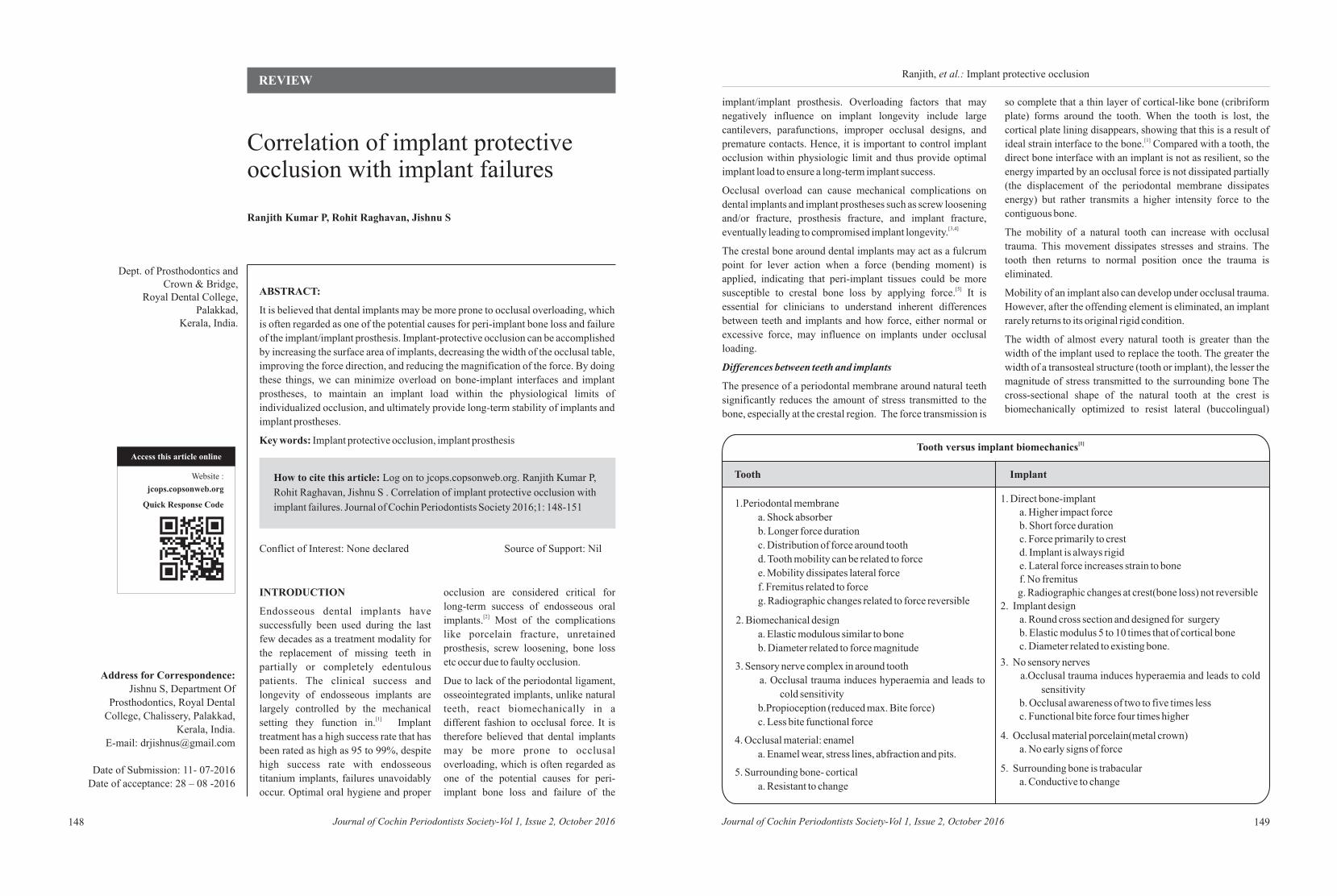
Ranjith Kumar P, Rohit Raghavan, Jishnu S
How to cite this article: Log on to jcops.copsonweb.org. Ranjith Kumar P,
Rohit Raghavan, Jishnu S . Correlation of implant protective occlusion with
implant failures. Journal of Cochin Periodontists Society 2016;1: 148-151
Conflict of Interest: None declared Source of Support: Nil
Journal of Cochin Periodontists Society-Vol 1, Issue 2, October 2016 Journal of Cochin Periodontists Society-Vol 1, Issue 2, October 2016148
Access this article online
Website :
jcops.copsonweb.org
Quick Response Code
Dept. of Prosthodontics and Crown & Bridge,
Royal Dental College, Palakkad,
Kerala, India.
Address for Correspondence:
Jishnu S, Department Of
Prosthodontics, Royal Dental
College, Chalissery, Palakkad,
Kerala, India.
E-mail: [email protected]
Date of Submission: 11- 07-2016
Date of acceptance: 28 – 08 -2016
REVIEW
ABSTRACT:
It is believed that dental implants may be more prone to occlusal overloading, which
is often regarded as one of the potential causes for peri-implant bone loss and failure
of the implant/implant prosthesis. Implant-protective occlusion can be accomplished
by increasing the surface area of implants, decreasing the width of the occlusal table,
improving the force direction, and reducing the magnification of the force. By doing
these things, we can minimize overload on bone-implant interfaces and implant
prostheses, to maintain an implant load within the physiological limits of
individualized occlusion, and ultimately provide long-term stability of implants and
implant prostheses.
Key words: Implant protective occlusion, implant prosthesis
149
Correlation of implant protective occlusion with implant failures
Ranjith, et al.: Implant protective occlusion
INTRODUCTION
Endosseous dental implants have
successfully been used during the last
few decades as a treatment modality for
the replacement of missing teeth in
partially or completely edentulous
patients. The clinical success and
longevity of endosseous implants are
largely controlled by the mechanical [1]setting they function in. Implant
treatment has a high success rate that has
been rated as high as 95 to 99%, despite
high success rate with endosseous
titanium implants, failures unavoidably
occur. Optimal oral hygiene and proper
occlusion are considered critical for
long-term success of endosseous oral [2]implants. Most of the complications
like porcelain fracture, unretained
prosthesis, screw loosening, bone loss
etc occur due to faulty occlusion.
Due to lack of the periodontal ligament,
osseointegrated implants, unlike natural
teeth, react biomechanically in a
different fashion to occlusal force. It is
therefore believed that dental implants
may be more prone to occlusal
overloading, which is often regarded as
one of the potential causes for peri-
implant bone loss and failure of the
implant/implant prosthesis. Overloading factors that may
negatively influence on implant longevity include large
cantilevers, parafunctions, improper occlusal designs, and
premature contacts. Hence, it is important to control implant
occlusion within physiologic limit and thus provide optimal
implant load to ensure a long-term implant success.
Occlusal overload can cause mechanical complications on
dental implants and implant prostheses such as screw loosening
and/or fracture, prosthesis fracture, and implant fracture, [3,4]eventually leading to compromised implant longevity.
The crestal bone around dental implants may act as a fulcrum
point for lever action when a force (bending moment) is
applied, indicating that peri-implant tissues could be more [5]susceptible to crestal bone loss by applying force. It is
essential for clinicians to understand inherent differences
between teeth and implants and how force, either normal or
excessive force, may influence on implants under occlusal
loading.
Differences between teeth and implants
The presence of a periodontal membrane around natural teeth
significantly reduces the amount of stress transmitted to the
bone, especially at the crestal region. The force transmission is
so complete that a thin layer of cortical-like bone (cribriform
plate) forms around the tooth. When the tooth is lost, the
cortical plate lining disappears, showing that this is a result of [1]ideal strain interface to the bone. Compared with a tooth, the
direct bone interface with an implant is not as resilient, so the
energy imparted by an occlusal force is not dissipated partially
(the displacement of the periodontal membrane dissipates
energy) but rather transmits a higher intensity force to the
contiguous bone.
The mobility of a natural tooth can increase with occlusal
trauma. This movement dissipates stresses and strains. The
tooth then returns to normal position once the trauma is
eliminated.
Mobility of an implant also can develop under occlusal trauma.
However, after the offending element is eliminated, an implant
rarely returns to its original rigid condition.
The width of almost every natural tooth is greater than the
width of the implant used to replace the tooth. The greater the
width of a transosteal structure (tooth or implant), the lesser the
magnitude of stress transmitted to the surrounding bone The
cross-sectional shape of the natural tooth at the crest is
biomechanically optimized to resist lateral (buccolingual)
[1]Tooth versus implant biomechanics
1.Periodontal membrane
a. Shock absorber
b. Longer force duration
c. Distribution of force around tooth
d. Tooth mobility can be related to force
e. Mobility dissipates lateral force
f. Fremitus related to force
g. Radiographic changes related to force reversible
2. Biomechanical design
a. Elastic modulous similar to bone
b. Diameter related to force magnitude
3. Sensory nerve complex in around tooth
a. Occlusal trauma induces hyperaemia and leads to
cold sensitivity
b.Propioception (reduced max. Bite force)
c. Less bite functional force
4. Occlusal material: enamel
a. Enamel wear, stress lines, abfraction and pits.
5. Surrounding bone- cortical
a. Resistant to change
1. Direct bone-implant a. Higher impact force b. Short force duration c. Force primarily to crest d. Implant is always rigid e. Lateral force increases strain to bone f. No fremitusg. Radiographic changes at crest(bone loss) not reversible
2. Implant design a. Round cross section and designed for surgery b. Elastic modulus 5 to 10 times that of cortical bone c. Diameter related to existing bone.
3. No sensory nerves a.Occlusal trauma induces hyperaemia and leads to cold
sensitivity b. Occlusal awareness of two to five times less c. Functional bite force four times higher
4. Occlusal material porcelain(metal crown) a. No early signs of force
5. Surrounding bone is trabacular a. Conductive to change
Tooth Implant
Journal of Cochin Periodontists Society-Vol 1, Issue 2, October 2016 Journal of Cochin Periodontists Society-Vol 1, Issue 2, October 2016150 151
Ranjith, et al.: Implant protective occlusionRanjith, et al.: Implant protective occlusion
loads because of the bending fracture resistance (moment of
inertia) of the tooth and the direction of occlusal forces.
After osseointegration, mechanical stresses and strains beyond
the physical limits of hard tissue have been suggested as the
primary cause of both initial and long-term bone loss around
implants. If the occlusal overload is not corrected, crestal bone
loss will continue until the implant fails. Occlusal overload is
often regarded as one of the main causes for peri-implant bone
loss and implant prosthesis failure, because it can cause crestal
bone loss, thus increasing the anaerobic sulcus depth and peri- [7]implant disease states if patients oral hygiene status is poor.
Differences in the magnitude, duration, direction, and
frequency of the applied occlusal load and the tolerance
threshold of the host are the underlying reasons for the
conflicting observations obtained through the clinical studies
on occlusal overload. Possible mechanisms of why occlusal
overload can lead to peri-implantitis are conceivable. Implants
are considered less tolerable to non-axial occlusal load
compared to teeth because of a lack of a periodontal ligament.
Finite element studies have suggested that the occlusal load is
concentrated at the implant marginal bone. Bone remodels in
response to the strain. Excessive stress can cause microfracture
within bone and eventual bone loss. [1]Consequences of biomechanical overload
1. Porcelain fracture
2. Prosthesis fracture
3. Uncemented or unretained restoration
4. Screw loosening
5. Early crestal bone loss
6. Intermediate to late implant failure
7. Peri implant disease
8. Component fracture
Principles of implant protective occlusion
1. No premature occlusal contact: During maximum
intercuspation, no occlusal contact should be premature.
Occlusal prematurity between maximum intercuspation and
centric relation occlusion should be taken into consideration
especially on an implant supported prostheses. This is because,
non-mobile implants bear the entire load of the prosthesis when
it comes in contact with the mobile natural teeth, hence during
the occlusal adjustment between implants and natural teeth,
premature occlusal contacts on the implants can occur as the [6]natural teeth can move away from the centric during function.
2. Influence of surface area: Sufficient surface area is required
to withstand the load transmitted to the prosthesis therefore
when an implant of decreased surface area, subjected to
increased load in magnitude, direction or duration, the stress
and strain in the interfacial tissue will increase. This can be
minimized by placing additional implants in the region of
concern, ridge augmentation, reduce crown height or by
increasing the implant width. Bidez et al have reported a study
showing that, forces distributed over 3 abutments results in less
stress on the crestal bone compared to 2 abutments
3. Mutually protected articulation: When the natural canines
are present, during excursions it allows the teeth to distribute
horizontal load and also the posterior tooth to disocclude. This
concept is known as canine guidance or mutually protected
articulation. However, there should be no contact on the
implant crown during excursion to the opposing side and also
during protrusion. The anterior guidance of implant prosthesis
with anterior implant should be shallow. This is because, the
steeper the incisal guidance the greater the force on the anterior
implants. Weinberg et al have reported a study stating, every
10- degree change in the angle of disclusion, there is a 30 % [1]difference in the load.
4. Cusp angle of crown: Natural dentition has steep cuspal
inclination whereas in denture teeth, the cuspal inclination
given is 30 %. Cusp inclination has been found to produce a [6]high level of torque . For every 10° increase in cusp
inclination, there is an approximately 30% increase in torque.
Weinberg et al in 1995 have reported a study regarding the
torque of a gold screw, abutment screw, and implant. They have
concluded that, the cuspal inclination produces the most
torque, followed by maxillary horizontal implant offset, while
implant inclination and apical implant offset produce minimal
torque. Occlusal contact over an implant crown should be on a
flat surface perpendicular to implant body. This is achieved by
increasing 2 to 3mm of the width of the central groove in the
posterior implant crowns and the opposing cusp is recontoured
to occlude the central fossa directly over the implant body.
5. Implant body angle to occlusal load: There can be different
impact on the bone and implant interface based on the direction
of the load applied even if it’s of same magnitude of force,
however implant is mainly designed for long axis load. A study
was reported by Binderman in 1970, where 50 endosteal
implant designs were assessed and found that all the design
sustained lesser under a long axis load. The greater the angle of
load to the implant long axis, the greater the compressive,
tensile and shear stresses which leads to bone loss and
unsuccessful bone re growth.
6. Crown height: Implant crown height is often greater than the
natural anatomical crown. As the implant crown height
becomes greater, the crestal moment with any lateral
component of force also becomes greater. Therefore any
harmful effect of any feebly selected cusp angle, angled
implant body, or angled load to the crown will be magnified by
the crown height measurements.
7. Cantilever: Cantilevers with unfavourable crown or implant
ratio, increase the amount of stress to the implant. These can
further lead to peri implant bone loss and prosthesis failure. The
magnitude of load obtained by the implants is approximately
proportional to the length of the cantilevers but it also varies
with the implant number, spacing, and location. Long
cantilevers are correlated with increase crestal bone lost.
8. Occlusal contact position: Occlusal contact position
determines the direction of force especially during
parafunctional activity. In different theories, the number of
occlusal contact varies. Occlusal theory by Peter K Thomas
suggest that there should be tripod contact on each occluding
cusp, on each marginal ridge and central fossa with 18 and 15
individual occlusal contacts on a mandibular and maxillary
molars whereas, the other oclusal contact scheme indicates
that, number of occlusal contact for molars can be reduced.
9. Implant crown contour: In maxilla, the edentulous ridge
resorbs gradually in the medial direction whereas in posterior
mandible, the resorption occurs in lingual direction. Center of
implant is placed in the center of the edentulous ridge because
the ridge resorbs lingually with resorption hence the implant is
mostly not kept under the buccal cusp tip but near the central
fossa or more lingually, under the lingual cusp of the natural
tooth. The size of the implant body which is the buccolingual [1]dimension is smaller than the natural tooth.
10. Occlusal material: Occlusal material fracture is one of the
most common complications of implant restoration therefore
consideration of the occlusal material restoration is very
essential for each patient. Occlusal material may be evaluated
by esthetic, impact force, static load, chewing efficiency,
fracture, wear, inter arch space requirement, and accuracy of [3]casting.
CONCLUSION
The objectives of implant occlusion are to minimize overload
on the bone–implant interface and implant prosthesis, to
maintain implant load within the physiological limits of
individualized occlusion, and finally to provide long-term
stability of implants and implant prostheses. To accomplish
these objectives, increased support area, improved force
direction,
and reduced force magnifications are indispensable factors in
implant occlusion.
REFERENCES
1. Dental Implant Prosthetics Carl E. Misch. Implant
Dentistry. 2005;14(1):11-12.
2. Kozlovsky A, Tal H, Laufer B, Leshem R, Rohrer M,
Weinreb M et al. Impact of implant overloading on the
peri-implant bone in inflamed and non-inflamed peri-
implant mucosa. Clin Oral Implants Res.2007;18(5):601-
10.
3. Misch CE, Bidez MW: Implant protected occlusion: a
biomechanical rationale, Compend Cont’m Dent Educ 15:
1330-1343, 1994.
4. Swaminathan Y. Implant Protected Occlusion. IOSR
Journal of Dental and Medical Sciences. 2013;11(3):20-
25.
5. Kim Y, Oh T-J, Misch CE, Wang H-L, Occlusal
considerations in implant therapy: clinical guidelines with
biomechanical rationale, Clin. Oral Impl, 16, 2005, 26–35.
6. Y Y Chen, C L Kuan, Y B Wang, Implant occlusion:
biomechanical considerations for implant supported
prostheses, J Dent Sci, 3(2), 2008, 65 -74.
7. Miyata T, Kobayashi Y, Araki H, Ohto T, Shin K, The
influence of controlled occlusal overload on peri-implant
tissue. Part 3: A histologic study in monkeys, Int J Oral
Maxillofac Implants, 15, 2000, 425-31.
Krishna V. Vijay, Maya George
How to cite this article: Log on to jcops.copsonweb.org. Krishna V. Vijay,
Maya George. Advances in dental local anesthesia techniques and devices.
Journal of Cochin Periodontists Society 2016;1:152-155
Conflict of Interest: None declared Source of Support: Nil
Journal of Cochin Periodontists Society-Vol 1, Issue 2, October 2016 Journal of Cochin Periodontists Society-Vol 1, Issue 2, October 2016152
Access this article online
Website :
jcops.copsonweb.org
Quick Response Code
Department Of Periodontics,
Amrita School of Dentistry,
Kochi, Kerala, India
Address for Correspondence:
Krishna V. Vijay, Department Of
Periodontics, Amrita School Of
Dentistry, Kochi, Kerala, India.
E- mail: [email protected]
Date of Submission: 4- 07-2016
Date of acceptance: 22-08-2016
REVIEW
ABSTRACT:
Pain and its successful management have been one of the cornerstones of Dentistry
worldwide since time immemorial. Although local anesthesia remains the backbone
of pain control in dentistry, researches are going on to seek new and better means of
managing the pain. It is imperative on our part to update our knowledge and skills in
using newer alternatives in pain control and management and ways of administering
them. Most of the researches are focused on improvement in the area of anesthetic
agents, delivery devices and techniques involved. Newer technologies have been
developed that can assist the dentist in providing enhanced pain relief with reduced
injection pain and fewer adverse effects. This overview will enlighten the practicing
dentists regarding newer devices and methods of rendering pain control compared
with the earlier used ones on the basis of research and clinical studies available.
Keywords:- Local anesthesia, CCLAD, intra-osseous anesthesia
153
Krishna, et al.: Advances in L.A. techniques
Advances in dental local anesthesia techniques and devices
INTRODUCTION
The most important skill required of all
dental practitioners is the ability to
provide safe and effective local
anesthesia (LA). The injection of local
anesthetic is perhaps the greatest source of patient fear and inability to obtain
adequate pain control with minimal
discomfort remains a significant concern [1,2,3,4,5]of dental practitioners. Over the
years, many futile attempts have been
made to provide clinically adequate pain
control without the need for injection of
[6]drugs. The agents and anesthetic
delivery equipments available today
provide the practitioner an array of
options to effectively manage the pain
associated with dental procedures. This
review focuses on the most recent
developments in dental LA techniques
and devices.
EMLA
Eutectic Mixture of Local Anesthetics: It
contains a mixture of lignocaine and
prilocaine bases, which forms an oil
phase in the cream and passes through
the intact skin. Clarke et al suggested the use of EMLA cream
for anesthetizing the skin prior to needle insertion as this [6]reduces the incidence of injection pain.
DENTIPATCH [INTRAORAL LIGNOCAINE PATCH]
Dentipatch contains 10-20% lidocaine, which is placed on
dried mucosa for 15 minutes. Hersh et al studied the efficacy of
this patch and recommended it for use in achieving topical
anesthesia for injections in both maxilla and mandible. It is not
recommended in children. Disadvantages include central [7]nervous system and cardiovascular system complications.
ELECTRONIC DENTAL ANESTHESIA (EDA)
This technique is based on transcutaneous electronic nerve
stimulation (TENS), where electronic waves are used to disrupt [8]neural pain transmission to brain. It can be used a supplement
to conventional local anesthesia. Some limitations of this
technique are increased salivary flow and inability to use metal
instruments freely. It is contraindicated in several conditions
such as heart disease, seizures, neurological disorders, brain
tumors, patients wearing pacemakers and cochlear implants.
JET INJECTORS
Jet-injection technology is based on the principle of using a
mechanical energy source to create a release of pressure
sufficient to push a dose of liquid medication through a very
small orifice, creating a thin column of fluid with enough force
so that it can penetrate soft tissue into the subcutaneous tissue
without a needle. Jet injectors are believed to offer advantages
over traditional needle injectors by being fast and easy to use,
with little or no pain, less tissue damage, and faster drug [9,10]absorption at the injection site. Controlled studies
evaluating efficacy are lacking, and reports are primarily
anecdotal. To date, the effectiveness of the technique in [11,12]dentistry has been reported to be limited.
COMPUTER-CONTROLLED LOCAL ANESTHETIC
DELIVERY SYSTEMS
In the mid-1990s, work began on the development of local
anesthetic delivery systems that incorporated computer
technology to control the rate of flow of the anesthetic solution
through the needle. This concept is now called computer-[13]controlled local anesthetic delivery (CCLAD). The first of
these CCLAD devices, the Wand™ (Milestone Scientific, Inc.,
Livingston, N.J.), was introduced in 1997. Subsequent versions
from same manufacturers were named Wand Plus and
CompuDent™, the current designation. In 2001, the Comfort
Control Syringe (Dentsply International, York, PA, USA) was
marketed as an alternative to the Wand. Examples of similar
products include the QuickSleeper and SleeperOne devices
(Dental Hi Tec, Cholet, France),Anaeject (Nippon Shika
Yakuhin, Shimonoseki, Japan) and Ora Star (Showa Uyakuhin
Kako, Tokyo, Japan) syringes.
WAND/COMPUDENT SYSTEM
This system enables the operator to accurately manipulate
needle placement with fingertip accuracy and deliver the LA
with a foot-activated control. The lightweight handpiece is held
in a pen-like grasp that provides the user with greater tactile
sensation and control, compared to a traditional syringe. The
available flow rates of LA delivery are controlled by a
computer and thus remain consistent from one injection to the
next. The greater control over the syringe and the fixed flow
rates of the LA drug are responsible for a significantly
improved injection experience, as demonstrated in many
clinical studies conducted with CCLAD devices in [14,15,16,17]dentistry. A growing number of clinical trials in
medicine also demonstrate measurable benefits of CCLAD [18,19]technology.
COMFORT CONTROL SYRINGE
The Comfort Control Syringe differs from the Milestone
products in that there is no foot pedal. It has two main
components: A base unit and a syringe. Several functions of the
unit, most importantly injection and aspiration, can be
controlled directly from the syringe, possibly making its use
easier to master for practitioners accustomed to the traditional
manual syringe. The Comfort Control Syringe has five pre-
programmed speeds for different injection techniques and can
be used for all injection techniques. Although, use of the
Comfort Control Syringe may be more perceptive than that of
the CompuDent system in the sense that the injection is
controlled by hand, the syringe is bulky and more cumbersome [20,21]to use than the Wand handpiece. A comparison between the
traditional dental syringe and the Comfort Control Syringe
revealed no meaningful differences in ease of administration, [5]injection pain and efficacy, and acceptance by patients.
SINGLE-TOOTH ANESTHESIA [STA]
In 2006, the manufacturers of the original CCLAD, the Wand,
introduced a new device, Single Tooth Anesthesia (STA™)
which incorporates dynamic pressure-sensing (DPS)
technology that provides a constant monitoring of the exit
pressure of the local anesthetic solution in real time during all [21]phases of the drug’s administration. STA with DPS
technology can be used to give Palatal Anterior Superior
Alveolar, Anterior and Middle Superior Alveolar, Periodontal
Ligament injections. It overcomes the problems associated
with the traditional PDL injections. The system can be utilized
for all traditional intraoral injection techniques. The DPS
system provides confirmation (in audible tones, visual displays
and spoken alerts) that the needle tip is in the desired location
and has not moved outside this area during drug administration.
DPS alerts the user if leakage of LA occurs (a common problem
Journal of Cochin Periodontists Society-Vol 1, Issue 2, October 2016 Journal of Cochin Periodontists Society-Vol 1, Issue 2, October 2016154 155
Krishna, et al.: Advances in L.A. techniquesKrishna, et al.: Advances in L.A. techniques
when traditional syringes are used for the PDL). Since the
pressure of the LA is strictly regulated by the STA system, a
greater volume of LA can be administered with increased
comfort and less tissue damage than seen with traditional [22]syringes or PDL pressure devices. It has 3 modes for rate of
injection: STA mode, normal mode and turbo mode.
INTRA-OSSEOUS ANESTHESIA SYSTEMS [IO-
SYSTEMS]
Aim of intra-osseous anesthesia is to inject local anesthesia
solution into cancellous bone adjacent to the apex of the tooth
by piercing buccal gingiva and bone in relation to the tooth to
be anesthetized. It can be used as a supplemental technique
with mandibular nerve blocks to enhance deep pulpal
anesthesia or as a primary technique so that patients do not
experience numb lips or tongues postoperatively. Systems
include Stabident, X-Tip and IntraFlow. Stabident, an intra-
osseous Injection delivery system has a disadvantage that it can
be used only in visible and readily accessible areas because
while giving intraoral injection, once the perforator is
withdrawn, it can be extremely difficult to locate the
perforation site with the anesthetic needle. To overcome this,
X-Tip uses the pilot drill which is a hollow tube through which
a 27-gauge needle can pass. The initial drill stays in place,
allowing the anesthetic to be placed without hunting for the
hole that was just created. The above two systems use a two-
step method. IntraFlow anesthesia system further eases the IO
injection by using a single-step method which allows entry into
the penetration zone, injection, and withdrawal in one
continuous step, without the need to relocate the perforation
site. Reemers et al reported that the IntraFlow system as a
primary technique provides reliable anesthesia of posterior [23]mandibular teeth in 13 of 15 subjects, compared to 9 of 15
with an inferior alveolar nerve block. Nusstein et al found
supplemental mandibular intraosseous injection using the
Stabident system and 1.8 mL of 2% lidocaine with 1:100,000
epinephrine was 88% successful in gaining total pulpal
anesthesia for posterior teeth diagnosed with irreversible [24]pulpitis.
INTRA NASAL LOCAL ANESTHESIA
In the past, the use of nasal mucosa was conventional due to the
high blood supply and ability to achieve the systemic effects of
drugs. Nowadays for the nasal mucosa and even upper teeth
numbness, anesthetic drugs (especially tetracaine) are used on
the nasal mucosa. Studies have shown that the use of intranasal
tetracaine with a vasoconstrictor such as oxymetazoline can
provide tooth anesthesia for the first molar on one side to the
first molar on the other side and dental procedures can be
performed for the teeth, without need to inject anesthetic drugs. [8]
TENS (TRANSCUTANEOUS ELECTRONIC NERVE
STIMULATION)
The result of this method in patient comfort and it provides less
pain during the injection. This has been demonstrated
especially for Inferior Alveolar Nerve block techniques, while
topical anesthesia does not cause significant changes to reduce
pain during the injection. This technique stimulates the nervous
system and it starts before injecting and the pulse rate increases
to make a good shake to the patient. The needle is inserted at an
area between the electrodes of TENS while generated impulses
are continuing at the same level. After withdrawing the
injection and removing the needle, pulses are slowly reduced [25]and stopped.
CONCLUSION
Many newer delivery systems for local anesthesia have
evolved and the dental practitioners must be well aware of their
usage and applications. The required armamentarium may be
chosen according to the patient’s needs. Dentists must be well
aware of these newer delivery systems, their usage and must
have an up-to-date knowledge, so as to provide the benefits of
latest technology to their patients. The ability to deliver
painless injections and a desirable level and duration of
anesthesia results in reduced patient fear, reduced patient stress
and therefore reduced stress for the clinician and can aid patient
compliance with dental treatment.
REFERENCES
1. Saxena P, Gupta SK, Newaskar V, Chandra A. Advances in
dental local anesthesia techniques and devices: An update.
Natl J Maxillofac Surg 2013;4:19-24
2. Milgrom P, Weinstein P, Getz T. 2nd ed. Seattle (WA):
Continuing Dental Education, University of
Washington; 1995. Treating fearful dental patients. A
patient management handbook.
3. Al-Omari WM, Al-Omiri MK. Dental anxiety among
university students and its correlation with their field of
study. J Appl Oral Sci. 2009;17:199–203.
4. Kaufman E, Weinstein P, Milgrom P. Difficulties in
achieving local anesthesia. J Am Dent Assoc.
1984;108:205–8.
5. Grace EG, Barnes DM, Reid BC, Flores M, George DL.
Computerized local dental anesthetic systems: Patient and
dentist satisfaction. J Dent. 2003;31:9–12
6. Kaufman E, Weinstein P, Milgrom P. Difficulties in
achieving local anesthesia. J Am Dent Assoc
1984;108:205 8
7. M P. Santhosh Kumar/ J. Pharm. Sci. & Res. Vol. 7(5),
2015, 252-255
8. Malamed SF. Handbook of local anesthesia: Elsevier
Health Sciences; 2013
9. Kaczmarzyk T, Stypulkowska J. Assessment of the
effectiveness of peripheral admin istration of morphine
with local articaine anaesthesia for surgery in inflamed oral
and maxillofacial tissues. Pain. 2005;115(3):348-54.
10. Ogle OE, Mahjoubi G. Advances in local anesthesia in
dentistry. Dent Clin North Am. 2011;55:481–99
11. Clark TM, Yagiela JA. Advanced techniques and
armamentarium for dental local anesthesia. Dent Clin
North Am. 2010;54:757–68.
12. Dabarakis N, Alexander V, Tsirlis AT, Parissis NA,
Nikolaos M. Needle-less local anesthesia: Clinical
evaluation of the effectiveness of the jet anesthesia Injex in
loca l anes thes ia in dent i s t ry. Quintessence
Int.2007;38:E572–6.
13. New Orleans, Louisiana, USA: 2008. Proceedings of the st1 Annual Computer-Controlled Local Anesthesia
Delivery (C-CLAD) System meeting. Introductory
remarks.
14. Gibson RS, Allen K, Hutfless S, Beiraghi S. The Wand vs.
traditional injection: A comparison of pain related
behaviors. Pediatr Dent. 2000;22:458–62. Nicholson JW,
Berry TG, Summitt JB, Yuan CH, Witten TM. Pain
perception and utility: A comparison of the syringe and
computerized local injection techniques. Gen Dent.
2001;49:167–72.
15. Fukayama H, Yoshikawa F, Kohase H, Umino M, Suzuki
N. Efficacy of anterior and middle superior alveolar
(AMSA) anesthesia using a new injection system: The
Wand. Quintessence Int. 2003;34:537–41.
16. Perry DA, Loomer PM. Maximizing Pain Control. The
AMSA Injection can provide anesthesia with few
injections and less pain. Dimens Dent Hyg. 2003;1:28–33.
17. Tan PY, Vukasin P, Chin ID, Ciona CJ, Orteqa AE,
Anthone GJ, et al. The Wand local anesthetic delivery
system: A more pleasant experience for anal anesthesia.
Dis Colon Rectum. 2001;44:686–9.
18. Anderson ZN, Podnos SM, Shirley-King R. Patient
satisfaction during the administration of local anesthesia
using a computer controlled local anesthetic delivery
system. Dermatol Nurs. 2003;15:329 –30.
19. Grace EG, Barnes DM, Reid BC, Flores M, George DL.
Computerized local dental anesthetic systems: Patient and
dentist satisfaction. J Dent. 2003;31:9–12.
20. Hochman M. Inventor: Pressure/force computer
controlled drug delivery system and the like. Assigned:
Milestone Scientific, Inc. US Patent 6,200,289. March
2001.
21. Ferrari M, Cagidiaco MC, Vichi A, Goracci C. Efficacy of
the Computer-Controlled Injection System STATM, and
the dental syringe for intraligamentary anesthesia in
restorative patients. Inter Dent SA. 2008;11(1):4-12.
22. Remmers T, Glickman G, Spears R, He J. The efficacy of
IntraFlow intraosseous injection as a primary anesthesia
technique. J Endod 2008;34:280-3.
23. Nusstein J, Reader A, Nist R, Beck M, Meyers WJ.
Anesthetic efficacy of the supplemental intraosseous
injection of 2% lidocaine. Journal of dentistry. 1998;26(5-
6):417-20.
24. Meechan JG, Gowans AJ, Welbury RR. The use of patient-
controlled transcutaneous electronic nerve stimulation
(TENS) to decrease the discomfort of regional anaesthesia
in dentistry: a randomised controlled clinical trial. Journal
of dentistry. 1998;26(5-6):417-20.
Shajahan P A, Rohit Raghavan, Monisha V S
How to cite this article: Log on to jcops.copsonweb.org. Shajahan P A, Rohit
Raghavan, Monisha V S.Biologic width – the Prosthodontic perspective.
Journal of Cochin Periodontists Society 2016;1:156-159
Conflict of Interest: None declared Source of Support: Nil
Journal of Cochin Periodontists Society-Vol 1, Issue 2, October 2016 Journal of Cochin Periodontists Society-Vol 1, Issue 2, October 2016156
Access this article online
Website :
jcops.copsonweb.org
Quick Response Code
Dept. of Prosthodontics and Crown
& Bridge, Royal Dental College,
Palakkad, Kerala, India.
Address for Correspondence:
Monisha V S, Dept. of Prosthodontics
and Crown & Bridge,Royal Dental
College, Palakkad,
Kerala, India.
E-mail: [email protected]
Date of Submission: 12- 01 -2016Date of acceptance: 20-03-2016
REVIEW
ABSTRACT:
The first and most basic objective of restorative dentistry is preservation of the tooth
structure. However, for the long-term survival of restoration, the periodontium must
also remain healthy or vice versa. An adequate understanding of the relationship
between periodontal tissues and restorative dentistry is paramount to ensure adequate
form, function, esthetics, and comfort of the dentition. While most clinicians are
aware of this important relationship, uncertainty remains regarding specific concepts
such as the biologic width and indications and applications for surgical crown
lengthening. This review discusses the concept of the biologic width and its
relationship to periodontal health and restorative dentistry. The importance of
restorative margin location, materials, and contours related to periodontal health is
also addressed.
Key words: Biologic width, periodontium, restoration, esthetics, subgingival
margins , tissue damage
157
Monisha, et al.: Biologic width
Biologic width:the Prosthodontic perspective
INTRODUCTION
Dentistry of the modern era is dominated
by restorative procedures that not only
retain the functions of lost structures but
also needs to retain the esthetics.
Functional and esthetic restorations can
gain complete patient’s and dentist’s
satisfaction only when these restorations
exist in harmonious relationship with the
surrounding structures. Often dental
practitioners come across to cases with
chief complaints of a problematic or
faulty restorations that appear to be
normal to an untrained eye. Consistent
complaint of the patient towards the
inconvenience, makes the dentist to
examine the regions with restorations
and such cases are diagnosed as the
restorations violating the biologic width.
BIOLOGICAL WIDTH
Biologic width is defined as the
dimension of the soft tissue, which is
attached to the portion of the tooth
coronal to the crest of alveolar bone.
Biologic width is the term applied to the dimensional width of
dentogingival junction (epithelial attachment and underlying
connective tissue.The concept of biologic width was initiated
by Gargiulo et al in 1961. The term biologic width was coined [1]by D Walter Cohen.
SIGNIFICANCE
The significance of biologic width is that, it acts as a barrier and
prevents penetration of microorganisms into the periodontium.
Maintenance of biologic width is essential to preserve the
periodontal health and to remove any irritation that may
damage the periodontium. It is said that a minimum of 3mm
space between the restoration margin and the alveolar bone is
required to permit adequate healing and to maintain a healthy
periodontium. This 3 mm consists of 1mm of supraalveolar
connective tissue, 1mm of junctional epithelium and 1mm of
sulcular depth. This allows for adequate biologic width
(2.04mm) even when the margins are placed 0.5mm within the
sulcus. The location, fit and finish of restorative margins are [1,2]critical factors in the maintenance of periodontal health.
CATEGORIES OF BIOLOGIC WIDTH
In order to operationally define biologic width, Kois suggested
that the restorative dentist must determine the total distance
from the gingival crest to the alveolar crest. This procedure is
termed bone sounding.The Glossary of Periodontal terms
describes sounding as the penetration of anesthetized soft
tissue by a probe in order to determine the topography of the
alveolar process.
The patient is anesthetized and the periodontal probe is placed
in the sulcus and pushed through the attachment apparatus until
the tip of the probe engages alveolar bone. The measurements
are made on anterior teeth mid-facially and at the facial or [3]interproximal line angles .
v Normal-crest patient
v High-crest patient
v Low-crest patient
IMPORTANCE OF DETERMINING THE CREST
CATEGORY
Determination of the crest category allows the operator to
determine the optimal position of margin placement, as well as
inform the patient of the probable long-term effects of the [4]crown margin on gingival health and esthetics.
BIOLOGICAL CONSIDERATION
Restorative clinician have a narrow margin of error in order to
achieve a good esthetic restoration which is fully functional as
well as best suited for patient health. Restorative dentist should
know the importance of biological width in preserving the
healthy and esthetically good looking gingival form around the
tooth and implant.
EFFECTS OF BIOLOGICAL WIDTH VIOLATION
The restorative procedure are technique sensitive and involves
a great deal of understanding of the anatomy, function and
condition of the teeth or implants and their surrounding
structure. Placing restorative margin within the biologic width [5]frequently leads to:
v Gingival Inflammation.
v Clinical Attachment Loss.
v Bone Loss.
Clinically these sign of biological width violation appear as a
pain around the restoration margin, bleeding from the inflamed
gingival margin area of involved tooth and gingival recession.
EVALUATION OF BIOLOGIC WIDTH VIOLATION
If a patient having discomfort when restorative margin levels
are assessed with a probe , it is a good indication for biologic
width violation. The most important diagnostic method is bone
sounding, which is done by probing under local anesthesia to
bone level. Biologic width is assessed by subtracting the
sulcular depth from the resulting bone sounding measurement.
If this distance is less than 2mm, then a violation of biologic
width can be diagnosed. Radiographic evaluation can assess
interproximal violation of biologic width. But it is not [1]diagnostic because of tooth superimposition.
CORRECTION OF BIOLOGIC WIDTH VIOLATION
Biologic width violation can be corrected surgically or
orthodontically. Surgical correction is aimed at removing the
bone away from the restorative margin while in orthodontic
correction, the tooth is moved coronally away from the bone.
Surgical correction is done by gingivectomy, apically
repositioned flap with or without ostectomy. Orthodontic
correction is done either by slow eruption or forced eruption [6]with supracrestalfiberotomy.
The mode of treatment is chosen based on the width of attached
gingiva present, biologic width measurements as obtained [1]from bone sounding, and esthetic requirements
TYPES OF RESTORATIVE MARGINS
The restoration margins can be grouped in any of the following
three categories: - supragingival, equigingival, and [6]subgingival.
Journal of Cochin Periodontists Society-Vol 1, Issue 2, October 2016 Journal of Cochin Periodontists Society-Vol 1, Issue 2, October 2016158 159
Monisha, et al.: Biologic widthMonisha, et al.: Biologic width
Supragingival margin
It is the least irritating to the periodontium and is easy to
prepare. The final fit and finish of the margins and removal of
excess cement are also the easiest to achieve. Though this type
of margin has the least impact to the periodontium, it is
unaesthetic and preferred only in non-esthetic areas.
Equigingival margin
Equigingival margin can be easily blended with the tooth and
can be finished easily to provide a smooth and polished
margins. But such margins are not desirable as they are thought
to favor more plaque accumulation and therefore result in
greater gingival inflammation.
Subgingival margin
Though it is esthetic, it is detrimental to periodontal health as it
acts as a permanent irritant to the periodontium. Many studies
have demonstrated qualitative and quantitative changes in
subgingival microbes, increased plaque index, gingival
recession and pocket depth.
Biologic width encroachment becomes more common when
planning for subgingival restorations in cases that are fractured
or carious, near the alveolar crest. Also esthetics demands often
require hiding of restorative margins below the gingival
margins i.e., pushing them down into the sulcus, which may [7]cause biologic width violation.
It is widely believed that the best biological place for a
restorative margin is supragingival. Supragingival margins
stay away from the periodontal tissues, and have the following
advantages:• Preservation of tooth structure during tooth
preparation.
• Impressions are more predictable, with minimal or no cord
packing.
• Provisional restorations are easier to make, and the soft
tissues will be healthier when the patient returns for
cementation of the final restoration.
• Removing excess cement is much easier when the margin
is visible.
Conventionally equigingival margins were not recommended
as they were thought to retain more plaque than supragingival
or subgingival margins and therefore cause greater gingival
inflammation. There was also the concern that any minor
gingival recession would create an unsightly margin display.
These concerns are not valid today, not only because the
restoration margins can be esthetically blended with the tooth
but also because restorations can be finished easily to provide a
smooth, polished interface at the gingival margin. From a
periodontal viewpoint, both supragingival and equigingival
margins are well tolerated. The greatest biologic risk occurs
when placing subgingival or equigingival margins for finishing
procedures, and in addition, if the margin is placed too far
below the gingival tissue crest, it violates the gingival
attachment apparatus. Not only do restorative margins placed
subgingivally risk invading the attachment apparatus, but also
unwanted tissue effects appear to result merely due to their
subgingival location, regardless of the depth of the sulcus [8]penetration.
[7,8]CRITERIA FOR PLACEMENT OF MARGIN
The following three rules can be used to place subgingival
margins:
· If the sulcus probes 1.5 mm or less, place the restoration
margin 0.5 mm below the gingival tissue crest. This is
especially important on the facial aspect.
· If the sulcus probes more than 1.5 mm, place the margin
one half the depth of the sulcus below the tissue crest. This
places the margin far enough below the tissue so that it still
is covered if the patient is at higher risk of recession.
· If a sulcus greater than 2 mm is found, especially on the
facial aspect of the tooth, then evaluate to see whether a
gingivectomy could be performed to lengthen the teeth and
create a 1.5 mm sulcus.
FINISH LINE/RESTORATION MARGIN DESIGN
Beveled shoulder
When porcelain-fused-to-metal (PFM) restorations were
introduced, the metal collar was considered an ideal margin for
this type of prosthesis. Thus, the prescribed finishing line was
the beveled shoulder, based on the notion that its use would
reduce the marginal opening of the gold casting.
Knife edge
For PFM restorations if a knife-edge finishing line is to be used
then the butt joint necessary to accommodate the porcelain has
to be created within the metal coping further coronally. Despite
its theoretical conservatism, combination of this finishing line
with PFM restorations tends to under-prepare the axial walls
leading to the resulting crown being bulky and unaesthetic.
Conversely, the preparation may become overtapered leading
to an unretentive final restoration.
Flat shoulder
This design has, over time, replaced the beveled shoulder as the
resulting butt joint permits the use of a bulk of porcelain at the
margin, thus removing the need for a metal collar.
A shoulder width of 1 mm to 1.5 mm at a 90° to 100° angle to the
root surface is ideal. The axial line angle should be rounded to
reduce stress concentration in that area. This design is
sometimes referred to as the radial shoulder..
Chamfer
This is now the finishing line of choice for most cast veneer
preparations and hence recommended for most ceramic
restorations. Chamfers are less likely to have undercuts and are
generally considered to be more conservative than shoulder
preparations although a similar degree of tooth reduction is
required.
CRITERIA FOR SELECTION OF FINISH LINE
The following criteria for margin selection seem reasonable:
a. The selected margin must provide a predictable level of
marginal integrity. The cervical margin designs that meet
this criterion include: The shoulder, the shoulder bevel,
and the slant shoulder.
b. The shoulder and shoulder bevel meet the criterion to
provide smooth materials to the gingival sulcus so as to
minimize plaque accumulation. The shoulder can be used
with a metal margin, which can be highly polished, or with
a porcelain margin, which results in glazed porcelain in the
sulcus.
c. In situations where esthetics are important, the clinician
has three options.
• For an all-ceramic crown, a shoulder margin with a
rounded internal angle or a deep chamfer should be
prepared to end at approximately 90 to the external angle of
the labial or buccal surface with a depth of 1 and 1.5 mm.
• Traditional metal-ceramic restorations are completely
opaque, thus preventing light from passing into the tooth
and root. This results in a root that appears dark, and the
margin appears gray; even the gingiva appears gray. When
using highly translucent feldspathic porcelain clinicians
can achieve a “contact lens” effect, making the margin
disappear. As a result there is no need to hide the margin
subgingivally.
• When using a more opaque zirconia crown the margin can
safely be placed at the gingival margin in the esthetic [8]zone.
CONCLUSION
The health of the periodontal tissues is dependent on properly
designed restorations. Undoubtedly it is preferable if margins
can remain coronal to the free gingival margin. Obviously,
subgingival margin placement is often unavoidable. If
restorative margins need to be placed near the alveolar crest,
crown-lengthening surgery or orthodontic extrusion should be
consideredto provide adequate tooth structure while
simultaneously assuring the integrity of the biologic width.
Although individual variations exist in the soft tissue
attachment around teeth, a minimum of 3 mm should exist from
the restorative margin to the alveolar bone, allowing for 2 mm
of biologic width space and 1 mm for sulcus depth.
REFERENCES
1. Rajendran M, Usha Rao G, Logarani. A, SudagaranM,
Badgujar S R. Biologic Width - Critical Zone for a Healthy
Restoration. IOSR Journal of Dental and Medical Sciences
(IOSR-JDMS) Feb 2014;13: 93-98
2. Shenoy A, Shenoyl N, Babannavar R. Periodontal
considerations determining the design and location of
marg ins in res to ra t ive den t i s t ry. Journa l o f
Interdisciplinary Dentistry Jan-Apr 2012;2:1
3. Kois JC. Altering Gingival Levels: The Restorative
Connection Part I: Biologic Variables. Journal of Esthetic
and Restorative Dentistry 1994;6:3-7.
4. Robins JW .Tissue management in restorative Dentistry,
Special Issue-dentalaegis, October 2007
5. Malathi K, Singh A.Biologic width: Understanding and its
preservation. International Journal of Medical and Dental
science Jan 2014;3:1
6. Nugala B, Kumar S, Sahitya S, Krishna P M. Biologic
width and its importance in periodontal and restorative
dentistry. Journal of Conservative Dentistry. 2012;15:12-
17.
7. Gavelis JR, Morency JD, Riley ED, Sozio RB. The effect
of various finish line preparations on the marginal seal and
occlusal seat of full crown preparations. J Prosthet Dent
1981;45:136.
8. Donovan T, Chee W. Cervical margin design with
contemporary esthetic restorations. Dent Clin North Am
2004;48:417-31.
Nisha S. Rajan, Ambili R, Seba Abraham, Presanthila Janam,
Greeshma Saseendran, P.S. Thaha
How to cite this article: Log on to jcops.copsonweb.org. Nisha .S. Rajan, Ambili. R, Seba Abraham, Presanthila Janam, P.S. Thaha. Intricacies in Osseoperception : A changing scenario from proprioception. Journal of Cochin Periodontists Society 2016;1: 160-165
Conflict of Interest: None declared Source of Support: Nil
Journal of Cochin Periodontists Society-Vol 1, Issue 2, October 2016 Journal of Cochin Periodontists Society-Vol 1, Issue 2, October 2016160
Access this article online
Website :
jcops.copsonweb.org
Quick Response Code
Dept. Of Periodontics,
PMS College of Dental Science
and Research, Trivandrum,
Kerala, India.
Address for Correspondence:
Nisha S Rajan, Dept. Of Periodontics,
PMS College of Dental Science and
Research, Vencode P.O.Vattapara,
Trivandrum, Kerala,
India. – 695028,
E-mail: [email protected]
Date of Submission: 4- 03 -2016Date of acceptance: 22-05-2016
REVIEW
ABSTRACT:
The periodontal ligament, which connects the root of the tooth to the jaw bone,
contains many mechanoreceptors that provides the tactile function and fine tuning of
jaw motor control. Extraction of tooth will remove these receptors and reduce the
intra-oral neural input to the brain. The rehabilitation of edentulism by means of
endosseous implants leads to an improvement in the sensory and motor functions but
fail to reach the same level of sensitivity as dentate subjects. ‘Osseoperception’ is
defined as a perception of external stimuli transmitted via the implant through the
bone by activation of receptors located in the peri-implant environment, the
periosteum, the skin, the muscles and/or the joints.
Keywords: Proprioception, osseointegration, osseopercetion, oral kinaesthesia
161
Nisha,et al.: Osseoperception
Intricacies in Osseoperception : A changing scenario from proprioception
INTRODUCTION
In humans, perception is enabled by
several kinds of sensory systems which
include vision, audition, balance, [1] somatic function, taste and smell. The
initial contact with the external world is
made through special neural structures
called sensory receptors or end organs.
The human somatic sensory system have
four types of afferent nerve fibres : Aa,
Aß, Ad and C. Based on the structure as
well as signalling properties, receptors
can be divided into several classes
including thermoreceptors, nociceptors [ 2 ]and mechanoreceptors . In the
periodontal ligament (PDL) free nerve
endings are present which may be
responsive to pain. The majority of the
receptors located within the periodontal
ligament are of the mechanoreceptive
type. The periodontal ligament acts as
the medium of occlusal force transfer
and it acts as a shock absorber for
occlusal loads and as a hydraulic damper
serving to reduce the magnitude of loads
that are transferred to the surrounding
alveolar bone. Based on this behaviour, it
is anticipated that the magnitudes of
strain and distribution in the alveolus
will be altered by removal of the PDL.
Additionally, the PDL acts as a highly
viscoelastic material. Any condition influencing the
periodontal mechanoreceptors, alters the sensory feedback
pathway and thus affect the tactile function and fine tuning of
jaw motor control.
Periodontal ligament and proprioception
Proprioception is the ability to sense stimuli arising within the
body regarding position, motion, and equilibrium. One of the
main functions of the periodontal ligament in masticatory cycle
is to provide sensory feedback during chewing. Humans are
capable of detecting the presence of even very small particles
between the occlusal surfaces of teeth. The teeth also can serve
as an excellent judge of assessing material properties. There are
proprioceptive sensors in the PDL that provide sensory [3]information about how fast and hard to bite. The unconscious
feedback mechanisms of the PDL play a significant role in the
proper mechanical function of the masticatory system by
assisting in particle identification and maintaining appropriate
force application.
Oral mechanoreceptors may be classified on the basis of their
response properties into two main categories: slowly adapting
(SA) types and rapidly adapting (RA) types. These two
categories are associated with the sensations of touch-pressure
(SA types) and flutter-vibration (RA types) The structures
which mediate mechanosensation in the mouth include Merkel
cell complexes, Ruffini type endings, Meissner endings and
Pacinian corpuscles. Refined mechanoreceptive properties of
the receptors in the periodontal ligament are likely to be related
to an intimate contact between collagen fibres and Ruffini –like [4]endings. Dense innervations of myelinated nerve fibres are
distributed heterogeneously in the human PDL and with [5,6]increased density in the loaded (i.e. the apical) areas.
Tooth extraction may results in alteration in oral motor [7]behaviour as well as natural masticatory function. Extraction
will also result in loss of periodontal mechanoreceptors as well [8] as intradental nociceptors. Replacement of the extracted tooth
can be done by either removable or fixed prostheses. The
introduction of osseointegration has led to the successful
placement of implant-supported restorations in edentulous and [9,10,11]partially edentulous patients.
Dental implants are being used to support removable and
fixed reconstructions, thus rendering long-term predictable
outcomes in fully and partially edentulous patients and in
patients with single tooth gaps. Even though there are several
advantages for implant prostheses, lack of proprioception is a
major disadvantage.
Concept of osseoperception
Osseoperception is defined as mechanoreception in the
absence of a functional periodontal mechanoreceptive input
.
and it is considered to be derived from TMJ, muscle,
cutaneous, mucosal, periosteal mechanoreceptors which
provide mechanosensory information for oral kinaesthetic
sensibility in relation to the jaw function and the contacts of [12,13]artificial teeth. The phenomenon of osseoperception of
implants was first noted by Haraldson et al in [14]1979. According to him bone anchored prostheses in
edentulous patients transmit sensory information via a
protective neuromuscular pathway.Perceptions of static jaw
position and velocity of jaw movement (whether imposed or
voluntarily generated ) and forces generated during
contractions of jaw muscles constitute oral kinaesthetic and [15]proprioceptive sensations. Extensive studies are available on
the neural basis of limb kinaesthetic sensibility, but only less
information is available on oral kinaesthesia in dentate
individuals as well as in patients with implant supported
prosthesis.
Bone innervation includes both myelinated and unmyelinated
nerve fibers located in the periosteum, bone cortex, Haversian
systems, Volkmann’s canals, and the marrow spaces. Peri-
implant neurogenesis is one of the underlying mechanisms
governing the phenomenon of osseoperception.
Histomorphometric evidence have shown the presence of
Myelinated nerve fibres which densely populates the peri-
implant crestal gingival and apical regions, although they were
also identified in the woven bone and in the osteons near the
implant threads, largely contribute to the osseoperception of
dental implants
The mechanism of oral kinaesthesia is similar to limb
kinaesthesia. The CNS has two mechanisms to operate for oral
kinaesthetic perception.
·The first is by monitoring a collateral discharge of the
descending central command to muscles. The function of
this mechanism is to provide the sensation of muscular
force or effort which accompanies centrally generated [15,5,16]voluntary motor commands. An input from Golgi
Tendon Organs (GTOs) which is associated with the jaw
closing muscles is important in the sensation of effort in
voluntary biting. The collateral discharge does not provide
a sensation of movement or altered position.
·The second mechanism is derived from mechanoreceptors
activated during jaw movements and different jaw
positions.
The response of oral mechanoreceptors to natural stimuli is
determined by morphological factors such as the nature of the
relationship between nerve ending and certain cellular
specializations, their distribution in the mucosa, the diameter of
their primary afferent nerve fibers, and the central distribution
of these fibers in the brainstem. Because of the morphological
Journal of Cochin Periodontists Society-Vol 1, Issue 2, October 2016 Journal of Cochin Periodontists Society-Vol 1, Issue 2, October 2016162 163
Nisha,et al.: OsseoperceptionNisha,et al.: Osseoperception
similarities to certain cutaneous mechanoreceptors, the
mucosal lining may be considered as an internal continuation
of the large “receptor sheet” for localization and detection of
the mechanical stimuli.
Mechanoreceptors contributing to Osseoperception
(a) Joint mechanoreceptors
These are low-threshold mechanoreceptors which are present [17,18,19] in the TMJs of experimental animals and humans and in
[13]other joints of the body. TMJ receptors may play a more [17,20]significant role in signalling movements and positions.
(b) Muscle mechanoreceptors
The two principal mechanoreceptors associated with muscle
are GTOs and muscle spindles, which are slowly adapting
receptors. Golgi tendon organs are found at the musculo-
tendinous junction in series with a small number of extrafusal
muscle fibers. The pull of the muscle fibers with muscle
contraction results in the activation of GTOs. Golgi tendon [21,17,22]organs have been reported in jaw muscles. They provide
detailed information concerning the magnitude of muscle
contraction. Besides they play an important role in regulating [23,24,13]muscle contraction and signals the intramuscular tension.
These receptors, along with corollary discharge, senses the
intramuscular tension generated during voluntary contractions
such as biting.
Muscle spindles are the most complex somatosensory receptor [25]in the body which provide detailed information on muscle
length and rate of length change. They lie in parallel with
extrafusal muscle fibers, have independent sensory
innervations, and contain specialized intrafusal fibers. Gamma
(d) motor neurons are the efferent innervation to the intrafusal
muscle fibers. With d-motoneurone discharge, the contraction
of intrafusal fibers will contribute to an increased spindle
afferent discharge.
During voluntary muscle contraction, both a and d
motoneurones are activated simultaneously (a-d co-
activation), which ensures that muscle spindles maintain their
high sensitivity without saturation. The intramuscular
receptors in jaw muscles and periodontal mechanoreceptors
perform function in the assessment of jaw position and
movement. The spindle muscle fibres have afferent discharge
that arises from ä motoneurone and as a result of an actual
change in muscle length. The Central Nervous system (CNS)
have specific mechanisms to distinguish between these afferent
discharge.
The ä motoneurone signals are identified by the CNS via
corollary discharge. The resultant signals are interpreted by the [26,27,28]CNS as muscle length and change in length. Hence, in the
absence of periodontal afferent input, the muscle
mechanoreceptors can provide detailed information on jaw
position and movement.
(c) Cutaneous mechanoreceptors
These receptors were shown to display a positional [29] sensitivity. Cutaneous receptors in the hairy skin overlying
the TMJ can respond to skin deformation which occurs during
various movements of condyle. This somatosensory input can
provide important proprioceptive information for the control of
facial muscles which lacks definitive muscle spindle [30,31,32] innervations. It is also likely that orofacial cutaneous
mechanoreceptors exhibit response properties and graded
increases in firing rate occurs with the magnitude of the applied [33,34]mechanical stimulus. These response properties may
therefore provide information to the CNS concerning the jaw
position and its movement.
(d) Mucosal mechanoreceptors
Periodontal mechanoreceptors are responsible for refined
interdental discriminative function in the presence of natural [35]teeth. In the case of implant-supported prostheses opposing
complete dentures, a contribution to oral kinaesthetic
perception could come from the activation of mucosal
receptors beneath the complete denture and possibly periosteal
and/or mucosal mechanoreceptors in the vicinity of the implant [36]fixture. Mucosal receptors show low thresholds and graded
responses to mechanical stimuli that could contribute to
assessments of position and velocity of jaw movement at tooth
contact, as well as force of muscular contraction.
(e) Periosteal mechanoreceptors
There are few physiological data on the potential role of
periosteal mechanoreceptors in oral kinaesthetic perception.
The periosteum contains free nerve endings, which include
complex encapsulated and unencapsulated endings. These free
nerve endings are activated either by pressure or stretching of
the periosteum by the action of the masticatory muscles and [37]skin.
Central processing of Somatosensory information for
Osseoperception
For mechanoreceptive information to reach conscious
perception, it must project to the cerebral cortex. The
somatosensory information obtained from the various
receptors which include cutaneous and mucosal
mechanoreceptors, muscle spindles, and GTOs associated with
jaw muscles, as well as TMJ and periodontal mechanoreceptors
have been demonstrated at variouslevels of the orofacial
somatosensory afferent pathway. These pathways include the
trigeminal sensory nucleus, the ventroposteromedial (VPM)
nucleus of the thalamus, and the primary somatosensory cortex [38,39,40,41](SI) in a number of animal species as well as in the
[42] [43,44]human. This information is used for reflexes for the [45,46]modulation of mastication and swallowing, for the control
of voluntary orofacial movements, oral kinaesthesia and tactile
perception. Much of this mechanoreceptive information is
capable of being transmitted along somatosensory afferent
pathways with high synaptic security.
In the absence of periodontal afferents, an extensive cortical
representation of cutaneous, mucosal, and deep somatosensory
afferents could contribute, to conscious perceptual experiences
associated with kinaesthetic sensibility. Patients with modified
somatosensory input following tooth extraction and the
subsequent restoration with implant-supported prostheses,
insteadof complete dentures, may accommodate to the new
intra-oral environment by selectively attending to specific
orofacial afferent information in the functional
accommodation of the new prosthetic appliances. The fitting of
implant-supported prostheses will evoke a change in
somatosensory input to the brain that will be markedly different
from that occurring with complete dentures. Somatosensory
cortical areas may play a role in focusing attention on these
specif ic sensory inputs derived from orofacial
mechanoreceptors and provide a framework for the individual
to accommodate to this altered intra-oral environment.
After the removal of periodontal input and provision of implant
supported prosthesis, plastic changes occur in somatotopic
maps in the face motor and somatosensory cortical regions.
These plastic changes may be directly associated with the’
ability of the individual to accommodate to their new
prostheses. Further, the extent of these changes, together with
specific treatment differences and individual oro-dental
characteristics, may explain why some individuals experience
more difficulty than others in the process of accommodating to
either fixed or removable prostheses. It is likely that the better
the quality of the prostheses in optimizing esthetics, form, and
function, the more readily will the sensory-motor system will
adapt. It follows that the closer the final prostheses come to
restoring original function(e.g.,implant-supported fixed
prostheses compared with complete dentures), the closer the
sensory-motor system will re-establish its original
characteristics.
Several studies have demonstrated that the tactile sensation [47,48]depends on the implant surface structure. He et al proposed
a novel design of dental implant, wherein nano-springs were
used to construct a stress-cushioning structure inside the [49]implant. This novel design, transfers the main biomechanical
interface of the implant from outside to inside, which some
extent compensates for the functions of lost periodontium in
stress cushioning and proprioception. Future research in the
field of implantology might develop new systems with
osseointegration and osseoperception.
CONCLUSIONS
Patients with implant- supported prostheses have improved
tactile discriminative capabilities and report improved motor
function in comparison with those wearing complete dentures
as well as removable partial dentures, although their sensory
and motor capabilities do not appear to match those of dentate
individuals. With the loss of dental and periodontal
mechanoreception, other peripheral receptors dominate in
afferent projections to the sensorimotor cortex and provide the
neural basis for perceptual abilities of patients with implant-
supported prostheses. The evidence available on the plasticity
of the CNS provides a possible neural basis for our
understanding of the accommodation of patients to these
changes in dental status. However, it is also likely that an
appropriately designed implant-supported restoration, being
fixed to bone, more closely resembles the dental status before
tooth loss, and this may more appropriately restore the optimal
motor as well as sensory functions of the masticatory system.
REFERENCES
1. Martin JH coding and processing of sensory information.
In: Kandel, E.R.,Schwartz JH, Jessel, T.M.,eds. Principles rdof Neural Science, 3 edn. Norwalk, CT : Appleton &
Lange, pp.1991;329-340.
2. Birder, L.A. Perl, E.R.. Cutaneous sensory receptors.
Journal of clinical Neurophysiology 1994;11, 534-552
3. Hannam AG, Sess leBJ .Temporomandibular
neurosensory and neuromuscular physiology. In:
Temporomandibular joint and masticatory muscle
disorders. Zarb GACarlsson GE, Sessle BJ, Mohl ND,
editors. Copenhagen : Munksgaard,1994; pp. 67-100.
4. Lambrichts, I., Creemers,J. & van Steenberghe, D.
Morphology of neural endings in the human periodontal
ligament: an electron microscopic study. Journal of
Periodontal Research 1992; 27: 191–196.
5. Trulsson, M. Sensory-motor function of human
periodontal mechanoreceptors. Journal of Oral
Rehabilitation 2006;33: 262–273.
6. Huang, Y., Corpas, L.S.,Martens, W., Jacobs, R.
&Lambrichts, I. Histomorphological study of myelinated
nerve ûbres in the periodontal ligament of human canine.
ActaOdontologicaScandinavica2011;69: 279–286.
7. Svensson, K.G., Grigoriadis, J. &Trulsson, M. Alterations
in intraoral manipulation and splitting of food by subjects
with tooth- or implant-supported ûxed prostheses. Clinical
Oral Implants Research 2013;24: 549–555.
Journal of Cochin Periodontists Society-Vol 1, Issue 2, October 2016 Journal of Cochin Periodontists Society-Vol 1, Issue 2, October 2016164 165
Nisha,et al.: OsseoperceptionNisha,et al.: Osseoperception
8. Mason, A.G. & Holland, G.R. The reinnervation of
healing extraction sockets in the ferret. Journal of Dental
Research 1993;72: 1215–1221.
9. Adell R, Lekholm U, Rockler B, Branemark PI. A 15-year
study of osseointegrated implants in the treatment of the
edentulous jaw. Int J Oral Surg. 1981;6:387–399.
10. Andersson B, Odman P, Lidvall AM, Lithner B. Single
tooth restoration supported by osseointegrated implants:
results and experience from a prospective study after 2 to 3
years. Int J Oral Maxillofac Implants. 1995;10:702–711.
11. Engquist B, Bergendal T, Kallus T, Linden U. A
retrospective multicenter evaluation of osseointegrated
implants supporting overdentures. Int J Oral Maxillofac
Implants. 1988;3:129–134.
12. Klineberg I, Murray G. Osseoperception: sensory
function and proprioception. Adv Dent Res.
1999;13:120-29.
13. Klineberg I, Calford MB, Dreher B, Henry P, Macefield
V, Miles T. A consensus statement on osseoperception.
Clin Exp Pharmacol Physiol. 2005;32:145-46.
14. Haraldson T,Carlsson GE, IngervalB : Functional state,
bite force and postural muscle activity in patients with
o s s e o i n t e g r a t e d o r a l i m p l a n t b r i d g e s .
ActaOdontolScand1979;37:195-206.
15. McCloskey DI :Kinesthetic sensibility. Physiol Rev
1978;58:763-820
16. Bennett MR : The consciousness of muscular effort and
movement. In: The idea of consciousness. The
Netherlands: Harvard Academic, pp.1997; 67-87.
17. KlinebergI, Greenfield BE, WykeBD : Afferent discharges
from temporomandibular articular mechanoreceptors. An
experimental analysis of their behavioural characteristics
in the cat. Arch Oral Biol 1971; 16:1463-1479.
18. ThilanderB : Innervation of the temporo-mandibular joint
capsule in man. Trans R Sch Dent Umed 1961; 7:1-67
19. Dubner R, Sessle BJ, Storey AT : The neural basis of oral
and facial function. New York: Plenum Press 1978
20. Lund JP, Matthews B : Responses of temporomandibular
joint afferents recorded in Gasserian ganglion of the rabbit
to passive movements of the mandible. In: Oral-facial
sensory and motor functions. Kawamura Y, Dubner R,
editors. Tokyo: Quintessence Publishing Company, Inc.,
pp. 1981 ;153-160.
21. Capra NF, Dessem D: Central connections of trigeminal
primary afferent neurons: topographical and functional
considerations. Crit Rev Oral Biol Med 1992; 4:1-52.
22. Lund JP, Richmond FJR, Touloumis C, Patry Y, LamarreY
: The distribution of Golgi tendon organs and muscle
spindles in masseter and temporalis muscles of the cat.
Neurosci1978;3:259-270.
23. ProskeU : The Golgi tendon organ. Properties of the
receptor and reflex action of impulses arising from tendon
organs. In: Neurophysiology IV. International Review of
Physiology. Vol. 25. Porter R, editor. Baltimore, MD:
University Park Press, pp. 1981;127-171.
24. Clark FJ, HorchKW :Kinesthesia. In: Handbook of
perception and human performance. Vol. I. Sensory
processes and perception. Boff KR, Kaufman L, Thomas
JP, editors. New York: John Wiley and Sons, pp. 1986;13-
1-13-62.
25. Barker D, Banks RW : The muscle spindle. In: Myology,
basic and clinical. Engel AG, Franzini-Armstrong C,
editors. New York: McGraw-Hill, Inc., pp.1994; 333-360.
26. McCloskey DI Kinesthetic sensibility. Physiol Rev 1978
;58:763-820
27. Clark FJ, HorchKW : Kinesthesia. In: Handbook of
perception and human performance. Vol. I. Sensory
processes and perception. Boff KR, Kaufman L, Thomas
JP, editors. New York: John Wiley and Sons, pp.1986; 13-
1-13-62.
28. Matthews PBC : Proprioceptors and their contribution to
somatosensory mapping: complex messages require
complex processing. Can J PhysiolPharmacol 1988;
66:430-438.
29. EdinBB : Quantitative analysis of static strain sensitivity
in human mechanoreceptors from hairy skin. J
Neurophysiol1992 ;67:1105-1113.
30. Johansson RS, Trulsson M, Olsson KA, WestbergKG
:Mechanoreceptive activity from the human face and oral
mucosa. Exp Brain Res 1988a;72:204-208.
31. Johansson RS, Trulsson M, Olsson KA, AbbsJH
:Mechanoreceptive afferent activity in the infraorbital
nerve in man during speech and chewing movements. Exp
Brain Res 1988b ;72:209-214.
32. McClean MD, Dostrovsky JO, Lee L, TaskerRR :
Somatosensory neurons in human thalamus respond to
speech-induced orofacial movements. Brain Res
1990;513:343-347
33. Darian-Smith I : Neural mechanisms of facial sensation.
Int Rev Neurobiol 1966; 9:301-395.
34. Darian-Smith I : The sense of touch: performance and
peripheral neural processes. In: Handbook of physiology.
The nervous system. Sensory processes. Bethesda, MD:
American Physiological Society, pp.1984; 739-788.
35. Linden RWA : An update on the innervation of the
periodontal ligament. EurJOrthod 1990; 12:91-100.
36. Jacobs R, van SteenbergheD : Comparative evaluation of
the oral tactile function by means of teeth or
implantsupported prostheses. Clin Oral Impl Res 1991;
2:75-80.
37. Sakada, S : Mechanoreceptors in fascia, periosteum and
periodontal ligament. Bulletin of the Tokyo Medical and
Dental university 21 (Suppl),1974; 11-13.
38. Dubner R, Sessle BJ, Storey AT : The neural basis of oral
and facial function. New York: Plenum Press;1978.
39. Huang C-S, Hiraba H, SessleBJ : Input-output
relationships of the primary face motor cortex in the
monkey (Macacafascicularis). J Neurophysiol1989;
61:350-362.
40. Manger PR, Woods TM, JonesEG : Representation of the
face and intraoral structures in area 3b of the squirrel
monkey {Saimirisciureus) somatosensory cortex, with
special reference to the ipsilateral representation. J Comp
Neurol 1995; 363:597-607.
41. Manger PR, Woods TM, Jones EG: Representation of face
and intra-oral structures in area 3b of macaque monkey
somatosensory cortex. J Comp Neurol 1996; 371:513-521.
42. McClean MD, Dostrovsky JO, Lee L, Tasker RR.
Somatosensory neurons in human thalamus respond to
speech-induced orofacial movements. Brain Res 1990;
513:343-347.
43. Dubner R, Sessle BJ, Storey AT : The neural basis of oral
and facial function. New York: Plenum Press 1978.
44. Lund JP :Specialization of the reflexes of the jaws. In:
Neurophysiology of the jaws and teeth. Taylor A, editor.
Hampshire: MacMillan Press, pp.1990; 142-161.
45. Jean A: Brainstem organization of the swallowing
network. Brain BehavEvol 1984; 25:109-116.
46. Rossignol S, Lund JP, Drew T: The role of sensory inputs in
regulating patterns of rhythmical movements in higher
vertebrates. In: Neural control of rhythmic movements in
vertebrates. Cohen A, Rossignol S, Grillner S, editors.
New York: John Wiley & Sons, pp.1988; 201-283.
47. Lobbezoo F, VanDerZaag J, Naeije M: Bruxism: its
multiple causes and its effects on dental implants - an
updated review. J Oral Rehabil 2006;33:293-300
48. Enkling N, Heussner S, Nicolay C, et al: Tactile sensibility
of single-tooth implants and natural teeth under local
anesthesia of the natural antagonistic teeth. Clin Implant
Dent Relat Res 2012;14:273-280
49. He H, Yao Y, Wang Y, et al: A novel bionic design of dental
implant for promoting its long-term success using nerve
growth factor (NGF): utilizing nano-springs to construct a
stress-cushioning structure inside the implant. Med
SciMonit 2012;18:42-46
Pallavi Menon, Jayachandran P
How to cite this article: Log on to jcops.copsonweb.org. Pallavi Menon,
Jayachandran P.Infection Control Practices in Dentistry: A Review. Journal of
Cochin Periodontists Society 2016;1: 166-170
Conflict of Interest: None declared Source of Support: Nil
Journal of Cochin Periodontists Society-Vol 1, Issue 2, October 2016 Journal of Cochin Periodontists Society-Vol 1, Issue 2, October 2016166
Access this article online
Website :
jcops.copsonweb.org
Quick Response Code
Department of Periodontics,
Amrita School of Dentistry,
Ernakulam,
Kerala, India
Address for Correspondence:
Pallavi Menon, Department of
Periodontics, Amrita School of
Dentistry, Ernakulam, Kerala, India.
E-mail: [email protected]
Date of Submission: 4- 07 -2016
Date of acceptance: 2-08-2016
REVIEW
ABSTRACT:
Infection control is an essential part of the dental set up, as the patient’s saliva mixed
with blood, pus, plaque and crevicular fluid is often aerosolized and spattered, thus
exposing the dental professional to potential infectious agents like HIV, HBV, herpes,
etc. Dentist must be aware of the infection control protocol and mandatorily follow
them in his dental set-up. This review article highlights the various infection control
protocols to be followed in dental settings which include the different practices of
sterilization, surface disinfection, personal protection and waste segregation.
Keywords: Infection control, sterilization, surface disinfection, personal protection
167
Pallavi,et al.: Infection control practice
Infection control practices in dentistry
INTRODUCTION :
Infection control is one of the
cornerstones of modern dentistry. It is
extremely important not to risk the
transmission of any disease. All
materials and conditions must be free of
bacteria to ensure low risk to patient, the
dentist, and the auxiliary personnel.
By definition, infection control is the
discipline concerned with preventing
nosocomial or health care associated [1] infection. It refers to procedures and
activities which aim to prevent or
minimise the risk of transmission of
infectious diseases.
In this review article, the practices of
sterilization, disinfection, personal
protection and waste segregation, are
discussed for the dental personnel who
are at risk of being exposed to infectious
materials like body fluids and
c o n t a m i n a t e d e q u i p m e n t s ,
environmental surfaces, water and air.
The various infection control practices,
if followed, can reduce the potential for
cross contamination, i.e., disease
transmission from patient to dental
professional, from dental professional to
patient, and from patient to patient, via
the var ious rou tes o f d i sease
transmission.
The most common routes of disease [2]transmission are:
a) Direct contact (e.g. blood)
b) Indirect contact (e.g. instruments)
c) Contact of oral mucosa with droplets generated from an
infected person (e.g. by coughing, sneezing, or talking);
d) Inhalation
Following are the most common and simple preventive
practices to be followed by the dental personnel in a routine
dental practice.
PERSONAL PROTECTION
Dental team professionals must adapt a series of precautions in
order to avoid the various infections to which they are exposed.
Education emphasizing the importance of sensibility in this
subject undoubtedly is the first and the most important step.
.A set of precautions called universal/standard precautions is
designed to prevent transmission of HIV, HBV, and other
blood-borne pathogens when providing first aid or health care.
According to Centers for Disease Control & Prevention, all
patients must be treated as potentially infectious and it involves
use of protective barriers & precautions to prevent injuries
caused by sharp instruments. The principles of standard
precautions include: a) Hand washing b) Use of protective
barriers i.e. the use, of personal protective clothing c)
Biomedical waste segregation at the time of generation and not
after completion of the procedure d) No recapping of needles e)
Effective cleaning, decontamination and sterilization of
equipments, instruments and environment f) Timely
management of spillages g) Use of appropriate disinfectants at [3]the correct working dilution.
Patient evaluation
A thorough medical history should be taken from each patient
and updated at each recall visit. While taking a history, the
practitioners should identify the infectious diseases of concern,
and relevant questions should be asked to disclose sensitive
personal information and identify patients who are either
particularly susceptible to infection or who are at risk of
transmitting infection, known as carriers of disease or by being [4]in a high-risk category.
Patients should be asked whether he/she has been tested
positively for following: AIDS, herpes, hepatitis A, hepatitis B,
respiratory illness, etc.; also whether he/she lost 10 pounds in
last 6 months without dieting; has he/she undergone any blood
transfusion before 1985; and whether any recurrent sores in
mouth or other parts are present.
HAND HYGIENE
Fingers are the most common vehicles of infection
transmission, hence, hand hygiene of all staff members who are
either directly or indirectly in contact with patients should be [5]scrupulous. Hand washing is done at the beginning of the
clinical session and soon after removal of gloves. Hands are
washed with water and an antimicrobial soap (having
chlorhexidine gluconate (CHG) at 0.75% to 4% concentration
that may be dispensed as liquid soap or foam,
parachlorometaxylenol (PCMX) liquid, iodine liquid or
triclosan liquid, gel or foam)
After handwashing, any cuts and bruises, if present should be
medicated and covered using a band aid/dressing.
Scrubbing hands all the way up to the elbow for about 2 to 6
minutes using a single-use disposable sponge or a soft scrub
brush removes the dead cells along with the bacteria resident on
the skin of the hands. [6]Steps in handwashing:
· Wet hands completely with warm water. Extremely hot or
cold water should be avoided, as temperature extremes
may increase the risk of dermatitis.
· Rub hands together thoroughly for at least 15 seconds,
making sure to cover all surfaces of the hands and fingers.
· Rinse hands thoroughly.
· Dry hands thoroughly.
· Turn off manual faucets using a disposable towel to
prevent recontamination of hand.
A clean sink should be provided for hand washing, and the taps
should be elbow, foot, or sensor operated . Keep finger nails
short and clean. Jewellery should be removed as they tend to [7]entrap organisms and damage gloves.
PERSONAL PROTECTIVE EQUIPMENT (PPE)
These are used to protect personnel from blood and body fluids
and chemical hazards, to control cross contamination and
prevent spread of microbes. Barrier protection includes use of
gloves, eye shields, face masks and rubber dam isolation.
OSHA regulations specify that all clinical personnel must wear
treatment gloves during all treatment procedures. Gloves must
meet the new Food and Drug Administration (FDA) regulation: [8]less than 4 per 100 can have a leak detectable by a water test.
Protective eyewear may consist of goggles, or glasses with
solid side shields which protect the eyes from droplets or
aerosols. Face masks prevent splatter from patient‘s mouth or
splashes of contaminated solutions of chemicals.
IMMUNIZATION
Hepatitis B virus (HBV) infection is a well-recognized
occupational risk for health care professional (HCP) including [9]dental professionals. Immunization is the choice to prevent
HBV infections in dental settings. Immunizations substantially
reduce both the number of dental professionals susceptible to
these diseases and the potential for disease transmission to
Journal of Cochin Periodontists Society-Vol 1, Issue 2, October 2016 Journal of Cochin Periodontists Society-Vol 1, Issue 2, October 2016168 169
Pallavi,et al.: Infection control practicePallavi,et al.: Infection control practice
other dental professionals and patients. Thus, immunizations
are an essential part of prevention and infection-control
programs for dental professionals, and a comprehensive
immunization policy should be implemented for all dental
health-care facilities. The schedule for immunization is three
doses of 0, 1 and 6 months and a booster dose after every 5
years.
STERILIZATION AND DISINFECTION
Patient-care items (dental instruments, devices, and
equipment) should be categorized as critical, semi-critical, or
noncritical, depending on the potential risk for infection [10]associated with their intended use, according to Spaulding.
Accordingly the proper procedures for sterilization and
disinfection should be selected. Various physical and chemical
methods of sterilization are available such as dry heat
sterilization (flaming, incineration, hot air oven), moist heat
sterilization (pasteurisation, boiling, autoclaving), glass bead
sterilization, radiation sterilization, filtration, sonic and
ultrasonic sterilization, ethylene oxide sterilization, etc. The
killing effect of dry heat is due to the protein denaturation,
oxidative damage and the toxic effects of the elevated levels of
electrolytes. The principle of autoclave is that when water
boils, its vapour pressure equals that of the surrounding
atmosphere. Hence, when pressure inside a closed vessel
increases, the temperature at which water boils also increases.
All surgical instruments can be sterilized using this method.
Saturated steam (steam in thermal equilibrium with water from
which it is derived) has penetrative power and acts as an
effective sterilizing agent. Steam for sterilization can be either
wet saturated steam (containing entrained water droplets) or
dry saturated steam (no entrained water droplets). Steam comes
into contact with cooler surface and condenses to give up its [11]latent heat.
Before sterilization, the contaminated instruments should
undergo grouping, pre-soaking, pre-sterilization cleaning,
drying, proper sealing and packaging. The biological indicators
(eg:- spore strips of Bacillus stearothermophillus) must be
checked for every sterilization cycle, if not at least once in a
week with physical and chemical methods of monitoring of
sterilization cycles.
Items that cannot be sterilized by heat or by other chemical
methods (examples — impression materials, casts, and some
mirrors used in intraoral photography) are disinfected by
immersion method, in which the bioburden is rinsed out, the
material is immersed in a disinfectant(eg:- glutaraldehyde,
chlorine dioxide, bleach), and the disinfectant is washed off.
Surfaces that cannot be immersed such as bracket table, light
handles, hoses, counter surfaces, chair controls, x-ray unit
head/handles/controls and other surfaces are disinfected by
spraying the disinfectant on the surface, followed by wiping the
disinfectant, spraying again and finally after the prescribed
disinfection time, the excess is wiped off.
SURFACE BARRIERS
On surfaces difficult to clean and disinfectant (dental light
handles, air-water syringe buttons, electrical toggle switches,
headrest etc.), barriers such as materials impervious to
moisture (eg: - thin plastic wraps, paper, aluminium foils)
should be placed. They should be removed and replaced [12]between patients.
DENTAL UNIT WATER CONTAMINATION CONTROL –
Most modern dental unit water systems are made up of a
complex maze of waterlines, control blocks, valves, barbs and
connectors that are of various sizes and composed of different
metals, plastics and rubbers.
The design of all dental unit water systems allows settling of
contaminants from water and air. Contaminants can be
inorganic materials such as salts from the hardness of the
source water that coat the lines and cause corrosion of metals
and allow settling of microbes.
MEASURES TO CONTROL WATER CONTAMINATION:
· Flush water lines at the beginning of the day for 30
seconds.
· Flush for several minutes between patients to remove
contaminants that can enter the water system during
patient treatment.
· In general, disinfectants are allowed to remain in the lines
overnight, and are then flushed from the lines the next
morning.
· If bleach is used, it should be left in the tubing for a short
time only, and the system should then be rinsed with
copious amounts of water and left to dry overnight.
· Disinfectants for dental unit waterlines include hydrogen [13]peroxide, chlorhexidine gluconate, and iodophors.
MANAGEMENT OF EXPOSURE TO BLOOD & BODY
FLUIDS :
Strategies for decontaminating spills of blood and other body
fluids differ by setting and volume of the spill. The person
assigned to clean the spill should wear gloves and other PPE as
needed.
Nonporous surfaces should be cleaned and then
decontaminated with a hospital disinfectant such as 1:100
dilution of sodium hypochlorite (approximately ¼ cup of
5.25% household chlorine bleach to 1 gallon of water) which is
an inexpensive and effective disinfecting agent.
MANAGEMENT OF SHARPS –
Inappropriate handling of sharps, both during and after
treatment, is the major cause of penetrating injuries which
involve potential exposure to blood-borne diseases in the
dental surgery. Sharp instruments such as scalpels and scalers
must never be passed by hand between dental staff members
and must be placed in a puncture-resistant tray or bowl after
each use.
Needles must not be re-sheathed unless an approved recapping
device or single-handed technique is used. Contaminated
needles must never be bent or broken by hand or removed from
disposable syringes. After use, the contaminated syringe with
needle is inserted in the destroyer which generates a high
temperature above 1600ÚC, thereby completely burning the
needle and reducing it to ashes in just two seconds.
WASTE SEGREGATION AND MANAGEMENT –
Biomedical waste is defined as any solid or liquid waste arising
from health care or health related facilities. Categories of
biomedical waste include: a. Non- infectious (waste not
contaminated with body fluids) b. Infectious waste: (waste [3]contaminated with body fluids and hazardous to others).
All waste generated in dental practice must be segregated into [7] one or other of these categories and disposed off appropriately.
Waste is segregated and colour coded .
C O N TA I N E R A N D C O L O U R C O D I N G F O R
SEGREGATING WASTE [Table 1]
• Black - Non infectious materials
• Yellow - Potentially infectious materials
• White - rigid and puncture proof sharps.
• Red – infected dressings, blood and body fluids.
All segregated waste should be packed in proper containers
with red labels monitoring details of biomedical waste and
biohazard sign.
CONCLUSION
The huge number of dental professionals working in the field of
oral care is prone to various infections like hepatitis, human
immunodeficiency viruses (HIV) etc. while taking care of the
patients. This enhances the need to follow the basic infection
prevention practices so as to avoid the infections amongst
themselves and between the patients. Each and every dental
examination and procedure should be defined step wise and
adhered to strictly guarding both dentist and patient.
REFERENCES
1. Bhanu M. & Deepali B. Infection control and Prevention in
Dentistry. Indian Journal of Dental Advancements, July-
Sep 2011, 3(3): 577-582. doi: 10.5866/3.3.577
2. Bolyard EA, Tablan OC, Williams WW, Pearson ML,
Shapiro CN, Deitchman SD, Hospital Infection Control
Practices Advisory Committee. Guideline for infection
control in health care personnel, 1998. Infect Control Hosp
Epidemiol. 1998 Jun, 19(6):407-63 PMID: 9669622
3. Mohammad Mukhit Kazi , Rajeev Saxena. Infection
Control Practices in Dental Settings - A Review. Journal of
Dental & Allied Sciences 2012;1(2):67-71 DOI:
10.4103/2277-4696.159148
4. Clare Connor. Cross-contamination control in
prosthodontic practice. Int J Prosthodont. 1991 Jul-
Aug;4(4):337-44 PMID: 1811627
5. Maki DG, Alvarado CJ, Hassemer CA, Zilz MA. Relation
of the inanimate hospital environment to endemic
nosocomial infection. N Engl J Med 1982;307:1562-6.
PMID: 6815529 DOI: 10.1056/NEJM198212163072507
6. Marie T. Fluent. Hand Hygiene in the Dental Setting:
Reducing the Risk of Infection. Compend Contin Educ
Dent. 2013 Sep; 34(8):624-7. PMID: 24564616
7. Dr Julie George Alapatt, Dr Neenu Mary Varghese, Dr Joy
P T,Dr Mohamed Saheer K, Dr Bennett Atlin Correya.
Infection Control In Dental Office: A Review. IOSR
Journal of Dental and Medical Sciences (IOSR-JDMS) e-
ISSN: 2279-0853, p-ISSN: 2279-0861.15(2) Ver. VIII
(Feb. 2016), PP 10-15
8. Council on dental materials, instruments, and equipment,
council on dental practice, council on dental therapeutics.
Infection control recommendations for the dental office
and the laboratory. J. Am. Dent. Assoc., 1998; 116:241 -
248 PMID: 3422675
9. Mast EE, Alter MJ. Prevention of hepatitis B virus
infection among health-care workers. Ellis RW, editor.
Hepatitis B vaccines in clinical practice. New York(NY):
Marcel Dekker, 1993:295-307
10. Spaulding EH. Chemical disinfection of medical and
surgical materials [Chapter 32]. Lawrence CA, Block SS,
editors. Disinfection, sterilization and preservation.
Journal of Cochin Periodontists Society-Vol 1, Issue 2, October 2016170
Pallavi,et al.: Infection control practice
Philadelphia, PA: Lea & Febiger, 1968: 517—31.
11. R. Ananthanarayan, CK Panicker.Sterilisation and
Disinfection [Chapter 3]. Seventh Edition. CK Jayaram
Paniker, editor. Textbook of Microbiology. Chennai. India:
Orient Longman Private Ltd, 2005: 24-33.
12. S.Anil, L.P.Samaranayake, Georges Krygier. Surface
asepsis and disinfection of dental equipments [chapter 8].
First Edition. Infection control in dental practice. New
Delhi, India: Virender Kumar Arya for AITBS Publishers
& Distributors (Regd.), 1999: 103-116
13. Anil Kohli, Raghunath Puttaiah. Dental unit water system
contamination control [Chapter 14]. Dental Infection
Control & Occupational Safety For Oral Health
Professionals For Oral Health Professionals. New Delhi,
India: Dental Council of India; 52-57.
171
ABSTRACT:
Dental occlusion is much more than the physical contact of the biting surface of
opposing teeth or their replacements. The materials used in conjunction with
restorative dentistry have been changing over the years. Considerable information
has been made available concerning their physical and mechanical characteristics.
However, factors concerning their effect on occlusion and equilibrium of teeth have
not received equal attention. This review is an attempt to provide a basic knowledge
of occlusion, as required for the successful placement of the smallest to the largest
restoration.
Key words: Occlusion, restorative dentistry, direct restoration, indirect restoration
Access this article online
Website :
jcops.copsonweb.org
Quick Response Code
How to cite this article: Log on to jcops.copsonweb.org. Pranitha Prabhakaran,
Annapoorna BM. Role of occlusion in restorative dentistry: A review. Journal of
Cochin Periodontists Society 2016;1: 171-174
Conflict of Interest: None declared Source of Support: Nil
REVIEW
Department of Conservative
Dentistry and Endodontics,
JSS Dental College and Hospital,
Mysore-15,
Karnataka, India
Address for Correspondence: Pranitha Prabhakaran, Department of
Conservative Dentistry and Endodontics, JSS Dental College and Hospital, SS Nagar, Mysore 570015,
Karnataka, India. E-mail: [email protected]
Date of Submission: 6 - 07 - 2016Date of acceptance: 30 – 08 -2016
Journal of Cochin Periodontists Society-Vol 1, Issue 2, October 2016
Pranitha Prabhakaran, Annapoorna BM
Role of occlusion in restorative dentistry
Introduction:
The basic objective of restorative
dentistry is to develop the form, function
and esthetics of the teeth to be in
harmony with the stomatognathic [1]system. Occlusion is comprehensively
defined biologically as the coordinated
functional interaction between the
various cell populations forming the
masticatory system as they differentiate, [2]model, remodel, fail and repair.
This review is an attempt to provide a
basic knowledge of occlusion and its
significance in restorative dentistry.
FEATURES TO BE ACHIEVED IN
AN OCCLUDING RESTORATION
All features of optimal occlusion should
be achieved. Any interfering tooth parts
should be eliminated. Incline contacts of
occlusion should not be present in
restored teeth in order to avoid ‘skids’ or
eccentric movements of mandible when [3]closing at intercuspal contact position.
The vertical dimension of occlusion
should be maintained. The contacts of
the cusps should be kept broad if the
holding cusps occlude with more than
one opposing tooth to prevent movement
of the opposing teeth in a non-axial [4]direction by the holding cusps.
Disclusion should occur starting
posteriorly and ending by canine
disclusion. Posterior disclusion should [4] be achieved in protrusive movements.
The features in a restoration must in
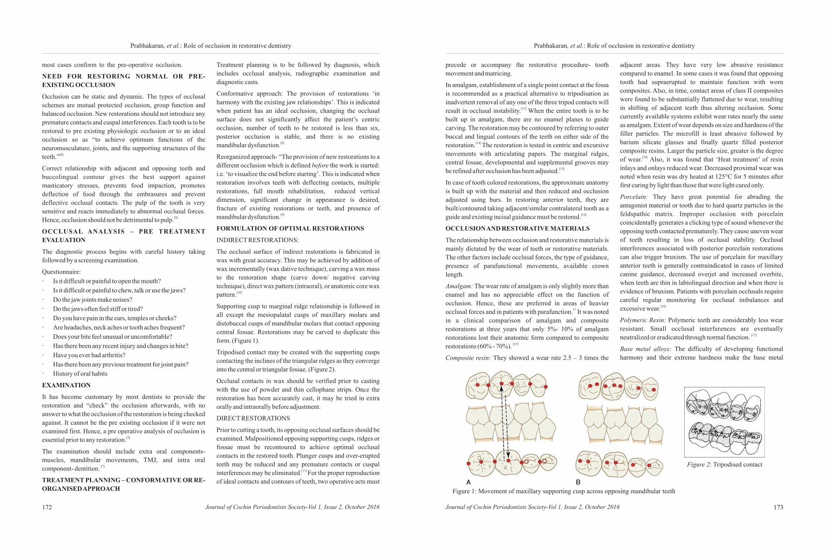
Journal of Cochin Periodontists Society-Vol 1, Issue 2, October 2016172 173Journal of Cochin Periodontists Society-Vol 1, Issue 2, October 2016
Prabhakaran, et al.: Role of occlusion in restorative dentistry Prabhakaran, et al.: Role of occlusion in restorative dentistry
most cases conform to the pre-operative occlusion.
NEED FOR RESTORING NORMAL OR PRE-
EXISTING OCCLUSION
Occlusion can be static and dynamic. The types of occlusal
schemes are mutual protected occlusion, group function and
balanced occlusion. New restorations should not introduce any
premature contacts and cuspal interferences. Each tooth is to be
restored to pre existing physiologic occlusion or to an ideal
occlusion so as “to achieve optimum functions of the
neuromusculature, joints, and the supporting structures of the [6]teeth.”
Correct relationship with adjacent and opposing teeth and
buccolingual contour gives the best support against
masticatory stresses, prevents food impaction, promotes
deflection of food through the embrasures and prevent
deflective occlusal contacts. The pulp of the tooth is very
sensitive and reacts immediately to abnormal occlusal forces. [5]Hence, occlusion should not be detrimental to pulp.
OCCLUSAL ANALYSIS – PRE TREATMENT
EVALUATION
The diagnostic process begins with careful history taking
followed by a screening examination.
Questionnaire:
· Is it difficult or painful to open the mouth?
· Is it difficult or painful to chew, talk or use the jaws?
· Do the jaw joints make noises?
· Do the jaws often feel stiff or tired?
· Do you have pain in the ears, temples or cheeks?
· Are headaches, neck aches or tooth aches frequent?
· Does your bite feel unusual or uncomfortable?
· Has there been any recent injury and changes in bite?
· Have you ever had arthritis?
· Has there been any previous treatment for joint pain?
· History of oral habits
EXAMINATION
It has become customary by most dentists to provide the
restoration and “check” the occlusion afterwards, with no
answer to what the occlusion of the restoration is being checked
against. It cannot be the pre existing occlusion if it were not
examined first. Hence, a pre operative analysis of occlusion is [5]essential prior to any restoration.
The examination should include extra oral components-
muscles, mandibular movements, TMJ, and intra oral [7]component- dentition.
TREATMENT PLANNING – CONFORMATIVE OR RE-
ORGANISED APPROACH
Treatment planning is to be followed by diagnosis, which
includes occlusal analysis, radiographic examination and
diagnostic casts.
Conformative approach: The provision of restorations ‘in
harmony with the existing jaw relationships’. This is indicated
when patient has an ideal occlusion, changing the occlusal
surface does not significantly affect the patient’s centric
occlusion, number of teeth to be restored is less than six,
posterior occlusion is stable, and there is no existing [8]mandibular dysfunction.
Reorganized approach- “The provision of new restorations to a
different occlusion which is defined before the work is started:
i.e. ‘to visualize the end before starting’. This is indicated when
restoration involves teeth with deflecting contacts, multiple
restorations, full mouth rehabilitation, reduced vertical
dimension, significant change in appearance is desired,
fracture of existing restorations or teeth, and presence of [9]mandibular dysfunction.
FORMULATION OF OPTIMAL RESTORATIONS
INDIRECT RESTORATIONS:
The occlusal surface of indirect restorations is fabricated in
wax with great accuracy. This may be achieved by addition of
wax incrementally (wax dative technique), carving a wax mass
to the restoration shape (carve down/ negative carving
technique), direct wax pattern (intraoral), or anatomic core wax [10]pattern.
Supporting cusp to marginal ridge relationship is followed in
all except the mesiopalatal cusps of maxillary molars and
distobuccal cusps of mandibular molars that contact opposing
central fossae. Restorations may be carved to duplicate this
form. (Figure 1).
Tripodised contact may be created with the supporting cusps
contacting the inclines of the triangular ridges as they converge
into the central or triangular fossae. (Figure 2).
Occlusal contacts in wax should be verified prior to casting
with the use of powder and thin cellophane strips. Once the
restoration has been accurately cast, it may be tried in extra
orally and intraorally before adjustment.
DIRECT RESTORATIONS
Prior to cutting a tooth, its opposing occlusal surfaces should be
examined. Malpositioned opposing supporting cusps, ridges or
fossae must be recontoured to achieve optimal occlusal
contacts in the restored tooth. Plunger cusps and over-erupted
teeth may be reduced and any premature contacts or cuspal [11] interferences may be eliminated. For the proper reproduction
of ideal contacts and contours of teeth, two operative acts must
precede or accompany the restorative procedure- tooth
movement and matricing.
In amalgam, establishment of a single point contact at the fossa
is recommended as a practical alternative to tripodisation as
inadvertent removal of any one of the three tripod contacts will [12] result in occlusal instability. When the entire tooth is to be
built up in amalgam, there are no enamel planes to guide
carving. The restoration may be contoured by referring to outer
buccal and lingual contours of the teeth on either side of the [14] restoration. The restoration is tested in centric and excursive
movements with articulating papers. The marginal ridges,
central fossae, developmental and supplemental grooves may [13]be refined after occlusion has been adjusted.
In case of tooth colored restorations, the approximate anatomy
is built up with the material and then reduced and occlusion
adjusted using burs. In restoring anterior teeth, they are
built/contoured taking adjacent/similar contralateral tooth as a [14]guide and existing incisal guidance must be restored.
OCCLUSION AND RESTORATIVE MATERIALS
The relationship between occlusion and restorative materials is
mainly dictated by the wear of teeth or restorative materials.
The other factors include occlusal forces, the type of guidance,
presence of parafunctional movements, available crown
length.
Amalgam: The wear rate of amalgam is only slightly more than
enamel and has no appreciable effect on the function of
occlusion. Hence, these are preferred in areas of heavier 17occlusal forces and in patients with parafunction. It was noted
in a clinical comparison of amalgam and composite
restorations at three years that only 5%- 10% of amalgam
restorations lost their anatomic form compared to composite [15]restorations (60% - 70%).
Composite resin: They showed a wear rate 2.5 – 3 times the
Figure 1: Movement of maxillary supporting cusp across opposing mandibular teeth
Figure 2: Tripodised contact
adjacent areas. They have very low abrasive resistance
compared to enamel. In some cases it was found that opposing
tooth had supraerupted to maintain function with worn
composites. Also, in time, contact areas of class II composites
were found to be substantially flattened due to wear, resulting
in shifting of adjacent teeth thus altering occlusion. Some
currently available systems exhibit wear rates nearly the same
as amalgam. Extent of wear depends on size and hardness of the
filler particles. The microfill is least abrasive followed by
barium silicate glasses and finally quartz filled posterior
composite resins. Larger the particle size, greater is the degree [18]of wear. Also, it was found that ‘Heat treatment’ of resin
inlays and onlays reduced wear. Decreased proximal wear was
noted when resin was dry heated at 125°C for 5 minutes after
first curing by light than those that were light cured only.
Porcelain: They have great potential for abrading the
antagonist material or tooth due to hard quartz particles in the
feldspathic matrix. Improper occlusion with porcelain
coincidentally generates a clicking type of sound whenever the
opposing teeth contacted prematurely. They cause uneven wear
of teeth resulting in loss of occlusal stability. Occlusal
interferences associated with posterior porcelain restorations
can also trigger bruxism. The use of porcelain for maxillary
anterior teeth is generally contraindicated in cases of limited
canine guidance, decreased overjet and increased overbite,
when teeth are thin in labiolingual direction and when there is
evidence of bruxism. Patients with porcelain occlusals require
careful regular monitoring for occlusal imbalances and [16]excessive wear.
Polymeric Resin: Polymeric teeth are considerably less wear
resistant. Small occlusal interferences are eventually [17]neutralized or eradicated through normal function.
Base metal alloys: The difficulty of developing functional
harmony and their extreme hardness make the base metal
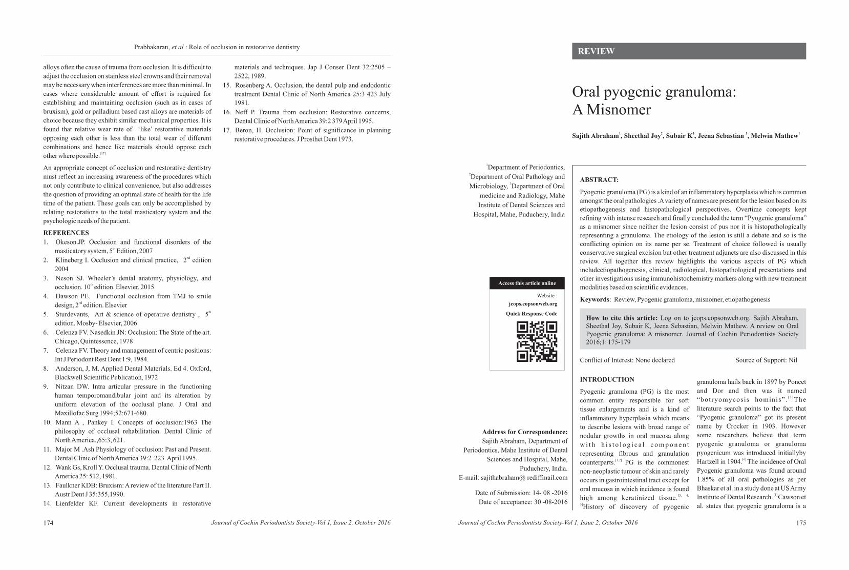
Journal of Cochin Periodontists Society-Vol 1, Issue 2, October 2016174
Prabhakaran, et al.: Role of occlusion in restorative dentistry
alloys often the cause of trauma from occlusion. It is difficult to
adjust the occlusion on stainless steel crowns and their removal
may be necessary when interferences are more than minimal. In
cases where considerable amount of effort is required for
establishing and maintaining occlusion (such as in cases of
bruxism), gold or palladium based cast alloys are materials of
choice because they exhibit similar mechanical properties. It is
found that relative wear rate of ‘like’ restorative materials
opposing each other is less than the total wear of different
combinations and hence like materials should oppose each [17]other where possible.
An appropriate concept of occlusion and restorative dentistry
must reflect an increasing awareness of the procedures which
not only contribute to clinical convenience, but also addresses
the question of providing an optimal state of health for the life
time of the patient. These goals can only be accomplished by
relating restorations to the total masticatory system and the
psychologic needs of the patient.
REFERENCES
1. Okeson.JP. Occlusion and functional disorders of the thmasticatory system, 5 Edition, 2007
nd2. Klineberg I. Occlusion and clinical practice, 2 edition
2004
3. Neson SJ. Wheeler’s dental anatomy, physiology, and thocclusion. 10 edition. Elsevier, 2015
4. Dawson PE. Functional occlusion from TMJ to smile nddesign, 2 edition. Elsevier
th5. Sturdevants, Art & science of operative dentistry , 5
edition. Mosby- Elsevier, 2006
6. Celenza FV. Nasedkin JN: Occlusion: The State of the art.
Chicago, Quintessence, 1978
7. Celenza FV. Theory and management of centric positions:
Int J Periodont Rest Dent 1:9, 1984.
8. Anderson, J, M. Applied Dental Materials. Ed 4. Oxford,
Blackwell Scientific Publication, 1972
9. Nitzan DW. Intra articular pressure in the functioning
human temporomandibular joint and its alteration by
uniform elevation of the occlusal plane. J Oral and
Maxillofac Surg 1994;52:671-680.
10. Mann A , Pankey I. Concepts of occlusion:1963 The
philosophy of occlusal rehabilitation. Dental Clinic of
North America.,65:3, 621.
11. Major M .Ash Physiology of occlusion: Past and Present.
Dental Clinic of North America 39:2 223 April 1995.
12. Wank Gs, Kroll Y. Occlusal trauma. Dental Clinic of North
America 25: 512, 1981.
13. Faulkner KDB: Bruxism: A review of the literature Part II.
Austr Dent J 35:355,1990.
14. Lienfelder KF. Current developments in restorative
materials and techniques. Jap J Conser Dent 32:2505 –
2522, 1989.
15. Rosenberg A. Occlusion, the dental pulp and endodontic
treatment Dental Clinic of North America 25:3 423 July
1981.
16. Neff P. Trauma from occlusion: Restorative concerns,
Dental Clinic of North America 39:2 379 April 1995.
17. Beron, H. Occlusion: Point of significance in planning
restorative procedures. J Prosthet Dent 1973.
175
ABSTRACT:
Pyogenic granuloma (PG) is a kind of an inflammatory hyperplasia which is common
amongst the oral pathologies .A variety of names are present for the lesion based on its
etiopathogenesis and histopathological perspectives. Overtime concepts kept
refining with intense research and finally concluded the term “Pyogenic granuloma”
as a misnomer since neither the lesion consist of pus nor it is histopathologically
representing a granuloma. The etiology of the lesion is still a debate and so is the
conflicting opinion on its name per se. Treatment of choice followed is usually
conservative surgical excision but other treatment adjuncts are also discussed in this
review. All together this review highlights the various aspects of PG which
includeetiopathogenesis, clinical, radiological, histopathological presentations and
other investigations using immunohistochemistry markers along with new treatment
modalities based on scientific evidences.
Keywords: Review, Pyogenic granuloma, misnomer, etiopathogenesis
Access this article online
Website :
jcops.copsonweb.org
Quick Response CodeHow to cite this article: Log on to jcops.copsonweb.org. Sajith Abraham, Sheethal Joy, Subair K, Jeena Sebastian, Melwin Mathew. A review on Oral Pyogenic granuloma: A misnomer. Journal of Cochin Periodontists Society 2016;1: 175-179
Conflict of Interest: None declared Source of Support: Nil
REVIEW
1Department of Periodontics, 2Department of Oral Pathology and
3Microbiology, Department of Oral
medicine and Radiology, Mahe
Institute of Dental Sciences and
Hospital, Mahe, Puduchery, India
Address for Correspondence:
Sajith Abraham, Department of
Periodontics, Mahe Institute of Dental
Sciences and Hospital, Mahe,
Puduchery, India.
E-mail: sajithabraham@ rediffmail.com
Date of Submission: 14- 08 -2016
Date of acceptance: 30 -08-2016
Journal of Cochin Periodontists Society-Vol 1, Issue 2, October 2016
1 2 1Sajith Abraham , Sheethal Joy , Melwin Mathew1 3Subair K , Jeena Sebastian ,
Oral pyogenic granuloma: A Misnomer
INTRODUCTION
Pyogenic granuloma (PG) is the most
common entity responsible for soft
tissue enlargements and is a kind of
inflammatory hyperplasia which means
to describe lesions with broad range of
nodular growths in oral mucosa along
w i t h h i s t o l o g i c a l c o m p o n e n t
representing fibrous and granulation [1,2] counterparts. PG is the commonest
non-neoplastic tumour of skin and rarely
occurs in gastrointestinal tract except for
oral mucosa in which incidence is found [3, 4, high among keratinized tissue.
5]History of discovery of pyogenic
granuloma hails back in 1897 by Poncet
and Dor and then was it named [ 2 ]“botryomycosis hominis” . The
literature search points to the fact that
“Pyogenic granuloma” got its present
name by Crocker in 1903. However
some researchers believe that term
pyogenic granuloma or granuloma
pyogenicum was introduced initiallyby [6] Hartzell in 1904. The incidence of Oral
Pyogenic granuloma was found around
1.85% of all oral pathologies as per
Bhaskar et al. in a study done at US Army [1] Institute of Dental Research. Cawson et
al. states that pyogenic granuloma is a
Journal of Cochin Periodontists Society-Vol 1, Issue 2, October 2016176 177Journal of Cochin Periodontists Society-Vol 1, Issue 2, October 2016
Abraham, et al.: Oral pyogenic granuloma Abraham, et al.: Oral pyogenic granuloma
common lesion and represents 0.5% of all skin nodules and
pregnancy tumour a variant occurs upto 5% in all [7]pregnancies. Oral pyogenic granuloma was found to be the
most frequent non-neoplastic lesion according to study by
Shamim et al accounting to nearly 52.7% of the lesions. [8]Therefore epidemiologic insights projects that this lesion is
frequent among oral pathologies in general.
Why a misnomer?
A misnomer is a word or term that suggests a meaning that is
known to be wrong. They often arise before the true nature was
unknown or because the nature of an earlier form is no longer a
norm. As the Latin word “etymon nominare” means “to name”;
a misnomermeans a special kind of mistake or misapplied or [9] wrong name. There were different terminologies put forward
to this lesion starting from the earliest “botryomycosis
hominis” and also includegranuloma pediculatum benignum,
granuloma telangiectactium, benign vascular tumor,
pregnancy tumor, vascular epulis and Crocker and Hartzell
disease as others. The lesion was subjected toan array of names
from past to present based on the etiology and
histopathological perspectives. Over time these concepts kept
refining with more researches in this area and finally
concluded; “Pyogenic Granuloma” as a misnomer since
neither the lesion containpus nordoes it represent a granuloma [1, 10,11]histopathologically.
Theories of Etiopathogenesis: Past to Present
Some investigators regard pyogenic granuloma as “infectious”
in origin. Staphylococcus and botryomycosis, foreign bodies
and localization of infection in blood vessel walls were
projected as factors of development of the lesion.Shafer et al.
suggested that either staphylococci or streptococci could
produce colonies with fungus like characteristics and oral [12 ] pyogenic granuloma arises as a result of these infection. As
per Reichart et al. granulation tissue in the lesion gets
contaminated by flora of the oral cavity and the surface gets [13]covered with fibrin which mimic pus. However suppuration
is not a characteristic feature of oral pyogenic granuloma to
support infectious origin and also in the inclusion of term “pyo”
in name of the lesion. Majority of the studies has pointed out
that even though the lesion has been associated to certain
species of Bartonella henselea species and Human herpes virus
type 8 in certain recurrent lesions; there is no evidence
confirming the presence of these microorganisms in larger [14] groups of pyogenic granuloma lesions. Currently
etiopathogenesis suggests that the lesion is result of some
minor trauma to tissues which provides pathway for invasion of [12]microscopic non-specific organisms.
Studies also stated that after any trauma wound healing is
initiated with formation of granulation tissue. The process of
wound healing is controlled by various cytokines which
include growth factors mainly basic fibroblast growth factor,
an angiogenic heparin binding angioprotein which is mitogenic
for capillary endothelial cells. Certain growth factors such as
inducible nitric oxide synthetase, vascular endothelial growth
factor, basic fibroblast growth factor or connective tissue
growth factor had integral roles in angiogenesis and rapid [15]growth of PG.
Researchers also considered pyogenic granuloma as a
“reactive” or “reparative” lesion. Regezi et al. has suggested
that pyogenic granuloma is an exuberant connective tissue
proliferation to a known stimulus or injury which includes
calculus or foreign materials within gingival crevice. The
reactive process can be to various stimuli which include
chronic low grade local irritation, trauma, hormonal factors or [16]certain kinds of drugs.
Richardson and Krotochovil suggested that hormonal
influence play basis in incidence of pregnancy tumour, which is
a variant of the lesion. Current studies have revealed the effect
of sex hormones which manifest a variety of biological and [17]immunological process. Estrogen increases vascular
endothelial growth factor (VEGF) production in macrophages
which is related to the development of lesion in pregnancy. This
effect is also seen antagonized by the androgens. Study by Yuan
et al. suggested increased levels of VEGF and basic fibroblast
growth factor expressions in pyogenic granuloma lesions.
Studies have proposed that in the absence of VEGF,
Angiopoetin -2 (Ang-2) causes the blood vessels to regress and
their levels were seen regulated by Tumor necrosis factor alpha
in all endothelial cells. It was seen that amount of VEGF was
high in granulomas of pregnancy and was absent or
undetectable after parturition. After parturition more of apoptic
cells and less of Ang-2 are seen expressed compared to
pregnancy. Therefore VEGF alone or in combination with Ang-
2 could be beneficial in protecting blood vessels from [18]apoptosis. Progesterone acts as an immunosuppressant in
gingival tissues in pregnancy as per study by Harri et al.
preventing rapid acute inflammatory reaction against plaque,
but allowing chronic tissue reaction resulting in exuberant [19]proliferation due to inflammation. However oral
contraceptives influence on vascular changes in periodontium
is still unclear due to absence of steroid receptors in the
periodontium as per findings by Nichols et al. which suggest
that estrogen or progesterone are not directly involved in [20]formation of the lesion.
Drugs like cyclosporine was seen to have a role in development
of the lesion in patients who were given the drug for graft [21]versus host disease. Inclusion bodies were found in the
fibroblasts of the lesion suggestive of disordered protein [22]metabolism according to the Davies et al.
Growth rate of the tumour is not only seen related to
proliferative activity of tumour cells but is also seen related to
the rate of cell death. Studies have suggested low apoptotic rate
in PG which is related to growth rate and regulation by Bcl2 [23]family proteins.
Clinical Presentations
Oral Pyogenic granuloma is the most common gingival
tumour, with predeliction for gingiva in 75% of all cases.
Extragingival lesions involve lips, tongue, buccal mucosa and
hard palate. Lesions are common in maxillary gingiva and have
predeliction towards anterior region than posterior
counterparts. These lesions are much common on the facial
aspect of the gingiva than the lingual aspect with some
extending between teeth and both facial and lingual gingiva.
Although PG occur at all age groups; highest incidence was
seen in second and fifth decades and females were slightly
more affected than males. Predeliction in second decade among
young females was possibly related to the vascular effects of [1, 2, 10,16]female sex hormones.
Clinical presentation of the lesion manifests from smooth or
exophytic small, red erythematous papule on a pedunculated
base or sessile, usually compressible and haemorrhagic. Size
varies from few millimetres to several centimetres rarely [11]exceeding 2.5cm in size. The clinical development of the
lesion starts slow, asymptomatic and painless but at times grow
rapidly. The surface is sometimes ulcerated which may be
covered with yellow fibrinous membrane. The colour ranges
from pink to red purple depending upon the age of the lesion
Pyogenic granuloma: A Clinical presentation
with younger PG’s appearing more reddish because of the
capillary predominance present in the hyperplastic granulation
tissue. Older lesions on the other hand are more collegenized [2, 10]and therefore appear pink.
Pregnancy tumour a variant of PG appears normally in the
second – third month of pregnancy, with a tendency to bleed
and possibility of interaction while masticating food. In the
initial months of pregnancy, the plaque influence persistently
producing catarrhal inflammation of gingiva that serve a base
for development of hyperplasia of the gingival tissue where as
in the last trimester modulation is mainly by the circulating [24]hormonal stimuli.
Radiological Presentations
The relevant radiological findings are usually absent in
pyogenic granuloma. In rare instances; long standing gingival
lesionsthere is someloss of the alveolar crest where the epulis
find attachment or it might produce saucer shape excavation of [10,12 25]variable depth
HistopathologicalPresentations
Briefly the natural history of the lesion follows three distinct [26]phases.
Initial/Cellular phase - Consist of lobules which are compact
and cellular with little lumen formation.
Capillary phase - The lobules becomes vascular with abundant
intraluminal red blood cells. One or more central vessels form
large lumen with thick muscular layer bearing resemblance to
veins.
Late/Involutionary phase – Increased tendencyfor intra and
perilobular fibrosis with increased venular differentiation.
Two distinct histological patterns of PG exist which include
lobular capillary hemangioma type and non –lobular type. The
first type is characterised by proliferating blood vessels that are
arranged in lobular pattern although superficially lesion does
not undergo specific changes like edema, capillary dilation or [27]inflammatory granulation tissue reaction. The latter non
–lobular type consists of highly vascular proliferation that
resembles granulation tissue with the central area consisting
significantly increased number of vessels with perivascular
mesenchymal cells non-reactive to alpha smooth muscle actin
and muscle specific actin than its lobular type counterpart.
Even though PG can undergo fibrous maturation over time; it is
suggested that it happened only in non-lobular capillary
haemangioma type of lesion clearly indicating different [10, 11]pathways of evolution amongst the two types of PG.
Other Investigations: IHCMarkers
Increased expression of basic fibroblast growth factor,Tie-2,
anti –CD34 and anti-alpha SMA antibodies, vascular
Journal of Cochin Periodontists Society-Vol 1, Issue 2, October 2016178
Abraham, et al.: Oral pyogenic granuloma
179Journal of Cochin Periodontists Society-Vol 1, Issue 2, October 2016
Abraham, et al.: Oral pyogenic granuloma
morphogenesis factors such as Angiopoetin -1, Angiopoetin -2,
Ephrin B2 and B4 respectively were seen evident in PG lesions.
Also low apoptotic rate expression of Bax/Bcl-2 proteins and
strong expression of phosphorylated mitogen activated protein
kinase and increased expression on vascular endothelial [23,28]growth factor (VEGF) were alsoseen significantly evident.
Differential Diagnosis
Differential diagnosis is helpful in further evaluation and
management of the patient. The differential diagnosis of PG
lesions include peripheral giant cell granuloma, peripheral
ossifying fibroma, peripheral odontogenic fibroma,
hemangioma, conventional granulation tissue, hyperplastic
gingival inflammation, Kaposi’s sarcoma, Bacillary
angiomatosis , angiosarcoma and non-Hodgkin’s [13,29]Lymphoma.
TREATMENT
Excisional biopsy is indicated treatment of PG, except when
procedure would produce marked deformity where incisional
biopsy becomes mandatory line of investigation. Therefore [30]management depends on the severity of symptoms. The usual
treatment option include conservative surgical excision and
removal of causal irritants like plaque, calculus, foreign [2,10]materials, source of trauma etc. Excision with 2mm margins
from its clinical periphery and to depth of periosteum or to [31] causative agent was recommended. Recently other treatment
protocols using lasers have been proposed because of the lower
risk of bleeding compared to other surgical techniques. Nd:
YAG lasers were superior to C02 lasers because of its superior [32, 33] coagulation properties. Other treatment modalities for
management of the lesion includes cryosurgery. Oral mucosa
because of its humidity and smoothness was seen ideal for [34] cryosurgery technique. Sclerotherapy with intralesional
injections using sodium tetradecyl sulphate, absolute ethanol
etc were seen to be other alternative treatment adjuncts used in
practice because of its simplicity and lack of scarring quality [35,36]but require multiple treatment sessions. Recurrence rate
after surgical excision ranged from 5.8% to 15.8% of PG [2,37] cases. Gingival lesions had higher recurrence rate
compared to other oral mucosal sites and showed no infiltrative
or malignant potential.
CONCLUSION
Oral PG is one among the commonest of all oral pathologies.
The etiopathogenesis of the lesion is still debatable and so is its
“name” making it different from the other oral lesions.This
article helps to highlight on various aspects of the oral pyogenic
granuloma based on scientific observations.
REFERENCES
1. Eversole LR 2002.Clinical outline of oral pathology:
rdDiagnosis and treatment.3 edition
2. BhaskarSN, JacowayJR. Pyogenic granuloma-clinical
features, incidence, histology and resultant treatment.
Report of 242 cases.J oral surgery 1966; 24:391-8
3. Vilmann A, Vilmann P, Vilmann H.Pyogenic granuloma:
evaluation of oral conditions.Br J Oral Maxillofacial
surgery1986; 24:376-82
4. Yao T, Nagai E, Utsunomiya T, TsuneyoshiM.An intestinal
counterpart of pyogenic granuloma of skin.A newly
proposed identity. Am J Surg Pathol 1995; 19:1054-1060.
5. Fowler EB,Cuenin MF, Thompson SH, Kudryk VL,
BillmanMA. Pyogenic granuloma associated with guided
tissue regeneration: a case reportJ.Periodontol1996;
67:1011-1015
6. HartzellMB.Granuloma pyogenicum.J Cutan Dis
Syph1904;22:520-525
7. Cawson RA,Binnie WH,Speight PM,Barrett AW,Wright thJM.Lucas Pathology of tumours of oral tissues.5
edition1998.
8. Shamim T, Varghese VI,Shameena PM,Sudha S.A
retrospective analysis of gingival biopsied lesions in South
Indian population:2001-2006 Med Oral Path Oral Car
Buccal 2008; 13:14-8.
9. https://en.m.wikipedia.org/accessed on 15/8/2016
10. Neville BW,Damm DD, Allen CM, Bouquot JE. Oral and thmaxillofacial pathology .4 edition
11. Bouquot JE, Nikai H.Lesions of oral cavity. In diagnostic
surgical Pathology of head and neck. Gnepp DR ed.2001th12. Shafers text book of oral pathology .6 edition
13. Peter A, Reichart.Hans Peter Philipsen. Colour atlas of
dental medicine Oral Pathology, 2000; 163
14. Jainer M. Infection and angiomatous cutaneous lesions.J
Mal Vasc 1999; 24: 135-38.
15. Murata M,Hara K, Saku T.Dynamic distribution of basic
fibroblast factor during epulis formation:An
immunohistochemical study in an enhanced healing
process of gingiva.J Oral Path Med 1997;26:224-32
16. Regezi JA, Sciubba JJ, Jordan RC. Oral Pathology: thClinical Pathologic considerations,4 edition 2003.
17. YihWY, Richardson L, Kratochvil FJ,Avera SP,Zieper
MB.Expression of estrogen receptors in desquamative
gingivitis .J Periodontol 2000; 71:482-7
18. Kanda N, Watanabe S.Regulatory roles of sex hormones in
cutaneous biology and immunology. J Dermatol Sci 2005;
38:1-7.
19. Ojanotko-HarriAO,Harri MP,Huritta HM,Sewon LA.
Altered Tissue metabolism of progesterone in pregnancy
gingivitis and granuloma.J Clin Perio1991; 18:262-266.
20. Nicholas GE,Gaffey MJ, Millis SE, Weiss LM.Lobular
Capillary hemangioma: An immuno- histochemical study
including steroid hormone receptor status.Am J Clin Path
1992; 97:770-75.
21. Bachemeyer C, DevergieA, Mansouri S et al.Pyogenic
granuloma of the tongue in chronic graft versus host
disease.Ann Dermatol Venereol 1996; 123:552-554
22. Davies MG,Barton S P, Atai F,Marks R.The abnormal
dermis in pyogenic granuloma .Histochemical and
ultrastructural observations. L Am Acad Dermatol 1980;
2:132-142
23. Nakamura T.Apoptosis and expression of Bcl2/Bax
Proteins in pyogenic granuloma: A comparative study with
granulation tissue and capillary hemangioma. J Cutan
Pathol 2000; 27:400-405
24. Boyarova TV, Dryankova MM, Bobeva AI,Genadiev
GI.Pregnancy and gingival hyperplasia. Folia Med 2001;
43:53-56
25. HM Worth. Principles and practice of oral radiographic
interpretation.1963.
26. Sternberg SS,Antonioli DA,Carter D,Millis SE,Obermann rdH. Diagnostic surgical pathology .3 edition. Lippincott
Williams & Wilkins 1999.
27. Mills SE, Cooper PH, Fechner RE. Lobular Capillary
hemangioma; the underlying lesion of pyogenic
granuloma. A study of 73 cases from oral and nasal mucous
membrane.Am J Surg Pathol 1980;4:470-79.
28. Sato H,Takeda Y,Satoh M.Expression of the endothelial
receptor tyrosine kinase Tie 2 in lobular capillary
hemangioma of the oral mucosa.An immunohistochemical
study.J Oral Pathol Med 2002;31:432-38.
29. Wood NK, Goaz PW. Differential Diagnosis of oral and thmaxillofacial lesions.5 edition, 1998.
30. Greenberg MS, Glick M. Burkets oral medicine: diagnosis thand treatment.2003.10 edition.
31. Marx RE, Stern D. Oral and Maxillofacial Pathology: A
rationale for diagnosis and treatment.Chicago
Quintessence Publishing 2003.
32. Powell JL, Bailey CL, Coopland AT, Otis CN, Frank JL,
Meyer I. Nd: YAG laser excision of a giant gingival
pyogenic granuloma of pregnancy. Lasers Surg Med 1994;
14:178-83.
33. White JM, Chaudhary SI, Kudler JJ, Sekandari N, and
Schoelch ML, Silverman S Jr. Nd: YAG and CO2 laser
therapy of oral mucosal lesions.J Clin Laser Med Surg
1998; 16:299-304.
34. Ishida CE, Ramose –e –Silva M. Cryosurgery in oral
lesions. Int J Dermatol 1998; 37:283-85.
35. Ichimiya M, Yoshikawa Y, Hamamoto Y, Muto M.
Successful treatment of pyogenic granuloma with
injection of absolute ethanol. J Dermatol 2004;31:342-44.
36. Moon SE, Hwang EJ, Cho KH. Treatment of pyogenic
granuloma by sodium tetradecyl sulfate Sclerotherapy.
Arch Dermatol 2005; 141:359-362.
37. Al-Khateeb T, Ababmeh F. Oral Pyogenic granuloma in
Jordanians. A retrospective analysis of 108 cases.J Oral
Maxillofac Surg 2003; 61:1285-88.
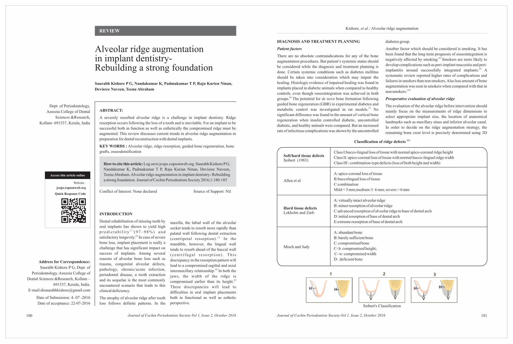
Saurabh Kishore P G, Nandakumar K, Padmakumar T P,
Devisree Naveen, Teenu Abraham
Raju Kurien Ninan,
How to cite this article: Log on to jcops.copsonweb.org. Saurabh Kishore P G,
Nandakumar K, Padmakumar T P, Raju Kurian Ninan, Devisree Naveen,
Teenu Abraham. Alveolar ridge augmentation in implant dentistry- Rebuilding
a strong foundation. Journal of Cochin Periodontists Society 2016;1:180-185
Conflict of Interest: None declared Source of Support: Nil
Journal of Cochin Periodontists Society-Vol 1, Issue 2, October 2016 Journal of Cochin Periodontists Society-Vol 1, Issue 2, October 2016180
Access this article online
Website :
jcops.copsonweb.org
Quick Response Code
Dept. of Periodontology,
Azeezia College of Dental
Sciences &Research,
Kollam- 691537, Kerala, India
Address for Correspondence:
Saurabh Kishore P G, Dept. of
Periodontology, Azeezia College of
Dental Sciences &Research, Kollam –
691537, Kerala, India.
E-mail:[email protected]
Date of Submission: 4- 07 -2016
Date of acceptance: 22-07-2016
REVIEW
ABSTRACT:
A severely resorbed alveolar ridge is a challenge in implant dentistry. Ridge
resorption occurs following the loss of a tooth and is inevitable. For an implant to be
successful both in function as well as esthetically the compromised ridge must be
augmented. This review discusses current trends in alveolar ridge augmentation in
preparation for dental reconstruction with dental implants.
KEY WORDS : Alveolar ridge, ridge resorption, guided bone regeneration, bone
grafts, osseodensification
181
Kishore, et al.: Alveolar ridge augmentation
Alveolar ridge augmentationin implant dentistry- Rebuilding a strong foundation
INTRODUCTION
Dental rehabilitation of missing teeth by
oral implants has shown to yield high [ 1 ]p r e d i c t a b i l i t y ( 9 7 – 9 8 % ) a n d
[2]satisfactory longevity. In case of severe
bone loss, implant placement is really a
challenge that has significant impact on
success of implants. Among several
reasons of alveolar bone loss such as
trauma, congenital alveolar defects,
pathology, chronic/acute infection,
periodontal disease, a tooth extraction
and its sequelae is the most commonly
encountered scenario that leads to this
clinical deficiency.
The atrophy of alveolar ridge after tooth
loss follows definite patterns. In the
maxilla, the labial wall of the alveolar
socket tends to resorb more rapidly than
palatal wall following dental extraction [ 3 ](centripetal resorption). In the
mandible, however, the lingual wall
tends to resorb ahead of the buccal wall
( c e n t r i f u g a l r e s o r p t i o n ) . T h i s
discrepancy in the resorption pattern will
lead to a compromised sagittal and axial [4]intermaxillary relationship. In both the
jaws, the width of the ridge is [5]compromised earlier than its height.
These discrepancies will lead to
difficulties in oral implant placements
both in functional as well as esthetic
perspective.
DIAGNOSIS AND TREATMENT PLANNING
Patient factors
There are no absolute contraindications for any of the bone
augmentation procedures. But patient’s systemic status should
be considered while the diagnosis and treatment planning is
done. Certain systemic conditions such as diabetes mellitus
should be taken into consideration which may impair the
healing. Histologic evidence of impaired healing was found in
implants placed in diabetic animals when compared to healthy
controls, even though osseointegration was achieved in both [6] groups. The potential for de novo bone formation following
guided bone regeneration (GBR) in experimental diabetes and [7]metabolic control was investigated in rat models. No
significant difference was found in the amount of vertical bone
regeneration when insulin controlled diabetic, uncontrolled
diabetic, and healthy animals were compared. But an increased
rate of infectious complications was shown by the uncontrolled
diabetes group.
Another factor which should be considered is smoking. It has
been found that the long term prognosis of osseointegration is [8]negatively affected by smoking. Smokers are more likely to
develop complications such as peri-implant mucositis and peri-[9]implantitis around successfully integrated implants. A
systematic review reported higher rates of complications and
failures in smokers than non smokers. Also less amount of bone
augmentation was seen in smokers when compared with that in [10]non smokers.
Preoperative evaluation of alveolar ridge
The evaluation of the alveolar ridge before intervention should
mainly focus on the measurements of ridge dimensions to
select appropriate implant size, the location of anatomical
landmarks such as maxillary sinus and inferior alveolar canal.
In order to decide on the ridge augmentation strategy, the
remaining bone crest level is precisely determined using 3D
[11]Classification of ridge defects
Soft/hard tissue defectsSeibert (1983)
Class I:bucco-lingual loss of tissue with normal apico-coronal ridge height
Class II: apico-coronal loss of tissue with normal bucco-lingual ridge width
Class III : combination-type defects (loss of both height and width)
A: apico-coronal loss of tissue
B:buccolingual loss of tissue
C:combination
Mild:< 3 mm;medium:3–6 mm; severe:> 6 mm
A: virtually intact alveolar ridge
B: minor resorption of alveolar ridge
C:advanced resorption of alveolar ridge to base of dental arch
D: initial resorption of base of dental arch
E:extreme resorption of base of dental arch
A: abundant bone
B: barely sufficient bone
C: compromised bone
C- h: compromised height;
C- w: compromised width
D: deficient bone
Allen et al
Hard tissue defectsLekholm and Zarb
Misch and Judy
Seibert's Classification
Journal of Cochin Periodontists Society-Vol 1, Issue 2, October 2016 Journal of Cochin Periodontists Society-Vol 1, Issue 2, October 2016182 183
Kishore, et al.: Alveolar ridge augmentationKishore, et al.: Alveolar ridge augmentation
radiographic methods.
BONE AUGMENTATION THERAPIES
Guided bone regeneration
The treatment concept of GBR is to exclude the soft tissues
from filling the osseous defect and allowing the bone forming
cells to occupy the defect space. Depending on the type of
defect, the space could be maintained by utilizing a block graft
or a particulated graft. Different natural and synthetic
biomaterials have been developed and used as graft materials in
augmentation of ridge defects.
Barrier Memberanes : They are broadly classified into non-
resorbable and resorbable.
Expanded polytetraflouroethylene (e-PTFE) is the most
frequently used non resorbable membrane. It has a flexible
property and external porous structure which allows tissue
integration and an internal occlusive layer which functions as a
barrier mechanism. A titanium scaffold is added between two
layers of e-PTFE to enhance the space making capacity of these
devices. But the disadvantage of these devices is that a second
intervention is needed to remove them. So in order to overcome
this difficulty resorbable membranes were developed.
Bioresorbable membranes are either natural (xenogeneic
collagen type I or III) or made of synthetic polymers, including
polyurethane, polyglactin 910, polylactic acid, polyglycolic
acid, polyorthoester, polyethylene glycol, and different
combinations of polylactic and polyglycolic acid.
Bioresorbable membranes when placed directly over the
implant threads, tend to collapse and occlude the space
available for bone regeneration. This problem can be avoided
by using a scaffold or graft material under the membrane that
provides the space for tissue in growth and subsequent bone
formation. When non resorbable and collagen resorbable
membranes, with and without the use of a scaffold, were
compared in an animal study, similar bone regenerative
outcomes was obtained for the non resorbable membranes and [12]the collagen resorbable membranes used with a scaffold.
Thus resorbable membranes are currently considered as the
gold standard when used in conjunction with an adequate
space-making graft material.
Bone grafts and bone substitutes:
Bone grafts
Autogenous bone grafts are considered as the gold standard in
bone regeneration therapies as they have osteoinductive, [13]osteoconductive and osteogenic properties. In alveolar ridge
defects, autografts are either used as a particulate or a block
graft. Particulate grafts are harvested from intra oral sites and
are used along with barrier membranes following the principles
of guided bone regeneration. Block grafts are harvested either
from intra oral sites or extra oral sites. They are used with
barrier membranes or alone but they require fixation to the
recipient site with micro screws in order to stabilize the graft
while healing.
Allografts are harvested from cadaver and are processed by
freezing or demineralization and freezing. It is then sterilized
and made available as particulate or as blocks. They are usually
used with barrier membranes following principles of GBR.
Xenografts are of animal origin, usually of bovine or porcine.
These graft materials are prepared by deproteinizing and
thereby avoiding the immunogenic property. These type of
graft materials are usually particulate and used according to the
principles of GBR.
Bone substitutes
Alloplasts are synthetic biocompatible bone substitutes.
Alloplastic material such as ² – tricalcium phosphate which is
resorbable is osteoneutral and is totally inert and serves only as
space fillers. They include different combinations of calcium
phosphates synthesized under different sintering conditions to
attain different physical properties and resorption rates. The
combination of ² – tricalcium phosphate and hydroxyapatite
provides both scaffolding as well as an osteoconductive
function. They are usually used as granules and should always
be used in conjunction with barrier membranes.
RIDGE AUGMENTATION PROCEDURES
Socket preservation
Socket preservation is a procedure which aims at minimizing
alveolar ridge atrophy after tooth extraction. A wide range of
socket preservation techniques and materials have been
reported that significantly reduce bone loss following
extraction. Atraumatic extraction leaving the socket wall intact [14]is enough to regenerate socket bone formation. This
procedure utilizes regenerative techniques such as resorbable /
non-resorbable membranes, resorbable membranes in
conjunction with bone substitutes, bone substitutes alone or
bone substitutes in combination with soft tissue grafts.
The subgroup analysis of a systematic review gave the [15]following conclusions:
• Use of membranes gave better results than for use of grafts
alone in terms of horizontal bone changes.
• A slight tendency towards less bone loss in the horizontal
direction was observed when the sockets healed by
primary intention.
• Flapped (primary closure) surgical procedures
demonstrated significantly less horizontal bone resorption
of the socket, when compared to flapless (healing by
secondary intention) surgeries.
Horizontal ridge augmentation
Horizontal ridge augmentation can be done by using either
particulate grafts or block grafts, with or without barrier
membranes. When there is enough bone width, it is indicated to
use particulate grafts together with barrier membranes to allow
good implant primary stability.
Both GBR and block grafts have been demonstrated to be a
successful and predictable treatment[16] modality to augment a horizontally deficient ridge. A
randomized clinical trial study has revealed that onlay graft
group attained more horizontal bone gain than GBR [17]group.
Since the use of autografts is limited due to morbidity while
procuring the graft and also the higher resorption rate, bone
substitutes, particularly xenografts along with barrier
membranes are used and have demonstrated good results.
Ridge splitting & ridge expansion
This is a technique used in the maxilla to augment bone width
through bone condensation. Summers first used this technique,
osteocondensation, to augment bone width and elevate sinus
floors in an attempt to avoid the lateral window sinus lift.
Chisels and osteotomes are used which will cause longitudinal
greenstick fractures in the bone and create osteotomy sites
without the need for drilling. This preserves the compromised
bone volume. The bone is compressed to the lateral surfaces
with the use of osteotomes of increasing diameters, thus
increasing itsstrength and density. The advantage of this
technique is that it allows for the ideal implant diameter to be
placed in the restoratively driven position. This procedure is
followed by the simultaneous placement of the implant.
Ridge splitting is essentially the fracture of the buccal cortical
plate and its displacement laterally to accommodate implant
placement. The spaces created between the cortical plates and
the implants are subsequently filled with particulate bone graft
materials.
Ridge splitting using piezosurgery
In the cortical mandible, bone splitting is much more
challenging, as mechanical trauma as well as the shape and
design of the cutting tools often fracture the thin and fragile
bone segments. In such cases, bone sectioning with
piezoelectric devices ensures micrometric osteotomies with no
macrovibrations and allows easy handling with an increased
maneuverability even in delicate cases.
Vertical ridge augmentation
Various studies during the last decade have reported good
results using vertical bone augmentation techniques with bone
graft materials, membranes, or both. However, vertical
augmentation may be less predictable than horizontal
augmentation.
A study in patients with atrophic edentulous ridges it was
demonstrated that up to 4 mm of vertical bone augmentation [18]can be predictably regenerated. The surgical protocol
included the use of a membrane reinforced with a titanium
structure. This membrane can be shaped to maintain the desired
form, thereby creating and preserving sufficient space between
the membrane and the bone defect.
Alveolar distraction osteogenesis
Distraction osteogenesis surgery is currently available
worldwide for managing orthopedic disorders. The process of
alveolar distraction osteogenesis involves mobilization,
transport, and fixation of a healthy segment of bone adjacent to
the deficient or deformed site.
A mechanical alveolar distraction device is used to provide
gradual, controlled transport of a mobilized alveolar segment.
Distraction is usually performed over a period of 30 days and [19]significant bone gain can be attained (4–7 mm). When the
desired repositioning of the bone segment is achieved, the
distraction device is left in a static mode to act as a fixation tool.
Because the soft tissue is left attached to the transport segment,
the movement of the bone also results in expansion of soft
tissue adjacent to the bone segment. As a result of the
distraction process, the volume of bone and soft tissue
increases. The reconstructed site is then suitable for further
rehabilitation with osseointegrated implants.
However, distraction osteogenesis has frequent complications,
such as fracture of the mandible or the moveable segment,
patient discomfort etc.
Molecular approach in bone augmentation
The use of growth factors has received more attention for
treatment of deficient alveolar ridges. Advances in molecular
cloning have made available unlimited quantities of
recombinant growth factors for applications in tissue
engineering. Recombinant growth factors such as platelet
derived growth factors (PDGF),insulin like growth factors
(IGFs), fibroblast growth factors (FGFs),and bone
morphogenetic proteins (BMP)’s have been used in clinical trials for the treatment of large ridge and alveolar deficiencies.
[20,21]
In vivo application of PDGF alone or in combination with IGF 1 [22],[23]enhances mineralized tissue repair. PDGF also has been
shown to have a significant regenerative impact on periodontal [24,25]ligament cells as well as on osteoblasts.
Journal of Cochin Periodontists Society-Vol 1, Issue 2, October 2016 Journal of Cochin Periodontists Society-Vol 1, Issue 2, October 2016184 185
Kishore, et al.: Alveolar ridge augmentationKishore, et al.: Alveolar ridge augmentation
Bone morphogenetic proteins (BMP) are a group of regulatory
glycoproteins that are members of the TGFa superfamily.
Investigations in animal models have shown the potential [26]repair of alveolar bony defects using rhBMP 12. In a clinical
trial, rhBMP 2 delivered by a bioresorbable collagen sponge
revealed significant bone formation in a human buccal wall
defect model following tooth extraction when compared to the [27]collagen sponge alone. Also BMP 7, stimulates bone
regeneration around teeth and endosseous dental implants, and [28]in maxillary sinus floor augmentation procedures.
OSSEODENSIFICATION – a new technique for ridge
expansion and enhanced implant stability
[30]
BONE AUGMENTATION IN FUTURE
There is a possible impact of tissue engineering on bone
regeneration in the future. A more predictable regeneration of
bone can be achieved by developing a therapeutic system
which can deliver the right cells at the right time in the presence
of growth factors. Various novel delivery scaffolding systems
are being extensively studied and fabricated, and are
Standard drill designs used in dental implantology are made to
excavate bone to create room for implant placement. They cut
away bone effectively but typically do not produce a precise
circumferential osteotomy. Moreover, osteotomies drilled into
narrow bone locations may produce dehiscence, buccally or
lingually, which also reduces primary stability and will require
an additional bone grafting procedure adding cost and healing
time to treatment.
Unl ike t rad i t iona l bone dr i l l ing technolog ies ,
osseodensification does not excavate bone tissue. Also, it
preserves bone bulk, so bone tissue is simultaneously
compacted and autografted in an outwardly expanding
direction to form the osteotomy. It is accomplished by using
proprietary densifying burs. When the densifying bur is rotated
at high speed in a reversed, non-cutting direction with steady
external irrigation (Densifying Mode), a dense compacted
layer of bone tissue is formed along the walls and base of the [29]osteotomy.
An in vivo study in sheep model compared conventional
drilling method as control and osseodensification method as
test group. The results revealed that a significant increase of
ridge width and bone volume percentage (%BV)
(approximately 30% higher) was detected in the test group.
Significantly better primary implant stability was achieved in
the test group.
Only few studies which have used this new technique are
reported. So more clinical trials should be carried out for
validating the technique and also for its incorporation in
practice of implant dentistry.
demonstrating capabilities to meet the challenges of current
regeneration therapy and probably can be used in near future.
One such technology that may evolve is 3D printing. Data from
computed tomography (CT) and magnetic resonance imaging
(MRI) are utilized to produce a 3D printed scaffold which fit
exactly into the defect.
CONCLUSION
This review has demonstrated that a wide range of surgical
procedures can be used to correct deficient edentulous ridges.
However, there are not enough data showing a single surgical
procedure which offer a better outcome than another, as far as
predictability of the augmentation and survival/success rates of
implants placed in augmented sites are concerned. Every
surgical procedure presents advantages and disadvantages,
which must be carefully evaluated before surgery. Ridge-
splitting techniques are mainly used as alternatives for grafting
to avoid morbidity and reduce the graft complications. But
these techniques are applicable only if there is enough
cancellous bone between the two cortices. Distraction
osteogenesis allows for more vertical bone augmentation than
other techniques. But patient compliance should be assured
throughout the treatment period.
REFRENCES
1. Berglundh, T., Persson, L. & Klinge, B. A systematic review
of the incidence of biological and technical complications in
implant dentistry reported in prospective longitudinal
studies of at least 5 years. Journal of Clinical
Periodontology 2002 ; 29:197–212.
2. Pjetursson, B.E., Thoma, D., Jung, R., Zwahlen, M. &
Zembic, A. A systematic review of the survival and
complication rates of implant supported fixed dental
prostheses (FDPs) after an observation period of at least 5
years. Clinical Oral Implants Research 2012 ; 23:22–38.
3. de Wijs FL, Cune MS. Immediate labial contour restoration
for improved esthetic: a radiographic study on bone splitting
in anterior single-tooth replacement. Int J Oral Maxillofac
Implants 1997;12:686-96.
4. Gaggl A, Rainer H, Chiari FM. Horizontal distraction of the
anterior maxilla in combination with bilateral sinus lift
operation — preliminary report. Int J Oral Maxillofac Surg
2005;34:37-44.
5. Cawood JI, Howell RA. A classification of the edentulous
jaws. Int J Oral Maxillofac Surg 1988;17:232-6.
6. Schlegel, K.A., Prechtl, C., Most, T. et al. Osseointegration
of SLActive implants in diabetic pigs. Clinical Oral
Implants Research 2013;24:128–134.
7. Retzepi, M., Lewis, M.P. & Donos, N. Effect of diabetes and
metabolic control on de novo bone formation following
guided bone regeneration. Clinical Oral Implants Research
2010;21:71–79.
8. Bain, C.A. & Moy, P.K. The association between the failure
of dental implants and cigarette smoking. International
Journal of Oral & Maxillofacial Implants 1993;8:609–615.
9. Roos Jansaker, A.M., Lindahl, C., Renvert, H. & Renvert, S.
Nine to fourteen year follow up of implant treatment. Part i:
Implant loss and associations to various factors. Journal of
Clinical Periodontology 2006;33:283–289.
10. Strietzel FP, Reichart PA, Kale A, Kulkarni M, Wegner B,
Ku¨chler I. Smoking interferes with the prognosis of dental
implant treatment: a systematic review and meta-analysis.
Journal of Clinical Periodontology 2007; 34: 523–544.
11. Hom-Lay Wang, Khalaf Al-Shammari. HVC Ridge
Deficiency Classification: A Therapeutically Oriented
Classification. Int J Periodontics Restorative Dent
2002;22:335–343
12. Hurzeler, M.B., Kohal, R.J., Naghshbandi, J. et al.
.Evaluation of a new bioresorbable barrier to facilitate
guided bone regeneration around exposed implant threads.
An experimental study in the monkey. International Journal
of Oral & Maxillofacial Surgery 1998;27:315–320.
13. Yukna, R.A. .Synthetic bone grafts in periodontics.
Periodontology 2000 1993;1:92–99.
14. Ohta Y. Comparative changes in microvasculature and bone
during healing of implant and extraction sites. J Oral
Implantol 1993;19:184-98.
15. Vignoletti, F., Matesanz, P., Rodrigo, D. et al. (2012)
Surgical protocols for ridge preservation after tooth
extraction. A systematic review. Clinical Oral Implants
Research 23 Suppl 5, 22–38.
16. Fiorellini, J.P. & Nevins, M.L. (2003). Localized ridge
augmentation/ preservation. A systematic review. Annals of
Periodontology 8, 321–327.
17. Chiapasco, M., Abati, S., Romeo, E. & Vogel, G. Clinical
outcome of autogenous bone blocks or guided bone
regeneration with e PTFE membranes for the reconstruction
of narrow edentulous ridges. Clinical Oral Implants
Research 1999;10:278–288.
18. Scipioni A, Bruschi G, Calesini G: The edentulous ridge
expansion technique: a five-year study, Int J Periodontics
Restorative Dent 1994;14:451–459.
19. Gaggl, A., Schultes, G. & Karcher, H. Vertical alveolar ridge
distraction with prosthetic treatable distractors: A clinical
investigation. International Journal of Oral & Maxillofacial
Implants 2000;15:701–710.
20. Fiorellini, J.P., Howell, T.H., Cochran, D. et al. Randomized
study evaluating recombinant human bone morphogenetic
protein 2 for extraction socket augmentation. Journal of
Periodontology 2005;76:605–613.
21. Nevins, M., Giannobile, W.V., McGuire, M.K. et al. Platelet
derived growth factor stimulates bone fill and rate of
attachment level gain: Results of a large multicenter
randomized controlled trial. Journal of Periodontology
2005;76:2205–2215.
22. Giannobile, W.V., Finkelman, R.D. & Lynch, S.E.
Comparison of canine and non human primate animal
models for periodontal regenerative therapy: Results
following a single administration of PDGF/IGF I. Journal of
Periodontology 1994;65:1158–1168.
23. Giannobile, W.V., Hernandez, R.A., Finkelman, R.D. et al.
Comparative effects of platelet derived growth factor BB
and insulin like growth factor i, individually and in
combination, on periodontal regeneration in macaca
fascicularis. Journal of Periodontal Research
1996;31:301–312.
24. Matsuda, N., Lin, W.L., Kumar, N.M., Cho, M.I. & Genco,
R.J. Mitogenic, chemotactic, and synthetic responses of rat
periodontal ligament fibroblastic cells to polypeptide
g r o w t h f a c t o r s i n v i t r o . J o u r n a l o f
Periodontology1992;63:515–525.
25. Oates, T.W., Rouse, C.A. & Cochran, D.L. Mitogenic
effects of growth factors on human periodontal ligament
cells in vitro. Journal of Periodontology 1993;64:142–148.
26. Wikesjo, U.M., Sorensen, R.G., Kinoshita, A. et al.
Periodontal repair in dogs: Effect of recombinant human
bone morphogenetic protein 12 (rhbmp 12) on regeneration
of alveolar bone and periodontal attachment. Journal of
Clinical Periodontology 2004;31:662–670.
27. Fiorellini, J.P., Howell, T.H., Cochran, D. et al. Randomized
study evaluating recombinant human bone morphogenetic
protein 2 for extraction socket augmentation. Journal of
Periodontology 2005;76:605–613.
28. van den Bergh, J.P., ten Bruggenkate, C.M., Groeneveld,
H.H., Burger, E.H. & Tuinzing, D.B. .Recombinant human
bone morphogenetic protein 7 in maxillary sinus floor
elevation surgery in 3 patients compared to autogenous
bone grafts. A clinical pilot study. Journal of Clinical
Periodontology 2000;27:627–636.
29. Meyer EG, Greenshields D, Huwais S.Osseodensification is
a Novel Implant Preparation Technique That Increases
Implant Primary Stability By Compaction and Auto-
Grafting Bone. American Academy of Periodontology. San
Francisco, California 2014
30. Trisi P, Berardini M, Falco A, Podaliri Vulpiani M. New
Osseodensification Implant Site Preparation Method to
Increase Bone Density in Low-Density Bone: In Vivo
Evaluation in Sheep. Implant Dent. 2016; 25(1):24-31
Bhavya B, Ashwini S, Vineeta Shaji
How to cite this article: Log on to jcops.copsonweb.org. Bhavya B, Ashwini
S, Vineeta Shaji. Newer periodontal pathogens and their potential role in
Periodontitis – A review.Journal of Cochin Periodontists Society 2016;
1:187- 192
Conflict of Interest: None declared Source of Support: Nil
Journal of Cochin Periodontists Society-Vol 1, Issue 2, October 2016 Journal of Cochin Periodontists Society-Vol 1, Issue 2, October 2016186
Access this article online
Website :
jcops.copsonweb.org
Quick Response Code
Department of Periodontics, Faculty of Dental Sciences, M.S.
Ramaiah University of Applied Sciences, M.S.R. Nagar,
Bangalore – 54, Karnataka, India.
Address for Correspondence:
Bhavya B, Department of
periodontics, Faculty of Dental
Sciences, M.S. Ramaiah University of
Applied Sciences, M.S.R. Nagar,
Bangalore – 54, Karnataka, India.
E-mail: [email protected]
Date of Submission: 4- 07-2016
Date of acceptance: 12 -08-2016
REVIEW
ABSTRACT:
Periodontitis, a biofilm-associated inflammatory disease of the periodontium, is a
major cause of tooth loss in the world. This disease appears to have multiple
etiologies, the most studied of which are microbial and immunological causes.
Socransky and Haffajee using checkerboard DNA-DNA hybridization gave us
clarity into the key microbial players in dental plaque associated biofilm. However
researchers have since then found out many other organisms which seem to play an
equally important role in the causation of periodontitis, be it adult periodontitis,
aggressive periodontitis or refractory periodontitis. This review aims to highlight
those newer periodontal pathogens which have been discovered but haven’t been
given due importance as causative factors of periodontitis. Knowledge about these
pathogens may be essential in the future to treat periodontitis in a site specific and
patient specific way.
Key words: Newer periodontal pathogens, checkerboard DNA-DNA hybridization,
oral synergistetes,
187
Bhavya, et al.: Newer periodontal pathogens
Newer periodontal pathogens and their potential rolein Periodontitis
INTRODUCTION
Periodontitis is a chronic inflammatory
disease of the supporting structures of
the periodontium thought to be caused by
an interplay of microbial invasion and
host immune inflammatory response
leading to loss of alveolar bone and
eventually tooth loss. From the
discovery of microbes on tooth surface
by Leuwenhoek to the dysbiotic theory
by Hajishengallis periodontology has
taught us that these organisms are not to
be ignored but to be taken very seriously
as they are the harbingers of disease
entity.
The establishment of a microorganism as
a true pathogen should be based on two
main levels of evidence: (1) the organism
should be present in higher prevalence
and/or levels in disease than in health
(“association” studies), and (2) its suppression or elimination [1]should reduce or stop disease progression.
HISTORY
The microbial etiology of periodontitis had been established
since 1976 when the non-specific plaque hypothesis was [2]proposed by Loesche. Socransky and Haffajee brought
greater clarity into the oral microbiota when they grouped
organisms into coloured complexes by checkerboard DNA-
DNA hybridization.In 2001, using cloning and Sanger [3]sequencing, Paster et alsuggested a possible role of cultivable
and not-yet cultivable/unrecognized microbial species in the
etiology of periodontitis, confirming the idea that the diversity
of the oral microbiota was more complex than previously
known. Several molecular approaches, sequencing techniques, [4]were published in the periodontal literature by Kumar et al;
[5] [6] [7] [8]Matarazzo et al; Teles et al; Griffen et al and Abusleme et [9]al.Haffajee et al attempted to study the complexes in
supragingival plaque adopting cluster analysis and community
ordination.The red complex observed in subgingival plaque
was strengthened with the addition of Eubacteriumno-datum
and Treponema socranskii. Similar additions were observed in [9]orange and yellow complex.
Normally pathogenic organisms like Porphyromona-
sgingivalis and Aggregatibacteractinomycetemcomitans being
[6,11]Organisms strongly implicated in periodontitis
Porphyromonas gingivalis
Tannerella forsythia
Aggregatibacteractinomycetemcomitans
Prevotella intermedia
Prevotella melaninogenica
Fusobacterium nucleatum
Eikenella corrodens
Prevotella nigrescens
Capnocytopha gagingivalis
Treponema denticola
Treponema socranskii
Eubacterium nodatum
Campylobacter rectus.
[6,11]Newer species implicated in the causation of periodontitis
Filifactora locis
Dialister pneumosintes
Fretibacterium fastidiosum
Jonquetella anthropi
Pyramido bacterpiscolens
Solobacterium moorei
Treponema lecithinolyticum
Parvimona smicra
Methanobrevi bacteroralis
Bulleidiaextructa, Slakiaexigua
Porphyromona sendodontalis
Prevotella histicola
Cryptobacterium curtum.
observed in healthy sites was contrary to specific plaque
hypothesis, microbial shift and keystone pathogen hypothesis. [10]The Polymicrobial synergy and dysbiotic theory gives credit
to the fact that these organisms can be present even at healthy
sites but can elevate the virulence of a community following
interactive communication with other pathogens like
Streptococcus mitis.
All speculations about different micro-organisms in a biofilm
still remain as more microbes get discovered and their
interactions with other groups of pathogens still remain
unexplored. However too much attention to red complex
organisms as being primarily responsible for disease has been
paid when hitherto undiscovered microbes may be the real
culprits.
Therefore the aim of this review is to shed light on these newer
micro-organisms which have been implicated in periodontitis
through various studies and which have clinical relevance as
etiological agents of periodontitis.[9]NEW ADDITIONS IN DIFFERENT COMPLEXES:
TREPONEMA SOCRANSKII
A new species, Treponema socranskii, and three new
subspecies, T. socranskii subsp. socranskii, T. socranskii
subsp. buccale, and T. socranskii subsp. paredis, were isolated
from supra gingival and sub gingival samples from patients
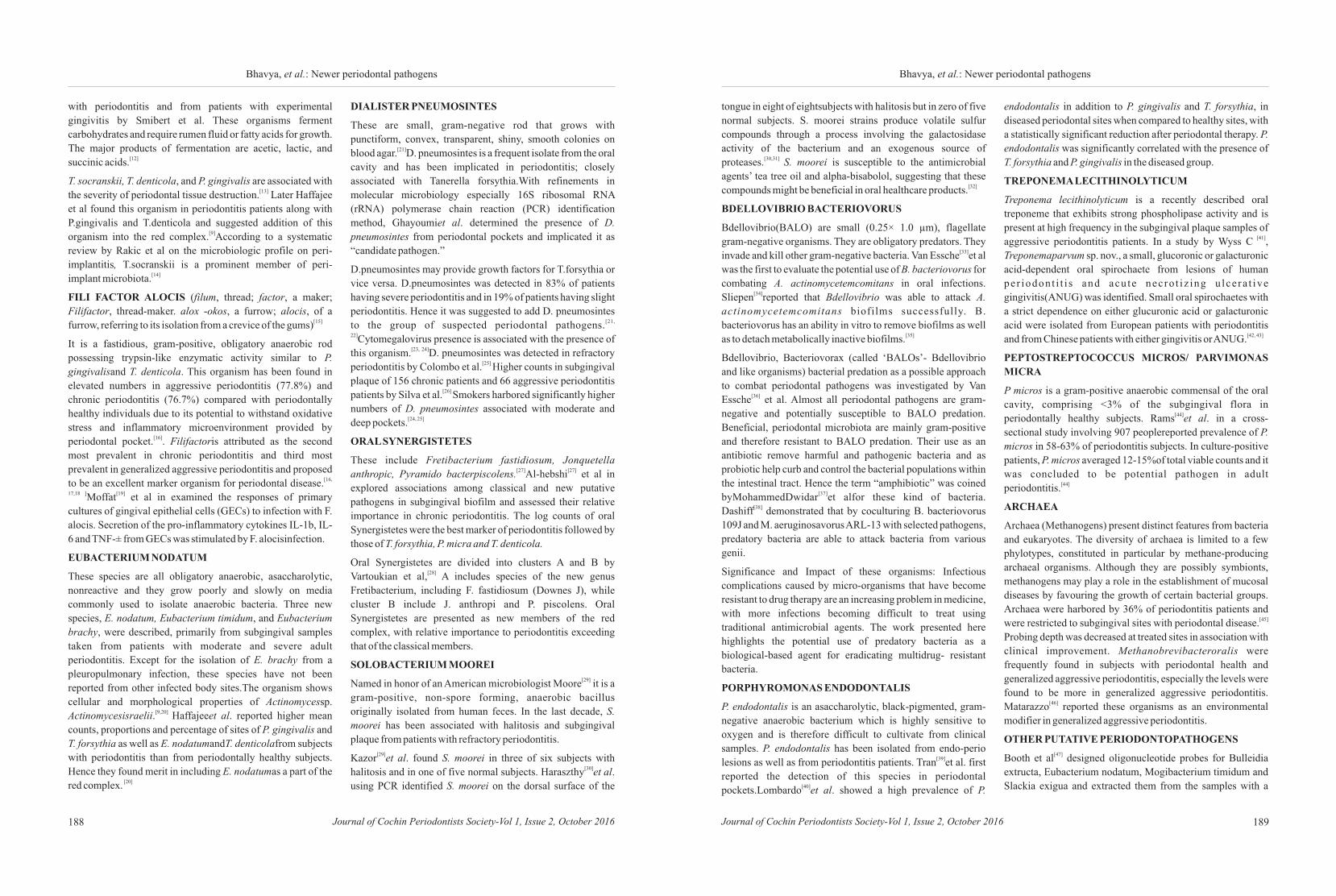
Journal of Cochin Periodontists Society-Vol 1, Issue 2, October 2016 Journal of Cochin Periodontists Society-Vol 1, Issue 2, October 2016188 189
Bhavya, et al.: Newer periodontal pathogensBhavya, et al.: Newer periodontal pathogens
with periodontitis and from patients with experimental
gingivitis by Smibert et al. These organisms ferment
carbohydrates and require rumen fluid or fatty acids for growth.
The major products of fermentation are acetic, lactic, and [12]succinic acids.
T. socranskii, T. denticola, and P. gingivalis are associated with [13]the severity of periodontal tissue destruction. Later Haffajee
et al found this organism in periodontitis patients along with
P.gingivalis and T.denticola and suggested addition of this [9]organism into the red complex. According to a systematic
review by Rakic et al on the microbiologic profile on peri-
implantitis, T.socranskii is a prominent member of peri-[14]implant microbiota.
FILI FACTOR ALOCIS (filum, thread; factor, a maker;
Filifactor, thread-maker. alox -okos, a furrow; alocis, of a [15]furrow, referring to its isolation from a crevice of the gums)
It is a fastidious, gram-positive, obligatory anaerobic rod
possessing trypsin-like enzymatic activity similar to P.
gingivalisand T. denticola. This organism has been found in
elevated numbers in aggressive periodontitis (77.8%) and
chronic periodontitis (76.7%) compared with periodontally
healthy individuals due to its potential to withstand oxidative
stress and inflammatory microenvironment provided by [16]periodontal pocket. . Filifactoris attributed as the second
most prevalent in chronic periodontitis and third most
prevalent in generalized aggressive periodontitis and proposed [16, to be an excellent marker organism for periodontal disease.
17,18 ] [19]Moffat et al in examined the responses of primary
cultures of gingival epithelial cells (GECs) to infection with F.
alocis. Secretion of the pro-inflammatory cytokines IL-1b, IL-
6 and TNF-± from GECs was stimulated by F. alocisinfection.
EUBACTERIUM NODATUM
These species are all obligatory anaerobic, asaccharolytic,
nonreactive and they grow poorly and slowly on media
commonly used to isolate anaerobic bacteria. Three new
species, E. nodatum, Eubacterium timidum, and Eubacterium
brachy, were described, primarily from subgingival samples
taken from patients with moderate and severe adult
periodontitis. Except for the isolation of E. brachy from a
pleuropulmonary infection, these species have not been
reported from other infected body sites.The organism shows
cellular and morphological properties of Actinomycessp. [9,20] Actinomycesisraelii. Haffajeeet al. reported higher mean
counts, proportions and percentage of sites of P. gingivalis and
T. forsythia as well as E. nodatumandT. denticolafrom subjects
with periodontitis than from periodontally healthy subjects.
Hence they found merit in including E. nodatumas a part of the [20]red complex.
DIALISTER PNEUMOSINTES
These are small, gram-negative rod that grows with
punctiform, convex, transparent, shiny, smooth colonies on [21]blood agar. D. pneumosintes is a frequent isolate from the oral
cavity and has been implicated in periodontitis; closely
associated with Tanerella forsythia.With refinements in
molecular microbiology especially 16S ribosomal RNA
(rRNA) polymerase chain reaction (PCR) identification
method, Ghayoumiet al. determined the presence of D.
pneumosintes from periodontal pockets and implicated it as
“candidate pathogen.”
D.pneumosintes may provide growth factors for T.forsythia or
vice versa. D.pneumosintes was detected in 83% of patients
having severe periodontitis and in 19% of patients having slight
periodontitis. Hence it was suggested to add D. pneumosintes [21, to the group of suspected periodontal pathogens.
22]Cytomegalovirus presence is associated with the presence of [23, 24]this organism. D. pneumosintes was detected in refractory
[25] periodontitis by Colombo et al. Higher counts in subgingival
plaque of 156 chronic patients and 66 aggressive periodontitis [26] patients by Silva et al. Smokers harbored significantly higher
numbers of D. pneumosintes associated with moderate and [24, 25]deep pockets.
ORAL SYNERGISTETES
These include Fretibacterium fastidiosum, Jonquetella [27] [27]anthropic, Pyramido bacterpiscolens. Al-hebshi et al in
explored associations among classical and new putative
pathogens in subgingival biofilm and assessed their relative
importance in chronic periodontitis. The log counts of oral
Synergistetes were the best marker of periodontitis followed by
those of T. forsythia, P. micra and T. denticola.
Oral Synergistetes are divided into clusters A and B by [28]Vartoukian et al, A includes species of the new genus
Fretibacterium, including F. fastidiosum (Downes J), while
cluster B include J. anthropi and P. piscolens. Oral
Synergistetes are presented as new members of the red
complex, with relative importance to periodontitis exceeding
that of the classical members.
SOLOBACTERIUM MOOREI[29]Named in honor of an American microbiologist Moore it is a
gram-positive, non-spore forming, anaerobic bacillus
originally isolated from human feces. In the last decade, S.
moorei has been associated with halitosis and subgingival
plaque from patients with refractory periodontitis.[29]Kazor et al. found S. moorei in three of six subjects with
[30]halitosis and in one of five normal subjects. Haraszthy et al.
using PCR identified S. moorei on the dorsal surface of the
tongue in eight of eightsubjects with halitosis but in zero of five
normal subjects. S. moorei strains produce volatile sulfur
compounds through a process involving the galactosidase
activity of the bacterium and an exogenous source of [30,31] proteases. S. moorei is susceptible to the antimicrobial
agents’ tea tree oil and alpha-bisabolol, suggesting that these [32]compounds might be beneficial in oral healthcare products.
BDELLOVIBRIO BACTERIOVORUS
Bdellovibrio(BALO) are small (0.25× 1.0 µm), flagellate
gram-negative organisms. They are obligatory predators. They [33]invade and kill other gram-negative bacteria. Van Essche et al
was the first to evaluate the potential use of B. bacteriovorus for
combating A. actinomycetemcomitans in oral infections. [34]Sliepen reported that Bdellovibrio was able to attack A.
actinomycetemcomitans biofilms successfully. B.
bacteriovorus has an ability in vitro to remove biofilms as well [35]as to detach metabolically inactive biofilms.
Bdellovibrio, Bacteriovorax (called ‘BALOs’- Bdellovibrio
and like organisms) bacterial predation as a possible approach
to combat periodontal pathogens was investigated by Van [36]Essche et al. Almost all periodontal pathogens are gram-
negative and potentially susceptible to BALO predation.
Beneficial, periodontal microbiota are mainly gram-positive
and therefore resistant to BALO predation. Their use as an
antibiotic remove harmful and pathogenic bacteria and as
probiotic help curb and control the bacterial populations within
the intestinal tract. Hence the term “amphibiotic” was coined [37]byMohammedDwidar et alfor these kind of bacteria.
[38]Dashiff demonstrated that by coculturing B. bacteriovorus
109J and M. aeruginosavorus ARL-13 with selected pathogens,
predatory bacteria are able to attack bacteria from various
genii.
Significance and Impact of these organisms: Infectious
complications caused by micro-organisms that have become
resistant to drug therapy are an increasing problem in medicine,
with more infections becoming difficult to treat using
traditional antimicrobial agents. The work presented here
highlights the potential use of predatory bacteria as a
biological-based agent for eradicating multidrug- resistant
bacteria.
PORPHYROMONAS ENDODONTALIS
P. endodontalis is an asaccharolytic, black-pigmented, gram-
negative anaerobic bacterium which is highly sensitive to
oxygen and is therefore difficult to cultivate from clinical
samples. P. endodontalis has been isolated from endo-perio [39]lesions as well as from periodontitis patients. Tran et al. first
reported the detection of this species in periodontal [40]pockets.Lombardo et al. showed a high prevalence of P.
endodontalis in addition to P. gingivalis and T. forsythia, in
diseased periodontal sites when compared to healthy sites, with
a statistically significant reduction after periodontal therapy. P.
endodontalis was significantly correlated with the presence of
T. forsythia and P. gingivalis in the diseased group.
TREPONEMA LECITHINOLYTICUM
Treponema lecithinolyticum is a recently described oral
treponeme that exhibits strong phospholipase activity and is
present at high frequency in the subgingival plaque samples of [41]aggressive periodontitis patients. In a study by Wyss C ,
Treponemaparvum sp. nov., a small, glucoronic or galacturonic
acid-dependent oral spirochaete from lesions of human
per iodont i t i s and acu te necro t iz ing u lcera t ive
gingivitis(ANUG) was identified. Small oral spirochaetes with
a strict dependence on either glucuronic acid or galacturonic
acid were isolated from European patients with periodontitis [42, 43]and from Chinese patients with either gingivitis or ANUG.
PEPTOSTREPTOCOCCUS MICROS/ PARVIMONAS
MICRA
P micros is a gram-positive anaerobic commensal of the oral
cavity, comprising <3% of the subgingival flora in [44]periodontally healthy subjects. Rams et al. in a cross-
sectional study involving 907 peoplereported prevalence of P.
micros in 58-63% of periodontitis subjects. In culture-positive
patients, P. micros averaged 12-15%of total viable counts and it
was concluded to be potential pathogen in adult [44]periodontitis.
ARCHAEA
Archaea (Methanogens) present distinct features from bacteria
and eukaryotes. The diversity of archaea is limited to a few
phylotypes, constituted in particular by methane-producing
archaeal organisms. Although they are possibly symbionts,
methanogens may play a role in the establishment of mucosal
diseases by favouring the growth of certain bacterial groups.
Archaea were harbored by 36% of periodontitis patients and [45] were restricted to subgingival sites with periodontal disease.
Probing depth was decreased at treated sites in association with
clinical improvement. Methanobrevibacteroralis were
frequently found in subjects with periodontal health and
generalized aggressive periodontitis, especially the levels were
found to be more in generalized aggressive periodontitis. [46]Matarazzo reported these organisms as an environmental
modifier in generalized aggressive periodontitis.
OTHER PUTATIVE PERIODONTOPATHOGENS[47]Booth et al designed oligonucleotide probes for Bulleidia
extructa, Eubacterium nodatum, Mogibacterium timidum and
Slackia exigua and extracted them from the samples with a
Journal of Cochin Periodontists Society-Vol 1, Issue 2, October 2016 Journal of Cochin Periodontists Society-Vol 1, Issue 2, October 2016190 191
Bhavya, et al.: Newer periodontal pathogensBhavya, et al.: Newer periodontal pathogens
chemiluminescent detection method. The levels of both E.
nodatum and S. exigua was significantly higher in deep than
shallow pockets. The level of E. nodatum, but not S. exigua,
was higher in patients than matched controls. Both E. nodatum
and S. exigua were associated with clinical indicators of
periodontal disease.
CRYPTOBACTERIUM CURTUM(Kryptos- hidden;
curtum- shortened; a hidden rod-shaped bacterium)
Cells are short gram-positive rods, occasionally gram-variable.
They are obligatory anaerobic, non-motile and non-sporing,
catalase negative and asaccharolytic. Individual cells occur
singly or in masses. C. curtum is characterized as an
opportunistic pathogen with a typical occurrence in the oral
cavity, involved in dental and oral infections such as [48]periodontitis, inflammation and abscess. Nakazawa et al.
isolated novel Eubacterium-like isolates, from the periodontal
pocket of an adult patient and necrotic dental pulp and named it
C. curtum.
PREVOTELLA HISTICOLA((histus: tissue; cola;
inhabitant, histicola inhabitant of tissue)
They are gram-negative nonmotile bacilli that are obligatory
anaerobes. Prevotellahisticola is found in the mucosal tissues
of the human oral cavity and is considered a normal flora of the
human oral microbiota. It is generally commensal but is known
to intrude the epithelial cells lining the cheeks. 16S rRNA gene
sequence analysis and DNA–DNA hybridization revealed that
the strains constituted a novel group within the genus
Prevotella, being most closely related to Prevotella [49] melaninogenica and Prevotella veroralis. Colonies are 1.5-
2.0mm in diameter, circular, entire, convex, cream-coloured
and opaque. Some strains produce black colonies in the
presence of metronidazole and other strains form bull’s-eye
colonies with reddish-brown pigmentation centers. Cells are
saccharolytic and are able to ferment common sugars. Major
amounts of acetic acid, succinic acid, lactic acid are produced [49]as end products of metabolism in peptone/yeast extract broth.
Newer species implicated in the causation of periodontitis are
uncultivated clones D084 and BH017 from the Deferribacte-
resphylum, AU126 from the Bacteroidete sphylum,
Megasphaeraclone BB166 and clone I025 from the TM7
phylum, and Eubacterium saphenum. . Species or phylotypes
more prevalent inperiodontal health included two uncultivated
phylotypes, clone W090 from the Deferri-bacteresphylum and
clone BU063 from the Bacteroidetes, and named species [11]Atopobiumrimae and Atopobiumparvulum.
SIGNIFICANCE OF KNOWING ABOUT NEWER
PATHOGENS
The subgingival pocket is a complex environment that
harbours a highly diverse microbiota. It seems evident that
other microorganisms might be involved in the onset and/or
progression of periodontitis. In periodontitis bacteria and other
pathogens could be described as the initiators of disease and the
host immune inflammatory response as the promoter of tissue
destruction. However owing to the chronic nature of
periodontitis early initiators might have long induction periods
of months or years because of which disease does not occur
immediately as soon as the bacteria learns to thrive in its
environment. Host immune response also throws its weight
around determining if the individual becomes susceptible to
disease or not.
However bacteria alone may not promote disease progression.
Viruses like human cytomegalovirus and herpes virus can alter
the subgingival environment increasing the pathogenicity of
harmless commensals in the periodontal pocket. For instance, [50]Slots showed that the conversion of a gingivitis lesion to a
periodontitis lesion and of a stable lesion to a progressing one
could reflect cycles of activity and latency in herpesvirus
infection of the periodontium.
It is the author’s view that our current knowledge about
pathogens in the dental plaque biofilm will help us to tailor-
make a treatment plan for every patient. Most of our current
treatment modalities still depend on the old non-specific plaque
hypothesis. By investigating the susceptibility of
periopathogens to mechanical therapy, probiotic therapy,
surgical intervention or any other therapy we will be able to
treat and control the epidemic of periodontitis without
depending wholly on chemotherapeutic agents. In order to
make progress of such an idea, advances have to be made in
targeting individual organisms rather than the community as a
whole. This will lead to majordisruption of the biofilm colony
formation (cutting off their major nutrient supply)and collapse
the biofilm thereby giving us huge leeway into treating
periodontitis. This translates in the form of reduction of
probing pocket depth or clinical attachment gain. This will also
help researchers find out exactly how much host responses
pave the way to progression of periodontitis.
CONCLUSION
The etiologic role of newer microorganisms would need to be
confirmed by risk assessment and interventional studies to
evaluate whether their reduction or elimination would be
accompanied by clinical improvements and whether their
persistence would lead to disease progression. In addition,
further investigation into their mechanisms of pathogenicity
and their ability to promote or evade host immune response
would be required. Let us hope that future researches into this
new world of microbial periodontics help us to treat the global
epidemic of periodontitis in a better fashion.
REFERENCES
1. Socransky, Sigmund S. “Criteria for the infectious agents
in dental caries and periodontal disease.” Journal of
clinical periodontology 6.7 (1979): 16-21.
2. Socransky SS, Haffajee AD, Cugini MA, Smith C, Kent
RL Jr. Microbial complexes in subgingival plaque. J
ClinPeriodontol 1998; 25:134-44.
3. Paster BJ, Boches SK, Galvin JL, Ericson RE, Lau CN,
Levanos VA, et al. Bacterial diversity in human
subgingival plaque. J Bacteriol 2001;183:3770-83.
4. Kumar PS, Griffen AL, Moeschberger ML, Leys EJ.
Identification of candidate periodontal pathogens and
beneficial species by quantitative 16S clonal analysis. J
ClinMicrobiol 2005; 43:3944-55.
5. Matarazzo, Flavia, et al. “Diversity and quantitative
analysis of Archaea in aggressive periodontitis and
periodontally healthy subjects.” Journal of clinical
periodontology 38.7 (2011): 621-627.
6. Teles R, Teles F, Frias-Lopez J, Paster B, Haffajee A.
Lessons learned and unlearned in periodontal
microbiology. Periodontol 2000 2013;62:95-162.
7. Griffen, Ann L., et al. “Distinct and complex bacterial
profiles in human periodontitis and health revealed by 16S
pyrosequencing.” The ISME journal 6.6 (2012): 1176-
1185.
8. Abusleme, Loreto, et al. “The subgingival microbiome in
health and periodontitis and its relationship with
community biomass and inflammation.” The ISME
journal 7.5 (2013): 1016-1025.
9. Haffajee AD, Socransky SS, Patel MR, Song X. Microbial
c o m p l e x e s i n s u p r a g i n g i v a l p l a q u e . O r a l
MicrobiolImmunol 2008;23:196-205.
10. Hajishengallis G, Lamont RJ. Beyond the red complex and
into more complexity: The polymicrobial synergy and
dysbiosis (PSD) model of periodontal disease etiology.
Mol Oral Microbiol 2012;27:409-19.
11. Kumar PS, Griffen AL, Barton JA, Paster BJ,
Moeschberger ML, Leys EJ. New bacterial species
associated with chronic periodontitis. J Dent Res
2003;82:338-44.
12. Smibert, Robert M., John L. Johnson, and Richard R.
R a n n e y. “ Tr e p o n e m a s o c r a n s k i i s p . n o v. ,
Treponemasocranskii subsp. socranskii subsp. nov.,
Treponemasocranskii subsp. buccale subsp. nov., and
Treponemasocranskii subsp. paredis subsp. nov. Isolated
from the Human Periodontia.” International Journal of
Systematic and Evolutionary Microbiology 34.4 (1984):
457-462.
13. Takeuchi, Yasuo, et al. “Treponemasocranskii,
Treponemadenticola, and Porphyromonasgingivalis are
associated with severity of periodontal tissue destruction.”
Journal of periodontology 72.10 (2001): 1354-1363.
14. Rakic, Mia, Maria Gabriella Grusovin, and Luigi Canullo.
“The Microbiologic Profile Associated with Peri-
lmplantitis in Humans: A Systematic Review.”
International Journal of Oral & Maxillofacial Implants
31.2 (2016).
15. Arora, Nupur, Ashank Mishra, and Samir Chugh.
“Microbial role in periodontitis: Have we reached the top?
Some unsung bacteria other than red complex.” Journal of
Indian Society of Periodontology 18.1 (2014): 9.
16. Schlafer S, Riep B, Griffen AL, Petrich A, Hübner J,
Berning M, et al. Filifactoralocis– Involvement in
periodontal biofilms. BMC Microbiol 2010;10:66.
17. Dahlén G, Leonhardt A. A new checkerboard panel for
testing bacterial markers in periodontal disease. Oral
MicrobiolImmunol 2006;21:6-11.
18. Aruni, A. Wilson, Francis Roy, and H. M. Fletcher.
“Filifactoralocis has virulence attributes that can enhance
its persistence under oxidative stress conditions and
m e d i a t e i n v a s i o n o f e p i t h e l i a l c e l l s b y
Porphyromonasgingivalis.” Infection and immunity 79.10
(2011): 3872-3886.
19. Moffatt, Catherine E., et al. “Filifactoralocis interactions
with gingival epithelial cells.” Molecular oral
microbiology 26.6 (2011): 365-373.
20. Haffajee AD, Teles RP, Socransky SS. Association of
Eubacteriumnodatum and Treponemadenticola with
human periodontitis lesions. Oral MicrobiolImmunol
2006;21:269-82.
21. Ghayoumi N, Chen C, Slots J. Dialisterpneumosintes, a
new putative periodontal pathogen. J Periodontal Res
2002;37:75-8.
22. Moore, W. E. C., and Lillian VH Moore. “The bacteria of
periodontal diseases.” Periodontology 2000 5.1 (1994):
66-77.
23. Contreras, Adolfo, Javier Enrique Botero, and Jørgen
Slots. “Biology and pathogenesis of cytomegalovirus in
periodontal disease.” Periodontology 2000 64.1 (2014):
40-56.
24. Slots, J., C. Sugar, and J. J. Kamma. “Cytomegalovirus
periodontal presence is associated with subgingival
Dialisterpneumosintes and alveolar bone loss.” Oral
microbiology and immunology 17.6 (2002): 369-374.
25. Colombo AP, Boches SK, Cotton SL, Goodson JM, Kent
R, Haffajee AD, et al. Comparisons of subgingival
microbial profiles of refractory periodontitis, severe
periodontitis, and periodontal health using the human oral
microbe identification microarray. J Periodontol 2009;
80:1421-32.
Journal of Cochin Periodontists Society-Vol 1, Issue 2, October 2016192
Bhavya, et al.: Newer periodontal pathogens
26. Silva-Boghossian CM, Neves AB, Resende FA, Colombo
AP. Suppuration associated bacteria in subjects with
chronic and aggressive periodontitis. J Periodontol 2013;
84:e9-16.
27. Al hebshi, N. N., et al. “Quantitative analysis of classical
and new putative periodontal pathogens in subgingival
biofilm: a case–control study.” Journal of periodontal
research 50.3 (2015): 320-329.
28. Vartoukian, Sonia R., Richard M. Palmer, and William G.
Wade. “The division “Synergistes”.” Anaerobe 13.3
(2007): 99-106.
29. Kazor CE, Mitchell PM, Lee AM, Stokes LN, Loesche WJ,
Dewhirst FE, et al. Diversity of bacterial populations on
the tongue dorsa of patients with halitosis and healthy
patients. J ClinMicrobiol 2003;41:558-63.
30. Haraszthy, V. I., et al. “Characterization and prevalence of
Solobacteriummoorei associated with oral halitosis.”
Journal of breath research 2.1 (2008): 017002.
31. Tanabe S, Grenier D. Characterization of volatile sulphur
compound production by Solobacteriummoorei. Arch Oral
Biol 2012;57:1639-43.
32. Forrer, Marcel, et al. “The antimicrobial activity of alpha-
bisabolol and tea tree oil against Solobacteriummoorei, a
Gram-positive bacterium associated with halitosis.”
Archives of oral biology 58.1 (2013): 10-16.
33. Van Essche, Mark, et al. “Bdellovibriobacteriovorus
attacks Aggregatibacteractinomycetemcomitans.” Journal
of dental research 88.2 (2009): 182-186.
34. S l i e p e n , I . , e t a l . “ I n t e r f e r e n c e w i t h
Aggregatibacteractinomycetemcomitans: colonization of
epithelial cells under hydrodynamic conditions.” Oral
microbiology and immunology 24.5 (2009): 390-395.
35. Dashiff, A., and D. E. Kadouri. “Predation of oral
pathogens by Bdellovibriobacteriovorus 109J. Mol. Oral
Microbiol. 26: 19–34.” (2011).
36. Van Essche, Mark, et al. “Killing of anaerobic pathogens
by predatory bacteria.” Molecular oral microbiology 26.1
(2011): 52-61.
37. Dwidar, Mohammed, Ajay KalanjanaMonnappa, and
Robert J. Mitchell. “The dual probiotic and antibiotic
nature of Bdellovibriobacteriovorus.” BMB reports 45.2
(2012): 71-78.
38. Dashiff, A., et al. “Predation of human pathogens by the
predatory bacteria Micavibrioaeruginosavorus and
Bdellovibriobacteriovorus.” Journal of applied
microbiology 110.2 (2011): 431-444.
39. T r a n T , F l y n n M J , C h e n C , S l o t s J .
Porphyromonasendodontalisin subgingival plaque. Clin
Infect Dis 1997;25Suppl 2:S222-3.
40. Lombardo Bedran TB, Marcantonio RA, Spin Neto R,
Alves Mayer MP, Grenier D, Spolidorio LC, et al.
Porphyromonasendodontalisin chronic periodontitis: A
clinical and microbiological cross-sectional study. J Oral
Microbiol 2012;4:10.
41. Wyss, C., et al. “Treponemalecithinolyticum sp. nov., a
small saccharolyticspirochaete with phospholipase A and
C activities associated with periodontal diseases.”
International Journal of Systematic and Evolutionary
Microbiology 49.4 (1999): 1329-1339.
42. Park KK, Heuner K, Göbel UB, Yoo YJ, Kim CK, Choi
BK. Cloning and characterization of a major surface
protein (MspTL) of Treponemalecithinolyticumassociated
with rapidly progressive periodontitis. FEMS
MicrobiolLett2002;207:185-92.
43. Moter A, Riep B, Haban V, Heuner K, Siebert G, Berning
M, et al. Molecular epidemiology of oral treponemes in
patients with periodontitis and in periodontitis-resistant
subjects. J ClinMicrobiol2006;44:3078-85.
44. Rams TE, Feik D, Listgarten MA, Slots J.
Peptostreptococcus micros in human periodontitis. Oral
MicrobiolImmunol 1992;7:1-6.
45. Lepp, Paul W., et al. “MethanogenicArchaea and human
periodontal disease.” Proceedings of the National
Academy of Sciences of the United States of America
101.16 (2004): 6176-6181.
46. Matarazzo, Flavia, et al. “Diversity and quantitative
analysis of Archaea in aggressive periodontitis and
periodontally healthy subjects.” Journal of clinical
periodontology 38.7 (2011): 621-627.
47. Booth, V., et al. “Gram positive anaerobic bacilli in human
periodontal disease.” Journal of periodontal research 39.4
(2004): 213-220.
48. Nakazawa, Futoshi, et al. “Cryptobacteriumcurtum gen.
nov., sp. nov., a new genus of Gram-positive anaerobic rod
isolated from human oral cavities.” International Journal
of Systematic and Evolutionary Microbiology 49.3
(1999): 1193-1200.
49. Downes, Julia, et al. “Prevotellahisticola sp. nov., isolated
from the human oral cavity.” International journal of
systematic and evolutionary microbiology 58.8 (2008):
1788-1791.
50. Slots J. Herpesviruses, the missing link between
gingivitisand periodontitis? J IntAcadPeriodontol2004: 6:
113–119.
193
ABSTRACT:
Purpose: The aim of this study was to evaluate the effect of diode LASER and
arginine containing desensitizing agent on coronal dentin as measured by dentinal
occlusion assessed by SEM analysis.
Materials and methods: 10 sound teeth extracted for orthodontic purpose from the
patients of age group 15 to 30 years. Specimens were divided into two groups each
with 5 teeth specimen. Group 1 Specimens were desensitized for 3 minutes by diode
LASER and group 2 specimens were brushed with arginine containing desensitizing
agent for twice in a day. The dentinal tubule occlusions of two group specimens were
analyzed by scanning electron microscope.
Results :The LASER treated teeth samples shows average result of 29 %
occluded dentinal tubules and the teeth samples which were treated with Arginine
desensitizing agent gave a result, of an average 22% occluded dentinal tubules
Conclusion: The pilot study results shows immediate positive effect by LASER
technique than the desensitizing agent used in our study. Thus we require a larger
sample size to prove the same.
Key words: Dentin hypersensitivity, diode LASER, desensitizing agent, scanning
electron microscope, dentinal tubule occlusion.
Access this article online
Website :
jcops.copsonweb.org
Quick Response Code
How to cite this article: Log on to jcops.copsonweb.org. Arun Narayanan, Ajay Bhat, Shabeer Ali K.. Comparative evaluation of the effect of diode LASER and arginine containing desensitizing agent: An in vitro SEM pilot study. Journal of Cochin Periodontists Society 2016;1: 193-196
Conflict of Interest: None declared Source of Support: Nil
BASIC RESEARCH
Department of Periodontology,
Kannur dental college,
Kannur, Karala, India
Address for Correspondence:
Shabeer ali K P , Department of Periodontology, Kannur dental
college, Kannur, India. E-mail: [email protected]
Date of Submission: 2- 06 -2016
Date of acceptance: 29 -07-2016
Journal of Cochin Periodontists Society-Vol 1, Issue 2, October 2016
Arun Narayanan, Ajay Bhat, Shabeer Ali K
Comparative evaluation of the effect of diode LASER and arginine containing desensitizing agent: An in vitro SEM pilot study
INTRODUCTION:
Dentin Hypersensitivity (DH) or
cervical dentin sensitivity may be
defined as pain arising from the exposed
dentin, typically in response to a
chemical, thermal, tactile, or osmotic
stimulus that cannot be explained as
arising from any other form of dental [1] defect or pathology. The prevalence of
this disorder has been reported in [2]different studies to vary from 4–73%.
The exposure of dentin and its sensitivity
may occur via one or both of two
processes: removal of enamel, or
denudation of the root surface due to loss
of the overlying cementum and [3]periodontal tissues.
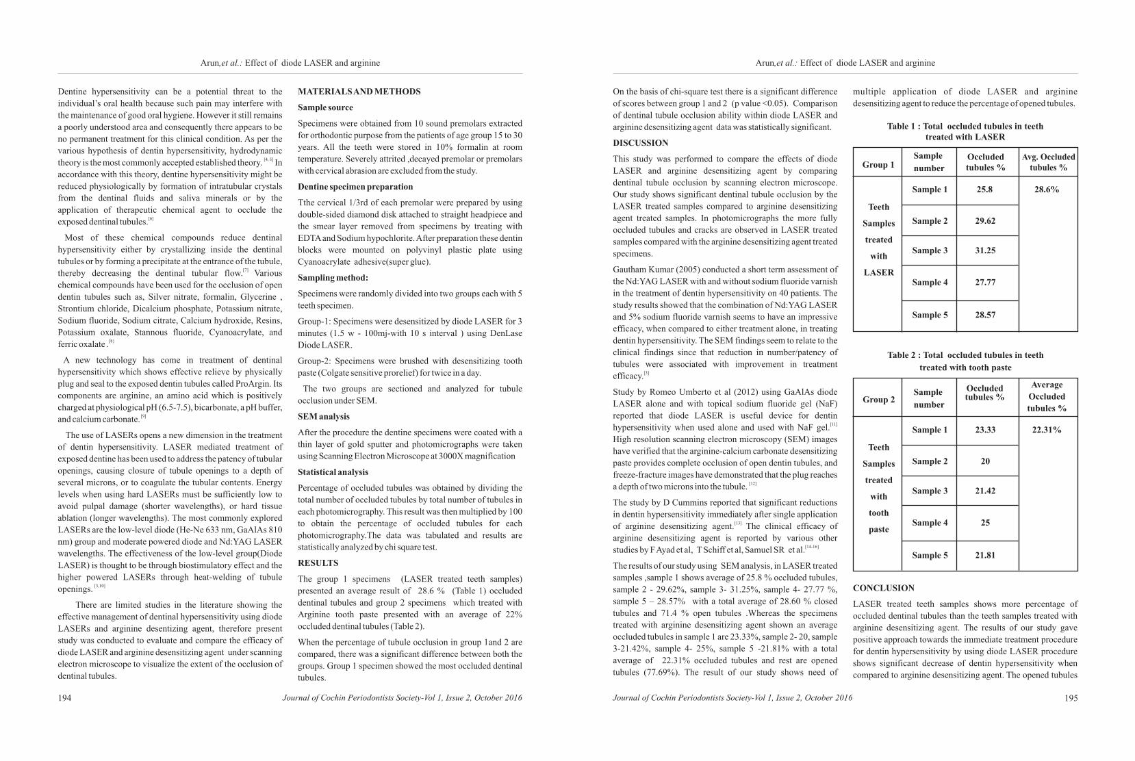
Journal of Cochin Periodontists Society-Vol 1, Issue 2, October 2016194 195
Arun,et al.: Effect of diode LASER and arginineArun,et al.: Effect of diode LASER and arginine
Dentine hypersensitivity can be a potential threat to the
individual’s oral health because such pain may interfere with
the maintenance of good oral hygiene. However it still remains
a poorly understood area and consequently there appears to be
no permanent treatment for this clinical condition. As per the
various hypothesis of dentin hypersensitivity, hydrodynamic [4, 5]theory is the most commonly accepted established theory. In
accordance with this theory, dentine hypersensitivity might be
reduced physiologically by formation of intratubular crystals
from the dentinal fluids and saliva minerals or by the
application of therapeutic chemical agent to occlude the [6]exposed dentinal tubules.
Most of these chemical compounds reduce dentinal
hypersensitivity either by crystallizing inside the dentinal
tubules or by forming a precipitate at the entrance of the tubule, [7] thereby decreasing the dentinal tubular flow. Various
chemical compounds have been used for the occlusion of open
dentin tubules such as, Silver nitrate, formalin, Glycerine ,
Strontium chloride, Dicalcium phosphate, Potassium nitrate,
Sodium fluoride, Sodium citrate, Calcium hydroxide, Resins,
Potassium oxalate, Stannous fluoride, Cyanoacrylate, and [8}ferric oxalate .
A new technology has come in treatment of dentinal
hypersensitivity which shows effective relieve by physically
plug and seal to the exposed dentin tubules called ProArgin. Its
components are arginine, an amino acid which is positively
charged at physiological pH (6.5-7.5), bicarbonate, a pH buffer, [9]and calcium carbonate.
The use of LASERs opens a new dimension in the treatment
of dentin hypersensitivity. LASER mediated treatment of
exposed dentine has been used to address the patency of tubular
openings, causing closure of tubule openings to a depth of
several microns, or to coagulate the tubular contents. Energy
levels when using hard LASERs must be sufficiently low to
avoid pulpal damage (shorter wavelengths), or hard tissue
ablation (longer wavelengths). The most commonly explored
LASERs are the low-level diode (He-Ne 633 nm, GaAlAs 810
nm) group and moderate powered diode and Nd:YAG LASER
wavelengths. The effectiveness of the low-level group(Diode
LASER) is thought to be through biostimulatory effect and the
higher powered LASERs through heat-welding of tubule [3,10]openings.
There are limited studies in the literature showing the
effective management of dentinal hypersensitivity using diode
LASERs and arginine desentizing agent, therefore present
study was conducted to evaluate and compare the efficacy of
diode LASER and arginine desensitizing agent under scanning
electron microscope to visualize the extent of the occlusion of
dentinal tubules.
MATERIALS AND METHODS
Sample source
Specimens were obtained from 10 sound premolars extracted
for orthodontic purpose from the patients of age group 15 to 30
years. All the teeth were stored in 10% formalin at room
temperature. Severely attrited ,decayed premolar or premolars
with cervical abrasion are excluded from the study.
Dentine specimen preparation
Tthe cervical 1/3rd of each premolar were prepared by using
double-sided diamond disk attached to straight headpiece and
the smear layer removed from specimens by treating with
EDTA and Sodium hypochlorite. After preparation these dentin
blocks were mounted on polyvinyl plastic plate using
Cyanoacrylate adhesive(super glue).
Sampling method:
Specimens were randomly divided into two groups each with 5
teeth specimen.
Group-1: Specimens were desensitized by diode LASER for 3
minutes (1.5 w - 100mj-with 10 s interval ) using DenLase
Diode LASER.
Group-2: Specimens were brushed with desensitizing tooth
paste (Colgate sensitive prorelief) for twice in a day.
The two groups are sectioned and analyzed for tubule
occlusion under SEM.
SEM analysis
After the procedure the dentine specimens were coated with a
thin layer of gold sputter and photomicrographs were taken
using Scanning Electron Microscope at 3000X magnification
Statistical analysis
Percentage of occluded tubules was obtained by dividing the
total number of occluded tubules by total number of tubules in
each photomicrography. This result was then multiplied by 100
to obtain the percentage of occluded tubules for each
photomicrography.The data was tabulated and results are
statistically analyzed by chi square test.
RESULTS
The group 1 specimens (LASER treated teeth samples)
presented an average result of 28.6 % (Table 1) occluded
dentinal tubules and group 2 specimens which treated with
Arginine tooth paste presented with an average of 22%
occluded dentinal tubules (Table 2).
When the percentage of tubule occlusion in group 1and 2 are
compared, there was a significant difference between both the
groups. Group 1 specimen showed the most occluded dentinal
tubules.
On the basis of chi-square test there is a significant difference
of scores between group 1 and 2 (p value <0.05). Comparison
of dentinal tubule occlusion ability within diode LASER and
arginine desensitizing agent data was statistically significant.
DISCUSSION
This study was performed to compare the effects of diode
LASER and arginine desensitizing agent by comparing
dentinal tubule occlusion by scanning electron microscope.
Our study shows significant dentinal tubule occlusion by the
LASER treated samples compared to arginine desensitizing
agent treated samples. In photomicrographs the more fully
occluded tubules and cracks are observed in LASER treated
samples compared with the arginine desensitizing agent treated
specimens.
Gautham Kumar (2005) conducted a short term assessment of
the Nd:YAG LASER with and without sodium fluoride varnish
in the treatment of dentin hypersensitivity on 40 patients. The
study results showed that the combination of Nd:YAG LASER
and 5% sodium fluoride varnish seems to have an impressive
efficacy, when compared to either treatment alone, in treating
dentin hypersensitivity. The SEM findings seem to relate to the
clinical findings since that reduction in number/patency of
tubules were associated with improvement in treatment [3]efficacy.
Study by Romeo Umberto et al (2012) using GaAlAs diode
LASER alone and with topical sodium fluoride gel (NaF)
reported that diode LASER is useful device for dentin [11] hypersensitivity when used alone and used with NaF gel.
High resolution scanning electron microscopy (SEM) images
have verified that the arginine-calcium carbonate desensitizing
paste provides complete occlusion of open dentin tubules, and
freeze-fracture images have demonstrated that the plug reaches [12]a depth of two microns into the tubule.
The study by D Cummins reported that significant reductions
in dentin hypersensitivity immediately after single application [13] of arginine desensitizing agent. The clinical efficacy of
arginine desensitizing agent is reported by various other [14-16]studies by F Ayad et al, T Schiff et al, Samuel SR et al.
The results of our study using SEM analysis, in LASER treated
samples ,sample 1 shows average of 25.8 % occluded tubules,
sample 2 - 29.62%, sample 3- 31.25%, sample 4- 27.77 %,
sample 5 – 28.57% with a total average of 28.60 % closed
tubules and 71.4 % open tubules .Whereas the specimens
treated with arginine desensitizing agent shown an average
occluded tubules in sample 1 are 23.33%, sample 2- 20, sample
3-21.42%, sample 4- 25%, sample 5 -21.81% with a total
average of 22.31% occluded tubules and rest are opened
tubules (77.69%). The result of our study shows need of
multiple application of diode LASER and arginine
desensitizing agent to reduce the percentage of opened tubules.
Journal of Cochin Periodontists Society-Vol 1, Issue 2, October 2016
Table 1 : Total occluded tubules in teeth treated with LASER
Teeth
Samples
treated
with
LASER
28.6%Sample 1
Sample 2
Sample 3
Sample 4
Sample 5
25.8
29.62
31.25
27.77
28.57
Teeth
Samples
treated
with
tooth
paste
Average
Occluded
tubules %
22.31%Sample 1
Sample 2
Sample 3
Sample 4
Sample 5
23.33
20
21.42
25
21.81
Group 2Occluded tubules %
Table 2 : Total occluded tubules in teeth
treated with tooth paste
CONCLUSION
LASER treated teeth samples shows more percentage of
occluded dentinal tubules than the teeth samples treated with
arginine desensitizing agent. The results of our study gave
positive approach towards the immediate treatment procedure
for dentin hypersensitivity by using diode LASER procedure
shows significant decrease of dentin hypersensitivity when
compared to arginine desensitizing agent. The opened tubules
Occluded tubules %
Avg. Occluded tubules %
Journal of Cochin Periodontists Society-Vol 1, Issue 2, October 2016196
Arun,et al.: Effect of diode LASER and arginine
in the both samples shows the need of further study about
multiple application and long duration of application of
LASER and desensitizing agent by using large sample size.
REFERENCES
1) Lopes A O, Aranha A C C.Comparative Evaluation of the
Effects of Nd:YAG LASER and a desensitizer agent on the
treatment of dentin hypersensitivity: a clinical study.
Photomedicine and LASER Surgery 2013; 31:132-37.
2) Mogharehabed A, Khademi H, Zamharir Z A, Bouraima
S A, Yaghini J, Poormoradi B.Comparative evaluation of
the effects of 5% sodium fluoride varnish and neodymium-
doped yttrium aluminium garnet (Nd:YAG) LASER and
their combined application on dentin hypersensitivity
treatment. J LASERs Med Sci 2012;3:109-14.
3) Kumar G, D.S. Mehta. Short-term assessment of the
Nd:YAG LASER with and without sodium fluoride
varnish in the treatment of dentin hypersensitivity – a
clinical and scanning electron microscopy study.J
Periodontol 2005;76:1140-47.
4) Kolker J L, Vargas M A, Armstrong, S.R , Dawson D V.
Effects of desensitizing agents on dentin permeability and
dentin tubule occlusion. J Adhes Dent 2002;4:211-21.
5) E.G.Absi, M.Addy, D.Adams. Dentin hypersensitivity-
the effect of tooth brushing and dietary compounds on
dentin invitro: an SEM study. J Oral Rehabil 1992; 19:101-
10.
6) Cuenin M F, Scheidt M J, O’Neal R B, Strong S L, Pashley
D H, Horner J A, Van Dyke T E. An invivo study of dentin
sensitivity: the relation of dentin sensitivity and the
patency of dentin tubules. J Periodontol 1991; 62:668-73.
7) Absi, E.G, Addy M, Adams D. Dentin hypersensitivity-
the development and evaluation of replica technique to
study sensitive and nonsensitive cervical dentin. J Clin
Periodontol 1989; 16:190-95.
8) Reinhart T C, Killoy W J, Love J, Overman P R, Sakumura
J.S . The effectiveness of a patient-applied tooth
desensitizing gel pilot study. J Clin Periodontol
1990;17:123-27.
9) Markowitz K, Pashley DH. Discovering new treatments
for sensitive teeth:The long path from biology to therapy. J
Oral Rehabil 2007;35:300-15.
10) Asnaashari M, Moeini M. Effectiveness of LASERs in the
Treatment of Dentin Hypersensitivity. J LASERs Med Sci
2013; 4(1):1-7.
11) Treatment of Dentine Hypersensitivity by Diode
LASER.A Clinical Study. Umberto R,Claudia R, Gaspare
P, Gianluca T, Alessandro D V. Hindawi Publishing
Corporation International Journal of Dentistry 2012.
12) Petrou I, Heu R, Stranick M, Lavender S, Zaidel L,
Cummins D, Sullivan, RJ, Hsueh C, Gimzewski JK: A
breakthrough therapy for dentin hyper- sensitivity: How
dental products containing 8% arginine and calcium car-
bonate work to deliver effective relief of sensitive teeth. J
Clin Dent 2009; 20: 23-31.
13) Cummins D.The Efficacy of a New Dentifrice Containing
8.0% Arginine,Calcium Carbonate, and 1450 ppm
Fluoride in Delivering Instant and Lasting Relief of Dentin
Hypersensitivity.J Clin Dent 2009 ;20: 109–14.
14) Ayad F et al.Comparing the efficacy in providing instant
relief of dentin hypersensitivity of a new oothpaste
containing 8.0% arginine,calcium carbonate, and 1450
ppm fluoride to a benchmark desensitizing toothpaste
containing 2% potassium ion and 1450 ppm fluoride,and to
a control toothpaste with 1450 ppm fluoride: A Three-Day
Clinical Study in Mississauga, Canada. J Clin Dent 2009
;20: 115-22.
15) Schiff T et al.The clinical effect of a single direct topical
application of a dentifrice containing 8.0% arginine,
calcium carbonate,and 1450 ppm fluoride on dentin
hypersensitivity: the use of a cotton swab applicator versus
the use of a fingertip. J Clin Dent 2009 ;20: 131-36.
16) Samuel SR, Khatri SG, Acharya S. Clinical Evaluation of
self and professionally applied desensitizing agents in
relieving dentin hypersensitivity after a single topical
application: A Randomized Controlled Trial. J Clin Exp
Dent. 2014;6(4): 339-43.
197
ABSTRACT
Aim: To assess the similarity in mineral composition of dental calculus and saliva.
Materials&Methods:The dental calculus collected from patients mouth wasassessed
under UV spectrometer &Inductively Coupled Plasma Mass Spectrometer (ICP-MS)
for its mineral contents.
Result: Within the limitations of the spectrometer used, the given sample of calculus
contained Calcium (11.43%), Magnesium (5080.64 mg/kg), Sodium (5096.60
mg/kg), Phosphorus (413 .64 mg/kg)&Zinc (140.22mg/kg).
Conclusion: Analysing the complete mineral content of calculus needs much better
assessment techniques. The spectrometers used could detect mainly inorganic
components like Calcium, Magnesium& Phosphorus and trace amount of Sodium
and Zinc. The mineral components were almost same as that of saliva
Keywords : Dental calculus, saliva, minerals, calcium, spectrometer, inductively
coupled plasma
Access this article online
Website :
jcops.copsonweb.org
Quick Response Code
How to cite this article: Log on to jcops.copsonweb.org. Suji A S, Anila Joseph, M K Saleena, Chaithra P, SwethaValsan, Elizabeth Kuruvilla. Analysis of mineral contents of dental calculus & assessment of its similarity with saliva. Journal of Cochin Periodontists Society 2016;1:
Conflict of Interest: None declared Source of Support: Nil
BASIC RESEARCH
Department of Clinical
Periodontology and Oral
Implantology, Royal Dental College,
Chalissery, Palakkad, Kerala, India
Address for Correspondence:
Suji A S, Department of Clinical
Peridontology and Oral
Implantology,Royal Dental College,
Chalissery, Palakkad- 679536, Kerala,
India. E-mail: [email protected]
Date of Submission: 4- 07 -2016
Date of acceptance: 22-07-2016
Journal of Cochin Periodontists Society-Vol 1, Issue 2, October 2016
Suji A S, Anila Joseph, M K Saleena, Chaithra P,
Elizabeth Kuruvilla
SwethaValsan,
Analysis of mineral contents of dental calculus & assessment of its similarity with saliva
INTRODUCTION:
Research Question: Is there any
similarity in the mineral composition of
dental calculus and saliva
Bacterial plaque is believed to be the
prime initiating factor of periodontal
disease along with other predisposing
factors including local, genetic, [1]environmental and acquired factors.
The dental calculus is formed by the
mineralization of the bacterial plaque,
resulting from its interaction with the
influx of mineral salts from saliva or [2]GCF. Saliva is known to be a reservoir
o f minera l s tha t con t inuous ly
remineralise the enamel and also
become a source of calcification of
plaque. The mineral content within
calculus is explained by periodic
differences in surrounding environment [3]which is primarily saliva. A study that
could assess the mineral content of
dental calculus could reveal its
relationship with the saliva. Such a study
was conducted her with a rationale of
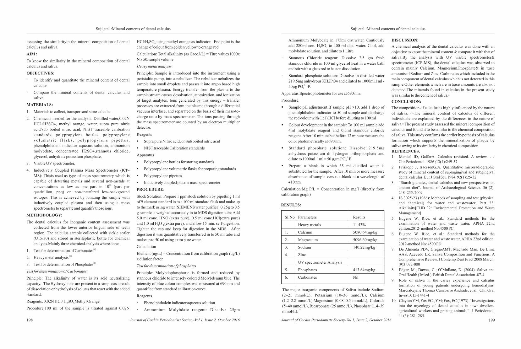
Journal of Cochin Periodontists Society-Vol 1, Issue 2, October 2016 Journal of Cochin Periodontists Society-Vol 1, Issue 2, October 2016198 199
Suji,etal.:Mineral contents of dental calculusSuji,etal.:Mineral contents of dental calculus
assessing the similarityin the mineral composition of dental
calculus and saliva.
AIM :
To know the similarity in the mineral composition of dental
calculus and saliva.
OBJECTIVES:
· To identify and quantitate the mineral content of dental
calculus
· Compare the mineral contents of dental calculus and
saliva.
MATERIALS:
1. Materials to collect, transport and store calculus
2. Chemicals needed for the analysis: Distilled water,0.02N
HCL/H2SO4, methyl orange, water, supra pure nitric
acid/sub boiled nitric acid, NIST traceable calibration
standards, polypropylene bottles, polypropylene
vo lume t r i c f l a sks , po lyp ropy lene p ipe t t e s ,
phenolphthalein indicator aqueous solution, ammonium
molybdate, concentrated H2SO4,stannous chloride,
glycerol, anhydrate potassium phosphate,
3. Visible UV spectrometer.
4. Inductively Coupled Plasma Mass Spectrometer (ICP-
MS): Thisis used as type of mass spectrometry which is
capable of detecting metals and several non-metals at 15concentrations as low as one part in 10 (part per
quadrillion, ppq) on non-interfered low-background
isotopes. This is achieved by ionizing the sample with
inductively coupled plasma and then using a mass
spectrometer to separate and quantify those ions.
METHODOLOGY:
The dental calculus for inorganic content assessment was
collected from the lower anterior lingual side of teeth
region. The calculus sample collected with sickle scaler
(U15/30) and stored in sterileplastic bottle for chemical
analysis.Mainly three chemical analysis where done[4]1. Test for determination of Carbonates
[5] 2. Heavy metal analysis [6]3. Test for determination of Phosphates
Test for determination of Carbonates:
Principle: The alkalinity of water is its acid neutralizing
capacity. The Hydroxyl ions are present in a sample as a result
of dissociation or hydrolysis of solutes that react with the added
standard.
Reagents: 0.02N HCI/ H SO Methyl Orange.2 4,
Procedure:100 ml of the sample is titrated against 0.02N
HCI/H SO using methyl orange as indicator. End point is the 2 4
change of colour from golden yellow to orange red.
Calculation: Total alkalinity (as Caco3/L) = Titre valuex1000x
N x 50/sample volume
Heavy metal analysis:
Principle: Sample is introduced into the instrument using a
peristaltic pump, into a nebulizer. The nebulizer nebulizes the
sample into small droplets and passes it into argon based high
temperature plasma. Energy transfer from the plasma to the
sample stream causes desolvation, atomization, and ionization
of target analytes. Ions generated by this energy – transfer
processes are extracted from the plasma through a differential
vacuum interface, and separated on the basis of their mass-to-
charge ratio by mass spectrometer. The ions passing through
the mass spectrometer are counted by an electron multiplier
detector.
Reagents
§Supra pure Nitric acid, or Sub boiled nitric acid
§NIST traceable Calibration standards
Apparatus
§Polypropylene bottles for storing standards
§Polypropylene volumetric flasks for preparing standards
§Polypropylene pipettes
§Inductively coupled plasma mass spectrometer
PROCEDURE:
Stock Solution: Prepare 1 ppmstock solution by pipetting 1 ml
of 9 element standard in to a 100 ml standard flask and make up
to the mark using water (SIEMENS water purifier).0.25g to 0.5
g sample is weighed accurately in to MDS digestion tube.Add
5.0 ml conc. HNO (extra pure), 0.5 ml conc.HCl(extra pure) 3
and 1.0 ml H O (extra pure), and allow 15 min. self-digestion. 2 2
Tighten the cap and keep for digestion in the MDS. After
digestion it was quantitatively transferred in to 50 ml tube and
make up to 50 ml using extra pure water.
Calculation
Element (ug/L) = Concentration from calibration graph (ug/L)
x dilution factor
Test for determination of phosphates
Principle: Molybdophosphoric is formed and reduced by
stannous chloride to intensely colored Molybdenum blue. The
intensity of blue colour complex was measured at 690 nm and
quantified from standard calibration curve.
Reagents
· Phenolphthalein indicator aqueous solution
· Ammonium Molybdate reagent: Dissolve 25gm
Ammonium Molybdate in 175ml dist.water. Cautiously
add 280ml con. H SO to 400 ml dist. water. Cool, add 2 4
molybdate solution, and dilute to 1 Litre.
· Stannous Chloride reagent: Dissolve 2.5 gm fresh
stannous chloride in 100 ml glycerol heat in a water bath
and stir with a glass rod to hasten dissolution.
· Standard phosphate solution: Dissolve in distilled water
219.5mg anhydrous KH2PO4 and diluted to 1000ml.1ml -3-50µg PO -P.4
Apparatus:Spectrophotometer for use at 690 nm.
Procedure:
§Sample pH adjustment:If sample pH >10, add 1 drop of
phenolphthalein indicator to 50 ml sample and discharge
the red colour with (1:1) HCl before diluting to 100 ml
§Colour development in the sample: To 100 ml sample add
4ml molybdate reagent and 0.5ml stannous chloride
reagent. After 10 minute but before 12 minute measure the
color photometrically at 690 nm.
§Standard phosphate solution: Dissolve 219.5mg
anhydrous potassium di hydrogen orthophosphate and 3-dilute to 1000ml. 1ml = 50 µgm PO P4
§Prepare a blank in which 35 ml distilled water is
substituted for the sample. After 10 min or more measure
absorbance of sample versus a blank at a wavelength of
410 nm.
Calculation:Mg P/L = Concentration in mg/l (directly from
calibration graph)
SI No Parameters Results
Heavy metals 11.43%
1. Calcium 5080.64mg/kg
2. Magnesium 5096.60mg/kg
3. Sodium 140.22mg/kg
4. Zinc
UV spectrometer Analysis
5. Phosphates 413.64mg/kg
6. Carbonates Nil
RESULTS:
The major inorganic components of Saliva include Sodium
(2–21 mmol/L), Potassium (10–36 mmol/L), Calcium
(1.2–2.8 mmol/L),Magnesium (0.08–0.5 mmol/L), Chloride
(5–40 mmol/L), Bicarbonate (25 mmol/L), Phosphate (1.4–39 [7]mmol/L).
DISCUSSION:
A chemical analysis of the dental calculus was done with an
objective to know the mineral content & compare it with that of
saliva.By the analysis with UV visible spectrometer&
spectrometer (ICP-MS), the dental calculus was observed to
contain mainly Calcium, Magnesium,Phosphate& in trace
amounts of Sodium and Zinc. Carbonates which included in the
main component of dental calculus which is not detected in this
sample.Other elements which are in trace amounts are also not
detected.The minerals found in calculus in the present study [8]was similar to the content of saliva.
CONCLUSION:
The composition of calculus is highly influenced by the nature [2,9,10]of saliva. The mineral content of calculus of different
individuals are explained by the differences in the nature of [3]saliva. The present study assessed the mineral composition of
calculus and found it to be similar to the chemical composition
of saliva. This study confirms the earlier hypothesis of calculus
formation which supports the mineralization of plaque by
saliva owing to its similarity in chemical composition.
REFERENCES:
1. Mandel ID, GaffarA. Calculus revisited. A review. . J ClinPeriodontol. 1986 ;13(4):249-57
2. Friskopp J, IsacssonG.A. Quantitative microradiographic study of mineral content of supragingival and subgingival dental calculus. Eur J Oral Sci. 1984, 92(1):25-32
3. “Starch granules, dental calculus and new perspectives on ancient diet”. Journal of Archaeological Science. 36 (2): 248–255. 2009.
4. IS 3025-23 (1986): Methods of sampling and test (physical and chemical) for water and wastewater, Part 23: Alkalinity[CHD 32: Environmental Protection and Waste Management]
5. Eugene W. Rice, et al.: Standard methods for the examination of water and waste water, APHA 22nd edition,2012- method No:4500 PC.
6. Eugene W. Rice, et al.: Standard methods for the examination of water and waste water, APHA 22nd edition; 2012-method No: 4500 PD.
7. De Almeida PDV, GregioAMT, Machado Man, De Lima AAS, Azevedo LR. Saliva Composition and Functions: A Comprehensive Review. J Contemp Dent Pract 2008 March; (9)3:072-080
8. Edgar, M.; Dawes, C.; O’Mullane, D. (2004). Saliva and Oral Health (3rd ed.). British Dental Association -87-4.
9. Role of saliva in the caries experience and calculus formation of young patients undergoing hemodialysis. MarciaRejane Thomas Canabarro Andrade, et al.: Clin Oral Invest; 015-1441-4
10. Clayton YM, Fox EC., YM; Fox, EC (1973). “Investigations into the mycology of dental calculus in town-dwellers, agricultural workers and grazing animals.”. J Periodontol. 44 (5): 281–285.
Gaining access to the Internet anytime and anywhere is the biggest benefit brought about by mobile Internet services, here COPS is taking advantage of it making its official journal online. In this new age of global interconnectivity and interdependence, we provide practitioners, professionals and students with state-of-the art knowledge through multi disciplinary high quality articles published through the journal.
Dr. Rajeev Simon K; Oral & Maxillofacial Dept And Oral Implantology Unit,; Poyanil Hospital,; Kozhencherry P O; Pathanamthitta Dist; Kerala - 689641. Email: [email protected]; Website: www.dentaura.com; Mob: 00 91 9447244072
The Journal publishes original contributions of high scientific merit in every aspect of dentistry and related sciences, with special affinity to the subject of Periodontology under the broad categories of reviews, original researches, case reports, case series with discussions, short communications & basic science short research reports (brief report of short research projects in 1-2 journal pages).
Preparation of Manuscripts
Manuscripts must be prepared in accordance with "Uniform requirements for Manuscripts submitted to Biomedical Journals" developed by the International Committee of Medical Journal Editors (October 2006). It is the responsibility of authors/ contributors to obtain permissions for reproducing any copyrighted material. A copy of the permission obtained must accompany the manuscript. Copies of any and all published articles or other manuscripts in preparation or submitted elsewhere that are related to the manuscript must also accompany the manuscript. The material should be sent to the address of Editor in Chief of the journal.
Original Research Articles: The text of original articles amounting to up to 3000 words (excluding Abstract, References and Tables) should be divided into sections with the headings Abstract, Key-words, Introduction, Material and Methods, Results, Discussion, References, Tables and Figure legends. When reporting studies on human beings, indicate whether the procedures followed were in accordance with the ethical standards of the responsible committee on human experimentation (institutional or regional) and with the Helsinki Declaration of 1975, as revised in 2000 (available at http://www.wma.net/e/policy/17-c_e.html). For prospective studies involving human participants, authors are expected to mention about approval of regional/ national/ institutional or independent Ethics Committee or Review Board.
Review Articles: It is expected that these articles would be written by individuals who have done substantial work on the subject or are considered experts in the field. The prescribed word count is up to 3500 words excluding Abstract, Tables and References.The manuscript may have about 90 references. The manuscript should have an unstructured Abstract (250 words) representing an accurate summary of the article with 3-5 Key-words. The journal expects the contributors to give post-publication updates on the subject of review.
Case Reports: New, interesting and rare cases can be reported. The manuscript could be of up to 1500 words (excluding references and abstract) and could be supported by 10-20 references. Case Reports could be authored by up to four authors.
Short Communications: Short communications are suitable for the presentation of interesting, well-documented, practice-based clinical cases aimed at providing the diagnostic or therapeutic knowledge and/or imparting the requisite clinical skills to the general practitioners and other specialists. New and rare cases with clinical significance or implications will be given priority. These communications should be of up to 800 words (excluding Abstract and references) and should have the following headings: Abstract (unstructured), Key-words, Introduction, and Communication Report (may or may-not have Discussion Section), Reference, Tables and Legends in that order. The manuscript should be strictly restricted to 800 words (excluding references and abstract) and should be supported by less than 10 references. Strictly should have two tables and/or figures. Short
Communications could be authored by up to three authors.
Basic Science Short Research Reports: It should be research reports of short research projects in the same format of original research articles, but limiting it to 1-2 journal pages.
Tables, Figures and figure legends: Maximum 5 figures / tables accepted. Attach separately, only high quality figures accepted. Photos if any should be in JPEG format with minimum 300dpi resolution
References: References should be numbered consecutively in the order in which they are first mentioned in the text (not in alphabetic order). Identify references in text, tables, and legends by Arabic numerals in superscript with square bracket after the punctuation marks. References cited only in tables or figure legends should be numbered in accordance with the sequence established by the first identification in the text of the particular table or figure. Use the style based on the formats used by the NLM in Index Medicus. The titles of journals should be abbreviated according to the style used in Index Medicus. Use complete name of the journal for non-indexed journals. Avoid using abstracts as references.
Covering Letter: Should be signed by all contributors, source of funding has to be mentioned and conflicts of interest disclosed
Authors: Last name and given name provided along with Middle name initials. Provide the full postal address and a valid e-mail address of each author. Clearly indicate Author of Correspondence. Provide the complete postal address and a valid e-mail address of the corresponding author. Numbers of contributors are restricted as per the instructions. Identity not to be revealed in paper except title page (e.g. name of the institute in Methods, citing previous study as 'our study', names on figure labels, name of institute in photographs, etc.)
Presentation and format
•Double spacing
•Margins 2.5 cm from all four sides
•Page numbers included at bottom
•Title page contains all the desired information
•Running title provided (not more than 50 characters)
•Abstract page contains the full title of the manuscript
•Abstract provided (structured abstract of 250 words for original articles, unstructured abstracts of about 150 words for all other manuscripts excluding letters to the Editor)
•Key words provided (three or more)
•Introduction of 75-100 words
•Headings in title case (not ALL CAPITALS)
•The references cited in the text should be after punctuation marks, in superscript with square bracket.
•References according to the journal's instructions, punctuation marks checked
•Send the article file without 'Track Changes'
Contributors' form: The form should be duly signed, scanned and attached with the manuscript.
INSTRUCTIONS TO AUTHORS SUBMITTING ARTICLES IN JCOPS


![Untitled-1 [] › wp-content › uploads › ...Pramodh Naidu Concept and Design GAAP Communications Pvt. Ltd. Creative Team GopdldLrishnd Ddmoddr Pramodh Naidu Roshan M , Ratheesh](https://img.pdfslide.us/doc/110x75/5f159321e64c873f23089e85/untitled-1-a-wp-content-a-uploads-a-pramodh-naidu-concept-and-design.jpg)