Embed Size (px)
Citation preview

Wireless Video Capsule Endoscopy
David Cave MD PhDProfessor of Medicine
University of Massachusetts Medical School
1st Invitational Workshop on Body Area Network Technology and Applications

DisclosuresDavid Cave
• Recipient of research funds from and consultant for Given Imaging
• Recipient of research funds from Intromedic• Consultant for Olympus Corp

Anatomy of the small intestine
Tools available
Esophago-gastro-duodenoscopyPush enteroscopyDeep enteroscopy
Colonoscopy Deep enteroscopy

What is Capsule Endoscopy?
• Capsule endoscopy (CE) allows for direct, non-invasive visual examination of the gastrointestinal tract
• Images are transmitted from a disposable, ingestible wireless video capsule and are downloaded for clinical review
• CE has become the gold standard in evaluating suspected disease of the small bowel

1999 20072000 2003
Hardware
Software
Capsule
Evolution of Capsule Endoscopy
OMOMMirocamCapsocam


Capsule Endoscopy Systems
Given PillCam SB2 Olympus EndoCapsule

Capsule Endoscopes
IntroMedicMiroCam
Given ImagingSB / SB2
OlympusEndoCapsule
Size 11x24mm 11x26mm 11x26mm
Weight 3.4g 3.45g --
Resolution 320 x 320 256 x 256 --
Rate 3 fps 2 fps 2 fps
Time 11 hours 8 hours 8 hours
Field of View 150° 140° / 156° 145°
Communication HBC RF RF
Real Time Viewer Yes Yes Yes

Clinical Applications for CE(ASGE Technology Status Evaluation Report)
Small Bowel Capsule Endoscopy:
• Obscure GI bleeding including iron deficiency anemia (IDA)
• Suspected Crohn’s disease
• Suspected small intestinal tumors and surveillance in patients with polyposis syndromes
• Suspected or refractory malabsorptive syndromes(celiac disease)
Gastrointestinal Endoscopy 2006;63(4):539-545
Clinical Applications for CE[Other]
• Chronic diarrhea
• Assessment of disease activity of Crohn’s disease.
• Detection of strictures
• Abdominal pain?????

The Problem
• Where is the lesion/capsule?
• Current localization algorithms are not useful

Anatomy of the small intestine


Video capsule desktop
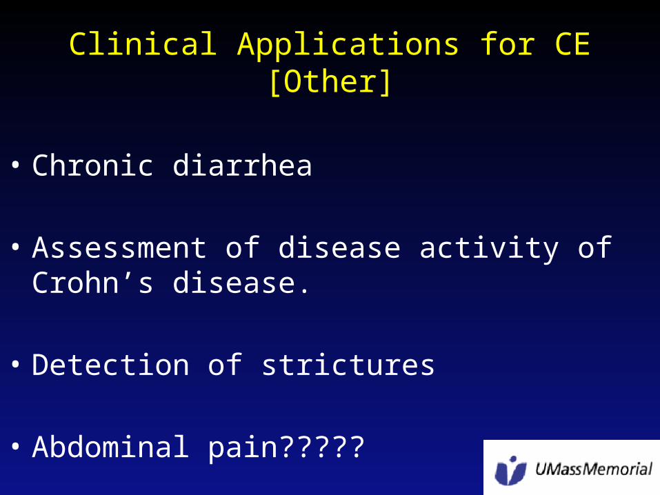
What do we use to assess position of the VCE?
• Time– Assumption: motion is linear between pylorus and
ileo-cecal valve• Factors that alter motility
– Disease process– Adhesions
• Blood stimulates gut motility• Transit incomplete 15-25% of the time

Assessment of motor function
• New insight into intestinal motor function via noninvasive endoluminal image analysis
• Malagelada C et al; Gastroenterology 2008 Oct;135(4):1155-62

Conclusions
• Is there a methodology that could improve location of a VCE?
• 1- longitudinally• 2- in 3 D with reference to fixed points