Embed Size (px)
Citation preview

Wilms’ tumor 1-associating protein regulates G2�Mtransition through stabilization of cyclin A2 mRNAKeiko Horiuchi*, Michihisa Umetani†, Takashi Minami*, Hiroto Okayama‡, Shinji Takada§, Masayuki Yamamoto¶,Hiroyuki Aburatani�, Patrick C. Reid*, David E. Housman**††, Takao Hamakubo*††, and Tatsuhiko Kodama*
*Laboratory for Systems Biology and Medicine and �Genome Science Division, Research Center for Advanced Science and Technology, University of Tokyo,Meguro, Tokyo 153-8904, Japan; †Howard Hughes Medical Institute, Department of Pharmacology, University of Texas Southwestern Medical Center, Dallas,TX 75390; ‡Department of Biochemistry and Molecular Biology, Graduate School of Medicine, University of Tokyo, Bunkyo-ku, Tokyo 113-0033, Japan;§Okazaki Institute for Integrative Biosciences, National Institute of Natural Sciences, Okazaki 444-8787, Japan; ¶Center for Tsukuba Advanced ResearchAlliance, University of Tsukuba, Tsukuba 305-8577, Japan; and **Center for Cancer Research and Department of Biology, Massachusetts Institute ofTechnology, Cambridge, MA 02139
Contributed by David E. Housman, September 22, 2006
Wilms’ tumor 1-associating protein (WTAP) has been reported tobe a ubiquitously expressed nuclear protein. Although a relation tosplicing factors has been postulated, its actual physiological func-tion still remains to be elucidated. To investigate the role of WTAP,we generated WTAP-knockout mice and performed small interfer-ing RNA (siRNA)-mediated knockdown analyses in primary cul-tured cells. In DNA microarrays using human umbilical vein endo-thelial cells, WTAP-targeted siRNA treatment resulted in markedlyreduced expression of cell-cycle-related genes. siRNA-mediatedWTAP knockdown down-regulated the stability of cyclin A2 mRNAthrough a nine-nucleotide essential sequence in cyclin A2 mRNA 3�
UTR. WTAP knockdown induced G2 accumulation, which is partiallyrescued by adenoviral overexpression of cyclin A2. Moreover,WTAP-null mice exhibited proliferative failure with death resultingat approximately embryonic day 6.5, an etiology almost identicalto cyclin A2-null mice. Collectively, these findings establish WTAPas an essential factor for the stabilization of cyclin A2 mRNA,thereby regulating G2�M cell-cycle transition.
mRNA stability � cell-cycle regulation
W ilms’ tumor 1-associating protein (WTAP) was identifiedas a protein that specifically interacts with Wilms’ tumor
1 (WT1) in both in vitro and in vivo assays (1). The Wilms tumorsuppressor gene WT1 is essential for the normal development ofthe bipotential gonad, and the primordial kidney (2–4) and isassociated with a common form of pediatric kidney cancer (5).WTAP and WT1 are present together throughout the nucleo-plasm as well as in nuclear speckles and partially colocalize withsplicing factors (1). WTAP was also identified as the mammalianhomologue of the Drosophila female-lethal-2-D [fl(2)D]. f l(2)Dis required for the female developmental pathway because of itsactivation of female-specific patterns of alternative splicing onSXL and transformer (tra) premRNA (6, 7). The presence of thelethal phenotype of the fl(2)D mutation in both sexes suggestsan additional function for the gene (8). In vitro splicing assaysdemonstrated that depletion of WTAP from HeLa nuclearextracts affects tra but not AdML (a model adenovirus RNA)splicing, suggesting a biochemical function in female-specificsplicing regulation (9). Furthermore, proteomic studies isolatedWTAP as one of 145 spliceosomal proteins assembled on twodistinct premRNAs, adenovirus major late and Fushi tarazu(10). While investigating GATA function in vascular endothelialcells, we serendipitously identified WTAP, in addition to GATA-associating proteins and hematopoietically expressed homeobox,from a yeast two-hybrid screen (11). In spite of these refinedstudies, the exact function of WTAP remains unknown.
In the present study, we investigated the function of WTAP invitro by using RNAi in primary cultured cells and in vivo bygenerating WTAP-knockout mice. Here, we demonstrate thatWTAP is a factor essential for cyclin A2 mRNA stabilization andG2�M transition, the understanding of which should yield sig-
nificant insight into posttranscriptional regulation, as well as cell-cycle regulation in normal and�or tumor cells.
ResultsWTAP-Knockdown Cells Display Markedly Reduced Expression ofCyclin A2 and B and Cell Division-Related Genes. To investigate thephysiological function of WTAP, we examined the relativeinfluence of WTAP on gene expression in human umbilical veinendothelial cells (HUVEC) by using an RNAi-based approach.To examine WTAP expression, we first generated an anti-WTAP antibody raised in rabbits by using bacterially expressedGST-WTAP protein. Its specificity for WTAP was tested byWestern blot analysis (Fig. 1A). A �50-kDa band correspondingto myc-tagged WTAP was detectable by both anti-myc and-WTAP antibodies (Fig. 1 A, arrow). Additionally, low levels ofendogenous WTAP were also detectable by anti-WTAP anti-body. In HUVEC cells, after WTAP siRNA transfection,WTAP protein levels were reduced to �20% of that in controlsiRNA-transfected cells (Fig. 1B). Subsequently, RNAs fromWTAP or control siRNA-treated cells were subjected to mi-croarray analysis. Of 38,500 genes present on a U133 Plus 2.0array, 329 genes were �2-fold down-regulated and 324 genes�2-fold up-regulated by WTAP knockdown confirmed in twoindependent experiments (Tables 3 and 4, which are publishedas supporting information on the PNAS web site). The top 30down- or up-regulated genes are listed in Tables 5 and 6, whichare published as supporting information on the PNAS web site,in which genes with unknown functions have been excluded.Interestingly, the major genes exhibiting decreased expressionare all related to the cell division cycle, including cyclins A2, B1,B2, and CDC20, whereas the up-regulated genes are related tometabolism, inflammation, or cell adhesion.
siRNA-Mediated WTAP Reduction Results in G2-Phase Accumulation.To examine the effect of WTAP reduction on cell cycle, prolif-eration assays and FACS analysis were performed. As shown inFig. 1C, HUVEC in the control group exhibited moderategrowth for the first 48 h after siRNA transfection, then increased3.5-fold in cell number by 72 h. In contrast, WTAP siRNA-
Author contributions: K.H., M.U., and T.M. contributed equally to this work; K.H., M.U.,T.M., H.O., S.T., M.Y., H.A., P.C.R., D.E.H., T.H., and T.K. designed research; K.H. and M.U.performed research; K.H. analyzed data; and K.H., T.M., P.C.R., and T.H. wrote the paper.
The authors declare no conflict of interest.
Freely available online through the PNAS open access option.
Abbreviations: WT1, Wilms’ tumor 1; siRNA, small interfering RNA; HUVEC, human umbil-ical vein endothelial cells; En, embryonic day n.
Data deposition: The data reported in this paper have been deposited in the GeneExpression Omnibus (GEO) database, www.ncbi.nlm.nih.gov�geo�(accession no. GSE2327).
††To whom correspondence may be addressed. E-mail: [email protected] or [email protected].
© 2006 by The National Academy of Sciences of the USA
17278–17283 � PNAS � November 14, 2006 � vol. 103 � no. 46 www.pnas.org�cgi�doi�10.1073�pnas.0608357103

treated cells underwent growth inhibition, as evidenced by thelack of an increase in cell number over time. Moreover, FACSanalysis revealed that WTAP siRNA-treated cells exhibited asignificantly higher proportion in G2 (21%) compared with thatof control cells (8%; Fig. 1D). These data demonstrate thatWTAP reduction leads to growth arrest in G2 or reduction inprogression through G2. To determine whether this effect isspecific to HUVEC, we next performed the same experiments inprimary human fibroblasts. G2 accumulation was also induced in
human neonatal dermal fibroblasts (Fig. 7, which is published assupporting information on the PNAS web site). This findingdemonstrates that the growth inhibition caused by WTAPreduction is not specific to endothelial cells but can be inducedin other cell types as well.
WTAP Regulates Cyclin A2 mRNA Stability Through 3�UTR. Among thegenes down-regulated by the knockdown of WTAP expression,cyclin A2 was the most prominently reduced. Interestingly, it has
Control siRNA
WTAP siRNA
WTAP
Cyclin A2
0Control siRNA
WTAP siRNA
100
80
60
40
20
Rel
ativ
e cy
clin
A2
mR
NA
A B
**
50kDa
50kDa
100
75
50
25
0Rel
ativ
e lu
cife
rase
act
ivity
100
75
50
25
0
Rel
ativ
e lu
cife
rase
act
ivity
3'3'UTR
5'luciferaseSV40
ControlsiRNA
ControlsiRNA
WTAP siRNA
WTAP siRNA
**
C
D
Perc
ent r
emai
ning
mR
NA
100
10
0 2 4 6 8Time after addition of AMD (h)
Control siRNA
WTAP siRNA*
E
109 1408 26015'UTR
CDS 3'UTR
cyclin A2
luciferase-UTR construct
F
1387 2555
1651
1662
2082
3'UTR full
fragment A
fragment B
fragment C
**
**
0 80604020 100
Relative luciferase activity
Control siRNA
WTAPsiRNA
0 80604020 100Relative luciferase activity
Control siRNA
WTAPsiRNA
**
**
**
1651fragment A
1387
fragment D 1507
fragment E 1565
del 1526-34
del 1535-40
Fig. 2. WTAP knockdown leads to the reduction of cyclin A2 mRNA and protein levels because of destabilization of cyclin A2 mRNA. (A) Quantitative real-timePCR analysis of cyclin A2 expression. Cyclin A2 mRNA levels were decreased to 10% that of controls 24 h after siRNA transfection. Values are WTAP RNAi vs. controlRNAi samples normalized to cyclophilin mRNA levels. (B) Western blot analysis of WTAP and cyclin A2. Cells were harvested 72 h after siRNA transfection. (C)The effect of WTAP knockdown on cyclin A2 promoter activity was studied by using a cyclin A2 promoter-luciferase reporter assay. The full promoter region ofcyclin A2, corresponding to �737 to �108 bp (start site, �1), was inserted into the pGL3-basic plasmid. (D) Actinomycin D (AMD) was added at 24 h after siRNAtransfection, and total RNA was prepared at each indicated time point. The remaining cyclin A2 mRNA was measured by quantitative real-time PCR andnormalized to rpL32 mRNA, which has a half-life of �25 h. (E) Influence of the 3�UTR of cyclin A2 mRNA on WTAP-mediated mRNA stability. A luciferase-cyclinA2 3�UTR chimeric plasmid was constructed by subcloning in the 3�UTR fragment of cyclin A2 immediately downstream of the firefly luciferase ORF in apGL3-control vector. At 12 h after siRNA transfection, HUVEC were transiently transfected with the chimeric luciferase-cyclin A2 3�UTR plasmid. Eighteen hourslater, dual-luciferase assays were performed. Relative luciferase activity (firefly�Renilla) was normalized to the relative basal luciferase activity obtained froma pGL3-control vector. (F) Responsible region for WTAP-mediated mRNA stability was analyzed by using deletion constructs of cyclin A2 mRNA 3�UTR. *, P � 0.05;**, P � 0.001 vs. control.
75kDa
50kDa
37kDa
+
anti-myc
anti-WTAP
WTAP-mycmyc(mock)
+-
ControlsiRNA
WTAPsiRNA
WTAP
beta-actin
**
WTAP siRNA
Cel
l num
ber
( x
10,0
00)
Control RNAi WTAP RNAi
100
80
60
40
20
Time after siRNA transfection (h)0
020 40 60 80
**Control siRNA
G0/G1
S
G2/M
G0/G1G2/M
S
79.29 +/- 5.78 % 8.32 +/- 1.36 %12.39 +/- 4.51 %
G0/G1G2/M
S
70.18 +/- 5.09 %21.31 +/- 2.65 % 8.52 +/- 4.39 %
A B C D-
++-
-
Fig. 1. WTAP siRNA-treated cells display growth inhibition in G2�M phase. (A) The specificity of anti-WTAP polyclonal antibody. COS7 cells transientlytransfected with a pcDNA3.1(�)-WTAP-myc-His or empty vector were subjected to Western blot analysis by using anti-WTAP or -myc antibody. Arrows indicatethe band corresponding to WTAP (�50 kDa), detected by an anti-myc antibody. (B) Efficient reduction of WTAP protein was confirmed by Western blots in WTAPsiRNA-transfected HUVEC. HUVEC were transfected with WTAP siRNA or control siRNA. Total proteins were extracted 72 h after transfection. (C) Cell growthrates in WTAP siRNA or control siRNA-treated HUVEC. Cell numbers were determined by using a hemacytometer. (D) Cell-cycle analysis was carried out by usingsiRNA-treated HUVEC. Forty-eight hours after siRNA transfection, cells were harvested and stained with propidium iodide and then analyzed for DNA contentwith a FACScalibur. *, P � 0.05; **, P � 0.001 vs. contol.
Horiuchi et al. PNAS � November 14, 2006 � vol. 103 � no. 46 � 17279
CELL
BIO
LOG
Y
been reported that cyclin A mutant Drosophila embryos undergocell-cycle arrest in G2 (12), and somatic mammalian cells areblocked at G2 on ablation of cyclin A (13). Hence, we hypoth-esized that the G2 accumulation brought about by WTAPreduction was because of the loss of cyclin A2 expression andfurther investigated how WTAP affects cyclin A2 mRNA ex-pression. We first confirmed that WTAP knockdown led to adecrease in cyclin A2 at the mRNA and protein levels. Quan-titative real-time PCR analysis revealed that the cyclin A2mRNA level was decreased to 10% that of controls within 24 hafter siRNA transfection and, as a result, reduction in the cyclinA2 protein level was observed by Western blot analysis (Fig. 2A and B). To investigate the mechanism responsible for thedecrease in cyclin A2 mRNA levels, we examined the effect ofWTAP on cyclin A2 promoter activity and mRNA stability inHUVEC. By using a cyclin A2 promoter-luciferase reporterconstruct, the effect of WTAP knockdown on cyclin A2 pro-moter activity was examined. The full promoter region of cyclinA2, corresponding to �737 to �108 bp (start site, �1), wasinserted into a pGL3-basic plasmid. As shown in Fig. 2C, RNAiof WTAP did not result in any substantial change in luciferaseactivity, suggesting that WTAP has no obvious effect on tran-scription from the cyclin A2 promoter. Next, we studied thehalf-life of cyclin A2 mRNA by using standard actinomycinD-based methods to investigate whether its low expression levelwas because of enhanced degradation of its mRNA. At 24 h aftersiRNA transfection, 5 �g�ml actinomycin D was added to themedium, and RNA was isolated at each indicated time point. Thecyclin A2 mRNA remaining was measured by quantitativereal-time PCR normalized against the stable rpL32 mRNA(T1/2 � 25 h). In the absence of WTAP, the cyclin A2 mRNAhalf-life significantly declined from 2.8 h, in control siRNA-treated cells, to 1.3 h (Fig. 2D). To further examine cyclin A2mRNA stability, we performed a reporter assay by using a fireflyluciferase-cyclin A2 3�UTR chimeric plasmid constructed byconjugating a 3�UTR fragment of cyclin A2 into a pGL3-controlvector. The reporter plasmids were cotransfected with a CMV-Renilla luciferase reporter plasmid into HUVEC 12 h aftersiRNA transfection. Eighteen hours later, dual-luciferase assayswere performed. The relative luciferase activity (firefly�Renilla)was normalized to the basal relative luciferase activity obtainedfrom the pGL3-control vector alone. A significant decrease inluciferase activity was observed in WTAP siRNA-treated cells(�40%) compared with that in control cells (Fig. 2E). Thesefindings suggest that WTAP regulates cyclin A2 mRNA stabi-lization through its effect on cyclin A2 3�UTR.
To determine the responsible region for WTAP-mediatedstabilization, we generated 3�UTR deletion constructs and usedreporter assays (Fig. 2F). There are three AUUUA motifs,sequences known to be involved in mRNA decay, in the 3�UTRof cyclin A2 mRNA at �1,686, �2,297, and �2,417. Notably, a
1,387�1,651 construct (fragment A) with no AUUUA motif gavea similar or greater decrease in relative luciferase activity(�40%) as the 3�UTR full construct in WTAP siRNA-treatedcells. Deletion of this region (fragment B) completely abolishedthe loss in luciferase activity induced by WTAP reduction. Asexpected, fragment C showed no effect after WTAP RNAi.
To identify the responsible region for stabilization by WTAPwithin fragment A, we undertook a luciferase assay using thedeletion constructs of fragment A (Fig. 2F). Construct (1,565�1,651; fragment E) eliminated the destabilizing activity ofWTAP knockdown, as evidenced by an increase in luciferaseactivity to control levels (Fig. 2F). These results suggest that theresponsible element confirming cyclin A2 mRNA stability ispresent between 1,507 and 1,565 bp of the 3�UTR. Deletion of1526–34 (del 1526–34) abolished the destabilizing activity,whereas the adjacent deletion of 1535–40 (del 1535–40) showedsimilar activity to fragment A. Taken together, these findingssuggest that ACAAAUUAU, which corresponds to the 3�UTR1526–34, is an essential element required for the WTAP-mediated stabilization of cyclin A2 mRNA, in a manner inde-pendent of AUUUA motifs.
Adenoviral-Mediated Expression of Cyclin A2 Protein Rescued the G2
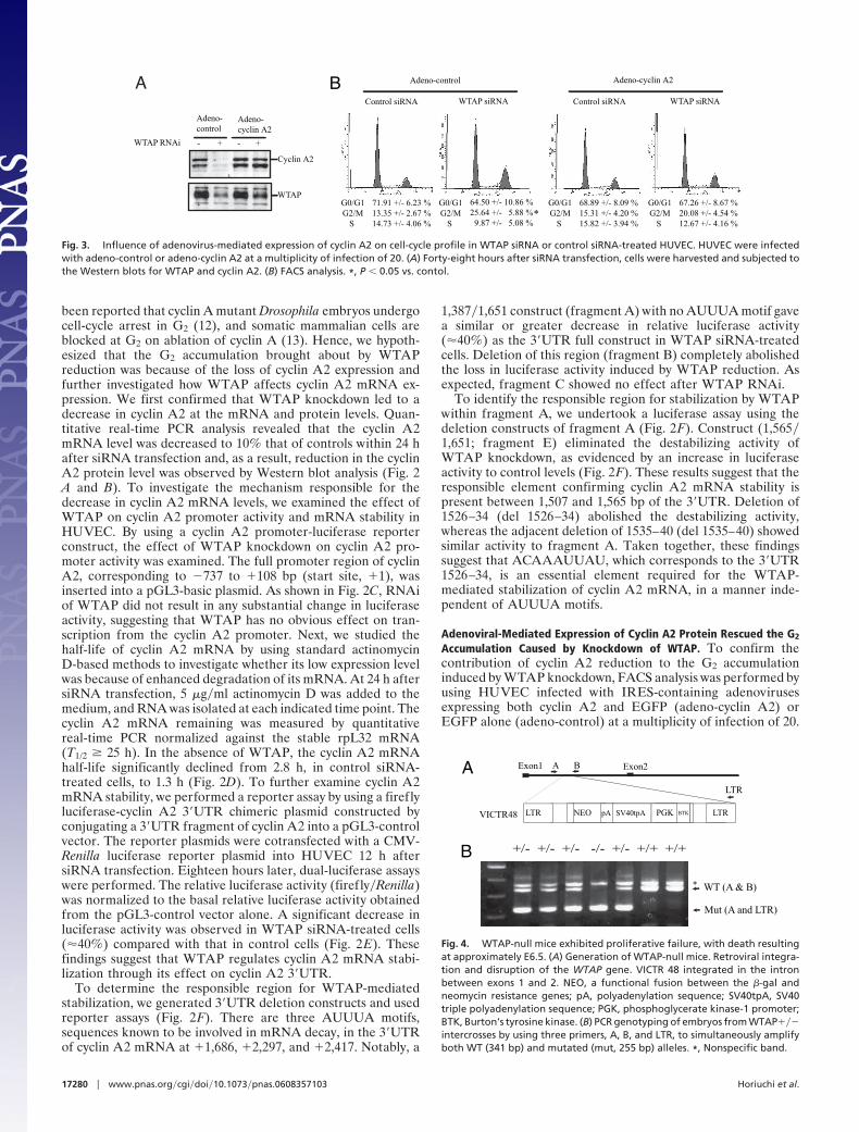
Accumulation Caused by Knockdown of WTAP. To confirm thecontribution of cyclin A2 reduction to the G2 accumulationinduced by WTAP knockdown, FACS analysis was performed byusing HUVEC infected with IRES-containing adenovirusesexpressing both cyclin A2 and EGFP (adeno-cyclin A2) orEGFP alone (adeno-control) at a multiplicity of infection of 20.
B +/- +/- +/- -/- +/- +/+ +/+
LTR NEO pA SV40tpA PGK BTK LTR
A Exon1 Exon2A B
LTR
WT (A & B)
Mut (A and LTR)
VICTR48
*
Fig. 4. WTAP-null mice exhibited proliferative failure, with death resultingat approximately E6.5. (A) Generation of WTAP-null mice. Retroviral integra-tion and disruption of the WTAP gene. VICTR 48 integrated in the intronbetween exons 1 and 2. NEO, a functional fusion between the �-gal andneomycin resistance genes; pA, polyadenylation sequence; SV40tpA, SV40triple polyadenylation sequence; PGK, phosphoglycerate kinase-1 promoter;BTK, Burton’s tyrosine kinase. (B) PCR genotyping of embryos from WTAP���intercrosses by using three primers, A, B, and LTR, to simultaneously amplifyboth WT (341 bp) and mutated (mut, 255 bp) alleles. *, Nonspecific band.
Adeno-control Adeno-cyclin A2
Control siRNA WTAP siRNA
*
WTAP
Cyclin A2
WTAP RNAi - + - +
Adeno-control
Adeno-cyclin A2
Control siRNA WTAP siRNA
B
G0/G1G2/M
S
71.91 +/- 6.23 %13.35 +/- 2.67 %14.73 +/- 4.06 %
G0/G1G2/M
S
64.50 +/- 10.86 %25.64 +/- 5.88 % 9.87 +/- 5.08 %
G0/G1G2/M
S
68.89 +/- 8.09 %15.31 +/- 4.20 %15.82 +/- 3.94 %
G0/G1G2/M
S
67.26 +/- 8.67 %20.08 +/- 4.54 %12.67 +/- 4.16 %
A
Fig. 3. Influence of adenovirus-mediated expression of cyclin A2 on cell-cycle profile in WTAP siRNA or control siRNA-treated HUVEC. HUVEC were infectedwith adeno-control or adeno-cyclin A2 at a multiplicity of infection of 20. (A) Forty-eight hours after siRNA transfection, cells were harvested and subjected tothe Western blots for WTAP and cyclin A2. (B) FACS analysis. *, P � 0.05 vs. contol.
17280 � www.pnas.org�cgi�doi�10.1073�pnas.0608357103 Horiuchi et al.

As shown in Fig. 3A, cells infected with adeno-cyclin A2 showedcyclin A2 expression both in the absence or presence of WTAPsiRNA. Adeno-cyclin A2 carries the cDNA encoding the humancyclin A2 coding region. These findings are consistent withWTAP-mediated regulation of cyclin A2 expression through thecyclin A2 mRNA 3�UTR. Adenovirus-mediated cyclin A2 ex-pression had no effect on the cell-cycle profile (Fig. 3B). The G2accumulation was partially rescued by the overexpression ofcyclin A2, whereas the knockdown of WTAP still led to the G2accumulation of adeno-control-infected cells.
WTAP-Null Mice Die and Exhibit Proliferative Failure at ApproximatelyEmbryonic Day 6.5 (E6.5). Finally, we extended our studies in vivousing WTAP-null mice. The WTAP-knockout mice were gen-erated by gene trapping by using a retroviral vector integratedbetween exons 1 and 2 (Fig. 4A). Genotyping by PCR using threeprimer sets, which lie on opposite sides of the insertion site, anda gene-trap vector (Fig. 4B) revealed that no newborn homozy-gous mice were yielded from heterozygous intercrosses (data notshown). This finding demonstrates that WTAP is required forembryonic development. To determine the timing of lethalityduring embryogenesis, we therefore performed genotyping andhistological analysis of embryos from the heterozygous matings.Table 1 depicts genotyping by using the yolk sac DNA ofembryos from E8.5 to E10.5. Homozygous WTAP��� embryosare obviously small and without an embryo body (Fig. 5 A andB; heterozygous WTAP ��� embryos at E8.5 and homozygouslittermate, respectively). Histological analyses of decidua duringearly stages of development revealed that abnormal embryoswere observed at E6.5 (Table 2). Morphologically abnormalembryos are small like blastocysts and exhibit defects in prolif-eration and an absence of the shapes characteristic of endodermand ectoderm (Fig. 5 D, E, and H), whereas normal embryos
displayed defined layers of embryonic ectoderm and endoderm(Fig. 5 C and F) by E6.5. Interestingly, these features arestrikingly similar to those seen in the cyclin A2 null mutant.Cyclin A2 null mutants cease development at approximatelyE6.5 and present a morphology reminiscent of blastocysts (14).
Immunostaining of serial sections with rabbit polyclonal anti-WTAP antibodies revealed that WTAP was detectable in thenuclei of morphologically normal embryos (Fig. 5G). In contrast,no specific WTAP staining was observed in morphologicallyabnormal embryos, confirming that these embryos are homozy-gous null mutants (Fig. 5I). In addition to the proliferativefailures observed in the WTAP��� embryos, some presented adilated proamniotic canal (Fig. 5E).
Cell-Cycle-Dependent WTAP Expression Is Synchronous with Cyclin A2.It has been reported that cyclin A2 mRNA stability varies withthe cell cycle, which helps account for its cell-cycle-dependentrelative abundance (15). Thus, we next examined the expressionof WTAP throughout the cell division cycle. To this end,HUVEC were subjected to cell-cycle synchronization experi-ments by serum starvation for 36 h, so that cells exhibitedG1-enriched distribution (�90%). Then cells were passaged andreleased from quiescence by exchanging the medium for com-plete medium EGM2, and cell-cycle progression was monitoredby FACS analysis. Protein was harvested at time points when themajority of the cells were in G1 (87%), S (76%), or G2�M (55%).As shown in Fig. 6A, WTAP expression varied with the cell cycle.WTAP was low in G1, elevated to 1.7-fold in S and 2.1-fold in G2.The cell-cycle-dependent expression of cyclin A2 was detected inonly the S- and G2-rich phases. The cell-cycle-dependent changesin the expression levels of cyclin A2 and WTAP were similar,
Table 1. Genotyping of mouse embryos from heterozygousmatings by PCR by using yolk sac DNA
Stage ofdevelopment
Numbers of embryosTotal
number��� ��� ��� Reabsorbed
E8.5 7 16 2 1 26E9.5 4 17 5 26E10.5 4 12 2 18
Fig. 5. Morphology of embryos from WTAP��� heterozygous matings. (A and B) Heterozygous WTAP��� embryo at E8.5 (A) and homozygous WTAP���littermate (B). Homozygous WTAP��� embryos are small and present no embryo body. (C–E) Representative H&E stainings of E6.5 normal (C) and abnormal (Dand E) embryos cross-sectioned. (E) Some abnormal embryos have a dilated proamniotic canal. (F–I) Representative H&E and WTAP staining of E6.5 embryossagittally sectioned . (F) Normal embryos exhibit defined layers of cells from embryonic ectoderm and endoderm. (H) Abnormal embryos are smaller and presentno defined layers. No specific WTAP stainings were observed in morphologically abnormal embryos (I, compare with G), confirming that these abnormal embryosare homozygous null mutants. (Scale bars: A and B, 500 �m; C–E, 200 �m; F and H, 100 �m.)
Table 2. Morphology of embryos from heterozygous matings
Stage ofdevelopment
Numbers of embryosTotal
numberNormal Abnormal Reabsorbed
E6.5 28 4 32E7.5 14 2 16E8.5 8 1 1 10E10.5 0 1 1
Abnormal embryos were not stained by immunostaining with anti-WTAPpolyclonal antibodies.
Horiuchi et al. PNAS � November 14, 2006 � vol. 103 � no. 46 � 17281
CELL
BIO
LOG
Y
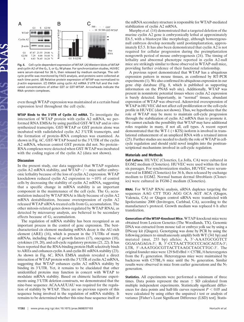
even though WTAP expression was maintained at a certain basalexpression level throughout the cell cycle.
WTAP Binds to the 3�UTR of Cyclin A2 mRNA. To investigate theinteraction of WTAP protein with cyclin A2 mRNA, we per-formed RNA EMSAs by using purified GST-WTAP and in vitrosynthesized transcripts. GST-WTAP or GST protein alone wasincubated with radiolabeled cyclin A2 3�UTR transcripts, andthe formation of protein–RNA complexes was examined. Asshown in Fig. 6C, GST-WTAP bound to the 3�UTR of the cyclinA2 mRNA, whereas control GST protein did not. No protein–RNA complexes were detected when GST-WTAP was incubatedwith the coding region of the cyclin A2 (data not shown).
DiscussionIn the present study, our data suggested that WTAP regulatescyclin A2 mRNA stability, and WTAP��� mice suffer embry-onic lethality because of the loss of cyclin A2 expression. WTAPknockdown reduced cyclin A2 expression to �10% of controlcells by decreasing the stability of cyclin A2 mRNA, suggestingthat a specific change in mRNA stability is an importantcomponent in the maintenance of the cell cycle. The G2 accu-mulation induced by WTAP RNAi is likely because of cyclin A2mRNA destabilization, because overexpression of cyclin A2released WTAP siRNA-treated cells from G2 accumulation. Theother mitosis-related genes down-regulated by WTAP RNAi, asdetected by microarray analysis, are believed to be secondaryeffects because of G2 accumulation.
The regulation of mRNA stability has been recognized as animportant process in the control of gene expression. The best-characterized cis element mediating mRNA decay is the AU-richelement (ARE) (16), which is present in the 3�UTRs of manymRNAs, including those of growth factors (17), oncogenes (18),cytokines (19, 20), and cell-cycle regulatory proteins (21, 22). It hasbeen reported that the RNA-binding protein HuR selectively bindsto AREs and enhances cyclin A and cyclin B1 mRNA stability (23).As shown in Fig. 6C, RNA EMSA analysis revealed a directinteraction of WTAP protein with the 3�UTR of cyclin A2 mRNA,suggesting that WTAP enhances cyclin A2 mRNA stability bybinding its 3�UTR. Yet, it remains to be elucidated that otherunidentified proteins may function in concert with WTAP tomodulate mRNA stability. Based on chimeric luciferase experi-ments using 3�UTR deletion constructs, we demonstrated that thenine-base sequence ACAAAUUAU was required for the regula-tion of stability by WTAP. There are no previous reports of thissequence being involved in the regulation of mRNA stability. Itremains to be determined whether this nine-base sequence itself or
the mRNA secondary structure is responsible for WTAP-mediatedstabilization of cyclin A2 mRNA.
Murphy et al. (14) demonstrated that a targeted deletion of themurine cyclin A2 gene is embryonically lethal at approximatelyE6.5, with a blastocyst like morphology, although homozygous-null embryos develop normally until postimplantation, approx-imately E5.5. It has also been demonstrated that cyclin A2 is notrequired for cellular progression during the preimplantationnongrowth period of mouse embryogenesis (24). The timing oflethality and abnormal phenotype reported in cyclin A2-nullmice are strikingly similar to those observed in WTAP-null mice,providing further evidence of their functional relationship.
A previous report demonstrated that WTAP has a ubiquitousexpression pattern in mouse tissues, as confirmed by RT-PCRexperiments (1). We also confirmed its ubiquitous expression in ourgene chip database (Fig. 8, which is published as supportinginformation on the PNAS web site). Additionally, WTAP waspresent in nonmitotic potential tissues where cyclin A2 expressionis barely detected. Importantly, in ‘‘normal’’ tissues, abundantexpression of WTAP was observed. Adenoviral overexpression ofWTAP in HUVEC did not affect cell proliferation or the cell-cycleprofile in HUVEC (data not shown). Thus, we hypothesize that therole of WTAP may be more to maintain cell-cycle progressionthrough the stabilization of cyclin A2 mRNA than to promote it.We cannot exclude the possibility that WTAP may have additionalroles requisite in many, if not all, cell types. A recent reportdemonstrated that the WT-1 (�KTS) isoform is involved in trans-lational enhancement of an unspliced RNA with a retained intron(25). Further studies are needed to clarify the role of WTAP in cell-cycle regulation and should yield novel insights into the posttran-scriptional mechanisms involved in cell-cycle regulation.
Materials and MethodsCell Culture. HUVEC (Clonetics, La Jolla, CA) were cultured inEGM2 medium (Clonetics). HUVEC were used within the firstsix passages. For synchronization studies, HUVEC were serum-starved in EBM2 (Clonetics) for 36 h, then released by exchangemedium to EGM2. Normal human dermal fibroblasts (Clonet-ics) were cultured in FGM-2 medium (Clonetics).
RNAi. For WTAP RNAi analysis, siRNA duplexes targeting thesequences AAG CTT TGG AGG GCA AGT ACA (Qiagen,Valencia, CA) or Qiagen control siRNA were transfected withlipofectamine 2000 (Invitrogen, Carlsbad, CA), according to themanufacturer’s protocol. Growth medium was replaced 6 h aftertransfection.
Generation of the WTAP-Knockout Mice. WTAP-knockout mice wereobtained from Lexicon Genetics (The Woodlands, TX). GenomicDNA was extracted from mouse tail or embryo yolk sac by using aDNeasy kit (Qiagen). Genotyping was done by PCR by using thefollowing primers to simultaneously amplify both WT (341 bp) andmutated (mut, 255 bp) alleles; A, 5�-AAATGCGGTCTGGAGAGAGA-3�; B, 5�-CCTAACTTGCCCCAGCAGTA-3�;LTR, 5�-AAATGGCGTTACTTAAGCTAGCTTGC-3�. Theoriginal founder mice were 129 SvEvBrd � C57BL�6 heterozygotesfrom the F2 generation. Heterozygous mice were maintained bybackcross with C57BL�6 mice until the N7 generation. Similarresults were observed in mice from earlier generations and the N7generation.
Statistics. All experiments were performed a minimum of threetimes. Data points represent the mean � SD calculated frommultiple independent experiments. Statistically significant differ-ences for data points and half-life curves represent P � 0.05 andwere calculated by using either the unpaired t test or analysis ofvariance [Fisher’s Least Significant Difference (LSD) test]. Statis-
B3
2
1
Rel
ativ
e ex
pres
sion
of
WTA
P
Cyclin A2
WTAP
β-actin
G1 S G2/M
G1G2/M
S
87.877.294.83
29.0854.6816.24
23.550.00
76.45
%%% G1 S G2/M
AGSTGST-WTAP- - 10 50 500
- 500 - - -
freeRNA
C
Fig. 6. Cell-cycle-dependent expression of WTAP. (A) Western blots of WTAPand cyclin A2 of the G1, S, or G2�M phase. For synchronization studies, HUVECwere serum-starved for 36 h, then released by medium exchange. The cell-cycle profile was monitored by FACS analysis, and proteins were collected ateach time point. (B) Relative protein expression of WTAP was normalized to�-actin expression. (C) EMSA using cyclin A2 mRNA 3�UTR full and the indi-cated concentrations of either GST or GST-WTAP. Arrowheads indicate theRNA–protein complexes.
17282 � www.pnas.org�cgi�doi�10.1073�pnas.0608357103 Horiuchi et al.
tical analyses were performed by using Statview version 5 software(Abacus Concepts, Berkeley, CA).
Further Details. Further details of the experimental proceduresused in this study are provided in Supporting Text, which ispublished as supporting information on the PNAS web site.
We thank Dr. K. Boru of Pacific Edit for review of the manuscript, Drs.
Yoshifumi Yamaguchi and Norihiko Ohbayashi for helpful discussion,and Naomi Saito for helpful technical assistance. This work is supportedin part by grants from the Program for Promotion of FundamentalStudies in Health Sciences of the National Institute of BiomedicalInnovation; from the Special Coordination Fund for Science and Tech-nology from the Ministry of Education, Culture, Sports, Science, andTechnology; and from the New Energy and Industrial TechnologyDevelopment Organization Bionanochip Project (P03023), Ministry ofEconomy, Trade, and Industry, Japan.
1. Little NA, Hastie ND, Davies RC (2000) Hum Mol Genet 9:2231–2239.2. Haber DA, Buckler AJ, Glaser T, Call KM, Pelletier J, Sohn RL, Douglass EC,
Housman DE (1990) Cell 61:1257–1269.3. Buckler AJ, Pelletier J, Haber DA, Glaser T, Housman DE (1991) Mol Cell Biol
11:1707–1712.4. Kreidberg JA, Sariola H, Loring JM, Maeda M, Pelletier J, Housman D,
Jaenisch R (1993) Cell 74:679–691.5. Hastie ND (2001) Cell 106:391–394.6. Granadino B, Campuzano S, Sanchez L (1990) EMBO J 9:2597–2602.7. Granadino B, Penalva LO, Sanchez L (1996) Mol Gen Genet 253:26–31.8. Granadino B, San Juan A, Santamaria P, Sanchez L (1992) Genetics 130:
597–612.9. Ortega A, Niksic M, Bachi A, Wilm M, Sanchez L, Hastie N, Valcarcel J (2003)
J Biol Chem 278:3040–3047.10. Zhou Z, Licklider LJ, Gygi SP, Reed R (2002) Nature 419:182–185.11. Minami T, Murakami T, Horiuchi K, Miura M, Noguchi T, Miyazaki J,
Hamakubo T, Aird WC, Kodama T (2004) J Biol Chem 279:20626–20635.
12. Lehner CF, O’Farrell PH (1990) Cell 61:535–547.13. Pagano M, Pepperkok R, Verde F, Ansorge W, Draetta G (1992) EMBO J
11:961–971.
14. Murphy M, Stinnakre MG, Senamaud-Beaufort C, Winston NJ, Sweeney C,Kubelka M, Carrington M, Brechot C, Sobczak-Thepot J (1997) Nat Genet15:83–86.
15. Maity A, McKenna WG, Muschel RJ (1997) Cell Growth Differ 8:311–318.16. Chen CY, Shyu AB (1995) Trends Biochem Sci 20:465–470.17. Shaw G, Kamen R (1986) Cell 46:659–667.18. Miller AD, Curran T, Verma IM (1984) Cell 36:51–60.19. Henics T, Sanfridson A, Hamilton BJ, Nagy E, Rigby WF (1994) J Biol Chem
269:5377–5383.20. Wodnar-Filipowicz A, Moroni C (1990) Proc Natl Acad Sci USA 87:777–781.21. Lal A, Mazan-Mamczarz K, Kawai T, Yang X, Martindale JL, Gorospe M
(2004) EMBO J 23:3092–3102.22. Lin S, Wang W, Wilson GM, Yang X, Brewer G, Holbrook NJ, Gorospe M
(2000) Mol Cell Biol 20:7903–7913.23. Wang W, Caldwell MC, Lin S, Furneaux H, Gorospe M (2000) EMBO J
19:2340–2350.24. Winston N, Bourgain-Guglielmetti F, Ciemerych MA, Kubiak JZ, Senamaud-
Beaufort C, Carrington M, Brechot C, Sobczak-Thepot J (2000) Dev Biol223:139–153.
25. Bor YC, Swartz J, Morrison A, Rekosh D, Ladomery M, Hammarskjold ML(2006) Genes Dev 20:1597–1608.
Horiuchi et al. PNAS � November 14, 2006 � vol. 103 � no. 46 � 17283
CELL
BIO
LOG
Y