Embed Size (px)
Citation preview

Types of Neck Dissection and the Post-Operative Neck
Daniel W. Williams III, MD
Outline
I. Primary site II. Neck dissections III. Myocutaneous flaps
1° Site Imaging Appearance
Conventional Open Surgery TORS*
(TransOral Robotic Surgery)
* Photos courtesy of Josh Waltonen MD
1° Site Imaging Appearance: Conventional Open Surgery
• Tumor (+ adjacent tissue) resected • Anatomic distortion • Enhancing granulation tissue (early) or scar
(later)
Post-op
Total Laryngectomy (Larynx SCCa)
Pre-op Pre-op Post-op
Parotidectomy (Mucoepidermoid Ca)

1° Site Imaging Appearance: TORS
• Depends on type of surgery (lat oropharyngectomy, PSGL, BOT resection etc…)
• Tumor resected (usually minimal margin) • Progressive retraction of lat. OP wall or BOT • Loss of fat planes around medial pterygoid muscle
and pterygomandibular raphe
“Transoral Robotic Surgery in Head and Neck Cancer: What Radiologists Need to Know about the Cutting Edge”, LALoevner MD et al, RadioGraphics 2013; 33:1759–1779
62 F, left tonsil SCCa s/p TORS (Radical Tonsillectomy)
Pre-op Post-op
88M, right BOT SCCA s/p TORS
Post-op 2015 Pre-op 2011
Outline
I. Primary site II. Neck dissections III. Myocutaneous flaps
Neck Dissection • Systematic removal of LN’s and
surrounding fibrofatty tissue from various compartments of the neck (cervical lymphadenectomy)
• Types
– Therapeutic: positive nodes by exam – Elective: suspected occult nodes
ND Rationale
• Each anatomic site has its own nodal spread pattern, and…
• Nodal involvement is (usually) orderly and predictable, and…
• Removing nodes involved or at risk improves loco-regional control and patient outcomes

• Grandfather of ND in N. America • Published a systematic approach to ND in 1905 • Cleveland Clinic founder
General George W. Crile Sr.
1864-1943 Crile building at Cleveland Clinic
Neck Dissection Classification *
1. Radical neck dissection (RND) 2. Modified RND 3. Selective ND (“SND”+ LN levels
removed) 4. Extended ND (RND plus) * KT Robbins et al. Neck dissection classification update. Arch Otolaryn Head Neck Surg 2002; 128: 751-758.
Radical Neck Dissection Structures removed
- LN levels I-V - SCM, IJV, SAN - SM gland
Standard procedure to which other ND’s compared
Cummings, 4th ed. 2005
Normal RND
Radical Neck Dissection Modified RND Structures removed - LN levels I-V
Structures preserved - 1 or more non- lymphatic structures (SCM, IJV, SAN) c/w RND
Cummings, 4th ed. 2005

Modified RND
IJV + SAN SCM + IJV SCM + SAN
Normal Modified RND
Modified RND (IJV & SCM removed)
Modified RND (Bilateral; SCM removed)
Selective Neck Dissection Preservation of 1 or more LN levels
(c/w RND): - Oral cavity: SND (I-III) - OP/HP & laryngeal: SND (II-IV) - Low ant. neck ML structures: SND (VI) - Skin Ca: SND (LN levels adj. to 1°)
SND for Oral Cavity Ca SND (I-III) *
Structures removed - LN levels I, II, III - SM gland
* Supraomohyoid SND

Normal SND Selective ND (I-III)
Imaging Appearance of ND’s • Depends on type of surgery • RND/some MRND’s: recognizable Δ’s
– absent structures, neck contour Δ, muscle denerv atrophy / hypertrophy
• Some MRND/most SND’s: subtle Δ’s – loss of fat planes, slight neck contour Δ,
surgical scar, ± absent structures
• Be alert for complications and pitfalls
Recognizing Neck Dissections
• Use neck symmetry! • Neck contour abnormality/scar? • SCM, IJV present? • Trapezius m. atrophy? • Fat planes around IJV/CA?
Read the Op Note!
Complications and Pitfalls
• Complications – Perioperative - bleeding, nerve injury, pntx, air
embolus/leak, infection/abscess – Postoperative – shoulder syndrome, fistulas,
CA rupture, IJV thrombosis, facial/cerebral edema, traumatic neuroma
• Pitfalls – MC flaps – Pseudotumors – IJV stump
Complications and Pitfalls
Neck abscess Pseudotumor (lev. scap. hypertrophy)

Outline
I. Primary site II. Neck dissections III. Myocutaneous flaps
Options in Head and Neck Reconstruction
• Healing by secondary intention • Primary closure • Skin grafts (split or full thickness) • Composite grafts • Flaps (local, distant pedicled, distant
free)
From Gurtner GC, Evans GR. Advances in head and neck reconstruction. Plast Reconstr Surg 2000;106:672-682; quiz 683
MC Flaps: Uses • Facilitate wound closure • Repair surgical defects • Enables more complete removal of 1° tumor • Restore optimal function (speech, breathing,
mastication, swallowing) • Cosmesis (recreation of facial aesthetics) • Protection (carotid artery during RT, skull
base, orbital apex)
MC Flaps: Classification
Flaps classified according to: • Blood supply (random, axial, free) • Type of tissue transferred (cutaneous,
myocutaneous, osteomyocutaneous) • Site of origin (local, regional,
microvascular free tissue transfer) • Flat vs. tubed
Rec. larynx ca w/ carotid invasion
Case: Regional flap (pectoralis major)
Thanks to D. Browne, MD
Carotid artery sacrificed
Saphenous vein graft

Harvested flap PM flap in place
SCCA alveolar ridge
Case: Free flap (iliac crest)
Thanks to D. Browne, MD
Skin paddle Surgical landmark
Harvested flap
Iliac crest flap; Titanium bar
Post-op
MC Flaps: Imaging Appearance
• Imaging changes usually obvious, but can be quite subtle
• Pearls: – Compare to pre-treatment exam – Read the op-note or call the surgeon !!!! – Be aware of potential complications and
pitfalls
MC Flaps: CT/MR findings
• Neck contour change • Fatty “mass” w/ muscle striations • Muscle denervation atrophy over time • Muscle enhancement on MR (often
intense, persists many months) • Rotational flaps - muscle origin remains
attached; vascular pedicle visible • “Unusual appearing” bones/other signs of
surgery
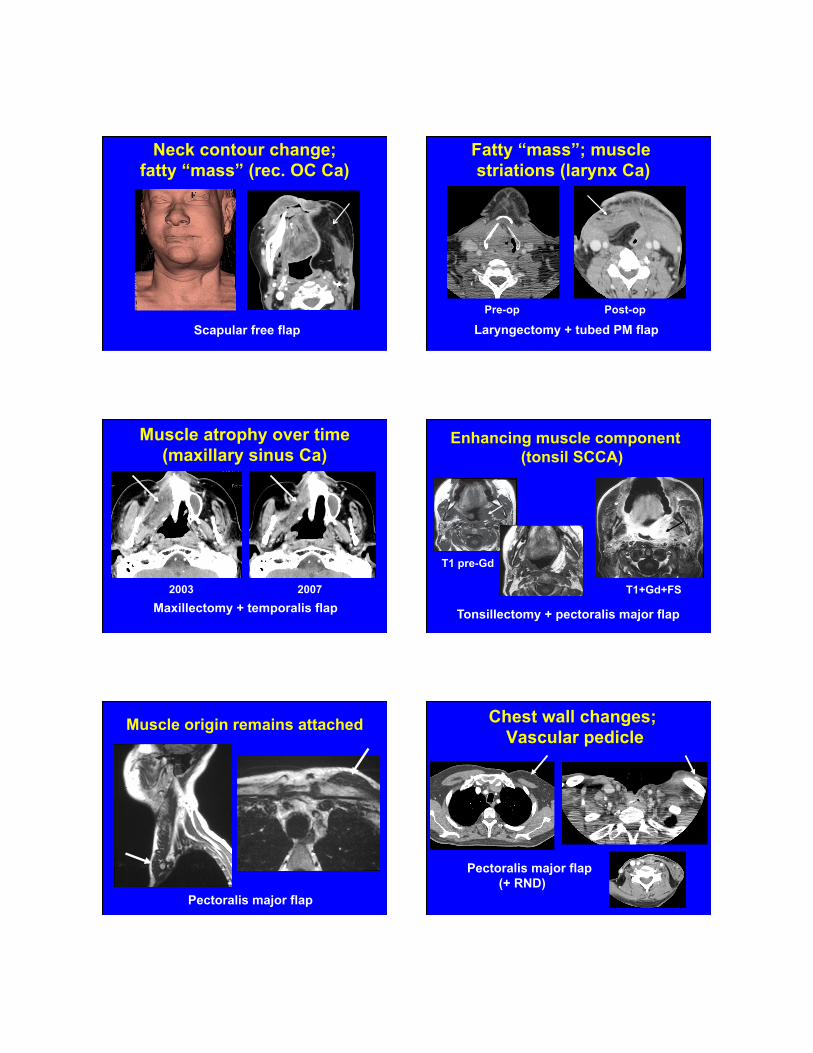
Scapular free flap
Neck contour change; fatty “mass” (rec. OC Ca)
Fatty “mass”; muscle striations (larynx Ca)
Pre-op Post-op
Laryngectomy + tubed PM flap
Muscle atrophy over time (maxillary sinus Ca)
Maxillectomy + temporalis flap 2003 2007
Enhancing muscle component (tonsil SCCA)
T1 pre-Gd
T1+Gd+FS
Tonsillectomy + pectoralis major flap
Muscle origin remains attached
Pectoralis major flap
Chest wall changes; Vascular pedicle
Pectoralis major flap (+ RND)

Scapular OMC free flap (s/p maxillectomy)
“Unusual appearing” bones + hardware Other signs of surgery
Temporalis flap
Zygomatic arch plate
MEDPOR® implant
Complications and Pitfalls • Fluid collections, fistulas, IJV thrombosis,
flap ischemia/necrosis, nerve injury, bone nonunion or infection
• Pseudotumors (muscle denerv. atrophy, muscle hypertrophy, enhancing flap muscle component)
• Changes or complications assoc. with primary tumor excision, ND or RT
• Hide early tumor recurrences from PEx
s/p resection inverted papilloma
Complication: Temporalis flap necrosis + Abscess
Pitfall: CN 12 injury (tongue denerv. atrophy)
Conclusions • Perform high quality exams • Learn types of ND’s and MCF’s used at your
institution • Recognizing post-Rx changes will help
detect recurrent H/N Ca and avoid imaging exam misinterpretation
• Above all, read Rx notes/speak to the surgeon and/or radiation oncologist
The End


![Cardboard Tubed Smoke Projectile[1]](https://img.pdfslide.us/doc/110x75/577d28d91a28ab4e1ea55e96/cardboard-tubed-smoke-projectile1.jpg)