Embed Size (px)
Citation preview
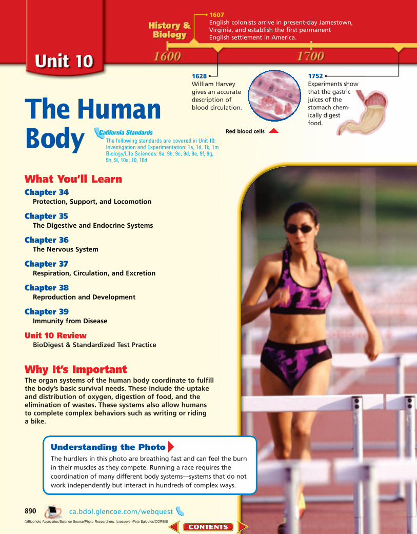
The HumanBodyWhat You’ll LearnChapter 34
Protection, Support, and Locomotion
Chapter 35The Digestive and Endocrine Systems
Chapter 36The Nervous System
Chapter 37Respiration, Circulation, and Excretion
Chapter 38Reproduction and Development
Chapter 39Immunity from Disease
Unit 10 ReviewBioDigest & Standardized Test Practice
Why It’s ImportantThe organ systems of the human body coordinate to fulfillthe body’s basic survival needs. These include the uptakeand distribution of oxygen, digestion of food, and theelimination of wastes. These systems also allow humansto complete complex behaviors such as writing or riding a bike.
1628William Harveygives an accuratedescription ofblood circulation.
Understanding the PhotoThe hurdlers in this photo are breathing fast and can feel the burnin their muscles as they compete. Running a race requires thecoordination of many different body systems—systems that do notwork independently but interact in hundreds of complex ways.
890
1607English colonists arrive in present-day Jamestown,Virginia, and establish the first permanentEnglish settlement in America.
1752Experiments show that the gastric juices of the stomach chem-ically digest food.
Red blood cells
(t)Biophoto Associates/Science Source/Photo Researchers, (crossover)Pete Saloutos/CORBIS
ca.bdol.glencoe.com/webquest
The following standards are covered in Unit 10:Investigation and Experimentation: 1a, 1d, 1k, 1mBiology/Life Sciences: 9a, 9b, 9c, 9d, 9e, 9f, 9g,9h, 9i, 10a, 10, 10d
California Standards
0890-0891 UO10 BDOL-829900 8/4/04 4:14 PM Page 890

1804Lewis and Clark begin theirexploration of the north-western United States.
1928AlexanderFleming acciden-tally discoversthe antibioticpenicillin.
1847An instrument thatmeasures blood pres-sure is developed.
1969The first artificialheart is implantedinto a human at ahospital in Texas.
1998The world’s first handtransplant is successfullyperformed.
1946The United Nations, an internationalpeace-keeping organization, has itsfirst meeting.
Alexander Fleming
1875The electrical activity of the brain is recorded for the first time.
891
Hulton Archive
0890-0891 UO10 BDOL-829900 8/4/04 4:16 PM Page 891

What You’ll Learn■ You will interpret the struc-
ture and functions of theintegumentary system.
■ You will identify the functionsof the skeletal system.
■ You will classify the differenttypes of muscles in the body.
Why It’s ImportantYour skin, skeleton, and muscleswork together to protect, sup-port, and move your body. Aknowledge of each system helpsyou understand how your bodyis able to accomplish such a variety of activities.
Visit to• study the entire chapter
online• access Web Links for more
information and activities on skin, bones, and muscles
• review content with theInteractive Tutor and self-check quizzes
Protection, Support, and Locomotion
Protection, Support, and Locomotion
892
The integration of the skeletaland muscular systems providesthe support and power these athletes need to perform. Theskin plays a role in regulatingtheir body temperatures as they compete.
Understandingthe Photo
Paul J. Sutton/Duomo/CORBIS
ca.bdol.glencoe.comca.bdol.glencoe.com
0892-0892 C34 CO BDOL-829900 8/5/04 2:36 AM Page 892

epidermis fromthe Greek wordsepi, meaning “on,”and derma, mean-ing “skin”; The epi-dermis covers otherlayers of skin.
34.1 SKIN: THE BODY’S PROTECTION 893
Skin: The Body’s Protection34.1SECTION PREVIEWObjectivesCompare the structuresand functions of the epider-mis and dermis.Identify the role of theskin in responding to exter-nal stimuli.Outline the healing processthat takes place when theskin is injured.
Review Vocabularyhomeostasis: regulation of
an organism’s internalenvironment to maintainconditions suitable for itssurvival (p. 9)
New Vocabularyepidermiskeratinmelanindermishair follicle
Epidermis
Dermis
Read and Write As you read Chapter 34, draw and label the layers and structures of the skin under the appropriate tab. Describe one function for each of the labeled structures.
Skin Structure and Function Make the following Foldable to help learn the structures and functions of the skin.
Fold a vertical sheet of paper in half from top to bottom.
Fold in half from side to side with the fold at the top.
Unfold the paper once. Cut only the fold of the top flap to make two tabs.
Turn the paper vertically and label the front tabs as shown.
STEP 1
STEP 3
STEP 2
STEP 4
Structure and Functions of the Integumentary System
Skin, the main organ of the integumentary (inh TE gyuh MEN tuh ree)system, is composed of layers of the four types of body tissues: epithelial,connective, muscle, and nervous. Epithelial tissue, found in the outerlayer of the skin, functions to cover surfaces of the body. Connective tis-sue, which consists of both tough and flexible protein fibers, serves as asort of organic glue, holding your body together. Muscle tissues interactwith hairs on the skin to respond to stimuli, such as cold and fright.Nervous tissue helps us detect external stimuli, such as pain or pressure.The skin is a flexible and responsive organ. Skin is composed of two prin-cipal layers—the epidermis and dermis. Each layer has a unique structureand performs a different function in the body.
Epidermis: The outer layer of skinThe epidermis is the outermost layer of the skin, and is made up of two
parts—an exterior and interior portion. The exterior layer of the epidermisconsists of 25 to 30 layers of dead, flattened cells that are continually beingshed. Although dead, these cells still serve an important function as theycontain a protein called keratin (KER uh tun). Keratin helps protect the liv-ing cell layers underneath from exposure to bacteria, heat, and chemicals.
Standard 10a Students know the role of the skin in providing nonspecificdefenses against infection.
California Standards
0893-0898 C34 S1 BDOL-829900 8/5/04 2:38 AM Page 893
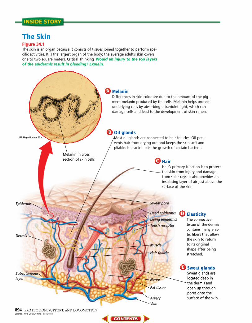
The SkinFigure 34.1The skin is an organ because it consists of tissues joined together to perform spe-cific activities. It is the largest organ of the body; the average adult’s skin coversone to two square meters. Critical Thinking Would an injury to the top layersof the epidermis result in bleeding? Explain.
Dermis
Subcutaneouslayer
Epidermis Sweat pore
Dead epidermis
Living epidermis
Touch receptor
Nerve
Fat tissue
ArteryVein
Muscle
Hair follicle
Melanin in cross section of skin cells
ElasticityThe connective tissue of the dermiscontains many elas-tic fibers that allowthe skin to returnto its originalshape after beingstretched.
DD
Sweat glandsSweat glands arelocated deep in the dermis andopen up throughpores onto the surface of the skin.
EE
894 PROTECTION, SUPPORT, AND LOCOMOTION
LM Magnification: 65�
Science Photo Library/Photo Researchers
MelaninDifferences in skin color are due to the amount of the pig-ment melanin produced by the cells. Melanin helps protectunderlying cells by absorbing ultraviolet light, which candamage cells and lead to the development of skin cancer.
AA
Oil glandsMost oil glands are connected to hair follicles. Oil pre-vents hair from drying out and keeps the skin soft andpliable. It also inhibits the growth of certain bacteria.
BB
HairHair’s primary function is to protectthe skin from injury and damagefrom solar rays. It also provides aninsulating layer of air just above thesurface of the skin.
CC
0893-0898 C34 S1 BDOL-829900 8/5/04 2:40 AM Page 894
The interior layer of the epidermiscontains living cells that continuallydivide to replace the dead cells. Someof these cells contain melanin, a pig-ment that colors the skin and helpsprotect body cells from damage bysolar radiation. As the newly formedcells are pushed toward the skin’s sur-face, the nuclei degenerate and thecells die. Once they reach the outer-most epidermal layer, the cells areshed. This entire process takes about28 days. Therefore, every four weeks,all cells of the epidermis are replacedby new cells.
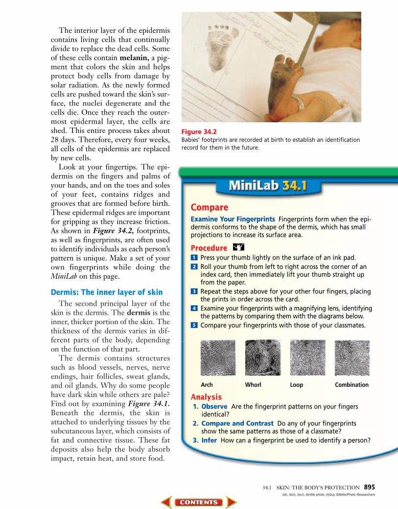
Look at your fingertips. The epi-dermis on the fingers and palms ofyour hands, and on the toes and solesof your feet, contains ridges andgrooves that are formed before birth.These epidermal ridges are importantfor gripping as they increase friction.As shown in Figure 34.2, footprints,as well as fingerprints, are often usedto identify individuals as each person’spattern is unique. Make a set of yourown fingerprints while doing theMiniLab on this page.
Dermis: The inner layer of skinThe second principal layer of the
skin is the dermis. The dermis is theinner, thicker portion of the skin. Thethickness of the dermis varies in dif-ferent parts of the body, dependingon the function of that part.
The dermis contains structuressuch as blood vessels, nerves, nerveendings, hair follicles, sweat glands,and oil glands. Why do some peoplehave dark skin while others are pale?Find out by examining Figure 34.1.Beneath the dermis, the skin isattached to underlying tissues by thesubcutaneous layer, which consists offat and connective tissue. These fatdeposits also help the body absorbimpact, retain heat, and store food.
Figure 34.2Babies’ footprints are recorded at birth to establish an identificationrecord for them in the future.
34.1 SKIN: THE BODY’S PROTECTION 895(bl), (bcl), (bcr), (br)file photo, (t)Guy Gillette/Photo Researchers
CompareExamine Your Fingerprints Fingerprints form when the epi-dermis conforms to the shape of the dermis, which has smallprojections to increase its surface area.
Procedure! Press your thumb lightly on the surface of an ink pad.@ Roll your thumb from left to right across the corner of an
index card, then immediately lift your thumb straight upfrom the paper.
# Repeat the steps above for your other four fingers, placingthe prints in order across the card.
$ Examine your fingerprints with a magnifying lens, identifyingthe patterns by comparing them with the diagrams below.
% Compare your fingerprints with those of your classmates.
Analysis1. Observe Are the fingerprint patterns on your fingers
identical?2. Compare and Contrast Do any of your fingerprints
show the same patterns as those of a classmate?3. Infer How can a fingerprint be used to identify a person?
Arch Whorl Loop Combination
0893-0898 C34 S1 BDOL-829900 8/5/04 2:40 AM Page 895
Hair, another structure of theintegumentary system, grows out ofnarrow cavities in the dermis calledhair follicles, as shown in Figure 34.3.As hair follicles develop, they are sup-plied with blood vessels and nerves andbecome attached to muscle tissue.Most hair follicles have an oil glandassociated with them. When oil anddead cells block the opening of the hairfollicle, pimples may form.
Functions of the integumentary system
One function of skin is to help main-tain homeostasis by regulating yourinternal body temperature. When yourbody temperature rises, the many smallblood vessels in the dermis dilate,blood flow increases, and body heat islost by radiation. This mechanism alsoworks in reverse. When you are cold,the blood vessels in the skin constrictand heat is conserved.
Another noticeable thing that hap-pens to your skin as your body heats upis that it becomes wet. Glands in thedermis produce sweat in response to anincrease in body temperature. As sweatevaporates, water changes state fromliquid to vapor and heat is lost. Thebody cools as a result of the heat loss.Investigate further the role of skin incooling the body by carrying out theProblem-Solving Lab on this page.
Of course, anyone who has everstepped on a sharp object or beenburned by a hot pot handle knows thatskin also functions as a sense organ.Nerve cells in the dermis receive stim-uli from the external environment andrelay information about pressure,pain, and temperature to the brain.
Skin also plays a role in producingessential vitamins. When exposed toultraviolet light, skin cells producevitamin D, a nutrient that aids theabsorption of calcium into the blood-stream. As a person’s exposure to Hair follicle
896
LM Magnification: 30�
Sheila Terry/Science Photo Library/Photo Researchers
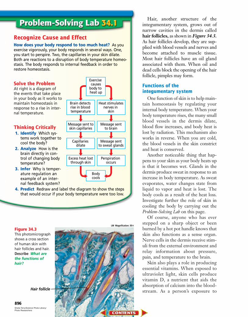
Recognize Cause and EffectHow does your body respond to too much heat? As youexercise vigorously, your body responds in several ways. One,you start to perspire. Two, the capillaries in your skin dilate.Both are reactions to a disruption of body temperature homeo-stasis. The body responds to internal feedback in order torestore homeostasis.
Solve the ProblemAt right is a diagram ofthe events that take placein your body as it works tomaintain homeostasis inresponse to a rise in inter-nal temperature.
Thinking Critically1. Identify Which sys-
tems work together tocool the body?
2. Analyze How is thebrain directly in con-trol of changing body temperature?
3. Infer Why is temper-ature regulation anexample of an inter-nal feedback system?
4. Predict Redraw and label the diagram to show the stepsthat would occur if your body temperature were too low.
Brain detects rise in blood temperature
Heat stimulatesnerves in
skin
Message sent toskin capillaries
Message sentto brain
Capillaries dilate
Message sentto sweat glands
Excess heat lostthrough skin
Perspiration occurs
Bodycools
Exercise causes
body to heat up
Figure 34.3This photomicrographshows a cross sectionof human skin withhair follicles and hair.Describe What arethe functions ofhair?
0893-0898 C34 S1 BDOL-829900 8/5/04 2:41 AM Page 896
34.1 SKIN: THE BODY’S PROTECTION 897
Blood clot
Scab New skin cells
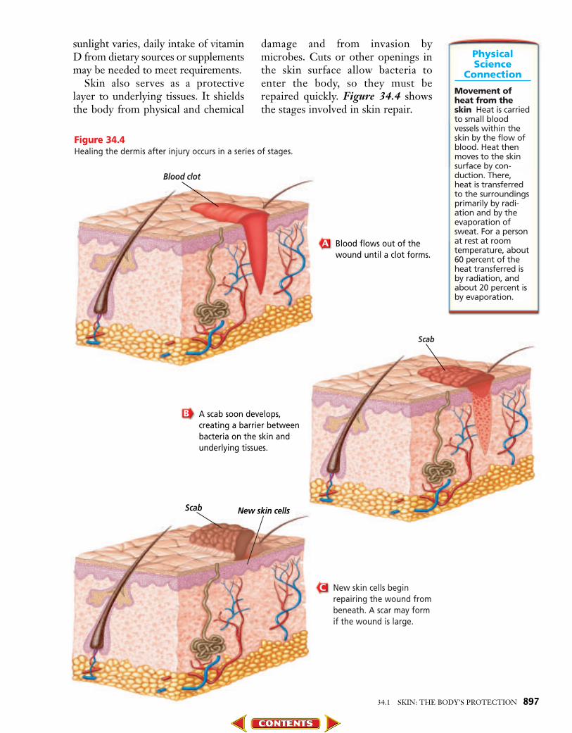
Figure 34.4Healing the dermis after injury occurs in a series of stages.
New skin cells beginrepairing the wound frombeneath. A scar may formif the wound is large.
C
Blood flows out of thewound until a clot forms.
A
Scab
A scab soon develops,creating a barrier betweenbacteria on the skin andunderlying tissues.
B
sunlight varies, daily intake of vitaminD from dietary sources or supplementsmay be needed to meet requirements.
Skin also serves as a protectivelayer to underlying tissues. It shieldsthe body from physical and chemical
damage and from invasion bymicrobes. Cuts or other openings inthe skin surface allow bacteria toenter the body, so they must berepaired quickly. Figure 34.4 showsthe stages involved in skin repair.
PhysicalScience
Connection
Movement ofheat from theskin Heat is carriedto small bloodvessels within theskin by the flow ofblood. Heat thenmoves to the skinsurface by con-duction. There,heat is transferredto the surroundingsprimarily by radi-ation and by theevaporation ofsweat. For a personat rest at roomtemperature, about60 percent of theheat transferred isby radiation, andabout 20 percent isby evaporation.
0893-0898 C34 S1 BDOL-829900 8/5/04 2:42 AM Page 897

Skin Injury and Healing
If you’ve ever had amild scrape, you knowthat it doesn’t take longfor the wound to heal.When the epidermis sus-tains a mild injury, such asa scrape, the deepest layerof epidermal cells divideto help fill in the gap leftby the abrasion. If, how-ever, the injury extendsinto the dermis, whereblood vessels are found,bleeding usually occurs.The skin then goesthrough a series of stagesto heal the damaged tis-sue. The first reaction ofthe body is to restore the
continuity of the skin, that is, to closethe break. Blood flowing from thewound soon clots. The wound is thenclosed by the formation of a scab,which prevents bacteria from enter-ing the body. Dilated blood vesselsthen allow infection-fighting whiteblood cells to migrate to the woundsite. Soon after, skin cells beneath thescab begin to multiply and fill in thegap. Eventually, the scab falls off toexpose newly formed skin. If a woundis large, high amounts of dense con-nective tissue fibers used to close thewound may leave a scar.
Have you ever suffered a painfulburn? Burns can result from exposureto the sun or contact with chemicalsor hot objects. Burns are ratedaccording to their severity.
First-degree burns, such as a mildsunburn, involve the death of epider-mal cells and are characterized byredness and mild pain. First-degreeburns usually heal in about one weekwithout leaving a scar. Second-degreeburns involve damage to skin cells ofboth the epidermis and the dermisand can result in blistering and scar-ring. The most severe burns arethird-degree burns, which destroyboth the epidermis and the dermis.With this type of burn, skin functionis lost, and skin grafts may be requiredto replace lost skin. In some cases,healthy skin can be removed fromanother area of the patient’s body andtransplanted to a burned area.
As people get older, their skinchanges. It becomes drier as glandsdecrease their production of lubricat-ing skin oils—a mixture of fats, cho-lesterol, proteins, and inorganic salts.As shown in Figure 34.5, wrinklesmay appear as the elasticity of the skindecreases. Although these changes arenatural, they can be accelerated byprolonged exposure to ultraviolet raysfrom the sun.
Summarize how theskin changes as people age.
Understanding Main Ideas1. Compare the structures and functions of the
epidermis and the dermis.2. Identify and interpret the functions of the integu-
mentary system.3. Compare how the skin interrelates with other organ
systems to maintain a constant body temperature.4. How does the skin respond to external stimuli?
Thinking Critically5. How could third-degree burns over a significant
portion of the skin affect the body as a whole?
6. Sequence Outline steps that occur as a cut in theskin heals. For more help, refer to Sequence in theSkill Handbook.
SKILL REVIEWSKILL REVIEW
898 PROTECTION, SUPPORT, AND LOCOMOTION
Figure 34.5As people age, theirskin loses its elasticityand begins to wrinkle.
Lawrence Migdale
ca.bdol.glencoe.com/self_check_quiz
0893-0898 C34 S1 BDOL-829900 8/5/04 2:43 AM Page 898
34.2SECTION PREVIEWObjectivesCompare the differenttypes of movable joints.Describe how bone isformed.Identify the structure andfunctions of the skeletal system.
Review Vocabularycartilage: tough, flexible
material that makes upportions of bony-animalskeletons (p. 799)
New Vocabularyaxial skeletonappendicular skeletonjointligamentbursatendoncompact boneosteocytespongy boneosteoblastred marrowyellow marrow
34.2 BONES: THE BODY’S SUPPORT 899
Bones: The Body’s Support
The Body’s FoundationFinding Main Ideas On a piece ofpaper, construct an outline about the skeletal system. Use the red and blue titles in the section as a guideline. As you read the paragraphs that follow the titles,add important information andvocabulary words to your outline.
Example:I. Skeletal System Structure
A. Joints1. Ball-and-socket joint2. Pivot joint
Use your outline to help youanswer questions in the SectionAssessment on page 904. For more help, refer to Outline in the Skill Handbook.
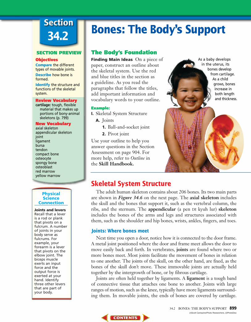
As a baby develops in the uterus, its
bones developfrom cartilage.
As a childgrows, bonesincrease inboth lengthand thickness.
(r)Scott Camazine/Photo Researchers, (l)PhotoDisc
PhysicalScience
Connection
Joints and leversRecall that a leveris a rod or plankthat pivots on afulcrum. A numberof joints in yourbody serve asfulcrums. Forexample, yourforearm is a leverthat pivots on theelbow joint. Thebiceps muscleexerts an inputforce and theoutput force isexerted at yourhand. Identifythree other leversthat are part ofyour body.
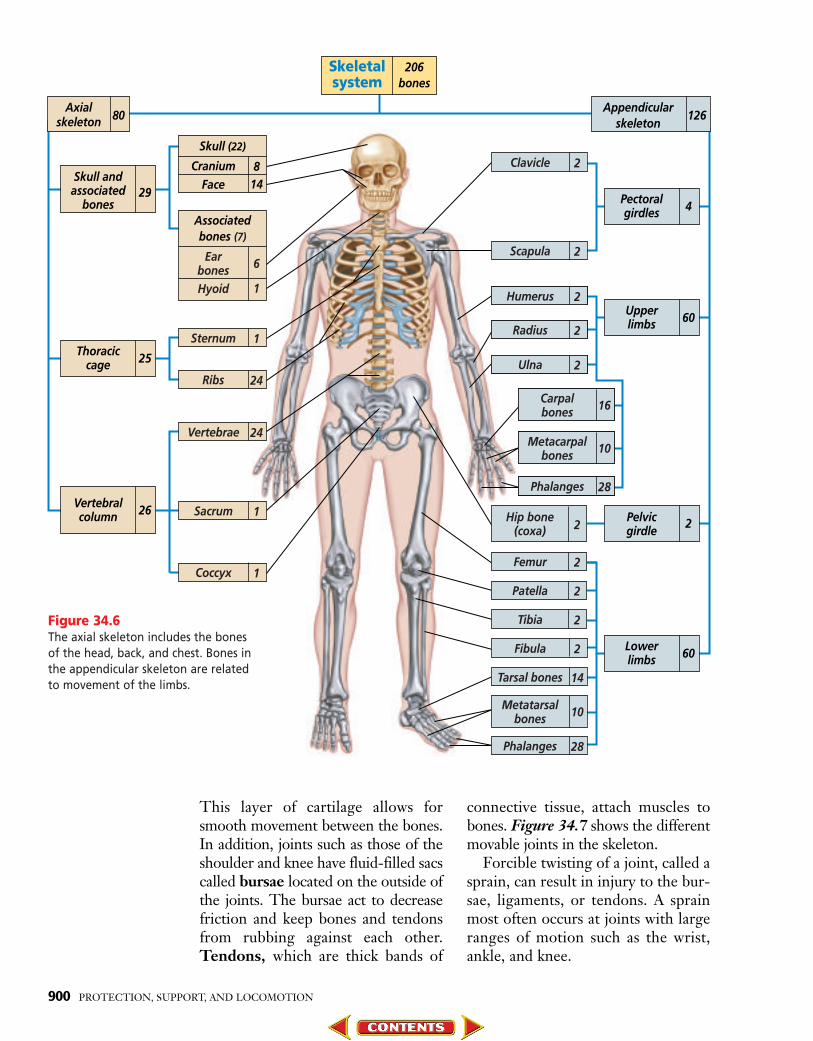
Skeletal System StructureThe adult human skeleton contains about 206 bones. Its two main parts
are shown in Figure 34.6 on the next page. The axial skeleton includesthe skull and the bones that support it, such as the vertebral column, theribs, and the sternum. The appendicular (a pen DI kyuh lur) skeletonincludes the bones of the arms and legs and structures associated withthem, such as the shoulder and hip bones, wrists, ankles, fingers, and toes.
Joints: Where bones meetNext time you open a door, notice how it is connected to the door frame.
A metal joint positioned where the door and frame meet allows the door tomove easily back and forth. In vertebrates, joints are found where two ormore bones meet. Most joints facilitate the movement of bones in relationto one another. The joints of the skull, on the other hand, are fixed, as thebones of the skull don’t move. These immovable joints are actually heldtogether by the intergrowth of bone, or by fibrous cartilage.
Joints are often held together by ligaments. A ligament is a tough bandof connective tissue that attaches one bone to another. Joints with largeranges of motion, such as the knee, typically have more ligaments surround-ing them. In movable joints, the ends of bones are covered by cartilage.
0899-0904 C34 S2 BDOL-829900 8/5/04 2:56 AM Page 899
Skeletalsystem
206bones
Axialskeleton 80
Appendicularskeleton
126
Skull andassociated
bones29
Thoraciccage 25
Upperlimbs 60
Pelvicgirdle 2
Lowerlimbs 60
Pectoralgirdles 4
Vertebralcolumn 26
Skull (22)
814
CraniumFace
Associatedbones (7)
6
1
EarbonesHyoid
1Sternum
24Vertebrae
28
10
Phalanges
2Femur
2Patella
2Tibia
2Fibula
28Phalanges
14Tarsal bones
1Sacrum
1Coccyx
24Ribs
Hip bone(coxa)
Carpalbones
Metacarpalbones
10Metatarsalbones
2Clavicle
2Scapula
2Humerus
2Radius
2Ulna
16
2
This layer of cartilage allows forsmooth movement between the bones.In addition, joints such as those of theshoulder and knee have fluid-filled sacscalled bursae located on the outside ofthe joints. The bursae act to decreasefriction and keep bones and tendonsfrom rubbing against each other.Tendons, which are thick bands of
connective tissue, attach muscles tobones. Figure 34.7 shows the differentmovable joints in the skeleton.
Forcible twisting of a joint, called asprain, can result in injury to the bur-sae, ligaments, or tendons. A sprainmost often occurs at joints with largeranges of motion such as the wrist,ankle, and knee.
900 PROTECTION, SUPPORT, AND LOCOMOTION
Figure 34.6The axial skeleton includes the bonesof the head, back, and chest. Bones inthe appendicular skeleton are relatedto movement of the limbs.
0899-0904 C34 S2 BDOL-829900 8/5/04 2:57 AM Page 900

Besides injury, joints are also sub-ject to disease. One common jointdisease is arthritis, an inflammation ofthe joints. It can be caused by infec-tions, aging, or injury. One kind ofarthritis results in bone spurs, or out-growths of bone, inside the joints.Such arthritis is especially painful,
and often limits a person’s ability tomove his or her joints.
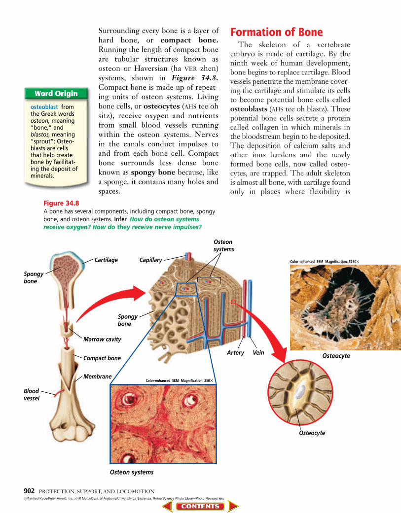
Compact and spongy boneAlthough bones may appear uni-
form, they are actually composed oftwo different types of bone tissue:compact bone and spongy bone.
34.2 BONES: THE BODY’S SUPPORT 901
arthritis from theGreek wordsarthron, meaning“joint,” and itis,meaning “swellingdisease”; Arthritisis a swelling dis-ease of the joints.
Pivot joints allow bones to twist around eachother. One example of a pivot joint is in your arm,between the ulna and the radius. It allows you totwist your lower arm around.
B
Hinge joints are found in the elbows, knees,fingers, and toes. They allow back-and-forthmovement like that of a door hinge.
C Gliding joints, found in the wrists and ankles,allow bones to slide past each other.
D
Ball-and-socket joints allow movement in alldirections. The joints of the hips and shoulders areball-and-socket joints; they allow you to swingyour arms and legs around in many directions.
A
Figure 34.7Body movements are made possible by joints that allow bones to move in several different directions.
0899-0904 C34 S2 BDOL-829900 8/5/04 2:57 AM Page 901
osteoblast fromthe Greek wordsosteon, meaning“bone,” and blastos, meaning“sprout”; Osteo-blasts are cells that help createbone by facilitat-ing the deposit ofminerals.
Surrounding every bone is a layer ofhard bone, or compact bone.Running the length of compact boneare tubular structures known asosteon or Haversian (ha VER zhen)systems, shown in Figure 34.8.Compact bone is made up of repeat-ing units of osteon systems. Livingbone cells, or osteocytes (AHS tee ohsitz), receive oxygen and nutrientsfrom small blood vessels runningwithin the osteon systems. Nerves in the canals conduct impulses to and from each bone cell. Compactbone surrounds less dense boneknown as spongy bone because, likea sponge, it contains many holes andspaces.
Formation of BoneThe skeleton of a vertebrate
embryo is made of cartilage. By theninth week of human development,bone begins to replace cartilage. Bloodvessels penetrate the membrane cover-ing the cartilage and stimulate its cellsto become potential bone cells calledosteoblasts (AHS tee oh blastz). Thesepotential bone cells secrete a proteincalled collagen in which minerals inthe bloodstream begin to be deposited.The deposition of calcium salts andother ions hardens and the newlyformed bone cells, now called osteo-cytes, are trapped. The adult skeletonis almost all bone, with cartilage foundonly in places where flexibility is
902 PROTECTION, SUPPORT, AND LOCOMOTION
Spongy bone
Cartilage
Osteocyte
Bloodvessel
Marrow cavity
Spongybone
Capillary
Osteonsystems
Artery VeinCompact bone
Membrane
Figure 34.8A bone has several components, including compact bone, spongybone, and osteon systems. Infer How do osteon systemsreceive oxygen? How do they receive nerve impulses?
Osteon systems
Color-enhanced SEM Magnification: 250�
Color-enhanced SEM Magnification: 5250�
Osteocyte
(l)Manfred Kage/Peter Arnold, Inc., (r)P. Motta/Dept. of Anatomy/University La Sapienza, Rome/Science Photo Library/Photo Researchers
0899-0904 C34 S2 BDOL-829900 8/5/04 2:59 AM Page 902
Yellow bone marrow
34.2 BONES: THE BODY’S SUPPORT 903KS Studios
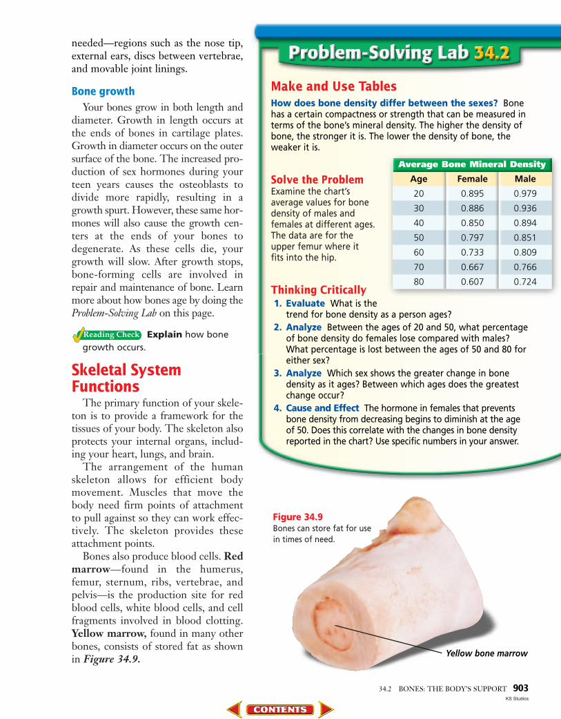
Make and Use TablesHow does bone density differ between the sexes? Bonehas a certain compactness or strength that can be measured interms of the bone’s mineral density. The higher the density ofbone, the stronger it is. The lower the density of bone, theweaker it is.
Solve the ProblemExamine the chart’s average values for bone density of males and females at different ages. The data are for the upper femur where it fits into the hip.
Thinking Critically1. Evaluate What is the
trend for bone density as a person ages? 2. Analyze Between the ages of 20 and 50, what percentage
of bone density do females lose compared with males?What percentage is lost between the ages of 50 and 80 foreither sex?
3. Analyze Which sex shows the greater change in bonedensity as it ages? Between which ages does the greatestchange occur?
4. Cause and Effect The hormone in females that preventsbone density from decreasing begins to diminish at the ageof 50. Does this correlate with the changes in bone densityreported in the chart? Use specific numbers in your answer.
Average Bone Mineral Density
Age Female Male
20 0.895 0.979
30 0.886 0.936
40 0.850 0.894
50 0.797 0.851
60 0.733 0.809
70 0.667 0.766
80 0.607 0.724
needed—regions such as the nose tip,external ears, discs between vertebrae,and movable joint linings.
Bone growthYour bones grow in both length and
diameter. Growth in length occurs atthe ends of bones in cartilage plates.Growth in diameter occurs on the outersurface of the bone. The increased pro-duction of sex hormones during yourteen years causes the osteoblasts todivide more rapidly, resulting in agrowth spurt. However, these same hor-mones will also cause the growth cen-ters at the ends of your bones todegenerate. As these cells die, yourgrowth will slow. After growth stops,bone-forming cells are involved inrepair and maintenance of bone. Learnmore about how bones age by doing theProblem-Solving Lab on this page.
Explain how bonegrowth occurs.
Skeletal SystemFunctions
The primary function of your skele-ton is to provide a framework for thetissues of your body. The skeleton alsoprotects your internal organs, includ-ing your heart, lungs, and brain.
The arrangement of the humanskeleton allows for efficient bodymovement. Muscles that move thebody need firm points of attachmentto pull against so they can work effec-tively. The skeleton provides theseattachment points.
Bones also produce blood cells. Redmarrow—found in the humerus,femur, sternum, ribs, vertebrae, andpelvis—is the production site for redblood cells, white blood cells, and cellfragments involved in blood clotting.Yellow marrow, found in many otherbones, consists of stored fat as shownin Figure 34.9.
Figure 34.9Bones can store fat for usein times of need.
0899-0904 C34 S2 BDOL-829900 8/5/04 3:00 AM Page 903

Understanding Main Ideas1. Distinguish between the appendicular skeleton
and the axial skeleton.
2. Compare and contrast the four main kinds ofmovable joints and provide an example of each.
3. How is compact bone structurally different fromspongy bone?
4. Identify and interpret the functions of the skeletalsystem.
Thinking Critically5. Why would it be impossible for bones to grow
from within?
6. Get the Big Picture Outline the steps involvedin bone formation and growth—from cartilage tothe cessation of bone growth. For more help,refer to Get the Big Picture in the Skill Handbook.
SKILL REVIEWSKILL REVIEW
904 PROTECTION, SUPPORT, AND LOCOMOTION
Figure 34.10The X ray on the leftshows a leg bone thathas completely frac-tured. The X ray on theright shows the bone(with a supporting rod)after it has healed. Thearrow indicates thearea where the breakhealed.
(l)Matt Meadows, (r)Matt Meadows
Bones store mineralsFinally, your bones serve as store-
houses for minerals, including cal-cium and phosphate. Calcium isneeded to form strong, healthybones and is therefore an importantpart of your diet. Sources of calciuminclude milk, yogurt, cheese, lettuce,spinach, and other assorted leafyvegetables.
Bone injury and diseaseBones tend to become more brittle
as their composition changes withage. For example, a disease calledosteoporosis (ahs tee oh puh ROH sus)
involves a loss of bone volume andmineral content, causing the bones tobecome more porous and brittle.Osteoporosis is most common inolder women because they producelesser amounts of estrogen—a hor-mone that aids in bone formation.
When bones are broken, as shownby the X-ray images in Figure 34.10,a doctor moves them back into posi-tion and immobilizes them with a castor splint until the bone tissueregrows. Read more about the use ofX rays in the diagnosis of brokenbones in the Connection to Physics atthe end of this chapter.
Wave types X raysare electromag-netic waves. Unlikesound waves,electromagneticwaves can travelthrough space andconsist of vibratingelectric and mag-netic fields. Micro-waves, visible light,and ultraviolet raysare also electro-magnetic waveswith longer wave-lengths than X rays.
ca.bdol.glencoe.com/self_check_quiz
PhysicalScience
Connection
0899-0904 C34 S2 BDOL-829900 8/5/04 3:00 AM Page 904
34.3SECTION PREVIEWObjectivesClassify the three types ofmuscles.Analyze the structure of amyofibril.Interpret the sliding fila-ment theory.
Review VocabularyATP: energy-storing mole-
cule in cells composed of an adenosine mole-cule, a ribose sugar, andthree phosphate groups(p. 222)
New Vocabularysmooth muscleinvoluntary musclecardiac muscleskeletal musclevoluntary musclemyofibrilmyosinactinsarcomeresliding filament theory
34.3 MUSCLES FOR LOCOMOTION 905
Classifying Muscles Concept Map Copy the concept map onto a separate sheet of paper.
Organize Information As you read this section, complete the concept map tocompare different types of muscles.
MuscleTypes
involuntarily
1. 2.
6.5.4.3.
cardiac
attached to bones
located controlled located controlled located controlled
Muscles for Locomotion
Skeletalmuscle fiber
Nucleus
Striation
Smooth muscle fibersappear spindle-shapedunder the microscope.
A Cardiac muscle fibersappear striated orstriped when magnified.
B Skeletal muscle fibers alsoappear striated whenmagnified.
C
LM Magnification: 100�
Cardiacmusclefiber
Striation
Nucleus
Color-enhanced TEM Magnification: 42 000� Color-enhanced TEM Magnification: 1100�
Smoothmusclefiber
Nucleus
Figure 34.11Muscles differ in structureand appearance.
(bl)M.I. Walker/Science Source/Photo Researchers, (bc)Don Fawcett/Visuals Unlimited, (br)J. Venable/Visuals Unlimited/D. Fawcett
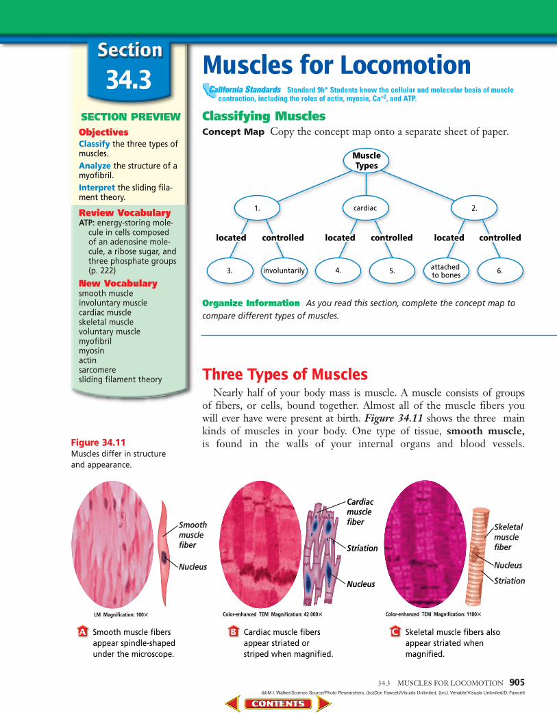
Three Types of MusclesNearly half of your body mass is muscle. A muscle consists of groups
of fibers, or cells, bound together. Almost all of the muscle fibers you will ever have were present at birth. Figure 34.11 shows the three mainkinds of muscles in your body. One type of tissue, smooth muscle,is found in the walls of your internal organs and blood vessels.
Standard 9h* Students know the cellular and molecular basis of musclecontraction, including the roles of actin, myosin, Ca+2, and ATP.
California Standards
0905-0915 C34 S3 BDOL-829900 8/5/04 7:25 AM Page 905

Smooth muscle is made up of sheets ofcells that are ideally shaped to form alining for organs, such as the digestivetract and the reproductive tract. Themost common function of smoothmuscle is to squeeze, exerting pressureon the space inside the tube or organ itsurrounds in order to move materialthrough it. Examples include themovement of food through the diges-tive system and the movement ofgametes through the reproductive sys-tem. Because contractions of smoothmuscle are not under conscious con-trol, smooth muscle is considered aninvoluntary muscle.
Another type of involuntary muscleis the cardiac muscle, which makes upyour heart. Cardiac muscle fibers areinterconnected and form a networkthat helps the heart muscle contractefficiently. Cardiac muscle is foundonly in the heart and is adapted to gen-erate and conduct electrical impulsesnecessary for its rhythmic contraction.
The third type of muscle tissue,skeletal muscle, is the type that isattached to and moves your bones.The majority of the muscles in yourbody are skeletal muscles, and, asyou know, you can control their con-tractions. A muscle that contractsunder conscious control is called avoluntary muscle. Compare thestructure and function of skin, mus-cle, and bone in the Problem-SolvingLab on this page.
Skeletal MuscleContraction
Whether you are playing tennis,pushing a lawn mower, or writing, somemuscles contract while others relax asthe action is performed. Figure 34.12shows the movement of the lower armas controlled by opposing muscles inthe upper arm. The majority of skeletalmuscles work in opposing pairs.
Compare and ContrastHow are skin, bone,and muscle cells different? Cells thatform skin (whichincludes epithelialcells), bone, and mus-cle are specialized toperform various func-tions. Each of thesesystems of the bodycontains differenttypes of cells thatwork together to carryout the function of the tissue or organ.
Solve the Problem Using the text and figures on pages 894, 902, 905, and 908,prepare a table that compares and contrasts the structure andfunction of skin, muscle, and bone cells.
Thinking CriticallyInfer Protein analysis of an unknown tissue sample yields ahigh level of myosin and actin. Is this sample skin, bone, ormuscle? What structures could you look for in an electronmicroscope to confirm the identity of this tissue?
Cell Structure and Function
Structure Function
Skin
Muscle
Bone
Relaxedbiceps
Contractingtriceps
Contractingbiceps
Relaxedtriceps
Figure 34.12When the biceps muscle contracts, the lower arm is moved upward (A).When the triceps muscle on the back of the upper arm contracts, the lower arm movesdownward (B).
AABB
906
0905-0915 C34 S3 BDOL-829900 8/5/04 1:54 AM Page 906

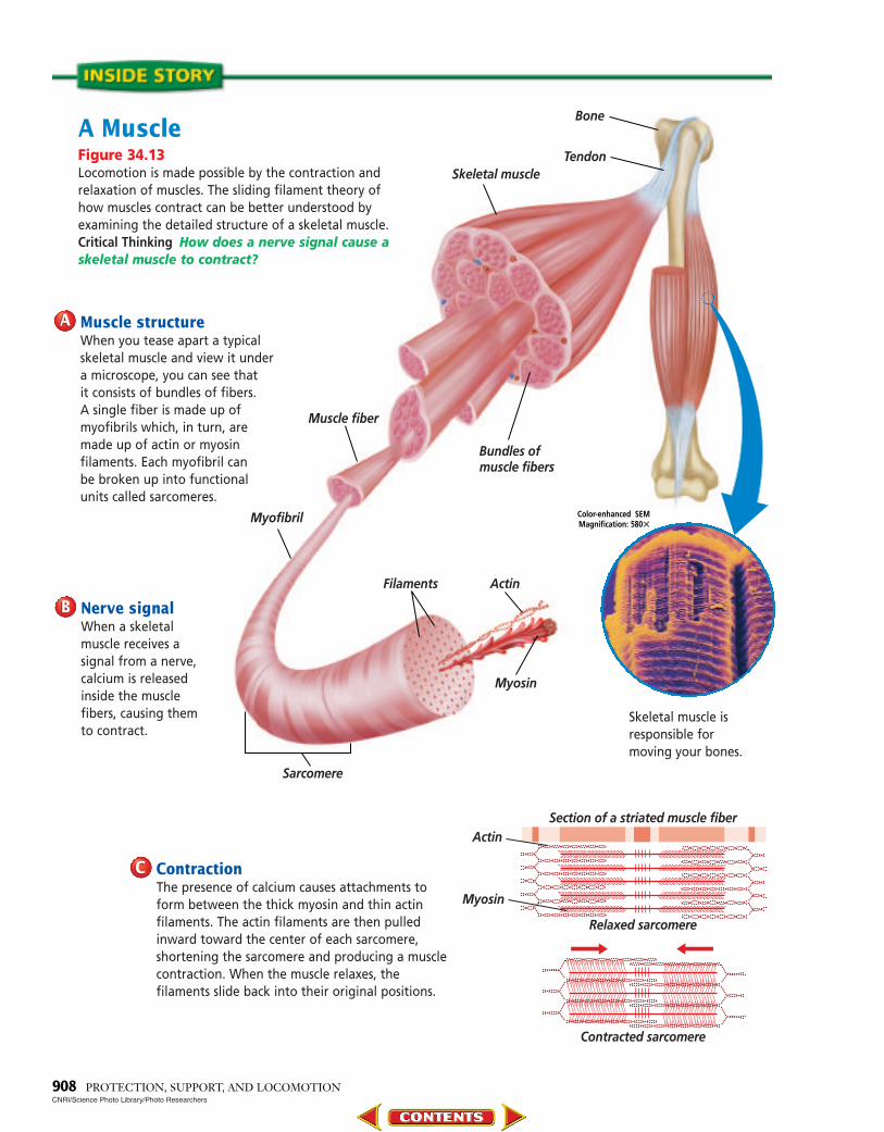
Muscle tissue is made up of musclefibers, which are actually just verylong, fused muscle cells. Each fiber ismade up of smaller units calledmyofibrils (mi oh FI brulz). Myo-fibrils are themselves composed ofeven smaller protein filaments thatcan be either thick or thin. Thethicker filaments are made of the pro-tein myosin, and the thinner fila-ments are made of the protein actin.Each myofibril can be divided intosections called sarcomeres (SAR kuhmeerz), the functional units of mus-cle. How do nerves signal muscles tocontract? Find out in Figure 34.13on the next page.
The sliding filament theory cur-rently offers the best explanation forhow muscle contraction occurs. Thesliding filament theory states that,when signaled, the actin filamentswithin each sarcomere slide towardone another, shortening the sarcom-eres in a fiber and causing the muscleto contract. The myosin filaments, onthe other hand, do not move. Learnmore about the sliding filament the-ory and muscle contraction in theMiniLab on this page.
Muscle Strength and Exercise
How can you increase the strengthof your muscles? Muscle strength doesnot depend on the number of fibers ina muscle. It has been shown that thisnumber is basically fixed before you areborn. Rather, muscle strength dependson the thickness of the fibers and onhow many of them contract at onetime. Regular exercise stresses musclefibers slightly; to compensate for thisadded workload, the fibers increase indiameter by adding myofibrils.
Recall that ATP is produced duringcellular respiration. Muscle cells arecontinually supplied with ATP from
both aerobic and anaerobic processes.However, the aerobic respirationprocess dominates when adequateoxygen is delivered to muscle cells,such as when a muscle is at rest or dur-ing moderate activity. When an ade-quate supply of oxygen is unavailable,such as during vigorous activity, ananaerobic process—specifically lacticacid fermentation—becomes the pri-mary source of ATP production.
Identify the twoprocesses the body uses to produce ATP.
34.3 MUSCLES FOR LOCOMOTION 907
InterpretExamining Muscle Contraction Sarcomeres in muscle fibersare composed of the protein filaments actin and myosin. Thesliding action of these filaments in relation to one anotherresults in muscle contraction.
Procedure! Look at diagrams A and B. Diagram A shows a sarcomere
in a relaxed muscle. Diagram B shows a sarcomere in a contracted muscle.
@ Using a centimeter ruler, measure and record the length ofa sarcomere, a myosin filament, and an actin filament indiagram A. Record your data in a table.
# Repeat step 2 for diagram B.
Analysis 1. Evaluate When a muscle contracts, do actin or myosin fil-
aments shorten? Use your data to support your answer.2. Infer How does the sarcomere shorten?
Myosin
Actin
Sarcomere
AA
BB
PhysicalScience
Connection
Muscles doingwork The workdone on an objectis the force appliedto the object timesthe distance itmoves. You do nowork if you push ona car and it doesn’tmove. You may feeltired if you pushlong enough due tothe production oflactic acid in yourmuscles.
0905-0915 C34 S3 BDOL-829900 8/5/04 7:26 AM Page 907
Muscle fiber
Bone
TendonSkeletal muscle
Myofibril
Filaments Actin
Myosin
Sarcomere
Bundles ofmuscle fibers
908 PROTECTION, SUPPORT, AND LOCOMOTION
Color-enhanced SEMMagnification: 580�
Muscle structureWhen you tease apart a typicalskeletal muscle and view it undera microscope, you can see that it consists of bundles of fibers. A single fiber is made up ofmyofibrils which, in turn, are made up of actin or myosinfilaments. Each myofibril can be broken up into functional units called sarcomeres.
AA
Relaxed sarcomere
Section of a striated muscle fiber
Contracted sarcomere
Actin
Myosin
CNRI/Science Photo Library/Photo Researchers
Nerve signalWhen a skeletalmuscle receives asignal from a nerve,calcium is releasedinside the musclefibers, causing themto contract.
BB
A MuscleFigure 34.13Locomotion is made possible by the contraction andrelaxation of muscles. The sliding filament theory ofhow muscles contract can be better understood byexamining the detailed structure of a skeletal muscle.Critical Thinking How does a nerve signal cause askeletal muscle to contract?
Skeletal muscle isresponsible formoving your bones.
ContractionThe presence of calcium causes attachments toform between the thick myosin and thin actinfilaments. The actin filaments are then pulledinward toward the center of each sarcomere,shortening the sarcomere and producing a musclecontraction. When the muscle relaxes, thefilaments slide back into their original positions.
CC
0905-0915 C34 S3 BDOL-829900 8/5/04 1:55 AM Page 908

Think about what happens whenyou are running in gym class or aroundthe track at school. Figure 34.14Aillustrates how an athlete’s need foroxygen changes as the intensity of hisor her workout increases. At somepoint, your muscles are not able to getoxygen fast enough to sustain aerobicrespiration and produce adequateATP. Thus, the amount of availableATP becomes limited. For your mus-cle cells to get the energy they need,they must rely on lactic acid fermenta-tion as well. Figure 34.14B indicateshow, at a certain intensity, the bodyshifts from aerobic respiration to theanaerobic process of lactic acid fer-mentation for its energy needs.
During exercise, lactic acid buildsup in muscle cells. As the excess lacticacid is passed into the bloodstream,the blood becomes more acidic andrapid breathing is stimulated. As youcatch your breath following exercise,adequate amounts of oxygen are sup-plied to your muscles and lactic acid isbroken down. Regular exercise canresult in improved performance ofmuscles. Do the BioLab at the end ofthe chapter to find out how musclefatigue affects the amount of exerciseyour muscles can accomplish.
Explain what hap-pens to lactic acid after exercise iscompleted.
Understanding Main Ideas1. Compare the structure and interpret the functions
of the three main types of muscles in the muscularsystem.
2. Summarize the sliding filament theory of musclecontraction.
3. How can exercise change muscle strength? Howcan it change muscle function?
4. What determines muscle strength?
Thinking Critically5. Why would a disease that causes paralysis of
smooth muscles be life threatening?
6. Interpret Scientific Illustrations Diagram the composition of muscle fibers as shown inFigure 34.13. For more help, refer to InterpretScientific Illustrations in the Skill Handbook.
SKILL REVIEWSKILL REVIEW
34.3 MUSCLES FOR LOCOMOTION 909
Rate
of o
xyge
nco
nsum
ptio
n
Work rate
Oxygen Consumption During Exercise
Bloo
d la
ctic
aci
d
Work rate
Blood Lactic Acid Levels During Exercise
Shift towardanaerobicprocess
Figure 34.14Athletic trainers use information about muscle functioning during exerciseto establish appropriate levels of intensity for training.
As an individual increases the intensity of his or her workout,the need for oxygen goes up in predictable increments.
A
During exercise, lactic acid concentrations can increase. Infer Whatdoes an increase in the presence of lactic acid in the blood-stream indicate about the amount of oxygen available tomuscle cells?
B
ca.bdol.glencoe.com/self_check_quiz
0905-0915 C34 S3 BDOL-829900 8/5/04 1:17 AM Page 909
Before You Begin
The movement of bodyparts results from the con-traction and relaxation ofmuscles. In this process,muscles use energy fromaerobic respiration andlactic acid fermentation.When exercise is contin-ued for a long period oftime, the waste productsof fermentation accumu-late and muscle fibers arestressed, causing fatigue.How does fatigue affectmuscles? In this lab youwill investigate the effectsof fatigue on the ability ofmuscles to perform a task.
910 PROTECTION, SUPPORT, AND LOCOMOTIONStudiohio
Does fatigue affect theability to perform anexercise?
ProblemHow does fatigue affect the number of repetitions of anexercise you can accomplish?
HypothesesHypothesize whether or not muscle fatigue has any effect onthe amount of exercise muscles can accomplish. Considerwhether fatigue occurs within minutes or hours.
ObjectivesIn this BioLab, you will:■ Hypothesize whether or not muscle fatigue affects the
amount of exercise muscles can accomplish.■ Measure the amount of exercise done by a group of muscles.■ Make a graph to show the amount of exercise done by a
group of muscles.
Possible Materialsstopwatch or clock graph paper
with second hand small weights
Safety PrecautionsCAUTION: Do not choose an exercise that is too difficult. Donot overexert yourself. Wear appropriate footwear andclothing for exercise.
Skill HandbookIf you need help with this lab, refer to the Skill Handbook.
1. Design a repetitive exercise for a particular group ofmuscles. Make sure you can count single repetitions ofthe exercise, for example, one jumping jack.
PLAN THE EXPERIMENTPLAN THE EXPERIMENT
PREPARATIONPREPARATION
0905-0915 C34 S3 BDOL-829900 8/5/04 1:19 AM Page 910

Matt Meadows
ANALYZE AND CONCLUDEANALYZE AND CONCLUDE
1. Make Inferences What effect did repeating theexercise over time have on the muscle group?
2. Compare and Contrast As you repeated theexercise over time, how did your muscles feel?
3. Recognize Cause and Effect What physio-logical factors are responsible for fatigue?
4. Think Critically How well do you think yourfatigued muscles would work after 30 minutes of rest? Explain your answer.
5. Hypothesize Form a hypothesis about how different amounts of resistance would affectthe rate of fatigue. Design an experiment totest your hypothesis. Identify the indepen-dent and dependent variables.
6. Compare your results tothose of other student groups. How can youexplain the differences in results? If you wereto perform this experiment again, how wouldyou improve it?
ERROR ANALYSIS
2. Work in pairs, with one member of the team beinga timekeeper and the other member performingthe exercise.
3. Compare your design with those of other groups.
Check the Plan1. Be sure that the exercises are ones that can be
done rapidly and cause a minimum of disruptionto other groups in the classroom.
2. Consider how long you will do the activity andhow often you will record measurements.
3. Make sure your teacher has approved yourexperimental plan before you proceed further.
4. Make a table in which you can record the numberof exercise repetitions per time interval.
5. Carry out the experiment.
6. On a piece of graph paper, plot the number ofrepetitions on the vertical axis and the time inter-vals on the horizontal axis.
Project Design an experiment that willenable you to measure the strength of muscle contractions.
Web Links To find out more about muscles, visit
34.3 MUSCLES FOR LOCOMOTION 911
ca.bdol.glencoe.com/muscles
0905-0915 C34 S3 BDOL-829900 8/5/04 1:23 AM Page 911

912 PROTECTION, SUPPORT, AND LOCOMOTION(t)Bettmann/CORBIS, (b)Courtesy Hologic, Inc.
X Rays—ThePainless Probe
Xrays are a form of radiation emitted by X-ray tubes and by some astronomical
objects such as stars. Machines that use X rays to view concealed objects are so common thatyou have probably had contact with one recently.Dentists use them to examine teeth, doctors toinspect bones and organs, and airports to lookinside your carry-on and checked baggage.
Wilhelm Roentgen, a German physics profes-sor, accidentally discovered the X ray in 1895. Ashe was studying cathode rays in a high-voltagevacuum tube, he noticed that a special screenlying nearby was giving off fluorescent light. Heeventually determined that rays given off by thetube were able to penetrate the black box thatenclosed it and strike the screen, causing it toglow. Because he did not know what these rayswere, he called them X rays, “X” standing for“unknown.” He made a film of his wife’s hand,exposing the bones—the first permanent X rayof a human. Two months later, he published ashort paper. Within a month of its publication,doctors in Europe and the U.S. were using X rays in their work.
Noninvasive diagnosis In medicine, X raysare passed through the body to photographicfilm. Bones and other dense objects show up aswhite areas on the film. As a result, the positionand nature of a break is clearly visible. The con-tours of organs such as the stomach can be seenwhen a patient ingests a high-contrast liquid;other organs can be marked with special dyes.
Another practical application of X raysincludes their use in tests that measure bonedensity. Recall that osteoporosis is a disease thatresults in bones becoming porous and brittle,causing them to fracture more easily. Bone den-sity scans use X rays to measure the density of an
individual’s bones, such as those found in the hipand the spine. These scans are painless, low-riskscans that yield highly accurate results.
Radiation treatments As X rays bombardatoms of tissues, electrons are knocked fromtheir orbits, resulting in damage to the exposedtissue cells. To protect healthy tissues, absorptivemetals are used as shields. You’ve probably had adental X ray where the dental assistant spread aheavy lead apron across your chest. The destruc-tive nature of high doses of X rays has provenuseful in the treatment of cancers, where cancer-ous cells are targeted and destroyed.
An X ray of BerthaRoentgen’s hand (above).Bone scan of a 55 year-old female’s hip (right).
Research Evaluate the impact of X rays on scien-tific thought and society by researching how physi-cians diagnosed skeletal disorders prior to theinvention of X rays. Investigate what other typesof painless probes are available today, such asthose used for facial recognition and iris scans.
To find out more about X rays, visitca.bdol.glencoe.com/physics
0905-0915 C34 S3 BDOL-829900 8/5/04 1:26 AM Page 912
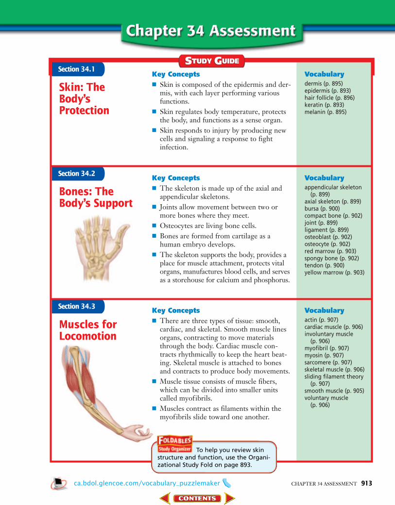
Section 34.1
Section 34.2
Vocabularydermis (p. 895)epidermis (p. 893)hair follicle (p. 896)keratin (p. 893)melanin (p. 895)
Vocabularyappendicular skeleton
(p. 899)axial skeleton (p. 899)bursa (p. 900)compact bone (p. 902)joint (p. 899)ligament (p. 899)osteoblast (p. 902)osteocyte (p. 902)red marrow (p. 903)spongy bone (p. 902)tendon (p. 900)yellow marrow (p. 903)
Bones: TheBody’s Support
STUDY GUIDESTUDY GUIDE
CHAPTER 34 ASSESSMENT 913
Skin: TheBody’sProtection
Section 34.3
Muscles forLocomotion
Vocabularyactin (p. 907)cardiac muscle (p. 906)involuntary muscle
(p. 906)myofibril (p. 907)myosin (p. 907)sarcomere (p. 907)skeletal muscle (p. 906)sliding filament theory
(p. 907)smooth muscle (p. 905)voluntary muscle
(p. 906)
To help you review skinstructure and function, use the Organi-zational Study Fold on page 893.
Key Concepts■ Skin is composed of the epidermis and der-
mis, with each layer performing variousfunctions.
■ Skin regulates body temperature, protectsthe body, and functions as a sense organ.
■ Skin responds to injury by producing newcells and signaling a response to fightinfection.
Key Concepts■ There are three types of tissue: smooth,
cardiac, and skeletal. Smooth muscle linesorgans, contracting to move materialsthrough the body. Cardiac muscle con-tracts rhythmically to keep the heart beat-ing. Skeletal muscle is attached to bonesand contracts to produce body movements.
■ Muscle tissue consists of muscle fibers,which can be divided into smaller unitscalled myofibrils.
■ Muscles contract as filaments within themyofibrils slide toward one another.
Key Concepts■ The skeleton is made up of the axial and
appendicular skeletons. ■ Joints allow movement between two or
more bones where they meet.■ Osteocytes are living bone cells.■ Bones are formed from cartilage as a
human embryo develops.■ The skeleton supports the body, provides a
place for muscle attachment, protects vitalorgans, manufactures blood cells, and servesas a storehouse for calcium and phosphorus.
ca.bdol.glencoe.com/vocabulary_puzzlemaker
0905-0915 C34 S3 BDOL-829900 8/5/04 2:06 AM Page 913
Review the Chapter 34 vocabulary words listed inthe Study Guide on page 913. Match the wordswith the definitions below.
1. the outermost layer of skin2. tough bands of connective tissue that attach
bone to bone3. living cells of compact and spongy bone4. type of muscle found in the walls of internal
organs5. protein that makes up the thick filaments of
myofibrils
6. Which of the following is a skin pigment thatprotects cells from solar radiation damage?A. keratin C. melaninB. epidermis D. dermis
7. Which of the following is nour-ished by blood vessels that runwithin this structure?A. dermisB. osteocyteC. epidermisD. sarcomere
8. All of the following are types of muscleexcept ________.A. epidermal C. smoothB. cardiac D. skeletal
9. Skin plays a role in ________.A. storing calciumB. regulating body temperatureC. manufacturing blood cellsD. supporting the body
10. The axial skeleton includes bones from________.A. the skull C. the sternumB. the ribs D. all of the above
11. Complete the concept map by using the fol-lowing vocabulary terms: actin, myofibrils,skeletal muscles, myosin.
12. Open Ended Compare the interrelations ofthe skeletal and muscular systems to eachother and to the body as a whole.
13. Open Ended How could an injury to a liga-ment affect the function of the joint withwhich it is associated?
14. Open Ended Analyze, critique, and reviewthe strengths and weaknesses of the slidingfilament theory of muscle contraction.
15. Infer You view three tissue slides under themicroscope. Slide A has an outer layer con-taining flat, dead cells. The cells of Slide Bhave nuclei and are striated. Slide C containsrepeating circular units with capillaries atthe center. Identify each slide as skin, bone,or muscle tissue, and explain the function of each.
16. Osteoporosis isa health threat for many Americans. Visit
to find out moreinformation about this disease. What are therisk factors? Why are the elderly at greaterrisk for this disease? Analyze the importanceof nutrition and exercise in the preventionof osteoporosis.
REAL WORLD BIOCHALLENGE
contain bundles of fibers made up of
1.
which are made of thin filaments
of the protein
2.
3. 4.
which are made of thick filaments
of the protein
914 CHAPTER 34 ASSESSMENT
Osteon system
ca.bdol.glencoe.com
ca.bdol.glencoe.com/chapter_test
0905-0915 C34 S3 BDOL-829900 8/5/04 2:07 AM Page 914

CHAPTER 34 ASSESSMENT 915
Constructed Response/Grid InRecord your answers on your answer document.
24. Open Ended What are the similarities and differences between first-degree burns, second-degree burns, and third-degree burns? Include in your response information about which layers of the skin are damaged, symptoms, and treatment.
25. Open Ended Describe how muscle cells are supplied with energy during exercise.
Multiple ChoiceUse the graph to answer questions 19–21.
19. The highest levels of calcium are found atapproximately what time?A. 10 milliseconds C. 30 millisecondsB. 50 milliseconds D. 70 milliseconds
20. Which conclusion could be reached aboutthe relationship between calcium and musclecontraction? A. Calcium is not involved in muscle
contraction. B. Calcium is released after the muscle has
finished contracting.C. Calcium is released before the muscle
reaches its greatest force of contraction.D. Calcium is released the entire time the
muscle contracts.
21. At what time is the force of the muscle contraction strongest?A. 10 millisecondsB. 50 millisecondsC. 30 millisecondsD. 70 milliseconds
Study the diagram and answer questions 22 and 23.
22. Which structure in the diagram is involvedin temperature regulation?A. 1 C. 3B. 2 D. 4
23. Which layer in the diagram above containsmelanin-producing cells?A. 1 C. 3B. 2 D. 4
1
4
3
2
Mu
scle
fo
rce
and
cal
ciu
m le
vels
Time (milliseconds)10 30 50 70 90 110 130 150 170 190
Calcium level
Muscle force
Calcium Levels in Contracting Muscle
17. Hypothesize How would the destruction ofred bone marrow affect other systems withinthe body?
18. Infer During summer months many peoplego barefoot and the skin on their feet thick-ens. Why does this thickening occur?
ca.bdol.glencoe.com/standardized_test
9h
9h
10a
9h
The assessed California standard appears next to the question.
0905-0915 C34 S3 BDOL-829900 8/5/04 2:08 AM Page 915