Embed Size (px)
Citation preview
Prac%cal Issues for Mul%modal Neuroimaging WhynHow 5/5/11
Ellen Lau Mar%nos Center
Mul%modal Imaging: Why and How
• Why do it? • How to design the study • How to approach data analysis • Why don’t the datasets match up??

Why do it?

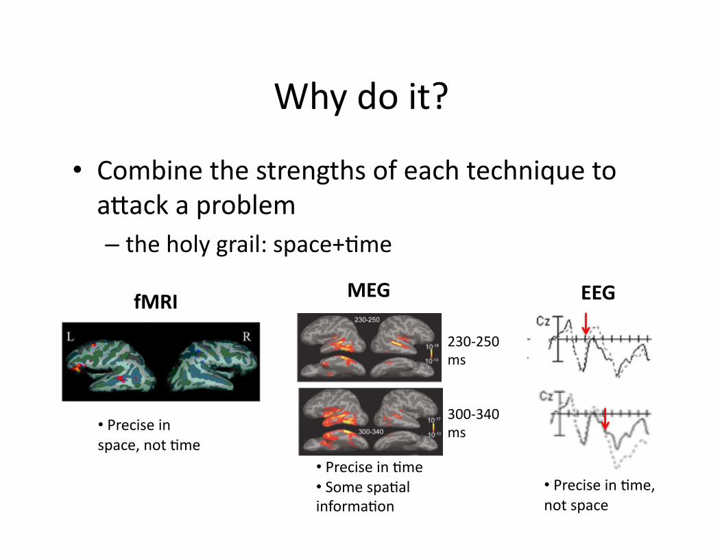
Why do it?
• Combine the strengths of each technique to aGack a problem • E.g. the holy grail: space+%me
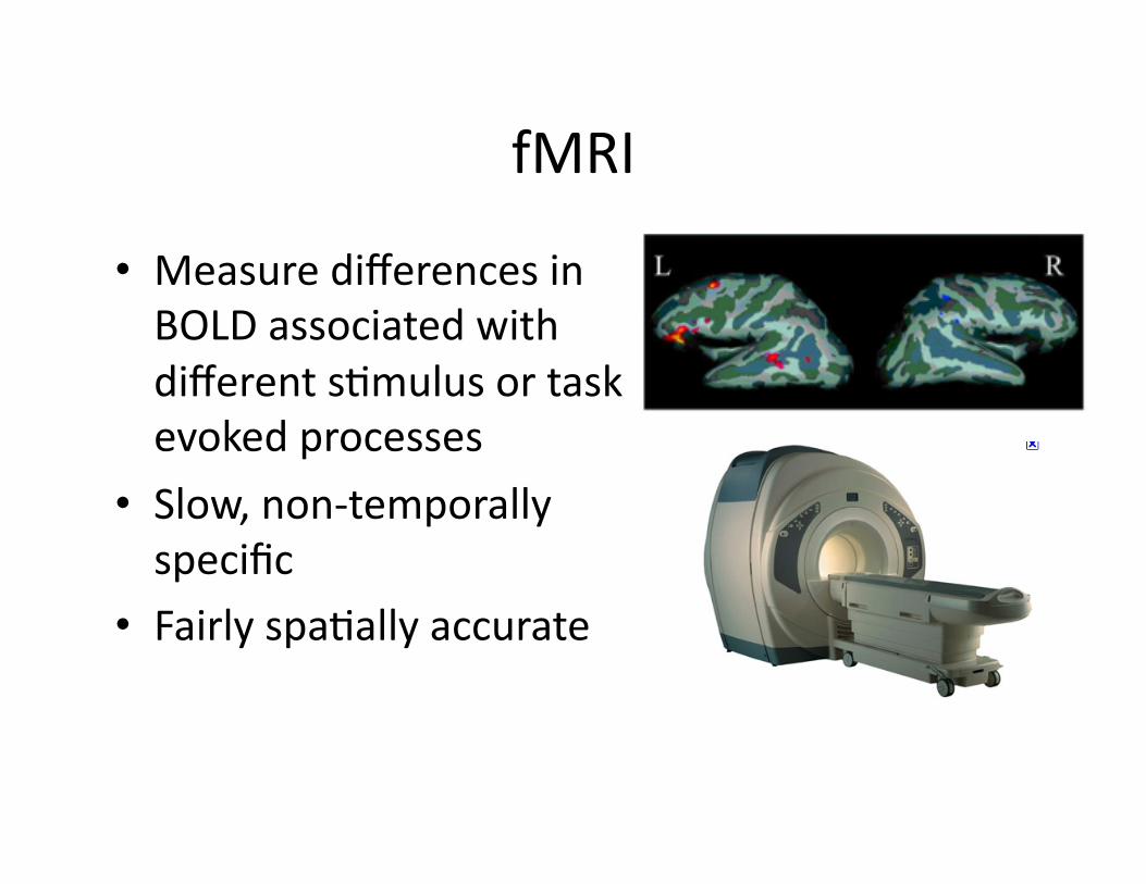
fMRI
• Measure differences in BOLD associated with different s%mulus or task evoked processes
• Slow, non-‐temporally specific
• Fairly spa%ally accurate
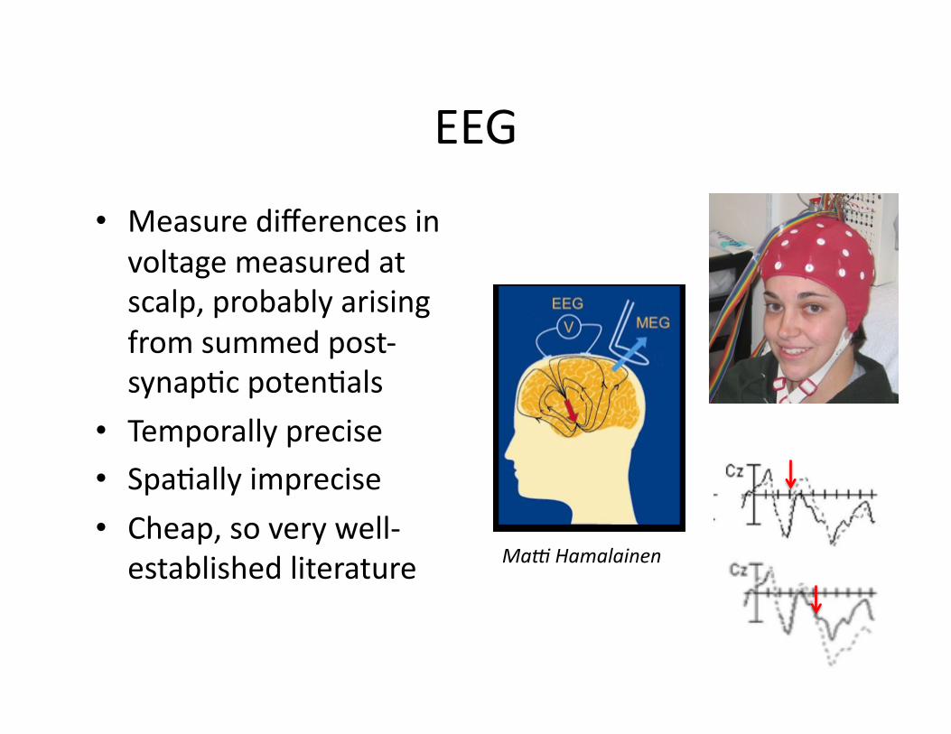
EEG
• Measure differences in voltage measured at scalp, probably arising from summed post-‐synap%c poten%als
• Temporally precise • Spa%ally imprecise
• Cheap, so very well-‐established literature Ma# Hamalainen
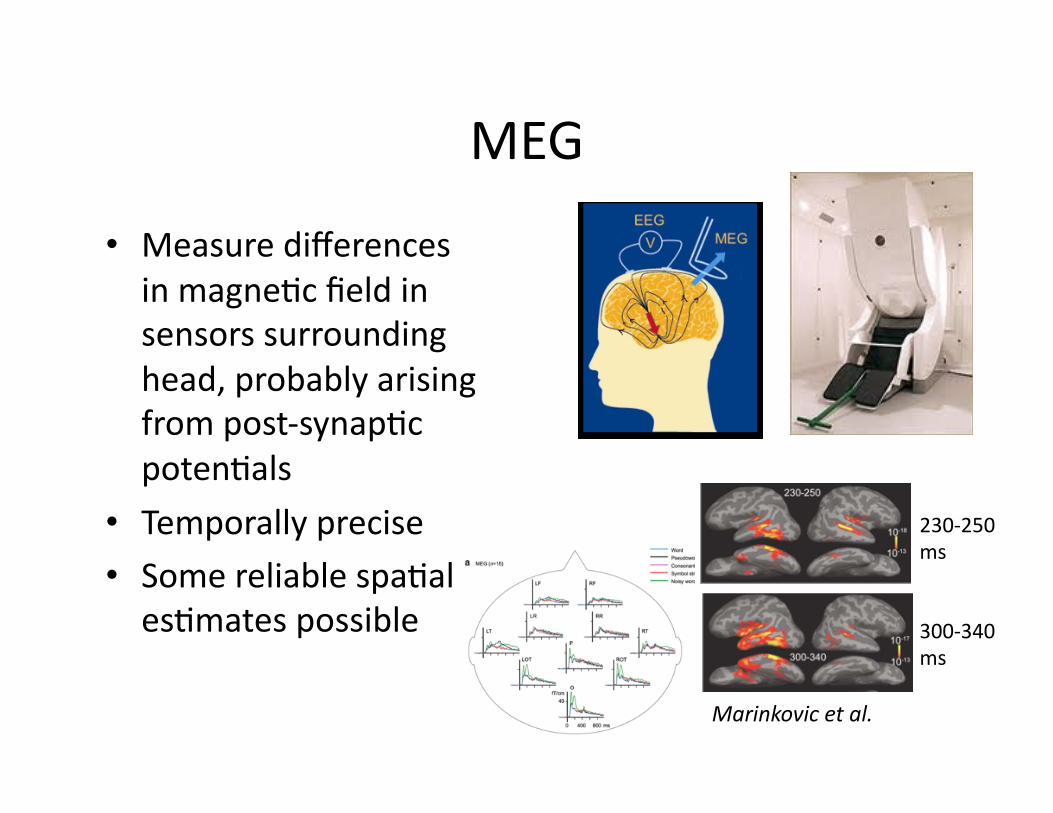
MEG
• Measure differences in magne%c field in sensors surrounding head, probably arising from post-‐synap%c poten%als
• Temporally precise • Some reliable spa%al es%mates possible
230-‐250 ms
300-‐340 ms
Marinkovic et al.
Why do it?
• Combine the strengths of each technique to aGack a problem – the holy grail: space+%me
fMRI
• Precise in space, not %me
EEG
• Precise in %me, not space
• Precise in %me • Some spa%al informa%on
MEG
230-‐250 ms
300-‐340 ms
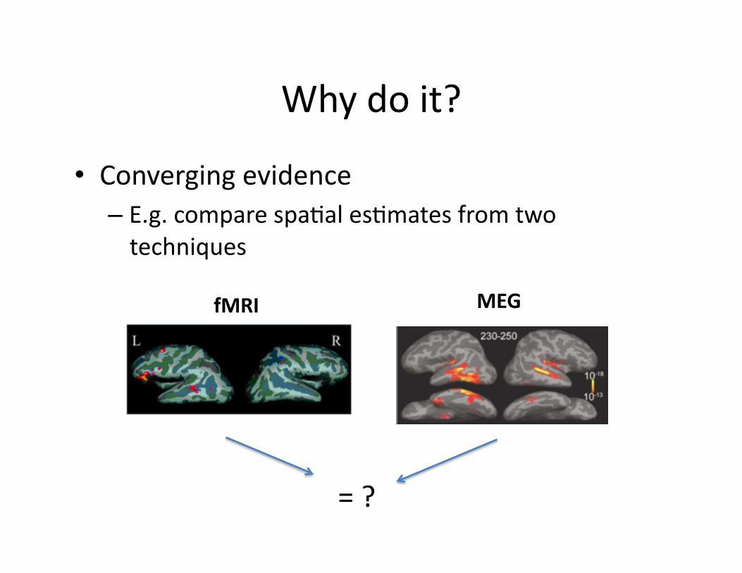
Why do it?
• Converging evidence – E.g. compare spa%al es%mates from two techniques
fMRI MEG
= ?
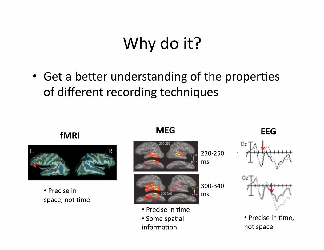
Why do it?
• Get a beGer understanding of the proper%es of different recording techniques
fMRI
• Precise in space, not %me
EEG
• Precise in %me, not space
• Precise in %me • Some spa%al informa%on
MEG
230-‐250 ms
300-‐340 ms

How to design the study
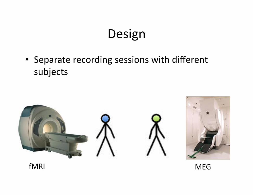
Design
• Separate recording sessions with different subjects
fMRI MEG
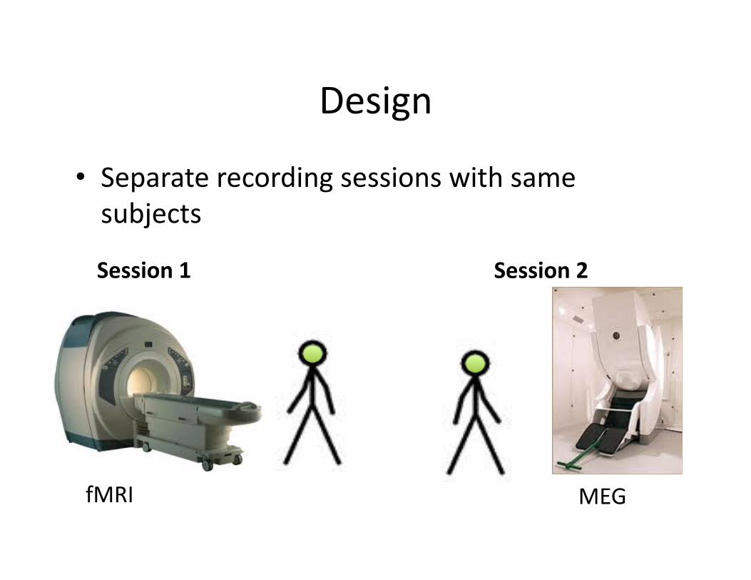
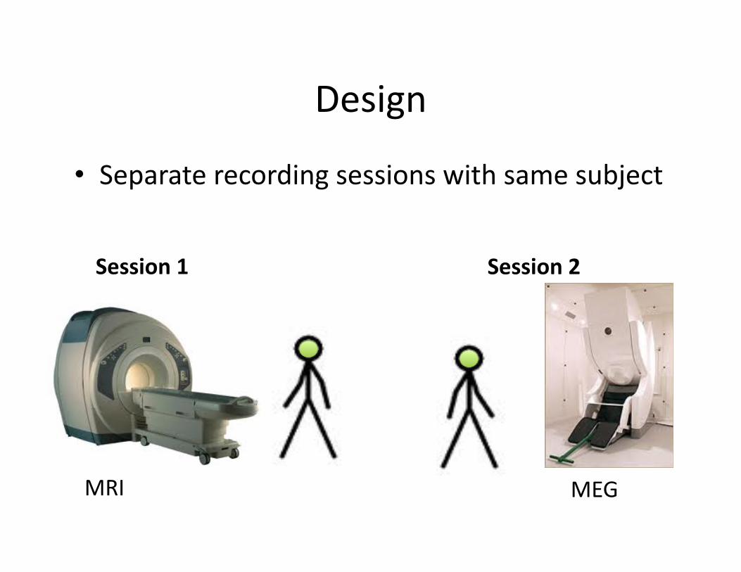
Design
• Separate recording sessions with same subjects
fMRI MEG
Session 1 Session 2

Design
• Simultaneous recording
MEG
EEG
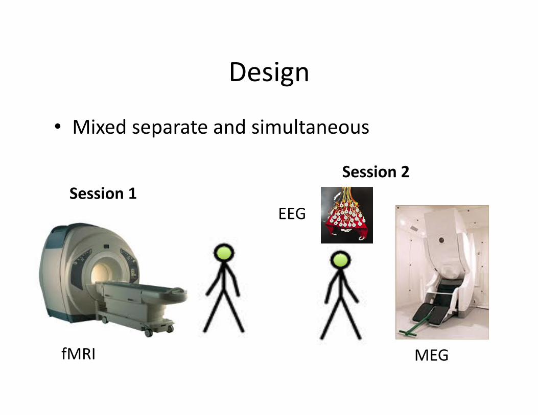
Design
• Mixed separate and simultaneous
fMRI
Session 1
MEG
Session 2
EEG
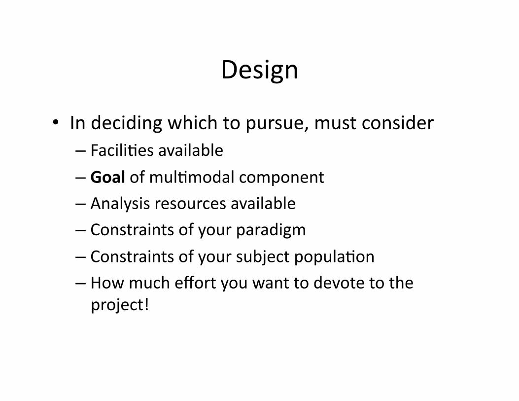
Design
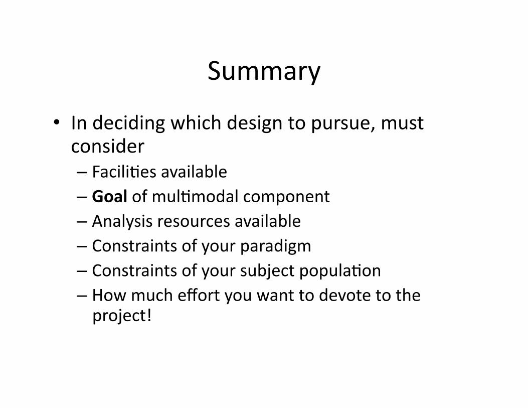
• In deciding which to pursue, must consider – Facili%es available – Goal of mul%modal component – Analysis resources available – Constraints of your paradigm
– Constraints of your subject popula%on – How much effort you want to devote to the project!
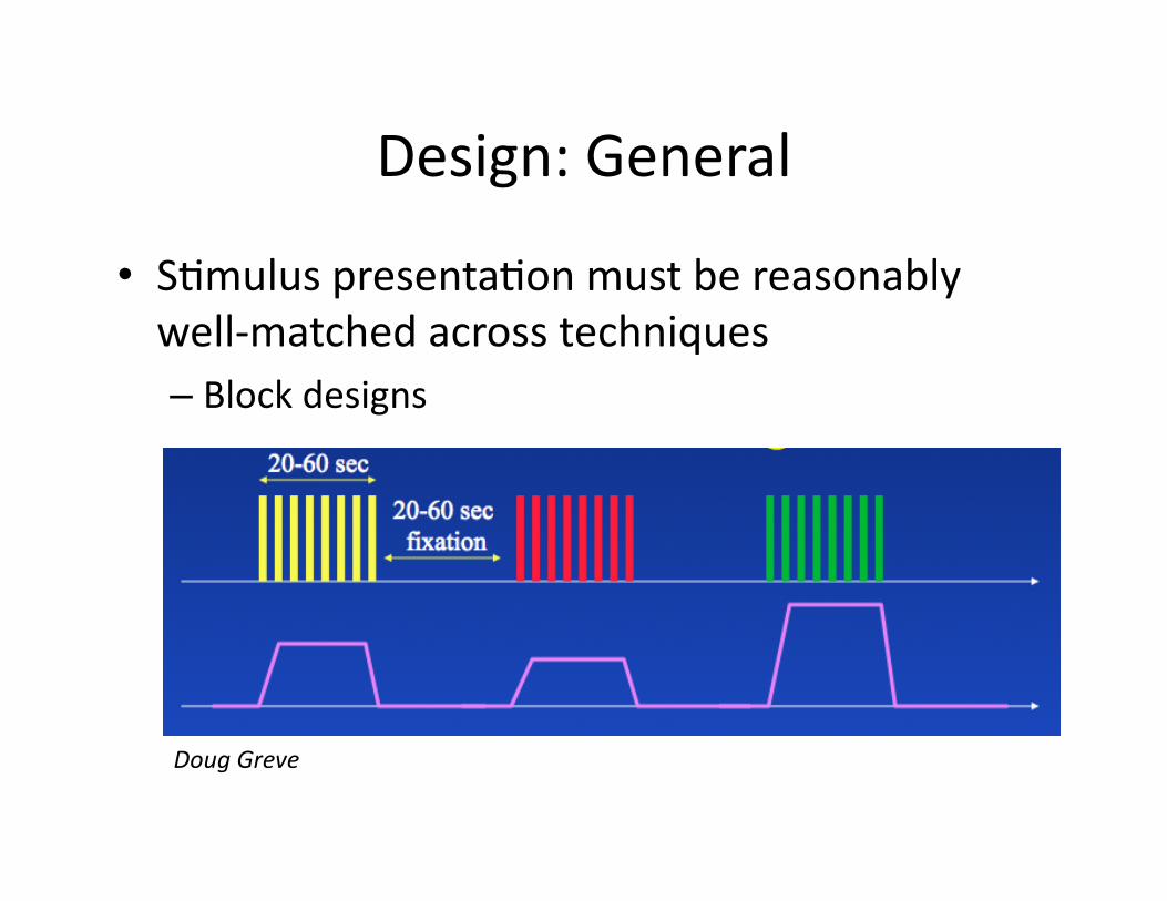
Design: General
• S%mulus presenta%on must be reasonably well-‐matched across techniques – Block designs
Doug Greve
Design: General
• S%mulus presenta%on must be reasonably well-‐matched across techniques – Block designs • Could work for EEG/MEG if you are looking at steady-‐state responses, i.e. whether power in a certain frequency band is different for A blocks and B blocks
• Won’t work if you are looking at s%mulus evoked responses in EEG/MEG
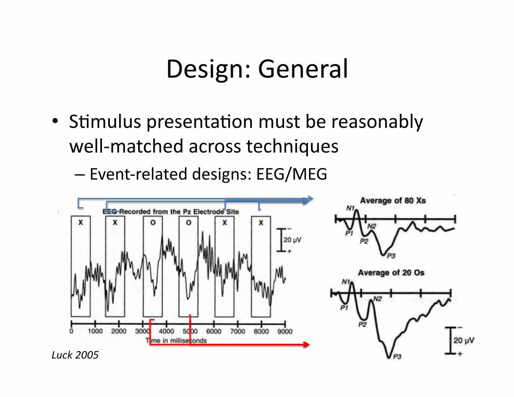
Design: General
• S%mulus presenta%on must be reasonably well-‐matched across techniques – Event-‐related designs: EEG/MEG
Luck 2005
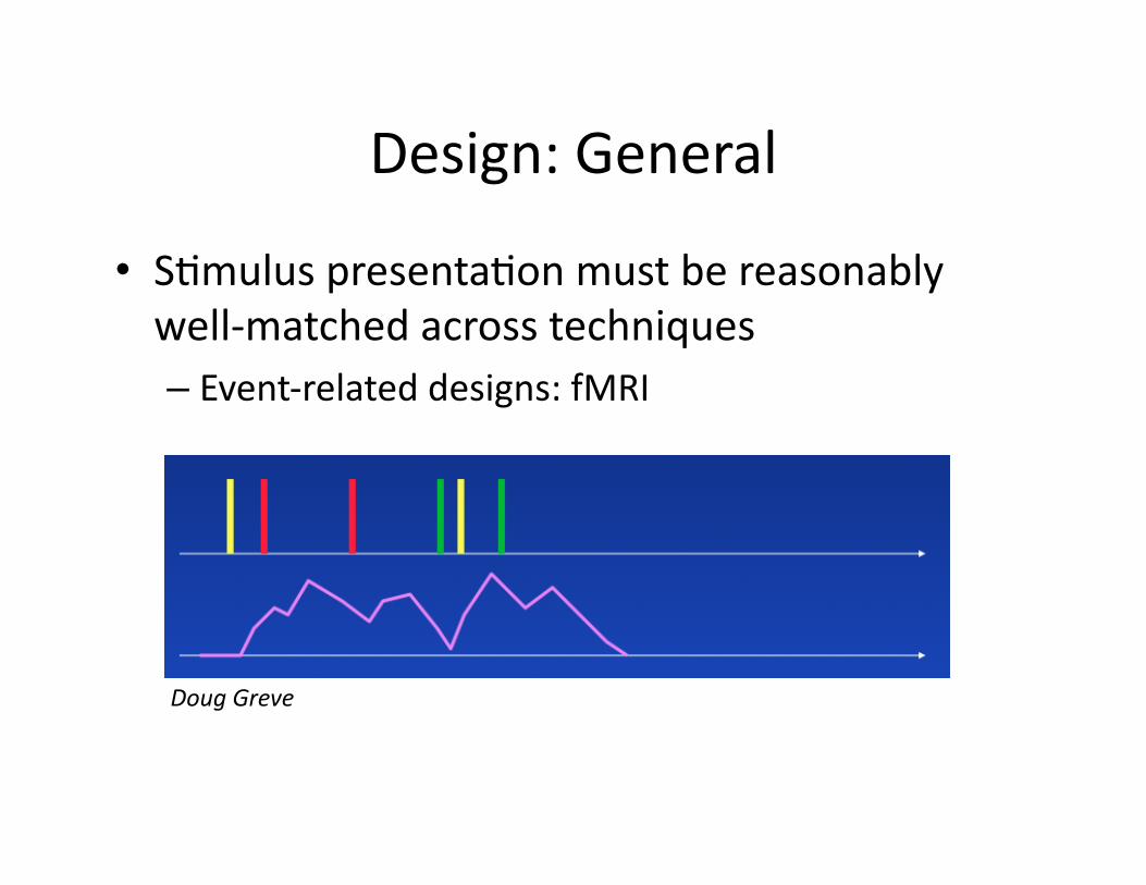
Design: General
• S%mulus presenta%on must be reasonably well-‐matched across techniques – Event-‐related designs: fMRI
Doug Greve
Design: General
• S%mulus presenta%on must be reasonably well-‐matched across techniques – Special requirements for s%muli in event-‐related fMRI • To get the most robust es%mates, need to op%mize order of s%mulus presenta%on such that sequence is not completely random (e.g. ‘optseq’ sodware) • Need to add ‘null’ periods of fixa%on for fMRI deconvolu%on
Design: General
• S%mulus presenta%on must be reasonably well-‐matched across techniques – Special requirements for event-‐related s%muli in MEG/EEG • Regular rest intervals for people to blink • May need a larger number of trials to es%mate source solu%ons
Design: General
• S%mulus presenta%on must be reasonably well-‐matched across techniques – Matching visual angle, resolu%on, and other low-‐level proper%es across very different projec%on setups
– And so on…
Design
MRI MEG or EEG
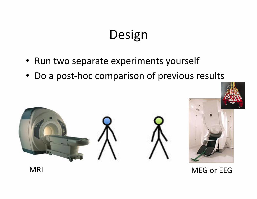
• Run two separate experiments yourself • Do a post-‐hoc comparison of previous results
Between-‐Subjects Design
• Need to have enough subjects for it to be reasonable to generalize to (sub)popula%on – For MEG, EEG, and fMRI, personal rule of thumb is about 16 good subjects drawn from approximately same age-‐range and educa%onal level
– Should be recruited in the same way
Design
• Separate recording sessions with same subject
MRI MEG
Session 1 Session 2
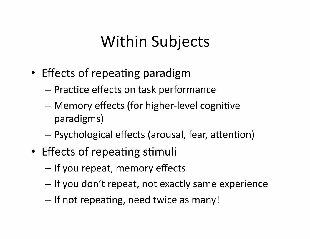
Within Subjects
• Effects of repea%ng paradigm – Prac%ce effects on task performance
– Memory effects (for higher-‐level cogni%ve paradigms)
– Psychological effects (arousal, fear, aGen%on) • Effects of repea%ng s%muli – If you repeat, memory effects – If you don’t repeat, not exactly same experience
– If not repea%ng, need twice as many!
Within Subjects
• Counterbalancing, counterbalancing… – Order of measures (fMRI-‐MEG, MEG-‐fMRI) most important
– Order of s%muli lists (if different)
– List x measure interac%on? – Can be difficult because of limited sample size
• Controlling delay between Session 1 and Session 2? – Can be difficult because of logis%cs!
Within Subjects
• Par%cipants – Double the possible exclusion factors
• E.g. metal fillings for MEG, ver%go/claustrophobia/body size for fMRI
– Par%cipant scheduling • ‘Need someone who can make it to our lab’s Thursday 2-‐4 MRI slot and our Monday 10-‐1 MEG slot’
– Par%cipant drop-‐out • Spend 1k on the fMRI and they don’t return for MEG!
– Uneven data quality • Subject keeps s%ll in the MRI but moves a lot in MEG
Within Subjects
• Par%cipants – Inevitably, you will end up with par%cipants that have one good dataset but not the other, so try to design experiment in such a way that data from each technique by itself is s%ll valuable
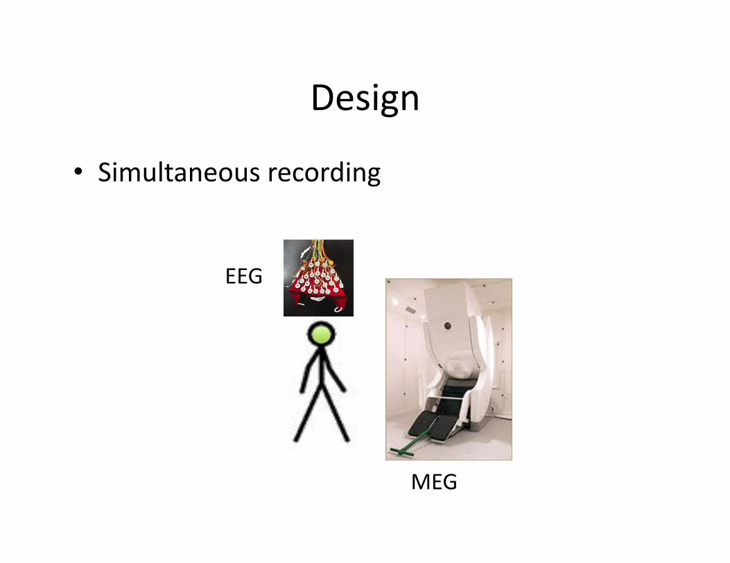
Design
• Simultaneous recording
MEG
EEG
Simultaneous Recording
• Recording is more difficult and more prone to error – More steps to remember, more pieces of equipment, more chances something will go wrong on a given day • Extreme organiza%on is absolutely necessary!
– Par%cipants may become more anxious or bored during a long setup
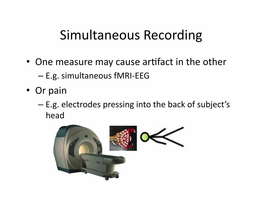
Simultaneous Recording
• One measure may cause ar%fact in the other – E.g. simultaneous fMRI-‐EEG
• Or pain – E.g. electrodes pressing into the back of subject’s head
How to approach the analysis
Summary
• In deciding which design to pursue, must consider – Facili%es available – Goal of mul%modal component – Analysis resources available – Constraints of your paradigm – Constraints of your subject popula%on – How much effort you want to devote to the project!
Analysis
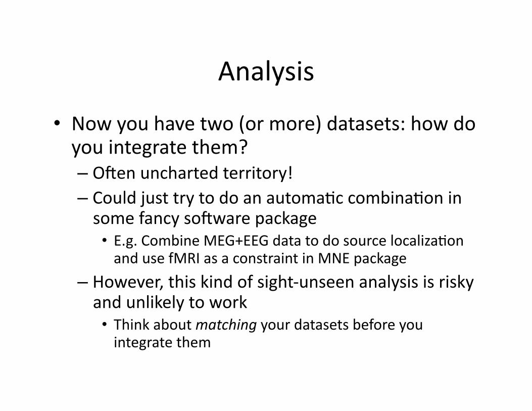
• Now you have two (or more) datasets: how do you integrate them? – Oden uncharted territory! – Could just try to do an automa%c combina%on in some fancy sodware package • E.g. Combine MEG+EEG data to do source localiza%on and use fMRI as a constraint in MNE package
– However, this kind of sight-‐unseen analysis is risky and unlikely to work • Think about matching your datasets before you integrate them
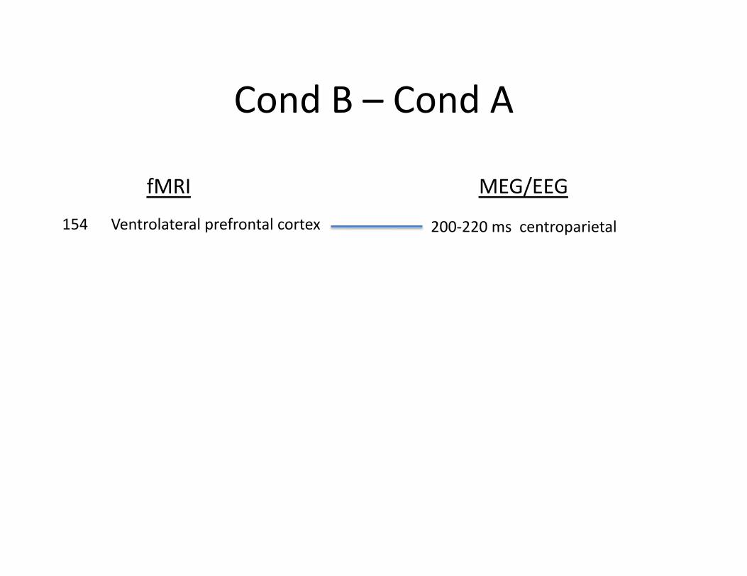
Cond B – Cond A
fMRI MEG/EEG
154 Ventrolateral prefrontal cortex 200-‐220 ms centroparietal
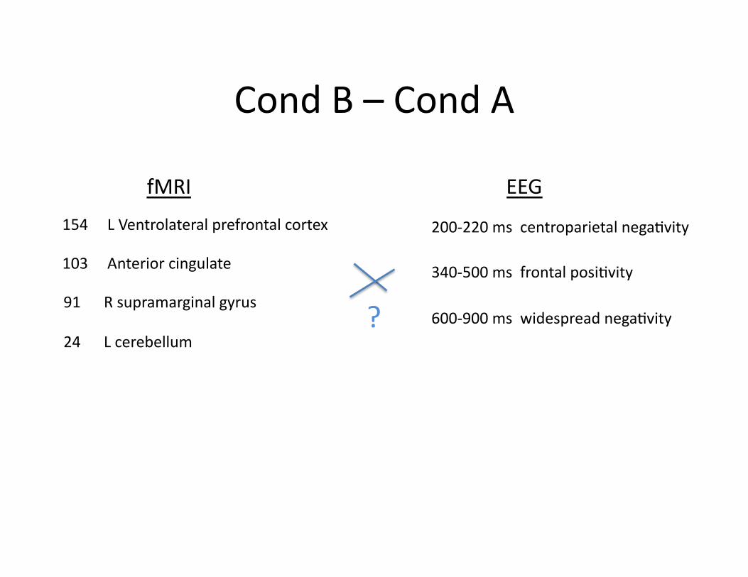
Cond B – Cond A
fMRI EEG
154 L Ventrolateral prefrontal cortex 200-‐220 ms centroparietal nega%vity
103 Anterior cingulate
91 R supramarginal gyrus
24 L cerebellum
340-‐500 ms frontal posi%vity
600-‐900 ms widespread nega%vity ?
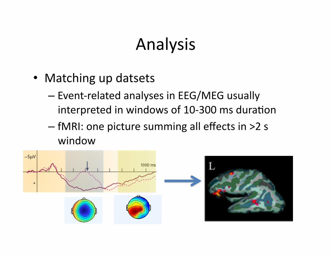
Analysis
• Matching up datsets – Event-‐related analyses in EEG/MEG usually interpreted in windows of 10-‐300 ms dura%on
– fMRI: one picture summing all effects in >2 s window
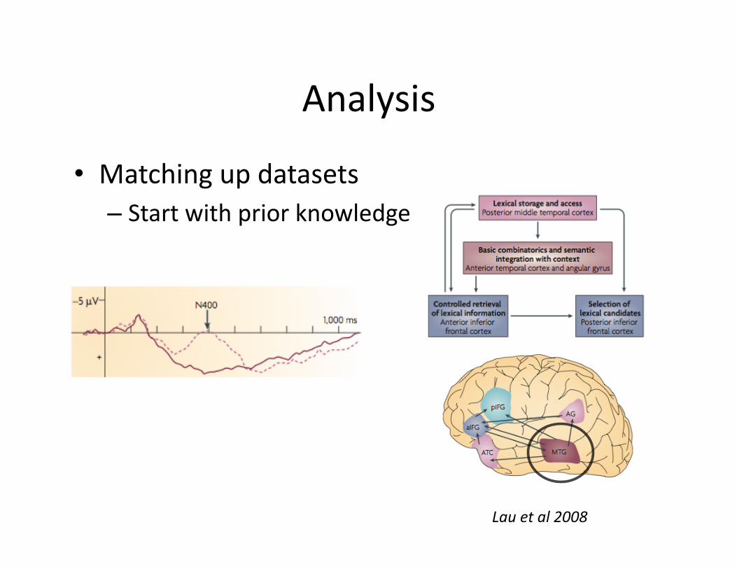
Analysis
• Matching up datasets – Start with prior knowledge
Lau et al 2008
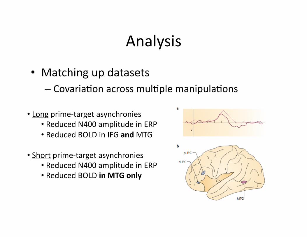
Analysis
• Matching up datasets – Covaria%on across mul%ple manipula%ons
• Long prime-‐target asynchronies • Reduced N400 amplitude in ERP • Reduced BOLD in IFG and MTG
• Short prime-‐target asynchronies • Reduced N400 amplitude in ERP • Reduced BOLD in MTG only

Analysis
• Matching up datasets: within-‐subjects designs – Can take advantage of within-‐subjects nature of the design with individual subject analyses, e.g. • Lateraliza%on index in each measure
• Loca%on of ac%vity in the individual rela%ve to the group average, in each measure
• Correla%ons in effect size in each measure across individuals
Analysis
• Matching up datasets: within-‐subjects designs – If you can figure out a reliable means of matching up the two datasets, you can now use one measure to aid in source localiza%on for the other measure on an individual basis (i.e., fMRI as a constraint on MEG/EEG source localiza%on)
Analysis
• Matching up datasets: simultaneous recording – Can take advantage of the fact that you have simultaneous data from individual trials—but how?

Analysis
• Matching up datasets: simultaneous recording – Correla%on analyses over individual trials • Note that many sodware programs may not be specialized to access individual trial informa%on
• Also have to decide what correla%ons to look for, and what will count as a meaningful correla%on

Summary
• Plan analysis ahead!
A few references to get started…
• Sharon et al. (2007) Neuroimage – fMRI + MEG/EEG within subjects
• Ahveninen et al. (2006) PNAS – fMRI + MEG within subjects
• Var%ainen et al. (2011) J. Neurosci – fMRI + MEG/EEG within subjects
• Kveraga et al. (2011) PNAS – fMRI + MEG in frequency domain, partly within subjects
• Ahveninen et al. (2011) PNAS – fMRI + MEG/EEG
Why might measures differ?
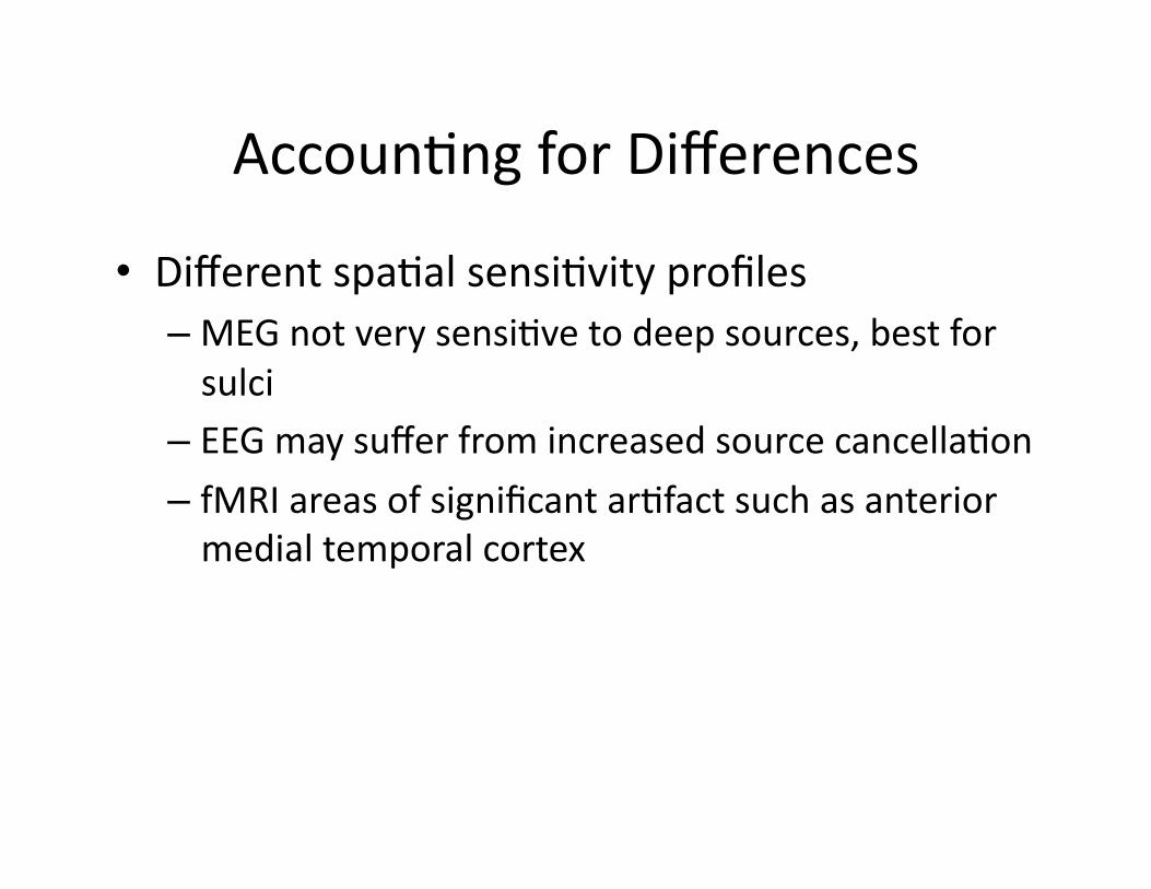
Accoun%ng for Differences
• Different spa%al sensi%vity profiles – MEG not very sensi%ve to deep sources, best for sulci
– EEG may suffer from increased source cancella%on
– fMRI areas of significant ar%fact such as anterior medial temporal cortex
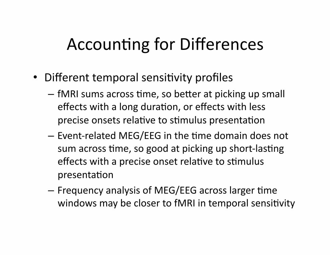
Accoun%ng for Differences
• Different temporal sensi%vity profiles – fMRI sums across %me, so beGer at picking up small effects with a long dura%on, or effects with less precise onsets rela%ve to s%mulus presenta%on
– Event-‐related MEG/EEG in the %me domain does not sum across %me, so good at picking up short-‐las%ng effects with a precise onset rela%ve to s%mulus presenta%on
– Frequency analysis of MEG/EEG across larger %me windows may be closer to fMRI in temporal sensi%vity
Accoun%ng for Differences
• Matching up datasets – Different levels of background variability • E.g., in sentence comprehension in fMRI, pulling out brain area involved in fixing viola%ons versus area involved in lexical access
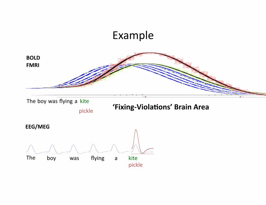
Example
The boy was flying a kite
pickle
The boy was flying a kite pickle
BOLD FMRI
EEG/MEG
‘Fixing-‐Viola@ons’ Brain Area
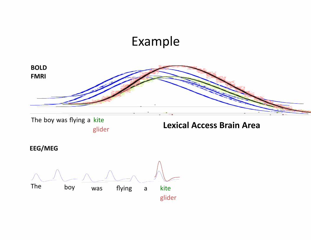
Example
The boy was flying a kite glider
The boy was flying a kite glider
BOLD FMRI
EEG/MEG
Lexical Access Brain Area
A few ‘mismatch’ references
• Var%ainen et al., 2011, J. Neurosci. • Yigal Agam (submiGed)
• Brem et al., 2009, Hum. Brain Mapp.
• Lau et al., 2008, Nat. Rev. Neurosci.
Mul%modal Neuroimaging is difficult…
• Don’t need to be an expert in each modality – But good to have an expert consultant for each
• Do need a lot of foresight, organiza%on, and planning – Not a ‘tack-‐on’
• Few cookie-‐cuGer analysis rou%nes available, so analysis needs to be planned ahead
…but rewarding!
• Can have great payoffs in beGer understanding both system of interest and the measures themselves