Embed Size (px)
Citation preview
Why study energy in plants?
Plants are the lungs and bread-bowl of the Earth. They make the food we eat, create the oxygen we breathe and remove our waste carbon dioxide from the air. The world’s plants actually produce six times more energy than humans use each year.
Understanding and getting the best out of plants is ever more important in a world faced with climate change and dwindling resources.
Plant energy biologists delve deep inside plants to study the tiny cells from which they are built. With the help of modern technology, we can actually watch what happens inside individual plant cells. Chloroplasts, mitochondria and peroxisomes are particularly interesting
to watch, as these compartments make energy in the cell. They “talk”
to each other to coordinate processes that keep the
whole plant running.
Plant energy biology in Australia
Researchers in Australia are paving the way towards harnessing the massive potential of plant energy systems.
The ARC Centre of Excellence in Plant Energy Biology (PEB) brings together over 70 world-class researchers and cutting edge technology. Its headquarters are located at The University of Western Australia (UWA) in Perth. PEB also has nodes at Australian National University (ANU) in Canberra and Flinders University in Adelaide.
At PEB, we think that plants are amazing. We want to share with you some of their incredible survival tactics and highlight some of the discoveries we have made. This photo display will take you on a journey through a plant’s world - from bushfires spanning hundreds of metres across, right down to cells and molecules spanning only micrometres. There are one million micrometres (µm) in every metre.

Surviving the drought
Plants can’t just pick up and move to find water during a drought. They must adapt or die. Tiny molecules inside plant cells can make a big difference in survival situations. PEB scientists have discovered that drought-stressed plants become more sensitive to a steroid hormone called brassinosteroid. The hormone helps them put energy into continued growth and survive in challenging circumstances. This helps plants complete their life cycle and pass on their genes to the next generation.
Image: Plants on steroids Image: Chris Brown

Smoke signals for plants
Australian plants have to be pretty tough when it comes to surviving bushfires. PEB and Kings Park scientists, along with UWA chemists, have discovered a surprising mechanism which allows plants to cope. They have discovered that plants respond to a chemical in smoke called karrikin. This “smoke signal” helps stimulate seed germination in dormant seeds after a fire. It also helps create sturdier plants that can grow in blackened conditions. These scientists are now investigating the use of karrikins to help germinate valuable seeds and restore mine sites.
Image: Bushfire aftermath, Eneabba Photography: Dr Ben Miller, Botanic Gardens and Parks Authority, Kings Park
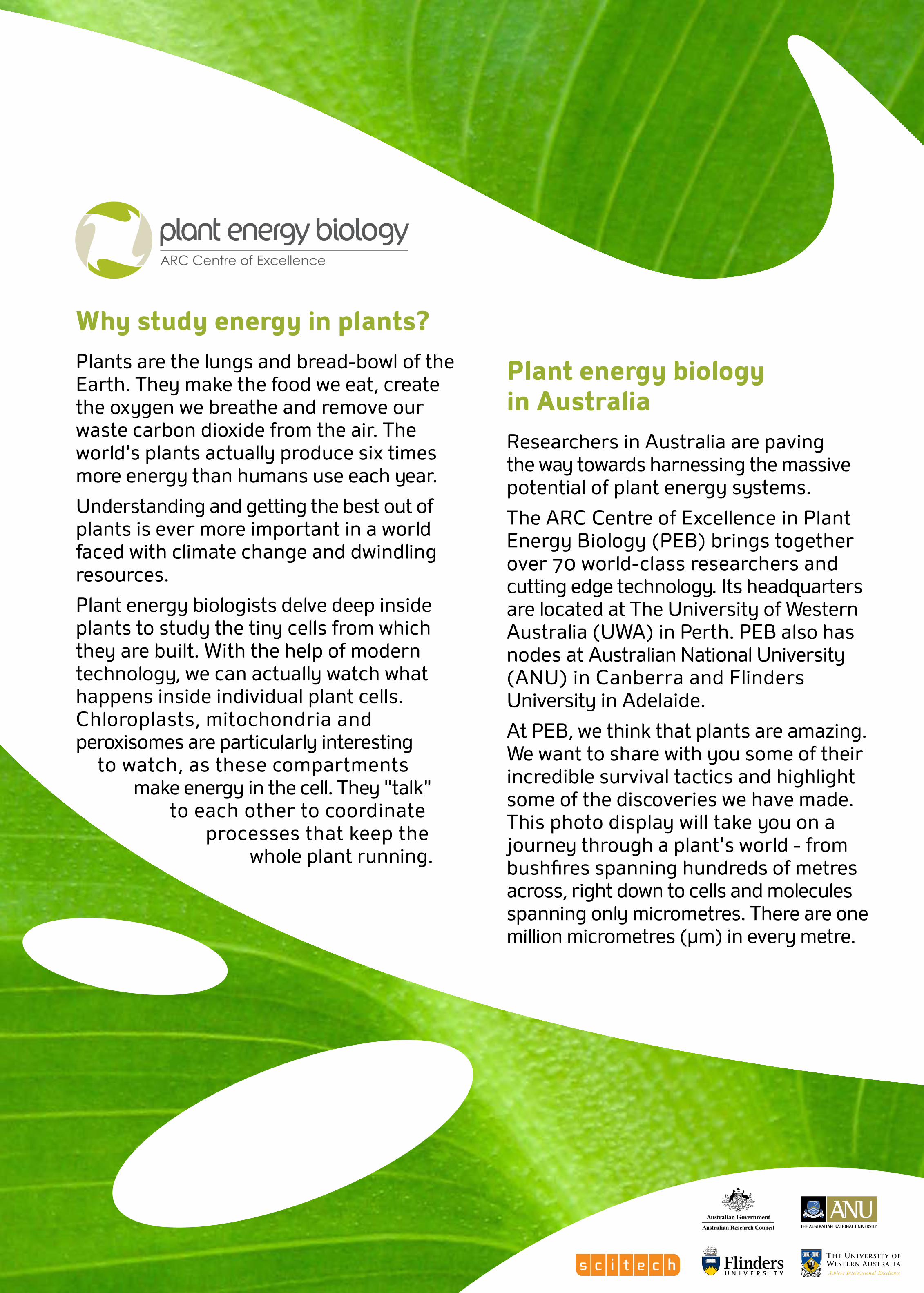
Energy from sunlight
Without the sun and without plants, we simply wouldn’t be here. Plants make oxygen for us to breathe and food for us to eat. They do this by using energy from the sun to turn water and carbon dioxide into plant sugars and oxygen. This process, called photosynthesis, is the single most important reaction for life on Earth.
Overall we know very little of the complex world inside a plant. Understanding how plants work helps us get the best out of them.
Image: Dew droplet on Coprosma repens (Mirror Bush) leaf Photography: Dr Sarah Rich, Plant Biology, UWA
Scale: 40mm across
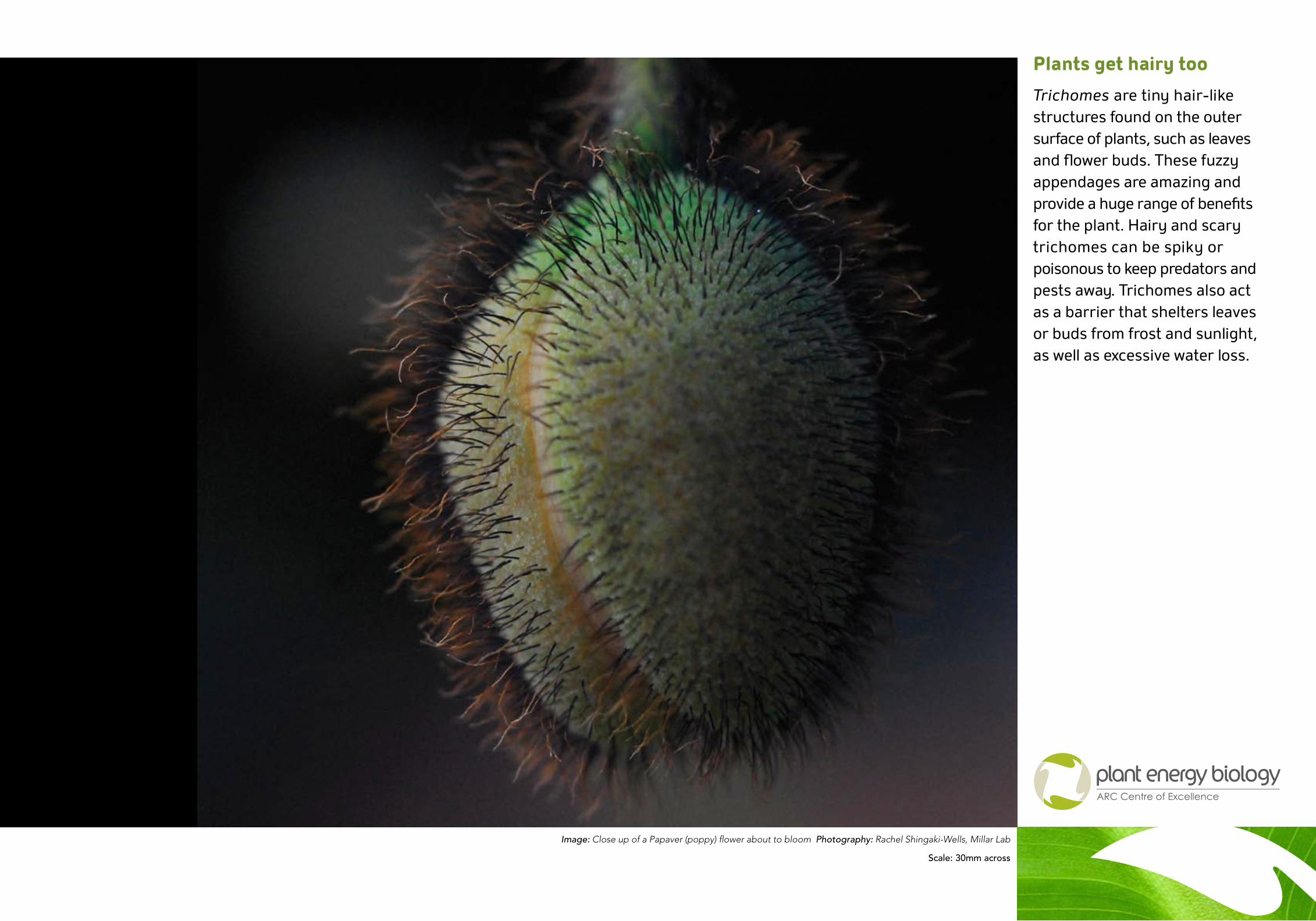
Plants get hairy too
Trichomes are tiny hair-like structures found on the outer surface of plants, such as leaves and flower buds. These fuzzy appendages are amazing and provide a huge range of benefits for the plant. Hairy and scary trichomes can be spiky or poisonous to keep predators and pests away. Trichomes also act as a barrier that shelters leaves or buds from frost and sunlight, as well as excessive water loss.
Image: Close up of a Papaver (poppy) flower about to bloom Photography: Rachel Shingaki-Wells, Millar Lab
Scale: 30mm across
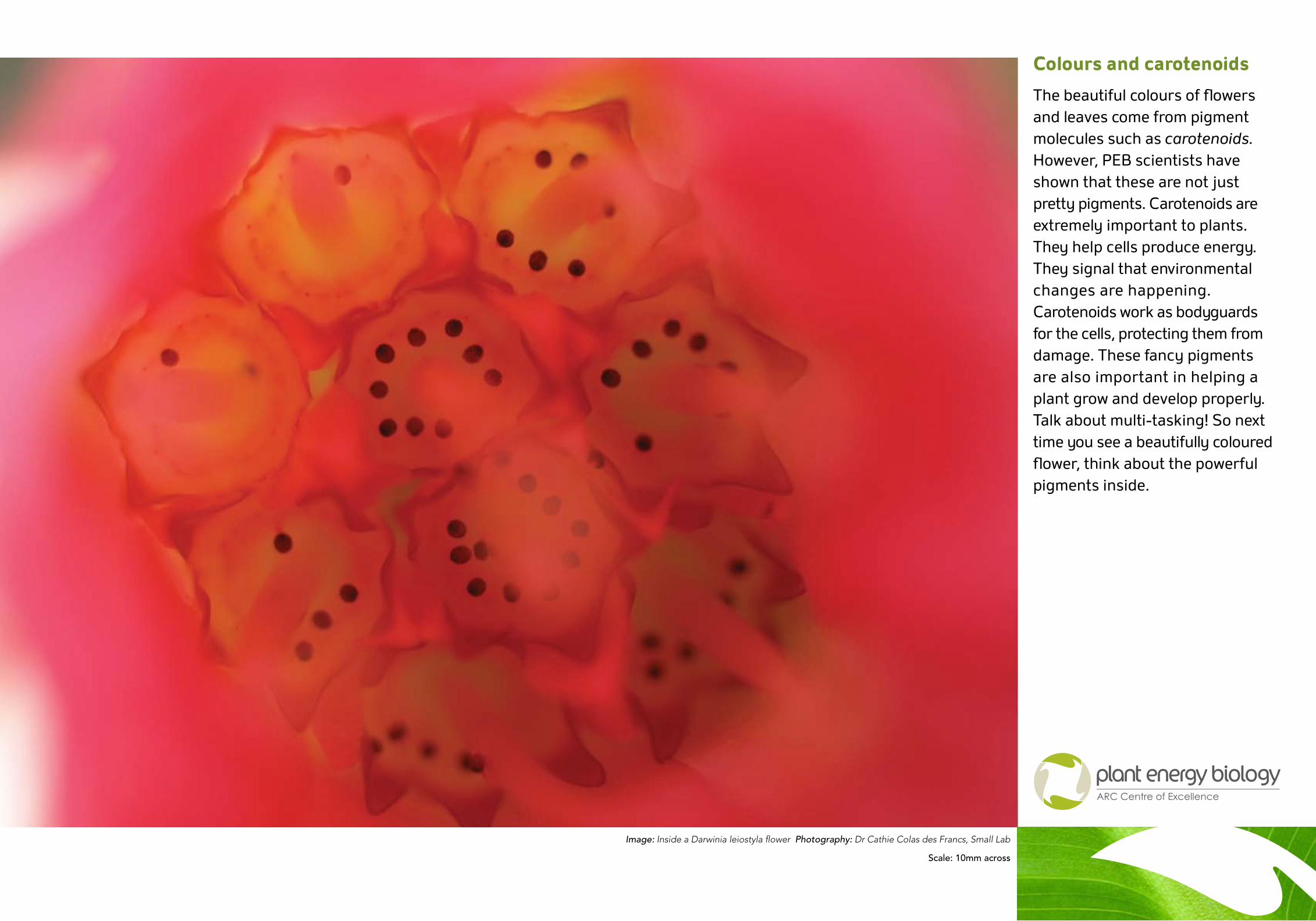
Colours and carotenoids
The beautiful colours of flowers and leaves come from pigment molecules such as carotenoids. However, PEB scientists have shown that these are not just pretty pigments. Carotenoids are extremely important to plants. They help cells produce energy. They signal that environmental changes are happening. Carotenoids work as bodyguards for the cells, protecting them from damage. These fancy pigments are also important in helping a plant grow and develop properly. Talk about multi-tasking! So next time you see a beautifully coloured flower, think about the powerful pigments inside.
Image: Inside a Darwinia leiostyla flower Photography: Dr Cathie Colas des Francs, Small Lab
Scale: 10mm across
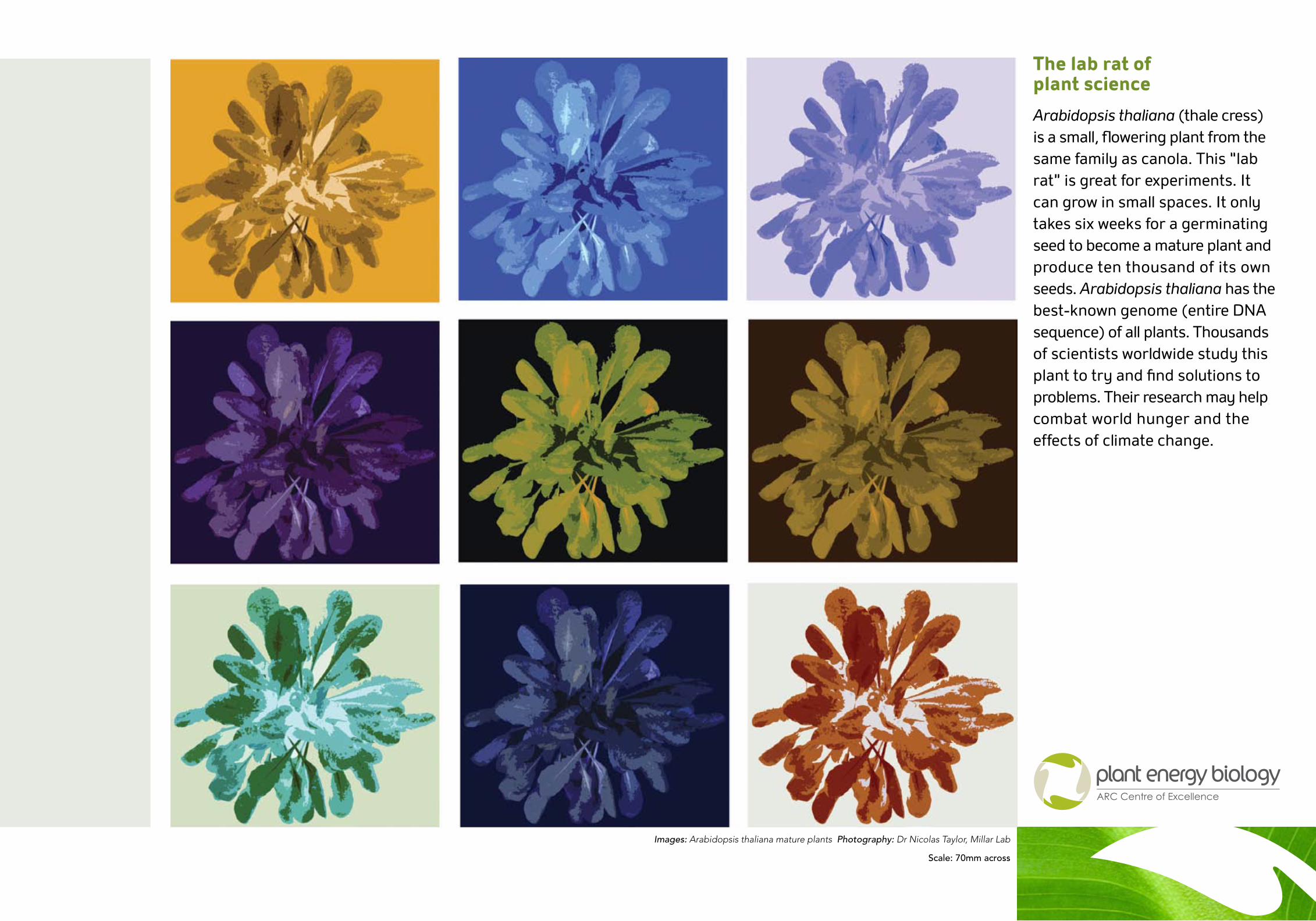
The lab rat of plant science
Arabidopsis thaliana (thale cress) is a small, flowering plant from the same family as canola. This “lab rat” is great for experiments. It can grow in small spaces. It only takes six weeks for a germinating seed to become a mature plant and produce ten thousand of its own seeds. Arabidopsis thaliana has the best-known genome (entire DNA sequence) of all plants. Thousands of scientists worldwide study this plant to try and find solutions to problems. Their research may help combat world hunger and the effects of climate change.
Images: Arabidopsis thaliana mature plants Photography: Dr Nicolas Taylor, Millar Lab
Scale: 70mm across
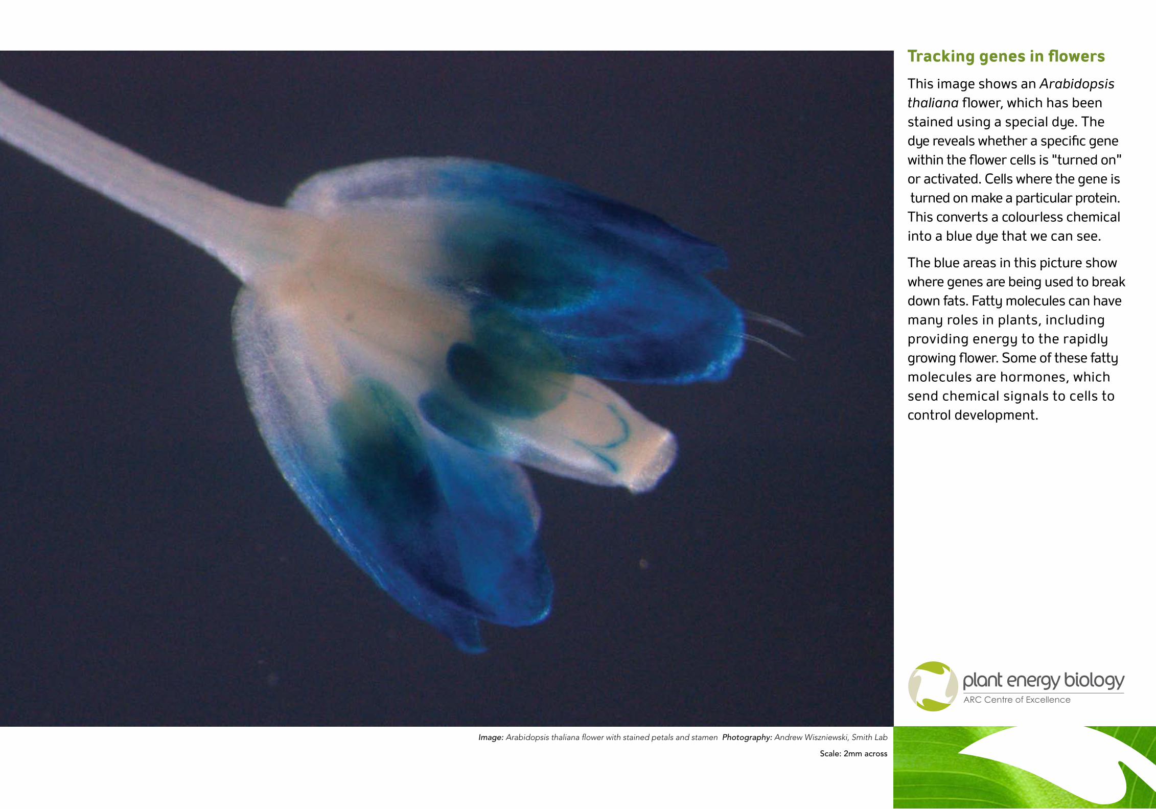
Tracking genes in flowers
This image shows an Arabidopsis thaliana flower, which has been stained using a special dye. The dye reveals whether a specific gene within the flower cells is “turned on” or activated. Cells where the gene is turned on make a particular protein. This converts a colourless chemical into a blue dye that we can see.
The blue areas in this picture show where genes are being used to break down fats. Fatty molecules can have many roles in plants, including providing energy to the rapidly growing flower. Some of these fatty molecules are hormones, which send chemical signals to cells to control development.
Image: Arabidopsis thaliana flower with stained petals and stamen Photography: Andrew Wiszniewski, Smith Lab
Scale: 2mm across
Learning more about genes
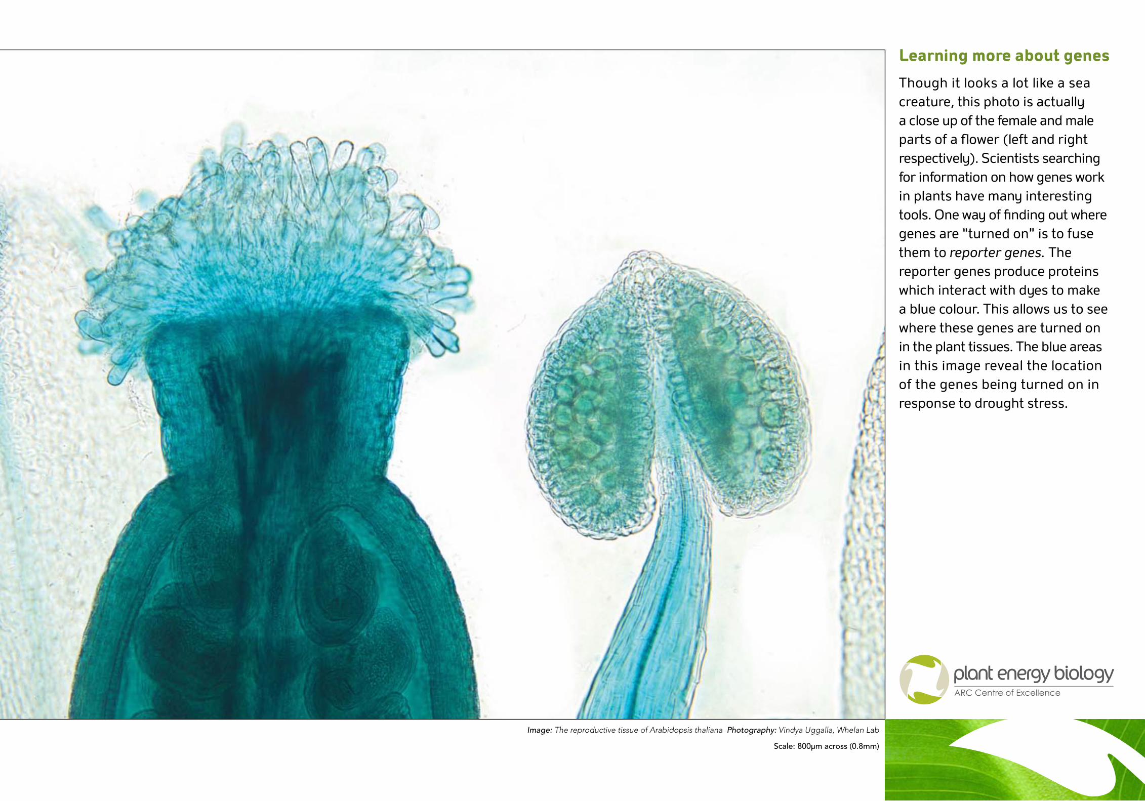
Though it looks a lot like a sea creature, this photo is actually a close up of the female and male parts of a flower (left and right respectively). Scientists searching for information on how genes work in plants have many interesting tools. One way of finding out where genes are “turned on” is to fuse them to reporter genes. The reporter genes produce proteins which interact with dyes to make a blue colour. This allows us to see where these genes are turned on in the plant tissues. The blue areas in this image reveal the location of the genes being turned on in response to drought stress.
Image: The reproductive tissue of Arabidopsis thaliana Photography: Vindya Uggalla, Whelan Lab
Scale: 800µm across (0.8mm)
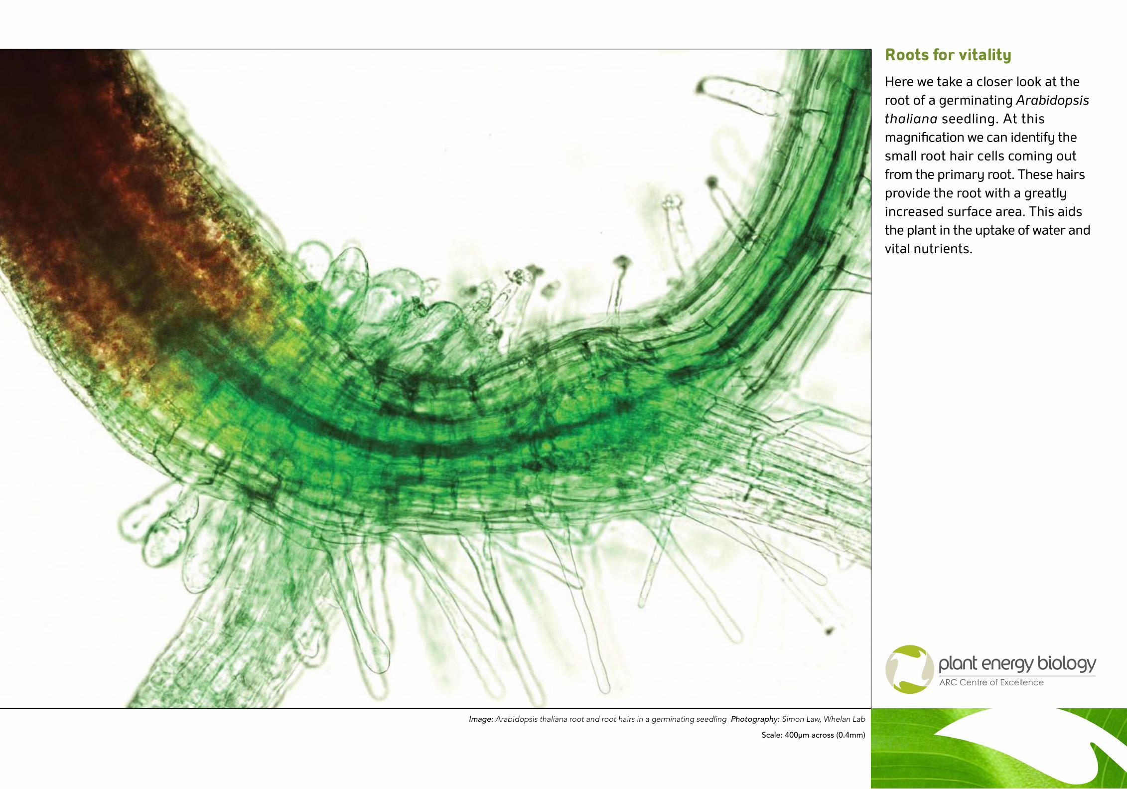
Roots for vitality
Here we take a closer look at the root of a germinating Arabidopsis thaliana seedling. At this magnification we can identify the small root hair cells coming out from the primary root. These hairs provide the root with a greatly increased surface area. This aids the plant in the uptake of water and vital nutrients.
Image: Arabidopsis thaliana root and root hairs in a germinating seedling Photography: Simon Law, Whelan Lab
Scale: 400µm across (0.4mm)
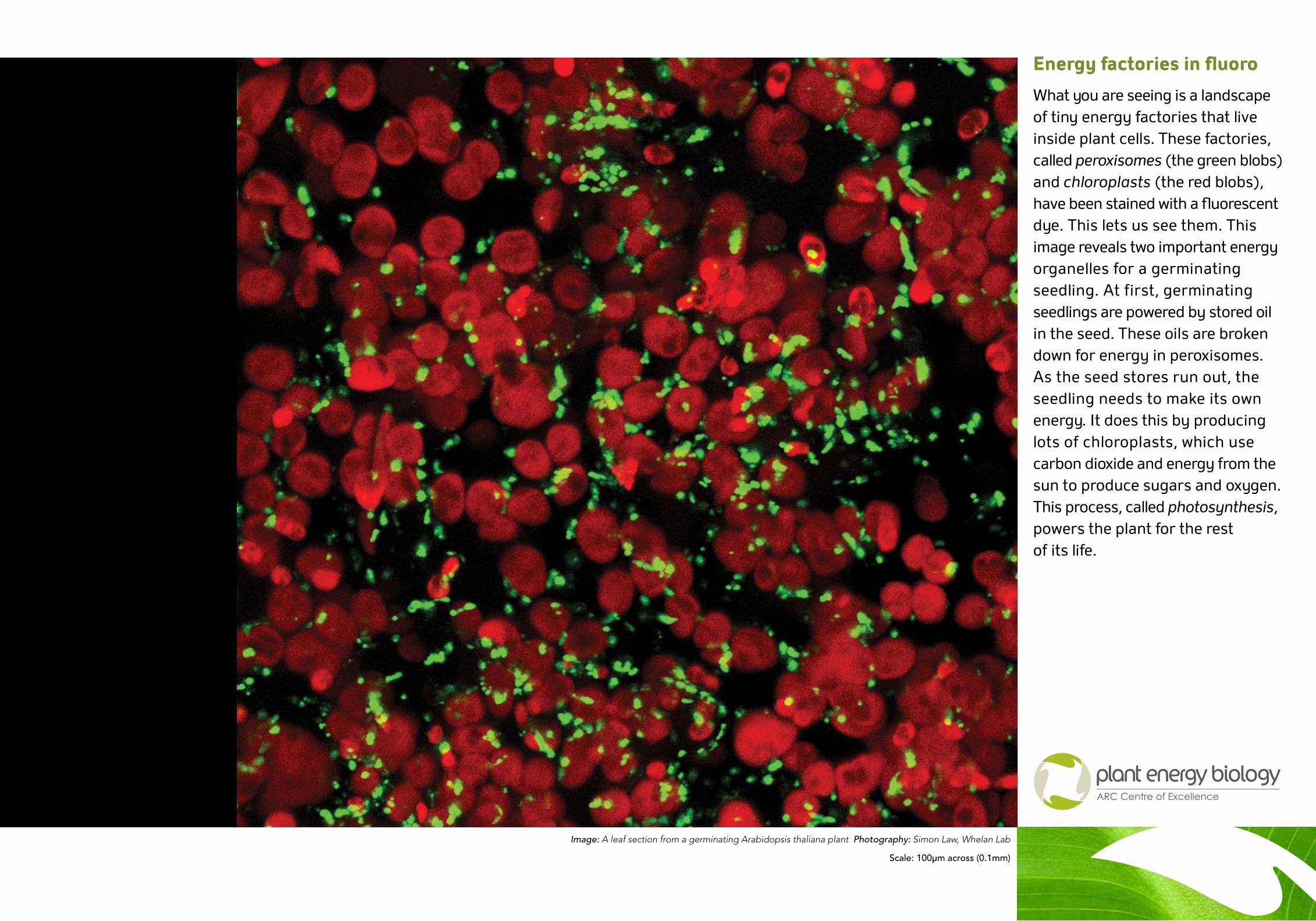
Energy factories in fluoro
What you are seeing is a landscape of tiny energy factories that live inside plant cells. These factories, called peroxisomes (the green blobs) and chloroplasts (the red blobs), have been stained with a fluorescent dye. This lets us see them. This image reveals two important energy organelles for a germinating seedling. At first, germinating seedlings are powered by stored oil in the seed. These oils are broken down for energy in peroxisomes. As the seed stores run out, the seedling needs to make its own energy. It does this by producing lots of chloroplasts, which use carbon dioxide and energy from the sun to produce sugars and oxygen. This process, called photosynthesis, powers the plant for the rest of its life.
Image: A leaf section from a germinating Arabidopsis thaliana plant Photography: Simon Law, Whelan Lab
Scale: 100µm across (0.1mm)
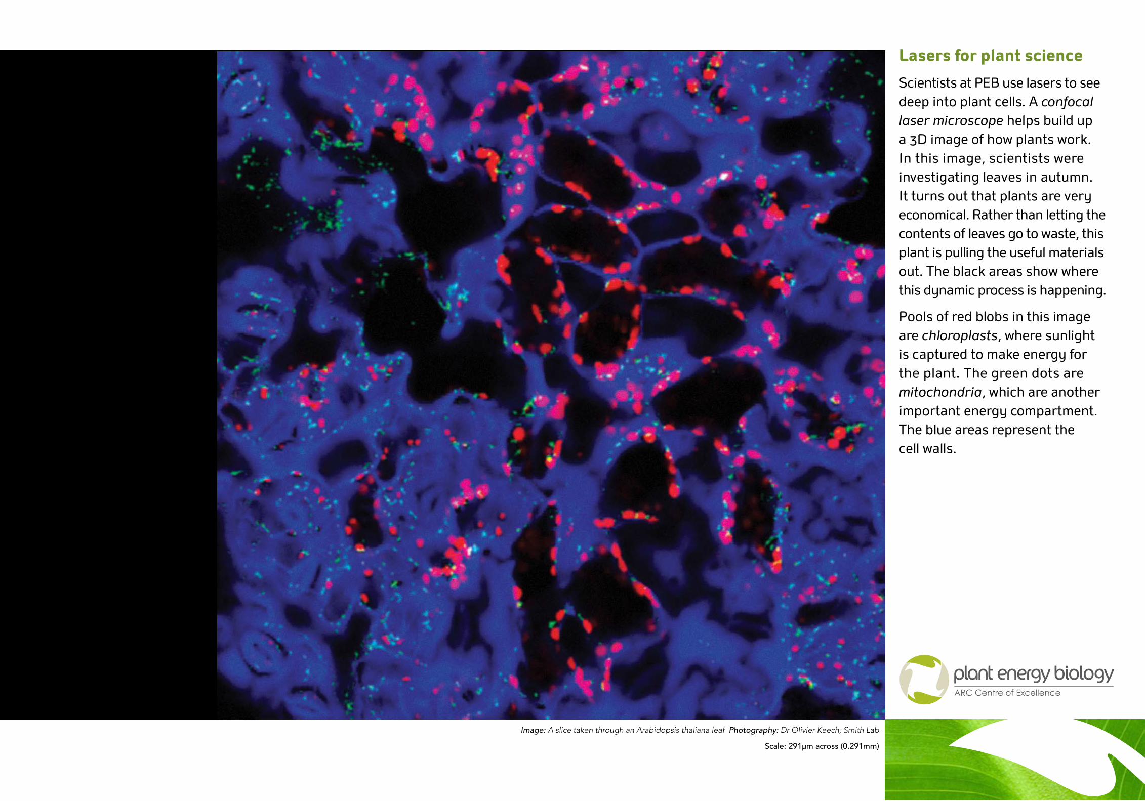
Lasers for plant science
Scientists at PEB use lasers to see deep into plant cells. A confocal laser microscope helps build up a 3D image of how plants work. In this image, scientists were investigating leaves in autumn. It turns out that plants are very economical. Rather than letting the contents of leaves go to waste, this plant is pulling the useful materials out. The black areas show where this dynamic process is happening.
Pools of red blobs in this image are chloroplasts, where sunlight is captured to make energy for the plant. The green dots are mitochondria, which are another important energy compartment. The blue areas represent the cell walls.
Image: A slice taken through an Arabidopsis thaliana leaf Photography: Dr Olivier Keech, Smith Lab
Scale: 291µm across (0.291mm)
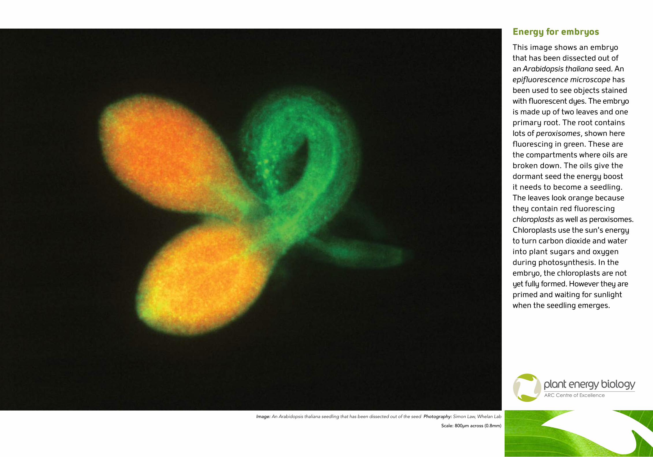
Energy for embryos
This image shows an embryo that has been dissected out of an Arabidopsis thaliana seed. An epifluorescence microscope has been used to see objects stained with fluorescent dyes. The embryo is made up of two leaves and one primary root. The root contains lots of peroxisomes, shown here fluorescing in green. These are the compartments where oils are broken down. The oils give the dormant seed the energy boost it needs to become a seedling. The leaves look orange because they contain red fluorescing chloroplasts as well as peroxisomes. Chloroplasts use the sun’s energy to turn carbon dioxide and water into plant sugars and oxygen during photosynthesis. In the embryo, the chloroplasts are not yet fully formed. However they are primed and waiting for sunlight when the seedling emerges.
Image: An Arabidopsis thaliana seedling that has been dissected out of the seed Photography: Simon Law, Whelan Lab
Scale: 800µm across (0.8mm)
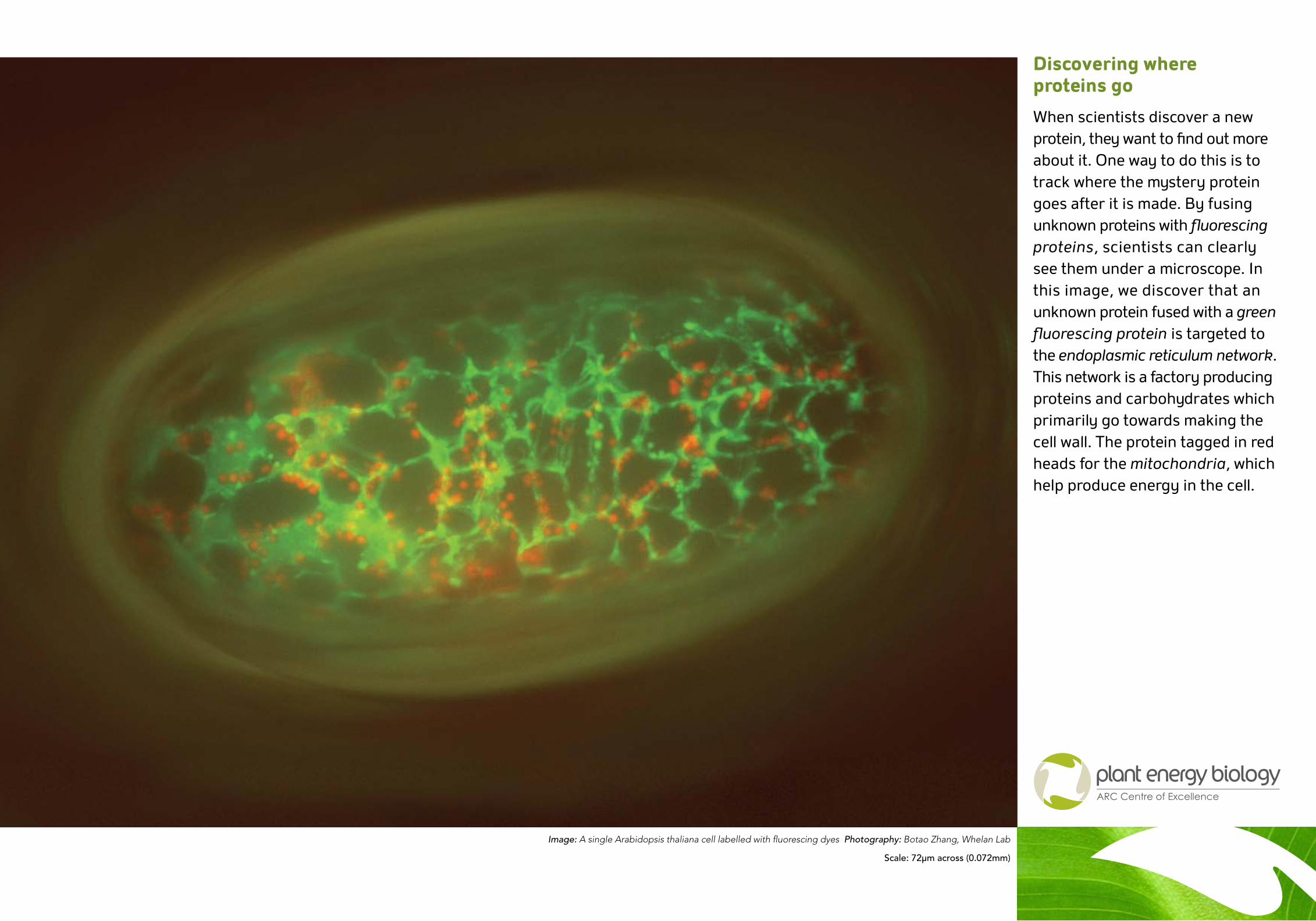
Discovering where proteins go
When scientists discover a new protein, they want to find out more about it. One way to do this is to track where the mystery protein goes after it is made. By fusing unknown proteins with fluorescing proteins, scientists can clearly see them under a microscope. In this image, we discover that an unknown protein fused with a green fluorescing protein is targeted to the endoplasmic reticulum network. This network is a factory producing proteins and carbohydrates which primarily go towards making the cell wall. The protein tagged in red heads for the mitochondria, which help produce energy in the cell.
Image: A single Arabidopsis thaliana cell labelled with fluorescing dyes Photography: Botao Zhang, Whelan Lab
Scale: 72µm across (0.072mm)
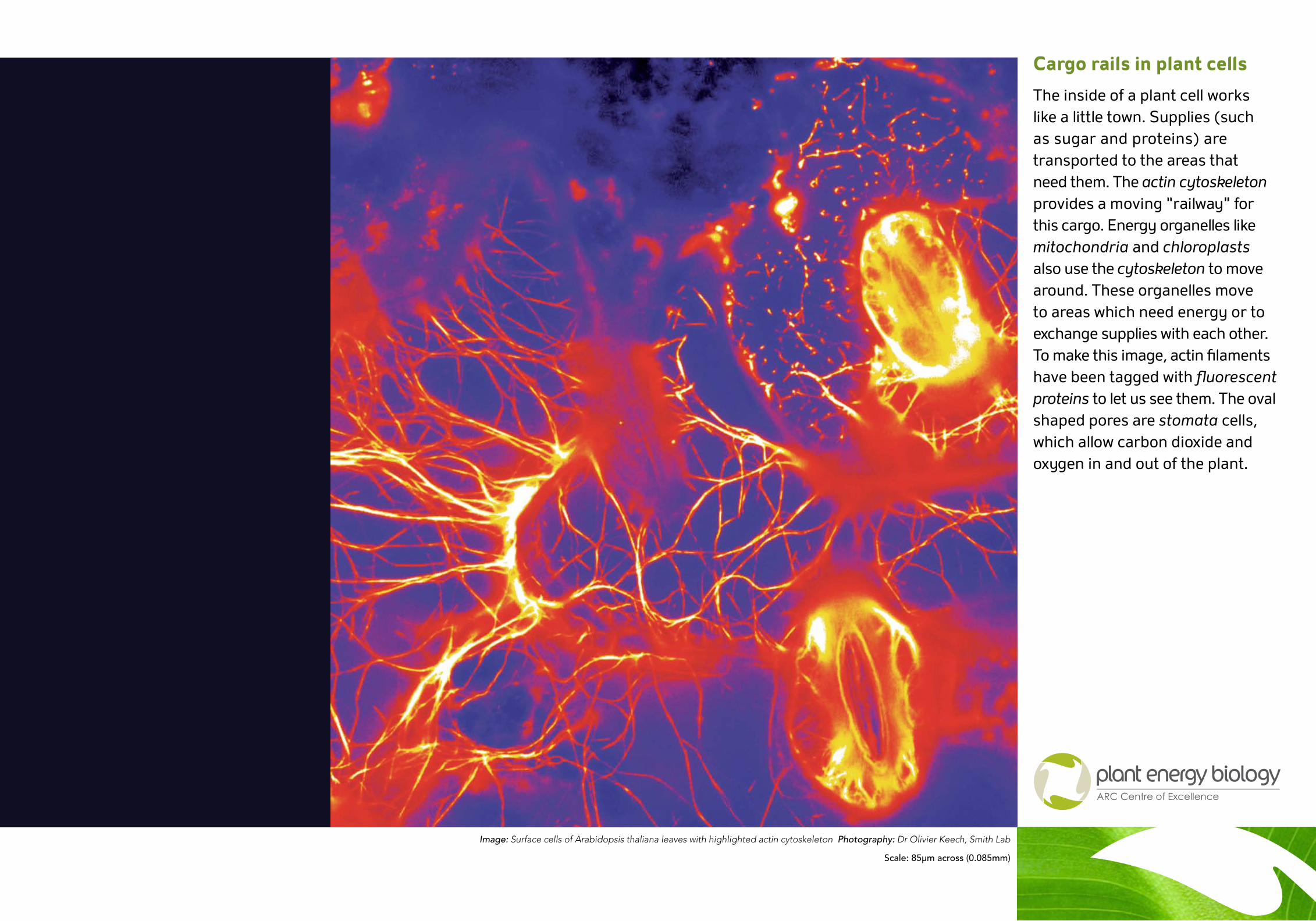
Cargo rails in plant cells
The inside of a plant cell works like a little town. Supplies (such as sugar and proteins) are transported to the areas that need them. The actin cytoskeleton provides a moving “railway” for this cargo. Energy organelles like mitochondria and chloroplasts also use the cytoskeleton to move around. These organelles move to areas which need energy or to exchange supplies with each other. To make this image, actin filaments have been tagged with fluorescent proteins to let us see them. The oval shaped pores are stomata cells, which allow carbon dioxide and oxygen in and out of the plant.
Image: Surface cells of Arabidopsis thaliana leaves with highlighted actin cytoskeleton Photography: Dr Olivier Keech, Smith Lab
Scale: 85µm across (0.085mm)