Embed Size (px)
Citation preview
REPORT
Whole Exome Sequencing and Homozygosity MappingIdentify Mutation in the Cell Polarity Protein GPSM2as the Cause of Nonsyndromic Hearing Loss DFNB82
Tom Walsh,1,4,* Hashem Shahin,2,4 Tal Elkan-Miller,3 Ming K. Lee,1 Anne M. Thornton,1 Wendy Roeb,1
Amal Abu Rayyan,2 Suheir Loulus,2 Karen B. Avraham,3 Mary-Claire King,1 and Moien Kanaan2,*
Massively parallel sequencing of targeted regions, exomes, and complete genomes has begun to dramatically increase the pace of
discovery of genes responsible for human disorders. Here we describe how exome sequencing in conjunction with homozygosity
mapping led to rapid identification of the causative allele for nonsyndromic hearing loss DFNB82 in a consanguineous Palestinian
family. After filtering out worldwide and population-specific polymorphisms from the whole exome sequence, only a single deleterious
mutation remained in the homozygous region linked to DFNB82. The nonsense mutation leads to an early truncation of the G protein
signaling modulator GPSM2, a protein that is essential for maintenance of cell polarity and spindle orientation. In the mouse inner ear,
GPSM2 is localized to apical surfaces of hair cells and supporting cells and is most highly expressed during embryonic development.
Identification of GPSM2 as essential to the development of normal hearing suggests dysregulation of cell polarity as a mechanism under-
lying hearing loss.
Homozygosity mapping in consanguineous kindreds with
nonsyndromic hearing loss has led to the discovery of
more than 50 recessive deafness loci.1 Genomic regions
linked to hearing loss by homozygosity mapping are gener-
ally multiple megabases in size and can contain hundreds
of genes. The identification of the critical allele from a large
number of candidates has heretofore been an expensive
and time-consuming process. Very recently, advances in
DNA enrichment and next-generation sequencing tech-
nology have made it possible to quickly and cost-
effectively sequence all the genes in the ‘‘exome,’’ the
protein-coding portion of the genome, and then to rapidly
identify alleles responsible for Mendelian disorders.2,3
We applied whole exome sequencing to identify the
gene responsible for nonsyndromic hearing loss DFNB82
in a Palestinian family.4 Hearing loss in affected individ-
uals was evaluated by pure-tone audiometry and was deter-
mined to be sensorineural, prelingual, bilateral, and severe
to profound. (Figure 1A). Vision and vestibular function
were normal, and there were no other findings on
ophthalmic testing and clinical exam. The DFNB82 region
spans 3.1 Mb on chromosome 1p13.1 and contains 50
annotated protein coding genes and 1 miRNA. The
DFNB82 region partially overlaps with the DFNB32 region
(MIM 608653) defined by a Tunisian family with congen-
ital profound hearing loss.5 The region of overlap contains
five genes: VAV3, SLC25A4, NBPF4, NBPF6, and FAM102B.
Genomic DNA from individual CG5 (Figure 1B) was ex-
tracted from blood, sonicated to approximately 200 bp
fragments, and used to make a library for paired-end
sequencing (Illumina). After quality control, the library
1Department of Medicine, Division of Medical Genetics, University of Wash
University, Bethlehem, Palestine; 3Department of Human Molecular Gene
Tel Aviv 69978, Israel4These authors contributed equally to this work
*Correspondence: [email protected] (T.W.), [email protected] (M.K.)
DOI 10.1016/j.ajhg.2010.05.010. ª2010 by The American Society of Human
90 The American Journal of Human Genetics 87, 90–94, July 9, 2010
was hybridized to biotinylated cRNA oligonucleotide baits
from the SureSelect Human All Exon kit (Agilent Technol-
ogies), purified by streptavidin-bound magnetic beads,
amplified, and sequenced.6 The exome design covers
38 Mb of human genome corresponding to the exons
and flanking intronic regions of 23,739 genes in the
National Center for Biotechnology Information Con-
sensus CDS database (September 2009 release) and also
covers 700 miRNAs from the Sanger v13 database and
300 noncoding RNAs.7 We generated a total of 5.6 Gb of
sequence as paired-end 76 bp reads from one lane of an
Illumina Genome Analyzer IIX with Illumina pipeline
v.1.6. We used MAQ v.0.7.18 to discard reads of inadequate
sequence quality or depth and to discard those with iden-
tical start and end sites.
In order to identify the allele responsible for DFNB82, we
focused on exonic and flanking intronic variants within
the linkage region on chromosome 1p13.3 between bp
108,307,327 and 111,450,408 (February 2009 human
reference sequence UCSC hg19/GRCh37). Alignment of
the sequence reads in this region revealed that 424 of the
458 targeted exons (93%) had >10 high-quality reads.
The fully evaluated exons include three genes (VAV3,
SLC25A4, and FAM102B) from the DFNB32/DFNB82 over-
lap region. The other two genes in the region (NBPF4 and
NBPF6) could not be evaluated because they lie on adjacent
segmental duplications that are >99% identical.
In the DFNB82 region, 80 variants, all single nucleotide
substitutions, passed our quality thresholds. All 80 substi-
tutions were predicted to be homozygous, as expected
given our SNP-based homozygosity mapping data.4
ington, Seattle, WA 98195, USA; 2Department of Life Sciences, Bethlehem
tics and Biochemistry, Sackler Faculty of Medicine, Tel Aviv University,
Genetics. All rights reserved.
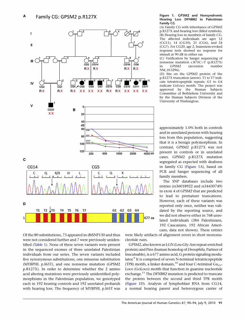
Figure 1. GPSM2 and NonsyndromicHearing Loss DFNB82 in PalestinianFamily CG(A) Family CG with inheritance of GPSM2p.R127X and hearing loss (filled symbols).(B) Hearing loss in members of family CG.The affected individuals are ages 12(CG11), 14 (CG10), 21 (CG6), and 24(CG7). For CG20, age 2, brainstem-evokedresponse tests showed no response forstimuli at 90 dB in either ear.(C) Verification by Sanger sequencing ofnonsense mutation c.875C>T (p.R127X)in GPSM2 (accession numberNM_013296).(D) Site on the GPSM2 protein of thep.R127X truncation (arrow). T1 to T7 indi-cate tetratricopeptide repeats; G1 to G4indicate GoLoco motifs. The project wasapproved by the Human SubjectsCommittee of Bethlehem University andby the Human Subjects Division of theUniversity of Washington.
Of the 80 substitutions, 73 appeared in dbSNP130 and thus
were not considered further and 7 were previously uniden-
tified (Table 1). None of these seven variants were present
in the sequenced exomes of three unrelated Palestinian
individuals from our series. The seven variants included
five synonymous substitutions, one missense substitution
(MYBPHL p.I65T), and one nonsense mutation (GPSM2
p.R127X). In order to determine whether the 2 amino
acid altering mutations were previously unidentified poly-
morphisms in the Palestinian population, we genotyped
each in 192 hearing controls and 192 unrelated probands
with hearing loss. The frequency of MYBPHL p.I65T was
The American Journal of H
approximately 1.0% both in controls
and in unrelated persons with hearing
loss from this population, suggesting
that it is a benign polymorphism. In
contrast, GPSM2 p.R127X was not
present in controls or in unrelated
cases. GPSM2 p.R127X mutation
segregated as expected with deafness
in family CG (Figure 1A), based on
PCR and Sanger sequencing of all
family members.
The SNP databases include two
entries (rs36018922 and rs34430749)
in exon 4 of GPSM2 that are predicted
to lead to premature truncations.
However, each of these variants was
reported only once, neither was vali-
dated by the reporting source, and
we did not observe either in 768 unre-
lated individuals (384 Palestinians,
192 Caucasians, 192 African Ameri-
cans, data not shown). These entries
were likely artifacts of alignment errors in short mononu-
cleotide runs.
GPSM2, also known as LGN (Leu-Gly-Asn repeat-enriched
protein) and Pins (human homolog of Drosophila, Partner of
Inscuteable), is a 677 amino acid, G protein signaling modu-
lator.9 It is comprised of seven N-terminal tetratricopeptide
(TPR) motifs, a linker domain,10 and four C-terminal Gai/o-
Loco (GoLoco) motifs that function in guanine nucleotide
exchange.11 The DFNB82 mutation is predicted to truncate
the protein between the second and third TPR motifs
(Figure 1D). Analysis of lymphoblast RNA from CG14,
a normal hearing parent and heterozygous carrier of
uman Genetics 87, 90–94, July 9, 2010 91
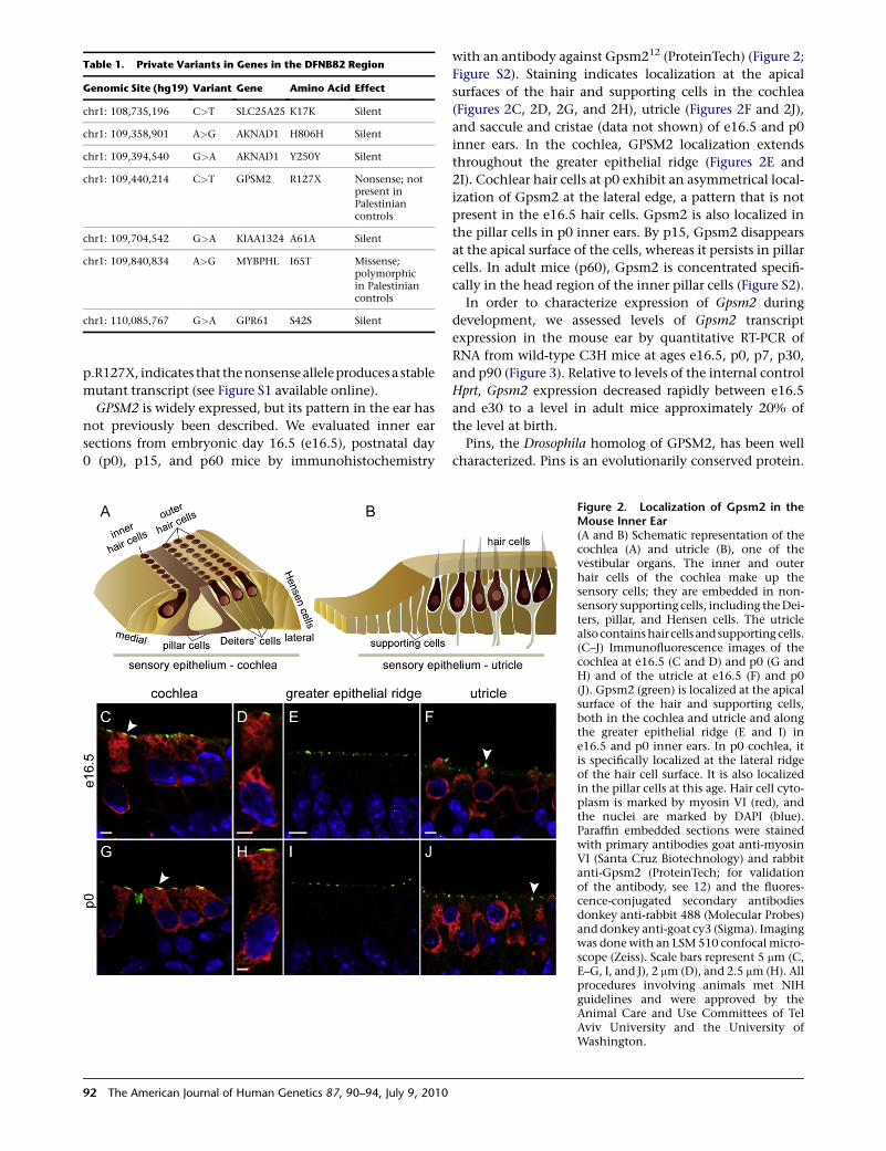
Table 1. Private Variants in Genes in the DFNB82 Region
Genomic Site (hg19) Variant Gene Amino Acid Effect
chr1: 108,735,196 C>T SLC25A25 K17K Silent
chr1: 109,358,901 A>G AKNAD1 H806H Silent
chr1: 109,394,540 G>A AKNAD1 Y250Y Silent
chr1: 109,440,214 C>T GPSM2 R127X Nonsense; notpresent inPalestiniancontrols
chr1: 109,704,542 G>A KIAA1324 A61A Silent
chr1: 109,840,834 A>G MYBPHL I65T Missense;polymorphicin Palestiniancontrols
chr1: 110,085,767 G>A GPR61 S42S Silent
p.R127X, indicates that thenonsense alleleproducesa stable
mutant transcript (see Figure S1 available online).
GPSM2 is widely expressed, but its pattern in the ear has
not previously been described. We evaluated inner ear
sections from embryonic day 16.5 (e16.5), postnatal day
0 (p0), p15, and p60 mice by immunohistochemistry
92 The American Journal of Human Genetics 87, 90–94, July 9, 2010
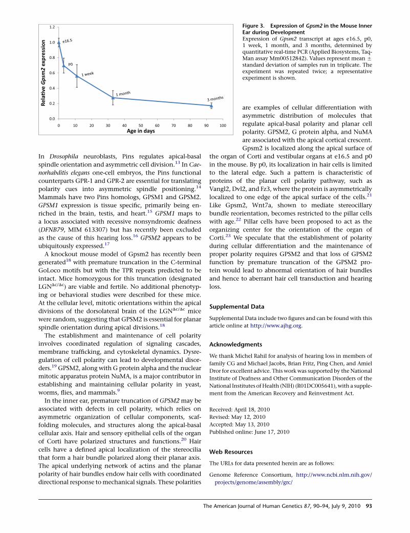
with an antibody against Gpsm212 (ProteinTech) (Figure 2;
Figure S2). Staining indicates localization at the apical
surfaces of the hair and supporting cells in the cochlea
(Figures 2C, 2D, 2G, and 2H), utricle (Figures 2F and 2J),
and saccule and cristae (data not shown) of e16.5 and p0
inner ears. In the cochlea, GPSM2 localization extends
throughout the greater epithelial ridge (Figures 2E and
2I). Cochlear hair cells at p0 exhibit an asymmetrical local-
ization of Gpsm2 at the lateral edge, a pattern that is not
present in the e16.5 hair cells. Gpsm2 is also localized in
the pillar cells in p0 inner ears. By p15, Gpsm2 disappears
at the apical surface of the cells, whereas it persists in pillar
cells. In adult mice (p60), Gpsm2 is concentrated specifi-
cally in the head region of the inner pillar cells (Figure S2).
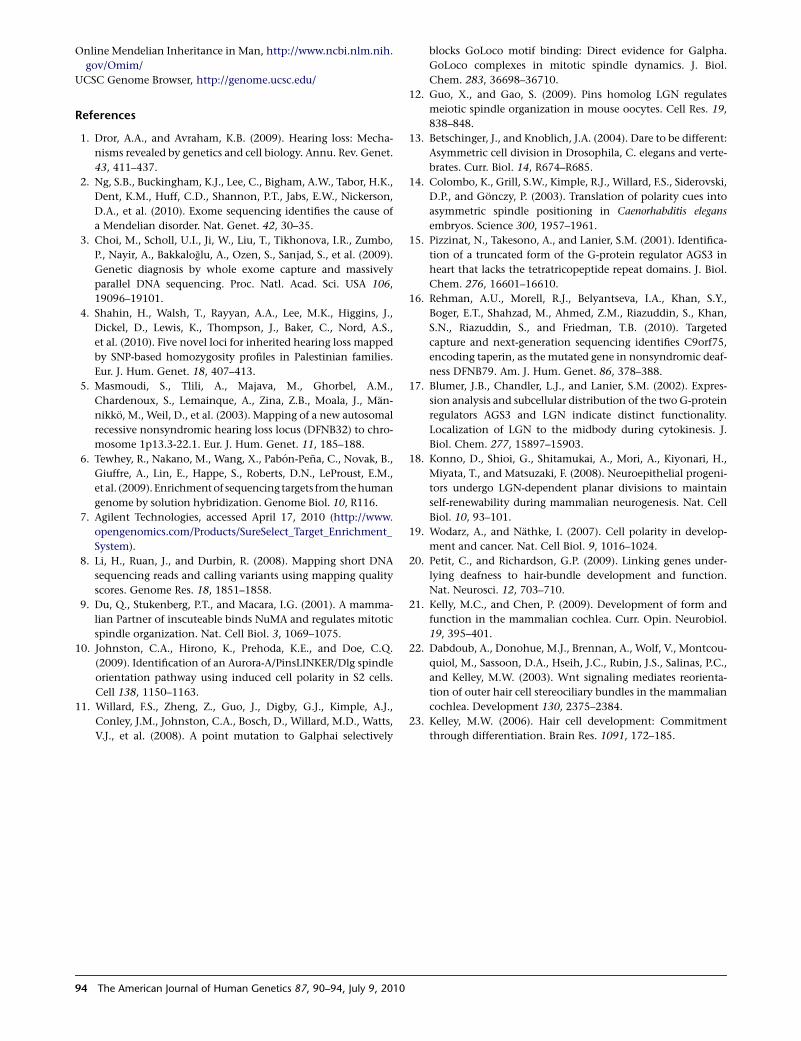
In order to characterize expression of Gpsm2 during
development, we assessed levels of Gpsm2 transcript
expression in the mouse ear by quantitative RT-PCR of
RNA from wild-type C3H mice at ages e16.5, p0, p7, p30,
and p90 (Figure 3). Relative to levels of the internal control
Hprt, Gpsm2 expression decreased rapidly between e16.5
and e30 to a level in adult mice approximately 20% of
the level at birth.
Pins, the Drosophila homolog of GPSM2, has been well
characterized. Pins is an evolutionarily conserved protein.
Figure 2. Localization of Gpsm2 in theMouse Inner Ear(A and B) Schematic representation of thecochlea (A) and utricle (B), one of thevestibular organs. The inner and outerhair cells of the cochlea make up thesensory cells; they are embedded in non-sensory supporting cells, including the Dei-ters, pillar, and Hensen cells. The utriclealsocontains haircells and supporting cells.(C–J) Immunofluorescence images of thecochlea at e16.5 (C and D) and p0 (G andH) and of the utricle at e16.5 (F) and p0(J). Gpsm2 (green) is localized at the apicalsurface of the hair and supporting cells,both in the cochlea and utricle and alongthe greater epithelial ridge (E and I) ine16.5 and p0 inner ears. In p0 cochlea, itis specifically localized at the lateral ridgeof the hair cell surface. It is also localizedin the pillar cells at this age. Hair cell cyto-plasm is marked by myosin VI (red), andthe nuclei are marked by DAPI (blue).Paraffin embedded sections were stainedwith primary antibodies goat anti-myosinVI (Santa Cruz Biotechnology) and rabbitanti-Gpsm2 (ProteinTech; for validationof the antibody, see 12) and the fluores-cence-conjugated secondary antibodiesdonkey anti-rabbit 488 (Molecular Probes)and donkey anti-goat cy3 (Sigma). Imagingwas done with an LSM 510 confocal micro-scope (Zeiss). Scale bars represent 5 mm (C,E–G, I, and J), 2 mm (D), and 2.5 mm (H). Allprocedures involving animals met NIHguidelines and were approved by theAnimal Care and Use Committees of TelAviv University and the University ofWashington.
0.0
0.2
0.4
0.6
0.8
1.0
1.2
0 10 20 30 40 50 60 70 80 90 100
Rela
�ve
Gps
m2
expr
essi
on
Age in days
Figure 3. Expression of Gpsm2 in the Mouse InnerEar during DevelopmentExpression of Gpsm2 transcript at ages e16.5, p0,1 week, 1 month, and 3 months, determined byquantitative real-time PCR (Applied Biosystems, Taq-Man assay Mm00512842). Values represent mean 5
standard deviation of samples run in triplicate. Theexperiment was repeated twice; a representativeexperiment is shown.
In Drosophila neuroblasts, Pins regulates apical-basal
spindle orientation and asymmetric cell division.13 In Cae-
norhabditis elegans one-cell embryos, the Pins functional
counterparts GPR-1 and GPR-2 are essential for translating
polarity cues into asymmetric spindle positioning.14
Mammals have two Pins homologs, GPSM1 and GPSM2.
GPSM1 expression is tissue specific, primarily being en-
riched in the brain, testis, and heart.15 GPSM1 maps to
a locus associated with recessive nonsyndromic deafness
(DFNB79, MIM 613307) but has recently been excluded
as the cause of this hearing loss.16 GPSM2 appears to be
ubiquitously expressed.17
A knockout mouse model of Gpsm2 has recently been
generated18 with premature truncation in the C-terminal
GoLoco motifs but with the TPR repeats predicted to be
intact. Mice homozygous for this truncation (designated
LGNDc/Dc) are viable and fertile. No additional phenotyp-
ing or behavioral studies were described for these mice.
At the cellular level, mitotic orientations within the apical
divisions of the dorsolateral brain of the LGNDc/Dc mice
were random, suggesting that GPSM2 is essential for planar
spindle orientation during apical divisions.18
The establishment and maintenance of cell polarity
involves coordinated regulation of signaling cascades,
membrane trafficking, and cytoskeletal dynamics. Dysre-
gulation of cell polarity can lead to developmental disor-
ders.19 GPSM2, along with G protein alpha and the nuclear
mitotic apparatus protein NuMA, is a major contributor in
establishing and maintaining cellular polarity in yeast,
worms, flies, and mammals.9
In the inner ear, premature truncation of GPSM2 may be
associated with defects in cell polarity, which relies on
asymmetric organization of cellular components, scaf-
folding molecules, and structures along the apical-basal
cellular axis. Hair and sensory epithelial cells of the organ
of Corti have polarized structures and functions.20 Hair
cells have a defined apical localization of the stereocilia
that form a hair bundle polarized along their planar axis.
The apical underlying network of actins and the planar
polarity of hair bundles endow hair cells with coordinated
directional response to mechanical signals. These polarities
The American Journal of Human Genetics 87, 90–94, July 9, 2010 93
l
l
-
are examples of cellular differentiation with
asymmetric distribution of molecules that
regulate apical-basal polarity and planar cell
polarity. GPSM2, G protein alpha, and NuMA
are associated with the apical cortical crescent.
Gpsm2 is localized along the apical surface of
the organ of Corti and vestibular organs at e16.5 and p0
in the mouse. By p0, its localization in hair cells is limited
to the lateral edge. Such a pattern is characteristic of
proteins of the planar cell polarity pathway, such as
Vangl2, Dvl2, and Fz3, where the protein is asymmetrically
localized to one edge of the apical surface of the cells.21
Like Gpsm2, Wnt7a, shown to mediate stereocillary
bundle reorientation, becomes restricted to the pillar cells
with age.22 Pillar cells have been proposed to act as the
organizing center for the orientation of the organ of
Corti.23 We speculate that the establishment of polarity
during cellular differentiation and the maintenance of
proper polarity requires GPSM2 and that loss of GPSM2
function by premature truncation of the GPSM2 pro-
tein would lead to abnormal orientation of hair bundles
and hence to aberrant hair cell transduction and hearing
loss.
Supplemental Data
Supplemental Data include two figures and can be found with this
article online at http://www.ajhg.org.
Acknowledgments
We thank Michel Rahil for analysis of hearing loss in members of
family CG and Michael Jacobs, Brian Fritz, Ping Chen, and Amie
Dror for excellent advice. This work was supported by the Nationa
Institute of Deafness and Other Communication Disorders of the
National Institutes of Health (NIH) (R01DC005641), with a supple
ment from the American Recovery and Reinvestment Act.
Received: April 18, 2010
Revised: May 12, 2010
Accepted: May 13, 2010
Published online: June 17, 2010
Web Resources
The URLs for data presented herein are as follows:
Genome Reference Consortium, http://www.ncbi.nlm.nih.gov/
projects/genome/assembly/grc/
Online Mendelian Inheritance in Man, http://www.ncbi.nlm.nih.
gov/Omim/
UCSC Genome Browser, http://genome.ucsc.edu/
References
1. Dror, A.A., and Avraham, K.B. (2009). Hearing loss: Mecha-
nisms revealed by genetics and cell biology. Annu. Rev. Genet.
43, 411–437.
2. Ng, S.B., Buckingham, K.J., Lee, C., Bigham, A.W., Tabor, H.K.,
Dent, K.M., Huff, C.D., Shannon, P.T., Jabs, E.W., Nickerson,
D.A., et al. (2010). Exome sequencing identifies the cause of
a Mendelian disorder. Nat. Genet. 42, 30–35.
3. Choi, M., Scholl, U.I., Ji, W., Liu, T., Tikhonova, I.R., Zumbo,
P., Nayir, A., Bakkaloglu, A., Ozen, S., Sanjad, S., et al. (2009).
Genetic diagnosis by whole exome capture and massively
parallel DNA sequencing. Proc. Natl. Acad. Sci. USA 106,
19096–19101.
4. Shahin, H., Walsh, T., Rayyan, A.A., Lee, M.K., Higgins, J.,
Dickel, D., Lewis, K., Thompson, J., Baker, C., Nord, A.S.,
et al. (2010). Five novel loci for inherited hearing loss mapped
by SNP-based homozygosity profiles in Palestinian families.
Eur. J. Hum. Genet. 18, 407–413.
5. Masmoudi, S., Tlili, A., Majava, M., Ghorbel, A.M.,
Chardenoux, S., Lemainque, A., Zina, Z.B., Moala, J., Man-
nikko, M., Weil, D., et al. (2003). Mapping of a new autosomal
recessive nonsyndromic hearing loss locus (DFNB32) to chro-
mosome 1p13.3-22.1. Eur. J. Hum. Genet. 11, 185–188.
6. Tewhey, R., Nakano, M., Wang, X., Pabon-Pena, C., Novak, B.,
Giuffre, A., Lin, E., Happe, S., Roberts, D.N., LeProust, E.M.,
et al. (2009). Enrichment of sequencing targets from the human
genome by solution hybridization. Genome Biol. 10, R116.
7. Agilent Technologies, accessed April 17, 2010 (http://www.
opengenomics.com/Products/SureSelect_Target_Enrichment_
System).
8. Li, H., Ruan, J., and Durbin, R. (2008). Mapping short DNA
sequencing reads and calling variants using mapping quality
scores. Genome Res. 18, 1851–1858.
9. Du, Q., Stukenberg, P.T., and Macara, I.G. (2001). A mamma-
lian Partner of inscuteable binds NuMA and regulates mitotic
spindle organization. Nat. Cell Biol. 3, 1069–1075.
10. Johnston, C.A., Hirono, K., Prehoda, K.E., and Doe, C.Q.
(2009). Identification of an Aurora-A/PinsLINKER/Dlg spindle
orientation pathway using induced cell polarity in S2 cells.
Cell 138, 1150–1163.
11. Willard, F.S., Zheng, Z., Guo, J., Digby, G.J., Kimple, A.J.,
Conley, J.M., Johnston, C.A., Bosch, D., Willard, M.D., Watts,
V.J., et al. (2008). A point mutation to Galphai selectively
94 The American Journal of Human Genetics 87, 90–94, July 9, 2010
blocks GoLoco motif binding: Direct evidence for Galpha.
GoLoco complexes in mitotic spindle dynamics. J. Biol.
Chem. 283, 36698–36710.
12. Guo, X., and Gao, S. (2009). Pins homolog LGN regulates
meiotic spindle organization in mouse oocytes. Cell Res. 19,
838–848.
13. Betschinger, J., and Knoblich, J.A. (2004). Dare to be different:
Asymmetric cell division in Drosophila, C. elegans and verte-
brates. Curr. Biol. 14, R674–R685.
14. Colombo, K., Grill, S.W., Kimple, R.J., Willard, F.S., Siderovski,
D.P., and Gonczy, P. (2003). Translation of polarity cues into
asymmetric spindle positioning in Caenorhabditis elegans
embryos. Science 300, 1957–1961.
15. Pizzinat, N., Takesono, A., and Lanier, S.M. (2001). Identifica-
tion of a truncated form of the G-protein regulator AGS3 in
heart that lacks the tetratricopeptide repeat domains. J. Biol.
Chem. 276, 16601–16610.
16. Rehman, A.U., Morell, R.J., Belyantseva, I.A., Khan, S.Y.,
Boger, E.T., Shahzad, M., Ahmed, Z.M., Riazuddin, S., Khan,
S.N., Riazuddin, S., and Friedman, T.B. (2010). Targeted
capture and next-generation sequencing identifies C9orf75,
encoding taperin, as the mutated gene in nonsyndromic deaf-
ness DFNB79. Am. J. Hum. Genet. 86, 378–388.
17. Blumer, J.B., Chandler, L.J., and Lanier, S.M. (2002). Expres-
sion analysis and subcellular distribution of the two G-protein
regulators AGS3 and LGN indicate distinct functionality.
Localization of LGN to the midbody during cytokinesis. J.
Biol. Chem. 277, 15897–15903.
18. Konno, D., Shioi, G., Shitamukai, A., Mori, A., Kiyonari, H.,
Miyata, T., and Matsuzaki, F. (2008). Neuroepithelial progeni-
tors undergo LGN-dependent planar divisions to maintain
self-renewability during mammalian neurogenesis. Nat. Cell
Biol. 10, 93–101.
19. Wodarz, A., and Nathke, I. (2007). Cell polarity in develop-
ment and cancer. Nat. Cell Biol. 9, 1016–1024.
20. Petit, C., and Richardson, G.P. (2009). Linking genes under-
lying deafness to hair-bundle development and function.
Nat. Neurosci. 12, 703–710.
21. Kelly, M.C., and Chen, P. (2009). Development of form and
function in the mammalian cochlea. Curr. Opin. Neurobiol.
19, 395–401.
22. Dabdoub, A., Donohue, M.J., Brennan, A., Wolf, V., Montcou-
quiol, M., Sassoon, D.A., Hseih, J.C., Rubin, J.S., Salinas, P.C.,
and Kelley, M.W. (2003). Wnt signaling mediates reorienta-
tion of outer hair cell stereociliary bundles in the mammalian
cochlea. Development 130, 2375–2384.
23. Kelley, M.W. (2006). Hair cell development: Commitment
through differentiation. Brain Res. 1091, 172–185.