Embed Size (px)
Citation preview
Whole-Body 1-131 Metastatic Survey as an Aide in Determining Functional Thyroid Metastases
Leo R. LaRocque
Hartford Hospital. Hartford. Connecticut
Whole-body metastatic sun•ey scans are performed in order to determine the extent of metastases, the quality of tumor uptake, and whether the patient will benefit from therapeutic doses of radioiodine. Periodic whole-body scans also help to evaluate the success of radioiodine therapy or the necessity to repeat therapeutic treatments. Using a dual-probe rectilinear scanner and supplementing with scintillation camera images, we have found this procedure useful in detecting metastatic and focal thyroid cancer.
Following surgery for some forms of thyroid carcinoma, patients may be evaluated with radioiodine [1111] sodium iodide for residual functioning tissue by various methods. These include imaging techniques, 1-131-PBI conversion rates, and excretion studies ( /). It is assumed that there will be little or no accumulation of radioiodine by any remaining thyroid tissue. These studies are also based on the premise that the tracer dose of 1-131 is cleared by the thyroid. kidneys. gastric mucosa, and the salivary glands. The presence of metastases and evidence that the lesion concentrates iodine sufficiently are two primary indications when selecting patients for radioiodine therapy.
Method and Materials
Whole-body scans are usually performed 48 or 72 hr following the oral administration of 1-3 mCi of[ 11 1J] sodium iodide. The images arc recorded by an OhioNuclear model 84 dual-probe rectilinear scanner (Ohio Nuclear. Solon, OH) using 38-M medium energy collimators. Whole-body minified scans arc obtained by using the 5: I ratio and 5: I light aperatures with a 1/x-in. line spacing (Fig. 1). Neither backgrou11d erase nor contrast enhancement is used in order that we may be able to detect all areas of radioiodine concentration. The scanner is set at a speed of 700 (53~ em min) and the total elapsed time of the procedure is approximately 40 min. The lower distal extremities are excluded.
At the conclusion of the scan the patient is requested to remain in the department until the images have been reviewed by a nuclear medicine physician. Additional images may be requested of abnormal areas of raJioiodine accumulation with either pinhole or medium energy collimators attached to a scintillation camera. When additional views are requested, we use the camera,
For rerrinb contact: Leo R. LaR<lC4liC. Dcrt. of Clinical :'..:uclcar
Medicine. Hartford Hosrital. Hartford. CT Oh 115.
whenever possible. for two reasons: it usually requires only 3-5 min to take camera images. and so minimal disruption in patient scheduling is achieved: and, at the same time. the scanner is readily available for the next patient. By performing a 48- or 72-hr study, we are allowing for some of the radioiodine to be excreted by the urinary system. thereby decreasing body background.
Discussion
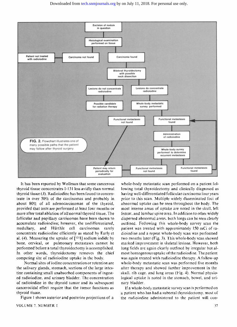
The best available treatment for carcinoma of the thyroid is surgery (2) and there are two important reasons why surgery should precede radioiodine therapy. First, a large specimen of the tumor is provided for pathological examination because some parts of the gland portion of the tumor may concentrate T-131 and other parts may not. Secondly, uptake of the remaining cancerous tissue is increased when part or all of the gland is removed. Depending upon the outcome of the pathological examination, a variety of paths may be taken (Fig. 2)
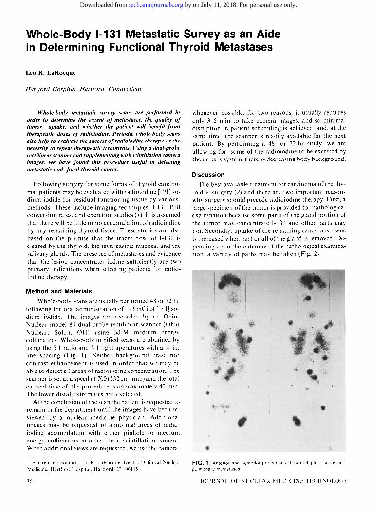
FIG. 1. Antenor and postenor prOJectrons show multrple osseous and
pulmonary metastases
.IOl'R-.;/\1 OF -.;tiCif/\R MfDIC!-.;f ITCH-.;01 OCiY
by on July 11, 2018. For personal use only. tech.snmjournals.org Downloaded from
It has been reported by W oilman that some cancerous thyroid tissue concentrates 1-131 less avidly than normal thyroid tissue (3). Radioiodine has been found to concentrate in over 50% of the carcinomas and probably in about 80% of all adenocarcinomas of the thyroid, provided that tests are performed at least four months or more after total ablation of all normal thyroid tissue. The follicular and papillary carcinomas have been shown to accumulate radioiodine; however, the undifferentiated, medullary, and Hlirthle cell carcinomas rarely concentrate radioiodine efficiently as stated by Early et a!. (4). Measuring the uptake of ['31I] sodium iodide by bone, cervical, or pulmonary metastases cannot be performed before a total thyroidectomy is accomplished. In other words, thyroidectomy removes the chief competing site of radioiodine uptake in the body.
Normal sites of iodine concentration or retention are the salivary glands, stomach, sections of the large intestine containing small unabsorbed components of ingested radioiodine, and urinary bladder. The concentration of radioiodine in the thyroid tumor and its subsequent cancerocidal effect require that the tumor functions as thyroid tissue.
Figure I shows anterior and posterior projections of a
VOLUME 7. NUMBER I
whole-body metastatic scan performed on a patient following total thyroidectomy and clinically diagnosed as having well-differentiated follicular carcinoma four years prior to this scan. Multiple widely disseminated foci of abnormal uptake can be seen throughout the body. The most intense areas of uptake are noted in the skull, left femur, and lumbar spine area. In addition to other widely dispersed abnormal areas, both lungs can be seen clearly outlined. Following this whole-body survey scan the patient was treated with approximately 150 mCi of radioiodine and a repeat whole-body scan was performed two months later (Fig. 3). This whole-body scan showed marked improvement in skeletal lesions. However, both lung fields are again clearly outlined by irregular but almost homogenous uptake ofthe radioiodine. The patient was again treated with radioiodine therapy. A follow-up whole-body metastatic scan was performed five months after therapy and showed further improvement in the skull, rib cage, and lung areas (Fig. 4). Normal physiological uptake is noted in the stomach, bowel, and urinary bladder.
If a whole-body metastatic survey scan is performed on a patient who has had a subtotal thyroidectomy, most of the radioiodine administered to the patient will con-
by on July 11, 2018. For personal use only. tech.snmjournals.org Downloaded from
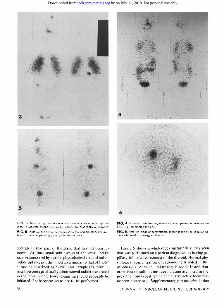
FIG. 3. Functioning thyroid metastatic disease is noted with improvement in skeletal lesions; pulmonary lesions are essentially unchanged.
FIG. 5. Anterior and posterior images show foci of radioiodine accumulation in neck. upper chest. and paravertebral area
centrate in that part of the gland that has not been removed. At times small subtle areas of abnormal uptake may be concealed by normal physiological areas of radioiodine uptake, i.e., the bowel area similar to that ofGa-67 citrate as described by Schall and Temple (5). Since a small percentage of orally administered iodide is excreted in the feces, proper bowel cleansing should probably be initiated if subsequent scans are to be performed.
38
FIG. 4. Follow-up whole-body metastatic scan performed five months following radioiodine therapy.
FIG. 6. Anterior image of paravertebral region taken by scintillation camera with medium energy collimator.
Figure 5 shows a whole-body metastatic survey scan that was performed on a patient diagnosed as having papillary-follicular carcinoma of the thyroid. Normal physiological concentration of radioiodine is noted in the oropharynx, stomach, and urinary bladder. In addition, other foci of radioiodine accumulation are noted in the neck and upper chest region and a large active focus may be seen posteriorly. Supplementary gamma scintillation
JOURNAL OF NUCLEAR MEDICINE TECHNOLOGY
by on July 11, 2018. For personal use only. tech.snmjournals.org Downloaded from
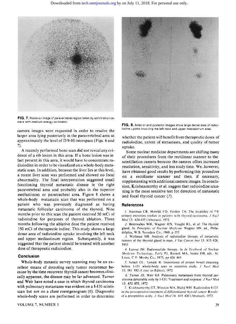
FIG. 7. Posterior image of paravertebral region taken by scintillation camera with medium energy collimator.
camera images were requested in order to resolve the larger area lying posteriorly in the paravertebral area at approximately the level of D 9-10 interspace (Figs. 6 and 7).
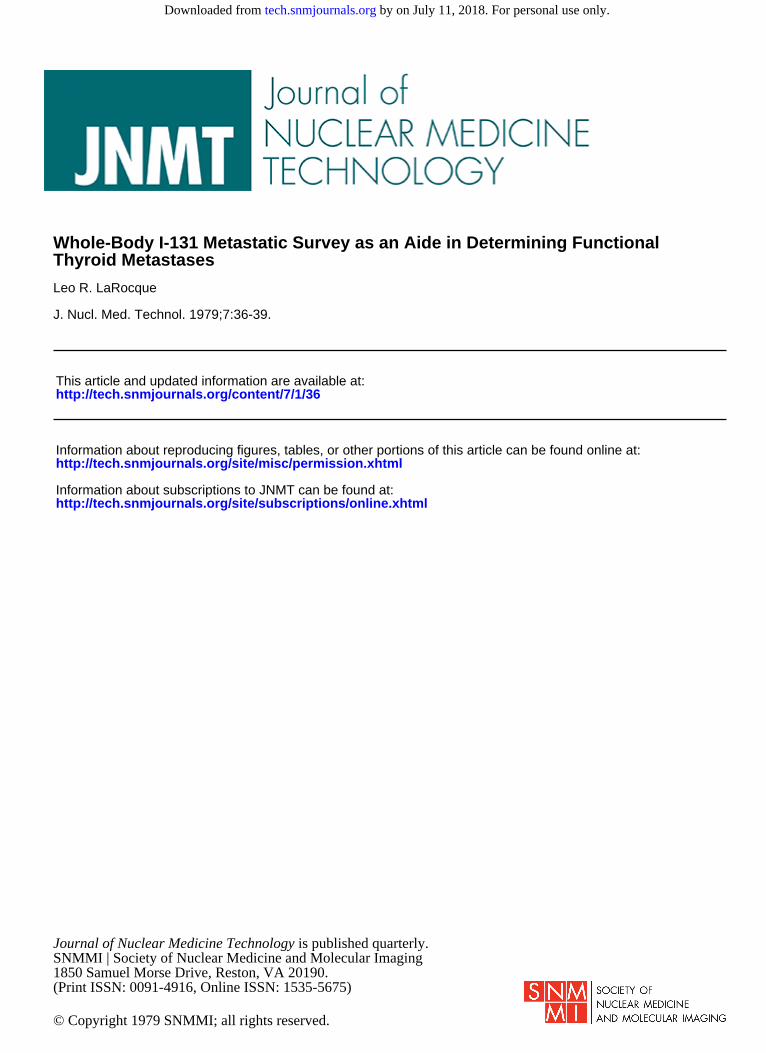
A recently performed bone scan did not reveal any evidence of a rib lesion in this area. If a bone lesion was in fact present in this area, it would have to concentrate radioiodine in order to be visualized on a whole-body metastatic scan. In addition, because the liver lies at this level, a recent liver scan was performed and showed no focal abnormality. The final interpretation suggested small functioning thyroid metastatic disease in the right paravertebral area and probably also in the superior mediastinum or paratracheal area. Figure 8 shows a whole-body metastatic scan that was performed on a patient who was previously diagnosed as having metastatic follicular carcinoma of the thyroid. Nine months prior to this scan the patient received 50 mCi of radioiodine for purposes of thyroid ablation. Three months following the ablative dose the patient received 150 mCi of therapeutic iodine. This study shows a large dense area of radioiodine uptake involving the left neck and upper mediastinum region. Subsequently, it was suggested that the patient should be treated with another dose of therapeutic radioiodine.
Conclusion Whole-body metastic survey scanning may be an ex
cellent means of detecting early tumor recurrence because by the time recurrent thyroid cancer becomes clinically apparent, the disease may be far advanced. Turner and Weir have noted a case in which thyroid carcinoma with pulmonary metastases was evident on a I-131 scintiscan but not on a chest roentgenogram (6). Diagnostic whole-body scans are performed in order to determine
VOLUME 7, NUMBER I
FIG. 8. Anterior and posterior images show large dense area of radioiodine uptake involving the left neck and upper mediastinum area.
whether the patient will benefit from therapeutic doses of radioiodine, extent of metastases, and quality of tumor uptake.
Some nuclear medicine departments are shifting many of their procedures from the rectilinear scanner to the scintillation camera because the camera offers increased resolution, sensitivity, and less study time. We, however, have obtained good results by performing this procedure on a rectilinear scanner and then, if necessary, supplementing with additional camera images. In conclusion, Krishnamurthy et a!. suggest that radioiodine scanning is the most sensitive test for detection of metastatic and focal thyroid cancer (7).
References
/. Hartman CR. Hofeldt FD. Verdon T A: The invalidity of 111 1 urinary excretion studies in patients with thyroid carcinoma . .I Nuel Med 13: 434-435 (Abstract). 1972
2. Beierwaltes WH, Wagner HN. Vought RL. et al: The thyroid gland. In Principles o( Nuclear Medicine. Wagner HN. ed .. Philadelphia, W.B. Saunders Co .. 1968. p 353
3. Wollman SH: Analysis of radioiodine therapy of metastatic tumors of the thyroid gland in man . .I Nat Cancer /nst 13: 815 828. 1953
4. !.arose JH: Radionuclide therapy. In In Texthook o( .Vue/ear Medicine Technologr. Early PJ. Rataak MA. Sodce DR. eds .. St. Louis. C.V. Mosby Co .. 1975. pp 456 463
5. Schall GI.. Temple R: Importance of proper bowel cleansing before 1-131 whole-body scan or retention study . .I l\'uel /11ed 13: 181 182 (Letter to Editor). 1972
6. Turner JE, Weir GJ: Pulmonary metastases from thyroid carcinoma detectable only by 1-131: Treatment and response . .I Nucl Med 13: 852-855, 1972
7. K rishnamurthy GT. Winston MA. Rlahd WH: Radioiodine 1-131 in the postoperative treatment of differentiated thyroid cancer: Result' of a prospective study . .I .Vue/ Med 14: 419 420 (Abstract). 1973
39
by on July 11, 2018. For personal use only. tech.snmjournals.org Downloaded from
1979;7:36-39.J. Nucl. Med. Technol. Leo R. LaRocque Thyroid MetastasesWhole-Body I-131 Metastatic Survey as an Aide in Determining Functional
http://tech.snmjournals.org/content/7/1/36This article and updated information are available at:
http://tech.snmjournals.org/site/subscriptions/online.xhtml
Information about subscriptions to JNMT can be found at:
http://tech.snmjournals.org/site/misc/permission.xhtmlInformation about reproducing figures, tables, or other portions of this article can be found online at:
(Print ISSN: 0091-4916, Online ISSN: 1535-5675)1850 Samuel Morse Drive, Reston, VA 20190.SNMMI | Society of Nuclear Medicine and Molecular Imaging
is published quarterly.Journal of Nuclear Medicine Technology
© Copyright 1979 SNMMI; all rights reserved.
by on July 11, 2018. For personal use only. tech.snmjournals.org Downloaded from