Embed Size (px)
Citation preview

2017JournalofBiomaterialsStKittsPrimatePaperBasedonFraudulentPrimateData
Theyknowmyvalues;andInVivoTherapeuticsknewnottoputmynameasanauthoronfraud
WHODOTHEYTHINKTHEYARE?
Iwasabsolutelyjawdroppedshockedtoseefalsedata;that’sisknowntobefalsetothemajorityoftheauthorspublishedintheJournalofBiomaterials.IwentfrombeingfirstauthortonotbeingnamedasanauthorbecauseIwouldn’tsignontofraud.Iwastheonlyscientistinvolvedineveryaspectofeverystudyfrom2007-2013.
Sincelate2010,thefabricatedprimatedatawhichisnowpublishedprimatedatawasheldforcriminalevidenceinfilesatInVivoTherapeutics.SinceIleftInVivoinAugust2013,theauthorsandInVivoTherapeuticshaverequestedthelocationofthereal/rawdataatleast10times.AsrecentlyassixmonthsagoIwasaskedtoprovidethelocationoftherealfiles,butaseveryoneknowstherealprimatedataistiedupintheReynoldsvs.InVivoTherapeuticslitigation,sotheypublishedknownfakedata.It’ssickening.
TheInVivoTherapeuticsStKittsprimatedatapublishedintheSlotkinetalpaperisknowntobefalsetomanyoftheauthors.TheoriginalprimatedataiscurrentlytiedupinlitigationReynoldsvs.InVivoTherapeutics.ThedatawasfoundinoldrecordsafterIleftInVivoTherapeutics.InAugust2013,theoriginalprimatedatawassecuredinInVivo’sSalem,NHdatacenteranditwaspreservedintheReynoldsvs.InVivoTherapeuticcase.TheoriginalprimatedatahasneverbeenmadeavailabletotheauthorssinceOctober2010.Thedataincludedintheprimatepaperwasfabricatedbytwo,w-2employee,andaBoardmemberofInVivoTherapeuticswhowantedtokilltheNeuroScaffoldproject.ThefabricatedprimatedatawaspreservedinfilesatInVivoTherapeuticsaslegalevidenceinvolvingscientificfraudinvolvingthosescientists.AsCEOofInVivoTherapeutics,bycontract.Iwastheonlypersonwithlegalcontroloftheprimatedata.IalwayshadtheoriginalprimatedatasowesimplystoredthefabricatedPricharddataanditwassecuredforlegalreasons.Authorshavebeeninmyfiancée’shometwiceinthelast18monthsaskingmefortheoriginalprimatedatabutagainitistiedupinlitigationanditwasnotavailableforanyoneelsetoreviewuntilafterlitigation.ThepapergaveacknowledgementtoHainingDai,butnotauthorshipbecauseHainingandIwouldNOTsignoffasauthorsonthepaperin2012,2013,2014,2015and2016.Nowit’spublished?Theauthorsknewthepaperdraftshadusedthefraudcasefilesthatwereheldasevidence.Theywerebothawareoftheemployeefraudincident.
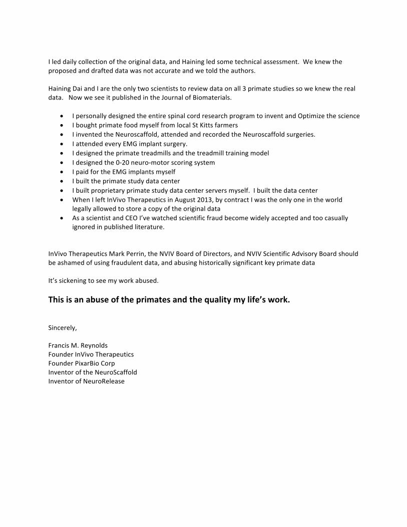
Ileddailycollectionoftheoriginaldata,andHainingledsometechnicalassessment.Weknewtheproposedanddrafteddatawasnotaccurateandwetoldtheauthors.HainingDaiandIaretheonlytwoscientiststoreviewdataonall3primatestudiessoweknewtherealdata.NowweseeitpublishedintheJournalofBiomaterials.
• IpersonallydesignedtheentirespinalcordresearchprogramtoinventandOptimizethescience• IboughtprimatefoodmyselffromlocalStKittsfarmers• IinventedtheNeuroscaffold,attendedandrecordedtheNeuroscaffoldsurgeries.• IattendedeveryEMGimplantsurgery.• Idesignedtheprimatetreadmillsandthetreadmilltrainingmodel• Idesignedthe0-20neuro-motorscoringsystem• IpaidfortheEMGimplantsmyself• Ibuilttheprimatestudydatacenter• Ibuiltproprietaryprimatestudydatacenterserversmyself.Ibuiltthedatacenter• WhenIleftInVivoTherapeuticsinAugust2013,bycontractIwastheonlyoneintheworld
legallyallowedtostoreacopyoftheoriginaldata• AsascientistandCEOI’vewatchedscientificfraudbecomewidelyacceptedandtoocasually
ignoredinpublishedliterature.InVivoTherapeuticsMarkPerrin,theNVIVBoardofDirectors,andNVIVScientificAdvisoryBoardshouldbeashamedofusingfraudulentdata,andabusinghistoricallysignificantkeyprimatedataIt’ssickeningtoseemyworkabused.Thisisanabuseoftheprimatesandthequalitymylife’swork.Sincerely,FrancisM.ReynoldsFounderInVivoTherapeuticsFounderPixarBioCorpInventoroftheNeuroScaffoldInventorofNeuroRelease

lable at ScienceDirect
Biomaterials 123 (2017) 63e76
Contents lists avai
Biomaterials
journal homepage: www.elsevier .com/locate/biomater ia ls
Biodegradable scaffolds promote tissue remodeling and functionalimprovement in non-human primates with acute spinal cord injury
Jonathan R. Slotkin a, Christopher D. Pritchard b, Brian Luque c, Janice Ye c,Richard T. Layer c, *, Mathew S. Lawrence d, Timothy M. O'Shea e, f, g, Roland R. Roy h, i,Hui Zhong i, Isabel Vollenweider j, V. Reggie Edgerton h, i, k, Gr�egoire Courtine j,Eric J. Woodard l, Robert Langer b, e, f, g
a Department of Neurosurgery, Geisinger Clinic, Danville, PA, USAb Department of Chemical Engineering, Massachusetts Institute of Technology, Cambridge, MA, USAc InVivo Therapeutics Corporation, Cambridge, MA, USAd RxGen, Hamden, CT, USAe HarvardeMassachusetts Institute of Technology, Division of Health Sciences and Technology, Massachusetts Institute of Technology, Cambridge, MA, USAf Institute for Medical Engineering and Science, Massachusetts Institute of Technology, Cambridge, MA, USAg David H. Koch Institute for Integrative Cancer Research, Massachusetts Institute of Technology, Cambridge, MA, USAh Brain Research Institute, University of California, Los Angeles, CA, USAi Department of Integrative Biology and Physiology, University of California, Los Angeles, CA, USAj Center for Neuroprosthetics and Brain Mind Institute, Swiss Federal Institute of Technology (EPFL), Lausanne, Switzerlandk Departments of Neurobiology and Neurology, University of California, Los Angeles, CA, USAl Department of Neurosurgery, New England Baptist Hospital, Boston, MA, USA
a r t i c l e i n f o
Article history:Received 8 April 2016Received in revised form8 December 2016Accepted 22 January 2017Available online 25 January 2017
Keywords:Polymeric scaffoldsTissue remodelingTissue engineeringFunctional improvementPrimatesSpinal cord injury
* Corresponding author. InVivo Therapeutics, One KEast, 4th Floor, Cambridge, MA, 02139, USA.
E-mail address: [email protected] (R.
http://dx.doi.org/10.1016/j.biomaterials.2017.01.0240142-9612/© 2017 Elsevier Ltd. All rights reserved.
a b s t r a c t
Tissue loss significantly reduces the potential for functional recovery after spinal cord injury. We pre-viously showed that implantation of porous scaffolds composed of a biodegradable and biocompatibleblock copolymer of Poly-lactic-co-glycolic acid and Poly-L-lysine improves functional recovery and re-duces spinal cord tissue injury after spinal cord hemisection injury in rats. Here, we evaluated the safetyand efficacy of porous scaffolds in non-human Old-World primates (Chlorocebus sabaeus) after a partialand complete lateral hemisection of the thoracic spinal cord. Detailed analyses of kinematics and muscleactivity revealed that by twelve weeks after injury fully hemisected monkeys implanted with scaffoldsexhibited significantly improved recovery of locomotion compared to non-implanted control animals.Twelve weeks after injury, histological analysis demonstrated that the spinal cords of monkeys with ahemisection injury implanted with scaffolds underwent appositional healing characterized by a signif-icant increase in remodeled tissue in the region of the hemisection compared to non-implanted controls.The number of glial fibrillary acidic protein immunopositive astrocytes was diminished within the innerregions of the remodeled tissue layer in treated animals. Activated macrophage and microglia werepresent diffusely throughout the remodeled tissue and concentrated at the interface between the pre-served spinal cord tissue and the remodeled tissue layer. Numerous unphosphorylated neurofilament Hand neuronal growth associated protein positive fibers and myelin basic protein positive cells mayindicate neural sprouting inside the remodeled tissue layer of treated monkeys. These results support thesafety and efficacy of polymer scaffolds in a primate model of acute spinal cord injury. A device sub-stantially similar to the device described here is the subject of an ongoing human clinical trial.
© 2017 Elsevier Ltd. All rights reserved.
endall Square, Building 1400
T. Layer).
1. Introduction
Traumatic spinal cord injury afflicts approximately 180,000persons worldwide each year [1], often leading to severe motordeficits, sensory impairments, and bowel, bladder, and sexual
J.R. Slotkin et al. / Biomaterials 123 (2017) 63e7664
dysfunctions. The functional deficits that follow a spinal cord injuryresult in part from damage to, or loss of, neurons and glia in theneural tissue surrounding the location of the initial mechanicaltrauma, as well as a second phase of tissue loss that persists forweeks to months after the injury [2]. Pathological processes thatcontribute to this tissue loss include ischemia, anoxia, free-radicalinitiated lipid peroxidation, excitotoxicity, activation of proteolyticenzymes, and inflammation [3e6]. The loss of tissue leads to theaccumulation of necrotic debris and other inhospitable materials inthe lesion site. In the weeks following initial injury, residentmicroglia and circulating leukocytes clear the lesion of debris,which leaves a fluid-filled cystic cavity [7]. This cystic cavity,coupled with the presence of a surrounding scar that contains avariety of molecules that inhibit the growth of neurites, constitutesa formidable barrier to the regeneration or plasticity of axon tracts.Secondary syrinx formation can result in a variety of worseningneuropathic symptoms. The development of strategies to overcomethese barriers and to minimize and replace lost tissue is a majorfocus of spinal cord injury research.
Therapeutic strategies to replace lost tissue and bridge cysticcavities primarily rely on the transplantation of cells such asSchwann cells, olfactory nervous system cells, embryonic CNS tis-sue, and embryonic or adult stem or progenitor cells [8]. The sur-vival of transplanted cells within injured CNS tissue, however, canbe low. A promising approach to improving the survival of trans-planted cells is to provide an artificial structure on which trans-planted cells can grow, divide, and differentiate [9e11]. The utilityof polymer-based biomaterial scaffolds to support transplanted cellsurvival within injured spinal cord tissue in pre-clinical models ofCNS injury has been demonstrated for a variety of cell typesincluding Schwann cells [9,12,13], neural stem and progenitor cells[14e18], and cells genetically engineered to express growth factors[19e22].
The ability of acellular polymeric implants to support regener-ation and remodeling of spinal cord tissue after injury also has beenexplored [23e29]. Teng et al. [16] showed that implantation ofporous polymer scaffolds, with and without seeded neural stemcells, promoted functional improvements in rats after a lateralthoracic hemisection. Histological and immunohistochemical ana-lyses suggested that the functional recovery was associated withreduced tissue loss, diminished glial scarring, and axonal sproutingthrough the scaffold. In that study, Teng et al. [16] utilized thepolymer PLGA-PLL, which is a block copolymer of Poly-lactic-co-glycolic acid (PLGA) and Poly-L-lysine (PLL). PLGA is a biodegrad-able and biocompatible polymer that is approved by the FDA forapplications such as surgical sutures. PLL is a polymer with lysinefunctional groups that promote cellular adhesion by creating apositively charged material substrate [30e32]. The resultingcopolymer PLGA-PLL is biocompatible and biodegradable, and canbe formulated into highly porous scaffolds that contain functionalgroups capable of promoting the conjugation of biologically activemolecules and cellular adhesion. The functional improvement andmitigation of tissue loss in a pre-clinical model of spinal cord injury[16] suggests that a PLGA-PLL implant placed in apposition to un-injured tissue, without the additional complexities of addingexogenous cells, may be sufficient to facilitate the remodeling andhealing of spinal cord tissue after injury and to promote functionalrecovery.
A variety of animal models have been developed to evaluate thepotential of neural repair interventions to promote functional re-covery after spinal cord injury [33]. Although the majority ofexperimental spinal cord injury research is conducted in rodentmodels, there is increasing awareness of the importance of deter-mining safety and efficacy in non-human Old World primatesbefore advancing promising potential therapies to clinical trials
[34]. Non-human primates more closely emulate the size, scale, andwork flow of clinical surgical implantation. Moreover, non-humanprimates and humans share many features in the organization oftheir neural structure and physiological processes, providing thepotential to predict safety and efficacy of spinal cord repair treat-ments, including biomaterials, in humans. Recently, we demon-strated the feasibility of implanting porous PLGA scaffolds in non-human primates utilizing a lateral hemisection model of thoracicspinal cord injury [35]. This preliminary evaluation demonstratedthe biocompatibility of cell-free PLGA based scaffolds and sug-gested that these implants could support the remodeling of tissuein the damaged primate spinal cord. In the present study, wesought to determine whether implantation of porous, bio-resorbable PLGA-PLL scaffolds promotes the remodeling of spinalcord tissue and functional improvement after lateral hemisectioninjury in African green monkeys.
2. Materials and methods
2.1. Overview of the experimental protocol
The safety and efficacy of biopolymer implants, including PLGA-PLL scaffolds, were evaluated in two studies of adult (5e10 years ofage; 4.5e7.0 kg) male African greenmonkeys (Chlorocebus sabaeus).The first study incorporated 8 monkeys, of which 4 were non-implanted control animals, and 4 were implanted with PLGA-PLLscaffolds. One of the non-implanted control animals in the firststudy was sacrificed prior to study termination due to bilateral legimpairment and the development of autophagy that was unre-sponsive to veterinary treatment. The second study incorporated 16monkeys, of which 8 were non-implanted control animals, and 8were implanted with PLGA-PLL scaffolds. Data from non-implantedand PLGA-PLL scaffold implanted animals from these studiesconstitute the present report.
The in vivo phase of the studies was carried out at RxGenresearch facilities on the St. Kitts Biomedical Research Foundationcampus (St. Kitts, W.I.) in accordance with the National Institutes ofHealth Guide for the Care and Use of Laboratory Animals, andfollowing approval by the Institutional Animal Care and Use Com-mittee. Prior to injury, animals were trained to walk on a motor-driven treadmill within a plexiglass enclosure. Following a one-month training period, wireless Konigsberg EMG telemetry de-vices were surgically implanted to establish a baseline for mea-surement of muscle activity and whole-body kinematics for allmonkeys participating in the trial [36]. After an additional trainingperiod baseline kinematics analyses were recorded for all animalsduring treadmill walking using real-time EMG recordings andvideo kinematics recording.
The animals then underwent lateral hemisection of the thoracic(T9-T10) spinal cord [35]. Monkeys were fully randomized using acomputer-derived randomization protocol, and surgeons wereblinded to treatment assignment until after the hemisection sur-gery. After completion of the hemisection, polymer implants wereinserted to completely fill the lesion gap in treated animals. Duringthe recovery period, video recordings of quadrupedal locomotionon the treadmill at 3 different speeds (1, 3, and 4 mph) werecaptured with four high-speed video cameras for observationalassessment of gait and locomotion beginning 2 days after injury.EMG datawere acquired simultaneous and linked to the high speedvideo recordings using Konigsberg telemetry units. Treadmill ses-sions were performed weekly for 12 weeks post-injury. Kine-maTracer instrumentation and software permitted thesynchronization of EMG recordings with high-speed videos. Loco-motionwas evaluated for 12weeks since similarly injuredmonkeysachieve a stable level of recovery by 12 weeks after injury [37] and

J.R. Slotkin et al. / Biomaterials 123 (2017) 63e76 65
the PLGA-PLL scaffold was expected to undergo full degradationand resorbtion.
2.2. Fabrication of scaffolds
PLGA-PLL scaffolds were prepared similarly to the previouslypublished protocols from our and other laboratories [16,35,38,39]using salt porogens sized between 212 and 500 mm resulting inthe formation of a highly interconnected porous structure. Scaffoldsprepared in our previous study had pores of approximately 300 mmin diameter when sectioned and viewed under electronmicroscope[35]. The PLGA-PLL scaffolds were engineered to biodegrade overthe course of the 12-week study. The scaffolds were packaged,sterilized, andmaintained in sterile packaging until aseptic deliverywithin the cord lesion.
2.3. EMG implants
The electrode implant procedure has been described in detailpreviously [36,40]. During the training period prior to lesion injury,the monkeys were implanted under a separate surgical procedurewith bipolar intramuscular EMG electrodes (Model T33F-1B,Konigsberg Instruments, Pasadena, CA, USA) to monitor the activ-ity of the rectus femoris, tibialis anterior, and soleus musclesbilaterally. Post-operative care consisted of intensive monitoringuntil the monkeys regained their equilibrium and were able to situpright. Wound healing was monitored closely by a dedicatedveterinary and animal care staff.
2.4. Surgical procedures
A lateral hemisection of the thoracic (T9-T10) spinal cord wascreated as previously described [35]. The unilateral restriction ofthe lesion was intended to spare bladder and bowel function.Briefly, under isoflurane general anesthesia and intubated venti-lator respiratory support, the spinal cord was exposed by lam-inectomy followed by microdissection and opening of the dura. Alateral hemisection (hemicordectomy) at the thoracic spine levelT9-T10 was created by making a 10 mm pial incision along themidline of the spinal cord using a #-11 scalpel. The incision wasdeveloped ventrally and extended from a rostral point at approxi-mately the T9 dorsal root exit zone to the T10 dorsal root. Metic-ulous care was taken to avoid the spinal arteries. Lateral cuts weremade with microscissors between the rostral and caudal extent ofthe midline incision, and intervening tissue was removed bymicro-suction aspiration; this created a lesion of approximately 8e10 mmin length (one spinal segment from exiting nerve root to exitingnerve root). Hydrated PLGA-PLL scaffolds were customized to fit thelesion site and implanted taking care to avoid exerting pressure onthe surrounding host tissue during and following insertion.Following implantation, the dura was approximated using 8.0monofilament nylon interrupted suture and the fascia and subcu-taneous layers were closed using absorbable suture. The skin wasclosed using cyanoacrylate adhesive. The animals were allowed torecover from general anesthesia in the standard fashion. Thebladders of each animal were checked at regular intervals for thefirst 48e72 h after surgery. For those animals with urine retention,the bladders were manually expressed until bladder tone andfunction returned.
2.5. Kinematics and EMG recordings
Video recordings of quadrupedal locomotion were collectedutilizing a custom motorized treadmill. A transparent plexiglassambulation chamber contained the animal on the treadmill while
allowing synchronous recording of continuous locomotion usingfour orthogonally positioned high-speed video cameras. Video re-cordings of approximately 10 steps from the animal while itmaintained a consistent position on the treadmill were captured ateach speed. Telemetric EMG signals (2 KHz) and video frame(100 Hz) recordings were synchronized through an integratedcomputer unit. A reflective white paint was directly applied on theshaved skin overlying the following body landmarks (bilaterally):the iliac crest, the greater trochanter, the knee joint, the lateralmalleolus, the 5th metatarsophalangeal and the outside tip of thefifth digit (Fig. 2A). The Kissei Comtec kinematics software wassubsequently utilized to obtain 3-D spatial coordinates of themarkers. The body was modeled as an interconnected chain of rigidsegments, and the joint angles were computed accordingly [36].
2.6. Kinematics and EMG analysis
An expert in primate locomotion and spinal cord injury whowasblinded to treatment of animals performed the EMG and kine-matics analyses (at pre-lesion baseline and 12 weeks). All methodsused have been detailed previously [36,40e42]. Table 1 lists the 102parameters (Table 1) that enabled quantification of kinematicsunderlying hindlimb stepping patterns for each monkey usingcustom written Matlab scripts. To evaluate the characteristics un-derlying stepping for the different experimental conditions, weused a multi-step statistical procedure based on principal compo-nent (PC) analysis [41]. Typically, the first principal component(PC1), which accounts for the largest variance in the dataset,distinguished pre-lesion gait patterns from stepping patternsrecorded in the sub-acute and chronic state of the spinal cordinjury. This overall encompassing measure, termed ’kinematicsrecovery,’ was used as a measure of locomotor performance [43].
2.7. Locomotor observational rating score (LORS)
Video recordings of at least 10 or more steps (where possible)were collected for review and rating by a qualified individual whowas experienced in the assessment of gait and locomotion in spinalcord injury models and masked to treatment group assignment. A36-point locomotor observational rating score (LORS) was designedto evaluate limb joint movement, limb weight support, forelimb-hindlimb (FL-HL) coordination, step stability, paw position, toeclearance, run speed, trunk stability, and tail position duringambulation on a treadmill (Table 2). The score is based on a pre-viously published scale designed for the characterization of non-forced, over-ground, locomotor activity in healthy African greenmonkeys in an activity chamber [35], which, in turn, was based onpreviously established methods for behavioral assessment afterspinal cord injury in rats [44] and monkeys [37].
The LORSmethod strives to follow the conventions of thewidelyused Basso-Beattie-Bresnahan scoring system [44]. It is designed tohelp the examiners to focus their observations and anticipatesubsequent categories (Table 2). Early improvements in locomotorfunction recovery are captured in the first category, i.e., limb jointmovement. Since limb joint movement is required for more com-plex functions such as weight support and stepping, scores in thiscategory determine how the animal will perform in subsequentcategories of the scale. In most cases, if a score of zero is observed inthe limb joint movement category, a score of zero will be observedfor the rest of the categories. Subsequent categories are similarlyarranged from basic tomore complex. Since the treadmill forces theanimal to ambulate if it is able to do so, animals that are able toambulate must develop a stepping pattern. An animal scoring zeroin the FL-HL coordination category will likely have a score of zero inthe rest of the categories. The LORS method combines categories in

Fig. 1. Relationships between lesion size and the degree of functional recovery. (A) Schematic representation of an intact coronal section at T10 spinal level and typical extent ofincomplete hemisection (blue) and complete hemisection (red); this color coding is maintained throughout the Figure. Thionine Nissl and Hematoxylin and Eosin (H&E) stainedspinal cord sections and presence of spared ventral white matter were used to segregate animals into two groups based on lesion size. The two sections displayed show the typicalmorphology of a complete and incomplete T9/T10 lateral hemisection. The dashed line indicates the midline. Scale bar, 2 mm. Animals with an incomplete hemisection exhibited asignificantly smaller lesion volume compared to animals with a complete hemisection. (B) Evolution of total motor score during the 12-week period of recovery after spinal cordinjury for monkeys with incomplete or complete hemisection lesions. (C) Histogram showing the average cumulative motor score for the two types of lesions. (D) Representation ofgait patterns in the chronic state of spinal cord injury in the 3-D (left-hand graph) and 2-D (right-hand graph) spaces created by the first, second, and third principal components(PC1-3). Each data point represents the gait patterns (mean value) of a given animal pre-lesion or at 12 weeks after spinal cord injury. (E) Histogram showing the extent of ki-nematics recovery measured as the score on PC1, in the chronic state of spinal cord injury. Grey circles correspond to individual animals. (F) Correlation between kinematicsrecovery and neural tissue sparing for the two types of lesion. Values are means ± SEM; *, P < 0.05; ***, P < 0.001; a.u., arbitrary unit.
Fig. 2. Representative kinematics and EMG patterns for the different experimental groups. (A) Representative kinematics and EMG activity during locomotion on a treadmillperformed before injury (pre-lesion), as well as (B) 1 week (sub-acute) and (C) 12 weeks post-injury (chronic). All PLGA-PLL scaffolds were implanted immediately after spinal cordinjury. For all panels, a stick diagram decomposition of hindlimb motion is shown together with color-coded trajectories of hindlimb endpoints. Vectors represent the direction andintensity of the hindlimb endpoint velocity at swing onset. The averaged (n ¼ 10 steps ± S.D) waveforms of hindlimb joint oscillations and rectified EMG activity of an ankle extensor(soleus) and an ankle flexor (tibialis anterior) muscle are shown below. Grey, red, and blank portions of the bars indicate the duration of the stance, drag, and swing phases,respectively. mtp, metatarsophalangeal.
J.R. Slotkin et al. / Biomaterials 123 (2017) 63e7666
which scores are generated for each hindlimb (limb joint move-ment, limb weight support, FL-HL coordination, step stability, pawposition, and toe clearance), with scores based on evaluation of the
animal as a whole (run speed, trunk stability, and tail position). Afully functional animal will score 16 for each hindlimb (32 total),and 4 for eachwhole animal category, for a total possible score of 36
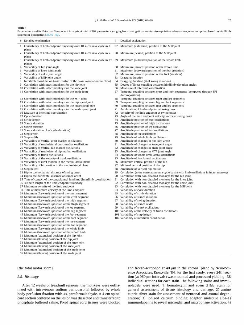
Table 1Parameters used for Principal Component Analysis. A total of 102 parameters, ranging from basic gait parameters to sophisticatedmeasures, were computed based on hindlimblocomotor kinematics [36,40e42].
# Detailed explanation # Detailed explanation
1 Consistency of limb endpoint trajectory over 10 successive cycle in Xplane
57 Maximum (extension) position of the MTP joint
2 Consistency of limb endpoint trajectory over 10 successive cycle in Yplane
58 Minimum (flexion) position of the MTP joint
3 Consistency of limb endpoint trajectory over 10 successive cycle in XYplanes
59 Maximum (outward) position of the whole limb
4 Variability of hip joint angle 60 Minimum (inward) position of the whole limb5 Variability of knee joint angle 61 Maximum (outward) position of the foot (rotation)6 Variability of ankle joint angle 62 Minimum (inward) position of the foot (rotation)7 Variability of MTP joint angle 63 Dragging duration8 Interlimb coordination (max r value of the cross correlation function) 64 Dragging duration (% of swing duration)9 Correlation with intact monkeys for the hip joint 65 Degree of linear coupling between hindlimb elevation angles10 Correlation with intact monkeys for the knee joint 66 Meansure of interlimb coordination11 Correlation with intact monkeys for the ankle joint 67 Temporal coupling between crest and tight segments (computed through FFT
decomposition)12 Correlation with intact monkeys for the MTP joint 68 Temporal coupling between tight and leg segments13 Correlation with intact monkeys for the hip speed joint 69 Temporal coupling between leg and foot segments14 Correlation with intact monkeys for the knee speed joint 70 Temporal coupling between foot and leg segments15 Correlation with intact monkeys for the ankle speed joint 71 Acceleration of limb endpoint at swing onset16 Measure of interlimb coordination 72 Velocity of the limb endpoint at swing onset17 Cycle duration 73 Angle of the limb endpoint velocity vector at swing onset18 Stride length 74 Amplitude position of crest oscillations19 Stance duration 75 Amplitude position of thigh oscillations20 Swing duration 76 Amplitude position of leg oscillations21 Stance duration (% of cycle duration) 77 Amplitude position of foot oscillations22 Step length 78 Amplitude of toe oscillations23 Step width 79 Amplitude of whole limb oscillations24 Variability of vertical crest marker oscillations 80 Amplitude of changes in hip joint angle25 Variability of mediolateral crest marker oscillations 81 Amplitude of changes in knee joint angle26 Variability of vertical hip marker oscillations 82 Amplitude of changes in ankle joint angle27 Variability of mediolateral hip marker oscillations 83 Amplitude of changes in MTP joint angle28 Variability of trunk oscillations 84 Amplitude of whole limb lateral oscillations29 Variability of the velocity of trunk oscillations 85 Amplitude of foot lateral oscillations30 Variability of crest motion in the medio-lateral plane 86 Maximum vertical position of the hip31 Variability of hip motion in the medio-lateral plane 87 Minimal vertical position of the hip32 Step height 88 Amplitude of vertical hip motion33 Hip to toe horizontal distance of swing onset 89 Correlation (cross correlation on a cycle basis) with limb oscillations in intact monkeys34 Hip to toe horizontal distance of stance onset 90 Correlation with non-disabled monkeys for the hip joint35 Time of contact of the contralateral hindlimb (interlimb coordination) 91 Correlation with non-disabled monkeys for the knee joint36 3D path length of the limd endpoint trajectory 92 Correlation with non-disabled monkeys for the ankle joint37 Maximum velocity of the limb endpoint 93 Correlation with non-disabled monkeys for the MTP joint38 Time of maximum velocity of the limb endpoint 94 Variability of cycle duration39 Maximum (forward) position of the crest segment 95 Variability of stride duration40 Minimum (backward) position of the crest segment 96 Variability of stance duration41 Maximum (forward) position of the thigh segment 97 Variability of swing duration42 Minimum (backward) position of the thigh segment 98 Variability of stance width43 Maximum (forward) position of the leg segment 99 Variability of trunk oscillations44 Minimum (backward) position of the leg segment 100 Variability of the velocity of trunk oscillations45 Maximum (forward) position of the foot segment 101 Variability of step height46 Minimum (backward) position of the foot segment 102 Variability of interlimb coordination47 Maximum (forward) position of the toe segment48 Minimum (backward) position of the toe segment49 Maximum (forward) position of the whole limb50 Minimum (backward) position of the whole limb51 Maximum (extension) position of the hip joint52 Minimum (flexion) position of the hip joint53 Maximum (extension) position of the knee joint54 Minimum (flexion) position of the knee joint55 Maximum (extension) position of the ankle joint56 Minimum (flexion) position of the ankle joint
J.R. Slotkin et al. / Biomaterials 123 (2017) 63e76 67
(the total motor score).
2.8. Histology
After 12 weeks of treadmill sessions, the monkeys were eutha-nized with intravenous sodium pentobarbital followed by wholebody perfusion fixation with 4% paraformaldehyde. A 4 cm spinalcord section centered on the lesionwas dissected and transferred tophosphate buffered saline. Fixed spinal cord tissues were blocked
and freeze-sectioned at 40 mm in the coronal plane by NeuroSci-ence Associates, Knoxville, TN. For the first study, every 24th sec-tion (at 960 mm intervals) was mounted and processed yielding ~38individual sections for each stain. The following stains and immu-nolabels were used: 1) hematoxylin and eosin (H&E) stain forgeneral assessment of tissue histology and damage; 2) aminocupric silver stain for assessment of neuronal and axonal degen-eration; 3) ionized calcium binding adaptor molecule (Iba-1)immunolabeling to reveal microglial andmacrophage activation; 4)

Table 2Locomotor Observational Rating Score Category Descriptions. Slight: partial joint movement through less than half of the range of joint motion; Extensive: movement throughmore than half of the range of joint motion; Dorsal Stepping: weight is supported on the dorsal surface of the paw; Plantar Stepping: weight is supported on the plantar surfaceof the paw; FL: forelimb; HL: hindlimb; FL-HL Coordination: for every forelimb step a hindlimb step is taken; NoWeight Support: minimal contraction of extensor muscles ofthe hindlimb, ambulation often characterized by “hopping” with the body weight supported by the contralateral limb; Stride length: distance between two successiveplacements of the same paw.
Category Description
Limb joint movement 0 No voluntary function1 Slight one or two joints movement2 Extensive one or two joints movement3 Extensive movement of all three joints
Limb weight support 0 No weight bearing/dorsal stepping1 Partial weight support/dorsal stepping2 Partial weight support/Mix dorsal and plantar stepping3 Partial weight support/plantar stepping4 Full weight supported/plantar stepping
FL-HL coordination 0 None or occasional FL-HL coordination during gait1 Frequent FL-HL coordination during gait (50e95%)2 Consistent weight supported with FL-HL coordination during gait
Step stability 0 Unstable/wobbly steps and unequal stride length steps1 Frequent stable steps or full range of stride length steps2 Frequent stable and full range of stride length steps3 Full range of stride length with consistent stable steps
Paw position 0 Rotated (internal/external) at initial contact1 Parallel at initial contact and rotated at lift off2 Parallel at initial contact and at lift off
Toe clearance 0 No toe clearance (<50%)1 Frequent toe clearance (50e95%)2 Consistent toe clearance and no foot drag
Run speed 0 Cannot keep up with the treadmill speed1 Keeps up with the treadmill speed with difficult/unstable steps2 Easily keeps up with the treadmill speed with stable steps
Trunk stability 0 Not consistent/unstable1 Consistently stable
Tail position 0 Tail consistently down (over 70%)1 Tail consistently up (over 70%)
J.R. Slotkin et al. / Biomaterials 123 (2017) 63e7668
glial fibrillary acidic protein (GFAP) immunolabeling to reveal as-trocytes (particularly reactive astrocytes) and glial scaring; 5)myelin basic protein (SMI-99) immunolabeling to assess myelinintegrity; and 6) solochrome cyanine stain to assess myelin integ-rity. For the second study, every 12th section (at 480 mm intervals)was mounted and processed yielding ~83 individual sections foreach stain. The following stains and immunolabels were used: 1)H&E stain; 2) thionine nissl stain; 3) Iba-1 immunolabeling; 4)GFAP immunolabeling; 5) non-phosphorylated neurofilament H(SMI-32) immunolabeling to assess the presence of axons; and 6)neuronal growth associated protein (GAP43) immunolabeling toassess the presence of sprouting neurites.
Harvested spinal cords were treated overnight with 20% glyceroland 2% dimethylsulfoxide to prevent freeze-artifacts. The cordsthen were multiply embedded in a gelatin matrix (NeuroScienceAssociates, Knoxville, TN). After curing, the block was rapidly frozenby immersion in isopentane chilled with crushed dry ice andmounted on a freezing stage of an AO 860 sliding microtome. Theblock was sectioned coronally at 40 mm. All sections cut werecollected sequentially into containers filled with Antigen Preservesolution (50% PBS pH 7.0, 50% ethylene glycol, 1% polyvinyl pyrro-lidone). Sections not stained immediately were stored at �20 �C.
For immunochemistry, the sections were stained free-floating.All incubation solutions from the blocking serum onward usedTris buffered saline with Triton X100 as the vehicle; all rinses werewith Tris buffered saline. After hydrogen peroxide treatment andblocking serum, the sections were immunostained with a primaryantibody overnight at room temperature. Vehicle solution con-tained 0.3% TritonX-100 for permeabilization. Following rinses abiotinylated secondary antibody (anti IgG of host animal in whichthe primary antibody was produced) was applied. To visualize thelocation of the binding site of the primary antibody an avidin-
biotin-HRP complex (details in Vectastain elite ABC kit, Vector,Burlingame, CA) was applied. After rinses, the sections were treatedwith diaminobenzidine tetrahydrochloride and hydrogen peroxideto create a visible reaction product and mounted on gelatinized(subbed) glass slides, air dried, dehydrated in alcohols, cleared inxylene, and cover-slipped. The sections stained for Iba-1 werelightly counter-stained with thionine-nissl to improve contrast.
For the H&E stain, sections were mounted on gelatinized (sub-bed) slides, air dried, and carried through the following sequence:95% ethanol, chloroform/ether/absolute ethanol (1:2:1), 95%ethanol, 10% HCl/95% ethanol, 95% ethanol, 70% ethanol, deionizedwater, hematoxylin (Richard Allen), running tap water, 1% HCl,running tap water, 1% ammonium hydroxide, running tap water,70% ethanol, Eosin (Richard Allen), 95% ethanol, 100% ethanol,xylene, and then cover-slipped.
For the Nissl (thionine) stain, sections were mounted ongelatinized (subbed) slides, air dried, and carried through thefollowing sequence: 95% ethanol, chloroform/ether/absoluteethanol (1:2:1), 95% ethanol,10% HCl/95% ethanol, 95% ethanol, 70%ethanol, deionized water, and stained in 0.05% Thionine/0.08Macetate buffer, pH 4.5. The slides then were rinsed with deionizedwater, 70% ethanol, 95% ethanol, and differentiated in 95% alcohol/acetic followed by dehydration in a standard alcohol series, clearedin xylene, and cover-slipped.
Images were analyzed using a Hamamatsu NanoZoomer 2.0-RSscanner system to scan the slides, creating digital images at a res-olution of 40x. Calculation of the size of the regions of sections ofprimate cord was performed using NDP view software (provided byHamamatsu) and a freehand tool to trace each image to getperimeter and area. To determine the vacuolation ratio, digitalimages were analyzed with Visiopharm image analysis software(version 4.3.2.2, Hoersholm, Denmark). The first 5 sections, at
J.R. Slotkin et al. / Biomaterials 123 (2017) 63e76 69
0.96 mm apart, from the rostral and caudal edges of the lesionwereevaluated. Sections were imaged, and the right and left sides weredifferentiated. An intensity threshold was set so that only areaswith a vacuole (excluding the central canal) were quantified (seeFig. 8B for an example). The vacuolation ratio was determined bydividing the area of vacuolation (the number of pixels determinedto be vacuole area) by the total area of the side (total number ofpixels in the cord section on that side).
2.9. Statistics
Values are presented as mean ± standard error of mean (SEM)and differences were considered statistically significant at theP < 0.05 level. Statistical analysis was performed using GraphPadPrism version 5.00 for Windows, GraphPad Software (San DiegoCalifornia USA, www.graphpad.com). For analysis of lesion size andremodeled tissue volume, the lesion or remodeled tissue area oneach slide in the hemisection region was determined. A numericintegration was used to determine lesion volume. Unpaired t-testsand two-way analysis of variance were used to compare differencesbetween groups. For analysis of total motor score as a measure offunctional recovery, scores for each animal were plotted by time(weeks) post-implant. A cumulative functional recovery (area un-der the curve) was calculated for each animal. Unpaired t-testswere used to compare differences in cumulative functional recov-ery between groups. For analysis of kinematics recovery, one-wayanalysis of variance with Tukey's multiple comparison test wasused to compare PC1 scores.
3. Results
3.1. Overall impact of spinal cord injury and implantation of PLGA-PLL scaffolds
The African green monkey hemisection model was used to
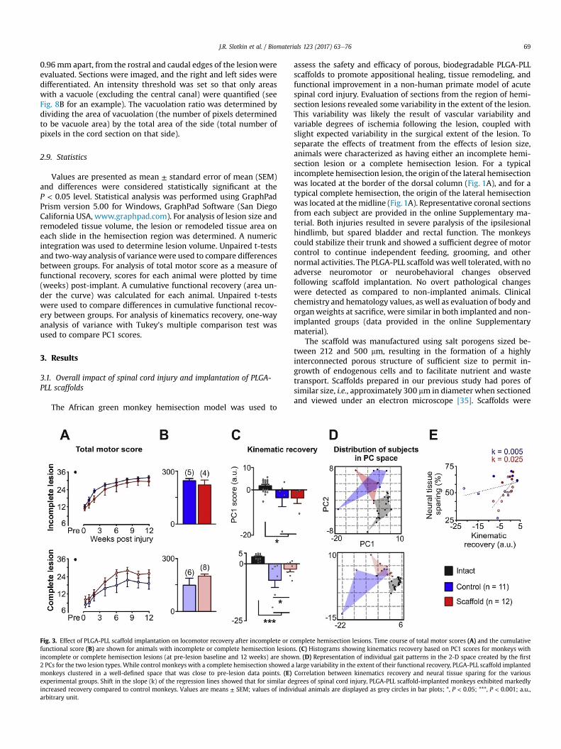
Fig. 3. Effect of PLGA-PLL scaffold implantation on locomotor recovery after incomplete or cfunctional score (B) are shown for animals with incomplete or complete hemisection lesionincomplete or complete hemisection lesions (at pre-lesion baseline and 12 weeks) are show2 PCs for the two lesion types. While control monkeys with a complete hemisection showed amonkeys clustered in a well-defined space that was close to pre-lesion data points. (E)experimental groups. Shift in the slope (k) of the regression lines showed that for similar deincreased recovery compared to control monkeys. Values are means ± SEM; values of indivarbitrary unit.
assess the safety and efficacy of porous, biodegradable PLGA-PLLscaffolds to promote appositional healing, tissue remodeling, andfunctional improvement in a non-human primate model of acutespinal cord injury. Evaluation of sections from the region of hemi-section lesions revealed some variability in the extent of the lesion.This variability was likely the result of vascular variability andvariable degrees of ischemia following the lesion, coupled withslight expected variability in the surgical extent of the lesion. Toseparate the effects of treatment from the effects of lesion size,animals were characterized as having either an incomplete hemi-section lesion or a complete hemisection lesion. For a typicalincomplete hemisection lesion, the origin of the lateral hemisectionwas located at the border of the dorsal column (Fig. 1A), and for atypical complete hemisection, the origin of the lateral hemisectionwas located at themidline (Fig.1A). Representative coronal sectionsfrom each subject are provided in the online Supplementary ma-terial. Both injuries resulted in severe paralysis of the ipsilesionalhindlimb, but spared bladder and rectal function. The monkeyscould stabilize their trunk and showed a sufficient degree of motorcontrol to continue independent feeding, grooming, and othernormal activities. The PLGA-PLL scaffold was well tolerated, with noadverse neuromotor or neurobehavioral changes observedfollowing scaffold implantation. No overt pathological changeswere detected as compared to non-implanted animals. Clinicalchemistry and hematology values, as well as evaluation of body andorganweights at sacrifice, were similar in both implanted and non-implanted groups (data provided in the online Supplementarymaterial).
The scaffold was manufactured using salt porogens sized be-tween 212 and 500 mm, resulting in the formation of a highlyinterconnected porous structure of sufficient size to permit in-growth of endogenous cells and to facilitate nutrient and wastetransport. Scaffolds prepared in our previous study had pores ofsimilar size, i.e., approximately 300 mm in diameter when sectionedand viewed under an electron microscope [35]. Scaffolds were
omplete hemisection lesions. Time course of total motor scores (A) and the cumulatives. (C) Histograms showing kinematics recovery based on PC1 scores for monkeys withn. (D) Representation of individual gait patterns in the 2-D space created by the firstlarge variability in the extent of their functional recovery, PLGA-PLL scaffold implantedCorrelation between kinematics recovery and neural tissue sparing for the variousgrees of spinal cord injury, PLGA-PLL scaffold-implanted monkeys exhibited markedlyidual animals are displayed as grey circles in bar plots; *, P < 0.05; ***, P < 0.001; a.u.,

Fig. 4. Scaffold implantation promotes tissue remodeling after incomplete hemisection lesion. Representative coronal sections from a non-implanted control animal (A-E) and aPLGA-PLL scaffold implanted animal (F-J) with an incomplete hemisection lesion stained with thionine nissl to evaluate cell density (A, F), GFAP to assess the activation of reactiveastrocytosis and glial scaring (B, G), Iba-1 to visualize macrophage and microglia activity (C, H), SMI-32 to reveal the presence of axons (D, I), and GAP-43 to visualize sproutingneurites (E, J) in the remodeled tissue. Scale bar, 2 mm (A-J) and 200 mm (a-j). Boxed regions in (A-J) are shown at higher magnification in (a-j).
J.R. Slotkin et al. / Biomaterials 123 (2017) 63e7670
fabricated using a PLGA-PLL copolymer that is designed to biode-grade by hydrolysis and resorb within 12 weeks after implantation.Consistent with our previous study [35], no evidence of residualPLGA-PLL scaffolds was observed in histological sections throughthe region of the hemisection at 12 weeks after implantation(Figs. 1A and 4e6).
3.2. Functional recovery as a function of lesion size
Animals were allocated into two groups based on the degree ofsparing of the dorsal column and ipsilesional ventral white matter.An evaluator blinded to treatment group characterized the mon-keys as having either an incomplete or a full hemisection lesionbased upon evaluation of coronal sections of the lesion area.Assessment of lesion volume confirmed significant size differences(P¼ 0.02) between incomplete hemisection lesion (34.6 ± 2.3 mm3,n ¼ 9 monkeys) and complete hemisection lesion (56.0 ± 6.6 mm3,
n ¼ 14 monkeys, Fig. 1A).Recovery of locomotionwas quantified using weekly-performed
visual assessments and detailed kinematics analyses conducted atthe end of the recovery period (12 weeks post-lesion). Total motorscores show a gradual recovery of hindlimb locomotion during thefirst 6 weeks post-injury in both groups of monkeys (Fig. 1B). Afterthis initial phase of rapid improvement, the motor scores reached aplateau with limited subsequent amelioration of locomotor ca-pacity. While the time course of recovery was similar for bothgroups of monkeys, cumulative motor scores revealed that mon-keys with incomplete hemisection lesion (237.3 ± 14.8, n ¼ 9monkeys) regained superior locomotor capacities (P ¼ 0.04)compared to monkeys with the complete hemisection lesion(176.4 ± 20.0, n ¼ 14 monkeys), regardless of the presence orabsence of PLGA-PLL scaffolding (Fig. 1C).
Detailed kinematics analyses confirmed these results. We firstcomputed 102 parameters that provided a comprehensive

Fig. 5. Scaffold implantation promotes tissue remodeling after a complete hemisection lesion. Representative coronal sections from both a non-implanted control (A-E) and a PLGA-PLL scaffold implanted (F-J) monkey with complete hemisection lesions are shown using the same convention as in Fig. 4. Scale bar, 2 mm (A-J) and 200 mm (a-j). Boxed regions in(A-J) are shown at higher magnification in (a-j).
J.R. Slotkin et al. / Biomaterials 123 (2017) 63e76 71
quantification of the features underlying the gait pattern for eachmonkey. We next applied a PC analysis on all of the computedparameters and represented gait patterns in the new de-noisedspace created by PC1-3 (Fig. 1D). Each experimental group clus-tered in a specific space. In this kind of representation, the distancebetween data points is proportional to the degree of discrepancybetween gait patterns. Accordingly, gait patterns for monkeys withincomplete hemisection lesion were located closer to pre-lesiondata than those for monkeys with complete hemisection lesion(Fig. 1D). The location of data points along the PC1 axis, whichaccounted for 22% of the total data variance, segregated gait pat-terns from the different types of lesion and time points. Thisquantification of kinematics recovery revealed that animals with anincomplete hemisection lesion (�0.8426 ± 0.5946, n ¼ 9 monkeys)showed a significantly higher recovery extent than animals with acomplete hemisection lesion (�4.563 ± 0.92, n ¼ 14;
F(2,44) ¼ 57.47, P < 0.0001) and were closer to values of intactanimals (2.978 ± 0.155, n ¼ 24 monkeys) (Fig. 1E). The degree ofkinematics recovery significantly correlated with the amount ofneural tissue spared at the injury site (r ¼ �0.5; P < 0.05) (Fig. 1F).
Prior to injury, all tested monkeys exhibited coordinated loco-motion on a treadmill with reciprocal activation between flexor andextensor muscles at the ankle, and highly reproducible trajectory ofthe hindlimb endpoint (Fig. 2A). Oneweek post-lesion, all monkeysdragged the ipsilesional hindlimb along the treadmill beltthroughout the gait cycle (Fig. 2B). Ankle muscles generallyremained quiescent, with no or rare bursts of EMG activity (Fig. 2B).In the chronic state (12 weeks), monkeys with either incomplete orfull hemisection lesions regained plantar stepping movements onthe ipsilesional side with appropriate recruitment of flexor musclesduring swing, and extensor muscles during stance (Fig. 2C). Mon-keys with the more severe complete hemisection, however,

Fig. 6. Epicenter of complete hemisection lesions at 12 weeks after injury. Coronal sections from a control (AeC) and a PLGA-PLL scaffold implanted monkey (DeF) were stainedwith hematoxylin and eosin to assess neuronal and axonal degeneration (A, D), GFAP to evaluate activation of reactive astrocytosis and glial scaring (B, E) and myelin basic protein(MBP) to detect the density of host myelin around axons (C, F). Scale bars, 2 mm (A-F) and 250 mm (c, f). Boxed regions (in C and F) are shown at higher magnification (in c and f).
Fig. 7. Increased volume of remodeled tissue in scaffold implanted monkeys. Histo-gram showing the remodeled tissue volume for non-implanted control and PLGA-PLLscaffold implanted monkeys with incomplete (blue) or complete (red) hemisectionlesions. A two-factor analysis of variance revealed that animals implanted with PLGA-PLL scaffolds had significantly more volume of remodeled tissue than non-implantedcontrol animals. Values are means ± SEM; ***, P < 0.001; the number of animals isindicated above each bar.
J.R. Slotkin et al. / Biomaterials 123 (2017) 63e7672
displayed extensive dragging during the swing phase and increasedvariability in joint angle oscillations as compared to monkeys withan incomplete hemisection lesion.
3.3. Functional recovery in monkeys after PLGA-PLL scaffoldimplantation
Due to the observed differences in the extent of the functionalrecovery based on lesion severity, we segregated the monkeys bylesion type and treatment group for subsequent analyses. Fig. 3shows the time course of the changes in the total motor score(Fig. 3A) and cumulative functional recovery score (Fig. 3B) for non-implanted control and PLGA-PLL scaffold implanted monkeys witheither incomplete or complete hemisection lesions. The kinematicsrecovery based on PC analysis for control and PLGA-PLL scaffoldimplanted monkeys with either incomplete or complete hemi-section is shown in Fig. 3C and D. Monkeys with incomplete
hemisection lesions regained locomotor capacities that were closeto those observed pre-injury: consequently, there were no differ-ences between the non-implanted and scaffold-implanted groupsin cumulative functional recovery (Fig. 3B) and kinematics analyses(Fig. 3C and D) in those animals with an incomplete hemisectionlesion.
In contrast, there was a significant difference in the extent of thekinematics recovery between non-implanted control and PLGA-PLLscaffold implanted monkeys that had a complete hemisection. Dueto the variability between the monkeys and the relatively modestdifferences between groups, the average cumulative functionalrecovery of the PLGA-PLL scaffold implanted group (197.8 ± 16.7,n ¼ 8 monkeys) did not reach statistical significance compared tothe control group (148.3 ± 40.1, n¼ 6 monkeys) (P ¼ 0.23, unpairedt-test; Fig. 3B). More detailed analysis, however, revealed thatscaffold implanted animals with a complete hemisection lesion(�2.39 ± 1.10, n ¼ 8) showed significantly improved kinematicsrecovery compared to non-implanted control animals with acomplete hemisection lesion (�7.29 ± 3.23, n ¼ 6) (F(2, 31) ¼ 21.18,P < 0.001, post hoc: P < 0.05, Fig. 3C). Most of scaffold implantedanimals with the complete hemisection lesion regained gait pat-terns that were similar to those observed in monkeys with the lesssevere incomplete lesion (Fig. 2C). In contrast, non-implantedmonkeys with a complete hemisection lesion showed extensivedragging of the ipsilesional hindlimb associated with a large vari-ability in joint angle oscillations and endpoint trajectory (Fig. 2C).While the extent of the functional recovery was dependent uponthe amount of neural tissue sparing (Fig. 3E), scaffold-implantedmonkeys showed clearly improved functional recovery comparedto non-implanted monkeys with similar tissue damage. Thisdistinct potential for recovery is highlighted by the differencesbetween the slopes of the regression lines associated with scaffold-implanted vs. non-implanted monkeys.
3.4. Histological assessment of PLGA-PPL scaffold implantation
In control monkeys (no implanted scaffold) with incomplete(Fig. 4) and complete hemisection lesions (Fig. 5), a thin,

Fig. 8. Reduced vacuolation in scaffold implanted monkeys with a complete hemisection lesion. Vacuolation typically results from demyelination and axonal degeneration and ismost prominent on the ipsilesional side. (A) Scheme displaying the regions of analysis rostral and caudal to the injury site. (B) Representative coronal sections from a control andPLGA-PLL scaffold implanted animal taken 2 mm rostral to the lesion epicenter in the chronic state of spinal cord injury. Scale bar, 2 mm. (C) Histogram showing the integratedvacuolation ratio relative to the total cord area in regions rostral (C-E) and caudal (F-H) to the injury site. The PLGA-PLL scaffold implanted group showed a decreased vacuolationratio in all analyzed regions, although the differences did not reach statistical significance. Values are means ± SEM.
J.R. Slotkin et al. / Biomaterials 123 (2017) 63e76 73
morphologically distinct layer of remodeled tissue was observed inthe region of the hemisection. This remodeled tissue stained darklywith thionine nissl (Figs. 4 and 5A) as well as hemotxylin and eosin(Fig. 6A). The density of GFAP reactivity was highest at theboundary between the preserved spinal cord tissue and theremodeled tissue layer (Figs. 4, 5 and 6B), but GFAP stainingappeared diffusely within the area of remodeled tissue (Figs. 4b and5b). Similarly, Iba-1 positive cells (macrophage/microglia) wereconcentrated at the interface between the preserved spinal cordtissue and the remodeled tissue layer (Figs. 4C and 5C). SMI-32(Figs. 4D and 5D) and GAP-43 (Figs. 4E and 5E) positive tissuewere observed mostly in the surviving grey matter of the sectionsin the region of the hemisection. SMI-32 strongly labeled the motorneurons of the ventral grey matter, and GAP-43 strongly labeledelements of the superficial dorsal horn. Discrete small spots ofstaining also appeared in the remodeled tissue on these SMI-32 andGAP-43 labeled sections.
In contrast, in PLGA-PLL scaffold implanted monkeys with
incomplete (Fig. 4) or complete hemisection lesions (Fig. 5), a muchlarger layer of morphologically distinct remodeled tissue wasobserved in the region of the hemisection. As observed in non-implanted monkeys, this remodeled tissue stained darkly withthionine nissl (Figs. 4F and 5F) as well as hematoxylin and eosin(Fig. 6D). The area of the darkly stained, remodeled tissue wasmeasured in each section in the region of the hemisection, and avolume was calculated using numerical integration methods.Monkeys implanted with PLGA-PLL scaffolds exhibited a signifi-cant, two-fold larger volume of remodeled tissue (18.6 ± 1.6 mm3,n ¼ 12 monkeys), compared to non-implanted monkeys(9.0 ± 0.9 mm3, n¼ 11 monkeys; P < 0.0001, unpaired t-test, Fig. 7).An increase in the volume of remodeled tissue in monkeysimplanted with PLGA-PLL scaffolds compared to non-implantedcontrols was observed for both severities of the lesion (two-factoranalysis of variance, F(1,19) ¼ 24.32, P < 0.0001). The amount ofremodeled tissue depended upon the lesion size, and animals withcomplete hemisection lesions exhibited significantly more

J.R. Slotkin et al. / Biomaterials 123 (2017) 63e7674
remodeled tissue than animals with incomplete hemisection le-sions (F(1,19) ¼ 6.16, P ¼ 0.02).
Monkeys with implanted scaffolds showed GFAP staining inclose proximity to the boundary between the preserved spinal cordtissue and the remodeled tissue layer (Figs. 4G, 5G and 6E). How-ever, GFAP reactivity was noticeably absent within the inner re-gions of the remodeled tissue layer (Figs. 4g and 5g). Iba-1 positivecells were concentrated at the interface between the preservedspinal cord tissue and the remodeled tissue layer, but they alsowere observed diffusely throughout the remodeled tissue (Figs. 4Hand 5H). No significant differences between PLGA-PLL scaffoldimplanted and non-implanted controls (with full hemisection le-sions) in the total positive area of either reactive astrocytes (GFAP)or macrophage/microglia (Iba-1) were observed in tissue sur-rounding the injury area (data not shown). In PLGA-PLL scaffoldimplanted animals, both SMI-32 positive (Figs. 4I and 5I) and GAP-43 (Figs. 4J and 5J) positive tissue were observed in the survivinggrey matter of the sections in the region of the hemisection. Unlikecontrol sections, however, numerous SMI-32 (Figs. 4i and 5i) andGAP-43 (Figs. 4j and 5j) positive fibers also were observed insidethe remodeled tissue layer. Similarly, in PLGA-PLL scaffoldimplanted animals, numerous myelin basic protein (SMI-99) posi-tive cells were observed inside the remodeled tissue layer (Fig. 6f),in addition to the surviving white matter of the sections in theregion of the hemisection (Fig. 6F).
3.5. Neuroprotection
Spinal cord injury leads to pronounced demyelination andWallerian axonal degeneration. This secondary damage leads to aprominent vacuolar degeneration associated with gliosis in theregions immediately rostral and caudal to the spinal cord injury(Fig. 8B). Using Visiopharm image analysis software, we calculatedthe vacuolation ratio for the ipsilesional and contralesional sides onthe first 5 sections (0.96 mm apart) immediately rostral and caudalto the spinal cord injury for monkeys with complete hemisectionlesions (Fig. 8CeH). On the ipsilesional side, vacuolation wasmaximal at the edge of the region of the hemisection, and pro-gressively decreased in the rostral and caudal directions from thelesion area (Fig. 8E and H). Vacuolation was less prominent on thecontralesional side (Fig. 8D and G). PLGA-PLL implanted groupswith complete hemisection lesion showed a consistently lowerintegrated vacuolation ratio than non-implanted controls (Fig. 8Cand F), but this difference did not reach statistical significance.
4. Discussion
We show that porous PLGA-PLL scaffolds can be safely implan-ted into the injured primate spinal cord, and that these implantsabet tissue remodeling and improve recovery of locomotion in OldWorld African green monkeys with a complete lateral thoracichemisection spinal cord injury. These results may suggest thatPLGA-PLL scaffolds contribute to creating a permissive environ-ment for the survival and growth of axons. The implantation of suchscaffolds may play an important role in the design of future stra-tegies for the treatment of acute spinal cord damage. An ongoinghuman clinical trial investigating a scaffold device substantiallysimilar to the one in this paper is underway (ClinicalTrials.govIdentifier: NCT02138110).
We utilized polymer scaffolds to promote appositional healingand three-dimensional tissue remodeling after an incomplete spi-nal cord injury. Polymer scaffolds promote cell migration andattachment, and enable diffusion of nutrients and materials criticalto cellular function [39]. An ideal polymer scaffold for tissue repairshould: (i) have high interconnected porosity to provide space and
the largest possible volume for fluid flow, waste removal andnutrient exchange; (ii) serve as an adhesive substrate for cells andpromote their survival and growth; and (iii) biodegrade at a rateproportional to new tissue formation [39]. Here, we exploitedPLGA-PLL, which is a block copolymer of Poly (lactic-co-glycolicacid) (PLGA) and Poly(L-lysine). PLGA is a biodegradable andbiocompatible polymer that is approved for human use by the FDAfor a variety of clinical applications such as surgical sutures. PLL is abiopolymer that includes positively charged primary amine func-tional groups at physiological pH designed to improve cell adher-ence [30e32]. Incorporation of positively charged functionalgroups has previously been shown to increase the effectiveness ofother biomaterial scaffolds [9,45]. PLGA and PLL are biocompatibleand biodegradable. As a block copolymer PLGA-PLL can be used tofabricate highly porous scaffolds using a solvent casting porogenleaching manufacturing technique [16]. The PLGA-PLL scaffoldsderive their bioresorptive character from the PLGA component,while the PLL confers charged properties that promote theadsorption of specific extracellular proteins. Positioning the porousscaffold in apposition to (in direct contact with) the spared ele-ments of the spinal cord allows local cell populations to adhere,spread, and migrate along the polymer substrate. PLGA-PLL scaf-folds, therefore, may offer a range of advantages to guide theappositional healing of injured tissue after spinal cord injury, asthey may (i) support the survival and regeneration of neurons andpassing fibers to preserve and restore functional communicationacross the site of injury; (ii) provide a permissive environment forthe survival and proliferation of oligodendrocytes capable of mye-linating sprouting and regenerating axons; and (iii) be seeded withneuronal stem cells or other active substances aimed at promotingneuroregeneration.
We showed that there is a larger volume of tissue in the PLGA-PLL scaffold treated group that is undergoing a process of structuralremodeling after injury than in non-implanted controls. In addi-tion, the bioabsorption of PLGA-PLL scaffolds coincided with theappearance of remodeled tissue. We showed that PLGA-PLL scaf-folds were resorbed completely at 12 weeks after the spinal cordinjury, and that the area where scaffolds had been placed wascharacterized by the presence of remodeled tissue organized into amatrix. This remodeled tissue presented a well-defined boundary,appeared fibrous in nature with concentric organization, and wasnot infiltrative. SMI-32 immunolabeling revealed the presence ofnon-phosphorylated neurofilament H, which is a main componentof the neuronal cytoskeleton. GAP-43 immunolabeling indicatesthe presence of sprouting neurites. MBP immunolabeling revealedthe presence of myelin producing cells such as oligodendrocytes.Not only did the remodeled tissue appear to fill the cystic cavityformed by the hemisection spinal cord injury, but the presence ofSMI-32 and GAP-43 positive fibers and MBP positive tissue indi-cated that this remodeled tissue could potentially support neuralsprouting as well as myelin-producing cells such as oligodendro-cytes. We did not evaluate whether the remodeled tissue wasnewly formed or preserved tissue. Therefore, the observedincreased volume of remodeled tissue may have been promoted bycells that migrated from the vascular or local periphery, by localpreserved CNS cells, or by both.
The ability of PLGA-PLL scaffolds to support growth permissivetissue remodeling after spinal cord injury may be due to the pres-ence of positively charged lysine residues. Positively charged sur-faces have been shown to promote the adhesion of primaryneuronal cell types as well as neuroblastoma cells in culture[46e51]. Poly-lysine modification also has been shown to supportincreased growth of neurofilament positive neural cortical cellsgrowing on PLGA films [52]. Hydrogels with positive surfacecharges have been shown to support connective tissue deposition

J.R. Slotkin et al. / Biomaterials 123 (2017) 63e76 75
[25] and axonal regeneration [9,25] following implantation intosites of spinal cord injury in rats.
We used both visual scoring systems and detailed kinematicsanalyses to evaluate the recovery of locomotion on a treadmill inthe non-human primates with spinal cord injury. Importantly, wefound a correlation between lesion size and functional recovery.Monkeys with incomplete hemisections, in which portions of thedorsal and ventral columns were intact, exhibited greater recoverythan monkeys with complete hemisections. One limitation of thisstudy is that this result prompted segregation of the monkeys intotwo groups based on lesion size. We found that while visual scoresdid not reach significance, PC analysis applied on a large number ofkinematics variables revealed that among the monkeys with acomplete lateral hemisection, those treated with PLGA-PLL scaf-folds had significantly greater locomotor recovery than non-implanted controls. PLGA-PLL scaffold mediated ameliorations ofgait were modest, and only manifested for larger lesions. In thepresent study, we utilized an incomplete spinal cord injury (lateralhemisection) after which there is significant recovery that occursspontaneously [53]. The degree of recovery significantly correlatedwith the amount of tissue sparing in both treated and non-treatedmonkeys. Considering the rapid and extensive recovery of basiclocomotor capacities in all animals that occurred spontaneously,the potential magnitude of the improvement that could be attrib-uted to the scaffolding was limited in the present study. In pri-mates, unlike in rats, recovery of supraspinal control of movementafter a lateral hemisection has been linked to extensive collateralsprouting of spinal cord midline-crossing corticospinal axons [42].In this injury model recovery of nearly 60% of pre-lesion axondensity in segments caudal to the lesion was observed [53].
5. Conclusions
We implanted PLGA-PLL scaffolds in the injured primate spinalcord to create an environment permissive for appositional healingand tissue remodeling, and showed that this strategy promotedmodest, yet significant improvement of locomotion in non-humanprimates with a complete lateral hemisection. One could speculate,therefore, that the tissue remodeling effect observed in the presentstudymay have enhanced recovery of motor function by facilitatingnew detour circuits through further midline crossing with scaf-folding within the surviving tissue post-lesion. These results sug-gest that implantation of polymer scaffolds, including the porousPLGA-PLL scaffold used in the present study, may abet tissueremodeling and improve recovery of function after an acute spinalcord injury, and may play an important role in the future design ofcombinatorial spinal cord injury treatment strategies [18,53e55].
Acknowledgment
This research was financially supported by InVivo Therapeutics.The authors thank Haining Dai for his excellent technical assistance.
Appendix A. Supplementary data
Supplementary data related to this article can be found at http://dx.doi.org/10.1016/j.biomaterials.2017.01.024.
References
[1] M. Fitzharris, R.A. Cripps, B.B. Lee, Estimating the global incidence of traumaticspinal cord injury, Spinal Cord. 52 (2014) 117e122.
[2] H.M. Bramlett, W.D. Dietrich, Progressive damage after brain and spinal cordinjury: pathomechanisms and treatment strategies, Prog. Brain Res. 161(2007) 125e141.
[3] D.C. Baptiste, M.G. Fehlings, Update on the treatment of spinal cord injury,
Prog. Brain Res. 161 (2007) 217e233.[4] M.S. Beattie, A.A. Farooqui, J.C. Bresnahan, Review of current evidence for
apoptosis after spinal cord injury, J. Neurotrauma 17 (2000) 915e925.[5] D.J. Donnelly, P.G. Popovich, Inflammation and its role in neuroprotection,
axonal regeneration and functional recovery after spinal cord injury, Exp.Neurol. 209 (2008) 378e388.
[6] J.C. Fleming, M.D. Norenberg, D.A. Ramsay, G.A. Dekaban, A.E. Marcillo,A.D. Saenz, et al., The cellular inflammatory response in human spinal cordsafter injury, Brain 129 (2006) 3249e3269.
[7] M.E. Schwab, Repairing the injured spinal cord, Science 295 (2002)1029e1031.
[8] S. Thuret, L.D. Moon, F.H. Gage, Therapeutic interventions after spinal cordinjury, Nat. Rev. Neurosci. 7 (2006) 628e643.
[9] B.K. Chen, A.M. Knight, N.N. Madigan, L. Gross, M. Dadsetan, J.J. Nesbitt, et al.,Comparison of polymer scaffolds in rat spinal cord: a step toward quantitativeassessment of combinatorial approaches to spinal cord repair, Biomaterials 32(2011) 8077e8086.
[10] H. Nomura, C.H. Tator, M.S. Shoichet, Bioengineered strategies for spinal cordrepair, J. Neurotrauma 23 (2006) 496e507.
[11] H. Kim, M.J. Cooke, M.S. Shoichet, Creating permissive microenvironments forstem cell transplantation into the central nervous system, Trends Biotechnol.30 (2012) 55e63.
[12] B.K. Chen, A.M. Knight, G.C. de Ruiter, R.J. Spinner, M.J. Yaszemski, B.L. Currier,et al., Axon regeneration through scaffold into distal spinal cord after tran-section, J. Neurotrauma 26 (2009) 1759e1771.
[13] M. Oudega, S.E. Gautier, P. Chapon, M. Fragoso, M.L. Bates, J.M. Parel, et al.,Axonal regeneration into Schwann cell grafts within resorbable poly(alpha-hydroxyacid) guidance channels in the adult rat spinal cord, Biomaterials 22(2001) 1125e1136.
[14] P.J. Johnson, A. Tatara, D.A. McCreedy, A. Shiu, S.E. Sakiyama-Elbert, Tissue-engineered fibrin scaffolds containing neural progenitors enhance functionalrecovery in a subacute model of SCI, Soft Matter 6 (2010) 5127e5137.
[15] H. Kim, T. Zahir, C.H. Tator, M.S. Shoichet, Effects of dibutyryl cyclic-AMP onsurvival and neuronal differentiation of neural stem/progenitor cells trans-planted into spinal cord injured rats, PLoS One 6 (2011) e21744.
[16] Y.D. Teng, E.B. Lavik, X. Qu, K.I. Park, J. Ourednik, D. Zurakowski, et al.,Functional recovery following traumatic spinal cord injury mediated by aunique polymer scaffold seeded with neural stem cells, Proc. Natl. Acad. Sci. U.S. A 99 (2002) 3024e3029.
[17] M.P. Vacanti, J.L. Leonard, B. Dore, L.J. Bonassar, Y. Cao, S.J. Stachelek, et al.,Tissue-engineered spinal cord, Transpl. Proc. 33 (2001) 592e598.
[18] P. Lu, Y. Wang, L. Graham, K. McHale, M. Gao, D. Wu, et al., Long-distancegrowth and connectivity of neural stem cells after severe spinal cord injury,Cell 150 (2012) 1264e1273.
[19] J.H. Brock, E.S. Rosenzweig, A. Blesch, R. Moseanko, L.A. Havton, V.R. Edgerton,et al., Local and remote growth factor effects after primate spinal cord injury,J. Neurosci. 30 (2010) 9728e9737.
[20] M. Gao, P. Lu, B. Bednark, D. Lynam, J.M. Conner, J. Sakamoto, et al., Templatedagarose scaffolds for the support of motor axon regeneration into sites ofcomplete spinal cord transection, Biomaterials 34 (2013) 1529e1536.
[21] T. Gros, J.S. Sakamoto, A. Blesch, L.A. Havton, M.H. Tuszynski, Regeneration oflong-tract axons through sites of spinal cord injury using templated agarosescaffolds, Biomaterials 31 (2010) 6719e6729.
[22] L. Schnell, A.S. Hunanyan, W.J. Bowers, P.J. Horner, H.J. Federoff, M. Gullo, etal., Combined delivery of Nogo-A antibody, neurotrophin-3 and the NMDA-NR2d subunit establishes a functional 'detour' in the hemisected spinalcord, Eur. J. Neurosci. 34 (2011) 1256e1267.
[23] F. Gelain, S. Panseri, S. Antonini, C. Cunha, M. Donega, J. Lowery, et al.,Transplantation of nanostructured composite scaffolds results in the regen-eration of chronically injured spinal cords, ACS Nano 5 (2011) 227e236.
[24] L. He, Y. Zhang, C. Zeng, M. Ngiam, S. Liao, D. Quan, et al., Manufacture of PLGAmultiple-channel conduits with precise hierarchical pore architectures andin vitro/vivo evaluation for spinal cord injury, Tissue Eng. Part C Methods 15(2009) 243e255.
[25] A. Hejcl, P. Lesny, M. Pradny, J. Sedy, J. Zamecnik, P. Jendelova, et al., Macro-porous hydrogels based on 2-hydroxyethyl methacrylate. Part 6: 3D hydro-gels with positive and negative surface charges and polyelectrolyte complexesin spinal cord injury repair, J. Mater Sci. Mater Med. 20 (2009) 1571e1577.
[26] K. Kataoka, Y. Suzuki, M. Kitada, T. Hashimoto, H. Chou, H. Bai, et al., Alginateenhances elongation of early regenerating axons in spinal cord of young rats,Tissue Eng. 10 (2004) 493e504.
[27] H. Kim, C.H. Tator, M.S. Shoichet, Chitosan implants in the rat spinal cord:biocompatibility and biodegradation, J. Biomed. Mater Res. A 97 (2011)395e404.
[28] V.M. Tysseling-Mattiace, V. Sahni, K.L. Niece, D. Birch, C. Czeisler,M.G. Fehlings, et al., Self-assembling nanofibers inhibit glial scar formationand promote axon elongation after spinal cord injury, J. Neurosci. 28 (2008)3814e3823.
[29] S. Woerly, P. Petrov, E. Sykova, T. Roitbak, Z. Simonova, A.R. Harvey, Neuraltissue formation within porous hydrogels implanted in brain and spinal cordlesions: ultrastructural, immunohistochemical, and diffusion studies, TissueEng. 5 (1999) 467e488.
[30] D. Barrera, E. Zylstra, P. Lansbury, R. Langer, Copolymerization and degrada-tion of poly(lactic acid-co-lysine), Macromolecules 28 (1995) 425e432.
[31] J. Hrkach, J. Ou, N. Lotan, R. Langer, Synthesis of poly(L-lactic acid-co-lysine)

J.R. Slotkin et al. / Biomaterials 123 (2017) 63e7676
graft copolymers, Macromolecules 28 (1995) 4736e4739.[32] E.B. Lavik, J.S. Hrkach, N. Lotan, R. Nazarov, R. Langer, A simple synthetic route
to the formation of a block copolymer of poly(lactic-co-glycolic acid) andpolylysine for the fabrication of functionalized, degradable structures forbiomedical applications, J. Biomed. Mater Res. 58 (2001) 291e294.
[33] A.R. Blight, Animal models of spinal cord injury, Top. Spinal Cord. Inj. Rehabil.6 (2000) 1e13.
[34] G. Courtine, M.B. Bunge, J.W. Fawcett, R.G. Grossman, J.H. Kaas, R. Lemon, etal., Can experiments in nonhuman primates expedite the translation oftreatments for spinal cord injury in humans? Nat. Med. 13 (2007) 561e566.
[35] C.D. Pritchard, J.R. Slotkin, D. Yu, H. Dai, M.S. Lawrence, R.T. Bronson, et al.,Establishing a model spinal cord injury in the African green monkey for thepreclinical evaluation of biodegradable polymer scaffolds seeded with humanneural stem cells, J. Neurosci. Methods 188 (2010) 258e269.
[36] G. Courtine, R.R. Roy, J. Raven, J. Hodgson, H. McKay, H. Yang, et al., Perfor-mance of locomotion and foot grasping following a unilateral thoracic corti-cospinal tract lesion in monkeys (Macaca mulatta), Brain 128 (2005)2338e2358.
[37] R. Suresh Babu, R. Muthusamy, A. Namasivayam, Behavioural assessment offunctional recovery after spinal cord hemisection in the bonnet monkey(Macaca radiata), J. Neurol. Sci. 178 (2000) 136e152.
[38] E. Lavik, Y.D. Teng, E. Snyder, R. Langer, Seeding neural stem cells on scaffoldsof PGA, PLA, and their copolymers, Methods Mol. Biol. 198 (2002) 89e97.
[39] A.G. Mikos, G. Sarakinos, S.M. Leite, J.P. Vacanti, R. Langer, Laminated three-dimensional biodegradable foams for use in tissue engineering, Biomaterials14 (1993) 323e330.
[40] G. Courtine, R.R. Roy, J. Hodgson, H. McKay, J. Raven, H. Zhong, et al., Kine-matic and EMG determinants in quadrupedal locomotion of a non-humanprimate (Rhesus), J. Neurophysiol. 93 (2005) 3127e3145.
[41] N. Dominici, U. Keller, H. Vallery, L. Friedli, R. van den Brand, M.L. Starkey, etal., Versatile robotic interface to evaluate, enable and train locomotion andbalance after neuromotor disorders, Nat. Med. 18 (2012) 1142e1147.
[42] L. Friedli, E.S. Rosenzweig, Q. Barraud, M. Schubert, N. Dominici, L. Awai, et al.,Pronounced species divergence in corticospinal tract reorganization andfunctional recovery after lateralized spinal cord injury favors primates, Sci.Transl. Med. 7 (2015), 302ra134.
[43] R. van den Brand, J. Heutschi, Q. Barraud, J. DiGiovanna, K. Bartholdi,M. Huerlimann, et al., Restoring voluntary control of locomotion after para-lyzing spinal cord injury, Science 336 (2012) 1182e1185.
[44] D.M. Basso, M.S. Beattie, J.C. Bresnahan, A sensitive and reliable locomotorrating scale for open field testing in rats, J. Neurotrauma 12 (1995) 1e21.
[45] P. Lesny, M. Pradny, P. Jendelova, J. Michalek, J. Vacik, E. Sykova, Macroporoushydrogels based on 2-hydroxyethyl methacrylate. Part 4: growth of rat bonemarrow stromal cells in three-dimensional hydrogels with positive andnegative surface charges and in polyelectrolyte complexes, J. Mater Sci. MaterMed. 17 (2006) 829e833.
[46] A. Soekarno, B. Lom, P.E. Hockberger, Pathfinding by neuroblastoma cells inculture is directed by preferential adhesion to positively charged surfaces,Neuroimage 1 (1993) 129e144.
[47] Y. Bledi, A.J. Domb, M. Linial, Culturing neuronal cells on surfaces coated by anovel polyethyleneimine-based polymer, Brain Res. Brain Res. Protoc. 5(2000) 282e289.
[48] D.W. Branch, B.C. Wheeler, G.J. Brewer, D.E. Leckband, Long-term mainte-nance of patterns of hippocampal pyramidal cells on substrates of poly-ethylene glycol and microstamped polylysine, IEEE Trans. Biomed. Eng. 47(2000) 290e300.
[49] S. Lakard, G. Herlem, A. Propper, A. Kastner, G. Michel, N. Valles-Villarreal, etal., Adhesion and proliferation of cells on new polymers modified bio-materials, Bioelectrochemistry 62 (2004) 19e27.
[50] E. Martin-Lopez, M. Nieto-Diaz, M. Nieto-Sampedro, Differential adhesivenessand neurite-promoting activity for neural cells of chitosan, gelatin, and poly-L-lysine films, J. Biomater. Appl. 26 (2012) 791e809.
[51] A.A. Oliva Jr., C.D. James, C.E. Kingman, H.G. Craighead, G.A. Banker, Patterningaxonal guidance molecules using a novel strategy for microcontact printing,Neurochem. Res. 28 (2003) 1639e1648.
[52] D.W. Han, M. Sub Lee, B.J. Park, J.K. Kim, J.C. Park, Enhanced neurite outgrowthof rat neural cortical cells on surface-modified films of poly(lactic-co-glycolicacid), Biotechnol. Lett. 27 (2005) 53e58.
[53] E.S. Rosenzweig, G. Courtine, D.L. Jindrich, J.H. Brock, A.R. Ferguson,S.C. Strand, et al., Extensive spontaneous plasticity of corticospinal projectionsafter primate spinal cord injury, Nat. Neurosci. 13 (2010) 1505e1510.
[54] M.S. Beattie, J.C. Bresnahan, J. Komon, C.A. Tovar, M. Van Meter, D.K. Anderson,et al., Endogenous repair after spinal cord contusion injuries in the rat, Exp.Neurol. 148 (1997) 453e463.
[55] N. Weidner, A. Ner, N. Salimi, M.H. Tuszynski, Spontaneous corticospinalaxonal plasticity and functional recovery after adult central nervous systeminjury, Proc. Natl. Acad. Sci. U. S. A 98 (2001) 3513e3518.