Embed Size (px)
Citation preview

7 WHITE PHOSPHORUS
2. HEALTH EFFECTS
2.1 INTRODUCTION
The primary purpose of this chapter is to provide public health officials, physicians, toxicologists, and
other interested individuals and groups with an overall perspective on the toxicology of white phosphorus
and white phosphorus smoke. It contains descriptions and evaluations of toxicological studies and
epidemiological investigations and provides conclusions, where possible, on the relevance of toxicity and
toxicokinetic data to public health.
There are three allotropic forms of elemental phosphorus: white, red, and black phosphorus. At room
temperature, pure white phosphorus is a tetrahedral crystal with a molecular formula of P4. In the pure
form, white phosphorus is an ivory-colored, waxy solid. The commercial product is 99.9% pure and may
have a slightly yellow color. In the literature, the commercial product is often referred to as yellow
phosphorus. In this chapter, the terms white phosphorus and phosphorus are used to refer to P4, which
includes white and yellow phosphorus.
White phosphorus is the most active allotropic form and is extremely toxic when inhaled, ingested, or
absorbed through burned areas (Eldad and Simon 1991). It is fat soluble, glows in yellow-green light, and
ignites spontaneously upon drying and exposure to air. Storage of white phosphorus in water prevents it
from burning spontaneously (Eldad and Simon 1991). White phosphorus can cause thermal injury and
hygroscopic damage by absorbing water from surrounding tissues. It reacts with oxygen and water to
form strong acids (H3PO2, H3PO3) and combines with metals like copper to form dark-colored inactive
salts (Eldad and Simon 1991).
White phosphorus particles can burn on the surface of the skin or penetrate deep into the tissues when
carried on shrapnel particles. Local destruction of tissues continues as long as white phosphorus is
exposed to oxygen. White phosphorus smoke with a garlic odor is characteristic of white phosphorus
burns (Eldad and Simon 1991). High mortality rates seen following white phosphorus burns can be due to
its absorption from the burned surface, which may result in multi-organ failure (mainly liver and kidneys),
hyperphosphatemia, hypocalcemia, and electrocardiogram (ECG) abnormalities (ST depression, QT
elongation, microvoltage of QRS and bradycardia) (Bowen et al. 1971; Eldad and Simon 1991). Copper
8 WHITE PHOSPHORUS
2. HEALTH EFFECTS
sulphate is a very effective in vitro neutralizer of white phosphorus and has been used to treat white
phosphorus burns (Eldad and Simon 1991). However, it is extremely toxic as copper can be absorbed
from the burn injury or wound after topical application of copper sulphate to the burnt surface (Bowen et
al. 1971; Summerlin et al. 1967). Acute copper intoxication is characterized by hemolytic anemia with
intravascular hemolysis, hematuria, proteinuria, glycosuria, oliguria, uremia, tachycardia, hypotension,
abnormal liver functions, and jaundice (Summerlin et al. 1967). The hemolytic anemia seen following
copper intoxication is a common cause of death. Only tap water irrigations were found to be effective in
preventing death after white phosphorus burns (Eldad and Simon 1991).
The garlic-like odor is also detected in the vomitus which is phosphorescent and visible when examined in
a dark room. If phosphorus is absorbed as the gas phosphine (PH3), death can occur rapidly due to cardiac
collapse (Blanke 1970). In most cases, white phosphorus is ingested accidentally or when trying to
commit suicide. Following absorption, white phosphorus stays in the blood for several days and is slowly
oxidized to hypophosphoms and phosphorous acids (Blanke 1970). If death occurs within l-3 days, no
significant changes are seen. However, in patients who survive for more than a week, the effect of
phosphorus damage is evident by the extreme fatty changes seen on many organs; alterations in both fat
and protein metabolism, a yellowish liver with marked fatty degeneration, and severe jaundice are usually
present (Blanke 1970).
White phosphorus has been used in the manufacture of rat and cockroach poisons, pesticides, matchheads,
firecrackers, and ammunitions in the military. However, other chemicals such as sulfur have replaced
phosphorus in matchheads. Phosphorus is also used as a fumigant in the storage of grain in the form of
aluminum phosphide pellets. Due to ease of application, pellets of aluminum or magnesium phosphide are
commonly used (Garry et al. 1989). Phosphine, a highly toxic gas, is generated from phosphide. The rate
of formation of phosphine (permissible exposure limit [PEL], 0.4 mg/m3) is dependent on the ambient
temperature and humidity. In the presence of water (humidity) or acid, the formation of phosphine is
greatly enhanced at any given temperature. Phosphine is released rapidly, and it is extremely-fatal to the
unprotected worker/person (Garry et al. 1989). An accidental death of a pregnant woman was related to
phosphine exposure from stored grain that had been fumigated with aluminum phosphide (AlP3) pellets
(Garry et al. 1993). Phosphine can also be generated when phosphorus is used as a dopant in the
microchip processing, where a small amount of phosphorus is added to another substance such as a
semiconductor to alter its properties (Garry et al. 1989).

9 WHITE PHOSPHORUS
2. HEALTH EFFECTS
White phosphorus smoke is generated by burning white phosphorus. The U.S. Army uses white
phosphorus smoke as a smoke/obscurant for training and testing activities. The smoke generated from
burning white phosphorus consists primarily of oxidation and hydrolysis products of phosphorus,
including phosphorus pentoxide and phosphorus trioxide. The moisture in the air reacts with these
phosphorus oxides to produce a dynamic mixture of polyphosphoric acids that eventually transform into
orthophosphoric acid, pyrophosphoric acid, and orthophosphorus acid. Wind-tunnel tests in which white
phosphorus was burned and oxygen was non-limiting produced an average aerosol mass concentration
between 2,500 and 3,000 mg/m3, with the major components being polyphosphates, phosphine, and
elemental phosphorus (Van Voris et al. 1987). It should be stressed that while residual-coated white
phosphorus is very biologically toxic, there are somewhat stable combustion intermediates (linear and
cyclic polyphosphates) that can be persistent under low oxygen conditions and may be toxic to biological
organisms.
A glossary and list of acronyms, abbreviations, and symbols can be found at the end of this profile.
2.2 DISCUSSION OF HEALTH EFFECTS BY ROUTE OF EXPOSURE
To help public health professionals and others address the needs of persons living or working near
hazardous waste sites, the information in this section is organized first by route of exposure - inhalation,
oral, and dermal; and then by health effect - death, systemic, immunological, neurological, reproductive,
developmental, genotoxic, and carcinogenic effects. These data are discussed in terms of three exposure
periods - acute (14 days or less), intermediate (15-364 days), and chronic (365 days or more).
Levels of significant exposure for each route and duration are presented in tables and illustrated in figures.
The points in the figures showing no-observed-adverse-effect levels (NOAELs) or lowest-observedadverse
effect levels (LOAELs) reflect the actual doses (levels of exposure) used in the studies. LOAELs
have been classified into “less serious” or “serious” effects. “Serious” effects are those that evoke failure
in a biological system and can lead to morbidity or mortality (e.g., acute respiratory distress or death).
“Less serious” effects are those that are not expected to cause significant dysfunction or death, or those
whose significance to the organism is not entirely clear. ATSDR acknowledges that a considerable
amount of judgment may be required in establishing whether an end point should be classified as a
NOAEL, “less serious” LOAEL, or “serious” LOAEL, and that in some cases, there will be insufficient
data to decide whether the effect is indicative of significant dysfunction. However, the Agency has

WHITE PHOSPHORUS 1 0
2. HEALTH EFFECTS
established guidelines and policies that are used to classify these end points. ATSDR believes that there is
sufficient merit in this approach to warrant an attempt at distinguishing between “less serious” and
“serious” effects. The distinction between “less serious” effects and “serious” effects is considered to be
important because it helps the users of the profiles to identify levels of exposure at which major health
effects start to appear. LOAELs or NOAELs should also help in determining whether or not the effects
vary with dose and/or duration, and place into perspective the possible significance of these effects to
human health.
The significance of the exposure levels shown in the Levels of Significant Exposure (LSE) tables and
figures may differ depending on the user’s perspective. Public health officials and others concerned with
appropriate actions to take at hazardous waste sites may want information on levels of exposure associated
with more subtle effects in humans or animals or exposure levels below which no adverse effects have
been observed. Estimates of levels posing minimal risk to humans (Minimal Risk Levels or MRLs) may
be of interest to health professionals and citizens alike.
Estimates of exposure levels posing minimal risk to humans (Minimal Risk Levels or MRLs) have been
made for white phosphorus. An MRL is defined as an estimate of daily human exposure to a substance
that is likely to be without an appreciable risk of adverse effects (noncarcinogenic) over a specified
duration of exposure. MRLs are derived when reliable and sufficient data exist to identify the target
organ(s) of effect or the most sensitive health effect(s) for a specific duration within a given route of
exposure. MRLs are based on noncancerous health effects only and do not consider carcinogenic effects.
MRLs can be derived for acute, intermediate, and chronic duration exposures for inhalation and oral
routes. Appropriate methodology does not exist to develop MRLs for dermal exposure.
Although methods have been established to derive these levels (Barnes and Dourson 1988; EPA 1990),
uncertainties are associated with these techniques. Furthermore, ATSDR acknowledges additional
uncertainties inherent in the application of the procedures to derive less than lifetime MRLs. As an
example, acute inhalation MRLs may not be protective for health effects that are delayed in development
or are acquired following repeated acute insults, such as hypersensitivity reactions, asthma, or chronic
bronchitis. As these kinds of health effects data become available and methods to assess levels of
significant human exposure improve, these MRLs will be revised.

11 WHITE PHOSPHORUS
2. HEALTH EFFECTS
A User’s Guide has been provided at the end of this profile (see Appendix A). This guide should aid in the
interpretation of the tables and figures for Levels of Significant Exposure and the MRLs.
2.2.1 Inhalation Exposure
White Phosphorus. Studies of human exposure to white phosphorus are limited to those examining
occupational exposure to white phosphorus for intermediate or chronic durations. Most of the information
on the effects of occupational exposure of humans to white phosphorus was from case reports, rather than
epidemiology studies. Phosphorus oxidizes rapidly when exposed to air, and fumes/vapors in phosphorus
factories probably contained phosphorus, phosphoric oxide, and phosphorus oxide (Heimann 1946;
Hughes et al. 1962; Ward 1928). Exposure levels for white phosphorus and phosphorus compounds were
not reported in the occupational studies. However, workers at phosphorus plants were probably also
exposed to white phosphorus by the dermal and oral routes.
One study was located regarding toxicity in animals after inhalation exposure to white phosphorus
(Ruzuddinov and Rys-Uly 1986). Rats were exposed for an intermediate duration to the atmosphere in a
phosphorus plant, reported to contain white phosphorus and its inorganic compounds, and changes in the
oral mucosa were examined. However, exposure levels for white phosphorus and phosphorus compounds
were not reported in the study.
White Phosphorus Smoke. Several studies have examined the toxicity of white phosphorus smoke in
humans and animals (White and Armstrong et al. 1935; Brown et al. 1980, 1981; Starke et al. 1982;
Walker et al. 1947). The Walker et al. (1947) study is a report of the health effects observed in workers
exposed to white phosphorus smoke during a factory fire. The other studies involve experimental
exposures. In these studies, the smoke was generated by burning either the felt that contained white
phosphorus or white phosphorus alone. White phosphorus-felt (WP/F) smoke was generated by forcing
military-grade white phosphorus under pressure into thick pieces of wool felt. This material was then cut
into cubes of specific weights. A cube of the white phosphorus felt on an aluminum-foil pan was placed
on an unlit electric hot plate within a chamber. The hot plate was a fast-heating unit capable of reaching
temperatures in excess of 700°F (Brown et al. 1980). The smoke was pumped into the exposure chamber
or the white phosphorus/felt was burned in the exposure chamber. In some experiments, the subjects were
placed in the exposure chamber prior to burning the white phosphorus, and in other experiments the test
atmosphere was generated prior to placing the subjects in the exposure chamber. In some of these studies,

12 WHITE PHOSPHORUS
2. HEALTH EFFECTS
subjects were not removed until the smoke had dissipated. Thus, the subjects had the potential to be
exposed to a wide range of concentrations of white phosphorus smoke. The temperature in the exposure
chambers was higher than room temperature. Another limitation of these inhalation studies is the
reporting of air concentrations. Typically air samples were collected on filters, diluted with distilled water,
and boiled to convert the phosphorus acids to orthophosphoric acids. The acid content was then
determined by titration, and the normality of the acid was then converted to orthophosphoric acid or
phosphorus pentoxide equivalents (Brown et al. 1980).
In the Brown et al. (1980, 1981) and Starke et al. (1982) studies, the air concentrations of white
phosphorus smoke were expressed in terms of orthophosphoric acid equivalents. In reviewing the
methods used to estimate the concentration of orthophosphoric acid, ATSDR detected a calculation
mistake. According to the authors’ equation for determining the orthophosphoric acid concentration of the
sample, the molecular weight was divided by 3 milliequivalents. At pH 9.6, the molecular weight should
be divided by 2 milliequivalents. A correction was made to exposure levels for the three studies. White
and Armstrong (1935) reported the white phosphorus smoke concentration in terms of phosphorus
pentoxide equivalents. In Section 2.2, the air concentrations for the White and Armstrong (1935) studies
are also expressed in terms of orthophosphoric acid equivalents. Differences in the exposure protocol
between the studies conducted by Brown et al. (1980, 198l), Starke et al. (1982), and White and
Armstrong (1935) make it difficult to make comparisons across studies. In White and Armstrong (1935),
the continuous-flow method was used, allowing the desired concentrations to be set up and maintained
under conditions which avoided exposure of the experimental animals to either excessive temperature rise,
oxygen deprivation, or other vitiation of the atmospheres breathed. Exposure duration was for 1 hour. In
Brown et al. (1980,1981), exposure times and concentration levels varied. Exposure durations ranged
from 5-90 minutes to 13 weeks, and target concentrations varied (200,500, or 1,000 mg/m3). In Starke et
al. (1982), rats were exposed to white phosphorus smoke or control air 15 minutes/day, 5 days/week, for
10 consecutive weeks at concentrations of 0,500, and 1,000 mg/m3.
2.2.1.1 Death
White Phosphorus. Two white phosphorus-related deaths were reported in a study of workers from three
plants involved in the production of fireworks (Ward 1928). Both workers were females exposed to white
phosphorus during the molding and wrapping of a paste containing 44% phosphorus. This step in the
production of fireworks involved continuous inhalation exposure to airborne white phosphorus, dermal

13 WHITE PHOSPHORUS
2. HEALTH EFFECTS
exposure to the paste, and likely ingestion of airborne white phosphorus and white phosphorus passed
from hand to mouth. Apparently, the phosphorus fumes/vapors contained white phosphorus and other
phosphorus compounds, including phosphoric oxide and phosphorus oxide. However, exposure levels
were not reported. Both women developed phossy jaw, a degenerative condition affecting the soft tissue,
bones, and teeth of the oral cavity, after chronic exposure to the atmosphere at the factory. The cause of
death in both cases was listed as septicemia, with abscess of a tooth and necrosis of the jaw listed as
contributory causes. Thus, death in both cases resulted from infections, probably secondary to the
degenerative effects of white phosphorus on the oral cavity (Ward 1928). It is likely that the development
of phossy jaw resulted from the local action of white phosphorus on the oral cavity (as discussed in
Section 2.2.2.2). It is not known whether white phosphorus inhaled and absorbed into the systemic
circulation contributed to the development of phossy jaw in these two workers.
No studies were located regarding death in animals after inhalation exposure to white phosphorus.
White Phosphorus Smoke. No deaths were observed in humans exposed to concentrations as high as
592 mg phosphorus pentoxide equivalents/m3 (817 mg orthophosphoric acid equivalents/m3) for
3.5 minutes or 514 mg phosphorus pentoxide/m3 (709 mg orthophosphoric acid equivalents/m3) for
15 minutes (White and Armstrong 1935).
Rats, mice, guinea pigs, and goats have died following acute-duration exposures to white phosphorus
smoke (Brown et al. 1980; White and Armstrong 1935). Following a single or multiple 5-60-minute
exposures, the lowest lethal concentrations identified in the species examined were 1,742 mg
orthophosphoric acid equivalents/m3 for pregnant rats (Brown et al. 1981; Starke et al. 1982), 1,794 mg
orthophosphoric acid equivalents/m3 for nonpregnant rats (Brown et al. 1980), 310 mg phosphorus
pentoxide equivalents/m3 (428 mg orthophosphoric acid equivalents/m3) for mice (White and Armstrong
1935), 264 mg orthophosphoric acid equivalents/m3 for guinea pigs (Brown et al. 1980), and 6,230 mg
phosphorus pentoxide equivalents/m3 (8,599 mg orthophosphoric acid equivalents/m3) for goats (White
and Armstrong 1935). For most studies, the cause of death was not determined. In mice, exposure to
white phosphorus smoke resulted in death within a few minutes after removal from low concentrations of
the smoke (White and Armstrong 1935). The increased mortality may have been the result of the severe
respiratory tract damage that was observed, most likely due to cerebral asphyxiation (the nares of the
animals were plugged with a heavy mucous discharge). Respiration was obstructed through irritation and
swelling of the mucous membranes lining the very small and constricted upper respiratory passages
14 WHITE PHOSPHORUS
2. HEALTH EFFECTS
(White and Armstrong 1935). This rapid death from acute exposure to the smoke was not observed in rats
or goats because of their larger upper respiratory tracts. However, much higher smoke concentrations
were necessary to produce asphyxial death in rats. In goats, with even larger respiratory tracts, relatively
enormous concentrations were required to cause asphyxial symptoms. As in the mice, the same white
mucous secretion was seen around their noses and mouths (White and Armstrong 1935).
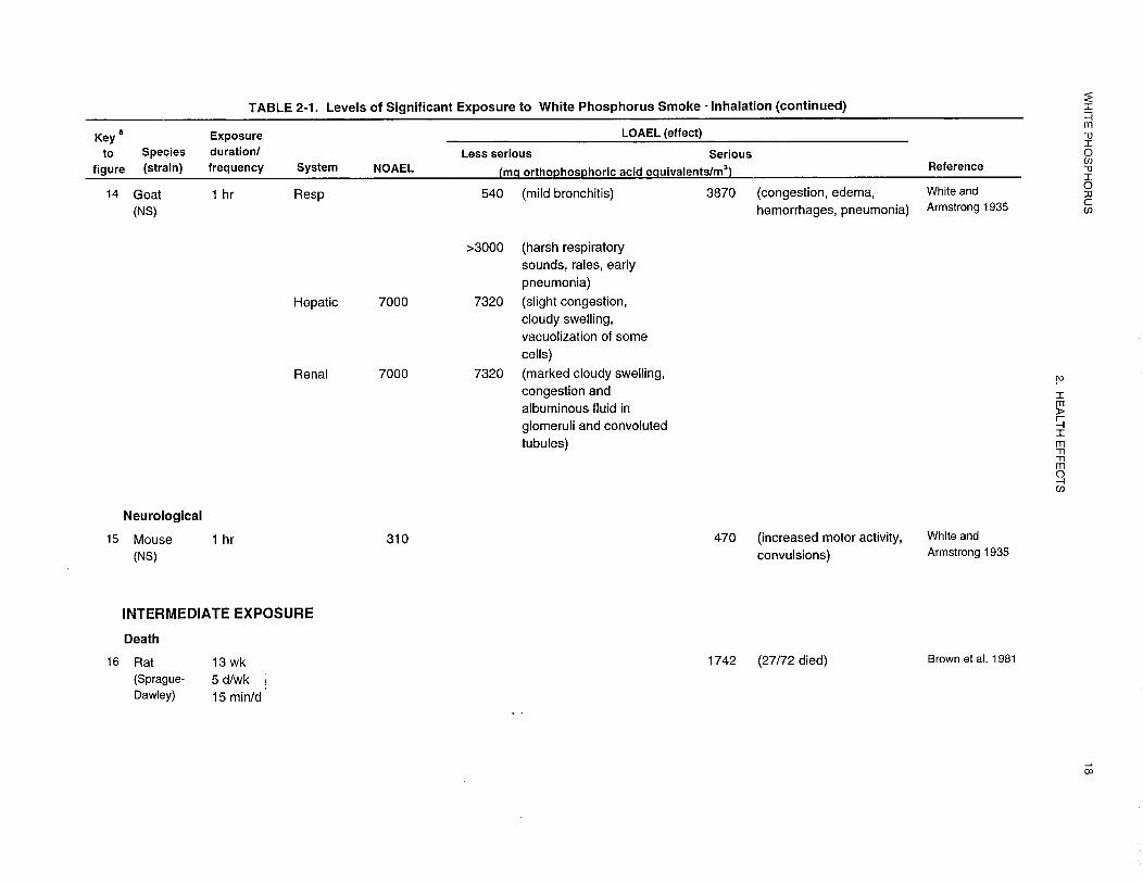
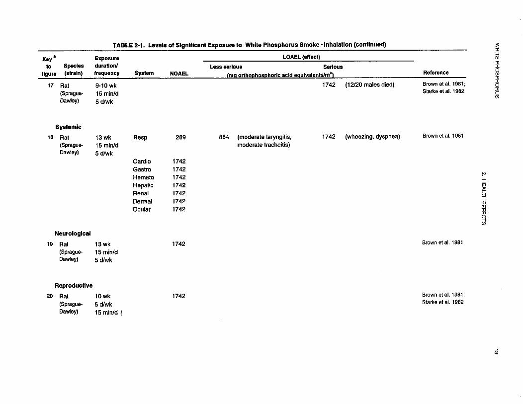
Increased mortality was also observed in rats exposed to 1,742 mg orthophosphoric acid equivalents/m3 of
white phosphorus smoke 15 minutes/day, 5 days/week, for 6-13 weeks (Brown et al. 1981) or 9-10 weeks
(Brown et al. 1981; Starke et al. 1982). As with the deaths occurring after acute exposure, the most severe
lesions were observed in the respiratory tract. Lesions were most extensive and severe in the larynx and
trachea. They consisted mainly of the thickening of the lamina propria (the connective tissue of the
mucous membrane) and submucosa by collagen, endothelial cell proliferation, and macrophage
infiltration. Occasionally, inflammatory cells were seen in the epithelium (Brown et al. 1981). No chronic
duration studies were located.
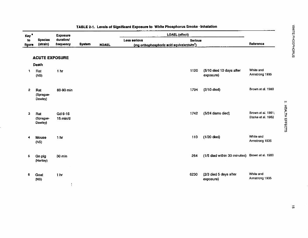
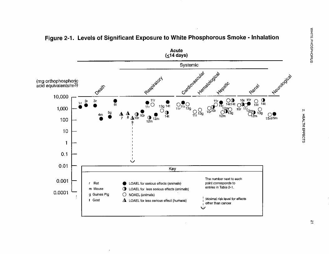
The LD50 values from each reliable study in each species and duration category are recorded in Table 2-1
and plotted in Figure 2-l.
2.2.1.2 Systemic Effects
White Phosphorus. No studies were located regarding gastrointestinal, dermal, or ocular effects in
humans or animals after inhalation exposure to white phosphorus.
White Phosphorus Smoke. Systemic effects of white phosphorus smoke in humans and animals after
inhalation exposure are discussed below. The highest NOAEL value and all reliable LOAEL values for
systemic effects in each species and duration category are recorded in Table 2- 1 and plotted in Figure 2-1.
No studies were located regarding musculoskeletal or other systemic effects in humans or animals after
inhalation exposure to white phosphorus smoke.


23 WHITE PHOSPHORUS
2. HEALTH EFFECTS
Respiratory Effects
White Phosphorus. In a study of 71 humans occupationally exposed to fumes/vapors and paste
containing white phosphorus for intermediate or chronic duration, an irritating cough was reported as
occurring in a large proportion of the employees (Ward 1928); no further information regarding
respiratory effects was reported. Details of this study are provided in Section 2.2.2.2; exposure levels of
white phosphorus and other compounds were not reported.
No studies were located regarding respiratory effects in animals after inhalation exposure to white
phosphorus.
White Phosphorus Smoke. In two acute-duration exposure studies, respiratory effects have been reported
by men inhaling white phosphorus smoke for 2-15 minutes (White and Armstrong 1935). At the lowest
concentration tested (187 mg phosphorus pentoxide equivalents/m3 [258 mg orthophosphoric acid
equivalents/m3] for 5 minutes), throat irritation during talking was reported. At higher concentrations
≥514 mg phosphorus pentoxide equivalents/m3 [709 mg orthophosphoric acid equivalents/m3 ]for
15 minutes), coughing and nose irritation were reported (White and Armstrong 1935). Coughing,
hoarseness, and erythema and edema of the larynx and vocal cords were reported in women exposed to an
unspecified amount of white phosphorus smoke for 15-20 minutes during a factory fire (Walker et al.
1947). No longer-term human exposure studies were located.
In animals, the respiratory tract is one of the primary targets of white phosphorus smoke toxicity. Slight to
intense congestion, edema, and hemorrhages were observed in the lungs of rats, mice, and goats (Brown et
al. 1980; White and Armstrong 1935). Exposure to 3,027 mg orthophosphoric acid equivalents/m3 for
90 minutes resulted in respiratory tract lesions in rats (Brown et al. 1980). “Unmistakable signs of
irritation” were observed in mice, rats, and goats exposed for 1 hour to 110,380, or 540 mg phosphorus
pentoxide equivalents/m3 (152,524, or 754 mg orthophosphoric acid equivalents/m3), respectively (White
and Armstrong 1935). Lung lesions were observed in all of the animals that died early and were
necropsied (White and Armstrong 1935). No respiratory tract effects were observed in guinea pigs
exposed to concentrations of white phosphorus smoke as high as 984 mg orthophosphoric acid
equivalents/m3 for 30 minutes (Brown et al. 1980). However, only one animal was examined at this
exposure level, and it was examined 2 weeks after exposure. Exposure to white phosphorus smoke for

24 WHITE PHOSPHORUS
2. HEALTH EFFECTS
6 or 13 weeks at a concentration of 884 mg orthophosphoric acid equivalents/m3 for 15 minutes/day,
5 days/week resulted in slight laryngitis and tracheitis in rats (Brown et al. 1981). Exposure to a higher
concentration (1,742 mg orthophosphoric acid equivalents/m3) resulted in wheezing, dyspnea, moderateto-
severe laryngitis and tracheitis, and minimal-to-severe interstitial pneumonia (Brown et al. 1981).
Cardiovascular Effects
White Phosphorus. No studies were located regarding cardiovascular effects in humans after inhalation
exposure to white phosphorus.
In rats exposed for an intermediate duration to an unknown concentration of airborne white phosphorus
from the furnace room of a phosphorus factory, an increase in permeability of capillary walls, lesions in
the walls of blood vessels, and evidence of impaired microcirculation were observed in the mouth
(Ruzuddinov and Rys-Uly 1986). Severe damage to the oral mucosa was also observed in these animals.
No information regarding effects on the heart was located in the animal studies.
White Phosphorus Smoke. No studies were located regarding cardiovascular effects in humans after
inhalation exposure to white phosphorus smoke.
No gross or histological alterations were observed in the hearts of rats exposed to white phosphorus smoke
at concentrations as high as 1,742 mg orthophosphoric acid equivalents/m3 15 minutes/day, 5 days/week
for 13 weeks (Brown et al. 1981).
Gastrointestinal Effects
White Phosphonrs Smoke. No studies were located regarding gastrointestinal effects in humans after
inhalation exposure to white phosphorus smoke.
Exposure of rats to concentrations of white phosphorus smoke as high as 1,742 mg orthophosphoric acid
equivalents/m3 for 15 minutes/day, 5 days/week for 13 weeks did not result in gross or histological
alterations in the gastrointestinal tract (Brown et al. 1981).

25 WHITE PHOSPHORUS
2. HEALTH EFFECTS
Hematological Effects
White Phosphorus. Anemia (marked decrease in red blood cells or hemoglobin) and leukopenia (very
low levels of white blood cells or leukocytes) were observed in workers chronically exposed to airborne
white phosphorus (Ward 1928). Because the workers handled white phosphorus contaminated rags, it is
possible that exposure occurred via oral and dermal routes also. No information on exposure levels was
provided. In another occupational exposure study, no alterations in hemoglobin or total or differential
leukocyte levels were observed (Hughes et al. 1962).
No studies were located regarding hematological effects in animals after inhalation exposure to white
phosphorus.
White Phosphorus Smoke. No studies were located regarding hematological effects in humans after
inhalation exposure to white phosphorus smoke.
Two weeks after exposure termination, no significant changes in erythrocyte, hematocrit, hemoglobin, or
total and differential leukocyte levels were observed in rats exposed to 3,027 mg orthophosphoric acid
equivalents/m3 for 90 minutes or guinea pigs exposed to 984 mg orthophosphoric acid equivalents/m3 for
10 minutes (Brown et al. 1980). No changes in erythrocyte, hematocrit, hemoglobin, or leukocyte (total or
differential) levels were observed in rats exposed to 1,742 mg orthophosphoric acid equivalents/m3 of
white phosphorus smoke for 15 minutes/day, 5 days/weeks, for 13 weeks (Brown et al. 1981).
Musculoskeletal Effects
White Phosphorus. Phossy jaw is a degenerative condition affecting the entire oral cavity including soft
tissue, teeth, and bones (Heimann 1946; Hughes et al. 1962; Ward 1928). This condition generally occurs
following long term exposure to airborne white phosphorus. The effects of phossy jaw can be extreme,
involving severe necrosis of soft tissue, teeth, and bones in the oral cavity. Massive life-threatening
infections often occur during the development of phossy jaw. With one exception (Jakhi et al. 1983) (see
Section 2.2.2.2), all reported cases of phossy jaw have resulted from occupational exposure to white
phosphorus fumes/vapors and/or dust. Because white phosphorus oxidizes rapidly, phosphorus
fumes/vapors may contain phosphoric oxide and phosphorus oxide, in addition to phosphorus (Heimann
1946; Hughes et al. 1962; Ward 1928).
26 WHITE PHOSPHORUS
2. HEALTH EFFECTS
In a study of workers exposed to white phosphorus for intermediate durations in three fireworks plants,
2 of 44 workers developed definite cases of phossy jaw (Ward 1928). These cases, described as slight
necrosis of the lower jaw, took up to 2 years for recovery. In the same study, 13 of 27 workers exposed to
white phosphorus for chronic durations developed necrosis of the upper and/or lower jaw, ranging from
slight to severe; 2 of the 13 workers developing phossy jaw died from complications related to the
necrosis. This study and several case reports discuss the progression of symptoms during the development
of phossy jaw (Heimann 1946; Hughes et al. 1962; Kennon and Hallam 1944). It is likely that the
development of necrosis of the jaw in workers exposed to airborne white phosphorus resulted from the
local action of phosphorus on the oral cavity. This information is discussed in detail in Section 2.2.2.2.
There is evidence that occupational exposure to white phosphorus affects bones other than those in the
jaw; this implies a systemic effect for inhaled white phosphorus. Two middle-aged men occupationally
exposed to white phosphorus for 20-30 years had a history of breaking their femurs in accidents not
normally expected to result in breakage of bones (Dearden 1899). One man had broken the right and left
femurs on two separate occasions by “tripping over a board,” while the other broke the right femur by
“stumbling down a single step,” and the left femur in “just as simple a manner.” Examination of bone
from the fingertip of one of the two workers indicated an increased “relative proportion of phosphoric acid
to lime” compared to healthy bone by nearly 1%. Thus, occupational exposure to white phosphorus may
change the composition of bone tissue, decreasing the bones ability to resist fracture; however, the
information reported in this study is insufficient to definitively attribute the observed effects to
occupational exposure to white phosphorus.
No studies were located regarding musculoskeletal effects in animals after inhalation exposure to white
phosphorus.
Hepatic Effects
White Phosphorus. Information on hepatotoxicity after exposure to airborne white phosphorus is limited
to an occupational exposure study. No alterations in liver function tests were observed in workers
chronically exposed to an unreported amount of airborne white phosphorus (Hughes et al. 1962).
No studies were located regarding hepatic effects in animals after inhalation exposure to white phosphorus.
27 WHITE PHOSPHORUS
2. HEALTH EFFECTS
White Phosphorus Smoke. No studies were located regarding hepatic effects in humans after inhalation
exposure to white phosphorus smoke.
Slight cloudy swelling was observed in the livers of rats exposed for 1 hour to 2 1,170 mg phosphorus
pentoxide equivalents/m3 (≥ 1,615 mg orthophosphoric acid equivalents/m3) (White and Armstrong 1935)
or 3,027 mg orthophosphoric acid equivalents/m3 for 90 minutes (Brown et al. 1980), mice exposed to
470 mg phosphorus pentoxide equivalents/m3 (649 mg orthophosphoric acid equivalents/m3) for 1 hour
(White and Armstrong 1935), and goats exposed for 1 hour to ≥7,320 mg phosphorus pentoxide
equivalents/m3) (≥10,104 mg orthophosphoric acid equivalents/m3) (White and Armstrong 1935). In the
White and Armstrong (1935) studies, only animals dying early were necropsied. Consequently, results
were only reported for some of the animals. No hepatic effects were observed in guinea pigs exposed to
984 mg orthophosphoric acid equivalents/m3 for 10 minutes; only one guinea pig exposed at this
concentration was examined 2 weeks after exposure (Brown et al. 1980). A NOAEL of 1,742 mg
orthophosphoric acid equivalents/m3 has been identified for hepatic effects in rats exposed for
15 minutes/day, 5 days/week for 13 weeks to white phosphorus smoke. At this concentration, no
alterations in the levels of triglyceride, cholesterol, serum aspartate aminotransferase (AST), or serum
alanine aminotransferase (ALT) or gross or histological lesions were observed (Brown et al. 1981).
Renal Effects
White Phosphorus. In an epidemiology study, 48 apparently healthy men working in a phosphorus plant
between 1 and 17 years had average creatinine levels in urine (141 mg/L) essentially identical to those of
28 workers (controls) not exposed to white phosphorus (Hughes et al. 1962). However, the groups were
apparently not well matched with respect to age and race (details not reported), as only 28 men (controls)
volunteered to allow the blood to be drawn (Hughes et al. 1962).
No studies were located regarding renal effects in animals after inhalation exposure to white phosphorus.
White Phosphorus Smoke. No studies were located regarding renal effects in humans after inhalation
exposure to white phosphorus smoke.
In rats, mice, and goats exposed to white phosphorus smoke for 1 hour, slight cloudy swelling was
observed in the kidneys at ≥1,170,470, and 7,320 mg phosphorus pentoxide equivalents/m3, respectively

28 WHITE PHOSPHORUS
2. HEALTH EFFECTS
(≥ 1,615, 649, or 10,104 mg orthophosphoric acid equivalents/m3) (White and Armstrong 1935). In the
White and Armstrong (1935) study, a white, mucous secretion was seen around the noses and mouths of
animals that died early. Upon necropsy, congestion, hemorrhage, edema, and pneumonia were the
principal lesions seen in the lungs, which are all evidence of respiratory obstruction (White and Armstrong
1935). No renal lesions were observed in rats exposed to 3,027 mg orthophosphoric acid equivalents/m3
for 90 minutes or guinea pigs exposed to 984 mg orthophosphoric acid equivalents/m3 for 10 minutes
(Brown et al. 1980). In the Brown et al. (1980) study, a small number of animals were examined 2 weeks
after exposure termination. Exposure to concentrations of white phosphorus smoke as high as 1,742 mg
orthophosphoric acid equivalents/m3 15 minutes/day, 5 days/week, for 13 weeks did not result in
significant changes in levels of serum urea nitrogen, creatinine, or uric acid or in alterations in the gross or
histological examination of the kidneys (Brown et al. 1981).
Dermal Effects
White Phosphoncs Smoke. No studies were located regarding dermal effects in humans after inhalation
exposure to white phosphorus smoke.
No alterations were observed in the skin of rats exposed to concentrations of white phosphorus smoke as
high as 1,742 mg orthophosphoric acid equivalents/m3 15 minutes/day, 5 days/week, for 13 weeks (Brown
et al. 1981).
Ocular Effects
White Phosphorus Smoke. No studies were located regarding ocular effects in humans after inhalation
exposure to white phosphorus smoke.
No alterations were observed in the eyes of rats exposed to concentrations of white phosphorus smoke as
high as 1,742 mg orthophosphoric acid equivalents/m3 15 minutes/day, 5 days/week, for 13 weeks (Brown
et al. 1981).

29 WHITE PHOSPHORUS
2. HEALTH EFFECTS
Other Systemic Effects
White Phosphorus. Decreased serum glucose levels were reported for some workers occupationally
exposed to white phosphorus for a chronic duration (Ward 1928). Details of this study are provided in
Section 2.2.2.2.
Rats received intermittent exposure to the atmosphere in the furnace room of a phosphorus factory for
l-4 months (Ruzuddinov and Rys-Uly 1986). Histology of rats killed monthly revealed progressive
morphological degeneration of the tongue and oral mucosa of the cheek, gum, and hard palate. It is likely
that the effects of white phosphorus in the oral cavity are local rather than systemic resulting from direct
contact of white phosphorus-containing atmosphere with tissues in the mouth. For this reason this study is
also discussed in Section 2.2.2.2.
2.2.1.3 Immunological and Lymphoreticular Effects
White Phosphorus. Limited information on the immunotoxicity of inhaled white phosphorus was located.
As discussed in Section 2.2.1.2, decreased leukocyte levels were observed in workers exposed to an
unknown concentration of white phosphorus via inhalation, oral, and dermal routes (Ward 1928). It is not
known if the decrease in leukocyte levels would result in impaired immune function.
No studies were located regarding immunological or lymphoreticular effects in animals after inhalation
exposure to white phosphorus.
White Phosphorus Smoke. No studies were located regarding immunological or lymphoreticular effects
in humans or animals after inhalation exposure to white phosphorus smoke.
2.2.1.4 Neurological Effects.
White Phosphorus. No studies were located regarding neurological effects in humans or animals after
inhalation exposure to white phosphorus.
White Phosphorus Smoke. No studies were located regarding neurological effects in humans after
inhalation exposure to white phosphorus smoke.

30 WHITE PHOSPHORUS
2. HEALTH EFFECTS
No histological alterations were observed in the brains of rats exposed to concentrations of white
phosphorus smoke as high as 1,742 mg orthophosphoric acid equivalents/m3 15 minutes/day, 5 days/week,
for 13 weeks (Brown et al. 1981). This NOAEL for neurological effects in rats exposed for an
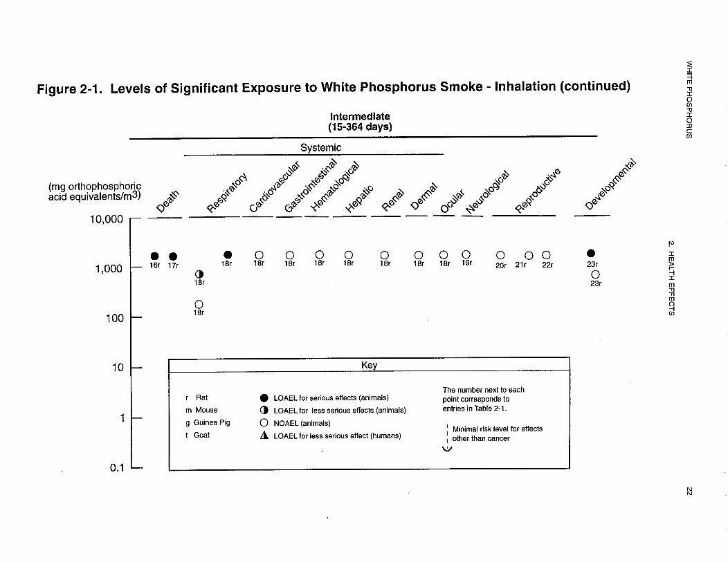
intermediate duration is recorded in Table 2- 1 and plotted in Figure 2-l.
2.2.1.5 Reproductive Effects
White Phosphorus. No studies were located regarding reproductive effects in humans or animals after
inhalation exposure to white phosphorus.
White Phosphorus Smoke. No studies were located regarding reproductive effects in humans after
inhalation exposure to white phosphorus smoke.
No effects on pregnancy rate or number of pups born alive were observed following the mating of male
rats exposed to 1,742 mg orthophosphoric acid equivalents/m3 of white phosphorus smoke
15 minutes/day, 5 days/week for 10 weeks with female rats exposed for 3 weeks (similar exposure
protocol) (Brown et al. 1981; Starke et al. 1982) or the mating of male rats exposed to 1,742 mg
orthophosphoric acid equivalents/m3 15 minutes/day, 5 days/week, for 13 weeks to unexposed female rats
(Brown et al. 1981; Starke et al. 1982). No exposure-related lesions were seen in the testis, epididymis,
ovary, and uterus of rats exposed to 1,742 mg orthophosphoric acid equivalents/m3 15 minutes/day,
5 days/week, for 13 weeks (Brown et al. 1981).
These LOAEL values from each reliable study for reproductive effects in rats in each duration category are
recorded in Table 2-l and plotted in Figure 2-l.
2.2.1.6 Developmental Effects
White Phosphorus. No studies were located regarding developmental effects in humans or animals after
inhalation exposure to white phosphorus.
White Phosphorus Smoke. No studies were located regarding developmental effects in humans after
inhalation exposure to white phosphorus smoke.
31 WHITE PHOSPHORUS
2. HEALTH EFFECTS
Increases in the incidence of right or reversed ductus arteriosus (9/18 or 50%) and ectopic testicles (3/18 or
33%) were observed in the offspring of unexposed male rats and female rats exposed to 1,742 mg
orthophosphoric acid equivalents/m3, 15 minutes/day on gestational days 6-15. Statistical analysis of
these data was not presented. No other developmental effects were observed in this study (Brown et al.
1981; Starke et al. 1982). In another study conducted by these authors, no significant increases in the
incidence of malformations or anomalies were observed in the offspring of rats exposed to 1,742 mg
orthophosphoric acid equivalents/m3, 15 minutes/day, 5 days/week during the 3-week premating period,
mating period, and gestation period. Thus, the authors did not consider these effects to be significant.
However continued exposure of the pups and dams to white phosphorus smoke (1,742 mg
orthophosphoric acid equivalents/m3) during the 3-week lactation period resulted in decreased pup growth,
pup survival, and pup viability. The authors suggested that the decreased pup growth and survival may
have been due to the dams not allowing the pups to nurse, not enough milk being produced, pups not
nursing due to respiratory tract irritation, or a direct compound-related effect on the pups (Brown et al.
1981; Starke et al. 1982).
These NOAEL and LOAEL values from each reliable study for developmental effects in rats are recorded
in Table 2-l and plotted in Figure 2-l.
2.2.1.7 Genotoxic Effects
White Phosphorus. No studies were located regarding genotoxic effects in humans or animals after
inhalation exposure to white phosphorus.
White Phosphorus Smoke. No studies were located regarding genotoxic effects in humans or animals
after inhalation exposure to white phosphorus smoke.
Genotoxicity studies are discussed in Section 2.5.
32 WHITE PHOSPHORUS
2. HEALTH EFFECTS
2.2.1.8 Cancer
White Phosphorus. No studies were located regarding cancer in humans or animals after inhalation
exposure to white phosphorus.
White Phosphonts Smoke. No studies were located regarding cancer in humans or animals after
inhalation exposure to white phosphorus smoke.
2.2.2 Oral Exposure
Studies reporting acute oral exposure of humans to white phosphorus were limited to case reports of
intentional or accidental ingestion of match heads, rat poison, cockroach poison, firecrackers, or from
military operations. Manufacturers of white phosphorus-containing rat poison have claimed that the only
active ingredient in the rat poison was white phosphorus (Peacock 1993). It is likely that white
phosphorus was the agent producing toxicity following ingestion of cockroach poison, match heads, and
fireworks, although the presence of other toxic compounds cannot be ruled out. Many of the case reports
involving acute oral exposure of humans to white phosphorus did not report intake levels. High doses of
white phosphorus nearly always induced vomiting, expelling much of the ingested white phosphorus from
the body. In addition, gastric lavage to remove white phosphorus from the stomach was performed on
many poisoned patients. Thus, doses could not be estimated for end points other than vomiting for all but
one of the case reports for humans receiving acute oral exposure to white phosphorus.
Several studies reporting intermediate oral exposure of children to white phosphorus were located. In
most cases the white phosphorus was administered as a treatment for rickets, but in some cases white
phosphorus was administered to healthy children to prevent the development of rickets. In studies
reporting the effects of white phosphorus on bones in children, the doses of white phosphorus..
administered (0.026-0.158 mg/kg/day) were several orders of magnitude lower than those reported
following intentional or accidental white phosphorus poisoning.
Humans exposed to white phosphorus in the workplace probably ingested some airborne white
phosphorus. One retrospective study indicated that oral exposure to white phosphorus passed from hand

33 WHITE PHOSPHORUS
2. HEALTH EFFECTS
to mouth was likely, because the workers constantly handled a paste containing 4-6% white phosphorus,
and washroom facilities at the plants were inadequate.
No studies were located regarding health effects in human or animals after oral exposure to white
phosphorus smoke.
2.2.2.1 Death
Numerous case reports of death following acute oral exposure of humans to white phosphorus were
located (Diaz-Rivera et al. 1950,196l; Dwyer and Helwig 1925; Hann and Veale 1910; Humphreys and
Halpert 1931; McCarron et al. 1981; Rao and Brown 1974; Rubitsky and Myerson 1949; Simmon and
Pickering 1976; Talley et al. 1972; Torrielli et al. 1974; Wechsler and Wechsler 1951; Wertham 1932;
Winek et al. 1973).
In one case report, circumstances following ingestion of white phosphorus allowed for estimation of dose
(Hann and Veale 1910). A woman consumed ≈3.9 g of rat poison containing 4% white phosphorus, but
did not vomit until the second day after the poisoning, and the vomitus at that time was clear. Thus, little
or none of the white phosphorus ingested was lost due to vomiting. The estimated single dose was
2 mg/kg/day. Four days after ingesting the rat poison, the woman died. The cause of death was not
reported, but autopsy revealed fatty degeneration and cell transformation in the liver (Hann and Veale
1910).
In case reports of 56 individuals intentionally ingesting large quantities of white phosphorus (0.19-6.3 g)
in rat poison, 48.2% of the individuals died, with a 90% death rate in patients ingesting ≥1.57 g of
phosphorus (Diaz-Rivera et al. 1950). Because white phosphorus at these oral exposure levels induced
rapid vomiting, the doses for these case reports could not be estimated. In patients that died, symptoms
prior to death included irreversible vascular collapse; cyanosis, ashen skin color, and deep pallor (probably
secondary to vascular collapse); coma; abnormal electrocardiogram readings; evidence of extreme liver
and kidney damage, and hypoglycemia (possible secondary to liver damage); and delirium, psychosis, and
hallucinations (possibly secondary to brain damage). The cause of death was not reported for each patient,
but appeared in most cases to be related to irreversible failure of the liver, kidney, brain, and/or
cardiovascular system (Diaz-Rivera et al. 1950). In other case reports, autopsy of patients dying from
white phosphorus poisoning nearly always revealed severe damage to one or more of those four systems

34 WHITE PHOSPHORUS
2. HEALTH EFFECTS
(Diaz-Rivera et al. 1961; Dwyer and Helwig 1925; Hann and Veale 1910; Humphreys and Halpert 1931;
McCarron et al. 1981; Rao and Brown 1974; Wechsler and Wechsler 1951; Wertham 1932). In some
cases, pulmonary edema and/or congestion were observed at autopsy (Rao and Brown 1974; Wechsler and
Wechsler 1951). In studies reporting the specific cause of death, death was attributed to cardiopulmonary
arrest (Diaz-Rivera et al. 1950; Rao and Brown 1974; Simon and Pickering 1976; Winek et al. 1973),
peripheral vascular collapse (Diaz-Rivera et al. 1950,1961), liver failure (Diaz-Rivera et al. 1961;
McCarron et al. 1981), hypoglycemia (Diaz-Rivera et al. 1961), and gastrointestinal hemorrhage and
hemorrhagic bronchopneumonia (Winek et al. 1973).
No deaths were reported in children treated with 0.0264.158 mg/kg/day white phosphorus for as much as
26 months (Compere 1930a; Phemister 1918). An infant became seriously ill during treatment with
0.083 mg/kg/day white phosphorus (timed-weighted average dose for 6 months), but recovered entirely
following discontinuation of the dose (Sontag 1938).
Humans occupationally exposed to phosphorus probably ingested some airborne white phosphorus. In a
study of 71 humans occupationally exposed to fumes/vapors and paste containing white phosphorus, oral
exposure to phosphorus passed from hand to mouth was likely, because the workers constantly handled a
paste containing 4-6% white phosphorus, and washroom facilities at the plants were inadequate (Ward
1928). White phosphorus-related deaths occurred in 0 of 44 and 2 of 27 of the workers exposed for
intermediate and chronic durations, respectively. In the two cases of death, the workers died from
complications related to phossy jaw, a degenerative condition affecting the soft tissue, bones, and teeth of
the oral cavity. In this condition, the toxic effects of white phosphorus probably result from the local
irritant action of white phosphorus on tissues in the mouth. Thus, white phosphorus paste passed from
hand to mouth and the local action of airborne white phosphorus on the oral cavity may have contributed
to the development of phossy jaw, and subsequent death, of these two workers. It is not known whether
white phosphorus ingested and absorbed into the systemic circulation contributed to the development of
phossy jaw in the two workers that died (Ward 1928). Details of this study are provided in
Section 2.2.2.2.
A mortality rate of 30% was observed in Wistar rats treated by gavage with 6 mg/kg white phosphorus
(Torrielli et al. 1974). The oral LD50 value for Charles-River rats was 3.03 mg/kg for females and
3.76 mg/kg for males (Lee et al. 1975). A mortality rate of 20-35% was observed in mice treated by
gavage with 5-6 mg/kg (Hurwitz 1972). LD50 values of 4.82 mg/kg and 4.85 mg/kg were reported for
35 WHITE PHOSPHORUS
2. HEALTH EFFECTS
female and male mice, respectively (Lee et al. 1975). In two separate one-generation reproduction studies
in rats (IRDC 1985; Bio/dynamics 1991), 30-47% and 53%, respectively, of pregnant females treated by
gavage with 0.075 mg/kg/day for 145-204 days (intermediate duration) died (or were killed due to
morbidity) in late gestation or during parturition; dams exposed to 0.015 mg/kg/day for similar durations
did not have an increased mortality rate (IRDC 1985). Compound-related deaths were not observed in
male rats exposed to 0.075 mg/kg/day for similar durations (Bio/dynamics 1991; IRDC 1985).
Mortality was observed in 9 of 21 dogs treated once by gavage with an unknown quantity of white
phosphorus from firecrackers (Dwyer and Helwig 1925). A cat died 2 hours after ingesting an unknown
amount of white phosphorus (Frye and Cucuel 1969).
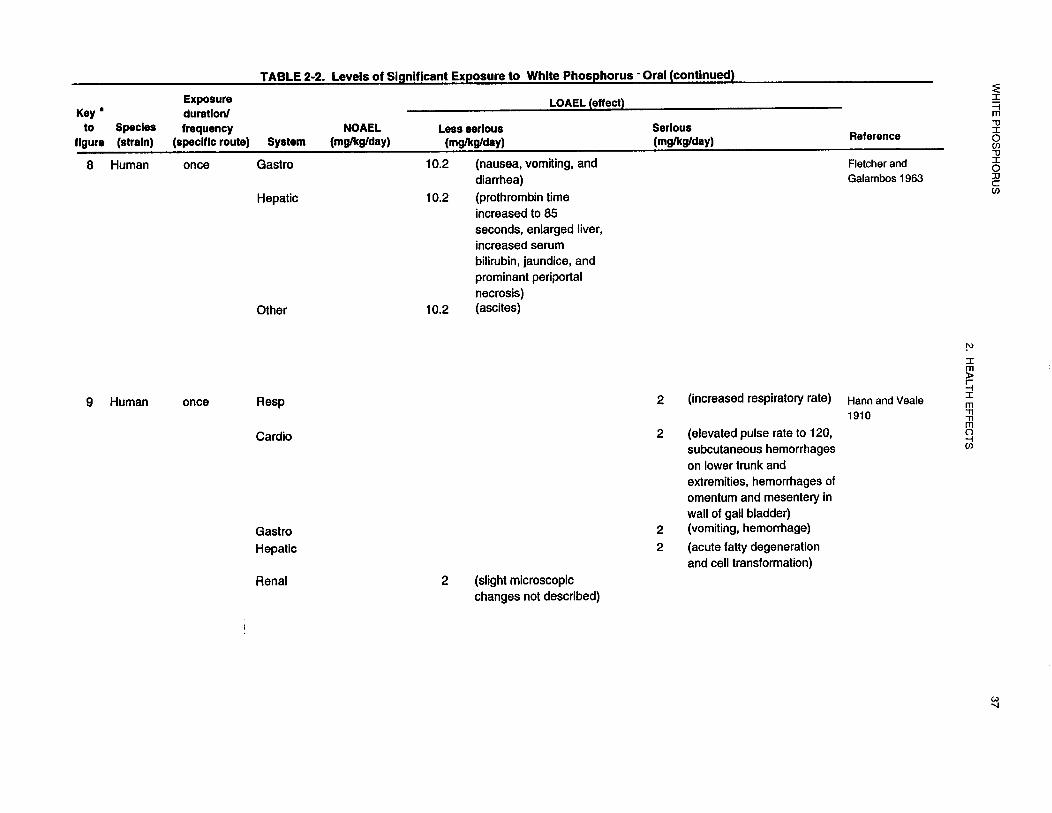
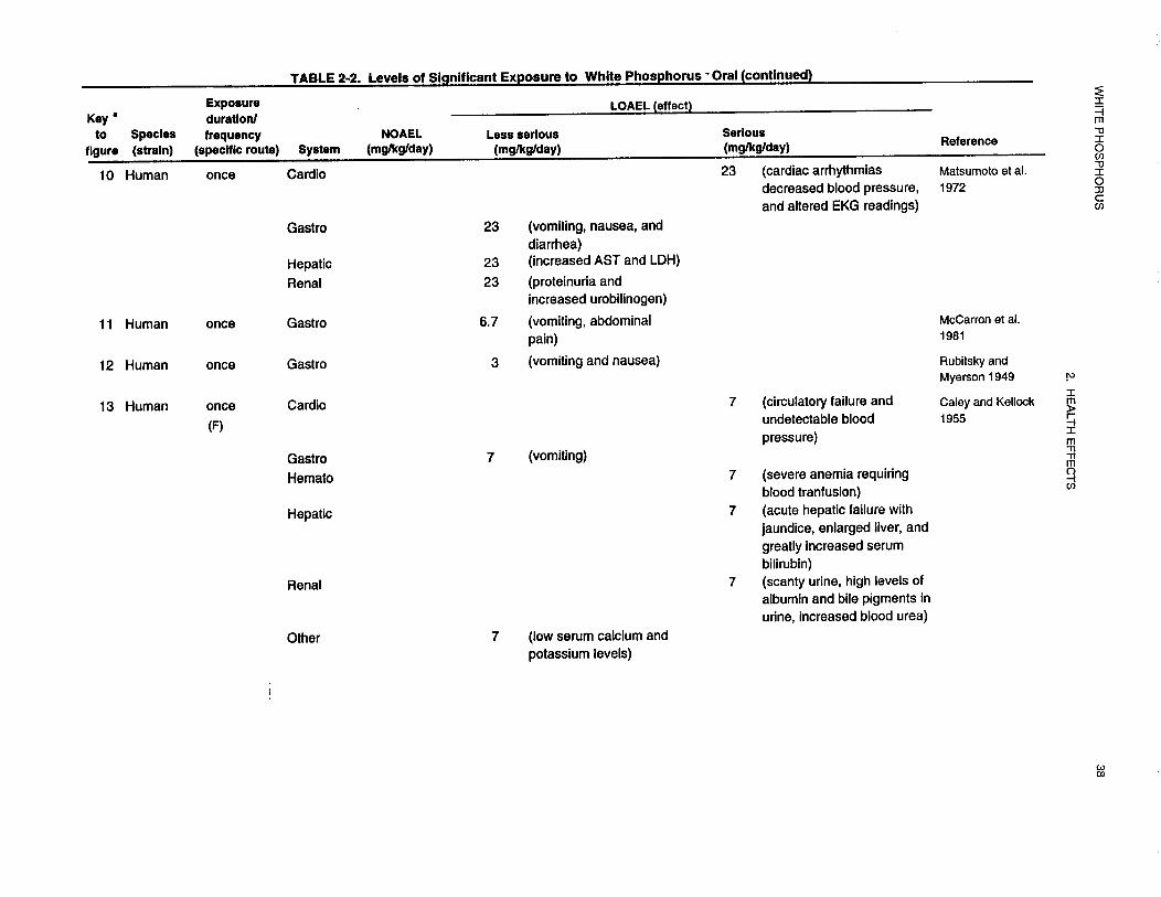
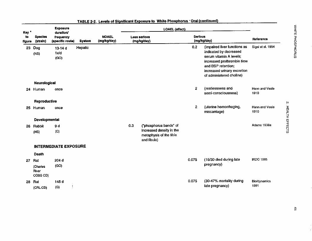
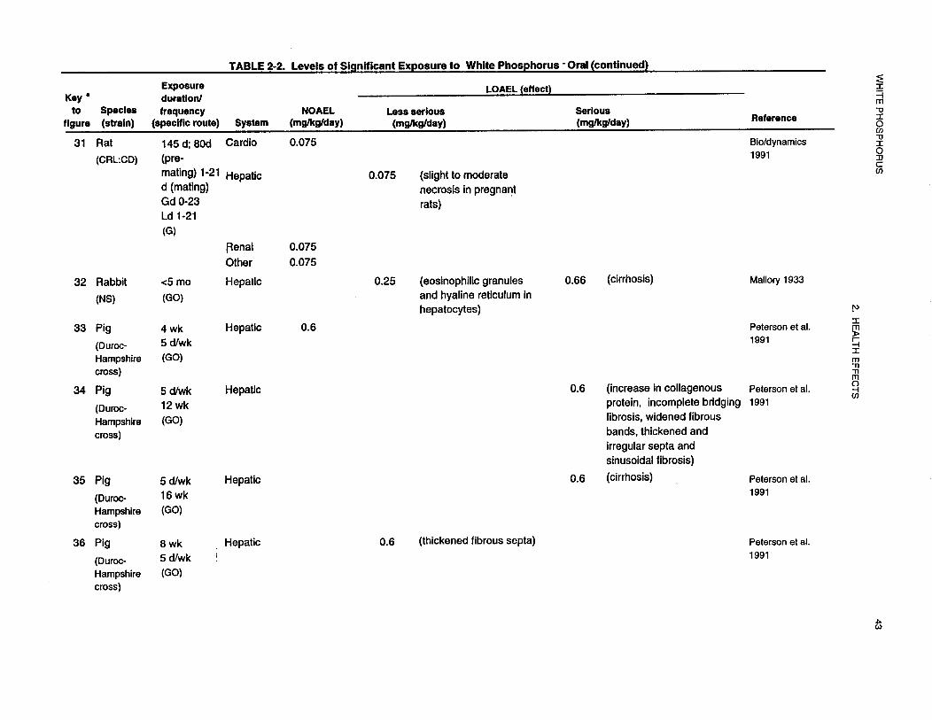
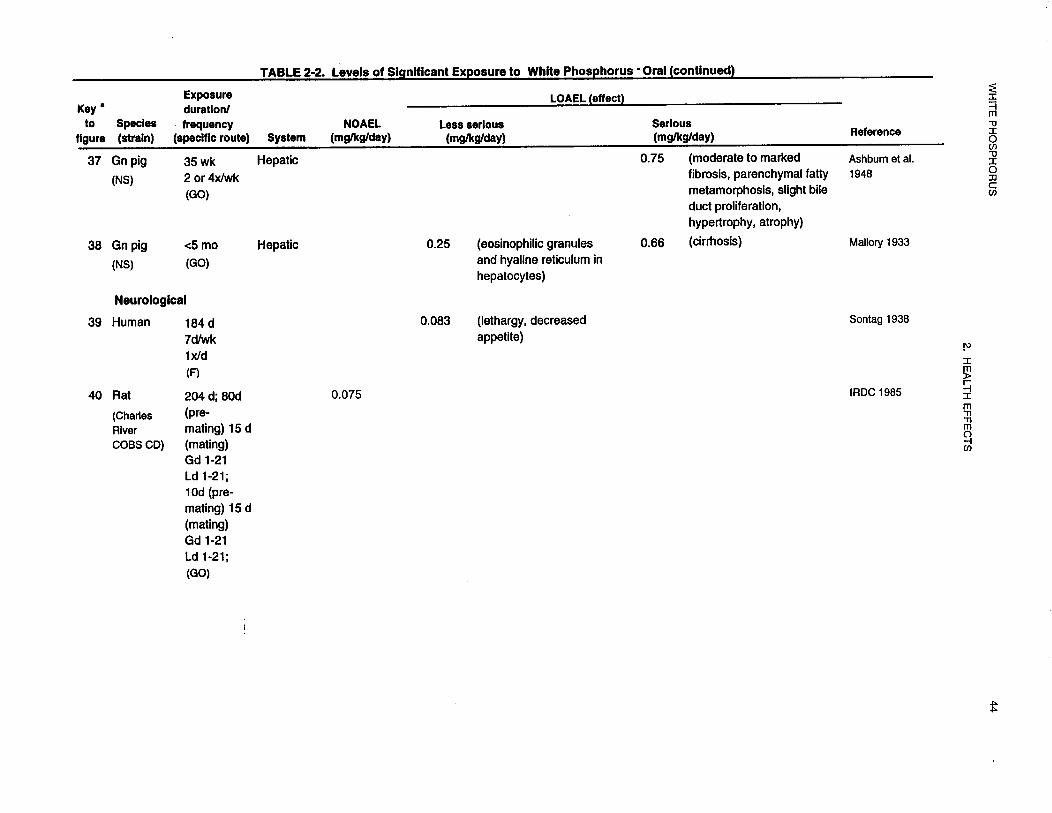
The LD50 values and doses associated with death in each species and duration category are recorded in
Table 2-2 and plotted in Figure 2-2.
2.2.2.2 Systemic Effects
Systemic effects of white phosphorus in humans and animals after oral exposure are discussed below. The
highest NOAEL value and all reliable LOAEL values from each reliable study for systemic effects in each
species and duration category are recorded in Table 2-2 and plotted in Figure 2-2.
No studies were located regarding ocular effects in humans or animals after oral exposure to white
phosphorus.
Respiratory Effects. Studies on respiratory effects following acute oral exposure of humans to white
phosphorus were limited to case reports of intentional or accidental consumption of materials containing
white phosphorus. Although intake of phosphorus was often reported, dose could be estimated for only
one study (Hann and Veale 1910), because vomiting and/or gastric lavage nearly always occurred soon
after poisoning, expelling much of the ingested phosphorus from the body.
Tachypnea (increased respiratory rate; 48 breaths/minute) was observed in a woman consuming rat poison
containing 4% white phosphorus (Hann and Veale 1910); the woman apparently did not vomit until the
second day, and the vomitus was clear. The estimated dose was 2 mg/kg. Four days after ingesting the rat


50 WHITE PHOSPHORUS
2. HEALTH EFFECTS
poison, the woman died, apparently from liver failure. Autopsy showed that the pleural cavity was filled
with a dark fluid, but no histological abnormalities were observed in the lungs (Hann and Veale 1910).
In the following studies, no doses could be estimated for respiratory effects because of vomiting and/or
gastric lavage. In a case report involving ingestion of rat poison containing white phosphorus, the patient
arrived at a hospital in a coma and displayed Cheyne-Stokes respirations and rales (Wechsler and
Wechsler 1951). The Cheyne-Stokes respirations increased to an extreme degree, and the patient died.
Autopsy revealed pulmonary congestion and edema throughout the stroma. Increased respiratory rate
(56 breaths/minute) and rales also were observed in an infant ingesting rat poison containing white
phosphorus (Rao and Brown 1974). The child died, and autopsy revealed evidence of pulmonary edema.
Rales were observed in a child ingesting a fatal dose of white phosphorus in fireworks; autopsy indicated
that the lungs were normal except for some fibrous adhesions (Dwyer and Helwig 1925). Hemorrhagic
bronchopneumonia was observed following autopsy of a man ingesting a fatal dose of rat poison
containing white phosphorus (Winek et al. 1973). Autopsy of a child who died following ingestion of a
firecracker revealed fatty deposition in parenchyma, bronchial epithelium, and tracheal epithelium and
cartilage (Humphreys and Halpert 1931). Death from cardiopulmonary failure was reported for a 63-year-old
woman (Winek et al. 1973), a 2-year-old boy (Simon and Pickering 1976), and a 3-year-old girl
(Simon and Pickering 1976) following ingestion of white phosphorus in rat poison; a respiratory rate of
44 breaths/minute was initially observed in the girl (Simon and Pickering 1976). Increased respiratory rate
was observed prior to death in two case reports involving ingestion of rat poison (Talley et al. 1972;
Winek et al. 1973). Shallow respirations and cyanosis were observed prior to death in an adult female
following ingestion of rat/roach poison containing white phosphorus (Rubitsky and Myerson 1949). Rales
were observed 1 day after intentional ingestion of rat poison by a 30-year-old man; 2 days later the patient
went into shock but survived the poisoning and eventually recovered (Pietras et al. 1968).
No treatment-related respiratory effects were reported in children treated with white phosphorus for
intermediate durations.
No treatment-related microscopic changes were observed in the lungs of rats exposed to 0.2 mg/kg/day
white phosphorus in the diet for a chronic duration (Fleming et al. 1942) or 0.075 mg/kg/day phosphorus
by gavage for an intermediate duration (LRDC 1985). Heavy breathing and apnea were reported following
ingestion of a fatal quantity of white phosphorus by a cat (Frye and Cucuell969). Necropsy revealed
hyperemia, hemorrhage and edema in the lungs.

51 WHITE PHOSPHORUS
2. HEALTH EFFECTS
Cardiovascular Effects. Alterations in electrocardiograms, such as altered or inverted T waves and
changes in the QRS complex, and other cardiac changes, such as tachycardia, arrhythmias, atrial
fibrillation, and decreased ventricular contractility, have been observed in individuals accidentally or
intentionally ingesting a single dose of white phosphorus (Dathe and Nathan 1946; Diaz-Rivera et al.
1950, 1961; Dwyer and Helwig 1925; Ehrentheil 1957; Matsumoto et al. 1972; McCarron et al. 1981;
Newburger et al. 1948; Pietras et al. 1968; Rao and Brown 1974; Simon and Pickering 1976; Talley et al.
1972). Damage to the myocardium was verified by a number of cases in which histological examination
of the heart was performed. Prominent cross striations in the myocardium (Dwyer and Helwig 1925),
fatty infiltration of muscle (Diaz-Rivera et al. 1961; Humphreys and Halpert 1931; Wertham 1932),
necrosis of myocardium (Wechsler and Wechsler 1951), markedly dilated cardiac chamber (Rao and
Brown 1974), and interstitial edema of the myocardium and vacuolation of cells (Talley et al. 1972) have
been observed. Because of vomiting and gastric lavage, doses cannot be calculated from the human
studies. No cardiac effects were reported in longer term human studies.
In addition to the effects on the heart, a number of vascular effects have been observed in humans acutely
exposed to white phosphorus. A markedly decreased or undetectable blood pressure (Caley and Kellock
1955; Dathe and Nathan 1946; McCarron et al. 1981; Rubitsky and Myerson 1949; Simon and Pickering
1976; Wechsler and Wechsler 1951), vascular collapse (Diaz-Rivera et al. 1950, 1961), undetectable or
decreased pulse (Dwyer and Helwig 1925; Rubitsky and Myerson 1949), and increased pulse (Dathe and
Nathan 1946; Hann and Veale 1910; McCarron et al. 1981; Wechsler and Wechsler 1951) have been
observed. In addition, individuals have died following cardiopulmonary arrest (Simon and Pickering
1976; Winek et al. 1973), which may be due to effects on the heart or vascular system. A dose of
2 mg/kg/day for vascular effects was identified from the Hann and Veale (1910) report of a woman
ingesting a single dose of white phosphorus. Dose levels cannot be estimated for the other case reports.
Hemorrhaging in internal organs, as well as the appearance of petechial hemorrhages on the skin, have
been reported in a number of acute human exposure cases (Hann and Veale 1910; Humphreys and Halpert
1931; Winek et al. 1973). It is not known whether these effects are due to impairment of the integrity of
the blood vessels or due to damage of the affected organ (e.g., liver, stomach) itself.
In rats administered 0.075 mg/kg/day white phosphorus for an intermediate duration, no histological
alterations were observed in the heart (Bio/dynamics 1991; IRDC 1985). In rats exposed for an
intermediate duration to an unknown concentration of airborne white phosphorus from the furnace room

52 WHITE PHOSPHORUS
2. HEALTH EFFECTS
of a phosphorus factory, an increase in permeability of capillary walls, lesions in the walls of blood vessels
and evidence of impaired microcirculation were observed in the mouth (Ruzuddinov and Rys-Uly 1986).
These effects probably resulted from the local action of white phosphorus on the oral cavity.
Gastrointestinal Effects. Most of the human case reports listed vomiting as an early effect following
ingestion of a single high dose of white phosphorus (Caley and Kellock 1955; Dathe and Nathan 1946;
Diaz-Rivera et al. 1950; Dwyer and Helwig 1925; Ehrentheil 1957; Fletcher and Galambos 1963;
Greenberger et al. 1964; Hann and Veale 1910; Humphreys and Halpert 193 1; Matsumoto et al. 1972;
McCarron et al. 1981; McIntosh 1927; Newburger et al. 1948; Pietras et al. 1968; Rubitsky and Myerson
1949; Simon and Pickering 1976; Wechsler and Wechsler 1951; Winek et al. 1973). The doses that
induced vomiting ranged from 2 to 23 mg/kg (Caley and Kellock 1955; Ehrentheil 1957; Fletcher and
Galambos 1963; Harm and Veale 1910; Matsumoto et al. 1972; McCarron et al. 1981; Newburger et al.
1948; Rubitsky and Myerson 1949). Vomiting generally started within hours after ingesting the white
phosphorus, and sometimes continued for many days. Other gastrointestinal effects included abdominal
cramps or pain (often severe) (Dwyer and Helwig 1925; Ehrentheil1957; Fletcher and Galambos 1963;
Greenberger et al. 1964; Humphreys and Halpert 1931; McCarron et al. 1981; Newburger et al. 1948;
Pietras et al. 1968), vomiting blood and/or pieces of the gastric mucosa (Dathe and Nathan 1946; Diaz-
Rivera et al. 1950; Rubitsky and Myerson 1949), necrosis and erosion of mucosa in the esophagus,
stomach, duodenum, and jejunum (Wechsler and Wechsler 1951), and gastrointestinal hemorrhage
(Dwyer and Helwig 1925; Hann and Veale 1910; Humphreys and Halpert 1931; Wertham 1932, Winek et
al. 1973). These effects, with the exception of necrosis, were probably due to the irritating effects of white
phosphorus on the mucosa of the gastrointestinal tract. Vomitus often contained white phosphorus,
indicating that vomiting generally occurred before all white phosphorus and/or its oxidation products had
been absorbed.
No gastrointestinal effects were reported in children receiving treatment with 0.026-0.158 mg/kg/day
white phosphorus for as much as 26 months (Phemister 1918; Compere 1930a). An infant became
seriously ill during treatment with 0.083 mg/kg/day white phosphorus (6-month time-weighted average
dose), but recovered entirely following discontinuation of the treatment (Sontag 1938). No vomiting or
diarrhea was observed during the treatment period.
Gastrointestinal effects were not reported in studies examining longer term occupational exposure to white
phosphorus (Heimann 1946; Hughes et al. 1962; Kennon and Hallam 1944; Ward 1928).
53 WHITE PHOSPHORUS
2. HEALTH EFFECTS
Erosion and hemorrhages in tissue in the esophagus and stomach were observed following ingestion of a
fatal unknown quantity of white phosphorus by a cat (Frye and Cucuel 1969). Vomiting was observed in
6 of 21 dogs treated by gavage with an unknown quantity of white phosphorus from firecrackers (Dwyer
and Helwig 1925). No gross or microscopic alterations were observed in the gastrointestinal tract of rats
treated by gavage with 0.075 mg/kg/day for 204 days (IRDC 1985).
Hematological Effects. Hematological effects have been reported in a number of case histories of
individuals accidentally or intentionally ingesting a single dose of white phosphorus contained in rat (and
cockroach) poisons or fireworks. Because most of the individuals vomited or received gastric lavage
shortly after ingestion, the amount of white phosphorus available for absorption is not known. Increases
in erythrocyte levels (Diaz-Rivera et al. 1950) and hemoglobin levels (Diaz-Rivera et al. 1950; McIntosh
1927); decreases in erythrocyte levels (Dwyer and Helwig 1925) and hemoglobin and/or hematocrit levels
(Simon and Pickering 1973); and anemia (Caley and Kellock 1955) have been observed in some of these
individuals. A number of individuals had no change in erythrocyte parameters (Ehrentheil 1957; Fletcher
and Galambos 1963; Newburger et al. 1948; Simon and Pickering 1976). The decreases in erythrocyte
parameters may be a reflection of the hemorrhages observed in specific tissues (e.g., gastrointestinal tract,
liver, skin) (Dathe and Nathan 1946; Hann and Veale 1910; Humphreys and Halpert 1931; Wechsler and
Wechsler 1951; Winek et al. 1973). In addition to these changes in erythrocyte parameters, changes in
total or differential leukocyte levels were reported in a number of individuals acutely exposed to white
phosphorus. Decreases in total leukocyte levels (Diaz-Rivera et al. 1950; Ehrentbeil 1957; Fletcher and
Galambos 1963; McCarron et al. 1981; Newburger et al. 1948; Pietras et al. 1968) and decreases or
increases in the percentage of polymorphonuclear leukocytes (neutrophils) have been reported (Ehrentheil
1957; McCarron et al. 1981; Pietras et al. 1968). No changes in leukocyte parameters were observed in a
number of individuals (Fletcher and Galambos 1963; Newburger et al. 1948; Simon and Pickering 1976).
Abnormally low protbrombin times or levels (hypo-prothrombinemia) and a moderate decrease in platelets
were observed in a number of individuals ingesting single doses of white phosphorus (Caley and Kellock
1955; Dathe and Nathan 1946; Ehrentheil 1957; Fletcher and Galambos 1963; McCarron et al 1981).
Most of the patients developed hypoprothrombinemia within 4-8 days (McCarron et al. 1981). This is
probably secondary to the liver damage rather than a direct effect on platelets. No changes in
hematological parameters were observed in a child ingesting phosphorized cod liver oil (0.083 mg/kg/day
phosphorus) for 184 days (Sontag 1938). Anemia and leukopenia were observed in individuals occupationally
exposed to white phosphorus chronically (Ward 1928). It is likely that workers were exposed
by the inhalation, oral, and dermal routes.
54 WHITE PHOSPHORUS
2. HEALTH EFFECTS
Because there is very little consistency regarding the length of time that elapsed between ingestion and
measurement of hematological parameters and the doses cannot be calculated, it is difficult to compare the
results of different studies. There is insufficient information to determine whether white phosphorus has a
direct effect on erythrocytes and/or leukocytes. The effects observed may be secondary effects. The
decrease in erythrocyte, hemoglobin, hematocrit and leukocyte levels may be secondary to hemorrhaging
or hematoemesis (Diaz-Rivera et al. 1950; Rubitsky and Myerson 1949) and the increase in erythrocytes
and hemoglobin may be a compensatory mechanism due to tissue anoxia. However, since red blood cell
synthesis takes 3-5 days, the observed effects may be direct if they are occurring within l-2 days.
A slight decrease in hemoglobin levels and increase in eosinophil levels were observed in a 30-year-old
man who performed magic shows that involved placing white phosphorus pellets in the mucobuccal folds
of his mouth for 15 years. He had no other personal habits that might adversely affect his health except
for occasional bidi smoking for about 8 years (Jakhi et al. 1983).
Information on hematological effects in animals is limited to one study in which a marked increase in total
leukocyte levels and the percentage of monocytes were observed in a guinea pig acutely exposed to
0.9-2.4 mg/kg/day of white phosphorus in a complex dosing regimen (Lawrence and Huffman 1929).
The study authors did not specify at which doses the effects occurred.
Musculoskeletal Effects. Following ingestion of a fatal dose of rat poison containing white
phosphorus by a woman, autopsy revealed fatty infiltration of essentially all tissues, including the
musculature (Wertham 1932). Similar effects were reported following the death of a male child who
accidentally ingested a firecracker containing white phosphorus; autopsy revealed fatty deposition in many
tissues, included the diaphragmic muscle (Humphreys and Halpert 1931).
Humans occupationally exposed to white phosphorus probably ingested some airborne white phosphorus.
In a study of 71 humans occupationally exposed to white phosphorus, oral exposure to white phosphorus
via hand-to-mouth activity was likely because the workers constantly handled a paste containing
4-6% white phosphorus and washroom facilities were inadequate (Ward 1928). In workers exposed to
white phosphorus for intermediate durations, 2 of 44 developed phossy jaw, described as slight necrosis in
the lower jaw. In workers exposed to white phosphorus for chronic durations, 12 of 27 developed phossy
jaw, with necrosis ranging from slight to severe; 2 of the 12 workers developing phossy jaw died from
complications related to the necrosis. The progression of the disease was similar in the cases described,

55 WHITE PHOSPHORUS
2. HEALTH EFFECTS
usually beginning with the extraction of one or more teeth, poor healing of the socket, followed by
necrosis of tissue in the jaw with severe pain and infection. Treatment consisted of repeated removal of
destroyed bone tissue and teeth, draining of abscesses, and reconstructive surgery. In severe cases,
extensive removal of necrotic bone tissue led to permanent disfigurement. However, exposure levels of
white phosphorus were not reported (Ward 1928). Case reports of development of phossy jaw following
intermediate or chronic occupational exposure to unreported levels of white phosphorus and phosphorus
compounds describe a similar progression of symptoms, with similar results; even in cases of early
diagnosis and prompt, intensive treatment of phossy jaw, recovery often took several years (Heimann
1946; Hughes et al. 1962; Kennon and Hallam 1944).
It is likely that the effect of white phosphorus in the oral cavity is local, resulting from contact of “inhaled”
white phosphorus particles with tissue in the mouth. White phosphorus may affect the oral mucosa. Dull,
red spots in the oral mucosa, an early sign of phossy jaw, have been reported to precede its development in
occupationally exposed workers (Kennon and Hallam 1944). The oral mucosa of workers exposed to
white phosphorus has been described as having a dull, red, unhealthy appearance (Hughes et al. 1962).
Exposed bones may be especially susceptible to the irritating affects of white phosphorus. It is not known
whether white phosphorus ingested and absorbed into the systemic circulation contributed to the
development of phossy jaw.
Not all workers exposed to white phosphorus for longer-term durations developed phossy jaw. In a study
of 71 workers exposed to airborne white phosphorus for intermediate or chronic durations, 4.5% and
44%, respectively, developed phossy jaw (Ward 1928). Forty-eight male workers with exposure to white
phosphorus ranging from 1 to 17 years were found to be normal and healthy with regards to many
parameters, including serum levels of calcium and phosphorus, and bone density; none of the men
developed phossy jaw (Hughes et al. 1962). Tooth loss often precedes and accompanies the progression
of development of phossy jaw (Heimann 1946; Hughes et al. 1962; Kennon and Hallam 1944; Ward
1928). Tooth loss during the later stages of phossy jaw clearly results from destruction of the-bone
structure supporting the teeth (Heimann 1946; Hughes et al. 1962; Kennon and Hallam 1944; Legge 1920;
Ward 1928). It is not known if tooth loss prior to diagnosis of phossy jaw or early in the development of
the condition is related to the white phosphorus exposure. Poor dental hygiene alone can result in tooth
loss, and in several case reports some of the workers were described as having poor dental hygiene
(Kennon and Hallam 1944). Tooth loss followed by poor healing of the socket often precedes
development of the necrosis (Heimann 1946; Hughes et al. 1962; Kennon and Hallam 1944; Ward 1928),
56 WHITE PHOSPHORUS
2. HEALTH EFFECTS
suggesting that poor dental hygiene and exposure to white phosphorus may both be contributing factors to
the development of phossy jaw. In a case report, five men developed “precursor signs” (delayed healing
of extracted tooth sites and residual sepsis) of phossy jaw developed following tooth extraction and
occupational exposure to white phosphorus (Hughes et al. 1962). However, the condition did not develop
into “classical” phossy jaw.
A man was repeatedly exposed to white phosphorus pellets, placed in the right mucobuccal cavity for
magic shows, for ≈ 15 years (Jakhi et al. 1983). After ≈ 14.5 years of this type of exposure to white
phosphorus, right maxillary molars became loose, and were subsequently lost. This was followed by a
lack of healing and development of fistulae in the sockets of the right maxillary molars. White
phosphorus necrosis of the jaw developed, with massive necrosis of the maxilla and floor of the antrum on
the right side of the mouth; perforations were present through which the maxillary sinus and nasal cavity
were visible. No effects were observed on the left side of the maxilla or on the mandible. Radiographs
revealed no evidence of pathology in the chest and long bones. The damage to the jaw was probably
caused by direct local contact of phosphorus with the soft tissue and bone in the oral cavity.
No microscopic or histological abnormalities were observed in the bone of rats treated by gavage with
0.075 mg/kg/day for 204 days (IRDC 1985). Rats exposed for a chronic duration to 0.2 mg/kg/day white
phosphorus in the diet had epiphyseal line thickening and greater extension of trabeculae into the
diaphysis of unspecified bones, compared to a control group (Fleming et al. 1942). This study is limited
by the failure to specify incidences of effects at interval during dosing and by the failure to state the dosing
duration explicitly.
Bone effects were observed in children (Compere 1930a; Phemister 1918; Sontag 1938) and young
animals (Adams 1938a, 1938b; Adams and Sarnat 1940; Whalen et al. 1973) following acute and
intermediate oral exposure to white phosphorus. Because white phosphorus-related effects were observed
in growing bones, these effects were considered developmental effects, and are discussed in.
Section 2.2.2.6.
Hepatic Effects. Hepatic effects have been observed in most individuals accidentally or intentionally
ingesting a single dose of white phosphorus. These effects include jaundice (Caley and Kellock 1955;
Diaz-Rivera et al. 1950, 1961; Ehrentheil 1957; Greenberger et al. 1964; Humphreys and Halpert 1931;
McCarron et al. 1981), hepatomegaly (Diaz-Rivera et al. 1950; Fletcher and Galambos 1963; Humphreys

57 WHITE PHOSPHORUS
2. HEALTH EFFECTS
and Halpert 1931; Rao and Brown 1974; Simon and Pickering 1976; Wechsler and Wechsler 1951),
increased levels of serum bilirubin (Caley and Kellock 1955; Fletcher and Galambos 1963; McCarron et
al. 1981; Pietras et al. 1968), impaired liver function test results (Fletcher and Galambos 1963; Newburger
et al. 1948; Pietras et al. 1968; Rubitsky and Myerson 1949), and increases in AST, ALT, and/or lactate
dehydrogenase (Ehrentheil 1957; Matsumoto et al. 1972; McCarron et al. 1981; Pietras et al. 1968). In
addition to these effects, autopsies or liver biopsies have revealed a number of histological alterations in
these individuals. Necrosis (Fletcher and Galambos 1963; Rao and Brown 1974; Wechsler and Wechsler
1951), degeneration (Dwyer and Helwig 1925; Greenberger et al. 1964; Wechsler and Wechsler 1951),
fibrosis (Greenberger et al. 1964), hemorrhages (Wechsler and Wechsler 1951), and fatty infiltration
(Dwyer and Helwig 1925; Hann and Veale 1910; Humphreys and Halpert 1931; Wertham 1932) have
been observed in the liver. In addition to these effects, altered prothrombin time or level has been
observed in a number of individuals ingesting a single dose of white phosphorus (Caley and Kellock 1955;
Dathe and Nathan 1946; Ehrentheill957; Fletcher and Galambos 1963; McCarron et al. 1981).
Prothrombin and other plasma proteins that are required for the efficient progression and regulation of
blood coagulation are primarily synthesized in the liver. A deficiency of these proteins is often observed
in individuals with severe liver disease. A prolongation of prothrombin time is in part due to this impaired
synthesis. Liver function tests were normal in workers chronically exposed to unreported levels of
airborne phosphorus (Hughes et al. 1962).
Similar hepatic alterations have been observed in animals acutely exposed to white phosphorus. Increases
in AST and ALT levels (Paradisi et al. 1984), impaired liver function tests (Ghoshal et al. 1969; Hurwitz
1972; Sigal et al. 1954) increased liver weight (Ghoshal et al. 1969; Seakins and Robinson 1964),
increased hepatic triglyceride levels (Ghoshal et al. 1969; Pani et al. 1972; Paradisi et al. 1984; Seakins
and Robinson 1964), decreased protein synthesis (Barker et al. 1963; Seakins and Robinson 1964),
disaggregation of polyribosomes (Pani et al. 1972), fatty degeneration (Ghoshal et al. 1969) and necrosis
(Ghoshal et al. 1969) have been observed. No NOAEL values for hepatic effects following acute animal
exposure were identified. In rats, the LOAEL value for liver effects was 6 mg/kg (Barker et al. 1963); in
mice it was 5 mgkglday (Hurwitz 1972); and in dogs it was 0.2 mg/kg/day (Sigal et al. 1954). The liver
effects occurred shortly after dosing; 3 hours after dosing, a significant decrease in protein synthesis was
observed in the liver (Barker et al. 1963), minimal hepatocytic fatty changes were observed after 4 hours
(Ghoshal et al. 1969), and severe hepatocytic fatty changes were observed after 12 hours (Ghoshal et al.
1969).

58 WHITE PHOSPHORUS
2. HEALTH EFFECTS
The following hepatic effects have been observed in animals orally exposed for an intermediate duration:
fatty infiltration in guinea pigs exposed to 0.75 mg/kg/day (Ashbum et al. 1948), presence of eosinophilic
granules at 0.25 mg/kg/day and cirrhosis at 0.66 mg/kg/day in rabbits and guinea pigs (Mallory 1933), and
fibrosis and cirrhosis in pigs exposed to 0.6 mg/kg for 5 days/week (Peterson et al. 1991). In the Peterson
et al. (1991) study, no liver effects were observed after 4 weeks of exposure; after 8 weeks, there were
early signs of fibrosis, and after 12 weeks, extensive fibrosis was observed. Exposure to 0.075 mg/kg/day
for an intermediate duration resulted in slight-to-moderate liver necrosis in dying pregnant rats
(Bio/dynamics 1991), but no hepatic effects in the surviving pregnant rats or in male rats (Bio/dynamics
1991). In another reproduction study, liver effects were not observed in dying pregnant rats exposed to
0.075 mg/kg/day (IRDC 1985). Both studies used similar exposure protocols and similar vehicles; the
difference in the occurrence of liver damage between the studies cannot be explained.
Renal Effects. Evidence of severe renal effects have been observed in a number of individuals
intentionally or accidentally ingesting a single dose of white phosphorus contained in rat (or roach) poison
or fireworks. Proteinuria (Matsumoto et al. 1972; Pietras et al. 1968; Rao and Brown 1974), albuminuria
(Dathe and Nathan 1946; Diaz-Rivera et al. 1950; Dwyer and Helwig 1925; Fletcher and Galambos 1963;
McCarron et al. 1981; Rubitsky and Myerson 1949), acetonuria (Pietras et al. 1968), increased
urobilinogen (Matsumoto et al. 1972), oliguria (Dathe and Nathan 1946; McCarron et al. 1981; Rao and
Brown 1974), increased blood levels of urea and/or nitrogen (Diaz-Rivera et al. 1950, 1961; McCarron et
al. 1981; Newburger et al. 1948; Pietras et al. 1968; Rao and Brown 1974; Rubitsky and Myerson 1949),
and increased blood creatinine levels (Dathe and Nathan 1946; McCarron et al. 1981) have been observed
in these individuals. Renal insufficiency may be due to a direct toxic effect of phosphorus on the kidneys
or to acute renal tubular necrosis from fluid loss and shock. Patients in shock may have a peculiar pallor
and cyanosis. These probably reflect extensive cellular damage with poor perfusion of the capillary beds,
and are a prognostic sign of serious health effects (Melamon et al. 1981). Several case reports have
reported no alterations in kidney function (Ehrentheil 1957; Fletcher and Galambos 1963; Greenberger et
al. 1964; Simon and Pickering 1976). Histological alterations have also been observed in a number of
humans ingesting a single dose of white phosphorus. Fatty changes in the tubules and loop of Henle
(Dwyer and Helwig 1925; Humphreys and Halpert 1931; Wertham 1932) and engorged glomeruli and
intratubular capillaries (Wechsler and Wechsler 1951) have been observed. Because most individuals
vomited shortly after ingesting the white phosphorus or were lavaged, accurate doses cannot be calculated
except for one study (Harm and Veale 1910). Histological alterations in the kidney were observed in an

59 WHITE PHOSPHORUS
2. HEALTH EFFECTS
individual ingesting 2 mg/kg/day, but the lesion was not described. Creatinine levels were similar among
unexposed workers and workers exposed to white phosphorus for chronic durations (Hughes et al. 1962).
In animals, fatty infiltration in the nephron and subcapsular hemorrhages were observed in dogs acutely
exposed to an unspecified amount of white phosphorus (Dwyer and Helwig 1925). No renal effects were
observed in rats exposed to 0.075 mg/kg/day for an intermediate duration (Bio/dynamics 1991; IRDC
1985).
No chronic exposure animal studies examining renal effects were located.
Dermal Effects. Transient toxic dermatitis (described as a scalartiniform rash) developed 9 days after a
man ingested a near-fatal dose of rat poison (Dathe and Nathan 1946). Edema of eyelids was reported in a
13-month-old child after ingestion of a fatal dose of white phosphorus (Rao and Brown 1974).
Subcutaneous hemorrhages were visible in the left foot in a woman after consumption of 3.9 g of rat
poison containing 4% phosphorus (Hann and Veale 1910). The woman died 4 days after the initial
poisoning. At this time, an enormous subcutaneous hemorrhage was visible below the waist line. In this
case report, the woman apparently did not expel (via vomiting) any of the ingested dose. Thus, it is likely
that the ingested dose (2 mg/kg) was representative of the effective dose. Scattered blue-green petechiae
were observed on the abdomen of a male child following accidental ingestion of a fatal dose of white
phosphorus mixed with other ingredients from a firecracker (Humphreys and Halpert 1931). The dose
level in this study could not be determined; the firecracker was a red composition of phosphorus mixed
with other ingredients and was thought to contain about 10% phosphorus (Humphreys and Halpert 1931).
No studies were located regarding dermal effects in animals after oral exposure to white phosphorus.
Other Systemic Effects. A number of other systemic effects have been observed in humans ingesting
a single dose of white phosphorus. The effects that are observed most consistently are hypoglycemia
(Diaz-Rivera et al. 1950; McCarron et al. 1981; McIntosh 1927; Wechsler and Wechsler 1951), an
increase in body temperature (mild pyrexia or fever) (Dathe and Nathan 1946; McIntosh 1927), and a
decrease in plasma calcium, potassium, and/or sodium levels (Caley and Kellock 1955; McCarron et al.
1981; Rao and Brown 1974). It is unclear whether the fever seen is a symptom of phosphorus poisoning
or a result of the treatment involved. In addition to these effects, metabolic acidosis (Rao and Brown
1974), hypothermia (Simon and Pickering 1976), damage to the spleen (Greenberger et al. 1964), ascites

60 WHITE PHOSPHORUS
2. HEALTH EFFECTS
(Fletcher and Galambos 1963), fatty infiltration of the pancreas (Humphreys and Halpert 193 l), and
necrosis of the adrenal medulla and cortex (Wechsler and Wechsler 1951) have been observed. In a child
ingesting 0.083 mg/kg/day white phosphorus for an intermediate duration, decreased appetite, impaired
body weight gain, and poor turgor (fullness or tension produced by the fluid content of blood vessels,
capillaries, and cells) were observed (Sontag 1938). Serum glucose levels were decreased in workers
occupationally exposed to white phosphorus for a chronic duration. It is likely that the workers were
exposed by the inhalation, oral, and dermal routes (Ward 1928).
In dogs acutely exposed to an unspecified amount of white phosphorus, hypoglycemia was observed
(Williamson and Mann 1923). Rats received intermittent exposure to the atmosphere in the furnace room
of a phosphorus factory for 14 months (Ruzuddinov and Rys-Uly 1986). Histology of rats killed
monthly revealed progressive morphological degeneration of the tongue and oral mucosa of the cheek,
gum, and hard palate. Epithelium and connective tissue from different parts of the oral cavity responded
similarly to the treatment. Changes in the epithelial layer, observed after only 1 month of exposure,
included increases in keratinization and numbers of cell layers, resulting in thickening and hyperkeratosis
in the epithelium of the mucosa. Over time, the thickening and hyperkeratosis in the epithelium increased
and histological changes were observed in the subepithelial connective tissue base. Eventually, the oral
cavity contained areas of thickening of the mucosa from hyperkeratosis and increased epithelial cell layers
interspersed with areas of decreased thickness of the epithelial layer due to atrophy, dystrophy, and
cellular necrosis. At this time, adverse changes in the subepithelial connective tissue were considered
pronounced. These effects occurred in most of the animals exposed to the atmosphere. It is likely that the
observed effects of phosphorus on the oral cavity were local rather than systemic, resulting from direct
contact of white phosphorus with tissues in the mouth. The study presented essentially no quantitative
data, and the types and exposure levels of chemicals in the atmosphere (thought to contain elementary
phosphorus and its inorganic compounds) were not reported (Ruzuddinov and Rys-Uly 1986).
2.2.2.3 Immunological and Lymphoreticular Effects.
There is limited information on the immunotoxicity of white phosphorus; however, there is some
information that suggests that the immune system may be a target. Thymic hemorrhages were observed in
two young children accidentally ingesting white phosphorus-containing fireworks (Dwyer and Helwig
1925; Humphreys and Halpert 1931). In one of these children, hyperplasia of lymphoid tissue in the
intestinal wall and abdominal lymph nodes and hyperplastic lymphoid corpuscles in the spleen were

61 WHITE PHOSPHORUS
2. HEALTH EFFECTS
observed (Humphreys and Halpert 193 1). Decreases in leukocyte levels were reported in a number of case
reports involving acute ingestion of rat poison or fireworks containing white phosphorus (Diaz-Rivera et
al. 1950; Ehrentheil 1957; Fletcher and Galambos 1963; McCarron et al. 1981; Newburger et al. 1948;
Pietras et al. 1968). A decrease (Pietras et al. 1968) or an increase in the percentage of polymorphonuclear
leukocytes (neutrophils) (McCarron et al. 1981) were also observed in individuals ingesting white
phosphorus. Because the individuals vomited shortly after ingesting the white phosphorus and/or received
gastric lavage, doses could not be estimated. In workers exposed to an unknown level of white
phosphorus via inhalation, oral, and dermal routes, a decrease in leukocyte levels was observed (Ward
1928).
No studies were located regarding immunological or lymphoreticular effects in animals after oral exposure
to white phosphorus.
2.2.2.4 Neurological Effects
A number of case reports of individuals accidentally or intentionally ingesting a single dose of white
phosphorus have reported neurological effects. Nonspecific neurological effects including lethargy (Dathe
and Nathan 1946; Fletcher and Galambos 1963; McCarron et al. 1981; Rao and Brown 1974; Rubitsky
and Myerson 1949; Simon and Pickering 1976; Talley et al. 1972), sleepiness (Dwyer and Helwig 1925;
Ehrentheil 1957; McCarron et al. 1981; McIntosh 1927), irritability (McCarron et al. 1981), restlessness
(Diaz-Rivera et al. 1950; Ehrentheil 1957; Harm and Veale 1910), and hypoactivity (Humphreys and
Halpert 1931) have been observed. Other symptoms of neurotoxicity that have been observed include
coma or semi-coma (Caley and Kellock 1955; Ehrentheil 1957; Hann and Veale 1910; McCarron et al.
1981; McIntosh 1927; Wechsler and Wechsler 1951), toxic delirium and psychosis (Diaz-Rivera et al.
1950), hemiplegia (Humphreys and Halpert 1931; McCarron et al. 1981), abnormal reflexes (Wechsler
and Wechsler 1951), hyperesthesia (Humphreys and Halpert 1931), coarse muscle fasciculations (Caley
and Kellock 1955), unresponsiveness to painful stimuli (Simon and Pickering 1976), and marked asterixis
(flapping tremor) (Greenberger et al. 1964). In addition to these overt signs of neurotoxicity, histological
damage in the brain was observed in four individuals ingesting a single dose of white phosphorus. Based
on this limited information, the types of cellular damage can be grouped into four categories: (1) cellular
changes resulting from ischemic damage found in the Purkinje cells and cerebral cortical cells of the
second and third layer of the cortex (Wertham 1932); (2) direct white phosphorus-induced cellular
damage to the dentate nucleus and inferior olives (Wertham 1932); (3) fatty infiltration in the ganglion
62 WHITE PHOSPHORUS
2. HEALTH EFFECTS
cells of the cortex, neuroglial cells, Golgi cells of the cerebellum, and the cells in the pia-arachnoid space
(Humphreys and Halpert 1931; Wertham 1932); and (4) cerebral edema (Rao and Brown 1974). It is not
known if the cerebral edema observed in this one individual was secondary to the other types of damage.
A child treated with 0.083 mg/kg/day white phosphorus for an intermediate duration became lethargic
3 months after beginning treatment and remained lethargic until treatment was discontinued ≈70 days
later. Following cessation of treatment, the child recovered very rapidly (Sontag 1938).
Overt signs of neurotoxicity were observed in a cat ingesting a single lethal dose (Fry and Cucuel 1969)
and in pregnant rats exposed to a lethal dose (0.075 mg/kg/day) of white phosphorus for an intermediate
duration (effects only observed during late gestation of parturition) (Bio/dynamics 1991). Tonoclonic
convulsions, increased salivation and weakness were observed in the cat (Frye and Cucuel1969), and
tremors were observed in pregnant rats (Bio/dynamics 1991). In another developmental toxicity study
(IRDC 1985), no signs of neurotoxicity were observed in pregnant rats.
All LOAEL values from each reliable study for neurological effects in each species and duration category
are recorded in Table 2-2 and plotted in Figure 2-2. Because vomiting occurred or the individuals
received gastric lavage shortly after ingestion, reliable dose estimations could only be made for one
individual acutely exposed to 2 mg/kg/day white phosphorus (Hann and Veale 1910).
2.2.2.5 Reproductive Effects
Extensive uterine hemorrhaging was observed in a 2-month pregnant woman following the intentional
ingestion of 2 mg/kg white phosphorus in rat poison (Hann and Veale 1910). Autopsy results showed that
the uterus was enlarged containing a hemorrhagic mole, which was consistent with a 2-month pregnancy.
No effects on reproductive performance or histological alterations in the ovaries, uterus, testis, or
epididymis were observed in rats administered 0.075 mg/kg/day or less in a one-generation reproduction
study (Bio/dynamics 1991; IRDC 1985).
The highest NOAEL value and all LOAEL values from each reliable study for effects in each species and
duration category are recorded in Table 2-2 and plotted in Figure 2-2.

63 WHITE PHOSPHORUS
2. HEALTH EFFECTS
2.2.2.6 Developmental Effects
A healthy infant was administered phosphorized cod liver oil (reported to contain 1.1 mg “pure”
phosphorus per fluid ounce) from ages l-7 months (Sontag 1938). The phosphorized cod liver oil was
apparently administered for the prevention of rickets. The time-weighted average dose for the 6-month
exposure was 0.083 mg/kg/day. During the first 3 months of treatment, the child appeared clinically
normal and grew at a normal rate. From the ages of ≈4 to 6 months, the child became clinically ill, gained
essentially no weight, and the rate of growth in height decreased from ≈0.1 to 0.04 cm/day. Following
replacement of the treatment with normal, nonphosphorized cod liver oil, the child appeared to recover
quickly, and began to grow at a normal rate. Radiograms taken at 6 months of age showed bands of
increased density at the end of all the long bones with increased thickness and density also observed in the
zones of calcification. Radiograms taken between 9 months and 5 years of age showed bands of increased
density in the diaphyses of the long bones, and in the pelvic, metacarpal, and metatarsal bones. This study
describes formation of “phosphorus” bands of increased density in the ends of long bones and possible
decreased growth in a child exposed to 0.083 mg/kg/day phosphorus for 6 months (Sontag 1938). It
should be noted that radiologic densities are common at the growing points of long bones in children.
However, lead poisoning, administration of nickel, certain chronic diseases like anemia, and hypervitaminosis
D may also produce bands in the ends of bones, but these are much thicker and heavier (Sontag
1938).
A child with Perthes’ disease was administered 0.056 mgkg/day of phosphorus for two periods of
intermediate duration, separated by a period with no exposure (Phemister 1918). “Phosphorus” bands of
increased density developed in the ends in the tibia, fibula, and femur during the two exposure periods,
without any improvement in the child’s condition. A male child with dyschondroplasia was administered
0.026 and 0.046 mg/kg/day white phosphorus for 3 and 8 months, respectively. “Phosphorus” bands of
increased density developed in the tibia, fibula, and femur. The density and thickness of the bands were
greater at the high-dose level and longer-treatment period. A male child with osteogenesis imperfecta was
administered 0.078, 0.063, and 0.059 mg/kg/day phosphorus for 26,3, and 18 months, respectively,
separated by a period of time with no white phosphorus exposure. Treatment with white phosphorus
produced marked changes, including bands of increased density at the ends of bones and increased
transverse diameters of the shafts of bones in the legs and arms (Phemister 1918). Four children with
moderate to severe cases of rickets were treated orally with 0.110-0.158 mg/kg/day white phosphorus for

64 WHITE PHOSPHORUS
2. HEALTH EFFECTS
durations ranging from 64 to 149 days (Compere 1930a). “Phosphorus” bands of increased thickness and
density were observed in the long bones of 1 of 2 of the children examined.
An arachitic child was treated with 0.119 mg/kg/day white phosphorus for 82 days (Compere 1930b).
Following treatment, the child had a “heavy phosphorus line” and increased density of cortices. Treatment
with white phosphorus did not generally improve the condition of the bones in children with rickets.
Because these children were sickly, the relevance of the observed effects to potential effects of white
phosphorus in normal, healthy children could not be ascertained.
Young, growing rabbits exposed to 0.3 mg/kg/day white phosphorus given as a pill for an acute duration
had transverse bands of increased density in metaphyseal regions of the tibia and fibula, compared to a
control group (Adams 1938a). However, the percentage of calcium and phosphorus, and the
calcium/phosphorus ratio in the metaphyseal and cortical regions of the right tibia was similar between
treated and control animals. Young, growing rabbits exposed to 0.3 mg/kg/day white phosphorus given as
a pill for an intermediate duration had average growth of the tibia of 0.27 mm/day, compared to
0.36 mm/day in the control group; however, no statistical analysis of the results was reported (Adams and
Samat 1940). One rabbit had histological abnormalities in the tibia including decreased size of epiphyseal
cartilage plate, as well as increased density in the metaphyseal zone with trabeculae that were greater in
number and extended further into the diaphysis to a greater extent, compared to a control rabbit. The
trabeculae were associated with a greater amount of calcified cartilage matrix. These effects probably
resulted from a decrease in the normal rate of bone resorption during bone growth, resulting in decreased
rate of growth of the tibia. Weanling rats exposed to 1.25 mg/kg/day white phosphorus in the feed for an
intermediate duration had widening of the metaphyseal trabeculae, broadened metaphysis, and a slightly
convex lateral contour of the proximal tibia, compared to a control group (Whalen et al. 1973).
Osteocytes were small and elongated compared to those in the control group, and osteocytic osteolysis and
chondrolysis were decreased or missing. In the treated rats, metaphyseal trabeculae extended deeper into
the diaphysis than in the controls. These effects probably resulted from decreased bone resorption during
bone growth, resulting in widening trabeculae and a denser metaphysis. Very similar results were
observed in studies on growing rats (Adams and Sarnat 1940) and rabbits, but not in an adult rabbit
(Adams 1938b). In rats, the doses varied from 0.002% to 0.05% yellow phosphorus (Adams and Sarnat
1940) and in rabbits, from 0.6 to 6 mg (Adams 1938b; Adams and Samat 1940).
65 WHITE PHOSPHORUS
2. HEALTH EFFECTS
A decrease in the number of viable pups and an increase in the number of stillborn pups was observed in
the F1a and F1b offspring of rats exposed to 0.075 mg/kg/day; however, the incidence was not significantly
(p<0.05) different from controls (IRDC 1985). These effects were not seen in a similarly designed
reproduction study in which rats were administered 0.075 mg/kg/day (Bio/dynamics 1991). Neither of
these studies found any significant differences in the occurrence of malformations or anomalies.
These NOAEL and LOAEL values from each reliable study for developmental effects in rats are recorded
in Table 2-2 and plotted in Figure 2-2.
2.2.2.7 Genotoxic Effects
No studies were located regarding genotoxic effects in humans or animals after oral exposure to white
phosphorus.
Genotoxicity studies are discussed in Section 2.5.
2.2.2.8 Cancer
No studies were located regarding cancer in humans or after oral exposure to white phosphorus.
In the only chronic duration oral study in animals, no treatment-related histopathological lesions were
observed in the lungs or other organs (not otherwise specified) in rats given ≤1.6 mg/kg/day white
phosphorus in the diet for up to 479 days (Fleming et al. 1942). Only six rats per dose group were used.
2.2.3 Dermal Exposure (Nonburn)
Studies regarding dermal (nonburn) exposure of humans to white phosphorus were limited tojhose
involving occupational exposure. In one study, the workers’ hands were regularly in contact with paste
containing phosphorus (Ward 1928). The extent of dermal exposure in the other occupational studies was
not clear, although it is likely there was dermal exposure to airborne particles (Heimann 1946; Hughes et
al. 1962; Kennon and Hallam 1944).

66 WHITE PHOSPHORUS
2. HEALTH EFFECTS
One study was located regarding health effects in animals after dermal exposure to white phosphorus; the
study tested dermal and ocular irritation of white phosphorus in rabbits (Lee et al. 1975).
No studies were located regarding health effects in humans or animals after dermal exposure to white
phosphorus smoke.
2.2.3.1 Death
Dermal exposure to white phosphorus most likely occurred in humans occupationally exposed to
fumes/vapors and paste containing white phosphorus. The workers constantly handled a paste containing
4-6% white phosphorus. The workers were most likely exposed by the inhalation, oral, and dermal
routes. White phosphorus-related deaths occurred in 0 of 44 and 2 of 27 of the workers exposed for
intermediate or chronic durations, respectively (Ward 1928).
No studies were located regarding death in animals after derma.l exposure to white phosphorus.
2.2.3.2 Systemic Effects
No studies were located regarding respiratory, cardiovascular, gastrointestinal, hepatic, or renal effects in
humans or animals after dermal exposure to white phosphorus. The highest NOAEL value and all reliable
LOAEL values for systemic effects in each species and duration are recorded in Table 2-3.
Hematological Effects. Anemia and a decrease in leukocytes were observed in workers occupationally
exposed to white phosphorus (Ward 1928). Because the workers handled rags coated with white
phosphorus, the workers were exposed by inhalation, oral, and dermal routes.
No studies were located regarding hematological effects in animals after dermal exposure to white
phosphorus.
Musculoskeletal Effects. Phossy jaw, described as slight-to-severe necrosis of the jaw, was observed
in workers exposed to white phosphorus via the inhalation, oral, and dermal routes for intermediate and
chronic durations. The workers constantly handled a paste containing 4-6% white phosphorus (Ward
1928). For more information on this effect, see Section 2.2.2.2.


70 WHITE PHOSPHORUS
2. HEALTH EFFECTS
No studies were located regarding musculoskeletal effects in animals after dermal exposure to white
phosphorus.
Dermal Effects. No studies were located regarding dermal effects in humans after dermal exposure to
white phosphorus.
No signs of skin irritation were observed in rabbits after a 0.1% solution of white phosphorus in peanut oil
was applied to the shaven intact skin (Lee et al. 1975). White phosphorus is highly reactive and is likely
to be an irritant. It is possible that the peanut oil vehicle was protective against these potential effects.
Ocular Effects. No studies were located regarding ocular effects in humans after dermal exposure to
white phosphorus.
No signs of eye irritation was observed in rabbits after a 0.1% solution of white phosphorus in peanut oil
was applied to the eyes (Lee et al. 1975). White phosphorus is highly reactive and is likely to be an
irritant. It is possible that the peanut oil vehicle was protective against this potential effect.
Other Systemic Effects. Decreased serum glucose levels were observed in workers occupationally
exposed to white phosphorus (Ward 1928). In addition to inhalation and oral exposure, the workers’ hands
were regularly in contact with paste containing white phosphorus.
No studies were located regarding other systemic effects in animals after dermal exposure to white
phosphorus.
2.2.3.3 Immunological and Lymphoreticular Effects
As discussed in Section 2.2.3.2, in workers exposed to an unknown level of phosphorus via inhalation,
oral, and dermal routes, a decrease in leukocyte levels was observed (Ward 1928). No other effects
suggestive of immunotoxicity were observed in humans.
No studies were located regarding immunological and lymphoreticular effects in animals after dermal
exposure to white phosphorus.
71 WHITE PHOSPHORUS
2. HEALTH EFFECTS
No studies were located regarding the following health effects in humans or animals after dermal exposure
(nonburn) to white phosphorus:
2.2.3.4 Neurological Effects
2.2.3.5 Reproductive Effects
2.2.3.6 Developmental Effects
2.2.3.7 Genotoxic Effects
Genotoxicity studies are discussed in Section 2.5.
2.2.3.8 Cancer
No studies were located regarding cancer in humans or animals after dermal exposure to white
phosphorus.
2.2.4 Dermal Exposure (Burn)
No studies were located regarding health effects in humans or animals after dermal exposure (burn) to
white phosphorus smoke.
2.2.4.1 Death
A high rate of mortality (12 of 27) in humans occurred following accidental explosions from ignited white
phosphorus in munitions factories (Walker et al. 1947). The workers that died had third-degree burns over
≈35-90% of their body surface. Those surviving had burns over ≤19% of the body surface. These burn
cases followed a course that was “indistinguishable” from that of nonphosphorus related third-degree
burns. Thus, death apparently resulted from the burns alone, with no contributing factor from white
phosphorus.
In animal studies using experimental white phosphorus burns, there is evidence that white phosphorus or
phosphorus compounds remaining in the burn site may contribute to the increased mortality. New
Zealand White rabbits received white phosphorus burns or branding burns (control group) over l0-20% of

72 WHITE PHOSPHORUS
2. HEALTH EFFECTS
the body surface (Bowen et al. 1971). The mortality rate was 65-85% in rabbits burned with white
phosphorus, compared to 0% in the control group. Most deaths occurred within the first 18-24 hours.
Clinical signs prior to death included a generalized depression and twitching. Results from serum
chemistry and EKG tests prior to death indicated that white phosphorus burns may produce a decrease in
the calcium/phosphorus ratio, with potentially lethal effects on heart function. Animals that died had
decreased serum calcium (80% of those that died) and increased serum phosphorus (100% of those that
died). Rabbits that died generally had major shifts in EKG readings, while survivors had minor EKG
changes.
Three other studies used rats as models of acute dermal burn. Doses of 29 mg/kg/day (Ben-Hur et al.
1972), 100 mg/kg/day (Ben-Hur and Appelbaum 1973), and ≈ 182 mg/kg/day (Eldad and Simon 1991)
resulted in 5 of 10 (50%), 4 of 8 (50%), and 16 of 16 (100%) deaths, respectively, in the groups that were
burned with white phosphorus. Although each of the studies using rats had a control group, none of the
studies reported incidences of mortality in the control group.
Changes in clinical serum and urinary parameters, as well as microscopic examination of tissues, generally
indicated severe liver and kidney damage. Acute renal and/or hepatic failure was the probable cause of
death. Severe hepatic, renal, and capillary damage was also indicated by light and phase-contrast
microscope (Ben-Hur et al. 1972; Ben Hur and Appelbaum 1973).
2.2.4.2 Systemic Effects
No studies were located regarding respiratory, gastrointestinal, or musculoskeletal effects in humans or
animals after dermal (burn) exposure to white phosphorus. The highest NOAEL value and all reliable
LOAEL values for systemic effects in each species and duration category are recorded in Table 2-3.
Cardiovascular Effects. Transient electrocardiogram alterations, indicative of myocardial-ischemia,
were observed in an individual burned by an unknown amount of white phosphorus (Summerlin et al.
1967). The electrocardiogram returned to normal 5 days after being burned.
In rabbits burned by an unknown amount of white phosphorus, electrocardiogram alterations
(prolongation of QT interval, ST segment depression, T-wave changes, bradycardia, and low voltage QRS

73 WHITE PHOSPHORUS
2. HEALTH EFFECTS
complex) indicative of myocardial damage were observed; however, no histological alterations were
observed in the heart (Bowen et al. 1971).
Hematological Effects. Anemia, hemolysis, and leukocytosis have been observed in individuals
burned by an unspecified amount of white phosphorus (Summerlin et al. 1967; Walker et al. 1947).
Because copper sulfate is often used to treat white phosphorus burns, it is difficult to determine whether
the anemia and hemolysis were due to copper or white phosphorus poisoning. Copper can be absorbed
from the burn injury or wound after topical application of copper sulfate to white phosphorus burn
surfaces (Bowen et al. 1971; Summerlin et al. 1967). Acute copper intoxication is characterized by
hemolytic anemia with intravascular hemolysis (Summerlin et al. 1967)
No studies were located regarding hematological effects in animals after dermal (burn) exposure to white
phosphorus.
Hepatic Effects. Jaundice, hepatomegaly, and increased serum bilirubin levels have been observed in
humans with white phosphorus-induced burns (Song et al. 1985; Summerlin et al. 1967; Walker et al.
1947).
In rats burned once with 29 or 100 mg/kg/day white phosphorus, an increase in ALT levels, necrosis,
ballooning degeneration of hepatocytes, and microthrombi in the portal veins have been observed (Ben-
Hur and Appelbaum 1973; Ben-Hur et al. 1972). In rabbits burned by white phosphorus (dose not
reported), semm calcium and phosphorus levels were normal, and no morphological damage was observed
(Bowen et al. 1971). No longer-term human and animal studies examining hepatic effects were found.
Renal Effects. Evidence of renal damage was observed in individuals burned once with white
phosphorus. Increased blood urea nitrogen (Summerlin et al. 1967), increased urinary levels of protein
and urea nitrogen (Walker et al. 1947), and signs of acute renal failure (Song et al. 1985) have been
observed. No longer term human studies were identified. Some of the blood/serum chemical changes are
also found in thermal burn patients and cannot necessarily be ascribed to white phosphorus toxicity.
However, controlled animal studies (discussed below) have shown similar effects that have been attributed
to white phosphorus.
74 WHITE PHOSPHORUS
2. HEALTH EFFECTS
The severe histological alterations that have been observed in animals acutely burned with
29-200 mg/kg/day white phosphorus, support the effects observed in humans. Necrosis and vascular
degeneration of the proximal tubule and ischemic changes in the glomerulus of rats were observed
(Applebaum et al. 1975; Ben-Hur and Applebaum 1973; Ben-Hur et al. 1972). In addition to these
histological alterations, increased blood urea nitrogen levels, excessive diuresis, oliguria, decreased
creatinine clearance, and renal failure have been observed (Ben-Hur et al. 1972). No histological
alterations were observed in the kidneys of rabbits burned once with an unreported amount of white
phosphorus (Bowen 1971). No longer-term derrnal (burn) animal studies were located.
Dermal Effects. Dermal effects have resulted from white phosphorus-induced burns during pesticide
manufacture and from incendiary munitions explosions (Konjoyan 1983; Song et al. 1985; Summerlin et
al. 1967; Walker et al. 1947). Many white phosphorus-induced burns are second and third degree. Burn
damage to the skin tissue is believed to result not only from heat but also from the corrosive action of
phosphoric acid and the hygroscopic (moisture-absorbing) properties of phosphorus pentoxide, which is
generated by the oxidation of white phosphorus (Ben-Hur and Appelbaum 1973). Severe white
phosphorus burns also tend to heal more slowly than other types of third-degree thermal burns.
Rat models of acute dermal burn exposure revealed necrosis of the skin at exposure levels of
29 mg/kg/day (Ben-Hur et al. 1972) and 100 mg/kg/day (Ben-Hur and Applebaum 1973), and delayed
wound healing at 100 mg/kg/day.
Ocular Effects. Ocular effects have resulted from white phosphorus-induced burns during pesticide
manufacture and from incendiary munitions explosions (Konjoyan 1983; Song et al. 1985; Summerlin et
al. 1967; Walker et al. 1947). Many white phosphorus-induced burns are second and third degree. Burn
damage to the skin tissue is believed to result not only from heat, but also the corrosive action of
phosphoric acid and the hygroscopic (moisture-absorbing) properties of phosphorus pentoxide which is
generated by oxidation of white phosphorus (Ben-Hur and Appelbaum 1973). Also, severe white
phosphorus burns tend to heal more slowly than other types of third-degree thermal burns.
Transient local necrosis and congestion were reported after smoking particles of white phosphorus were
discovered in the tarsal and bulbar conjunctival sacs of a dermal burn patient (Scherling and Blondis
1945). The conjunctival effects were completely absent by 4 days post-exposure.

75 WHITE PHOSPHORUS
2. HEALTH EFFECTS
No studies were located regarding ocular effects in animals after dermal (burn) exposure to white
phosphorus.
Other Systemic Effects. No studies were located regarding other systemic effects in humans after
dermal (burn) exposure to white phosphorus.
In rabbits burned once with an unknown amount of white phosphorus, serum hypocalcemia and
hyperphosphatemia were observed (Bowen et al. 1971). No longer term dermal (burn) exposure studies
were located.
2.2.4.3 Immunological and Lymphoreticular Effects
Leukocytosis was observed in individuals burned by an unspecified amount of white phosphorus (Walker
et al. 1947).
No studies were located regarding immunological or lymphoreticular effects in animals after dermal (burn)
exposure to white phosphorus.
2.2.4.4 Neurological Effects
An individual lapsed into a deep coma a number of hours after being burned by white phosphorus (Walker
et al. 1947). Depression, poor responsiveness to stimuli, shivering, twitching, and anorexia were observed
in rabbits burned by white phosphorus (no dose reported) (Bowen et al. 1971).
No studies were located regarding the following health effects in human or animals after dermal (burn)
exposure to white phosphorus:
2.2.4.5 Reproductive Effects
2.2.4.6 Developmental Effects
2.2.4.7 Genotoxic Effects
Genotoxicity studies are discussed in Section 2.5.
76 WHITE PHOSPHORUS
2. HEALTH EFFECTS
2.2.4.8 Cancer
No studies were located regarding cancer in humans or animals after dermal (burn) exposure to white
phosphorus.
2.3 TOXICOKINETICS
The toxicokinetics of white phosphorus are poorly understood. There are several reasons for this. First,
both organic and inorganic molecules containing phosphorus perform a vastly intricate web of functions in
the animal body. Organic phosphorus is metabolically important for energy storage and transfer in
adenosine triphosphate (ATP) and phosphocreatine, for information transfer and ultimately protein
synthesis in nucleotides and nucleic acids, and for carbohydrate metabolism in hexose- and triosephosphates
(Latner 1975). Phospholipids have an important structural role, forming integral parts of cell
and organelle membranes, including the matrix that properly orients mitochondrial oxidative enzymes
(Dryer 1970). Inorganic phosphorus occurs as phosphate in the blood’s buffering system, in interstitial
fluid, and in intracellular fluid where it is the major anionic constituent (Guyton 1976). Second, a method
for quantifying white phosphorus per se in body tissues has not been developed. Therefore, quantitative
assessments of white phosphorus absorption, distribution, metabolism, and excretion are necessarily
measurements of possible white phosphorus metabolites and not of white phosphorus itself. Even the
qualitative detection of white phosphorus in bodily fluids and tissues is equivocal. The one published
method for detecting the presence of white phosphorus in tissue gives unequivocal negative results but can
give false positives with unreported frequency (Blanke 1970). Also, phosphorescence observed in tissues
of white phosphorus-exposed animals may or may not be indicative of the presence of white phosphorus
as such.
The fate of white phosphorus following exposure by any route is an open question. White phosphorus is
an inorganic chemical that is poorly soluble in water, soluble in nonpolar organic solvents such as
benzene, soluble in more polar organic solvents such as CS2 (Cotton and Wilkinson 1966), and is lipid-soluble.
White phosphorus is the most reactive allotropic form of elemental phosphorus, oxidizing
spontaneously in the air at room temperature, and hydrating under certain conditions (Cotton and
Wilkinson 1966). Although it has not been demonstrated, it is probable that, since white phosphorus is
highly reactive in the presence of oxygen, it is rapidly converted to its oxidation products prior to

77 WHITE PHOSPHORUS
2. HEALTH EFFECTS
absorption into the body. Inorganic conversion (e.g., to phosphates, phosphorus peroxide, and
orthophosphates) may occur as white phosphorus sits on the skin exposed to the air, travels through the air
to the moist surfaces of the lungs and mouth, or moves through the highly acidic and then basic
environments of the mammalian gut. However, absorption of some white phosphorus by oral, inhalation,
and dermal routes is likely since it is lipid-soluble. If white phosphorus as such is absorbed, then the
inorganic reactions may occur in vivo in the blood, interstitial fluid, and intracellular fluid, although this
has not been demonstrated. Further, there are no studies either definitively supporting or refuting the
possibility that white phosphorus is broken down enzymatically.
The formation of white phosphorus metabolites is probably limited by the inorganic, aqueous dissociation
of white phosphorus. Following the dissociation of white phosphorus (either prior to absorption or in the
body fluids), the individual phosphorus atoms are probably incorporated first into phosphates and then
into a variety of biochemicals as secondary metabolites. The fate of the phosphorus would then follow
that of all common phosphorus-containing molecules in the body. At least 96% of excreted phosphorus
(both urine and feces) is excreted as inorganic phosphate, and the remainder is organic phosphorus (e.g.,
phosphoproteins, nucleoproteins, nucleotides, and phospholipids) (Latner 1975).
The toxicokinetics of white phosphorus smoke are likewise unknown. White phosphorus smoke is
primarily oxides and acids of phosphorus, with some residual unburnt white phosphorus (refer to
Chapter 3 for a detailed description of the composition of white phosphorus smoke). The fates of airborne
white phosphorus, and the phosphorus oxides and phosphorus-containing acids originating from the
combustion of white phosphorus, are largely unknown.
2.3.1 Absorption
2.3.1.l Inhalation Exposure
White Phosphorus. No studies were located regarding absorption in humans or animals after inhalation
exposure to white phosphorus. Human serum concentrations of phosphate (relevance to absorption of
white phosphorus is unknown) following inhalation exposure are discussed in Section 2.3.3 (Metabolism).
White Phosphorus Smoke. No studies were located regarding absorption in humans or animals after
inhalation exposure to white phosphorus smoke. White phosphorus smoke probably contains some
78 WHITE PHOSPHORUS
2. HEALTH EFFECTS
residual unburnt white phosphorus (see Chapter 3 for composition information). Human serum
concentrations of phosphate (relevance to absorption of white phosphorus smoke is unknown) following
occupational inhalation exposure to white phosphorus are discussed in Section 2.3.3 (Metabolism). Health
effects observed after inhalation of white phosphorus smoke are most likely portal of entry effects, and,
therefore, do not indicate that absorption of white phosphorus occurred. However, the oxides and acids of
white phosphorus that occur in the smoke are probably absorbed to an unknown degree.
2.3.1.2 Oral Exposure
White Phosphorus. No studies were located that quantify absorption of white phosphorus in humans
following oral exposure to white phosphorus. Qualitative evidence of oral absorption in humans abounds
in case reports of intentionally and accidentally ingested white phosphorus from rat poison or fireworks.
Systemic signs of toxicity following such ingestion range from simple gastrointestinal upset to liver failure
to death within hours of ingestion (see Section 2.2 for further details). These reports suggest that either
white phosphorus, or one of its inorganic breakdown products, is absorbed.
Orthophosphate is a stable end-product of the inorganic oxidation or hydrolysis of white phosphorus (see
Section 2.3.3 [Metabolism]), and is possibly produced in the gut to some degree prior to absorption.
During normal digestion, phosphate is released from phosphate-containing biochemicals in food and
actively absorbed in the upper small intestine, and somewhat less in the more basic environment of the
lower small intestine (Latner 1975). Phosphate absorption is significantly higher in acid pH environments
than in alkaline environments (Tietz 1970). Human and animal data on serum levels of phosphate
(technically, one of two ionic forms of orthophosphate [H3PO4]) are included for completeness in
Section 2.3.3 (Metabolism). Intestinal absorption of phosphate normally fluctuates widely. It is decreased
by high intake of calcium, magnesium, or iron, which form insoluble phosphates in the gut, by unusually
high intestinal alkalinity, and by low vitamin D intake (Latner 1975). Thus, the relevance of serum
phosphate levels as they relate to absorption of white phosphorus is not known.
Renwick (1989) notes species differences in gastric pH, ranging from 1.9 in rabbits to between 3.8 and
5.0 in rats. Such differences in gut environments may affect species differences in absorption rate of white
phosphorus or its inorganic breakdown products, although quantitative comparisons between species were
not located.

79 WHITE PHOSPHORUS
2. HEALTH EFFECTS
Absorption of 32P-labeled white phosphorus or one of its breakdown products is very rapid following oral
administration to animals. Cameron and Patrick (1966) observed 32P systemically in female rats, female
rabbits, and male mice for 48 hours following a single oral dose by gavage of 32P-labeled white
phosphorus. Absorption of 32P was high in the liver, renal cortex, bowel mucosa, epidermis, hair follicles,
pancreas, and adrenal cortex, and was low in the brain, striped muscle, myometrium, fat, and bone.
In a later study, Ghoshal et al. (1971) demonstrated that phosphorus was rapidly absorbed in male rats
following a single oral gavage of 32P-labeled white phosphorus mixed with a “toxic dose” of unlabeled
white phosphorus. By 15 minutes post-dosing, radioactivity was detected in the blood and liver (<5% of
administered 32P). At 2-3 hours, the percentage of total dose in the liver reached its maximum of 65-70%.
At that time, the percentages of administered 32P recovered from blood (12%), kidney (4%), spleen (0.4%),
pancreas (0.4%), and brain (0.39%) were significantly lower than for the liver (p<0.001; n=4 for each).
The fraction of the administered dose accounted for in these tissues at 2-3 hours post-dosing was
≈82-87%.
Finally, Lee et al. (1975) gavaged rats once with ≈10% of the oral LD50 of 32P-labeled white phosphorus
(LD50 in males = 3.76 mg/kg LD50; in females = 3.03 mg/kg) and found that total 32P absorbed reached a
maximum of 60-65% of the administered radioactivity at =24 hours post-dosing.
White Phosphoncs Smoke. No studies were located regarding absorption in humans or animals after oral
exposure to white phosphorus smoke or white phosphorus smoke condensates.
2.3.1.3 Dermal Exposure
White Phosphorus. No studies were located regarding absorption in humans or animals after dermal (no
burn) exposure to white phosphorus.
No studies were located that quantify absorption in humans or animals following exposure to white
phosphorus by dermal burning. Qualitative evidence of absorption in humans and animals in the form of
systemic effects suggests absorption of white phosphorus or one of its combustion products following
dermal white phosphorus burns (see Section 2.2). Human and animal serum concentrations of phosphate
following dermal white phosphorus burns are discussed in Section 2.3.3 (Metabolism), although their

80 WHITE PHOSPHORUS
2. HEALTH EFFECTS
White Phosphorus Smoke. No studies were located regarding absorption in humans or animals after
dermal exposure to white phosphorus smoke or white phosphorus smoke condensates.
2.3.2 Distribution
Naturally occurring phosphorus is widely distributed in the healthy human body. Approximately 80% of
phosphorus in the human body is bound with calcium in the bones and teeth. Pyrophosphates and
polyphosphates are associated with bone formation by maintaining the mineral phase of bone,
hydroxyapatite (3Ca3(PO4)2Ca(OH)2), in supersaturated solution in the extracellular fluid (Latner 1975).
Phosphorus-containing organic compounds in the blood and muscle such as proteins, lipids, and
carbohydrates constitute another 10% of body phosphorus. Nearly all organic forms of phosphorus in
blood, such as 2,3-diphosphoglyceric acid, adenosine triphosphate, and fructose 1,6-diphosphate, occur in
erythrocytes (Henry 1967). The remaining 10% has an extensive distribution in the fluids of the body
(Harper 1969). Serum inorganic phosphates are present at levels of 3.0-4.5 mg/l00 mL (Harper 1969) as
H2PO4-1 (80%) and as HPO4
-2 (20%) at normal pH of 7.4 (Tietz 1970). Walser and Mudge (1960) report
that ≈7% of plasma phosphate is in direct association with calcium. Transient hypo- or
hyperphosphatemia frequently occurs in the healthy human body following meals. Plasma inorganic
phosphate increases following ingestion of calcium and decreases during periods of increased
carbohydrate metabolism (Latner 1975). Vitamin D can increase phosphate absorption in the gut; during
vitamin D deficiency, plasma phosphate falls (Latner 1975). In ill individuals, hypophosphatemia is
caused by vomiting and severe diarrhea, and is associated with various liver diseases (Latner 1975).
2.3.2.1 Inhalation Exposure
White Phosphorus. No studies were located regarding distribution in humans or animals after inhalation
exposure to white phosphorus. Human serum concentrations of phosphate (relevance to distribution is
unknown) following inhalation exposure are discussed in Section 2.3.3 (Metabolism).
White Phosphorus Smoke. No studies were located regarding distribution in humans or animals after
inhalation exposure to white phosphorus smoke. White phosphorus smoke probably contains some
residual unburnt white phosphorus (see Chapter 3 for composition information). Human serum
concentrations of phosphate (relevance to distribution of white phosphorus, oxides of phosphorus, or acids
of phosphorus is unknown) following occupational inhalation exposure to white phosphorus smoke are
81 WHITE PHOSPHORUS
2. HEALTH EFFECTS
discussed in Section 2.3.3 (Metabolism). Health effects observed in humans and animals after inhalation
of white phosphorus smoke (see Section 2.2) are most likely portal of entry effects, and, therefore, do not
indicate that absorption and subsequent distribution of white phosphorus occurred. However, a percentage
of the oxides and acids of white phosphorus that occur in the smoke is probably absorbed and distributed
systemically.
2.3.2.2 Oral Exposure
White Phosphorus. No studies were located that quantify distribution in humans following oral exposure
to white phosphorus, but several studies have examined the distribution of 32P in animals following oral
administration white phosphorus. Human and animal serum concentrations of phosphate (relevance to
distribution is unknown) following oral exposure are discussed in Section 2.3.3 (Metabolism).
An acute exposure study by Cameron and Patrick (1966) showed qualitatively and quantitatively that the
48 hour distribution of 32P after oral administration of 32P-labeled white phosphorus was similar in rats,
rabbits, and mice. Using qualitative autoradiography results, they assigned the liver, renal cortex, bowel
mucosa, epidermis, hair follicles, pancreas, and adrenal cortex to the high uptake category. The ovary,
renal medulla, spleen, endometrium, myocardium, thymus, lung, and adrenal medulla were assigned to the
medium uptake category following autoradiography. Striated muscle, brain, myometrium, fat, and bone
were each assigned to the low uptake category on the basis of autoradiography. Autoradiography showed
that in both the kidney and adrenal gland, the cortex was more heavily labeled than the medulla, and that
the centrilobular region of the liver showed greater uptake of 32P than other areas of the liver.
Cameron and Patrick (1966) generally confirmed their own autoradiography results quantitatively. At 48-
hours post-dosing, data showed the same distribution of radioactivity for both perfused and unperfused
tissue samples. A comparison of radioactivity concentration between blood and other tissues was not
possible, since the units of radioactivity concentration in the blood were unclear. Among the tissues other
than blood, the bowel had the highest level of radioactivity, followed in generally decreasing order by the
liver, kidney, spleen, lung, heart, muscle, pancreas, adrenal, brain, thymus, thyroid, testes, ovary, uterus,
fat, bone, aorta, trachea, and pituitary.
In a later acute exposure study, Goshal et al. (1971) examined both gross distribution and subcellular
hepatic distribution of 32P at 2-3 hours post-dosing. At 2-3 hours, the percentage of total dose in the liver

82 WHITE PHOSPHORUS
2. HEALTH EFFECTS
reached its maximum of 65-70%. At that time, the percentages of administered 32P recovered from blood
(12%), kidney (4%), spleen (0.4%), pancreas (0.4%), and brain (0.39%) were significantly lower than for
the liver (p<.001; n=4 for each). A substantial amount (≈40%) of the administered 32P remained in the
liver for several hours. The subcellular distribution of 32P within the liver at 2 hours post-dosing was 54%,
18%, 16%, and 10% of total liver radioactivity in the supematant, microsomal, nuclear, and mitochondrial
fractions, respectively. Each of these subcellular fractions was treated in turn with 10% trichloracetic acid
(TCA) to precipitate essentially all macromolecules in the fraction, leaving only the water-soluble
components in solution. The radioactivity of TCA-precipitable material in the supematant, microsomal,
nuclear, and mitochondrial fractions was approximately 1.7%, 5.1% (significantly higher than in the other
fractions; p<0.0l), 1.2%, and 0.3% of total hepatic radioactivity, respectively. Most of the radioactivity of
the liver was recovered from the phosphate fraction after TCA treatment. Although the radioactivity
recovered from total hepatic lipids increased up to 2-3 hours post-dosing, total hepatic lipids represented a
maximum of only 1.4-3% of the administered dose of 32P. Lipids were also extracted from the
microsomal fraction of the liver homogenate at the 4- and 10-hour sacrifices (n=4 for each). Total
radioactivity of hepatic microsomal lipid significantly increased (p<0.025; n=4) between 4 and 10 hours
post-dosing, whether expressed as percent of total liver activity (increased from about 2.7% to 4.1%) or as
a percent of total 32P administered (increased from about 1.1% to 1.9%)
Following a single oral administration of 32P-labeled white phosphorus (dose not specified) to female rats,
Lee et al. (1975) found that administered 32P was distributed unevenly among sampled tissues, and the
distribution among tissues changed with the timing of sacrifice after dosing. The liver consistently had the
highest total 32P as a percent of the administered dose, with 16.1%, 16.9%, and 6.3% at 4 hours, 1 day, and
5 days post-dosing. The percent of the administered 32P recovered from whole blood, liver, and lungs
decreased as the time between dosing and sacrifice increased. The percent of the administered 32P
recovered from skeletal muscle increased slightly with time after dosing. Administered 32P recovered from
kidneys, spleen, and brain tissue remained relatively constant from 4 hours to 5 days post-dosing. The
tissue/plasma radioactivity radio (radioactivity in 1 g wet tissue per radioactivity in 1 mL plasma)
increased in all tissues examined (liver, kidney, spleen, brain, lung, skeletal muscle, and bone) during the
period 4 hours to 5 days post-dosing. Relative 32P concentration in the liver was far higher than in any
other tissue from 4 hours to 5 days post-dosing, and increased from 18.7 to 103.2 times the concurrent
plasma 32P concentration. The greatest increase in relative 32P concentration during that period was seen in
bone, from under twice to over 65 times the concurrent plasma concentration. Total radioactivity at

83 WHITE PHOSPHORUS
2. HEALTH EFFECTS
24 hours after a single oral dose was 19.3, 5.4,4.8, 2.9, 2.4, 2.2, 0.68, and 0.25 dpm(x10-5)/g in liver,
kidney, bone, blood, spleen, lungs, skeletal muscle, and brain, respectively.
In another acute exposure study, Lee et al. (1975) reported that the distribution of radioactivity in these
tissues was similar whether rats received a single dose or five daily doses. They measured total
radioactivity at 24 hours after the last of five daily doses to be 79.4,39.3,36.8,28.4,22.2, 17.7,5.9, and
2.6 dpm(xl0-5)/g in liver, kidney, bone, blood, lungs, spleen, skeletal muscle, and brain, respectively.
Although the highest absolute level of radioactivity was in the liver, the increase in radioactivity 24 hours
after the last of the five daily doses compared to a single dose, was 10.5, 10.2,9.8, 8.7, 7.7,7.4,7.2, and
4.1 times the single-dose level in the brain, lungs, blood, skeletal muscle, bone, spleen, kidney, and liver,
respectively.
White Phosphorus Smoke. No studies were located regarding distribution in humans or animals after oral
exposure to white phosphorus smoke or smoke condensates.
2.3.2.3 Dermal Exposure
White Phosphorus. No studies were located regarding distribution in humans or animals after dermal (no
burn) exposure to white phosphorus.
No studies were located that quantify distribution in humans or animals following exposure to white
phosphorus by dermal burning. Human and animal serum concentrations of phosphate following dermal
white phosphorus burns are discussed in Section 2.3.3 (Metabolism). Their relevance to white phosphorus
distribution is unknown.
White Phosphorus Smoke. No studies were located regarding distribution in humans or animals after
dermal exposure to white phosphorus smoke or smoke condensates.
2.3.2.4 Other Routes of Exposure
White Phosphorus. Whitely et al. (1953) compared phosphorus (allotropic form not specified) uptake in viva by
rabbit skin between areas of actively growing hair and areas where hair is not growing. Accumulation
of 32P was measured in the skin only. Rabbits were administered 75 µCi of 32P/kg body weight in

84 WHITE PHOSPHORUS
2. HEALTH EFFECTS
phosphate-buffered saline by intravenous injection. Accumulation of 32P in the skin following intravenous
injection was more rapid in areas with actively growing hair than in areas where hair was not growing, and
the ratio of radioactivity between those areas increased with time after exposure, up to 72 hours post-
dosing. The ratio of total radioactivity between the growing and quiescent areas as estimated by
autoradiography was approximately 1.6 at 5 minutes (three rabbits), 2.4 at 1 hour (nine rabbits, 2.7 at
24 hours (four rabbits), and 3.4 at 72 hours (number of rabbits not reported). A similar pattern over time
between the zones of hair growth was also observed in the total radioactivity as calculated from liquid
scintillation counter data. The ratio of total radioactivity between the growing and quiescent areas as
estimated by scintillation counting (or electron flux calculated in part from counts per minute) was
approximately 1.8 at 5 minutes, 2.3 at 1 hour, and 2.9 at 24 hours.
In a more detailed examination of 32P distribution in rabbit skin, Whitely et al. (1953) examined the
“phosphate fractions” of the skin following intravenous injection. The mean tissue concentration of total
phosphate in nucleic acid is significantly greater in the growing zone than in the quiescent zone (p<0.01),
both in terms of weight (0.46 and 0.27 mg phosphate/g skin wet weight, respectively) and area (0.07 and
0.02 mg phosphate/cm3 skin, respectively). The mean tissue concentration of total acid-soluble phosphate
was significantly greater in the growing zone than in the quiescent zone (p<0.0l), only when expressed in
terms of the area of skin examined (0.07 and 0.04 mg phosphate/cm3 skin, respectively). The difference
between the growing and quiescent zones in terms of mean specific activity of total phosphate (counts per
minute/mg phosphate; this appears to be a relative index of the fraction of total phosphate that is
radiolabeled) grew over time in the nucleic acid fraction and decreased in the acid-soluble fraction.
White Phosphorus Smoke. No studies were located regarding distribution in humans or animals after
other routes of exposure to white phosphorus smoke or smoke condensates.
2.3.3 Metabolism
Studies that specifically address the metabolism of white phosphorus are very limited. Nothing is known
about the enzymatic catalysis of white phosphorus. The inorganic chemistry of white phosphorus has
been fully described, but its relevance in vivo has not been specifically addressed.
White phosphorus is the most reactive allotropic form of elemental phosphorus, combusting (oxidizing)
spontaneously in the presence of oxygen to form P4O10 and P4O6 (Figure 2-3), which in turn are easily

85 WHITE PHOSPHORUS
2. HEALTH EFFECTS
hydrated to form oxo acids of phosphorus (Cotton and Wilkinson 1966). Orthophosphate (H3PO4) is the
most prevalent oxo acid of phosphorus found in the blood (Guyton 1976). In turn, orthophosphate occurs
in human serum in its monobasic (HPO4-2) and dibasic (H2PO4) forms in a ratio of about 4: 1 (Tietz 1970).
Blanke (1970) claims that phosphorus is slowly oxidized in the blood to hypophosphorous and
phosphorous acids (hypophosphite and phosphite, respectively), but does not cite a source, and does not
address the problem of special thermal or pH conditions. Hypophosphorous and phosphorous acids in
turn may ultimately be oxidized (heat requirement not specified) to orthophosphate in the presence of
water (Cotton and Wilkinson 1966). A variety of other higher oxo acids of phosphorus exist, such as
pyrophosphates and polyphosphates (which are involved in bone formation) (Latner 1975), cyclic
phosphates, and cyclic polyphosphates (Cotton and Wilkinson 1966). It is conceivable that any
combination of these may be formed either prior to absorption, or in the blood as minor reaction products
in the formation of orthophosphate from white phosphorus, although there is no solid evidence that these
co-products are formed.
White phosphorus also may be hydrolyzed in an alkaline aqueous environment with the addition of heat
(see Figure 2-3) to give a mixture of hypophosphite and phosphite, with hydrogen and phosphine,
respectively, as co-products (Cotton and Wilkinson 1966). Hudson (1965) reports similar reactions, but
does not mention that added heat is required, implying instead that a highly alkaline environment may
drive the reaction. The jejunum of the mammalian gut may provide an appropriate environment for either
of these reactions. Although the reactions forming these lower 0x0 acids of phosphorus seem unlikely to
occur in human serum due to the extreme thermal or pH requirements, it is possible that enzymatic
catalysis may occur. Indeed, there is some circumstantial evidence suggesting that these reactions do
occur in human blood.
Phosphine is a co-product of the formation of phosphite (HP03-2) by alkaline hydrolysis of white
phosphorus (Hudson 1965) and is a highly toxic gas. Phosphine can cause cardiac collapse (Blanke
1970), and severe cardiac problems have been observed in several cases involving human ingestion of
white phosphorus (see Section 2.2). It is also genotoxic in humans (Garry et al. 1989). An accidental
death of a pregnant woman was related to phosphine exposure from stored grain which had been
fumigated with aluminum phosphide (AIP3) pellets (Garry et al. 1993).


87 WHITE PHOSPHORUS
2. HEALTH EFFECTS
In in vitro studies, phosphine decreased red blood cell or plasma cholinesterase activity, and similar effects
were seen in vivo in workers using phosphine fumigant (Potter et al. 1993). Phosphine also reacted in vitro with intact red blood cells to form dense aggregates of denatured hemoglobin known as “Heinz
bodies” (Potter et al. 1991).
No studies were located regarding metabolism in humans or animals after inhalation, dermal, or other
routes of exposure to white phosphorus smoke or smoke condensates. White phosphorus smoke and
condensates of the smoke probably contain some residual unburnt white phosphorus (see Chapter 3 for
composition information). For further discussion of white phosphorus metabolism, see Sections 2.3.3.1,
2.3.3.2, and 2.3.3.3 below.
2.3.3.1 Inhalation Exposure
No studies were located that specifically address white phosphorus metabolism in humans or animals after
inhalation exposure. However, since orthophosphate is a stable end-product of the inorganic oxidation
and hydrolysis of white phosphorus, it is appropriate to examine data on serum phosphate levels in
humans and animals (these data are discussed further in Section 2.2).
An epidemiology study (Hughes et al. 1962) compared the mean serum phosphate level of five
occupationally-exposed phosphorus plant workers with the mean serum phosphate level of five healthy
control men not exposed to phosphorus. The exposure duration ranged from 1 to 17 years in the exposed
group. Although the route of exposure was not reported, it is assumed to be inhalation of airborne
phosphorus. It is likely that oral exposure (ingestion of airborne phosphorus) also occurred. The serum
phosphorus levels of phosphorus plant workers and controls were reported to be 2.85 and 2.9 mg/l00mL,
respectively. Both values are below the normal range for adult humans of 3.0-4.5 mg/l00 mL (Harper
1969). There was no statistical difference between the workers’ and controls’ serum phosphate levels (test
and p value not reported).
The serum phosphate level of a worker with phossy jaw was within the normal range of values for an adult
human (Hughes et al. 1962). Excretion of phosphate via urinary and fecal routes was reported
qualitatively to be approximately normal. The daily output of phosphorus in the feces was about l/4 to
l/3 of the total output in both urine and feces.

88 WHITE PHOSPHORUS
2. HEALTH EFFECTS
2.3.3.2 Oral Exposure
No studies were located that specifically address white phosphorus metabolism in humans after oral
exposure. However, since orthophosphate is a stable end-product of the inorganic oxidation and
hydrolysis of white phosphorus, it is appropriate to examine data on serum phosphate levels in humans
following oral ingestion of white phosphorus (these data are discussed further in Section 2.2). Although
orthophosphate is the stable end-product, linear and cyclic phosphorus compounds do exist that are stable
for relatively long periods, and these may be metabolic blockers especially where phosphorus metabolism
is on-going.
Generally, there were no obvious patterns in human serum phosphate level with respect to oral dose or
time of measurement after ingestion. Many case reports of adults who intentionally ingested rat poison
containing white phosphorus are discussed in Section 2.2. Over 25 of these cases reported serum
phosphate levels ranging from 1.2 to 8.6 mg/l00 mL 3 hours to 36 days after exposure. Twelve cases
reported sufficient data to calculate oral doses, which ranged between 7.1 and 22.9 mg/kg/day. The
normal serum concentrations of phosphate in adult humans are reported to range from 3.0 to
4.5 mg/l00 mL (Harper 1969). However, the doses in these adult suicide cases are confounded by the fact
that virtually all the persons either vomited or were lavaged fairly soon after dosing. At least 10 additional
case reports involve children who accidentally ingested single unspecified doses of white phosphorus, or
were administered phosphorus (between 0.095 and 0.212 mg/kg/day) (allotropic form not explicitly stated)
medicinally for rickets for an intermediate duration. Serum phosphate levels following ingestion in
children were similar to normal serum phosphate levels in children [4.0-7.0 mg/l00 mL (Harper 1969)].
Limited quantitative and qualitative evidence of the metabolic fate of white phosphorus after acute oral
exposure was located. In an acute oral study in rats with 32P-labeled white phosphorus, Lee et al. (1975)
determined that urinary and fecal elimination routes respectively accounted for 17.1% and 2%, 34.5% and
16.6%, and 46.7% and 33.0% of the administered dose at 4 hours, 1 day, and 5 days post-dosing,
respectively. White phosphorus is insoluble in water, and about 96% of the phosphorus bound in urine is
inorganic phosphate (Latner 1975). Based on this, it is reasonable to conclude that ≈20% of the
administered white phosphorus is excreted as phosphate in urine within 4 hours post-dosing, showing that
in viva metabolism of white phosphorus is extremely rapid.

89 WHITE PHOSPHORUS
2. HEALTH EFFECTS
Lee et al. (1975) also qualitatively studied urinary metabolites of 32P-labeled white phosphorus in rats.
They used thin-layer chromatography (TLC) at 4 and 24 hours after a single oral dose to show that
radioactive urinary metabolites consisted of two classes of compounds. One of the compounds
corresponded to inorganic phosphate, the other compound was less polar and suggested an organic
phosphate, although the composition of this class of metabolites was not determined. TLC analysis of
liver extract also showed two classes of compounds with similar properties.
Since phosphate is a metabolite of the breakdown of white phosphorus and since phosphate is
incorporated into a variety of organic molecules, these biomolecules may be considered molecular
successors (or secondary metabolites of white phosphorus) and may provide information regarding the fate
of white phosphorus after absorption. Ghoshal et al. (1971) studied the effect of acute oral administration
of white phosphorus in male Wistar rats on hepatic microsomal glucose-6-phosphatase activity. The
activity was reported as milligram inorganic phosphate split from glucose-6-phosphate in 20 minutes per
equivalent gram of microsomes. Glucose-6-phosphatase activity was significantly increased by 29%
(p<0.025; n=6) and 39% (p<0.01%; n=6) over control values at 12 and 24 hours post-dosing, respectively.
Activity was not significantly different from controls at 4 hours post-dosing (p value not reported; n=6).
2.3.3.3 Dermal Exposure
No studies were located regarding metabolism in humans or animals after dermal (no burn) exposure to
white phosphorus.
No studies were located that specifically address white phosphorus metabolism in humans or animals
following dermal white phosphorus burns. However, orthophosphate is a stable end-product of the
oxidation and hydrolysis of white phosphorus. Thus, it is appropriate to examine data on serum phosphate
and urinary phosphate in humans and animals following dermal white phosphorus burns (these data are
discussed further in Section 2.2).
Serum phosphate was reported in three human cases of dermal white phosphorus burn following explosion
of incendiary munitions. Serum phosphate ranged between 1.34 and 8.7 mg/l00 mL. The normal range
of adult human semm phosphate is 3.0-4.5 mg/l00 mL (Harper 1969). No patterns with respect to burn
intensity or time after exposure were evident.

90 WHITE PHOSPHORUS
2. HEALTH EFFECTS
Urinary phosphate was measured in eight human cases of dermal white phosphorus burn following
explosion of incendiary munitions. It was not possible to estimate doses. The rate of urinary phosphate
excretion varied widely, ranging between 0.08 and 5.83 g/day. The normal adult human output of
inorganic phosphate in urine is 0.34-1.0 g/day (Henry 1967). In five of these cases, levels were measured
daily for up to 27 of the first 30 days post-exposure. Overall, the rate of urinary excretion of phosphate
was not related to the percent of the area of body burned, the percent of the body with third-degree burns,
the survival of the patients, or the time after exposure.
In a series of controlled studies using rats (Applebaum et al. 1975; Ben-Hur and Applebaum 1973; Ben-
Hur et al. 1972) significant increases (p<0.01) in 72-hour serum phosphate ranging from 100% to 120%
over controls were observed. Anesthetized animals were burned in inguinal incisions after application of
26-200 mg/kg white phosphorus.
Anesthetized rabbits were burned on intact skin with 5,700 mg/kg white phosphorus (Bowen et al. 1971).
Those that died after white phosphorus burns showed significant (p<0.001) increases in serum phosphate
levels over pre-burn levels. The pre-burn levels ranged between 4.5 and 5.5 mg/l00 mL, and postexposure
levels measured at 12 hours to 3 days ranged between 6.5 and 10.5 mg/l00 mL. Phosphate
levels in phosphorus-burned animals that survived remained normal throughout the study.
2.3.4 Excretion
2.3.4.1 Inhalation Exposure
White Phosphorus. No studies were located regarding excretion in humans or animals after inhalation
exposure to white phosphorus. Human urinary excretion of phosphate (relevance to absorption of white
phosphorus is unknown) following inhalation exposure is discussed in Section 2.3.3 (Metabolism).
White Phosphorus Smoke. No studies were located regarding excretion in humans or animals after
inhalation exposure to white phosphorus smoke.

91 WHITE PHOSPHORUS
2. HEALTH EFFECTS
2.3.4.2 Oral Exposure
White Phosphorus. No studies were located that specifically address white phosphorus excretion in
humans after oral exposure However, two animal studies (Cameron and Patrick 1966; Lee et al. 1975)
indicate rapid urinary and fecal excretion of white phosphorus, metabolites, or unabsorbed inorganic
breakdown products.
Lee et al. (1975) measured urinary and fecal elimination of 32P in rats after oral administration of labeled
white phosphorus. The total radioactivity excreted in urine and feces was assessed in three different
groups of rats sacrificed at 4 hours or 1 or 5 days after dosing. Total excretion of 32P was far higher via the
urinary route by 4 hours post-dosing. Excretion of 32P via the fecal route increased rapidly between
4 hours and 5 days post-dosing. By 5 days post-dosing, combined urinary and fecal excretion accounted
for ≈ 80% of the administered dose of 32P.
Cameron and Patrick (1966), showed that radioactivity in the urine of rabbits at 48 hours post-dosing was
five times the level observed in the blood, while radioactivity in the feces of rabbits, rats, and mice was
higher than the tissue concentration in the bowel by a factor of = 13.
2.3.4.3 Dermal Exposure
White Phosphoras. No studies were located regarding excretion in humans or animals after derrnal (no
burn) exposure to white phosphorus.
No studies were located regarding excretion in animals after derrnal burn exposure to white phosphorus.
Human urinary excretion of phosphate (relevance to absorption is unknown) following dermal burn
exposure is discussed in Section 2.3.3 (Metabolism).
White Phosphorza Smoke. No studies were located regarding excretion in humans or animals after
dermal exposure to white phosphorus smoke or smoke condensates.

92 WHITE PHOSPHORUS
2. HEALTH EFFECTS
2.4 MECHANISMS OF ACTION
No quantitative information on absorption, distribution, metabolism, and excretion of white phosphorus
following inhalation, oral, dermal, and dermal burn exposure was located. Studies in which 32P-labeled
white phosphorus was orally administered to animals demonstrated that the label was widely distributed
throughout the body, with some of the highest concentrations in the liver, kidney, blood, spleen, and brain
(Cameron and Patrick 1966; Lee et al. 1975).
Liver damage in animals exposed to white phosphorus progresses rapidly. Four hours after receiving a
single oral dose of white phosphorus, minimal fatty changes in hepatocytes were observed; by 12 hours
fatty changes were extensive (Ghoshal et al. 1969). Exposure to white phosphorus has been shown to
damage the rough endoplasmic reticulum and cause a disaggregation of polyribosomes (Ganote and Otis
1969; Pam et al. 1972). This damage results in impairment of protein synthesis, in particular, a decrease in
the synthesis of the apolipoprotein portion of very low density lipoproteins (VLDL), which are required
for the transport of triglycerides. A significant decrease in protein synthesis has been detected as early as
3 hours after oral exposure (Barker et al. 1963). The smooth endoplasmic reticulum is also involved in the
formation of the VLDLs, and damage to the smooth endoplasmic reticulum also impairs the formation of
VLDLs. The net result of these ultrastructural changes is an accumulation of triglycerides in the liver
(Ghoshal et al. 1969). This results in steatosis and fibrosis, which is one of the mechanisms involved in
the hepatotoxicity of white phosphorus. The mechanism behind the damage to the endoplasmic reticulum
is not known; also, it is not known whether white phosphorus itself or a metabolite of white phosphorus is
the damaging agent. In addition to these damages, white phosphorus or a metabolite causes damage to the
mitochondria and nuclei in the livers of animals orally exposed to white phosphorus (Ghoshal et al. 1969).
The damage to the mitochondria may impair the cell’s ability to produce ATP, thus resulting in necrosis of
the cell.
Fatty infiltration and/or cellular damage has also been observed in the kidney, brain, and hear& It is
possible that white phosphorus (or a metabolite) also impairs the ability of cells in these organs to produce
ATP. The mitochondrial damage may also inhibit fatty acid oxidation (also contributing to the decreased
availability of ATP) which could result in an accumulation of fat in the organs.
Normal growth of long bones involves bone deposition and bone resorption. Bone deposition during
growth, also known as endochondral ossification, involves the formation of osseous tissue (bony tissue)

93 WHITE PHOSPHORUS
2. HEALTH EFFECTS
with cartilage. In long bones, endochondral ossification is seen at the epiphyseal cartilage plate by
formation of bone trabeculae on a frame work of unresorbed cartilage, by the action of osteoblasts. The
trabeculae extend out from the epiphysis towards the diaphysis or shaft of the bone. The trabeculae and
cartilage make up an intercellular calcified cartilage framework. The area of the bone containing this
framework is the metaphysis or metaphyseal zone. Normal bone growth involves the resorption of
intercellular calcified cartilage matrix, which is left when the cartilage cells are released and disappear on
the diaphyseal side of the epiphyseal cartilage plate. Normally a large percentage of this matrix is
reabsorbed, leaving only a few spicules on which bone is deposited. The process of tubulation (or
formation of the tube in a growing bone) is dependent on the peripheral and central resorption of
metaphyseal bone and cartilage. Additional new bone is deposited only in the portion designed to become
the cortex of the shaft (Guyton 1981).
The effects of orally administered phosphorus on growth of the long bones has been well documented in
the animal literature, and to a lesser extent in humans. Phosphorus apparently decreases the absorption of
intercellular calcified cartilage matrix by osteoclasts, in the metaphyseal region of growing bones.
Administration of phosphorus to growing animals or children produces “phosphorus bands” of increased
bone density and thickness that are visible grossly or from radiograms (Adams 1938a, 1938b; Adams and
Samat 1940; Compere 1930a; Sontag 1938; Whalen 1973). The “phosphorus bands” are observed in the
metaphyseal region of growing bones, and represent areas of decreased absorption of the calcified
cartilage matrix. Histological examination of “phosphorus bands” in young, growing rabbits revealed
decreased size of epiphyseal cartilage plate, as well as increased density in the metaphyseal zone, with
trabeculae that were greater in number and extended further into the diaphysis, compared to a control
rabbit (Adams and Sat-t-tat 1940).
The trabeculae were associated with a greater amount of calcified cartilage matrix. Growing rats exposed
to phosphorus had widening of the metaphyseal trabeculae, broadened metaphysis, and a slightly convex
lateral contour of the proximal tibia, compared to a control group (Whalen et al. 1973). Osteocytes were
small and elongated compared to those in the control group, and osteocytic osteolysis and chondrolysis
were decreased or missing. Metaphyseal trabeculae extended deeper into the diaphysis than seen in
controls. These effects probably resulted from decreased bone resorption during bone growth, resulting in
widening trabeculae and a denser metaphysis. Because normal growth of the bone depends on the
resorption of the calcified cartilage matrix, phosphorus decreases the rate of growth of long bones (Adams
94 WHITE PHOSPHORUS
2. HEALTH EFFECTS
1938a, 1938b; Adams and Samat 1940). These “phosphorus bands” were not observed in an adult rabbit
or at repaired fracture sites in long bone (Adams 1938b).
The mechanism of action of white phosphorus on the oral cavity has been determined primarily from
human occupational studies. The condition progresses slowly and has only been observed in workers
exposed for intermediate or chronic durations (Heinmann 1946; Hughes et al. 1962; Kennon and Hallam
1944). It is likely that the effect of phosphorus in the oral cavity is local, resulting from contact of
“inhaled” phosphorus particles with tissue in the mouth. Rats exposed to the atmosphere in a phosphorus
factory displayed progressive histopathological degeneration of the oral mucosa, reported as pronounced
after only 4 months of exposure (Ruzuddinov and Rys-Uly 1986). The oral mucosa of occupationally
exposed workers has been described as having a dull, red, unhealthy appearance (Hughes et al. 1962;
Kennon and Hallam 1944). These effects on the oral mucosa may result from the general irritant effects of
white phosphorus. The condition usually begins with the extraction of one or more teeth, poor healing of
the socket, followed by necrosis of tissue in the jaw with severe pain and infection (Heinmann 1946;
Hughes et al. 1962; Kennon and Hallam 1944). Tooth loss is thought to contribute to the condition, by
exposing the bone in the socket to the irritant effects of white phosphorus. It is not known whether tooth
loss (possibly related to poor dental hygiene) precedes the condition, or whether the tooth loss is the result
of white phosphorus exposure. It is also not known whether phosphorus ingested and absorbed into the
systemic circulation contributed to the development of this condition known as phossy jaw. Evidence
exists that long bones of occupationally exposed workers fracture more easily under stress, suggesting a
systemic effect of white phosphorus on bones.
2.5 RELEVANCE TO PUBLIC HEALTH
Phosphorus is found in every cell of the body, but most of it (about 80% of the total) is combined with
calcium as Ca3(PO4)2 in the bones and teeth (Harper 1969; Tietz 1970). Phosphorus is present in cells
mainly as organic phosphate, with a small amount in serum as inorganic phosphate (Tietz 1970).
Phosphorus is involved in the intermediary metabolism of carbohydrates (Tietz 1970). About 10% is
found in combination with proteins, phospholipids, and carbohydrates and in other compounds in the
blood and muscle (Harper 1969). The remaining phosphorus is widely distributed in various chemical
compounds such as nucleic acids, nucleotides, and adenosine triphosphate (ATP) (Tietz 1970).
95 WHITE PHOSPHORUS
2. HEALTH EFFECTS
The metabolism of phosphorus (P) is largely related to that of calcium (Ca). The Ca:P ratio in the diet
affects the absorption and excretion of these elements (Harper 1969). Any increase in serum phosphorus
results in a decrease of serum calcium by mechanisms which are still unknown. For example, increased
serum phosphorus levels and decreased serum calcium levels are seen in uremia (renal retention of
phosphorus), hypoparathyroidism, hypocalcemia (decreased serum calcium levels), and hyperphosphatemia
(increased serum phosphorus levels), and the reverse is seen in hypercalcemia (increased serum
calcium levels) and hyperparathyroidism. Hypophosphatemia (low serum phosphorus levels) is seen in
ricketts (vitamin D deficiency) (Harper 1969; Tietz 1970).
The recommended ratio of phosphorus to calcium is 1:1, except in infants it is 2:1. For older infants, the
recommended intake of phosphorus is increased to 80% of the calcium requirement, so that the ratio is
similar to cow’s milk (Harper 1969). Both phosphorus and calcium are distributed similarly in foods,
hence a sufficient intake of calcium ensures a sufficient intake of phosphorus. The exception is cows’
milk, which contains more phosphorus than calcium (Harper 1969). The adult daily requirement for
phosphorus is about 700 mg. A balanced diet provides sufficient amounts of phosphorus because it is
commonly found in foods (phosphoproteins and phospholipids, inorganic phosphate), especially milk and
milk products, wheat, meats and fish (Latner 1975). In the body, normal serum (inorganic) phosphorus
levels are 4-7 mg/l00 mL in children and 34.5 mg/l00 mL in adults and the elderly. In body fluids and
tissues, normal serum phosphorus levels found are 40, 170-250, 360, and 22,600 mg/l00 mL in blood,
muscle, nerve, and both bones and teeth, respectively (Harper 1969; Tietz 1970).
White Phosphorus. White phosphorus does not naturally occur in the environment. It has been
manufactured in the past for use in such products as matches, fireworks, pest poisons, and incendiary
munitions. It is primarily in the manufacture and use of these products where human exposure has
occurred. White phosphorus is also commonly called yellow phosphorus.
Chemically, white phosphorus is an allotropic form of elemental phosphorus containing four phosphorus
atoms. It is a solid at room temperature and may be stored as a solid in water without breaking down or
significantly dissolving, yet it is soluble in oils and lipids. It is unstable in air, either volatilizing or
spontaneously cornbusting at room temperature. Oxidized inorganic forms of phosphorus include
phosphorus pentoxide, which is a known hygroscopic compound. The oxidized forms of phosphorus are
also reactive, forming various higher oxo acids in the presence of water, and various lower oxo acids in an
alkaline aqueous environment with the addition of heat.

96 WHITE PHOSPHORUS
2. HEALTH EFFECTS
Biologically, white phosphorus is highly lipid-soluble indicating that it is probably absorbed easily via all
routes of exposure, although this has not yet been demonstrated. Most of the absorbed white phosphorus
is then probably quickly broken down by some of the inorganic reactions described above.
White phosphorus is highly toxic. People have attempted suicide by ingesting matches, fireworks, roach
poison, or rat poison containing white phosphorus. Unless emergency poison treatments are applied
within 2-3 hours, death is likely. Animal data are consistent with human data after acute oral exposure.
Further, a life-threatening condition called phossy jaw has been described following intermediate or
chronic occupational exposure to white phosphorus.
Systemic effects following oral ingestion in humans and animals usually begin with severe gastrointestinal
distress within several hours, probably due to extreme irritation of the gastrointestinal lining. This may be
followed during the next 3 weeks by life-threatening organ impairments that are manifested by severe
cardiovascular, hepatic, renal, hematological, and neurological effects. Respiratory, dermal, ocular,
reproductive, and immunological effects have generally not been severe after acute oral exposure.
Histological changes in the mucous membranes of the mouth and susceptibility to death among pregnant
females during birth are also possible following longer term exposure.
Similar systemic effects have been observed following dermal white phosphorus-induced burns. This
route of exposure also inflicts 2nd and 3rd degree burns that may be slow to heal.
White Phosphorus Smoke. There is limited information on the toxicity of white phosphorus smoke.
Based on this information, the respiratory tract appears to be the most sensitive target. Because white
phosphorus smoke contains a number of phosphorus compounds and a small amount of white phosphorus,
the toxicity of white phosphorus smoke cannot be extrapolated from human and animal studies involving
exposure to white phosphorus.
Minimal Risk Levels for White Phosphorus
Inhalation MRLs
No MRLs have been derived for inhalation exposure to white phosphorus because none of the inhalation
studies reported exposure levels.
97 WHITE PHOSPHORUS
2. HEALTH EFFECTS
Oral MRLs
• An MRL of 2x10⁻⁴ mg/kg/day has been derived for intermediate-duration oral exposure to white
phosphorus based on a NOAEL of 0.015 mg/kg/day (IRDC 1985).
In animals, the lowest LOAEL value was 0.075 mg/kg/day (Bio/dynamics 1991; IRDC 1985). At
this dose level, there were significant increases in mortality during late pregnancy and parturition
(Bio/dynamic 1991; IRDC 1985), liver necrosis (Bio/dynamics 1991), and neurotoxicity
(Bio/dynamics 1991). The IRDC (1985) study also identified a NOAEL of 0.075 mg/kg/day for
systemic, developmental, and reproductive effects, and the Bio/dynamics (1991) study identified a
NOAEL of 0.075 mg/kg/day for developmental and reproductive effects. At first examination, it
appeared that no intermediate oral MRL could be derived because the lowest dose indicated in
Table 2-2 was 0.075 mg/kg/day, which was associated with increased mortality in pregnant rats in
two reproductive studies one by Bio/dynamics (1991) and one by IRDC (1985). However, in the
IRDC (1985) study, 0.075 mg/kg/day was a NOAEL for systemic end points (Table 2-2); therefore,
the lower doses in the study did not appear in the LSE table. Further examination of the study
revealed no effects in rats at lower doses of 0.005 and 0.015 mg/kg/day. Therefore, the 0.015
mg/kg/day dose, which was not associated with increased mortality and produced no other effects,
was the NOAEL for the intermediate database. In the Bio/dynamics (1991) study, only the 0.075
mg/kg/day dose was used, which in addition to increased mortality, was associated with hepatic
toxicity (Table 2-2). Therefore, the critical end point is hepatic. It should be noted that the MRL
was actually based on a NOAEL of 0.015 mg/kg/day since 0.075 mg/kg/day was associated with
hepatic effects in the Biojdynamics (1991) study. The resultant intermediate MRL would be
2x10⁻⁴ mg/kg/day. It was noted that the EPA derived an RfD from the same NOAEL of
0.015 mgikg/day in the same study.
The lowest LOAEL value (0.083 mg/kg/day) in healthy humans ingesting white phosphorus for an
intermediate duration was identified in the Sontag (1938) study. Lower LOAEL values have been
identified in children with rickets (Phemister 1918). Because these children had a pre-existing
condition, these data were not considered reliable. Other systemic effects and neurological effects
were observed at this dose. This study was not selected as the basis of an intermediate-duration oral

98 WHITE PHOSPHORUS
2. HEALTH EFFECTS
MRL because it is a case report of a single child and no assessment of hepatic or renal toxicity (liver
and kidneys are two primary targets of white phosphorus toxicity) was made.
No acute oral MRL could be derived for exposure to white phosphorus. A number of case reports of
individuals accidentally or intentionally ingesting a single dose of white phosphorus identified LOAEL
values for gastrointestinal effects. No NOAEL values were identified in the human studies. The lowest
LOAEL value for systemic and reproductive effects identified in humans was 2 mg/kg/day (Harm and
Veale 1910); however, this is a lethal dose. The lowest LOAEL values in animals were 0.2 mg/kg/day for
impaired liver function in dogs (Sigal et al. 1954) and 0.3 mg/kg/day for developmental effects in rabbits
(Adams 1938a); both studies reported data for a small number of animals. No acute oral MRL could be
derived because serious effects (impaired liver function) occurred in dogs at 0.2 mg/kg/day, the lowest
dose in the acute oral database.
No chronic-duration MRL for oral exposure to white phosphorus could be derived. Information on
exposure levels was not reported in the human chronic-duration studies. A LOAEL value of
0.2 mg/kg/day was identified in a chronic dog study for skeletal effects (Fleming et al. 1942). However,
this study was not selected as the basis for a chronic-duration oral MRL because the study authors did not
specify which organs were examined, and thus it is not known whether the liver and kidneys (two primary
targets of white phosphorus toxicity) were examined. In addition, this LOAEL value is higher than the
intermediate LOAEL value for increased mortality in pregnant dams (Bio/dynamics 1991; IRDC 1985).
Minimal Risk Levels for White Phosphorus Smoke
Inhalation MRLs
• An MRL of 0.02 mg/m3 has been derived for acute-duration inhalation exposure to white
phosphorus smoke from a minimal LOAEL of 187 mg/m3 for 5 minutes for throat irritation in
humans (White and Armstrong 1935). Although a 5minute exposure duration is usually too brief to
consider for MRLs and expanding over a 24-hour period would result in an exposure level of
0.6 mg/m3, further experiments indicated exposure for longer durations would result in more severe
effects. In the field, white phosphorus smoke was generated at 0.1 mg/m3 to protect soldiers from
detection. In addition, the OSHA PEL is 0.1 mg/m3. Therefore, expanding the 5minute duration
over 24 hours is reasonable.

99 WHITE PHOSPHORUS
2. HEALTH EFFECTS
The acute toxicity of airborne white phosphorus smoke has been studied in humans and a number of
animal species. The human exposure studies assessed respiratory toxicity only (Walker et al. 1947;
White and Armstrong 1935). The animal exposure studies were primarily lethality studies (Brown et
al. 1980; White and Armstrong 1935). In the Brown et al. (1980) study, rats and guinea pigs were
killed 2 weeks after the single exposure, and a small number of animals were examined. The White
and Armstrong (1935) study examined a limited number of end points in rats, mice, and goats which
died during or after exposure. The human and animal studies suggest that the respiratory tract is the
most sensitive end point of toxicity; however, because of study limitations, other sensitive end points
cannot be eliminated.
No intermediate-duration inhalation MRL was derived. Only one intermediate-duration inhalation study
was identified. In this study, rats were exposed to several concentrations of white phosphorus smoke for
15 minutes/day (Brown et al. 1981). The study suggests that the respiratory tract is the most sensitive end
point of toxicity.
No chronic-duration inhalation exposure studies were located; therefore, an MRL for chronic-duration
exposure to white phosphorus smoke could not be derived.
Death
White Phosphorus. White phosphorus is highly toxic via the oral route. Many case reports of deaths
resulting from intentional or accidental ingestion of white phosphorus in rat and cockroach poison and
firecrackers were located (Diaz-Rivera et al. 1950, 1961; Dwyer and Helwig 1925; Hann and Veale 1910;
Humphreys and Halpert 1931; McCarron et al. 1981; Rao and Brown 1974; Rubitsky and Myerson 1949;
Simon and Pickering 1976; Tally et al. 1972; Torrielli et al. 1974; Wechsler and Wechsler 1951; Wertham
1932; Winek et al. 1973). The classical progression of symptoms from fatal oral white phosphorus
poisoning in humans involves three stages (McCarron et al. 1981). Symptoms in the first stage include
vomiting (with vomitus sometimes containing blood and/or pieces of the gastric mucosa) and extreme
abdominal cramps and pain. These symptoms probably result from the extreme irritant effects of white
phosphorus on the gastrointestinal lining. In the second stage the patient improves symptomatically and
appears to be recovering. The third stage usually consists of a rapid decline in condition, with death
resulting from failure of one or more organ systems, usually the liver, kidney, and cardiovascular and
central nervous systems. Autopsy often reveals massive damage to one or more of these organ systems

100 WHITE PHOSPHORUS
2. HEALTH EFFECTS
(Diaz-Rivera et al. 1961; Dwyer and Helwig 1925; Harm and Veale 1910; Humphreys and Halpert 1931;
McCarron et al. 1981; Rao and Brown 1974; Wechsler and Wechsler 1951; Wertham 1932). Not all cases
of fatal white phosphorus poisoning follow the classic scenario. Death from cardiac arrest may occur
rapidly (Diaz-Rivera et al. 1961). Because vomiting often expels much of the ingested dose, the effective
dose usually cannot be estimated. In one case report, a woman ingested a fatal dose of white phosphorus,
but did not expel the ingested dose before dying (Hann and Veale 1910). In this case the ingested dose
(2 mg/kg/day) probably approximates the effective dose producing death. This study identifies a LOAEL
of 2 mg/kg/day for a single oral dose of white phosphorus in humans. No deaths occurred in children
receiving daily doses of ≤0.158 mg/kg/day for intermediate durations (Compere 1930a; Phemister 1918;
Sontag 1938). No studies were located regarding death in humans after chronic oral exposure to white
phosphorus.
Longer term occupational exposure to white phosphorus can result in a condition (phossy jaw) that is
potentially life-threatening. Two white phosphorus-related deaths were reported in a study of 71 workers
from three plants involved in the production of fireworks (Ward 1928). Both workers developed phossy
jaw, a degenerative condition affecting the soft tissue, bones, and teeth of the oral cavity, after chronic
exposure to the atmosphere at the factory. It is likely that white phosphorus-related necrosis results from
a direct local effect following contact of phosphorus with tissues in the oral cavity. The cause of death in
both cases was listed as septicemia, with abscess of a tooth and necrosis of the jaw listed as contributory
causes. Thus, death in both cases resulted from infections, probably secondary to the degenerative effects
of white phosphorus on the oral cavity (Ward 1928).
Animal data support the acute oral toxicity of white phosphorus. Mortality rates of ≥20% were reported
for rats (Lee et al. 1975; Torrielli et al. 1974) and mice (Hurwitz 1972) exposed to single gavage doses
ranging from 3 to 6 mg/kg phosphorus, compared to the LOAEL of 2 mg/kg identified in the case report
of a woman intentionally ingesting rat poison (Hann and Veale 1910). In two one-generation reproduction
studies in rats, 30-47% (IRDC 1985) and 53% (Bio/dynamics 1991) of the pregnant females treated by
gavage with 0.075 mg/kg/day for an intermediate duration (145-204 days) died (or were killed due to
morbidity) in late gestation or parturition. Dams exposed to 0.015 mg/kg/day for similar durations did not
have an increased mortality rate (IRDC 1985). Compound-related deaths were not observed in male rats
exposed to 0.075 mg/kg/day for similar durations (IRDC 1985; Bio/dynamics 1991). The high mortality
rates in pregnant rats may indicate a parturition-related sensitivity to the toxic effects of white phosphorus.
Upon histopathological evaluation of selected tissues (heart, liver, kidneys, uterus, ovaries, and
101 WHITE PHOSPHORUS
2. HEALTH EFFECTS
testes/epididymides), the only finding considered treatment related was an increased incidence of
centrilobular liver necrosis in 8/30 treated females (Bio/dynamics 1991).
No studies were located regarding death in animals after inhalation or dermal (nonburn) exposure to white
phosphorus.
Information that was located regarding the contribution of white phosphorus to death after acute white
phosphorus-induced burns suggests a possible difference between humans and animal models. A high rate
of mortality (l2/27) in humans occurred following accidental explosions from ignited white phosphorus in
munitions factories (Walker et al. 1947). The workers that died had third-degree burns over ≈35-90% of
their body surface. Those surviving had burns over ≤19% of the body surface. The authors noted that
these burn cases followed a course that was “indistinguishable” from that of nonphosphorus related third-
degree burns. The contribution of white phosphorus to the increased mortality is not known.
In animal studies using experimental white phosphorus burns, there is evidence that phosphorus
compounds remaining in the burn site may contribute to the increased mortality. Experimental burns with
white phosphorus have resulted in abnormal EKGs in rabbits (Bowen et al. 1971) and extensive renal and
hepatic damages in rats (Ben-Hur et al. 1972; Ben-Hur and Appelbaum 1973). Increased mortality in
these studies was attributed to the systemic effects of white phosphorus or phosphorus compounds, rather
than to the toxicity of the burn. White phosphorus is probably absorbed to a much greater degree from
severe burns than from normal dermal exposure.
White Phosphorus Smoke. No deaths were reported in humans inhaling white phosphorus smoke at
concentrations as high as 592 mg phosphorus pentoxide equivalents/m3 (817 mg orthophosphoric acid
equivalents/m3) for 3.5 minutes or 514 mg pentoxide equivalents/m3 (709 mg orthophosphoric acid
equivalents/m3) for 15 minutes (White and Armstrong 1935). In animals exposed to white phosphorus
smoke, deaths have been observed following acute- and intermediate-duration inhalation exposure or acute
oral exposure. The lowest lethal concentrations identified in animals exposed once to white phosphorus
smoke are 1,943 mg orthophosphoric acid equivalents/m3 for rats (Brown et al. 1980), 310 mg phosphorus
pentoxide equivalents/m3 (428 mg orthophosphoric acid equivalents/m3) for mice (White and Armstrong
1935), 677 mg orthophosphoric acid equivalents/m3 for guinea pigs (Brown et al. 1980), and 6,230 mg
phosphorus pentoxide equivalents/m3 (8,599 mg orthophosphoric acid equivalents/m3) for goats (White
and Armstrong 1935). Similar lethal concentrations (1,742 mg orthophosphoric acid equivalents/m3) were

102 WHITE PHOSPHORUS
2. HEALTH EFFECTS
observed in rats exposed to white phosphorus smoke 15 minutes/day, 5 days/week, for 6-13 weeks
(Brown et al. 1981; Starke et al. 1982). For the most part, the cause of death was not determined. An
exception is the mouse acute exposure study. A thick mucous discharge was observed in the nares of
dying mice. This discharge plugged the nares, and the mice died of asphyxiation (White and Armstrong
1935). For the other species tested, the most prominent nonlethal effect was moderate-to-severe
respiratory tract irritation. It is possible that the respiratory tract damage was severe enough to be life-
threatening.
Based on this information on deaths in animals, it is likely that exposure to high concentrations of white
phosphorus smoke would be fatal to humans.
Systemic Effects
Respiratory Effects
White Phosphorus. In a study of 71 humans occupationally exposed to fumes/vapors and paste
containing white phosphorus for intermediate or chronic durations, an irritating cough was reported as
occurring in a large proportion of the employees (Ward 1928). No quantitative information was provided.
No information on respiratory effects was reported in other occupational exposure studies (Heinmann
1946; Hughes et al. 1962; Kennon and Hallam 1944). White phosphorus has an irritating effect on other
soft tissues, including the oral mucosa and gastrointestinal tract. Thus, inhalation of white phosphorus
might be expected to produce irritation in the tissue of the lungs.
No studies were located regarding respiratory effects in animals after inhalation exposure to white
phosphorus.
Various changes in respiration were reported in humans following intentional or accidental ingestion of
life-threatening doses of white phosphorus. During the initial stage following oral white phosphorus
poisoning, the patient is usually anxious and in a great deal of pain. Increases are observed in many vital
signs, including respiratory rate (Hann and Veale 1910; Rao and Brown 1974; Simon and Pickering 1976;
Talley et al. 1972; Winek et al. 1973). Rales have been reported in several cases of serious white
phosphorus poisoning (Dwyer and Helwig 1925; Pietras et al. 1968; Rao and Brown 1974; Wechsler and
Wechsler 1951). If the condition worsens, the patient often becomes comatose and may display decreased,

103 WHITE PHOSPHORUS
2. HEALTH EFFECTS
shallow respirations (Rubitsky and Myerson 1949) or Cheyne-Stokes respiration (Wechsler and Wechsler
195 1). Autopsy has revealed pulmonary damage including a small amount of dark fluid in the pleural
cavity (Hann and Veale 1910), pulmonary congestion and edema throughout the stroma (Wechsler and
Wechsler 1951), hemorrhagic bronchopneumonia (Winek et al. 1973), and fatty deposition in
parenchyma, bronchial epithelium, and tracheal epithelium and cartilage (Humphreys and Halpert 1931).
Death from cardiopulmonary failure was reported following ingestion of white phosphorus in rat poison
(Simmon and Pickering 1976; Winek et al. 1973); however, the pulmonary effect was probably secondary
to cardiovascular failure. Death due to acute oral white phosphorus poisoning is generally attributed to
organ systems other than the respiratory tract (Diaz-Rivera et al. 1961; Dwyer and Helwig 1925; Hann and
Veale 1910; Humphreys and Halpert 1931; McCarron et al. 1981; Rao and Brown 1974; Wechsler and
Wechsler 1951; Wertham 1932).
No treatment-related microscopic changes were observed in the lungs of rats exposed to 0.2 mg/kg/day
white phosphorus in the diet for a chronic duration (Fleming et al. 1942) or 0.075 mg/kg/day white
phosphorus by gavage for an intermediate duration (IRDC 1985). Heavy breathing and apnea were
reported following ingestion of a fatal quantity of white phosphorus by a cat (Frye and Cucuell969).
Necropsy revealed hyperemia, hemorrhage, and edema in the lungs.
No studies were located regarding respiratory effects following dermal exposure, both for burn and
nonburn.
White Phosphorus Smoke. Respiratory tract irritation has been observed in humans exposed to white
phosphorus smoke for 2-15 minutes. Throat irritation during talking, coughing, nose irritation, and
erythema and edema of the larynx and vocal cords have been reported (Walker et al. 1947; White and
Armstrong 1935).
Respiratory tract irritation has been observed at concentrations of 187 mg phosphorus pentoxide
equivalents/m3 (258 mg orthophosphoric acid equivalents/m3) for 5 minutes or longer (White and
Armstrong 1935). Damage to the respiratory tract has also been observed in animals exposed to white
phosphorus smoke for acute and intermediate durations. Slight-to-intense congestion, edema, and
hemorrhages were observed in the lungs of rats, mice, and goats dying during or following a l-hour
exposure to concentrations of 1,350,470, and 3,870 mg phosphorus pentoxide equivalents/m3,
respectively (1,863,649, and 5,342 mg orthophosphoric acid equivalents/m3) (White and Armstrong
104 WHITE PHOSPHORUS
2. HEALTH EFFECTS
1935) and rats exposed to 3,027 mg orthophosphoric acid equivalents/m3 for 90 minutes (Brown et al.
1980). Exposure to 1,742 mg orthophosphoric acid equivalents/m3 15 minutes/day, 5 days/week, for
13 weeks resulted in minimal-to-severe interstitial pneumonia in rats (Brown et al. 1981). In addition to
these effects in the lungs, slight tracheitis and laryngitis has been observed in rats exposed to 884 mg
orthophosphoric acid equivalents/m3 for 15 minutes/day, 5 days/week, for 6-13 weeks. The severity of
these effects on the trachea and larynx increased at higher concentrations (Brown et al. 1981). Dermal
studies examining the respiratory tract were not located. Because the respiratory tract effects observed
following inhalation exposure to white phosphorus smoke are probably the result of direct contact with the
respiratory tissue, it is not likely that similar respiratory tract effects would be observed following dermal
exposure.
Cardiovascular Effects
White Phosphorus. Effects on the myocardium have been observed in humans exposed to a single oral
dose of white phosphorus, humans exposed to molten phosphorus, and rabbits burned by phosphorus.
Alterations in electrocardiogram readings, tachycardia, arrhythmias, atrial fibrillation, and decreased
ventricular contractility have been observed in humans orally exposed to white phosphorus or burned by
phosphorus (Dathe and Nathan 1946; Diaz-Rivera et al. 1950,196l; Dwyer and Helwig 1925; Ehrentheil
1957; Matsumoto et al. 1972; McCarron et al. 1981; Newburger et al. 1948; Pietras et al. 1968; Rao and
Brown 1974; Simon and Pickering 1976; Summerlin et al. 1967; Talley et al. 1972) and rabbits burned by
white phosphorus (Bowen et al. 1971). These alterations may be due to a direct effect on the heart as
evidenced by the fatty infiltration, necrosis, cross striations, and interstitial edema that have been observed
in the hearts of affected individuals (Diaz-Rivera et al. 1961; Dwyer and Helwig 1925; Humphreys and
Halpert 1931; Talley et al. 1972; Wechsler and Wechsler 1951; Wertham 1932) or may be secondary to
peripheral vascular collapse that can cause a decrease in the coronary blood flow resulting in severe
myocardial ischemia. Longer-term human studies did not examine cardiac end points. No evidence of
myocardial damage were observed in rats orally exposed to relatively low concentrations of white
phosphorus (Bio/dynamics 1991; IRDC 1985).
Effects on the vascular system have also been observed in humans and animals. One of the more
prominent effects following acute human exposure to white phosphorus is shock, manifested by a marked
decrease in blood pressure, vascular collapse, marked decrease in pulse, cyanotic nail beds, cold clammy
skin, and cardiopulmonary arrest (Caley and Kellock 1955; Dathe and Nathan 1946; Diaz-Rivera et al.

105 WHITE PHOSPHORUS
2. HEALTH EFFECTS
1950,196l; Dwyer and Helwig 1925; Hann and Veale 1910; McCarron et al. 1981; Rubitsky and
Myerson 1949; Simon and Pickering 1976; Wechsler and Wechsler 1951; Winek et al. 1973). The
mechanism involved in the development of shock in these individuals is not known. Hemorrhaging in
internal organs as well as the appearance of petechial hemorrhages on the skin have been reported in a
number of acute human exposure cases (Harm and Veale 1910; Humphreys and Halpert 1931; Winek et al.
1973). In addition, an increase in permeability of capillary walls and lesions in the walls of blood vessels
have been observed in the mouth of rats exposed for an intermediate duration to an unknown
concentration of airborne phosphorus (Ruzuddinov and Rys-Uly 1986), and proliferation of the tunica
intima and occlusion of the blood vessel lumen were observed in the cortical blood vessels of rabbits
receiving intravenous injections of ≥15 mg/kg/day of white phosphorus for an intermediate duration
(Ferraro et al. 1938).
White Phosphorus Smoke. There is limited information on the potential of white phosphorus smoke to
induce cardiovascular effects in humans. No human exposure studies examining the cardiovascular
system were located. No gross or histological alterations were observed in the hearts of rats exposed to
white phosphorus smoke at concentrations as high as 1,742 mg orthophosphoric acid equivalents/m3
15 minutes/day, 5 days/week, for 13 weeks (Brown et al. 1981). No dermal exposure studies examining
the cardiovascular system were located.
Gastrointestinal Effects
White Phosphorus. No information on the gastrointestinal effects in humans or animals following
inhalation, dermal, or dermal burn exposure is available. In humans acutely ingesting poisons containing
white phosphorus, the most prominent gastrointestinal effect was vomiting (Caley and Kellock 1955;
Dathe and Nathan 1946; Diaz-Rivera et al. 1950; Dwyer and Helwig 1925; Ehrentheil 1957; Fletcher and
Galambos 1963; Greenberger et al. 1964; Hann and Veale 1910; Humphreys and Halpert 1931;
Matsumoto et al. 1972; McCarron et al. 1981; McIntosh 1927; Newburger et al. 1948; Pietraset al. 1968;
Rubitsky and Myerson 1949; Simon and Pickering 1976; Wechsler and Wechsler 1951; Winek et al.
1973). Reported doses that induced vomiting ranged from 2 to 23 mg/kg/day. The vomiting usually
started shortly after ingesting the white phosphorus and persisted for several days. Abdominal pain or
cramps often accompanied the vomiting. The effects on the gastrointestinal tract were probably due to the
irritating effects of white phosphorus. This is supported by the necrosis and erosion of the esophagus,
stomach, duodenum, and jejunum (Wechsler and Wechsler 1951) and the gastrointestinal hemorrhage

106 WHITE PHOSPHORUS
2. HEALTH EFFECTS
(Dwyer and Helwig 1925; Hann and Veale 1910; Humphreys and Halpert 1931; Wertham 1932; Winek et
al. 1973) that have been observed in humans. Gastrointestinal effects have not been observed in children
ingesting white phosphorus for an intermediate duration (Compere 1930a; Phemister 1918; Sontag 1938).
Similar gastrointestinal effects have been observed in animals ingesting white phosphorus. Vomiting was
observed in dogs ingesting an unreported amount of white phosphorus (Dwyer and Helwig 1925) and
erosion of the esophagus and stomach was observed in a cat ingesting a single dose (amount not reported)
of phosphorus (Fry and Cucuel 1969). No gross or microscopic alterations were observed in rats exposed
to a relatively low dose (0.075 mg/kg/day) of white phosphorus for an intermediate duration (IRDC 1985).
White Phosphorus Smoke. No studies were located regarding gastrointestinal effects in humans after
exposure to white phosphorus smoke. No gross or histological evidence of gastrointestinal tract damage
was observed in rats exposed to 1,742 mg orthophosphoric acid equivalents/m3 15 minutes/day,
5 days/week for 13 weeks (Brown et al. 1981). No dermal exposure studies examining gastrointestinal
effects were located.
Hematological Effects
White Phosphorus. Hematological effects have been observed in a number of individuals accidentally or
intentionally ingesting white phosphorus containing poisons or fireworks. The effects on erythrocytes
appear to be inconsistent. Increases and decreases in erythrocyte levels, increases and decreases in
hemoglobin levels, and anemia have been observed (Diaz-Rivera et al. 1950; Dwyer and Helwig 1925;
Caley and Kellock 1955; McIntosh 1927; Simon and Pickering 1976). The changes in erythrocyte
parameters may be a reflection of the hemorrhages observed in a number of individuals (Datbe and Nathan
1946; Hann and Veale 1910; Humphreys and Halpert 1931; Wechsler and Wechsler 1951; Winek et al.
1973) or a compensatory mechanism because of tissue anoxia. Anemia has also been observed in
individuals occupationally exposed to airborne white phosphorus via inhalation, ingestion, an-d dermal
contact (Ward 1928). Increases or decreases in the levels or percentage of polymorphonuclear leukocytes
(neutrophils) have been observed in individuals acutely exposed to ingested white phosphorus (Diaz-
Rivera et al. 1950; Ehrentheil 1957; Fletcher and Galambos 1963; McCarron et al. 1981; Newburger et al.
1948; Pietras et al. 1968). No hematological effects were observed in a number of individuals acutely
exposed to phosphorus (Fletcher and Galambos 1963; Simon and Pickering 1976) or in a child exposed
for an intermediate duration (Sontag 1938).

107 WHITE PHOSPHORUS
2. HEALTH EFFECTS
Anemia, hemolysis, and leukocytosis have been observed in individuals burned by white phosphorus
(Summerlin et al. 1967; Walker et al. 1947). Because copper sulfate is often used to treat white
phosphorus burns, it is difficult to determine whether the anemia and hemolysis were due to copper or
white phosphorus poisoning.
A small number of the available animal studies have measured hematological effects. An increase in total
leukocyte and monocyte levels were observed in guinea pigs acutely exposed to ingested white
phosphorus (Lawrence and Huffman 1929) An increase in the levels of monocytes was also observed in
guinea pigs receiving white phosphorus via subcutaneous injection for an acute duration (Lawrence and
Huffman 1929). Hemosiderosis has been observed in the spleens of rats subcutaneously exposed to
1.2 mg/kg/day for an acute duration, exposed to 0.8 mg/kg/day for an intermediate duration, and
0.05 mg/kg/day for a chronic duration (Fleming et al. 1942).
White Phosphorus Smoke. Hematological end points were not examined in the three human exposure
studies that were located (Walker et al. 1947; White and Armstrong 1935). No significant changes in
erythrocyte, hematocrit, hemoglobin, or total and differential leukocyte levels were observed in rats
exposed to 3,027 mg orthophosphoric acid equivalents/m3 for 90 minutes (Brown et al. 1980), guinea pigs
exposed to 984 mg orthophosphoric acid equivalents/m3 for 10 minutes (Brown et al. 1980), or rats
exposed to 1,742 mg orthophosphoric acid equivalents/m3 of white phosphorus smoke for 15 minutes/day,
5 days/week for 13 weeks (Brown et al. 1981). No dermal exposure studies examining hematological
effects were located. This information is not adequate for determining the potential of white phosphorus
smoke to induce hematological effects in humans.
Musculoskeletal Effects
White Phosphorus. White phosphorus produces two entirely different effects on bone, depending on the
type of exposure. Longer-term occupational exposure to airborne phosphorus can produce a degenerative
condition (phossy jaw) affecting the entire oral cavity including soft tissue, teeth, and bones (Heimann
1946; Hughes et al. 1962; Ward 1928). The effects of phossy jaw can be extreme, involving severe
necrosis of soft tissue, teeth, and bones in the oral cavity. Massive life-threatening infections often occur
during the development of phossy jaw. The progression of the disease was similar in the cases described,
usually beginning with the extraction of one or more teeth, poor healing of the socket, followed by
necrosis of tissue in the jaw with severe pain and infection. Treatment consisted of repeated removal of

108 WHITE PHOSPHORUS
2. HEALTH EFFECTS
destroyed bone tissue and teeth, draining of abscesses, and reconstructive surgery. In severe cases,
extensive removal of necrotic bone tissue led to permanent disfigurement. It is likely that the effect of
white phosphorus in the oral cavity is local, resulting from contact of inhaled white phosphorus particles
with tissue in the mouth. Workers in phosphorus factories have oral mucosa described as having a dull,
red, unhealthy appearance (Hughes et al. 1962). Placement of a white phosphorus pellet in the right
mucobuccal cavity of a man employed as a magician, eventually (after ≈ 14 years) resulted in massive
necrosis of the maxilla and floor of the antrum on the right side of the mouth; perforations were present
through which the maxillary sinus and nasal cavity were visible. No effects were observed on the left side
of the maxilla or on the mandible. Radiographs revealed no evidence of pathology in the chest and long
bones. The damage to the jaw was highly localized affecting only the side the mouth exposed to the
pellet. Exposed bones, such as sockets following tooth extraction, may be especially susceptible to the
irritating affects of white phosphorus. Thus, poor dental hygiene might be a contributing factor to the
development of phossy jaw. It is not known whether white phosphorus ingested and absorbed into the
systemic circulation contributed to the development of phossy jaw. There is evidence that occupational
exposure to white phosphorus weakens the long bones in the body, as indicated by fractures following
minimal stress (Dearden 1899).
Ingested white phosphorus does not affect the oral cavity. However, bone effects were observed in
children (Compere 1930a; Phemister 1918; Sontag 1938) and young animals (Adams 1938a, 1938b;
Adams and Surnat 1940; Whalen et al. 1973) following acute- and intermediate-duration oral exposure to
phosphorus. Because white phosphorus-related effects were observed in growing bones, these effects
were considered developmental in nature and are described under Developmental Effects in
Section 2.2.2.4.
No studies were located regarding musculoskeletal effects in animals after inhalation exposure to white
phosphorus.
Chronic oral exposure to 0.2 mg/kg/day white phosphorus in the diet resulted in epiphyseal line thickening
and greater extension of trabeculae into the diaphysis of unspecified bones, compared to a control group
(Fleming et al. 1942). This study is limited by the failure to specify incidences of effects at intervals
during dosing and by the failure to state the dosing duration explicitly. Thus, it is not known if these bone
effects occurred in young or adult rats.

109 WHITE PHOSPHORUS
2. HEALTH EFFECTS
White Phosphorus Smoke. No studies were located regarding musculoskeletal effects in humans and
animals after inhalation or dermal exposure to white phosphorus smoke.
Hepatic Effects
White Phosphorus. One of the primary targets of white phosphorus is the liver. Hepatotoxicity has been
observed in humans orally exposed to white phosphorus and burned by white phosphorus and in animals
exposed orally and parenterally and burned by white phosphorus. No information on hepatotoxicity in
animals following exposure to airborne white phosphorus was located. The following indicators of
hepatic damage have been noted in humans acutely exposed to white phosphorus: jaundice,
hepatomegaly, increased serum levels of bilirubin, impaired liver function tests, and increases in AST and
ALT (Caley and Kellock 1955; Diaz-Rivera et al. 1950, 1961; Ehrentheil 1957; Fletcher and Gahunbos
1963; Greenberger et al. 1964; Humphreys and Halpert 1931; Matsumoto et al. 1972; McCarron et al.
1981; Newburger et al. 1948; Pietras et al. 1968; Rao and Brown 1974; Rubitsky and Myerson 1949;
Simon and Pickering 1976; Wechsler and Wechsler 1951). Autopsies or liver biopsies were performed in
a number of these individuals, and marked liver damage was observed. Necrosis, fibrosis, hemorrhages
and fatty infiltration were observed (Dwyer and Helwig 1925; Fletcher and Galambos 1963; Greenberger
et al. 1964; Harm and Veale 1910; Humphreys and Halpert 1931; Rao and Brown 1974; Wechsler and
Wechsler 1951). Similar hepatic effects were observed in humans burned by white phosphorus (Song et
al. 1985; Summerlin et al. 1967; Walker et al. 1947). No alterations in liver function tests were observed
in workers chronically exposed to an unreported amount of airborne white phosphorus (Hughes et al.
1962). Although there is extensive information on the hepatotoxicity of white phosphorus in humans
ingesting a single dose of white phosphorus, there is very limited information on dose-effect relationships.
Most of the individuals vomited or received gastric lavage shortly after ingesting the white phosphorus;
thus, reliable doses could not be calculated. No dose information is available for the inhalation, dermal, or
dermal burn routes.
Similar hepatic effects have been observed in animals orally exposed for acute and intermediate durations
(Ashburn et al. 1948; Ghoshal et al. 1969; Hurwitz 1972; Mallory 1933; Pani et al. 1972; Paradisi et al.
1984; Peterson et al. 1991; Seakins and Robinson 1964; Sigal et al. 1954) and dermally burned with white
phosphorus once (Ben-Hur et al. 1972; Ben-Hur and Appelbaum 1973). The lowest LOAEL value for
hepatic effects identified in animals orally exposed to white phosphorus is 0.2 mg/kg/day (Sigal et al.
1954) for acute duration and 0.25 mg/kg/day (Mallory 1933) for intermediate duration. No NOAEL value

110 WHITE PHOSPHORUS
2. HEALTH EFFECTS
for acute oral exposure was identified. As discussed in Section 2.2.2.2, conflicting results in the
Bio/dynamics (1991) and IRDC (1985) studies preclude identifying the 0.075 mg/kg/day dose as a
NOAEL or LOAEL value. The lowest LOAEL value for dermal burn exposure is 29 mg/kg/day. No
NOAEL values for dermal burn exposure were identified.
Animal data provide information on the progression of the hepatic effects. Four hours after dosing,
minimal hepatocytic fatty changes were observed; by 12 hours extensive hepatocytic fatty changes were
observed (Ghoshal et al. 1969). The severity of the liver effects was duration related, with early signs of
fibrosis being detected after 8 weeks of exposure and extensive fibrosis after 12 weeks (Peterson et al.
1991). The steatosis (fatty infiltration) observed in the livers of humans and animals is likely to be the
result of impaired protein synthesis of the liver. One of the earliest signs of liver toxicity is a decrease in
protein synthesis (Barker et al. 1963); the impaired protein synthesis is probably due to the damage to the
rough endoplasmic reticulum and disaggregation of polyribosomes (Garrote and Otis 1969; Pani et al.
1972). The decrease in synthesis of apolipoproteins results in a decrease in the transport of triglycerides
out of the liver. Twelve hours after ingestion of a single dose of white phosphorus, significant increases in
liver triglyceride and decreases in plasma triglyceride levels were observed (Ghoshal et al. 1969). The
damage observed in the smooth endoplasmic reticulum (site for conjugation of different components of
lipoproteins) (Ganote and Otis 1969; Ghoshal et al. 1969) may also play a role in the decreased ability to
remove triglycerides from the liver. Other damage in the liver (e.g., fibrosis) may be due to the
ultrastructural changes that are observed in the mitochondria (focal matrical rarefaction, loss of cristae, and
rupture of peripheral membranes) (Ghoshal et al. 1969).
White Phosphorus Smoke. There is limited information on hepatotoxicity following exposure to white
phosphorus smoke. No human exposure studies examining hepatic end points were identified. In animals,
cloudy swelling of the liver was observed following 60-90-minute exposures to 1,170 mg phosphorus
pentoxide equivalents/m3 (1,615 mg orthophosphoric acid equivalents/m3) (White and Armstrong 1935) or
3,027 mg orthophosphoric acid equivalents/m3 in rats (Brown et al. 1980), 470 mg phosphorus pentoxide
equivalents/m3 (649 mg orthophosphoric acid equivalents/m3) in mice (White and Armstrong 1935), and
7,320 mg phosphorus pentoxide equivalents/m3 (10,104 mg orthophosphoric acid equivalents/m3) in goats
(White and Armstrong 1935). Hepatic effects were not observed in rats exposed to 1,742 mg
orthophosphoric acid/m3 15 minutes/day, 5 days/week for 6 or 13 weeks (Brown et al. 1981). No studies
examining hepatic end points following dermal exposure were located.

111 WHITE PHOSPHORUS
2. HEALTH EFFECTS
Renal Effects
White Phosphorus. No changes in urinary creatinine levels were observed in workers exposed to an
unspecified amount of airborne white phosphorus (Hughes et al. 1962). Evidence of severe renal effects
have been observed in humans orally exposed to white phosphorus and burned by white phosphorus. In
animals, renal effects have been observed following oral and dermal burn exposure. There is no
information on the potential of white phosphorus to induce renal effects in humans dermally exposed to
white phosphorus or animals exposed by inhalation and dermal routes.
The presence of protein, albumin, and acetone in the urine and increases in blood levels of urea nitrogen,
nonprotein nitrogen, and creatinine have been observed in individuals acutely ingesting rat (or roach)
poisons or fireworks containing white phosphorus (Dathe and Nathan 1946; Diaz-Rivera et al. 1950;
Dwyer and Helwig 1925; Fletcher and Galambos 1963; Matsumoto et al. 1972; McCarron et al. 1981;
Newburger et al. 1948; Pietras et al. 1968; Rao and Brown 1974; Rubitsky and Myerson 1949). These
clinical symptoms suggest a severe decrease in renal function. In addition to these indicators of renal
function, histological damage consisting of fatty changes in the tubules and loop of Henle and engorged
glomeruli and intratubular capillaries have been observed in humans acutely exposed to white phosphorus
(Dwyer and Helwig 1925; Humphreys and Halpert 1931; Wechsler and Wechsler 1951; Wertham 1932).
Several case reports have reported no alterations in kidney function (Ehrentheil 1957; Fletcher and
Galambos 1963; Greenberger et al. 1964; Simon and Pickering 1976). Histological alterations have also
been observed in a number of humans ingesting single dose of white phosphorus. Fatty changes in the
tubules, loop of Henle (Dwyer and Helwig 1925; Humphreys and Halpert 1931; Wertham 1932) and
engorged glomeruli and intratubular capillaries (Wechsler and Wechsler 195 1) have been observed.
Indicators of impaired renal function (increased blood urea nitrogen, and increased urinary protein and
urea nitrogen) have also been observed in humans acutely burned by white phosphorus (Song et al. 1985;
Summerlin et al. 1967; Walker et al. 1947). No longer-term human oral or dermal burn studies examining
renal effects were located.
Histological alterations in the kidneys have been observed in animals acutely ingesting white phosphorus
and burned by white phosphorus. Fatty infiltration in the nephron and subcapsular hemorrhages were
observed in dogs orally exposed to an unspecified amount of white phosphorus (Dwyer and Helwig 1925).
Necrosis and vascular degeneration of the proximal tubule and ischemic changes in the glomerulus were
observed in animals burned once with 29-200 mg/kg/day white phosphorus (Appelbaum et al. 1975; Ben
112 WHITE PHOSPHORUS
2. HEALTH EFFECTS
Hur and Appelbaum 1973; Ben-Hur et al. 1972). Evidence of impaired renal function (increased blood
urea nitrogen levels, excessive diuresis, oliguria, decreased creatinine clearance) have also been observed
in animals burned with white phosphorus (Ben-Hur et al. 1972). No renal effects were observed in rats
exposed to 0.075 mg/kg/day for an intermediate duration (Bio/dynamics 1991; IRDC 1985). No chronic
oral exposure studies or intermediate and chronic dermal burn exposure studies examining the renal
system were located.
The mechanism of action of white phosphorus on the kidney is not known. Changes in the integrity of
blood vessels have been observed in humans and animals exposed to phosphorus (see Cardiovascular
discussion). It is possible that changes in the glomemlar capillaries allow protein from the blood to enter
the glomemlar filtrate. The mechanism behind the necrosis in the proximal tubules is not known.
These data suggest that the kidneys are one of the primary targets of white phosphorus toxicity. It is likely
that white phosphorus, which is absorbed through the lungs and skin, would also affect the kidneys
The severe histological alterations that have been observed in animals acutely burned with
29-200 mg/kg/day white phosphorus, support the effects observed in humans. No histological alterations
were observed in the kidneys of rabbits burned once with an unreported amount of white phosphorus
(Bowen et al. 1971). No longer-term dermal burn animal studies were located.
White Phosphorus Smoke. Renal effects were not examined in the three acute-duration white phosphorus
smoke inhalation human studies (Walker et al. 1947; White and Armstrong 1935). Slight cloudy swelling
in the kidneys was observed in rats, mice, and goats exposed to white phosphorus smoke for 1 hour at
concentrations of 1,170,470, and 7,320 mg phosphorus pentoxide equivalents/m3, respectively (1,615,
649, and 10,104 mg orthophosphoric acid equivalents/m3) (White and Armstrong 1935). No renal lesions
were observed in rats exposed to 3,027 mg orthophosphoric acid/m3 for 90 minutes (Brown et al. 1980), or
rats exposed to 1,742 mg orthophosphoric acid equivalents/m3 15 minutes/day, 5 days/week, for 6 or
13 weeks (Brown et al. 1981). Because of differences in the exposure protocols, comparisons between the
White and Armstrong (1935) study and the Brown et al. (1980, 1981) studies cannot be made. No dermal
exposure studies examining renal effects were located.

113 WHITE PHOSPHORUS
2. HEALTH EFFECTS
Dermal Effects
White Phosphorus. There is limited information on the dermal toxicity of white phosphorus. Dermal
effects were not reported in humans or animals following inhalation exposure. Very few human studies
reported dermal effects following acute ingestion of white phosphorus. Toxic dermatitis (Dathe and
Nathan 1946) and subcutaneous hemorrhages (Hann and Veale 1910; Humphreys and Halpert 1931) have
been reported. It is not known if the dermatitis is related to phosphorus exposure, or due to a pre-existing
condition. The subcutaneous hemorrhages are consistent with other studies which found hemorrhages in
the liver, brain, and kidneys. No evidence of skin irritation was observed in animals exposed to white
phosphorus in peanut oil placed on the skin (Lee et al. 1975). The peanut oil vehicle may have been
protective against the potential irritating effects of white phosphorus.
Following dermal burn exposure to white phosphorus, damage to the skin has been reported in humans
and animals (Konjoyan 1983; Song et al. 1985; Summerlin et al. 1967; Walker et al. 1947). Many white
phosphorus-induced burns are second and Third degree. Burn damage to the skin tissue is believed to
result not only from heat but also from the corrosive action of phosphoric acid and the hygroscopic
(moisture-absorbing) properties of phosphorus pentoxide, which is generated by oxidation of white
phosphorus (Ben-Hur and Appelbaum 1973). Also, severe white phosphorus burns tend to heal more
slowly than other types of third-degree burns.
Rat models of acute dermal burn exposure revealed necrosis of the skin at 29 mg/kg/day (Ben-Hur et al.
1972) and 100 mg/kg/day (Ben-Hur and Appelbaum 1973), and delayed wound healing at 100 mg/kg/day.
White Phosphorus Smoke. There is limited information on the potential of white phosphorus smoke to
induce dermal effects. No human exposure studies examining dermal end points were located. In rats, no
histological damage in the skin was observed following inhalation exposure to 1,742 mg orthophosphoric
acid equivalents/m3 15 minutes/day, 5 days/week, for 13 weeks (Brown et al. 1981).
Ocular Effects
White Phosphorus. There is limited information on the ocular toxicity of white phosphorus. Ocular
effects were not reported in humans or animals following inhalation exposure. Very few human studies
reported ocular effects following acute ingestion of white phosphorus. Edema of the eyelids (Rao and
114 WHITE PHOSPHORUS
2. HEALTH EFFECTS
Brown 1974) has been reported. It is not known if the edema is related to phosphorus exposure, or due to
a pre-existing condition. No evidence of eye irritation was observed in animals exposed to white
phosphorus in peanut oil placed on the eye (Lee et al. 1975). The peanut oil vehicle may have been
protective against the potential irritating effects of white phosphorus.
Transient local necrosis and congestion were reported after smoking particles of white phosphorus were
discovered in the tarsal and bulbar conjunctival sacs of a dermal burn patient (Scherling and Blondis
1945). The conjunctival effects were completely absent by 4 days post-exposure.
No studies were located regarding ocular effects after dermal burn exposure.
White Phosphorus Smoke. There is limited information on the potential of white phosphorus smoke to
induce ocular effects in humans. No human exposure studies examining ocular end points were located.
In rats, no histological damage in the eye was observed following exposure to 1,742 mg orthophosphoric
acid equivalents/m3 15 minutes/day, 5 days/week, for 13 weeks (Brown et al. 1981).
Other Systemic Effects.
White Phosphorus. A number of other systemic effects have been observed in humans and animals
exposed to white phosphorus. Hypoglycemia was observed in humans ingesting a single dose of white
phosphorus (Diaz-Rivera et al. 1950; McCarron et al. 1981; McIntosh 1927; Wechsler and Wechsler
1951) and in dogs acutely ingesting an unspecified amount of white phosphorus (Williamson and Mann
1923). The hypoglycemia is probably the result of a decrease in the ability for the liver to regulate and/or
synthesize glucose.
A decrease in plasma electrolyte levels (calcium, potassium and/or sodium) has been observed in humans
acutely ingesting white phosphorus (Caley and Kellock 1955; McCarron et al. 1981; Rao and-Brown
1974), in children ingesting approximately 0.1 mg/g/day white phosphorus for an intermediate duration
(Compere 1930a), and in rabbits burned once with white phosphorus (Bowen et al. 1971). No reliable
dose information was available from acute duration studies. The alteration in electrolyte levels may be
secondary to the vomiting and diarrhea observed following ingestion of white phosphorus. The metabolic
acidosis that was observed in an individual ingesting a single dose of white phosphorus (Rao and Brown
1974) and the decrease in pH observed in a child consuming approximately 0.1 mg/kg/day white

115 WHITE PHOSPHORUS
2. HEALTH EFFECTS
phosphorus for an intermediate duration (Compere 1930a) may be related to an alteration in electrolyte
levels.
Other systemic effects have been observed in a small number of individuals intentionally or accidentally
ingesting white phosphorus containing poison or firework. Some of these effects, such as fatty infiltration
of the pancreas (Humphreys and Halpert 1931), splenomegaly (Greenberger et al. 1964), and necrosis of
the adrenal medulla and cortex (Wechsler and Wechsler 1951), are consistent with effects that have been
widely reported in other tissues. Other effects such as ascites (Fletcher and Galambos 1963), hyperthermia
(Dathe and Nathan 1946; McIntosh 1927; McIntosh 1927), and hypothermia (Simon and Pickering 1976),
may be the result of a pre-existing condition. No reliable dose information is available from these studies.
The decreased appetite, impaired weight gain, and poor turgor observed in a child who ingested
0.08 mg/kg/day white phosphorus for an intermediate duration (Sontag 1938) may have been caused by
damage to the gastrointestinal tract.
Degeneration of the tongue and oral mucosa of the cheek, gum, and hard palate was observed in rats
exposed to the atmosphere in a furnace room or a phosphorus factory for an intermediate duration. These
effects were most likely the result of a direct contact of white phosphorus with tissues of the mouth an&or
indirect contact through particles deposited on the fur which are then ingested by preening (Ruzuddinov
and Rys-Uly 1986).
No other systemic effects were reported for humans exposed via inhalation or dermal (nonburn) exposure,
humans burned with white phosphorus, or animals dermally (nonburn) exposed.
White Phosphorus Smoke. No studies were located regarding other systemic effects in humans and
animals after inhalation or dermal exposure to white phosphorus smoke.
Immunological and Lymphoreticular Effects
White Phosphorus. Information available on the immunotoxicity of white phosphorus is limited.
Hemorrhages in the thymus of two young children (Dwyer and Helwig 1925; Humphreys and Halpert
193l), hyperplasia of abdominal lymphoid tissue, lymph nodes, and splenic lymphoid corpuscles in a
young child (Humphreys and Halpert 1931), decreases in neutrophil levels (Diaz-Rivera et al. 1950;
116 WHITE PHOSPHORUS
2. HEALTH EFFECTS
Fletcher and Galambos 1963; McCarron et al. 1981; Newburger et al. 1948; Pietras et al. 1968), and a
decrease (Pietras et al. 1968) or an increase (McCarron et al. 1981) in the percentage of polymorphonuclear
leukocytes (neutrophils) were observed in individuals ingesting a single dose of white phosphorus.
Because the individuals vomited shortly after ingesting the white phosphorus and/or received gastric
lavage, doses could not be estimated. A decrease in neutrophil levels was also observed in workers
exposed to an unknown level of white phosphorus via inhalation, oral, and dermal routes (Ward 1928) and
in individuals burned by white phosphorus (Walker et al. 1947). The alterations suggest that the immune
system may be a target of toxicity. No information on immunotoxicity in animals was located.
White Phosphorus Smoke. No studies were located regarding immunological or lymphoreticular effects
in humans and animals after inhalation or dermal exposure to white phosphorus smoke.
Neurological Effects
White Phosphorus. Signs of neurotoxicity have been observed in a number of individuals ingesting a
single dose of white phosphorus. These signs include lethargy, sleepiness, irritability, restlessness,
hypoactivity, coma, toxic delirium and psychosis, hyperesthesia, coarse muscle fasciculations, marked
asterixis, unresponsiveness to painful stimuli, and hemiplegia (Caley and Kellock 1955; Dathe and Nathan
1946; Diaz-Rivera et al. 1950; Dwyer and Helwig 1925; Ehrentheil 1957; Fletcher and Galambos 1963;
Greenberger et al. 1964; Hann and Veale 1910; Humphreys and Halpert 1931; McCarron et al. 1981;
McIntosh 1927; Rao and Brown 1974; Rubitsky and Myerson 1949; Simon and Pickering 1976; Talley et
al. 1972; Wechsler and Wechsler 1951). Coma was also reported in an individual burned by white
phosphorus (Walker et al. 1947). Similar signs of neurotoxicity have been observed in animals receiving
oral, dermal burn, or parenteral exposure of white phosphorus (Bio/dynamics 1991; Bowen et al. 1971;
Bumell et al. 1976; Ferraro et al. 1938; Frye and Cucuel 1969). Histological damage in the brain has also
been observed in humans acutely ingesting white phosphorus (Humphreys and Halpert 1931; Rao and
Brown 1974; Wertham 1932) and in rabbits receiving intravenous injections of phosphorus far an
intermediate duration (Ferraro et al. 1938).
Liver damage was observed in most of the human and animals exhibiting signs of neurotoxicity. In a
compilation of case reports of individuals intentionally ingesting single doses of white phosphorus,
neurotoxicity was frequently observed in individuals exhibiting signs of liver toxicity (McCarron et al.
1981). Some of the neurological effects observed may have been secondary to the liver damage.

117 WHITE PHOSPHORUS
2. HEALTH EFFECTS
Disturbances in consciousness (ranging from confusion to stupor to deep coma) and fluctuating
neurological signs such as rigidity, hyperreflexia, asterixis, and, rarely, seizures have been observed in
individuals with hepatic failure. Histologic alterations observed in these individuals with hepatic failure
include hyperplasia of astrocytes (principally in the cortex), cerebral edema, and band-like cerebral cortical
necrosis. Many of these histological alterations were observed in the limited number of white phosphorus
poisoned individuals in which histopathological examination of the brain was performed (Rao and Brown
1974; Wertham 1932).
However, not all of the histological alterations in the brain appeared to be secondary to liver damage.
Fatty deposition (an effect observed in a number of tissues) has been observed in humans (Humphreys and
Halpert 1931; Wertham 1932) but not in animals (Ferraro et al. 1938) exposed to white phosphorus.
Alterations secondary to ischemic damage were observed in an individual ingesting white phosphorus
(Wertham 1932). Proliferation of the tunica intima of small cortical blood vessels that occasionally
resulted in occlusion of the vessel was observed (Ferraro et al. 1938). The hemiplegia observed in two
individuals may have been the result of occlusion of cortical blood vessels (Humphreys and Halpert 1931;
McCarron et al. 1981). Damage to microglial cells (hypertrophy and effects associated with acute
swelling) (Ferraro et al. 1938) and damage to cells of the inferior olives (observed in humans and animals)
(Ferraro et al. 1938; Wertham 1932) may be a direct effect of white phosphorus.
White Phosphorus Smoke. There is limited information on the neurotoxicity of white phosphorus smoke.
No human exposure studies were located. No lesions were observed in the brains of rats exposed to
1,742 mg orthophosphoric acid equivalents/m3 of white phosphorus smoke 15 minutes/day, 5 days/week
for 13 weeks (Brown et al. 1981). No other studies examining neurological end points were observed.
Reproductive Effects
White Phosphorus. There is limited information on the reproductive effects of white phosphorus in
humans. Uterine hemorrhaging was reported in a 2-month pregnant woman who intentionally ingested a
lethal dose (2 mg/kg) of phosphorus (Harm and Veale 1910). In male and female rats orally exposed to
white phosphorus, no effects on reproductive performance were observed at relatively low doses
(0.075 mg/kg/day) (Bio/dynamics 1991; IRDC 1985). In addition, no histological alterations were
observed in reproductive organs of male rats receiving subcutaneous injections of 3.2 mg/kg/day for
l-l1 days (Fleming et al. 1942), subcutaneous injections of 0.8 mg/kg/day for 140 days (Fleming et al.
118 WHITE PHOSPHORUS
2. HEALTH EFFECTS
1942), or subcutaneous injections of 0.4 mg/kg/day for 340-610 days (Fleming et al. 1942). Male and
female rats orally exposed to 0.075 mg/kg/day for 145 or 204 days (Bio/dynamics 1991; IRDC 1985) and
male guinea pigs receiving 0.4 mg/kg/day via subcutaneous injections for 720 days (Fleming et al. 1942)
likewise showed no effects in reproductive organs. Increased mortality was observed during late gestation
and parturition in rats receiving white phosphorus for an intermediate duration. These effects were
discussed previously in Section 2.5 under “Death.” No dermal exposure studies examining reproductive
toxicity were located. The studies that examined reproductive performance used relatively low doses; it is
not known if exposure to higher doses would result in reproductive toxicity. Thus, the potential of white
phosphorus to cause reproductive effects in humans cannot be determined.
White Phosphorus Smoke. Reproductive end points were not examined in the available human exposure
studies (Walker et al. 1947; White and Armstrong 1935). No histological damage was observed in
reproductive tissues of male and female rats exposed to 1,742 mg orthophosphoric acid equivalents/m3
15 minutes/day, 5 days/week, for 13 weeks (Brown et al. 1981). In addition, no effects on reproductive
performance were observed in male or female rats exposed to 1,742 mg orthophosphoric acid
equivalents/m3 for 15 minutes/day, 5 days/week, for 3-13 weeks (Brown et al. 1981; Starke et al. 1982).
Dermal exposure studies examining reproductive end points were not located.
Developmental Effects
White Phosphorus. Oral white phosphorus administered to two healthy children for intermediate
durations for the prevention of rickets resulted in a decreased growth rate in one child (Sontag 1938) and
the development of transverse bands of increased density at the ends of the long bones. Increased
thickness and density were observed in the zones of calcification in both children (Compere 1930a; Sontag
1938). Following cessation of treatment, the growth rate returned to normal; however, the “phosphorus”
bands were still visible 4.5 years later (Sontag 1938).
Young, growing rabbits and rats exposed orally to phosphorus for acute or intermediate durations
developed similar transverse bands of increased density in metaphyseal regions of the long bones,
compared to a control group (Adams 1938a, 1938b; Adams and Sarnat 1940; Whalen et al. 1973). In
addition, some animals had a decreased rate of growth of the long bones (Adams and Samat 1940).
Normal growth of long bones requires bone deposition and bone resorption. White phosphorus apparently
decreases the absorption of intercellular calcified cartilage matrix by osteoclasts, in the metaphyseal region
119 WHITE PHOSPHORUS
2. HEALTH EFFECTS
of growing bones. The process of tubulation (or formulation of the tube in a growing bone) is dependent
on the peripheral and central resorption of metaphyseal bone and cartilage. During white phosphorus
administration, areas of increased density are formed and bone growth is inhibited.
Information on the developmental toxicity of white phosphorus, other than effects on growing bones, is
limited to two oral exposure studies (Bio/dynamics 1991; IRDC 1985), which administered a relatively
low dose of white phosphorus. A nonsignificant (p>0.05) decrease in the incidence of viable pups and
increase in the incidence of stillbirths was observed in the offspring of rats exposed to 0.075 mg/kg/day.
These effects were not observed in a similarly designed study in which rats were exposed to
0.075 mg/kg/day (Bio/dynamics 1991). Anomalies or malformations were not observed in either of these
studies. Because these studies used relatively low doses, their usefulness in predicting whether exposure
to white phosphorus would result in developmental toxicity is limited.
White Phosphorus Smoke. No data on developmental effects in humans exposed to white phosphorus
smoke were located. No developmental effects were observed in rats exposed in utero to concentrations of
white phosphorus smoke as high as 1,742 mg orthophosphoric acid equivalents/m3 for 15 minutes/day
(Brown et al. 1981; Starke et al. 1982). However, exposure of the dams and pups to white phosphorus
smoke in utero and during lactation resulted in an 8% decrease in pup body weight, a 68% decrease in pup
survival, and a 35% decrease in viability (Brown et al. 1981; Starke et al. 1982). These results may be due
to interference with the pups suckling, decreased milk production, decreased suckling due to respiratory
tract irritation in the pups, or another compound-related effect. No dermal exposure developmental studies
were located.
Genotoxic Effects
White Phosphorus. No studies were located regarding in vivo genotoxic effects in humans or animals
after inhalation, oral, dermal (nonburn), and dermal (burn) exposure.
Genotoxicity of a saturated solution of white phosphorus in water was evaluated in vitro in a standard
Ames assay both with and without microsomal activation (Ellis et al. 1978). The tests used Salmonella typhimurium tester strains TA1535, TA1537, TA1538, TA98, and TAl00. White phosphorus was not
genotoxic in these test systems.
120 WHITE PHOSPHORUS
2. HEALTH EFFECTS
White Phosphorus Smoke. No studies were located regarding in vivo or in vitro genotoxic effects in
humans or animals after inhalation and dermal exposure.
Cancer
White Phosphorus. No information on carcinogenicity of white phosphorus in humans has been located.
Data in animals are very limited. In a chronic oral exposure study in rats, carcinogenic effects were not
reported (Fleming et al. 1942). However, this study provides limited information on which tissues were
examined.
White Phosphorus Smoke. No human or animal exposure studies examining cancer following inhalation
or dermal exposure to white phosphorus smoke were located.
2.6 BIOMARKERS OF EXPOSURE AND EFFECT
Biomarkers are broadly defined as indicators signaling events in biologic systems or samples. They have
been classified as markers of exposure, markers of effect, and markers of susceptibility (NAS/NRC 1989).
Due to a nascent understanding of the use and interpretation of biomarkers, implementation of biomarkers
as tools of exposure in the general population is very limited. A biomarker of exposure is a xenobiotic
substance or its metabolite(s), or the product of an interaction between a xenobiotic agent and some target
molecule(s) or cell(s) that is measured within a compartment of an organism (NAS/NRC 1989). The
preferred biomarkers of exposure are generally the substance itself or substance-specific metabolites in
readily obtainable body fluid(s) or excreta. However, several factors can confound the use and
interpretation of biomarkers of exposure. The body burden of a substance may be the result of exposures
from more than one source. The substance being measured may be a metabolite of another xenobiotic
substance (e.g., high urinary levels of phenol can result from exposure to several different aromatic
compounds). Depending on the properties of the substance (e.g., biologic half-life) and environmental
conditions (e.g., duration and route of exposure), the substance and all of its metabolites may have left the
body by the time samples can be taken. It may be difficult to identify individuals exposed to hazardous
substances that are commonly found in body tissues and fluids (e.g., essential mineral nutrients such as
copper, zinc, and selenium). Biomarkers of exposure to white phosphorus are discussed in Section 2.6.1.

121 WHITE PHOSPHORUS
2. HEALTH EFFECTS
Biomarkers of effect are defined as any measurable biochemical, physiologic, or other alteration within an
organism that, depending on magnitude, can be recognized as an established or potential health
impairment or disease (NAS/NRC 1989). This definition encompasses biochemical or cellular signals of
tissue dysfunction (e.g., increased liver enzyme activity or pathologic changes in female genital epithelial
cells), as well as physiologic signs of dysfunction such as increased blood pressure or decreased lung
capacity. Note that these markers are not often substance specific. They also may not be directly adverse,
but can indicate potential health impairment (e.g., DNA adducts). Biomarkers of effects caused by white
phosphorus are discussed in Section 2.6.2.
A biomarker of susceptibility is an indicator of an inherent or acquired limitation of an organism’s ability
to respond to the challenge of exposure to a specific xenobiotic substance. It can be an intrinsic genetic or
other characteristic or a preexisting disease that results in an increase in absorbed dose, a decrease in the
biologically effective dose, or a target tissue response. If biomarkers of susceptibility exist, they are
discussed in Section 2.8, “Populations That Are Unusually Susceptible.”
2.6.1 Biomarkers Used to Identify or Quantify Exposure to White Phosphorus
White Phosphorus. White phosphorus affects nearly every organ system to some degree, depending on
the duration and route of exposure (see Section 2.2). Most of these effects appear to be shared effects and
may not be useful individually as characteristic indicators of white phosphorus exposure. However,
established clinical diagnostic principles as well as the weight of evidence from this report (see
Section 2.2) suggest that a sequence of effects following exposure and a few individual clinical signs or
effects may be useful biomarkers of exposure. Data were sufficient to suggest biomarkers indicative of
acute oral, acute dermal, and chronic occupational exposures (see Section 2.2 for further details). Data
were not sufficient for other routes of exposure.
There are no known quantitative biomarkers unique to acute oral white phosphorus poisoningin an
individual. Unfortunately, the metabolism of white phosphorus is not well understood (refer to
Section 2.3 for details of metabolism). Studies that identify metabolites of white phosphorus were not
located. It seems, however, that most metabolites of white phosphorus are probably inorganic or organic
molecules that are commonly found in body tissues and fluids. Two obvious potential biomarkers of
exposure are serum and urine phosphate levels, which are parameters that are included in some routine
clinical test series. The normal range for human serum phosphate is 3.0-45 mg/l00 mL (Harper 1969),

122 WHITE PHOSPHORUS
2. HEALTH EFFECTS
and the normal range for the rate of human urinary phosphate excretion is 0.34-1.0 g/day (Henry 1967).
However, the weight of evidence upon examination of pertinent data (see Sections 2.2 and 2.3) indicates
that these parameters are probably not good biomarkers of exposure. Both of them commonly have a high
degree of variability within individuals. Also, both the mean and the range of an individuals values vary
within a population of individuals. Thus, serum and urine phosphate are contraindicated as biomarkers of
exposure. An odor of garlic in the inhaled breath of an individual, possibly accompanied by a pale bluegreen
phosphorescence in the vomitus or the feces, and smoke coming out of the mouth and feces are
indicative of acute oral white phosphorus poisoning. White phosphorus itself volatilizes and produces the
metallic odor of garlic, while the phosphorescence is thought to be evolved during the oxidation of white
phosphorus to phosphorus pentoxide (Rabinowitch 1943).
White Phosphorus Smoke. The lack of information on the metabolism and mechanism of action of white
phosphorus smoke and the limited information on the toxicity of white phosphorus smoke precludes
identifying biomarkers of exposure.
2.6.2 Biomarkers Used to Characterize Effects Caused by White Phosphorus
White Phosphorus. The primary sites of white phosphorus toxicity are the gastrointestinal tract, liver,
kidney, cardiovascular system, and bone. The bone effects are only observed following longer-term
exposure to white phosphorus, whereas hepatic, renal and cardiovascular effects are observed shortly after
exposure to white phosphorus. Following exposure to white phosphorus, a dense phosphorus line can be
identified on the bones. The appearance of this phosphorus line may be unique to phosphorus; however, it
may only occur in growing bones. The most extensive information on the effects of white phosphorus is
from the acute human oral exposure database, primarily case reports of individuals intentionally or
accidentally consuming poisons or fireworks containing phosphorus. The biomarkers of liver effects
include jaundice, impaired bromsulfophthalein (BSP) (liver function test) results, increased levels of
bilirubin, and increased in AST and ALT levels. Increases in blood levels of urea nitrogen and nonprotein
nitrogen, proteinuria, albuminuria, and oliguria are some of the biomarkers of kidney damage that have
been observed in humans. Indicators of cardiovascular effects include a marked decrease in (or
undetectable) blood pressure and/or pulse and hemorrhaging. Shortly after ingesting white phosphorus,
most individuals vomit, and often blood is present in the vomitus. Although these biomarkers are not
specific for phosphorus exposure, a combination of vomiting, altered serum chemistry values, suggestive
of liver and kidney damage, and symptoms of shock may be useful in identifying phosphorus exposure.

123 WHITE PHOSPHORUS
2. HEALTH EFFECTS
Quantitative biomarkers of acute oral exposure that are shared with a variety of other toxic compounds are
numerous (see Section 2.2 for more details). Clinical pathologic findings include hypoglycemia,
decreased total leukocytes, increased serum bilirubin, altered prothrombin time, increased AST and ALT,
impaired liver function, increased lactate dehydrogenase, decreased serum triglyceride levels, proteinuria,
albuminuria, oliguria, increased blood urea and/or nitrogen, and increased blood creatinine levels. A
postmortem quantitative biomarker is increased total hepatic lipids and hepatic triglyceride levels.
Clinical signs of oral toxicity may be the most reliable set of biomarkers of acute oral exposure to white
phosphorus. After acute oral exposure to white phosphorus, a typical set of clinical signs ensue (these are
discussed in more detail in Section 2.2). Severe gastrointestinal distress follows within several hours,
sometimes with gastrointestinal swelling. Usually, it involves persistent and/or violent vomiting,
sometimes accompanied by diarrhea. Following these typical initial signs of acute oral exposure to white
phosphorus, a number of nonspecific signs may appear within 12 hours of ingestion. These range from no
further effects to severe hepatomegaly, jaundice, decreased urine volume, increased respiratory rate,
vascular collapse or cardiac arrest, lethargy or sleepiness, transient hemiplegia, shock, coma, and/or death,
among others (see Section 2.2 for more detail).
Qualitative biomarkers of acute oral exposure that are shared with other toxic compounds include a variety
of electrocardiogram alterations. Postmortem biomarkers include fatty hepatic degeneration, pulmonary
edema and/or congestion, widespread internal hemorrhaging, widespread intracellular fatty deposits,
various myocardial damage, hepatic necrosis, hepatic fibrosis, and increased liver weight (see Section 2.2
for more detail).
Quantitative biomarkers of dermal burn exposure are generally similar to those seen after acute oral
exposure (see Section 2.2 for details). Qualitative biomarkers are also generally similar, but differ on a
few relatively obvious points. In white phosphorus burn exposures , gastrointestinal upset is less common
and generally less severe, and phosphorescent particles are frequently visible in the area of a white
phosphorus burn rather than in vomitus and/or feces. Also if white phosphorus is still present in the burn
wound following cleansing procedures, white phosphorus - induced burns may re-ignite spontaneously
There are no known quantitative in-life or postmortem biomarkers that are unique to chronic (primarily
inhalation, but also oral and dermal) exposure to white phosphorus. There are also no postmortem
quantitative biomarkers of chronic exposure. In-life quantitative biomarkers that are shared with other
124 WHITE PHOSPHORUS
2. HEALTH EFFECTS
toxic compounds include anemia, decreased total leukocytes, and hypoglycemia (see Section 2.2 for
details).
Qualitative in-life biomarkers that are characteristic of chronic exposure to white phosphorus include
progressive destruction of the jaw bones (phossy jaw), brittleness of long bones, and poor healing of oral
cavity lesions including tooth sockets after tooth extraction (see Section 2.2 for details). In-life biomarkers
that are probably shared with other toxic compounds include increased permeability of capillary walls and
impaired microcirculation. Postmortem qualitative biomarkers include hyperkeratosis of the epithelium of
the oral mucosa and lesions of the capillary walls (see Section 2.2 for details). Hyperkeratosis is a microscopic
morphological finding that can be seen in biopsy material from a living patient or from an autopsy
and is seen in association with phosphorus intoxication.
White Phosphorus Smoke. Based on the limited information on the toxicity of airborne white
phosphorus, the respiratory tract appears to be the primary site of toxicity. However, similar respiratory
tract effects have been observed following exposure to other airborne irritants.
Additional information regarding biomarkers for effects can be found in OTA (1990) and CDC/ATSDR
(1990). A more detailed discussion of the health effects caused by white phosphorus and white
phosphorus smoke can be found in Section 2.2 of Chapter 2.
2.7 INTERACTIONS WITH OTHER SUBSTANCES
White Phosphorus. Information that was located on interactive effects of white phosphorus with other
compounds focuses on absorption and hepatotoxicity.
Gastrointestinal absorption of white phosphorus may be enhanced by interaction with a liquid vehicle.
This is suggested by an increase in mortality rate after ingestion of rat poison, roach poison, or fireworks
in a study of 51 suicide cases (Diaz-Rivera et al. 1950). A possible mechanism is the facilitation of the
passage of the white phosphorus to the duodenum, where more rapid absorption may occur when the
medium of ingestion is itself digested and absorbed. However, the interpretation of the effect of a liquid
medium on phosphorus absorption is confounded by the timing of lavage, timing of post-ingestion
vomiting, and perhaps interactions between components of the poisons.

125 WHITE PHOSPHORUS
2. HEALTH EFFECTS
In contrast, gastrointestinal absorption was impeded by an interaction with physiologically inert liquid
petrolatum (a cathartic) orally administered to dogs (Atkinson 1921). Adverse systemic effects were
completely prevented or delayed. Apparently white phosphorus preferentially dissolves in liquid
petrolatum, which is itself not digested, and therefore passes through the gastrointestinal tract.
The interactive effects of copper sulfate associated with dermal absorption are equivocal. Information was
located suggesting antagonistic effects of copper sulfate solutions in water, glycerol, and oil with respect
to phosphorus absorption (Goldblatt and Oakeshott 1943; Jalenko 1974; Rabinowitch 1943). Emulsions
of 3% copper sulfate (with 1% hydroxyethyl cellulose, 5% sodium bicarbonate, and 1% lauryl sulfate)
applied to white phosphorus burns also appeared to demonstrate an antagonistic interaction in one study
by preventing white phosphorus-induced death in rats (Ben-Hur and Appelbaum 1973). However, the
same emulsion solution had no beneficial effect in a later repeat of the experiment (Eldad and Simon
1991). The mechanism by which copper sulfate theoretically impedes dermal absorption of phosphorus
following white phosphorus burn is the formation of a copper-phosphorus complex (Cu3P2) that will not
be absorbed, is unstable and may decompose to ionic copper and phosphate when in contact with oxygen
in the air (Sontag et al. 1985).
A safer and potentially more reliable antagonist to white phosphorus dermal absorption is a solution of
silver nitrate (AgNO3). The mechanism is not known with certainty (Song et al. 1985) but is hypothesized
to be formation of Ag3P, the toxic properties of which are not reported.
Increases in hepatic triglycerides after white phosphorus treatment were apparently potentiated in male rats
by pretreatment with intraperitoneal injection of phenobarbital (Jacqueson et al. 1979). The effect was not
observed in females. White phosphorus and phenobarbital individually induced statistically significant
increases in hepatic triglycerides. The potentiation may occur by phenobarbital increasing the hydroxylation
of testosterone, a steroid hormone, or it could also be due to proliferation of the smooth endoplasmic
reticulum (ER). A different steroid, 19-nortestosterone phenylpropionate (NTPP), was shown to be
moderately antagonistic to increases in white phosphorus-induced hepatic triglyceride levels (Jacqueson et
al. 1978). NTPP did not change the mortality rate or the incidence of other systemic effects after white
phosphorus treatment (Jacqueson et al. 1978).
Phenobarbitone is not only a potentiator of white phosphorus-induced fatty liver, but is also an antagonist
of white phosphorus-induced mortality. Pretreatment with phenobarbitone prevented death in

126 WHITE PHOSPHORUS
2. HEALTH EFFECTS
phosphorus-treated male rats, while white phosphorus alone caused 40% mortality in males (Jacqueson et
al. 1979). The mechanism for this is unclear.
Glutathione (GSH) and propyl gallate (PG) are antioxidants and free radical scavengers. Pretreatment
with each of these chemicals was antagonistic to white phosphorus-induced increases in hepatic
triglycerides and increases in hepatic polyribosome disaggregation (Pani et al. 1972). The overall effect of
intraperitoneal pre-treatment with glutathione or propyl gallate was maintenance of hepatic protein
synthesis in spite of white phosphorus treatment.
The interaction of phetharbital and white phosphorus is also antagonistic with respect to liver function.
Four daily treatments of mice with phetharbital following a single administration of white phosphorus
facilitated the return of BSP retention to control levels (Hurwitz 1972).
Three studies examined the mediating effect of phenobarbital on white phosphorus-induced liver function
impairment. Pre-treatment with four or five intraperitoneal injections of phenobarbital showed no effect
on white phosphorus-induced triglyceride accumulation at 12 hours after white phosphorus administration
(Pani et al. 1972), but had an antagonistic effect on white phosphorus-induced increases in BSP retention
at 24 hours after treatment with white phosphorus (Hurwitz 1972). The antagonistic effect of
phenobarbital pre-treatment on white phosphorus-induced BSP retention disappeared by 48 hours after
white phosphorus administration. Another study conducted by Hurwitz (1972) showed the antagonistic
effect of 4 daily intraperitoneal post-treatments with phenobarbital on white phosphorus-induced
mortality.
White Phosphorus Smoke. No information on the interactive effects of white phosphorus smoke and
other compounds was located.
2.8 POPULATIONS THAT ARE UNUSUALLY SUSCEPTIBLE
A susceptible population will exhibit a different or enhanced response to white phosphorus than will most
persons exposed to the same level of white phosphorus in the environment. Reasons include genetic
make-up, developmental stage, age, health and nutritional status (including dietary habits that may
increase susceptibility, such as inconsistent diets or nutritional deficiencies), and substance exposure
history (including smoking). These parameters may result in decreased function of the detoxification and

127 WHITE PHOSPHORUS
2. HEALTH EFFECTS
excretory processes (mainly hepatic, renal, and respiratory) or the pre-existing compromised function of
target organs (including effects or clearance rates and any resulting end-product metabolites). For these
reasons we expect the elderly with declining organ function and the youngest of the population with
immature and developing organs will generally be more vulnerable to toxic substances than healthy adults.
Populations who are at greater risk due to their unusually high exposure are discussed in Section 5.6,
“Populations With Potentially High Exposure.”
White Phosphorus. Studies have shown that pregnant rats are more susceptible than nonpregnant female
and male rats to the lethal effects of white phosphorus during late gestation or parturition. It is not known
if pregnant women would also represent an unusually susceptible population. Human exposure to white
phosphorus has shown that the liver, kidney, and cardiovascular systems are some of the primary targets of
toxicity. Individuals with pre-existing liver, kidney, heart, or circulatory disorders may be unusually
susceptible to white phosphorus toxicity.
White Phosphorus Smoke. There is no information on populations that would be usually susceptible to
the toxicity of white phosphorus smoke. Based on human and animal inhalation studies, it is possible that
individuals with pre-existing respiratory problems may be more sensitive.
2.9 METHODS FOR REDUCING TOXIC EFFECTS
This section describes clinical practice and research concerning methods for reducing toxic effects of
exposure to white phosphorus. However, because some of the treatments discussed may be experimental
and unproven, this section should not be used as a guide for treatment of exposures to white phosphorus.
When specific exposures have occurred, poison control centers and medical toxicologists should be
consulted for medical advice.
2.9.1 Reducing Peak Absorption Following Exposure
White Phosphorus. The first 2 hours following oral ingestion of white phosphorus containing
compounds appear to be critical. Mortality rates increase rapidly if gastric lavage is not administered
within 2-3 hours after ingestion (Diaz-Riviera et al. 1950). Although emesis has occasionally been
contraindicated because white phosphorus may be corrosive to the esophagus and mouth, a study of
51 cases of white phosphorus ingestion indicates that vomiting within 1 hour appears to substantially

128 WHITE PHOSPHORUS
2. HEALTH EFFECTS
increase the probability of survival, and an 80% mortality rate was indicated among individuals who did
not vomit at all (Diaz-Rivera et al. 1950). Supporting animal data show that white phosphorus in mineral
oil is absorbed in male rats within 15 minutes of ingestion, and within 2-3 hours 82-87% of the
administered dose had reached internal organs and fluids (Ghoshal et al. 1971).
Various lavage solutions have been suggested, including mineral oil, saline, and dilute copper sulfate
solution. Dilute copper sulfate has been suggested for its emetic properties, as well as for its ability to
form an apparently inert complex with phosphorus that reduces white phosphorus absorption (Eldad and
Simon 1991; Summerlin et al. 1967).
It has been suggested that dermal absorption may be impeded with dilute aqueous copper sulfate solutions
by temporarily inactivating white phosphorus until the copper-phosphorus complexes can be removed by
washing or with forceps (Rabinowitch 1943; Goldblatt and Oakeshott 1943; Jelenko 1974). However,
mitigation with copper sulfate solution has since been contraindicated because of its own toxicity as a
potent hemolytic agent (Eldad and Simon 1991). Indeed, even the effectiveness of 3% copper sulfate
emulsions to interfere with dermal absorption is in question, since contradictory results have been reported
(Ben-Hur and Appelbaum 1973; Eldad and Simon 1991). Indeed, water was the only flushing treatment
that was effective in preventing death in one of the studies (Eldad and Simon 1991). A solution of copper
sulfate in oil or glycerol is suggested to deactivate liquid paraffin or benzene-rubber solutions of
phosphorus, after successful trials with animals (Goldblatt and Oakeshott 1943). The oil solution
reportedly also effectively deactivates solid phosphorus and phosphorus dissolved in carbon sulfide or
benzene. The toxicity of the oil solution of copper sulfate itself was not reported. Silver nitrate was
suggested as an alternative to copper sulfate, and was successfully applied in 13 cases of white phosphorus
burns during 1978-1985 (Song et al. 1985).
It is suggested that oils and greases be excluded from white phosphorus burn areas, since they may
dissolve the white phosphorus and assist its penetration into the wound (Rabinowitch 1943). An apparent
exception to this is suggested in studies in animals subjected to white phosphorus burns and treated with
oil-phosphorus solution. The oil-phosphorus solution was very effective in inactivating white phosphorus
(Goldblatt and Oakeshott 1943; Rabinowitch 1943). In cases of limited white phosphorus burn, flushes
with aqueous solutions of dilute copper sulfate or sodium bicarbonate are suggested (Rabinowitch 1943).
Covering the affected area with liquid petrolatum has also been suggested (Jelenko 1974). Surgical

129 WHITE PHOSPHORUS
2. HEALTH EFFECTS
removal of phosphorus particles with forceps concurrent with the flushes is suggested, since they may reignite
once the burn area dries (Rabinowitch 1943).
Common treatments for eye exposure to white phosphorus fumes/vapors include holocaine and epinephrine
ointments, a camphor and epinephrine solution, and a solution with epinephrine alone. All of these
have been suggested as successful therapeutic agents following exposure of the eye, but their effect on
absorption through the eye area is unknown (Scherling and Blondis 1945).
White Phosphorus Smoke. There is no information on methods for reducing peak absorption following
exposure to white phosphorus smoke.
2.9.2 Reducing Body Burden
White Phosphorus. The retention of white phosphorus following absorption is poorly understood. Most
of the phosphorus is probably quickly converted to orthophosphate, which in turn is rapidly eliminated
from the body in urine (see Section 2.3). Supporting animal data show that up to 17% of administered
radiolabeled phosphorus was eliminated in the urine of rats 4 hours after oral administration (Lee et al.
1975). By 5 days post-dosing in the same study, 79% of administered radiolabeled phosphorus was
excreted in the urine and feces, combined. The fate of the remaining phosphorus is unknown.
Some evidence suggests that white phosphorus or its metabolites are present in the human body after
5 days. A latent period of 4 days to 3 weeks following initial gastrointestinal distress, and preceding
severe systemic effects and/or death, has been well described. It is not known whether the relapse is due
to retention of active phosphorus or one of its metabolites rather than to delayed effects from an organ
impairment that occurred during the initial intoxication.
No studies on body burden reduction methods were located. The state of definitive knowledge of white
phosphorus metabolism is too limited to permit extensive speculation on methods for reducing body
burden. However, it is possible that increasing selective excretion of phosphate may increase the rate of
inorganic conversion of white phosphorus to phosphate (this conversion is described in detail in
Section 2.3). Since phosphate is a naturally occurring component of the blood’s buffering system, this
would effectively deactivate the phosphorus. No methods for selectively increasing phosphate excretion
were located.
130 WHITE PHOSPHORUS
2. HEALTH EFFECTS
White Phosphorus Smoke. There is no information on methods to reduce the body burden of toxic
components of white phosphorus smoke.
2.9.3 Interfering with the Mechanism of Action for Toxic Effects
White Phosphorus. One manifestation of liver damage following ingestion of white phosphorus is an
increase in sulfobromophthalein (BSP) retention, indicating impairment of biliary excretion. Both
phetharbital and phenobarbital seem to mitigate the white phosphorus-induced increase in BSP retention.
Four daily intraperitoneal treatments of mice with either phetharbital or phenobarbital following a single
administration of white phosphorus facilitated the return of BSP retention to control levels at 5 days after
white phosphorus ingestion (Hurwitz 1972). However, the usefulness of these two compounds as
mitigating factors may be very limited, since white phosphorus-induced body weight changes and
mortality rate were not affected.
The primary mechanisms of action in the liver are an impairment of protein synthesis (particularly a
decrease in apolipoprotein), resulting in an accumulation of triglycerides, which eventually leads to
fibrosis and cirrhosis, and mitochondrial damage, which results in diminished ATP levels, and cell
necrosis. Similar mechanisms of action probably occur in the kidney, heart, and brain. A compound that
would interfere with the white phosphorus-induced ultrastructural damage would mitigate these effects;
no compound has been identified that would interfere with this mechanism of action.
White Phosphorus Smoke. There is no information on methods for interfering with the mechanism of
action for white phosphorus smoke-induced toxic effects.
2.10 ADEQUACY OF THE DATABASE
Section 104(I)(5) of CERCLA, as amended, directs the Administrator of ATSDR (in consultation with the
Administrator of EPA and agencies and programs of the Public Health Service) to assess whether adequate
information on the health effects of white phosphorus and white phosphorus smoke are available. Where
adequate information is not available, ATSDR, in conjunction with the National Toxicology Program
(NTP), is required to assure the initiation of a program of research designed to determine the health effects
(and techniques for developing methods to determine such health effects) of white phosphorus.

131 WHITE PHOSPHORUS
2. HEALTH EFFECTS
The following categories of possible data needs have been identified by a joint team of scientists from
ATSDR, NTP, and EPA. They are defined as substance-specific informational needs that if met would
reduce the uncertainties of human health assessment. This definition should not be interpreted to mean
that all data needs discussed in this section must be filled. In the future, the identified data needs will be
evaluated and prioritized, and a substance-specific research agenda will be proposed.
2.10.1 Existing Information on Health Effects of White Phosphorus
The existing data on health effects of inhalation, oral, and dermal exposure of humans and animals to
white phosphorus are summarized in Figures 2-4 and 2-5. The purpose of this figure is to illustrate the
existing information concerning the health effects of white phosphorus. Each dot in the figure indicates
that one or more studies provide information associated with that particular effect. The dot does not
necessarily imply anything about the quality of the study or studies, nor should missing information in this
figure be interpreted as a “data need.” A data need, as defined in ATSDR’s Decision Guide for Identifying
Substance-Specific Data Needs Related to Toxicological Profiles (ATSDR 1989), is substance-specific
information necessary to conduct comprehensive public health assessments. Generally, ATSDR defines a
data gap more broadly as any substance-specific information missing from the scientific literature.
Most of the information on the toxicity of white phosphorus in humans comes from case reports of
individuals who intentionally or accidentally ingested a single dose of phosphorus that was a component
of poison or fireworks. These case reports provide information on acute systemic effects, possible
immunological effects, neurological effects, reproductive effects, and death in humans. In addition to
these case reports of single exposures, there are several case reports of children ingesting white phosphorus
for an intermediate duration; these studies provide information on intermediate systemic effects
and developmental effects. Information on chronic oral and dermal exposure in humans is limited to
occupational exposure studies in which workers were exposed to white phosphorus via inhalation, oral, or
dermal routes. Some limited information on chronic systemic effects is available from these studies.
There is limited information on the toxicity of inhaled white phosphorus in humans. Several occupational
exposure studies are available; however, only a limited number of parameters were assessed in these
studies. Some information on health effects in humans following dermal burn exposure is available. As
with the occupational exposure studies identified for inhalation, oral, and dermal (nonburn), these studies
examined a limited number of systemic parameters.


134 WHITE PHOSPHORUS
2. HEALTH EFFECTS
Information on the toxicity of inhaled white phosphorus in animals is limited to a study that examined
some parameters of systemic toxicity following intermediate-duration exposure. Information on death,
acute, intermediate, and chronic systemic effects, neurological effects, reproductive effects, and
developmental effects has been located. Studies on animals dermally (nonburn) exposed to white
phosphorus are limited to an acute exposure study which monitored for dermal and ocular effects. Several
dermal burn studies were identified which provided data on death and a limited number of acute systemic
effects. Neurological effects were also observed in an acute animal dermal burn study.
White Phosphorus Smoke. There is a limited amount of available information on human toxicity of white
phosphorus smoke. These acute-duration human exposure studies monitored for systemic effects
following inhalation exposure. Death, systemic effects, and developmental effects have been observed in
animals exposed to airborne white phosphorus smoke for acute and intermediate durations. Reproductive
and neurological end points have also been monitored following intermediate-duration inhalation
exposure. No dermal exposure studies were located.
2.10.2 Identification of Data Needs
Acute-Duration Exposure
White Phosphorus. No acute-duration inhalation exposure data in humans and animals were located;
thus, an acute-duration inhalation MRL cannot be derived. There is extensive information on the
acute-duration effects of white phosphorus following oral exposure in humans (Caley and Kellock 1955;
Dathe and Nathan 1946; Diaz-Rivera et al. 1950,196l; Dwyer and Helwig 1925; Ehrentheil 1957;
Fletcher and Galambos 1963; Greenberger et al. 1964; Hann and Veale 1910; Humphreys and Halpert
1931; Matsumoto et al. 1972; McCarron et al. 1981; Newburger et al. 1948; Pietras et al. 1968; Rao and
Brown 1974; Rubitsky and Myerson 1949; Simon and Pickering 1976; Wechsler and Wechsler 1951).
The primary targets of toxicity are liver, kidneys, cardiovascular system, and gastrointestinal system.
Acute-duration oral exposure data in animals support the identification of these target organs. An acute-duration
oral MRL cannot be derived because most individuals vomit shortly after ingestion of white phosphorus; thus,
the dose cannot be calculated. Animal studies cannot be used as the basis of the MRL because the studies
that identified the lowest LOAEL values reported data for only a small number of animals (Adams
1938a; Sigal et al. 1954). Only one acute-duration dermal (nonburn) study was located
(Lee et al. 1975); this animal study only tested for dermal and ocular effects and did not use multiple
135 WHITE PHOSPHORUS
2. HEALTH EFFECTS
doses. Several human acute-duration dermal burn exposure studies reported effects following exposure to
white phosphorus (Konjoyan 1983; Obermer 1945; Scherling and Blondis 1945; Song et al. 1985;
Summerlin et al. 1967; Walker et al. 1947). Based on these human studies and animal studies
(Appelbaum et al. 1975; Ben-Hur and Appelbaum 1973; Ben-Hur et al. 1972; Bowen et al. 1971), the
primary targets following dermal burn exposure to white phosphorus appear to be the skin, liver, and
kidneys. Additional studies involving acute exposure to white phosphorus would be helpful to identify the
target organs following inhalation and dermal (nonburn) exposure and dose-response relationships for all
routes of exposure. There are populations surrounding hazardous waste sites that might be exposed to
white phosphorus for brief periods; children living near phosphorus-containing hazardous waste sites may
be exposed to white phosphorus by dirt ingestion and/or skin contact while playing at unrestricted
dumpsites. Therefore, this information is important.
White Phosphorus Smoke. In humans and animals exposed to airborne white phosphorus smoke for
acute durations, the respiratory tract appears to be the most sensitive end point (Brown et al. 1980; Walker
et al. 1947; White and Armstrong 1935). An acute inhalation MRL of 0.02 mg/m3 was derived based on
throat irritation in humans (White and Armstrong 1935). Other effects observed in animals include death
and hepatic and renal effects (Brown et al. 1980; White and Armstrong 1935). These studies have several
limitations; a limited number of end points were examined in the White and Armstrong (1935) study, and
animals were held for 2 weeks following exposure termination in the Bowen et al. (1980) study. In
addition, these studies expressed air concentrations in terms of orthophosphoric acid or phosphorus
pentoxide equivalent concentrations making it difficult to assess the health risk to humans following
exposure to white phosphorus smoke. Acute-duration inhalation exposure studies examining a number of
end points would be useful in assessing human health risk. No acute-duration dermal exposure studies
were located. Dermal exposure studies examining a number of end points would be useful in determining
the targets of white phosphorus smoke toxicity.
Intermediate-Duration Exposure
White Phosphorus. Several human intermediate-duration inhalation studies were identified (Hughes et al.
1962; Legge 1920; Ward 1928). Phossy jaw was observed in these workers. Only one animal inhalation
study was identified (Ruzuddinov and Rys-Uly 1986). Because these studies only examined a limited
number of end points (primarily focused on occurrence of phossy jaw), they cannot be used to determine
the targets of toxicity. Several intermediate duration studies in which children were administered white
136 WHITE PHOSPHORUS
2. HEALTH EFFECTS
phosphorus reported abnormalities in the bone (Compere 1930a; Phemister 1918; Sontag 1938). In
addition, a study in which workers were exposed to airborne white phosphorus (with some oral and dermal
exposure) reported phossy jaw (Ward 1928). Several intermediate-duration animal studies were identified
(Adams and Sarnat 1940; Ashbum et al. 1948; Bio/dynamics 1991; IRDC 1985; Mallory 1933; Peterson
et al. 1991; Sollmann 1925; Whalen et al. 1973). Based on the human and animal studies, the primary
targets of intermediate-duration exposure to white phosphorus appear to be the bone and liver. Most of
the animal studies did not examine the renal system; however, based on acute data, it is likely that this is
also a target of toxicity following intermediate-duration exposure. An intermediate-duration oral MRL of
2x10⁻⁴ mg/kg/day was derived based on a NOAEL of 0.015 mg/kg/day which increased mortality or other
effects in pregnant rats (Bio/dynamics 1991; IRDC 1985). As discussed above, in the workers examined
by Ward (1928) (exposed via inhalation, oral, and dermal routes), phossy jaw was observed. No
intermediate-duration dermal animal studies were identified. The targets of toxicity following dermal
exposure cannot be identified because of the limited number of end points examined in the Ward (1928)
study. No intermediate-duration dermal burn studies were identified. Inhalation, oral, and dermal
(nonburn and burn) exposure studies would be useful to determine the primary targets of white
phosphorus toxicity and dose-response relationships. There are populations surrounding hazardous waste
sites that might be exposed to white phosphorus for similar durations.
White Phosphorus Smoke. No human intermediate-duration inhalation exposure studies were located. In
rats exposed to white phosphorus smoke for 6-13 weeks, death and respiratory effects were observed
(Brown et al. 1981). The very short daily exposure duration (15 minutes/day) precludes using these
animal exposure studies as the basis of an intermediate-duration inhalation MRL. It is not known if a
longer daily exposure to white phosphorus smoke would be more toxic; intermediate-duration exposure
studies examining this effect would be useful. No human or animal intermediate-duration dermal
exposure studies were located. Dermal exposure studies examining a number of end points would be
useful in determining targets of white phosphorus smoke toxicity.
Chronic-Duration Exposure and Cancer
White Phosphorus. There is limited information on the chronic toxicity of white phosphorus. Increased
mortality (Ward 1928), phossy jaw (Hughes et al. 1962; Heimann 1946; Kennon and Hallam 1944; Legge
1920; Ward 1928), chronic cough (Ward 1928), and alterations in hematological parameters (Ward 1928)
have been observed in workers chronically exposed to airborne white phosphorus. Frequently these
137 WHITE PHOSPHORUS
2. HEALTH EFFECTS
workers were exposed by oral and dermal routes as well as the inhalation route. No chronic-duration
animal inhalation studies were located. The chronic-duration inhalation exposure data suggest that the
musculoskeletal system is one of the primary targets of phosphorus; however, because these studies only
examined a limited number of end points, other possible targets of toxicity cannot be excluded. The
occupational exposure studies (Heimann 1946; Hughes et al. 1962; Kennon and Hallam 1944; Legge
1920; Ward 1928) did not report exposure levels; thus, a chronic-duration inhalation MRL cannot be
derived. Phossy jaw and altered hematological parameters were observed in an individual orally exposed
to white phosphorus (Jakhi et al. 1983). In the Ward (1928) study, workers were also exposed orally; the
results of this study are presented above. One chronic-duration animal study was identified. In this study,
musculoskeletal effects were observed (Fleming et al. 1942). Bone changes consisting of thickening of
the epiphyseal line and extension of trabeculae into the shaft were noted in all the animals, but not in the
controls (Fleming et al. 1942). The skeletal system appears to be a target of white phosphorus toxicity.
Other potential targets of toxicity (i.e., liver and kidney) cannot be identified because the Jakhi et al.
(1983) and Ward (1928) human studies examined a limited number of end points, and it is unclear which
tissues were examined in the Fleming et al. (1942) study. Because targets of toxicity have not been fully
identified, a chronic-duration oral MRL could not be derived. Information on chronic-duration toxicity of
white phosphorus following dermal (nonburn) exposure to white phosphorus is limited to the occupational
exposure studies discussed above for inhalation. No chronic-duration nonburn dermal animal studies were
identified. No chronic-duration dermal burn human or animal studies were located. Studies designed to
identify potential targets of chronic white phosphorus toxicity could be useful because water and soil
sources near hazardous waste sites can be contaminated with white phosphorus.
No information on the carcinogenic potential of white phosphorus was located. Studies designed to assess
carcinogenicity in animals after inhalation, oral, and dermal (nonburn and burn) exposure would be useful.
White Phosphorus Smoke. No chronic-duration inhalation or dermal exposure studies for humans and
animals were located. The available intermediate-duration inhalation exposure study (Brown-et al. 1981)
did not examine carcinogenic end points. Chronic-duration inhalation and dermal exposure studies that
examine a number of end points as well as carcinogenicity would be useful in determining the targets of
white phosphorus smoke toxicity as well as its carcinogenic potential.

138 WHITE PHOSPHORUS
2. HEALTH EFFECTS
Genotoxicity
White Phosphorus. No studies were located regarding in vivo genotoxic effects in humans or animals
after inhalation, oral, or dermal (nonburn and burn) exposure. In a standard in vitro Ames assay, white
phosphorus was not genotoxic. Studies testing the in vitro and in vivo genotoxicity of white phosphorus
would be useful.
White Phosphorus Smoke. No information is available on the genotoxicity of white phosphorus smoke.
Studies that examined the in vitro and in vivo genotoxicity of white phosphorus smoke would be useful.
Reproductive Toxicity
White Phosphorus. There is limited information on the reproductive toxicity of white phosphorus.
Uterine hemorrhaging and spontaneous abortion were observed in a woman ingesting a lethal dose of
phosphorus (Hann and Veale 1910). Intermediate-duration oral exposure studies in rats have shown no
effect on reproductive performance or histological damage to reproductive organs following exposure to
relatively low doses (Bio/dynamics 1991; IRDC 1985). Acute, intermediate, and chronic duration
parenteral studies have shown no histological alterations in the testes (Fleming et al. 1942). Additional
studies could help determine the potential of white phosphorus to induce reproductive effects at higher
doses. Inhalation and dermal studies would provide information on reproductive toxicity by these routes.
White Phosphorus Smoke. No reproductive performance effects or histological lesions on reproductive
tissues were observed in male and female rats exposed to white phosphorus smoke for intermediate
durations (Brown et al. 1981). A limitation of this study is that the rats were exposed for a very short daily
duration (15 minutes/day). Studies that involved longer daily inhalation exposures or dermal exposures
would be useful to determine the potential for reproductive toxicity in humans exposed to white
phosphorus smoke.
Developmental Toxicity
White Phosphorus. No information on developmental toxicity in humans was located. In two onegeneration
reproduction studies, the incidence of developmental effects in rats orally exposed to white
phosphorus was not significantly different from the incidence in vehicle-only controls (Bio/dynamics

139 WHITE PHOSPHORUS
2. HEALTH EFFECTS
1991; IRDC 1985). These studies administered relatively low doses of white phosphorus, and additional
oral studies utilizing higher exposure levels could help determine the potential developmental toxicity of
white phosphorus. Inhalation and dermal studies would provide information on developmental toxicity by
these routes.
White Phosphorus Smoke. Decreased body weight and survival were observed in pups exposed to white
phosphorus smoke in utero and during the lactation period (Brown et al. 1981; Starke et al. 1982). The
authors suggested that these effects on the pups may be the result of impaired suckling. A study that tested
this hypothesis would be useful in determining the potential of white phosphorus smoke to induce
developmental effects. No dermal developmental toxicity studies were located; studies examining this
route would be useful in assessing human health risk.
lmmunotoxicity
White Phosphorus. Information on the immunotoxicity of white phosphorus is limited to case reports
involving decreases in total leukocyte levels or changes in differential leukocyte counts following
ingestion of a single dose of white phosphorus (Diaz-Rivera et al. 1950; Dwyer and Helwig 1925; Fletcher
and Galambos 1963; McCarron et al. 1981; Newburger et al. 1948; Pietras et al. 1968), changes in total
leukocyte levels in workers exposed to white phosphorus (inhalation, oral, and dermal exposures) for
chronic duration (Ward 1928), hemorrhages in the thymus in children ingesting a single dose of white
phosphorus (Dwyer and Helwig 1925; Humphreys and Halpert 1931), hyperplasia of lymphoid tissue and
lymph nodes in a child ingesting a single dose of white phosphorus (Humphreys and Halpert 193 l),
hyperplasia of splenic lymphoid corpuscles in a child ingesting a single dose of white phosphorus
(Humphreys and Halpert 193 l), and leukocytosis in individuals burned by white phosphorus (Walker et
al. 1947). No information on immunotoxicity in animals was located. The human data suggest that the
immune system is a target of white phosphorus toxicity; however, no information on the potential of white
phosphorus to impair immune function is available. Animal studies assessing the results of a battery of
immune function tests could be useful in determining the immunotoxic potential of white phosphorus.
Information on different routes of exposure could be useful in assessing if effects are route specific.
White Phosphorus Smoke. No studies examining immunotoxicity following inhalation or dermal
exposure to white phosphorus smoke were located. Animal studies assessing the results of a battery of

140 WHITE PHOSPHORUS
2. HEALTH EFFECTS
immune function tests would be useful in determining the immunotoxic potential of white phosphorus
smoke following inhalation or dermal exposure.
Neurotoxicity
White Phosphorus. There is extensive information on overt neurotoxicity in humans acutely ingesting
white phosphorus (Caley and Kellock 1955; Dathe and Nathan 1946; Diaz-Rivera et al. 1950; Dwyer and
Helwig 1925; Ehrentheil 1957; Fletcher and Galambos 1963; Greenberger et al. 1964; Hann and Veale
1910; Humphreys and Halpert 1931; McCarron et al. 1981; McIntosh 1927; Rao and Brown 1974;
Rubitsky and Myerson 1949; Simon and Pickering 1976; Talley et al. 1972; Wechsler and Wechsler 1951)
and several autopsy reports on histological damage in the brain (Humphreys and Halpert 193 1; Rao and
Brown 1974; Wertham 1932). Neurotoxicity has also been observed in animals exposed to white
phosphorus for acute or intermediate duration (Bio/dynamics 1991; Ferraro et al. 1938; Frye and Cucuel
1969). The study conducted by Ferraro et al. (1938) demonstrated that the severity of the neurological
damage was dose related. It is not known if the severity of the effects would also be duration-related; a
study that examined this relation could be useful. In addition, no information on neurotoxicity following
inhalation or dermal exposure in humans or animals was located. Inhalation and dermal studies would be
useful in determining whether the effects are route-specific.
White Phosphorus Smoke. Information on the neurotoxicity of white phosphorus smoke is limited to an
intermediate-duration study that examined the brain for histological lesions (Brown et al. 1981).
Inhalation and dermal exposure studies examining a battery of neurological end points (including
neurobehavioral effects) would be useful in assessing the neurotoxic potential of white phosphorus smoke.
Epidemiological and Human Dosimetry Studies
White Phosphorus. There are a great number of acute-duration human oral studies (Caley and, Kellock
1955; Dathe and Nathan 1946; Diaz-Rivera et al. 1950, 1961; Dwyer and Helwig 1925; Ehrentheil 1957;
Fletcher and Galambos 1963; Greenberger et al. 1964; Hann and Veale 1910; Humphreys and Halpert
1931; Matsumoto et al. 1972; McCarron et al. 1981; Newburger et al. 1948; Pietras et al. 1968; Rao and
Brown 1974; Rubitsky and Myerson 1949; Simon and Pickering 1976; Wechsler and Wechsler 1951) and
several occupational exposure studies (Heimann 1946; Hughes et al. 1962; Kennon and HaIlam 1944;
Legge 1920; Ward 1928). Because most individuals vomited shortly after ingestion of white phosphorus,
141 WHITE PHOSPHORUS
2. HEALTH EFFECTS
the amount of white phosphorus available for absorption is not known. In the occupational exposure
studies, the concentration of airborne white phosphorus was not reported. White phosphorus is still used
in the munitions industry and further studies of these workers may yield more useful information on
doseresponse relationships and provide quantifiable data that could be used to monitor individuals living near
hazardous waste sites.
White Phosphorus Smoke. There is limited information on the human toxicity of white phosphorus
smoke. Respiratory effects have been observed following acute-duration exposure to white phosphorus
smoke (Walker et al. 1947; White and Armstrong 1935).
Biomarkers of Exposure and Effect
White Phosphorus
Exposure. The major data insufficiency with respect to biomarkers is the lack of quantitative factors that
can be measured either in-life or postmortem, and that are uniquely indicative of white phosphorus
poisoning. This deficiency is related to the lack of definitive information regarding white phosphorus
toxicokinetics. Because little is known about the fate of white phosphorus in the body, there are no
substance-quantity or substance-presence tests that are currently available that indicate white phosphorus
intoxication.
Effect. There are a number of biomarkers of exposure for white phosphorus. Most of these biomarkers
are not unique for white phosphorus; however, the combination of biomarkers of effect for several targets
may be specific for white phosphorus. Studies that identified unique biomarkers for exposure and effect
would be useful.
White Phosphorus Smoke
Exposure. No biomarkers of exposure were identified for white phosphorus smoke. In addition, no
information on the metabolism of white phosphorus smoke were located. Studies designed to assess the
metabolism of white phosphorus smoke would be useful in identifying biomarkers of exposure.
Biomarkers of exposure are useful in facilitating future medical surveillance that can lead to early
detection and possible treatment.
142 WHITE PHOSPHORUS
2. HEALTH EFFECTS
Effect. There is limited available information on biomarkers of effect for white phosphorus smoke.
Studies that assessed a number of sensitive biomarkers of respiratory irritation would be useful for the
early detection of white phosphorus smoke-induced respiratory tract effects.
Absorption, Distribution, Metabolism, and Excretion
White Phosphorus. The pharmacokinetics database is inadequate. No quantitative information was
located regarding absorption, distribution, metabolism, or excretion following inhalation, dermal, and
dermal burn exposure to white phosphorus. Definitive quantitative data on metabolic pathways following
oral exposure to white phosphorus also are lacking. Data that were located on absorption, distribution, and
excretion following oral exposure were helpful. They provided some time-related data, but provided no
information regarding comparisons between various dose levels.
White Phosphorus Smoke. No information on the absorption, distribution, metabolism, and excretion of
white phosphorus smoke were located. Toxicokinetic studies would be useful in assessing the risk to
human health following exposure to white phosphorus smoke.
Comparative Toxicokinetics
White Phosphorus. No studies were located that provided information regarding target organs following
inhalation and dermal (no burn) exposure to white phosphorus. Similarities in organ-specific systemic
effects between humans and animals indicate that the same organs are targeted following oral and dermal
burn exposure to white phosphorus. No toxicokinetic studies have been performed on humans. Thus, the
appropriateness of animals as models of white phosphorus toxicokinetics in humans is unknown. A
similar tissue distribution of orally administered radiolabeled white phosphorus was observed in rats,
rabbits, and mice. It seems reasonable to expect that tissue distribution in humans would be similar. No
other multiple species studies were located.
White Phosphorus Smoke. Based on the limited available information on the toxicity of airborne white
phosphorus smoke, it appears that humans and animals have similar targets of concern. Toxicokinetic
studies in a variety of animal species would be useful in determining which animal species is an
appropriate model for human toxicity to white phosphorus smoke.

143 WHITE PHOSPHORUS
2. HEALTH EFFECTS
Methods for Reducing Toxic Effects
White Phosphorus. There is limited information on the mechanisms of absorption of white phosphorus
for any of the routes of exposure. Information on reducing peak absorption following acute oral exposure
(Diaz-Rivera 1950), after white phosphorus-induced dermal burns (Rabinowitch 1943; Goldblatt and
Oakeshott 1943; Jelenko 1974; Eldad and Simon 1991), and after acute eye exposure (Scherling and
Blondis 1945) is available. No information on reducing absorption following inhalation exposure was
located.
No studies were located regarding methods for reducing body burden or methods for interfering with the
mechanism of action for toxic effects. Also, no definitive studies were located regarding the metabolic
fate of white phosphorus after absorption. Therefore, studies elucidating metabolic pathways would be
helpful as baseline data for developing methods for reducing body burden.
White Phosphorus Smoke. There are limited data for assessing methods for reducing the toxic effects of
white phosphorus smoke. Studies that examine absorption would be useful in assessing methods for
preventing absorption of white phosphorus smoke. Studies examining the distribution and metabolism of
the white phosphorus smoke would be useful for determining methods for reducing the body burden of
toxic compounds following exposure to white phosphorus smoke. Studies designed to establish methods
for the mitigation of the respiratory tract effects observed in humans and animals exposed to airborne
white phosphorus smoke would be useful in treating individuals exposed to white phosphorus smoke.
2.10.3 On-going Studies
White Phosphorus. No on-going research studies pertaining to white phosphorus were located.
White Phosphorus Smoke. No on-going research studies pertaining to white phosphorus smoke were
located.