Embed Size (px)
Citation preview
1825ISSN 1746-0913Future Microbiol. (2015) 10(11), 1825–1836
part of
10.2217/fmb.15.100 © 2015 Future Medicine Ltd
PERSPECTIVE
Which microbial factors really are important in Pseudomonas aeruginosa infections?
Audrey Crousilles1, Eve Maunders1, Sean Bartlett2, Catherine Fan1, Emem-Fong Ukor3, Yassmin Abdelhamid1, Ysobel Baker2, Andres Floto3, David R Spring2 & Martin Welch*,1
1Department of Biochemistry (Hopkins Building), Tennis Court Road, Cambridge, CB2 1QW, UK 2Department of Chemistry, Lensfield Road, Cambridge, CB2 1EW, UK 3Department of Respiratory Medicine, Cambridge Biomedical Campus, Wellcome Trust/MRC Building, Hills Road, Cambridge, CB2 0XY, UK*Author for correspondence: [email protected]
Over the last two decades, tens of millions of dollars have been invested in understanding virulence in the human pathogen, Pseudomonas aeruginosa. However, the top ‘hits’ obtained in a recent TnSeq analysis aimed at identifying those genes that are conditionally essential for infection did not include most of the known virulence factors identified in these earlier studies. Instead, it seems that P. aeruginosa faces metabolic challenges in vivo, and unless it can overcome these, it fails to thrive and is cleared from the host. In this review, we look at the kinds of metabolic pathways that the pathogen seems to find essential, and comment on how this knowledge might be therapeutically exploited.
KEYWORDS antimicrobial agents cystic fibrosis infection metabolism Pseudomonas aeruginosa RNASeq TnSeq virulence
A changing landscapePseudomonas aeruginosa is an opportunistic human pathogen – one that has the dubious accolade of featuring regularly among the ‘top 10’ offenders in lists of common hospital ‘superbugs’ world-wide. The organism is capable of causing infections in a wide range of tissue types (especially in immunocompromised hosts) although it exhibits a particular predilection for soft tissues, where the resulting infections can become either acute or chronic. By definition, chronic infections fail to clear through normal immune intervention or following antibiotic treatment, and can often persist for months or even years without resolution. For example, by their “teens, the airways of many cystic fibrosis (CF) patients often become chronically colonized by P. aeruginosa, and these infections can persist for decades, in spite of regular aggressive antibiotic scourings” [1]. By contrast, acute infections often begin locally, but can rapidly spread to become systemic. Such infections can kill in a matter of days, and for this reason, P. aeruginosa remains the scourge of hospital burns units. The key question is why are the outcomes of these two types of infection – chronic and acute – so radically different? Recent efforts to address this question have forced us to reconsider our previously rather ‘anthropocentric’ notions about which microbial factors really are important for maintain-ing successful infection. It turns out that the answer(s) to this question are not necessarily what we thought. In this review, we assess how these recent contributions may lead to a change in our perception of the ‘virulence paradigm’ that has guided research into P. aeruginosa pathogenicity for the last few decades.
Some historyFirst, a bit of background. P. aeruginosa has long been known to be the archetypal ‘secretor’ and its large (ca. 6.4 Mbp) genome encodes an extensive repertoire of secreted virulence factors [2].
For reprint orders, please contact: [email protected]

Future Microbiol. (2015) 10(11)1826
PERSPECTIVE Crousilles, Maunders, Bartlett et al.
future science group
Of these, proteases seem to play a particularly important role. Indeed, of the 5568 open reading frames encoded in the genome of the type strain, PAO1, 155 (2.8%) are listed as proteases in the MEROPS database [3]. Moreover, in the right conditions, the manifold secretory systems (six at the last count) encoded by P. aeruginosa churn out vast quantities of these tissue-damaging exo-enzymes, which in addition to the proteases, also include phospholipases, exotoxins and endotox-ins (Figure 1). A vivid snapshot of the complexity and diversity of the secretome can be garnered by inspection of any of the manifold 2D-PAGE analyses of P. aeruginosa exoproteins that have been carried out over the years [4,5]. The impor-tance of these virulence factors in infection was inferred from that fact that mutants defective in their synthesis, regulation or secretion often displayed reduced pathogenicity in for example, rodent pulmonary models of infection. This was especially true of ‘pleiotropic’ mutants affecting multiple virulence phenotypes. For example, the cell–cell signaling mechanism, quorum sensing (QS) has been shown to exert decisive control over the synthesis of many secreted virulence factors in P. aeruginosa [4,6]. Consistent with this, QS mutants display drastically reduced virulence in vivo [7,8]. The inescapable conclu-sion from these studies was that ‘virulence fac-tors’ are a (if not the) key tactical weapon in the infection strategy of P. aeruginosa, and this line of thinking has dominated the direction of research in the area since its inception.
In parallel with the work being carried out on virulence factors, an influential hypothesis began to develop which posited that infection type (acute vs chronic) may be integrally linked with growth mode. Essentially (so the argument goes) chronic infections are mostly associated with the sessile or ‘biofilm-like’ mode of micro-bial growth, whereas acute infections involve cells whose physiology more closely resembles that observed in planktonic cultures [9,10] (Figure 2). This was (and remains) an attractive hypoth-esis for two reasons. First, planktonic cultures of P. aeruginosa are known to secrete virulence factors in far greater quantity than their sessile counterparts; a feature that correlated well with the apparently more aggressive nature of acute infections. (Recent work has also shown that bio-films not only secrete fewer exoproteins, but also that the spectrum of proteins secreted is differ-ent [4]). Second, due to their polysaccharide coat-ing and altered physiology, biofilms are known
to be highly resistant to antibiotics and also display reduced clearance by the host immune system. These are precisely the features that we associate with chronic infections. Moreover, and consistent with the overall notion that chronic infections are associated with reduced virulence, P. aeruginosa isolates harvested from CF patients frequently display impaired virulence factor pro-duction (often due to mutation of the master pleiotropic regulator of QS, lasR [11]) and/or increased antibiotic resistance, for example, due to the acquisition of mutations in genes encod-ing repressors of normally cryptic antibiotic efflux pumps [12].
In the light of the above, and if we had access to technologies that would allow us to ‘ask the cell’ which of its hardware really is important for infection, we would expect to see a list dom-inated by virulence factors, genes involved in lifestyle decisions, antibiotic resistance, ‘social functions’ (QS) and so on. Fortunately, such a technology does now exist: TnSeq [13] (Figure 3). This technique exploits the quantitative nature of next-generation DNA sequencing to measure the abundance of a particular trans-poson (Tn) insertion mutant among a library comprising hundreds of thousands of individ-ual mutants disrupted in different genes. The utility of TnSeq is that if a library of mutants is introduced into an infection model, those mutants containing Tn insertions in genes that are conditionally essential for infection should be negatively selected, and this will be reflected by a reduction in the corresponding abundance of DNA sequence reads associated with those gene(s) compared with either the input pool or with DNA harvested from a similarly inoculated in vitro axenic culture. Based on recent TnSeq analyses (see below), it seems that our precon-ceptions about infection may have been unneces-sarily skewed toward the role(s) played by viru-lence factors, and that other areas of microbial physiology make an equal – if not greater – con-tribution. Moreover, the same work also put paid to the widely held assumption that gene expres-sion (transcript levels, measured by RNASeq for example) during infection are a good proxy for the relative importance of the correspond-ing gene in the infection process (as revealed by TnSeq). It turns out that this is not true.
A different picture emergesLast year, Turner et al. published a ground-breaking study which combined both TnSeq
1827
Figure 1. Virulence factors and their associated secretion systems in P. aeruginosa. The genes encoding type I secretion systems (‘T1’ in the figure) are generally tightly associated with their respective substrates (e.g., the aprDEF cluster encodes the alkaline protease T1 secretion system, which is located immediately adjacent to the genes encoding its secreted substrates, AprX and AprA). By contrast, the passenger proteins secreted through the type II secretion systems (T2) are distributed around the genome and carry cleavable N-terminal signal sequences or Tat signals. Two T2 systems have been described in P. aeruginosa; the Xcp system [48] (which transports most of the proteins shown in the figure) and the Hxc system [49] which exports the alkaline phosphatase, LapA. The T3 and T6 systems are ‘injectisomes,’ which export proteins directly from the bacterial cytosol into the recipient cell [50], which may be either a host cell or another bacterium. P. aeruginosa encodes one dedicated type III secretion system (T3SS) comprised of around 40 genes and 3 T6SSs (HSI-I, HSI-II and HSI-III) each comprising around 15–20 genes. However, the flagellar apparatus (also a virulence factor in its own right, but not shown for clarity in the figure) can arguably be defined as a T3SS too. Finally, P. aeruginosa also encodes two varieties of T5SS [51]; the T5a autotransporter secretes the cell surface-associated esterase EstA whereas the T5b ‘two-partner’ systems secrete, for example, the protease LepA (which targets NF-κB) or the chaperone usher protein, CupB5. There are four other T5b systems known to be encoded in the P. aeruginosa genome. In addition, P. aeruginosa also secretes polysaccharides such as Psl [15] and Pel [52], which are thought to constitute the biofilm/pellicle matrix, as well as alginate (whose production is essentially pathognomic of CF). CF: Cystic fibrosis.
LasB (elastase)LasAPhospholipase C (PlcH, PlcN)PrpL proteaseLipases (LipA, LipC)ToxALapA (via Hxc T2SS)Mep72
ExoS, ExoY, ExoT,[ExoU], ExsE
HCNPyocyaninPyoverdineRhamnolipidsAlkyl quinolonesN-acyl homoserineLactones
Tse1 (amidase)Tse3 (muramidase)Tse2VgrG
Esterase (EstA)CupB5Exoprotease (Lep)
Alkaline proteaseHasAp
Alginate
Psl
Tat
T2
T3
T6T5
T1
Sec?
Which microbial factors really are important in Pseudomonas aeruginosa infections? PERSPECTIVE
future science group www.futuremedicine.com
(i.e., mutant fitness profiling) and RNASeq (transcriptome profiling) to investigate which P. aeruginosa genes are conditionally important for acute and chronic infections in mice [14]. In their murine model, they used a subcutane-ously infected dorsal burn to mimic an acute infection. This type of infection led to rapid sepsis and 100% mortality in 48 h. By contrast,
a surgically delivered dorsal excision was used to mimic a chronic infection. When covered with an adhesive bandage to prevent contractile healing such wounds deposit granulation tissue, are recalcitrant to antibiotic clearance, and can persist for many weeks. As a reference sample for comparison of their in vivo RNASeq and TnSeq data, these workers used planktonic cultures
Future Microbiol. (2015) 10(11)1828
Figure 2. Reciprocal regulation of virulence factors in laboratory cultures: the ‘lifestyle paradigm’. In laboratory growth conditions, planktonic cultures of P. aeruginosa secrete large quantities of tissue-damaging exoproteins, primarily due to the operation of the T1 and T2 secretion systems (see Figure 1). Expression of the T1 and T2 secretion systems and of many of their substrates is controlled by QS [5,6]. By contrast, expression of the T3 system is only minimally affected by QS. Instead, T3 secretion – which leads to the subversion of host cell function – is thought to be primarily turned ‘on’ as a consequence of RetS signalling. In the activated state, RetS forms inhibitory heterodimers with the sensor kinase, GacS. This leads to less GacA phosphorylation and consequently, less expression of the RsmA antagonistic RNA species, rsmZ and rsmY. RsmA activity therefore becomes unfettered, leading to increased T3 secretion and motility and a concomitant decrease in T6 secretion/psl gene expression [45]. This reciprocal regulation is further fine-tuned by cyclic di-GMP levels (low cyclic-di-GMP concentrations favor motility and T3 secretion). By contrast, when a biofilm forms, LadS signaling dominates. This leads to a decrease in free RsmA levels, and combined with high cyclic-di-GMP concentrations, promotes increased T6 secretion and psl gene expression. Oddly, QS has been posited as being important for biofilm formation too, yet in vitro-grown biofilms exhibit only low levels of T1/T2-dependent protein secretion [4]. It is not yet clear why QS should impact so differently on protein secretion during planktonic and biofilm growth. QS: Quorum sensing.
Type VI secretion “ON”Type III secretion “OFF”Type I and II secreted protein: LOW
Type III secretion “ON”Type VI secretion “OFF”Type I and II secreted protein: HIGH
Psl matrix
Virulencefactors
T3S
Planktonic cells acute infection Biofilm cells chronic infection
PERSPECTIVE Crousilles, Maunders, Bartlett et al.
future science group
grown to mid-log phase in MOPS-buffered defined medium with succinate as a sole carbon source – a condition in which the physiology of P. aeruginosa is well defined.
RNASeq dataInterestingly, the RNASeq analysis revealed that 14 and 19% of open reading frames were differentially transcribed (>four-fold change) in the acute (24 h postinfection) and chronic (3 days postinfection) models, respectively. Moreover, there was substantial overlap in the genes that were differentially regulated in each case, suggesting that the cues sensed by the bacteria in both types of infection are similar. Reassuringly, many of the modulated genes – the
‘usual suspects’ – were involved in virulence or protein secretion. Indeed, some of the greatest fold changes in expression were associated with known virulence factors, although the direc-tion of modulation was not always as expected. For example, transcripts encoding pyochelin (siderophore) synthesis, rhamnolipid synthesis and alkaline protease (all known virulence fac-tors) were strongly upregulated in both acute and chronic infection models, whereas the psl biosyn-thetic genes were strongly downregulated. The latter was unexpected because Psl polysaccharide is the main matrix component of P. aeruginosa biofilms [15], and as noted earlier, biofilms are thought to be associated with the development of chronic infections. However, these were
1829
Figure 3. Overview of the TnSeq method. A library of in vitro-generated Tn mutants is prepared and is used to inoculate a laboratory culture and an animal infection model. After the requisite amount of time, bacterial DNA is harvested from each sample and the DNA flanking the Tn insertions is PCR amplified. Bar-coded sequencing primers are then ligated to each PCR product and the mixture is sent for DNA sequencing (usually using an Illumina sequencer). The quantitative nature of Illumina sequencing means that the number of reads associated with each Tn insertion can be determined, allowing the representation of each Tn mutant in the output pool (cf. the input pool or control growth condition) to be established. In the figure, Tn insertions in ‘gene B’ are strongly negatively selected in the mouse infection model, so the read frequency of Tn insertions in that gene/condition are negligible. This indicates that gene B is essential for in vivo infection.
Quantitative Illumina sequencing
PCR amplify regions flanking Tn insertions in DNA extracted from each sample, ligate Illumina sequencing/bar coding adapters
Tn
Gene A
Gene A
Gene B
Gene B
Gene C
Gene C
Gene D
Gene D
Extract DNA
Tn
read
s (n
)T
n re
ads
(n)
Which microbial factors really are important in Pseudomonas aeruginosa infections? PERSPECTIVE
future science group www.futuremedicine.com
minor incongruities compared with the picture emerging from the parallel TnSeq analyses.
TnSeq dataAs in the RNASeq, a sizeable proportion of the mutants displayed differential fitness in the acute and chronic infection models, and some interest-ing general patterns became evident. For exam-ple, the TnSeq data indicated that flagellum-based motility is clearly extremely important in acute but not chronic infection. By contrast, the type III and type VI secretion systems were important for fitness in chronic, but not acute infections. This was somewhat unexpected since laboratory experiments have shown that these two secretion systems are reciprocally regulated in vitro: type III secretion is thought to be pri-marily associated with planktonic growth (and
therefore, with the development of acute infec-tions) whereas type VI systems are thought to be primarily associated with biofilm growth (i.e., chronic infection (Figure 2) and [9,10]). Presumably, living cells in ‘real’ infections are less dogmatic. Alternatively, the distinction between what constitutes planktonic and biofilm-like growth in vivo is more blurred than we think. Another intriguing finding was that the genes encoding the psl biosynthetic cluster contribute strongly to fitness in both the acute and chronic infection models. Recalling that RNASeq indi-cated that transcription of the psl gene cluster was downregulated in these infection types, this too was unexpected. However, the psl example was not unique – indeed, for most classes of gene, RNASeq was generally found to be a very poor predictor of genes important for fitness in
Future Microbiol. (2015) 10(11)1830
PERSPECTIVE Crousilles, Maunders, Bartlett et al.
future science group
the wound models. Critically though, there were some notable exceptions to this trend – primarily involving genes that encode enzymes involved in metabolism – and for the remainder of this review, we will focus on these.
You are what you eat…For some functional categories of gene – espe-cially those involved in primary metabolism – expression levels were found to be a good predictor of mutant fitness. Indeed, it seems that P. aeruginosa ‘rewires’ its metabolic path-ways during infection to take advantage of the particular set of nutrients available in the host environment. So what kinds of nutrient does P. aeruginosa eat as it tucks into a nice meal of mammal? The RNASeq/TnSeq analysis of Turner et al. allowed – for the first time – the in vivo dietary preferences of P. aeruginosa to be inferred. Projection of the RNASeq data for metabolic genes onto the KEGG pathways map for P. aeruginosa revealed some unexpected results. It turns out that P. aeruginosa – pre-viously thought to be an amino acid lover [16] (which would be commensurate with the plethora of proteases encoded in its genome) – is particularly partial to long-chain fatty acids. The faoAB genes (encoding the 2-enoyl-CoA hydratase/3-hydroxyacyl-CoA dehydrogenase subunit [FaoA] and 3-oxoacyl-CoA thiolase subunit [FaoB] of the fatty acid oxidase com-plex [17]) were upregulated in the acute infec-tion model, and the same genes displayed fit-ness defects when disrupted by Tn insertions. Moreover, a defined faoA deletion mutant was attenuated in both chronic and acute infection models, suggesting that fatty acid oxidation plays a key role in survival in vivo.
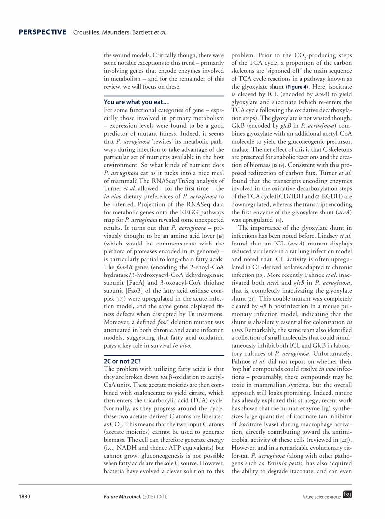
2C or not 2C?The problem with utilizing fatty acids is that they are broken down via β-oxidation to acetyl-CoA units. These acetate moieties are then com-bined with oxaloacetate to yield citrate, which then enters the tricarboxylic acid (TCA) cycle. Normally, as they progress around the cycle, these two acetate-derived C atoms are liberated as CO2. This means that the two input C atoms (acetate moieties) cannot be used to generate biomass. The cell can therefore generate energy (i.e., NADH and thence ATP equivalents) but cannot grow; gluconeogenesis is not possible when fatty acids are the sole C source. However, bacteria have evolved a clever solution to this
problem. Prior to the CO2-producing steps of the TCA cycle, a proportion of the carbon skeletons are ‘siphoned off ’ the main sequence of TCA cycle reactions in a pathway known as the glyoxylate shunt (Figure 4). Here, isocitrate is cleaved by ICL (encoded by aceA) to yield glyoxylate and succinate (which re-enters the TCA cycle following the oxidative decarboxyla-tion steps). The glyoxylate is not wasted though; GlcB (encoded by glcB in P. aeruginosa) com-bines glyoxylate with an additional acetyl-CoA molecule to yield the gluconeogenic precursor, malate. The net effect of this is that C skeletons are preserved for anabolic reactions and the crea-tion of biomass [18,19]. Consistent with this pro-posed redirection of carbon flux, Turner et al. found that the transcripts encoding enzymes involved in the oxidative decarboxylation steps of the TCA cycle (ICD/IDH and α-KGDH) are downregulated, whereas the transcript encoding the first enzyme of the glyoxylate shunt (aceA) was upregulated [14].
The importance of the glyoxylate shunt in infections has been noted before. Lindsey et al. found that an ICL (aceA) mutant displays reduced virulence in a rat lung infection model and noted that ICL activity is often upregu-lated in CF-derived isolates adapted to chronic infection [20]. More recently, Fahnoe et al. inac-tivated both aceA and glcB in P. aeruginosa, that is, completely inactivating the glyoxylate shunt [21]. This double mutant was completely cleared by 48 h postinfection in a mouse pul-monary infection model, indicating that the shunt is absolutely essential for colonization in vivo. Remarkably, the same team also identified a collection of small molecules that could simul-taneously inhibit both ICL and GlcB in labora-tory cultures of P. aeruginosa. Unfortunately, Fahnoe et al. did not report on whether their ‘top hit’ compounds could resolve in vivo infec-tions – presumably, these compounds may be toxic in mammalian systems, but the overall approach still looks promising. Indeed, nature has already exploited this strategy; recent work has shown that the human enzyme Irg1 synthe-sizes large quantities of itaconate (an inhibitor of isocitrate lyase) during macrophage activa-tion, directly contributing toward the antimi-crobial activity of these cells (reviewed in [22]). However, and in a remarkable evolutionary tit-for-tat, P. aeruginosa (along with other patho-gens such as Yersinia pestis) has also acquired the ability to degrade itaconate, and can even
1831
Figure 4. Overview of the glyoxylate shunt pathway in P. aeruginosa. In the glyoxylate shunt (pale orange arrows and metabolites) carbon skeletons are redirected away from the CO2 producing steps of the TCA cycle and retained for gluconeogenesis. The glyoxylate shunt requires two enzymes: isocitrate lyase (ICL) and malate synthase (GlcB) and the net effect of their activity is to convert two molecules of acetyl-CoA into one molecule of malate (a gluconeogenic precursor). In the chronic and acute wound models, the acetyl-CoA is generated as a consequence of FaoAB-mediated fatty acid oxidation. Unlike E. coli, in P. aeruginosa there are two isocitrate dehydrogenase enzymes (ICD and IDH), only one of which (ICD) is likely to be inhibitable by AceK-mediated phosphorylation [53].
C
O
HOOC CH2 COOH
CH2CH2
COOH
COOHC
OH
HOOC
C
COOH
H
COOH
Citrate
Isocitrate
CH2
OH
CHHOOC
CH2 CH2 COOHCHOOC
O
C
H
OH
HOOC COOHCH2 H C C S
H
H
O
CoA
C H
O
HOOC
HOOC CH2CH2 COOH
COOHHOOC CH CHGlcB
ICL
IDH
ICD
Gluconeogenesis
Fumarate
Malate
Oxaloacetate
α-ketoglutarateCO2
CO2
Succinate
Which microbial factors really are important in Pseudomonas aeruginosa infections? PERSPECTIVE
future science group www.futuremedicine.com
live off this compound as a sole C source [23]. Furthermore, recent data suggest that P. aer-uginosa can also degrade other ICL inhibitors, such as nitropropionic acid (produced by some plants and fungi) via the nitronate monooxy-genase activity of the gene encoded by PA4202 (nmoA) [24].
Interestingly, recent 13C flux analyses have indicated that unlike the situation in Escherichia coli, where aceA is only expressed during growth on acetate (or acetate-producing substrates), in P. aeruginosa, there is significant flux through the pathway even during growth on glucose [25]. This suggests that the shunt may play a more general role in the physiology of the organism beyond simply redirecting C skeletons for glu-coneogenesis. Indeed, inhibition of the glyoxy-late shunt may deliver a double whammy to P. aeruginosa; our own team recently showed that ICL activity stimulates type III secretion dur-ing microaerobic growth, suggesting that the absence of the glyoxylate shunt not only results in metabolic insufficiency – it also diminishes
virulence per se [26]. Moreover, itaconate could suppress type III secretion when added to micro-aerobic cultures in vitro. Crucially in this regard, the RNASeq/TnSeq data of Turner et al. strongly suggest that oxygen limitation plays a key role in infection – the anaerobic regulator [27] encoded by anr was among the most strongly attenuated Tn mutants that they identified, especially dur-ing acute infection [14]. These data suggest that oxygen limitation may be a common feature of many P. aeruginosa infections.
Quite why P. aeruginosa chooses a metabolic strategy that is dependent upon the glyoxy-late shunt (instead of utilizing more ‘available’ nutrients) is not clear. However, inspection of the rank-ordered list of attenuated Tn mutants obtained by Turner et al. reveals that insertions in glpD (glycerol 3-phosphate dehydrogenase) were also essentially avirulent in vivo. GlpD converts glycerol 3-phosphate into dihydroxy-acetone phosphate, and a mutant in glpD was recently shown to produce lower levels of cer-tain virulence determinants (pyocyanin and

Future Microbiol. (2015) 10(11)1832
PERSPECTIVE Crousilles, Maunders, Bartlett et al.
future science group
pyoverdine) in vitro [28]. One major possible source of glycerol in vivo is phospholipids, which are broken down to yield fatty acids and glycerol.
To get a better insight into what kinds of nutrient are on the P. aeruginosa menu in a mouse, Turner et al. compared the biosynthetic pathways expressed during growth on minimal succinate media (where most biosynthetic path-ways are necessarily ‘on’) with those expressed in vivo. The logic here was that biosynthetic pathways are generally only transcribed when the corresponding pathway product is not available from the environment (after all, why waste valuable metabolic resources synthesizing say, an amino acid, if the same amino acid is plentiful in the growth media?). Therefore, by comparing these two datasets, they could infer which nutrients might be available to P. aer-uginosa in vivo during wound infections. The results were surprising; nonavailable metabolites included certain amino acids (glutamate, aspar-tate, tyrosine, phenylalanine and asparagine) as well as purines, p-aminobenzoate and B-group vitamins (the precursors for many cofactors). Indeed, and further confirming these results, in vitro-constructed pabC and purA mutants (deficient in p-aminobenzoate and purine bio-synthesis, respectively) were avirulent in vivo. In contrast, mutants defective in metabolites pre-dicted to be ‘available’ in vivo were unaffected in virulence. This striking result strongly sug-gests that components of the pathways required for synthesis of ‘nonavailable’ metabolites may make excellent targets for the development of new antipseudomonal agents.
The situation is slightly different in CF infectionsMore recently, Turner et al. have been using Monte Carlo simulations to rigorously inter-rogate a new set of TnSeq data in which the growth requirements of P. aeruginosa in CF sputum have been investigated. These data have enabled them to refine the composition of artificial CF sputum medium [29]. The gene fitness requirements in this new medium now so closely resemble that in ‘real’ CF sputum that they are all but indistinguishable, thereby providing a far better laboratory model for the study of CF-associated P. aeruginosa infections than previously possible. In the same study, the authors also compared the list of genes that are conditionally essential for the survival of two strains of P. aeruginosa (PAO1 and PA14)
when grown in minimal medium containing authentic CF sputum as a sole nutrient source. Interestingly, most of the conditionally essen-tial genes were located in the conserved ‘core’ genome of both strains. However, clear differ-ences were observed in the relative essentiality of different genes in each strain. Moreover, and although common pathways (e.g., those for chorismate, panthothenate, diaminopime-late, purines, pyridoxal phosphate, riboflavin and certain polyamines) were deemed essen-tial for infection in both wound [14] and CF infections [29], some important differences were apparent. For example, as outlined earlier, fatty acid oxidation is essential in wound infections but seems to play a much less important role during growth in CF sputum. In general, the authors concluded that CF sputum presents a far less harsh environment compared with that encountered in wound infections, although cynically, this may simply be a reflection of the fact that the in vitro CF model used by those workers contains no immune cells, whereas presumably, an exuberant immune response is mounted in the in vivo wound models. Nevertheless, the results of Turner et al. [14,29] and Fahnoe et al. [21] strongly suggest that the targeting specific domains of metabolism may offer a real opportunity to develop some truly innovative antimicrobial interventions. In this regard, it is ironic to note that one of the first antibiotics, sulfanilimide (introduced in the 1930s), targets p-aminobenzoate metabolism.
Real-life infections are more complexOne fly-in-the-ointment in the case for target-ing specific metabolic nodes is that P. aerugi-nosa isolates which are auxotrophic for certain amino acids arise frequently in chronic CF infections [30]. Indeed, we too have identified auxotrophs in CF sputum that are defective in the synthesis of one or more of the 20 common amino acids, as well as in the synthesis of cer-tain cofactors (MW/E-FU/AF [Welch M et al., Unpublished data]). This is surprising given that the availability of several amino acids (e.g., Gln, Tyr, Asn, Asp) is predicted by Turner et al. to be limiting in CF sputum [29]. However, and as noted by Barth and Pitt, CF sputum is cer-tainly rich enough in amino acids to support the growth of some auxotrophs [31]. Additionally, there is even evidence to suggest that high total amino acid concentrations in sputum coincide with acute pulmonary exacerbation episodes and
1833
Which microbial factors really are important in Pseudomonas aeruginosa infections? PERSPECTIVE
future science group www.futuremedicine.com
an increased prevalence of auxotrophs [32]. One possibility is that these sputum amino acids are host-derived. If this is so, then the case for tar-geting P. aeruginosa amino acid biosynthesis for possible therapeutic intervention is weakened. Alternatively, the sputum amino acids could be microbially derived. In this regard, biofilm and anaerobic planktonic cultures of P. aer-uginosa (PAO1) grown in defined medium in vitro are known to accumulate large quantities of amino acids [33], presumably due to overflow metabolism. (We note here that the sputum samples used in the study of Tuner et al. that were found to be limiting for Gln/Tyr/Asn/Asp were [understandably] obtained from a patient with a low bacterial load [29].) This raises the possibility that the population structure of P. aeruginosa in any given chronically infected CF patient may be self-stabilized via intraspecies cross-feeding. For example, Tyr- P. aeruginosa auxotrophs could be sustained off the tyrosine provided by Tyr+ prototrophs, and so on. If this is indeed the case, then amino acids (and pre-sumably, other metabolites too) can be viewed as bacterially produced ‘public goods’ in much the same way that virulence factors have been [34,35]. While this possibility has not yet been rigorously tested experimentally, there is good evidence to suggest that the population of P. aeruginosa in a given sputum sample is both phenotypically and genomically heterogeneous [36–39]. If true and the P. aeruginosa population structure is stabilized by cross-feeding of nutrients, then by targeting the synthesis of one or more of these nutrients, the network of interactions that main-tains population cohesion should collapse (or at least undergo radical remodeling). However, a potential counterargument lies in the fact that CF infections are rarely associated with P. aeruginosa monocultures; a veritable zoo of microbes can be identified (usually from their 16S rDNA signatures) in many CF sputa [40,41], raising further opportunities for cross-feeding. Consequently, drugs targeting specific domains of metabolism may need to have rather broad specificity, allowing them to target enzymes in multiple genera of bacteria.
ConclusionP. aeruginosa is not a professional pathogen; in spite of its genomic arsenal of putative virulence determinants, it is primarily an opportunist that has to adapt to the host environment in order to survive. The TnSeq analyses of Turner et al.
have yielded insights into what the cell itself finds important during this process. The pre-cise physiological adaptations that appear to be truly important are tuned to some extent by the subtle nuances attending the different types of infection in different tissues. However, there is enough common ground between the require-ments in these different infection types to raise the hope that generic new antipseudomonal compounds might be discovered through this avenue of investigation. This notwithstanding, and aside from the issues raised earlier in this discussion, there are caveats. One is the issue of redundancy. For example, although FaoA/FaoB clearly play an important role in wound infec-tions, the acyl-CoA dehydrogenases that neces-sarily precede the action of these enzymes were not identified as being conditionally essential in infection. Presumably, this is because multiple copies of genes encoding acyl-CoA dehydroge-nases are present in the P. aeruginosa genome (seven genes are annotated as putative acyl-CoA dehydrogenases in PAO1) and inactivation of any one of these genes may be compensated by the residual activity of the other isozymes present. A similar argument could be made for ppGpp gen-erating enzymes. The stress alarmone, ppGpp, is required for amino acid biosynthetic pathway expression [42] and has been implicated in viru-lence in many bacterial pathogens (reviewed in [43]). However, for a noticeable virulence phenotype to be manifest, both ppGpp-gen-erating enzymes (RelA and SpoT) need to be inactivated [44]. Similarly, cyclic di-GMP is also known to play a key role in determining lifestyle and virulence in P. aeruginosa, yet this small molecule is made and sensed by a multitude of gene products [45]. Another potential issue relates to the nature of the medium employed for growth of the TnSeq reference samples (recall that Turner et al. used MOPS-buffered succinate medium). This is because the ‘essential gene set’ varies somewhat during growth in vitro in dif-ferent media [46]. Although there is an appar-ent ‘core’ set of ca. 350 genes (many of which are involved in central carbon metabolism) that seem to be essential in most common laboratory growth media, Lee et al. identified ca. 200 genes that appear to be conditionally essential in dif-ferent growth media. Thus, the conclusions of Turner et al. will be inevitably biased somewhat by their specific choice of MOPS-succinate for growth of the reference sample. However, and commensurate with the results discussed earlier,
Future Microbiol. (2015) 10(11)1834
PERSPECTIVE Crousilles, Maunders, Bartlett et al.
future science group
Lee et al. also found that CF sputum is likely to be rich in amino acids and unsaturated fatty acids [46].
Future perspectiveThese arguments aside, the results of Turner et al. must at the very least cause us to reassess our notions about P. aeruginosa virulence. The past two decades of P. aeruginosa virulence research have been dominated by the role(s) played by QS and biofilm formation. However, it turns out that although the ‘key regulators’ associated with these phenotypes (retS, ladS, lasR, gacA, gacS, rsmA, rsmY, rsmZ etc. – [9,10,47]) do feature on the ‘TnSeq attenuated-list,’ for the most part they do not make the ‘top 200’ for any infection type. That is not to say that they do not play an important role in infection under some circum-stances – they do, and we know that. It is just that some rather more mundane (and perhaps, overlooked?) phenotypes seem to be more impor-tant. This may actually be good news; unlike many of the pleiotropic virulence regulators that have become a favored target for antimicrobial research over the last two decades, the enzymes of central metabolism are generally highly con-served across the bacterial genera, and contain discrete ‘drugable’ binding pockets that are ame-nable to drug development. Moreover, we have
historical proof of principle that the targeting of metabolic nodes (re: the sulphonamides) works. Given the dearth of new antimicrobials in the pipeline, coupled with the inexorable spread of resistance to existing antibiotics, it seems likely that over the next few years we will see a renewed interest in targeting central metabolism.
Financial & competing interests disclosureWork in the M Welch laboratory is funded by the BBSRC (grant BB/M019411/1) and the EU (Marie Curie Educational Training Network ‘INTEGRATE’). A Crousilles is supported by the Cambridge Trusts. E Maunders is funded by a studentship from the MRC. S Bartlett is sup-ported by a Hershel Smith studentship. E-F Ukor is a clinical research fellow funded by the CF Trust (UK), Papworth Hospital NHS Trust and the Wellcome Trust. Y Abdelhamid is supported by a scholarship from the Yosef Jameel Foundation. Y Baker is an EPSRC-funded PhD student. A Crousilles is supported by the Cambridge Commonwealth, European and International Trust. Work in the laboratory of A Floto is sup-ported by the Wellcome Trust. Work in the DR Spring labora-tory is supported by the EPSRC. The authors have no other relevant affiliations or financial involvement with any organi-zation or entity with a financial interest in or financial con-flict with the subject matter or materials discussed in the manuscript apart from those disclosed.
No writing assistance was utilized in the production of this manuscript.
EXECUTIVE SUMMARY ● Pseudomonas aeruginosa is an opportunistic pathogen which produces a wealth of secreted virulence factors and
secondary metabolites. These secreted factors have been assumed to play a pivotal role in infection. ● New technologies are allowing us to ‘ask the bacterial cell’, which of its genes are important for infection in mammalian
systems. For example, RNASeq allows quantitation of transcript levels whereas TnSeq reports on the contribution of specific genes toward fitness in vivo.
● In a recent analysis, RNASeq and TnSeq were used in parallel to investigate which P. aeruginosa genes are required for optimal virulence in a murine infection model. Surprisingly, for most classes of gene (including known virulence factors) this analysis revealed that there was very little correlation between transcript abundance and the contribution of the corresponding gene toward overall fitness. However, for certain ‘metabolic genes’, these factors were strongly correlated.
● During some types of infection, P. aeruginosa exhibits a predilection for metabolizing fatty acids. Mutants in the fatty acid oxidase complex are severely attenuated in virulence. In addition, the glyoxylate shunt (an anaplerotic pathway which bypasses the oxidative decarboxylation steps of the TCA cycle) makes an important contribution toward fitness in vivo.
● Certain metabolites (amino acids and vitamins) are not available to P. aeruginosa during in vivo infections, making the biosynthetic pathways producing these compounds excellent potential targets for the development of novel antimicrobials.
● Not all types of infection elicit the same metabolic requirements in P. aeruginosa. Moreover, the relative essentiality of different genes varies between different P. aeruginosa strains.

1835future science group www.futuremedicine.com
Which microbial factors really are important in Pseudomonas aeruginosa infections? PERSPECTIVE
ReferencesPapers of special note have been highlighted as:
1 Foweraker J. Recent advances in the microbiology of respiratory tract infection in cystic fibrosis. Br. Med. Bull. 89, 93–110 (2009).
2 Stover CK, Pham XQ, Erwin AL et al. Complete genome sequence of Pseudomonas aeruginosa PAO1, an opportunistic pathogen. Nature 406, 959–964 (2000).
3 Hoge R, Pelzer A, Rosenau F, Wilhelm S. Weapons of a pathogen: proteases and their role in virulence of Pseudomonas aeruginosa. In: Current Research: Technology And Education Topics In Applied Microbiology And Microbial Biotechnology. Mendez-Vilas A (Ed.).
4 Passmore IJ, Nishikawa K, Lilley KS et al. Mep72, a metzincin protease that is preferentially secreted by biofilms of Pseudomonas aeruginosa. J. Bacteriol. 197, 762–773 (2015).
5 Nouwens AS, Beatson SA, Whitchurch CB et al. Proteome analysis of extracellular proteins regulated by the las and rhl quorum sensing systems in Pseudomonas aeruginosa PAO1. Microbiology 149, 1311–1322 (2003).
6 Pearson JP, Pesci EC, Iglewski BH. Roles of Pseudomonas aeruginosa las and rhl quorum-sensing systems in control of elastase and rhamnolipid biosynthesis genes. J. Bacteriol 179, 5756–5767 (1997).
7 Pearson JP, Feldman M, Iglewski BH, Prince A. Pseudomonas aeruginosa cell-to-cell signaling is required for virulence in a model of acute pulmonary infection. Infect Immun. 68, 4331–4 (2000).
8 Rumbaugh KP, Griswold JA, Iglewski BH, Hamood AN. Contribution of quorum sensing to the virulence of Pseudomonas aeruginosa in burn wound infections. Infect Immun. 67, 5854–5862 (1999).
9 Goodman AL, Kulasekara B, Rietsch A et al. A signaling network reciprocally regulates genes associated with acute infection and chronic persistence in Pseudomonas aeruginosa. Dev Cell 7, 745–754 (2004).
10 Ventre I, Goodman AL, Vallet-Gely I et al. Multiple sensors control reciprocal expression of Pseudomonas aeruginosa regulatory RNA and virulence genes. Proc. Natl Acad. Sci. USA 103, 171–176 (2006).
11 D’Argenio DA, Wu M, Hoffman LR, Kulasekara HD et al. Growth phenotypes of Pseudomonas aeruginosa lasR mutants adapted to the airways of cystic fibrosis patients. Mol. Microbiol. 64, 512–533 (2007).
12 Smith EE, Buckley DG, Wu Z, Saenphimmachak C et al. Genetic adaptation by Pseudomonas aeruginosa to the airways of cystic fibrosis patients. Proc. Natl Acad. Sci. USA 103, 8487–8492 (2006).
13 van Opijnen T, Bodi KL, Camilli A. Tn-seq: high-throughput parallel sequencing for fitness and genetic interaction studies in microorganisms. Nat. Methods 6, 767–772 (2009).
14 Turner KH, Everett J, Trivedi U et al. Requirements for Pseudomonas aeruginosa acute burn and chronic surgical wound infection. PLoS Genet. 10(7), e1004518 (2014).
et al.
in vivo.
P. aeruginosa
15 Jackson KD, Starkey M, Kremer S et al. Identification of psl, a locus encoding a potential exopolysaccharide that is essential for Pseudomonas aeruginosa PAO1 biofilm formation. J. Bacteriol. 186, 4466–4475 (2004).
16 Palmer KL, Aye LM, Whiteley M. Nutritional cues control Pseudomonas aeruginosa multicellular behavior in cystic fibrosis sputum. J. Bacteriol. 189, 8079–8087 (2007).
17 Sato S, Hayashi M, Imamura S et al. Primary structures of the genes, faoA and faoB, from Pseudomonas fragi B-0771 which encode the two subunits of the HDT multienzyme complex involved in fatty acid beta-oxidation. J. Biochem. 111, 8–15 (1992).
18 Cozzone AJ. Regulation of acetate metabolism by protein phosphorylation in enteric bacteria. Annu Rev Microbiol. 52, 127–164 (1998).
19 de la Pena Mattozzi M, Kang Y, Keasling Feast JD. Choking on acetyl-CoA, the glyoxylate shunt, and acetyl-CoA-driven metabolism. In: Handbook of Hydrocarbon and Lipid Microbiology. Timmis KN (Ed.). Springer-Verlag, Berlin, Heidelberg (2010).
20 Lindsey TL, Hagins JM, Sokol PA, Silo-Suh LA. Virulence determinants from a cystic fibrosis isolate of Pseudomonas aeruginosa include isocitrate lyase. Microbiology 154, 1616–1627 (2008).
21 Fahnoe KC, Flanagan ME, Gibson G et al. Non-traditional antibacterial screening approaches for the identification of novel inhibitors of the glyoxylate shunt in gram-negative pathogens. PLoS ONE 7(12), e51732 (2012).
P. aeruginosa
22 Ménage S, Attrée I. Metabolism: pathogens love the poison. Nat. Chem. Biol. 10, 326–327 (2014).
23 Sasikaran J, Ziemski M, Zadora PK et al. Bacterial itaconate degradation promotes pathogenicity. Nat. Chem. Biol. 10, 371–377 (2014).
24 Vercammen K, Wei Q, Charlier D et al. Pseudomonas aeruginosa LysR PA4203 regulator NmoR acts as a repressor of the PA4202 nmoA gene, encoding a nitronate monooxygenase. J. Bacteriol. 197, 1026–1039 (2015).
25 Berger A, Dohnt K, Tielen P, Jahn D, Becker J, Wittmann C. Robustness and plasticity of metabolic pathway flux among uropathogenic isolates of Pseudomonas aeruginosa. PLoS ONE 9(4), e88368 (2014).
26 Chung JC, Rzhepishevska O, Ramstedt M, Welch M. Type III secretion system expression in oxygen-limited Pseudomonas aeruginosa cultures is stimulated by isocitrate lyase activity. Open Biol. 3, 120131 (2013).
P. aeruginosa
27 Trunk K, Benkert B, Quäck N et al. Anaerobic adaptation in Pseudomonas aeruginosa: definition of the Anr and Dnr regulons. Environ. Microbiol. 12, 1719–1733 (2010).
28 Daniels JB, Scoffield J, Woolnough JL, Silo-Suh L. Impact of glycerol-3-phosphate dehydrogenase on virulence factor production by Pseudomonas aeruginosa. Can. J. Microbiol. 60, 857–863 (2014).
29 Turner KH, Wessel AK, Palmer GC et al. Essential genome of Pseudomonas aeruginosa in cystic fibrosis sputum. Proc. Natl Acad. Sci. USA 112, 4110–5 (2015).
P. aeruginosa
30 Barth AL, Pitt TL. Auxotrophic variants of Pseudomonas aeruginosa are selected from prototrophic wild-type strains in respiratory
Future Microbiol. (2015) 10(11)1836
PERSPECTIVE Crousilles, Maunders, Bartlett et al.
future science group
infections in patients with cystic fibrosis. J. Clin. Microbiol. 33, 37–40 (1995).
31 Barth AL, Pitt TL. The high amino-acid content of sputum from cystic fibrosis patients promotes growth of auxotrophic Pseudomonas aeruginosa. J. Med. Microbiol. 45, 110–119 (1996).
32 Thomas SR, Ray A, Hodson ME, Pitt TL. Increased sputum amino acid concentrations and auxotrophy of Pseudomonas aeruginosa in severe cystic fibrosis lung disease. Thorax 55, 795–797 (2000).
33 Patell S, Gu M, Davenport P, Givskov M, Waite RD, Welch M. Comparative microarray analysis reveals that the core biofilm-associated transcriptome of Pseudomonas aeruginosa comprises relatively few genes. Environ. Microbiol. Rep. 2, 440–448 (2010).
34 Rainey PB, Rainey K. Evolution of cooperation and conflict in experimental bacterial populations. Nature 425, 72–74 (2003).
35 Diggle SP, Griffin AS, Campbell GS, West SA. Cooperation and conflict in quorum-sensing bacterial populations. Nature 450, 411–414 (2007).
36 Chung JC, Becq J, Fraser L et al. Genomic variation among contemporary Pseudomonas aeruginosa isolates from chronically infected cystic fibrosis patients. J. Bacteriol. 194, 4857–4866 (2012).
37 Williams D, Evans B, Haldenby S et al. Divergent, coexisting Pseudomonas aeruginosa lineages in chronic cystic fibrosis lung infections. Am. J. Respir. Crit. Care Med. 191, 775–785 (2015).
38 Mowat E, Paterson S, Fothergill JL et al. Pseudomonas aeruginosa population diversity and turnover in cystic fibrosis chronic infections. Am. J. Respir. Crit. Care Med. 183, 1674–1679 (2011).
P. aeruginosa
39 Foweraker JE, Laughton CR, Brown DF, Bilton D. Phenotypic variability of Pseudomonas aeruginosa in sputa from patients with acute infective exacerbation of cystic fibrosis and its impact on the validity of antimicrobial susceptibility testing. J. Antimicrob. Chemother. 55, 921–927 (2005).
40 Twomey KB, Alston M, An SQ et al. Microbiota and metabolite profiling reveal specific alterations in bacterial community structure and environment in the cystic fibrosis airway during exacerbation. PLoS ONE 8, e82432 (2013).
41 Lynch SV, Bruce KD. The cystic fibrosis airway microbiome.. Cold Spring Harb. Perspect. Med. 3, a009738 (2013).
42 Traxler MF, Summers SM, Nguyen HT et al. The global, ppGpp-mediated stringent response to amino acid starvation in Escherichia coli. Mol. Microbiol. 68, 1128–1148 (2008).
43 Dalebroux ZD, Svensson SL, Gaynor EC, Swanson MS. ppGpp conjures bacterial virulence. Microbiol. Mol. Biol. Rev. 74, 171–199 (2010).
44 Bowden SD, Eyres A, Chung JC et al. Virulence in Pectobacterium atrosepticum is regulated by a coincidence circuit involving quorum sensing and the stress alarmone, (p)ppGpp. Mol. Microbiol. 90, 457–471 (2013).
45 Moscoso JA, Mikkelsen H, Heeb S et al. The Pseudomonas aeruginosa sensor RetS switches type III and type VI secretion via c-di-GMP signalling. Environ. Microbiol. 13, 3128–3138 (2011).
46 Lee SA, Gallagher LA, Thongdee M et al. General and condition-specific essential
functions of Pseudomonas aeruginosa. Proc. Natl Acad. Sci. USA 112, 5189–5194 (2015).
P. aeruginosain vitro.
47 Brencic A, McFarland KA, McManus HR et al. The GacS/GacA signal transduction system of Pseudomonas aeruginosa acts exclusively through its control over the transcription of the RsmY and RsmZ regulatory small RNAs. Mol. Microbiol. 73, 434–445 (2009).
48 Filloux A, Michel G, Bally M. GSP-dependent protein secretion in Gram-negative bacteria: the Xcp system of Pseudomonas aeruginosa. FEMS Microbiol. Rev. 22, 177–198 (1998).
49 Ball G, Durand E, Lazdunski A, Filloux A. A novel type II secretion system in Pseudomonas aeruginosa. Mol. Microbiol. 43, 475–485 (2002).
50 Bleves S, Viarre V, Salacha R, Michel GP, Filloux A, Voulhoux R. Protein secretion systems in Pseudomonas aeruginosa: a wealth of pathogenic weapons. Int. J. Med. Microbiol. 300, 534–543 (2010).
51 Filloux A. Protein secretion systems in Pseudomonas aeruginosa: an essay on diversity, evolution, and function. Front. Microbiol. 2, 155 (2011).
52 Franklin MJ, Nivens DE, Weadge JT, Howell PL. Biosynthesis of the Pseudomonas aeruginosa extracellular polysaccharides, alginate, Pel, and Psl. Front. Microbiol. 2, 167 (2011).
53 LaPorte DC, Koshland DE Jr. Phosphorylation of isocitrate dehydrogenase as a demonstration of enhanced sensitivity in covalent modification. Nature 305 , 286–290 (1983).