Embed Size (px)
Citation preview
W. Joseph MacInnes, Roopali Bhatnagar
WHERE DOES ATTENTION GO
WHEN FACILITATION IS
ABSENT?
BASIC RESEARCH PROGRAM
WORKING PAPERS
SERIES: PSYCHOLOGY
WP BRP 85/PSY/2017
This Working Paper is an output of a research project implemented at the National Research
University Higher School of Economics (HSE). Any opinions or claims contained in this
Working Paper do not necessarily reflect the views of HSE
1
W. Joseph MacInnes1, Roopali Bhatnagar
2
WHERE DOES ATTENTION GO WHEN FACILITATION IS ABSENT?
Attending a location in space facilitates responses to targets at that location when the time
between cue and target is short. Certain types of exogenous cues – such as sudden peripheral onsets –
have been described as reflexive and automatic. Recent studies however, have been showing many
cases where exogenous cues are less automatic than previously believed and do not always result in
facilitation. Given a lack of the behavioural facilitation, we propose to test whether this also suggests a
lack of underlying attention to that location. We test exogenous cueing with saccadic responses at a
range of cue-target onset asynchronies (CTOAs) , but also alternate measures linked to the allocation of
attention such as saccadic curvature, microsaccades and pupil size. As expected, we find no early
facilitation at short CTOAs, and likewise no impact of the cue on microsaccade direction or pupil size.
We do observe a small dip in the frequency of microsaccades after the cue as well as a tendency for
saccadic curvature away from the cued location at short CTOAs. We interpret these results as evidence
of reduced attention at the cued location that is removed or inhibited too quickly to be measured as
facilitation of saccadic responses.
Keywords: Attention; Eye Movements; microsaccades; facilitation; Inhibition of return
JEL Classification: Z
1 Assistant Professor, [email protected], National Research University, Higher School of Economics, Moscow, Russia 2 Graduate Student, [email protected], National Research University, Higher School of Economics, Moscow, Russia
2
Introduction
Selective attention allows our visual system to preferentially process some information over
others. Theories of attentional control often revolve around the dichotomy between top-down and
bottom-up control processes, also described as endogenous and exogenous attention. Endogenous
attention represents goal-driven processes (Beauchamp, Cox, & Deyoe, 1997; Bressler, Tang,
Sylvester, Shulman, & Corbetta, 2008; Giesbrecht, Woldorff, Song, & Mangun, 2003) while exogenous
is guided by stimulus-properties (Schreij, Owens, & Theeuwes, 2008; Theeuwes, 1991, 1992). Other
factors have been suggested to supplement or modulate this dichotomy such as selection history (Awh,
Belopolsky, & Theeuwes, 2012), associated reward (Anderson, Laurent, & Yantis, 2011; Theeuwes &
Belopolsky, 2012), context learning & memory (Chun & Jiang, 2003); task demands and complexity
(Castel, Pratt, Chasteen, & Scialfa, 2005; Lupiáñez & Milliken, 1999; Lupiáñez et al., 1997), prior
experience (Leber, Kawahara, & Gabari, 2009); or a temporal continuum of top down and bottom up
processes (van Zoest, Donk, & Theeuwes, 2004).
Posner’s cueing paradigm (Posner et al., 1980) has served as the backdrop in understanding
spatial and temporal interaction of visual attention by adjusting cue/target properties to see how they
affect responses to attended locations (mental chronometry). Typically, a location in space is cued with
a peripheral onset or central arrow followed by a target at the cued or uncued location. Short cue-target
onset asynchronies (CTOAs) will result in a facilitatory effect (faster RTs for cued targets) and longer
CTOAs in an inhibitory effect termed inhibition of return (IOR: slower RTs for cued targets). The
switch from facilitation to inhibition, is usually observed around a CTOA of 250-300 milliseconds (ms)
though this may depend on task demands (Lupiáñez, Milliken, Solano, Weaver, & Tipper, 2001). The
facilitatory effect can be explained as an orientation of attention towards the cued location and
improving further processing of the following target onset. At longer CTOAs, however, after visual
attention is disengaged from the cued location facilitation gives way to inhibition of return (IOR;
Posner & Cohen,1984). Spatial cueing effects using predictive cues have been demonstrated in other
species as well – monkeys (Cook & Maunsell, 2002), rats (Marote and Xavier, 2011), honeybees
(Eckstein, et al., 2013), archer fish (Gabay et al., 2013; Saban, Sekely, Klein, & Gabay, 2017)
highlighting their potential role in species survival. IOR is thus, seen as a ‘foraging facilitator’ (Klein &
MacInnes, 1999) and has been suggested to improve search efficiency by reducing the likelihood of
attention returning to already fixated locations (Klein & Macinnes, 1999; Bays & Hussain, 2012;
MacInnes et al., 2014; but see Smith & Henderson, 2011). So, it may seem that looking at a relevant
3
location twice may be part of human fixation selection strategy which is in fact a trade-off between
foraging for novelty and fully understanding the relevant parts (Wilming, Harst, Schmidt, & König,
2013). Although facilitation from exogenous orienting is often described as reflexive and automatic, a
number of studies have reported no facilitation at shorter CTOAs but instead early onset IOR (Tassinari
& Berlucchi, 1993; Tassinari et al., 1994). Danziger & Kingstone (1999) for example, observe IOR
within 50ms at the cued location and Maruff et al., (1999) observed facilitation at short CTOAs but
only if the cue and target overlapped temporally. Pratt, Hillis & Gold (2001) demonstrated the
influence of spatial overlap and physical characteristics of stimuli on cueing effects by using three
different types of cues. Out of the three experiment conditions, only one showed typical cueing effects
while the others showed insignificant or zero facilitation at short CTOAs. Pratt, Sekuler & McAuliffe
(2001) suggested an influence of attentional set on early facilitation. Taylor, Chan, Bennet, & Pratt
(2015) observed no facilitation and early IOR when potential target locations were not marked with
placeholders. MacInnes (2017) tested the spatial and temporal gradient of IOR with continuous random
CTOAs and also found no early facilitation for either manual or saccadic responses. Malevich,
Ardasheva, Krueger and MacInnes (2017) tested the influence of temporal expectations on cueing
effects and found no facilitation when the multiple CTOAs were random or mixed, but only observed
facilitation when the CTOAs were blocked. There seems little doubt that attentional set and top down
expectation can modulate the appearance of facilitation but what remains uncertain, is whether
attention was allocated to the cued location and removed too early to influence reaction times (RTs;
Klein, 2000; Malevich et al., 2017) or whether attention is absent completely.
Alternative measures of attention deployment
Saccadic curvature
Although RTs have become a standard in measuring the deployment of spatial attention, a
number of alternative methods have been proposed. Saccades to target locations are generally not
straight, and the curvature deviation from a straight path has been shown to be influenced by covert
attention (Sheliga, Riggio, Craighero, & Rizzolatti, 1995; Van der Stigchel & Theeuwes, 2007).
Additionally, the strength of saccadic deviation reflects the amount of attention to a particular location
as measured by target RTs. The trajectories of saccades deviating away from an attended location has
been consistently seen in studies, but this effect does not translate to hand movements (Van der
Stigchel, Meeter, & Theeuwes, 2007b).
4
The temporal aspects of saccadic deviations show the same biphasic pattern as reaction times
over increasing CTOAs (McSorley, Haggard, & Walker, 2006). McSorley et al., 2006 reported that
deviations away from a distractor were observed for longest latencies and deviations towards a
distractor in case of shorter latencies with the transition point around latency of 200ms. The same,
however, does not hold for anti-saccades and longest latencies did not correspond to greatest distractor
caused deviations (van Zoest, Van der Stigchel, & Barton, 2008). Saccadic deviations are also
influenced by the distance of the distractor to the target (McSorley, Cruickshank, & Inman, 2009; Van
der Stigchel & Theeuwes, 2005), vertical distance of the distractor from the fixation (Van der Stigchel,
Meeter, & Theeuwes, 2007a) and the target hemifield (Van der Stigchel & Theeuwes, 2008).
Also, similar to reaction times, curvature deviations may change based on prior knowledge
about the task (Walker, McSorley, & Haggard, 2006). In scenarios where target locations were known
or predictable, saccade trajectories were found to be deviating away from the distractors and scenarios
where target locations were unpredictable, saccades curved towards distractors
Microsaccades
The human visual system has been adapted to detect motion and so any stationary, unchanging
scene would cause perceptual fading as the retina adapts to it. To counter this effect, oculomotor
system generates micro movements (drifts, tremors and microsaccades) during a fixation.
Microsaccades are fixational eye movements that are involuntary and ballistic with an average rate of
1-3 per second, magnitude of 12 to 15 minutes of arc and a typical duration of less than 10ms (but see
Kowler, 2011 for an overview and why these sizes have been increasing). Microsaccades and saccades
seem to be kinematically similar, existing on a functional continuum, implicating similar neural
circuitry (Hafed, 2011). It has been shown that microsaccades occur not just during fixation but also
during search and exploratory tasks (Martinez-Conde, Otero-Millan & Macknik, 2013).
Recent results suggest that microsaccades are modulated by visual attention in spatial cueing
paradigms (Engbert & Kliegl, 2003; Hafed & Clark, 2002). Engbert & Kliegl (2003) reported that
microsaccades tend to be biased towards the cued location in a spatial cueing task, but many other
studies have shown microsaccade bias both towards and away from cue direction (Galfano, Betta, &
Turatto, 2004; Hafed & Clark, 2002; Laubrock, Engbert, & Kliegl, 2005; Rolfs, Engbert, & Kliegl,
2004). An interaction with the cue type has also been noted – endogenous attentional cues biasing
microsaccade direction towards the cue, as governed by attentional shifts (Gowen et al., 2007;
Laubrock, Kliegl, Rolfs, & Engbert, 2010; Pastukhov & Braun, 2010) and exogenous attentional cues
5
biasing microsaccade direction away from the cue, as per IOR (Galfano et al., 2004). Attentional cues
also affect microsaccade rate (Laubrock et al., 2005; Cui, Wilke, Logothetis, Leopold, & Liang, 2009)
as does task difficulty (Pastukhov & Braun, 2010). This has lead Laubrock, J., Engbert, R., Rolfs, M.,
& Kliegl, R. (2007) to propose that both microsaccade direction and RTs are strong indicators of spatial
attention (but see Horowitz et al. 2007)). Interestingly, microsaccades show biphasic modulation; that
is, at stimulus onset, microsaccade rate drops to zero immediately and then recovers. This is known as
‘microsaccadic inhibition’ (Engbert & Kliegl, 2003; Rolfs, 2009) and is interpreted as a top down effect
on microsaccades to modulate sensory signal quality.
Pupil size
Pupil size changes are a result of the interaction of the parasympathetic and sympathetic
components of the autonomic nervous systems (ANS). The primary pupillary function being regulation
of light entering the eye, resulting in pupillary light reflex (PLR). Pupil dilations have been noted due
to factors other than luminance changes, like individual differences, cognitive load (Beatty & Wagoner,
1978; Kahneman & Beatty, 1966), emotions (Partala & Surakka, 2003), attention (Beatty, 1977) and
stimulus probability (Reinhard & Lachnit, 2002), along with color perception and faces.
Pupil size, on average, is about 3 mm, which can increase by more than double (approx. 120%)
due to change in illumination, but only by 0.5 mm due to cognitive factors (Beatty & Lucero-Wagoner,
2000). Koss (1986) suggests there is a strong link between the locus coeruleus norepinephrine (LC-NE)
system and the pupillary response, hence a change in LC activation can be tracked through changes in
pupil size. LC-NE neurons project to a large number of brain areas, especially areas associated with
attention – superior colliculus, parietal cortex, pulvinar nucleus.
Gabay, Pertzov, & Henik (2011), measured pupillary response in monkeys in localization and
discrimination tasks and suggested a correlation between pupil size and IOR at cue onset. Mathôt and
colleagues (2013), tie the PLR to modulations in covert attention and suggest that this may provide a
measure of behavioral cueing effects.
Proposal
In our study, we follow the continuous CTOA design (MacInnes, 2017; Malevich et al., in
press) with a saccadic response and four possible target locations. With target validity and CTOA
selection chosen randomly within each block, we expect to observe robust IOR at mid to late CTOAs
6
but expect no facilitation at early CTOAs. With four possible equi-eccentric placeholders for cue and
target (top, bottom, left and right), invalid target locations are possible at opposite and orthogonal to the
cued location. This design allows us to test the impact of attention at valid and invalid spatial locations
using a number of metrics not related to saccadic reaction time, i.e. saccadic trajectory, microsaccades
and pupil size. If exogenous attention is automatically pulled to the cued location it is possible that we
will see its influence in microsaccades or pupil size at short CTOA, or in the saccadic curvature in trials
where the cue is orthogonal to the target. If attention is not allocated to the exogenous cue, we expect
to see no impact of target validity on any of our saccadic metrics.
Methods
Participants
Thirty participants (one excluded due to insufficient data; 14 males, 15 females in the age range
19 – 44 years; mean = 25 years) took part in the experiment. All participants reported normal or
corrected-to-normal vision and no color blindness. Written informed consent was provided and an
honorarium of 200 Rubles was given at the end of the session. The experiment was conducted with the
approval of the Higher School of Economics (HSE) ethics board.
Stimuli and Apparatus
Stimuli were presented on an ASUS VG248QE LCD monitor running at 120 Hz with a
1920x1080 pixels resolution and eye movements were recorded with SR-Research EyeLink II system
(SR Research, Mississauga, Ontario, Canada) at a temporal resolution of 1000 Hz. Stimuli were
generated using MATLAB (MathWorks, Natick, MA, USA) and Psychophysics Toolbox extension
(Brainard, 1997; Pelli, 1997). A nine-point eye tracker calibration and validation procedure were done
for each participant at the beginning of the session. The participant’s head was placed in a chinrest so
that the eyes are at a distance of 80 cm from the screen. The stimuli were viewed binocularly, but eye
movements from the right eye only, were analyzed. Stimuli and an experimental procedure are
illustrated in Figure 1. Each trial consisted of a fixation display with a gray background (B2B2B2), for
a duration of 400ms, showing a black (000000) central fixation cross and four black square
placeholders (top, bottom, right, and left; 5 degrees of visual angle from fixation). The exogenous cue
(white flash) appeared at any one of the four equi-eccentric locations (equal probability) for a duration
7
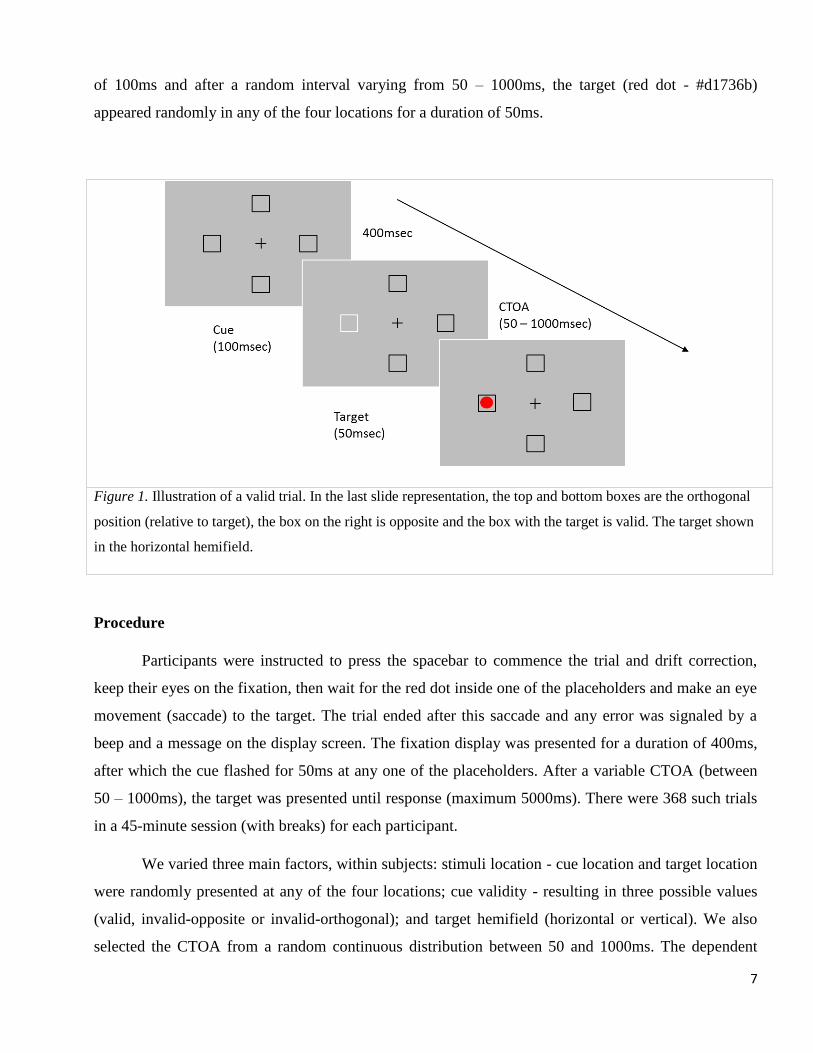
of 100ms and after a random interval varying from 50 – 1000ms, the target (red dot - #d1736b)
appeared randomly in any of the four locations for a duration of 50ms.
Figure 1. Illustration of a valid trial. In the last slide representation, the top and bottom boxes are the orthogonal
position (relative to target), the box on the right is opposite and the box with the target is valid. The target shown
in the horizontal hemifield.
Procedure
Participants were instructed to press the spacebar to commence the trial and drift correction,
keep their eyes on the fixation, then wait for the red dot inside one of the placeholders and make an eye
movement (saccade) to the target. The trial ended after this saccade and any error was signaled by a
beep and a message on the display screen. The fixation display was presented for a duration of 400ms,
after which the cue flashed for 50ms at any one of the placeholders. After a variable CTOA (between
50 – 1000ms), the target was presented until response (maximum 5000ms). There were 368 such trials
in a 45-minute session (with breaks) for each participant.
We varied three main factors, within subjects: stimuli location - cue location and target location
were randomly presented at any of the four locations; cue validity - resulting in three possible values
(valid, invalid-opposite or invalid-orthogonal); and target hemifield (horizontal or vertical). We also
selected the CTOA from a random continuous distribution between 50 and 1000ms. The dependent
8
measures were the saccadic reaction time (SRT in ms), saccadic curvature, microsaccades between
presentation of cue and target, and pupil size changes to the cue (pre- to post- onset).
Data analysis
Anticipatory responses or RTs < 100 ms (4.13%), keyboard press errors (0.77%), fixation errors
(7.30%), outliers with RTs > 3 SD from mean (1.2%) and trials with blinks (1.09%) were excluded.
Hence, 14.5% trials excluded using these criteria.
Saccadic reaction time was defined as the latency from target onset to saccade initiation, in
milliseconds. Saccadic amplitude was defined as the shortest distance between saccade start and end
co-ordinates in degrees of visual angle, and direction was defined as the angular deviation of saccade
direction taken from the initial fixation location to the final endpoint in polar coordinates. The direction
and magnitude of saccadic curvature was calculated by finding the area under the saccade trajectory
curve (Ludwig & Gilchrist, 2002), which was further normalized by dividing by the saccadic amplitude
to get curvature per unit. For trials in which the target was orthogonal to the cued location, trajectories
were signed as positive if they deviated towards the cued location and those deviating away from the
cue were assigned negative curvature values.
Pupil size was measured as difference score, comparing pre-cue baseline measurement with
post-cue measurement, reflecting pupil size change linked to cue onset. Using the change in pupil size
removes potential confounds due to ambient lighting, stimuli luminance and individual differences. If
pupil size represents arousal due to awareness of the cue, then larger changes in pupil size resulting
from that cue might predict the existence or magnitude of facilitation in those trials. Microsaccades
measured in the duration between cue onset and target onset, were investigated monocularly and
analyzed using the velocity-based detection algorithm specified by Engbert & Mergenthaler (2006).
The time series of gaze co-ordinates was first converted to velocity in the horizontal and vertical space
and separate detection thresholds were set for both components. Microsaccades were detected as
outliers in the velocity domain as defined by these detection thresholds.
Statistical analysis was done using the linear mixed effects model (lme4; Winter, 2013; Bates,
Maechler, Bolker, & Walker, 2015; Baayen, Davidson, & Bates, 2008) in the R statistical package (R
Core Team, 2017). For the linear mixed effects model, we first defined a null model, only with random
factors (participants), and incrementally added fixed effects and random slopes (target hemifield, cue-
target location, CTOA and pupil size change) to the model to see if their inclusion improved the model.
We used the chi-squared (χ2) test to check if a new model was an improvement over the previous one.
9
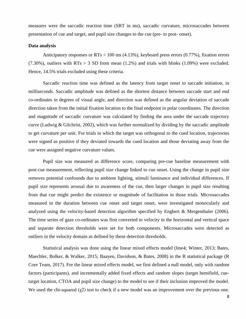
The model was tested for both slopes and intercepts, with by participant random slopes for CTOA
improving the model fit (χ2 (2) = 295, p<0.001), with all other fixed effects being intercepts. There was
no difference between the two invalid trial options (opposite and orthogonal, t < 1.0: and see figure 2a)
so these were combined for a single ‘invalid’ option for target location. Zero condition for all models
was selected as invalid, horizontal hemifield and 50ms CTOA.
Results
The mean model reaction time was 323ms, standard error 8.5ms (for the baseline condition of
horizontal target hemifield, 50ms CTOA, invalid trial) for the final model. There was a significant main
effect of cue validity (χ2 (1) = 148, p<0.001) as validly cued locations had slower RTs (25ms, SE 2ms)
than invalid trials (Fig. 2.). This effect of IOR was significant from the outset of 50ms with no validity
by CTOA interaction (χ2 (1) = 0.02, p=.876, see also figure 2, B). CTOA was also significant (χ2 (1) =
45, p<0.001) with faster RTs (6.3ms/100ms CTOA, SE .6ms) at late CTOAs. We observed strong
significant effect (χ2 (1) = 397, p<0.001) of target hemifield on the saccadic RT with saccades made in
the vertical hemifield slower (37ms, SE 2ms) than those made in the horizontal hemifield.
A) B)
Figure 2. Main effect of IOR with valid trials slower than invalid trials. A) Invalid trials did not differ between
the opposite and orthogonal location. B) Experiment 1 - SRT results. The valid and invalid RTs (ms) at each
CTOA (ms), demonstrating lack of facilitation and early onset IOR.
10
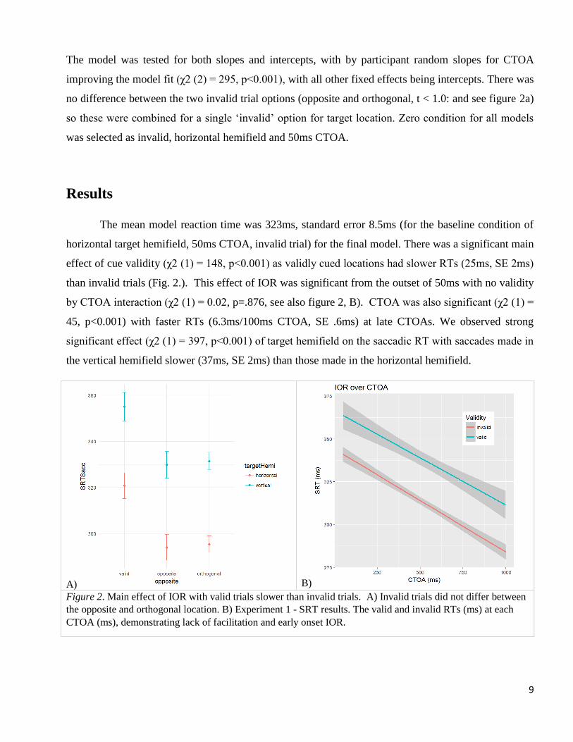
We also tried to fit the change in pupil size as a fixed effect to see if there was a change in
overall cue awareness as measured by pupil size. Although an interaction of pupil size and target
hemifield does help predict overall SRT (χ2 (2) = 8.9, p=0.011), pupil size does not interact with
validity to differentially influence valid cues at any CTOA.
A) B)
Figure 3. Saccadic curvature results. A) Sample saccades for a participant, centered at x, y, co -ordinates (0, 0).
B) The orthogonal cue invoked greater negative curvature at early CTOAs with no curvature bias at late CTOAs
.
For saccadic curvature analyses, a LME was tested for orthogonal cues specifically with fixed
effects of CTOA and hemifield. There was a small but significant effect of CTOA (χ2 (3) = 3.9,
p=0.048) with early CTOAs showing a negative curvature (-.98 away from the cued location, SE .45)
and becoming positive (toward the cue) as CTOA increases (Fig. 3). No other effects were significant
on saccadic curvature.
11
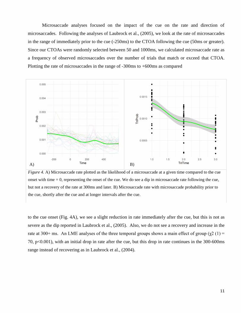
Microsaccade analyses focused on the impact of the cue on the rate and direction of
microsaccades. Following the analyses of Laubrock et al., (2005), we look at the rate of microsaccades
in the range of immediately prior to the cue (-250ms) to the CTOA following the cue (50ms or greater).
Since our CTOAs were randomly selected between 50 and 1000ms, we calculated microsaccade rate as
a frequency of observed microsaccades over the number of trials that match or exceed that CTOA.
Plotting the rate of microsaccades in the range of -300ms to +600ms as compared
to the cue onset (Fig. 4A), we see a slight reduction in rate immediately after the cue, but this is not as
severe as the dip reported in Laubrock et al., (2005). Also, we do not see a recovery and increase in the
rate at 300+ ms. An LME analyses of the three temporal groups shows a main effect of group (χ2 (1) =
70, p<0.001), with an initial drop in rate after the cue, but this drop in rate continues in the 300-600ms
range instead of recovering as in Laubrock et al., (2004).
A) B)
Figure 4. A) Microsaccade rate plotted as the likelihood of a microsaccade at a given time compared to the cue
onset with time = 0, representing the onset of the cue. We do see a dip in microsaccade rate following the cue,
but not a recovery of the rate at 300ms and later. B) Microsaccade rate with microsaccade probability prior to
the cue, shortly after the cue and at longer intervals after the cue.
12
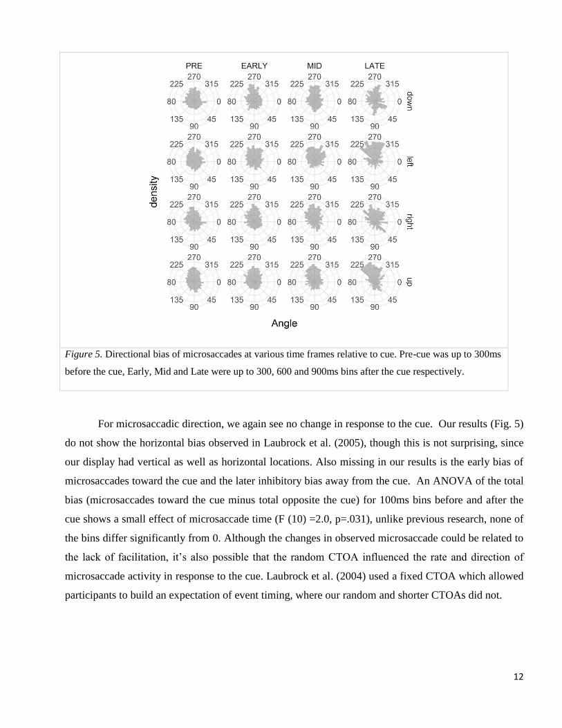
Figure 5. Directional bias of microsaccades at various time frames relative to cue. Pre-cue was up to 300ms
before the cue, Early, Mid and Late were up to 300, 600 and 900ms bins after the cue respectively.
For microsaccadic direction, we again see no change in response to the cue. Our results (Fig. 5)
do not show the horizontal bias observed in Laubrock et al. (2005), though this is not surprising, since
our display had vertical as well as horizontal locations. Also missing in our results is the early bias of
microsaccades toward the cue and the later inhibitory bias away from the cue. An ANOVA of the total
bias (microsaccades toward the cue minus total opposite the cue) for 100ms bins before and after the
cue shows a small effect of microsaccade time (F (10) =2.0, p=.031), unlike previous research, none of
the bins differ significantly from 0. Although the changes in observed microsaccade could be related to
the lack of facilitation, it’s also possible that the random CTOA influenced the rate and direction of
microsaccade activity in response to the cue. Laubrock et al. (2004) used a fixed CTOA which allowed
participants to build an expectation of event timing, where our random and shorter CTOAs did not.
13
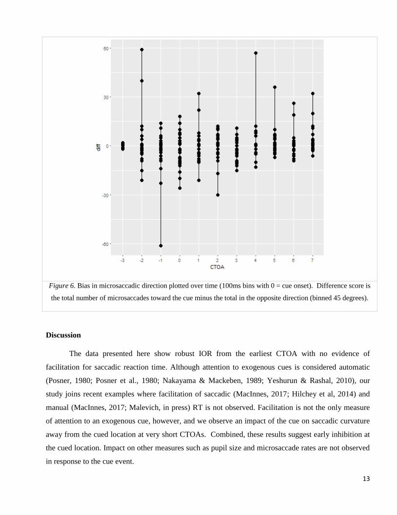
Figure 6. Bias in microsaccadic direction plotted over time (100ms bins with 0 = cue onset). Difference score is
the total number of microsaccades toward the cue minus the total in the opposite direction (binned 45 degrees).
Discussion
The data presented here show robust IOR from the earliest CTOA with no evidence of
facilitation for saccadic reaction time. Although attention to exogenous cues is considered automatic
(Posner, 1980; Posner et al., 1980; Nakayama & Mackeben, 1989; Yeshurun & Rashal, 2010), our
study joins recent examples where facilitation of saccadic (MacInnes, 2017; Hilchey et al, 2014) and
manual (MacInnes, 2017; Malevich, in press) RT is not observed. Facilitation is not the only measure
of attention to an exogenous cue, however, and we observe an impact of the cue on saccadic curvature
away from the cued location at very short CTOAs. Combined, these results suggest early inhibition at
the cued location. Impact on other measures such as pupil size and microsaccade rates are not observed
in response to the cue event.
14
Saccadic curvature deviations have been explained in terms of population coding theory
(Tipper, Howard, & Houghton, 2000; McSorley, Haggard, & Walker, 2004), which states that each
neuron in the motor map aligns to a vector, coding movement towards the corresponding location. Eye
movements are generated in the direction of the average of the vectors in the oculomotor system. When
there are two objects close to each other, the average movement vector would point to an intermediate
location. In the case where a single object has to be selected (target), the average movement vector will
involve suppression of one vector and hence, a deviation away from the distractor. On the other hand, if
the suppression is weak, it results in a deviation towards a distractor. Another interpretation, on the
basis of neurophysiological results, states that saccade trajectories are initiated on the basis of weighted
average of the corresponding vectors (Robinson, 1972). These results focus, primarily, on the superior
colliculus (SC) area of the brain which represents a vector map of the external world and receives
inputs from other cortical areas. So, stimulation of particular cells in the SC result in saccades to the
location corresponding to the stimulated location. Saccade deviations are seen as competitive
interaction within the layers of the SC and a measure of the oculomotor activity.
Other explanations of saccadic trajectories involve effect of distractor, temporal and spatial
aspects of oculomotor inhibition (Van der Stigchel, 2010). In distractor paradigm, deviation towards
the distractor indicates that the distractor activity has not been fully inhibited, deviation away from the
distractor represents complete inhibition of distractor activity. This inhibition is further influenced by
the strength of the stimulus and the distractor location. Walker, McSorley, & Haggard (2006) suggest
that top down inhibitory processes, originating in the frontal eye field (FEF), are applied before
stimulus onset when target location is known in advance. In unpredictable target conditions like our
paradigm, we should not see this preparatory inhibition process and should expect a greater possibility
of saccades deviating towards the distractor. Given the early deviation away from the uninformative
(distractor) cue in our results, we are likely seeing an early suppression of the oculomotor system with
no automatic attention to the cued location.
Although microsaccades are typically reflexive, they can be controlled voluntarily without
retinal fading and in some high-acuity tasks they do get suppressed automatically. There seems to be a
fixed relation between the micro-saccadic amplitude (degrees) and velocity (degrees/sec) which is seen
in a typical linearly increasing pattern called ‘main sequence’ (Bahill, Clark & Stark, 1975), indicating
a common generation mechanism for saccades and microsaccades (Otero-Millan, Troncoso, Macknik,
Serrano-Pedraza, & Martinez-Conde, 2008; Rolfs, Laubrock & Kliegl, 2008; Zuber, Stark & Cook,
1965). While some have observed more horizontal and vertical than oblique microsaccades (Engbert,
15
2006), this seems to be task dependent as we observe no effect here with horizontal and vertical targets
equally likely.
The distinction between pupillary dilation (driven by LC) and PLR (controlled by SC activity)
has been highlighted as they refer to different underlying mechanisms (Loewenfeld, 1958). It has been
demonstrated that PLR is modulated by higher level cognition or covert attention and should be
considered similar to spatial eye movements, seeing that both saccades and PLR have their origins in
SC (Wang et al., 2012; Wang, Brien & Munoz, 2015). Although there may have been an influence of
PLR on the cognitive demands of our task, we do not see a differential impact on cue validity.
Facilitation of reaction times to exogenous cues has been a key measure of spatial attention
since Posner’s original demonstration (Posner, 1980). Given a lack of observed facilitation, however,
it’s possible that the cue was not attended or that attentional control settings encouraged an early
removal of attention from the cue (Klein, 2000). The analyses presented here suggest that the cue was
at most briefly attended and then inhibited based on the early curvature of saccades away from the cue.
Any attention at the cued location was insufficient or too short lived to observe other results such as
facilitation or impact on microsaccades. Microsaccade rates have recently been suggested as a result of
automatic attention causing early directional bias toward the cue followed by spatial inhibition resulting
in a bias away from the cue. Given that we see minimal dip in frequency and no later rebound, we
suggest that attention to cue was minimized by some process other than oculomotor inhibition.
Likewise, the directional analysis shows no change in behavior to the cue as suggested by the
attentional account.
Although we do not observe early facilitation, we do observe robust IOR throughout the CTOA
range including early CTOAs. Hilchey et al., (2014) have suggested a dual account of IOR to explain
similar results (their Expt 1: peripheral). Rather than an attentional account, they propose an early
oculomotor facilitation caused by residual activity in the intermediate layers of the SC (Boehnke et al.,
2011; Dorris et al, 2002; Fecteau & Munoz, 2005). In their first experiment (similar to the method
presented here), their lack of faciliatory effect is explained by an early, stronger IOR possibly caused
by ‘input’ sensory adaptation. In fact, they only observe oculomotor driven facilitation when the target
saccade is signaled by a central arrow. This adaptation is observed neutrally in monkeys and has
worked in models of the SC (Wang et al, 2012; Trappenberg et al., 2001) and depends on whether the
oculomotor system is inhibited (Hilchey et al., 2014). This account, however would require a second
type of ‘output’ IOR for the later CTOAs since sensory adaptation is short lived, measured in a few
16
hundred milliseconds. Given our lack of interaction between validity and CTOA, we believe a single
underlying mechanism is more likely.
Although exogenous, ‘bottom-up’ attention is often considered automatic, it can be modulated
by top-down attention (Folk, Remington, & Johnston, 1992; Folk & Remington, 1998; Yantis, 2000;
Godijn & Theeuwes, 2002) Attentional control settings influenced by experiment design could
influence the degree of attentional allocated to the cue and reduce its impact when the likelihood of its
usefulness is low. Malevich et al. (2017), for example observe no early facilitation for manual
responses across a number of experiments with random and mixed CTOAs, and only see facilitation
when the CTOAs are blocked and temporally predictive. Pratt, Sekuler and McAuliffe (2001)
demonstrated a reduced impact of cues with reduced cue-target feature similarity. This also provides
evidence for the suggestion that the mechanisms for both IOR and facilitation exist very shortly after
the cue, but that IOR is typically masked by early facilitation when the bi-phasic pattern is observed
(Danziger & Kingstone, 1999; Pratt et al., 2001).
17
References
Anderson, B. A. & Halpern, M. (2017). On the value-dependence of value-driven attentional capture.
Attention, Perception, & Psychophysics, 1-11.
Anderson, B. A., Laurent, P. A., & Yantis, S. (2011b). Value-driven attentional capture. Proceedings of
the National Academy of Sciences, 108, 10367–10371. doi:10.1073/pnas.1104047108.
Aston-Jones, G. & Cohen, J. (2005). An integrative theory of locus coeruleus-norepinephrine function:
Adaptive gain and optimal performance. Annual Review of Neuroscience, 28, 403–450.
Aston-Jones, G., Rajkowski, J., & Cohen, J. (1999). Role of locus coeruleus in attention and behavioral
flexibility. Biol. Psychiatry 46:1309–20.
Awh, E., Belopolsky, A. V., & Theeuwes, J. (2012). Top-down versus bottom-up attentional control: A
failed theoretical dichotomy. Trends in Cognitive Sciences, 16, 437–443.
Baayen, R. H., Davidson, D. J., Bates, D. M. (2008). Mixed-effects modeling with crossed random
effects for subjects and items. Journal of Memory and Language. 59:390–412. doi:
10.1016/j.jml.2007.12.005.
Bahill, A. T., Clark, M. R., & Stark, L. (1975). The main sequence. A tool for studying human eye
movements. Math. Biosci., 24: 191–204.
Bates, D., Maechler, M., Bolker, B., & Walker, S. (2015). Fitting Linear Mixed-Effects Models Using
lme4. Journal of Statistical Software, 67(1), 1-48. doi:10.18637/jss.v067.i01.
Bays, P. M. & Husain, M. (2012). Active inhibition and memory promote exploration and search of
natural scenes. Journal of Vision, 12(8), 8. http://doi.org/10.1167/12.8.8.
Beatty, J. (1977). Activation and attention. In M. C. Wittrock, J. Beatty, J. E. Bogen, M. S. Gazzaniga,
H. J. Jerrsion, S. D. Krashen, R. D. Nebes, & T. Teyler (Eds.), The human brain (pp. 63–85).
Englewood Cliffs, NJ: Prentice-Hall.
Beatty, J. & Lucero-Wagoner, B. (2000). The pupillary system. In J. T. Cacioppo, L. G. Tassinary, &
G. Berntson (Eds.), Handbook of psychophysiology (pp. 142–162). Cambridge, MA:
Cambridge University Press.
Beatty, J. & Wagoner, B. L. (1978). Pupillometric signs of brain activation vary with level of cognitive
processing. Science, 199, 1216–1218.
Beauchamp, M. S., Cox, R. W., & Deyoe, E. A. (1997). Graded effects of spatial and featural attention
on human area MT and associated motion processing areas. Journal of Neurophysiology, 78(1),
516–520.
Belopolsky, A. V. & Theeuwes, J. (2012). Updating the premotor theory: The allocation of attention is
not always accompanied by saccade preparation. Journal of Experimental Psychology: Human
Perception and Performance, 38, 902–914. http://dx.doi.org/10.1037/a0028662.
Boehnke, S. E., Berg, D. J., Marino, R. A., Baldi, P. F., Itti, L., & Munoz, D. P. (2011). Visual
adaptation and novelty responses in the superior colliculus. European Journal of Neuroscience.
34(5):766–779.
Borji, A. & Itti, L. (2013). State-of-the-art in visual attention modeling, IEEE Transactions on Pattern
Analysis and Machine Intelligence, vol. 35, no. 1, pp. 185-207.
18
Brainard, D. H. (1997). The Psychophysics Toolbox. Spatial Vision, 10, 433–436.
doi:10.1163/156856897X00357.
Bressler, S. L., Tang, W., Sylvester, C. M., Shulman, G. L., & Corbetta, M. (2008). Top-down control
of human visual cortex by frontal and parietal cortex in anticipatory visual spatial attention.
Journal of Neuroscience, 28(40), 10056–10061.
Broadbent, D. (1958). Perception and communication. New York; Pergamon.
Bruce, N. D. & Tsotsos, J. K. (2009). Saliency, attention, and visual search: An information theoretic
approach. Journal of Vision, 9 (3): 5, 1–24, http://www.journalofvision.org/contents/9/3/5,
doi:10.1167/9.3.5.
Carlson T. A., Hogendoorn H., & Verstraten F. A. (2006). The speed of visual attention: what time is
it? J. Vis. 6, 1406–1411.
Carrasco, M. (2011). Visual attention: The past 25 years. Vision Research, 51, 1484–1525.
Castel, A. D., Pratt, J., Chasteen, A. L., & Scialfa, C. T. (2005). Examining task difficulty and the time
course of inhibition of return: Detecting perceptually degraded targets. Canadian Journal of
Experimental Psychology, 59, 90–98.
Cave, K. R., & Wolfe, J. M. (1990). Modeling the role of parallel processing in visual search.
Cognitive Psychology, 22, 225-271.
Cheal, M. L., Lyon, D. R., & Hubbard, D. C. (1991). Does attention have different effects on line
orientation and line arrangement discrimination. Quarterly Journal of Experimental Psychology:
A, Human Experimental Psychology, 43(4), 825–857.
Chun, M. M., & Jiang, Y. (2003). Implicit, long-term spatial contextual memory. Journal of
experimental Psychology: Learning, Memory, and Cognition, 29, 224–234. doi:10.1037/0278-
7393.29.2.224.
Collewijn, H., & Kowler, E. (2008). The significance of microsaccades for vision and oculomotor
control. Journal of Vision, 8(14):20, 1–21, http://journalofvision.org/8/14/20/,
doi:10.1167/8.14.20.
Connor, C. E., Egeth, H. E., & Yantis, S. (2004). Visual attention: Bottom–up versus top–down.
Current Biology, 14(19), R850−R852.
Cook, E. P., & Maunsell, J. H. R. (2002). Dynamics of neuronal responses in macaque MT and VIP
during motion detection. Nat. Neurosci. 5:985–994.
Cornsweet, T. N. (1956). Determination of the stimuli for involuntary drifts and saccadic eye
movements. Journal of the Optical Society of America, 46, 987–993.
Cui, J., Wilke, M., Logothetis, N. K., Leopold, D. A. & Liang, H. (2009). Visibility states modulate
microsaccade rate and direction. Vision Res. 49, 228–236.
Danziger, S., & Kingstone, A. (1999). Unmasking the inhibition of return phenomenon. Percept.
Psychophys. 61 1024–1037. 10.3758/BF03207610.
Dodd, M. D., Van der Stigchel, S., & Hollingworth, A. (2009). Novelty is not always the best policy:
Inhibition of return and facilitation of return as a function of visual task. Psychological Science,
20, 333–339. doi:10.1111/j.1467-9280.2009.02294.x.
Dorris, M. C., Klein, R. M., Everling, S., & Munoz, D. P. (2002). Contribution of the primate superior
colliculus to inhibition of return. Journal of Cognitive Neuroscience. 14(8):1256–1263.
19
Doyle, M., & Walker, R. (2001). Curved saccade trajectories: Voluntary and reflexive saccades curve
away from irrelevant distractors. Experimental Brain Research, 139, 333–344.
Duhamel, J. R., Colby, C. L., & Goldberg, M. E. (1992). The updating of the representation of visual
space in parietal cortex by intended eye movements. Science, 255(5040), 90–92.
Dukewich, K. R. (2009). Reconceptualizing inhibition of return as habituation of the orienting
response. Psychonomic Bulletin & Review, 16(2), 238–251.
Duncan, J. (1984). Selective attention and the organization of visual information. Journal of
Experimental Psychology. General, 113, 501-517.
Duncan, J., Ward, R., Shapiro, K. (1994). Direct measurement of attentional dwell time in human
vision. Nature. 369:313–315.
Eckstein, M. P., Mack, S. C., Liston, D. B., Bogush, L., Menzel, R., & Krauzlis, R. J. (2013).
Rethinking human visual attention: Spatial cueing effects and optimality of decisions by
honeybees, monkeys and humans. Vision Research, 85, 5–19.
http://doi.org/10.1016/j.visres.2012.12.011
Eckstein, M. P., Thomas, J. P., Palmer, J., & Shimozaki, S.S. (2000). A signal detection model predicts
the effects of set size on visual search accuracy for feature, conjunction, triple conjunction, and
disjunction displays. Percept. Psychophys. 62, 425–451.
Egly, R., Driver, J., & Rafal, R. D. (1994). Shifting visual attention between objects and locations:
Evidence from normal and parietal lesion subjects. Journal of Experimental Psychology.
General, 123, 161-177.
Engbert, R. (2006). Microsaccades: A microcosm for research on oculomotor control, attention, and
visual perception. Progress in Brain Research, 154, 177–192.
Engbert, R. & Kliegl, R. (2003). Microsaccades uncover the orientation of covert attention. Vision
Res., 43: 1035–1045.
Engbert, R. & Kliegl, R. (2004) Microsaccades keep the eyes’ balance during fixation. Psychol. Sci.,
15: 431–435.
Engbert, R., & Mergenthaler, K. (2006). Microsaccades are triggered by low retinal image slip.
Proceedings of the National Academy of Sciences of the United States of America, 103, 7192–
7197.
Engbert, R., Mergenthaler, K., Sinn, P. & Pikovsky, A. (2011). An integrated model of fixational eye
movements and microsaccades. Proc. Natl Acad. Sci. USA 108, E765–E770.
Eriksen, C. W. & St. James, J. D. (1986). Visual attention within and around the field of focal
attention—a zoom lens model. Perception and Psychophysics, 40(4):225–240.
Eriksen, C. W. & Yeh, Y. (1985). Allocation of attention in the visual field. J. Exp. PsychoL Hum
Percept. Perf 11:583-97.
Fecteau, J. H., & Munoz, D. P. (2005). Correlates of capture of attention and inhibition of return across
stages of visual processing. Journal of Cognitive Neuroscience. 17(11):1714–1727.
Folk, C. L. & Remington, R. (1998). Selectivity in distraction by irrelevant featural singletons:
Evidence for two forms of attentional capture. Journal of Experimental Psychology: Human
Perception & Performance, 24, 847–858.
20
Folk, C. L., Remington, R. W., & Johnston, J. C. (1992). Involuntary covert orienting is contingent on
attentional control settings. Journal of Experimental Psychology: Human Perception and
Performance, 18(4), 1030.
Gabay, S., Leibovich, T., Ben-Simon, A., Henik, A., & Segev, R. (2013). Inhibition of return in the
archer fish. Nature Communications, 4, 1657. http://doi.org/10.1038/ncomms2644.
Gabay, S., Pertzov, Y., and Henik, A. (2011). Orienting of attention, pupil size, and the norepinephrine
system. Atten. Percept. Psychophys. 73, 123–129.
Galfano, G., Betta, E. and Turatto, M. (2004) Inhibition of return in microsaccades. Exp. Brain Res.,
159: 400–404.
Giesbrecht, B., Woldorff, M., Song, A., & Mangun, G. (2003). Neural mechanisms of top-down control
during spatial and feature attention. Neuroimage, 19(3), 496–512.
Girard, B. & Berthoz, A. (2005). From brainstem to cortex: computational models of saccade
generation circuitry. Prog. Neurobiol., 77, 215–251.
Godijn, R., & Theeuwes, J. (2002). Programming of endogenous and exogenous saccades: evidence for
a competitive integration model. J Exp Psychol: Hum Percept Perform 28(5):1039–1054.
Gowen, E., Abadi, R. V., Poliakoff, E., Hansen, P. C. & Miall, R. C. (2007). Modulation of saccadic
intrusions by exogenous and endogenous attention. Brain Res. 1141, 154–167.
Hafed, Z. M. (2011). Mechanisms for generating and compensating for the smallest possible saccades.
Eur J Neurosci 33: 2101–2113.
Hafed, Z. M. & Clark, J. J. (2002). Microsaccades as an overt measure of covert attention shifts. Vision
Res., 42: 2533–2545.
Hall, N. J. & Colby, C. L. (2011). Remapping for visual stability. Philos Trans R Soc Lond B Biol Sci
366:528-539.
Hein, E., Rolke, B., & Ulrich, R. (2006). Visual attention and temporal discrimination: Differential
effects of automatic and voluntary cueing. Visual Cognition, 13(1), 29–50.
Hilchey, M. D., Klein, R. M., & Satel, J. (2014). Returning to “inhibition of return” by dissociating
long-term oculomotor IOR from short-term sensory adaptation and other nonoculomotor
“inhibitory” cueing effects. Journal of Experimental Psychology: Human Perception and
Performance, 40(4), 1603-1616.
Hoffman, J. E. (1979). A two-stage model of visual search. Perception & Psychophysics,25, 319–327.
Hooge, I. T., Over, E. A., van Wezel, R. J., & Frens, M. A. (2005). Inhibition of return is not a foraging
facilitator in saccadic search and free viewing. Vision Research, 45, 1901–1908.
Horowitz, T. S., Fine, E. M., Fencsik, D. E., Yurgenson, S. & Wolfe, J. M. (2007). Fixational eye
movements are not an index of covert attention. Psychol. Sci. 18, 356–363.
Horowitz, T. S. & Wolfe, J. M. (1998). Visual search has no memory. Nature 394, 575–577.
Itti, L., & Baldi, P. (2009). Bayesian surprise attracts human attention. Vision Research, 49, 1295–
1306. https://doi.org/10.1016/j.visres.2008.09.007.
Itti, L. & Koch, C. (2001). Computational modelling of visual attention. Nat Rev Neurosci 2(3):194–
203.
21
Itti, L., Koch, C., & Niebur, E. (1998). A Model of Saliency-Based Visual Attention for Rapid Scene
Analysis IEEE Transactions on Pattern Analysis and Machine Intelligence 20(11):1254-1259.
James, W. (1890). Principles of Psychology, 406. New York: Holt.
Jonides, J. (1981). Voluntary versus automatic control over the mind's eye's movement. In J.B. Long &
A.D. Baddeley (Eds.), Attention & Performance, Vol. 9. Hillsdale, N.J.: Erlbaum.
Jonides, J., Irwin, D. E., & Yantis, S. (1982). Integrating visual information from successive fixations.
Science, 215 (4529), 192–194.
Kahneman, D., & Beatty, J. (1966). Pupil diameter and load on memory. Science, 154, 1583–1585.
Kahneman, D., & Henik, A. (1981). Perceptual organization and attention. In M. Kubovy & J. R.
Pomerantz (Eds.), Perceptual organization (pp. 181-211). Hillsdale, N J: Erlbaum.
Kanizsa, G. (1976). Subjective Contours. Scientific American, 234(4), 48–52,138.
Koehler, K., Guo, F., Zhang, S., & Eckstein, M. P. (2014). What do saliency models predict? Journal of
Vision; 14(3):14. doi: 10.1167/14.3.14.
Khayat, P. S., Spekreijse, H., & Roelfsema, P. R. (2004). Visual information transfer across eye
movements in the monkey. Vision Research, 44 (25), 2901–2917.
https://doi.org/10.1016/j.visres.2004.06.018.
Khayat, P. S., Spekreijse, H., & Roelfsema, P. R. (2006). Attention lights up new object representations
before the old ones fade away. J Neurosci 26:138 –142.
Klein, R. M. (1988). Inhibitory tagging system facilitates visual search. Nature, 334, 430–431.
doi:10.1038/334430a0.
Klein, R.M. (2000). Inhibition of return. Trends in Cognitive Sciences, 4, 138–147. doi:10.1016/S1364-
6613(00)01452-2.
Klein, R. M. & MacInnes, W. J. (1999). Inhibition of return is a foraging facilitator in visual search.
Psychological Science, 10, 346–352.
Koch, C. & Ullman, S. (1985). Shifts in selective visual attention: towards the underlying neural
circuitry. Hum Neurobiol 4(4):219–227.
Koss, M. (1986). Pupillary dilation as an index of central nervous system α2-adrenoceptor activation.
Journal of Pharmacology Methods, 15, 1–19.
Kosslyn, S. M. (1994). Image and Brain. Cambridge,MA: MIT Press.
Kowler, E. Eye movements: the past 25 years. Vision Res. 51, 1457–1483 (2011).
Kowler, E. & Steinman, R. M. (1979). Miniature saccades: Eye movements that do not count. Vision
Research, 19, 105–108.
Kowler, E. & Steinman, R. M. (1980). Small saccades serve no useful purpose: Reply to a letter by
R.W. Ditchburn. Vision Research, 20, 273–276.
Kristjánsson, A. (2000). In search of remembrance: Evidence for memory in visual search.
Psychological Science, 11, 328-332.
Laubrock, J., Engbert, R., & Kliegl, R. (2005). Microsaccade dynamics during covert attention. Vision
Research, 45, 721–730.
22
Laubrock, J., Engbert, R., Rolfs, M., & Kliegl, R. (2007). Microsaccades are an index of covert
attention: commentary on Horowitz, Fine, Fencsik, Yurgenson, and Wolfe. Psychol Sci 18:364
–366; discussion 367–368.
Laubrock, J., Kliegl, R., Rolfs, M., & Engbert, R. (2010). When do microsaccades follow spatial
attention? Atten Percept Psychophys 72:683– 694.
Leber, A. B. & Egeth, H. E. (2006a). Attention on autopilot: Past experience and attentional set. Visual
Cognition, 14, 565–583.
Leber, A. B. & Egeth, H. E. (2006b). It’s under control: Top-down search strategies can override
attentional capture. Psychonomic Bulletin & Review, 13(1), 132–138.
Leber, A. B., Kawahara, J.-I., & Gabari, Y. (2009). Long-term abstract learning of attentional set.
Journal of Experimental Psychology: Human Perception and Performance, 35, 1385–1397.
doi:10.1037/a0016470.
Leigh, R. J. & Zee, D. S. (2006). The Neurology of Eye Movements. Oxford University Press, New
York.
Ling, S., & Carrasco, M. (2006). Sustained and transient covert attention enhance the signal via
different contrast response functions. Vision Research, 46(8–9), 1210–1220.
Liu, T., Stevens, S. T., & Carrasco, M. (2007). Comparing the time course and efficacy of spatial and
feature-based attention. Vision Research, 47(1), 108–113.
Loewenfeld IE (1958) Mechanisms of reflex dilatation of the pupil. Doc Ophthalmol 12: 185–448.
Ludwig, C. J. H., & Gilchrist, I. D. (2002). Measuring saccade curvature: A curve-fitting approach.
Behavior Research Methods, Instruments, & Computers, 34, 618–624.
doi:10.3758/BF03195490.
Lupiáñez, J., Milán, E.G., Tornay, F., Madrid, E., & Tudela, P. (1997). Does Inhibition of Return occur
in discrimination tasks? Yes, it does, but later. Perception & Psychophysics, 59, 1241-1254.
Lupiáñez, J. & Milliken, B. (1999). Inhibition of return and the attentional set for integrating versus
differentiating information. The Journal of General Psychology, 126(4), 392–418.
doi:10.1080/00221309909595373.
Lupiáñez, J., Milliken, B., Solano, C., Weaver, B., & Tipper, S. P. (2001). On the strategic modulation
of the time course of facilitation and inhibition of return. Quarterly Journal of Experimental
Psychology: Human Experimental Psychology, 54A, 753–777.
Motter, B. C. & Belky, E. J. (1998). The guidance of eye movements during active visual search.
Vision Research, 38, 1805–1815.
Macinnes, W. J. (2017). Multiple Diffusion Models to Compare Saccadic and Manual Responses for
Inhibition of Return. Neural Computation, 29(3), 804-824. doi:10.1162/neco_a_00904.
MacInnes, W. J., Hunt, A. R., Hilchey, M. D., Klein, R. M. (2014). Driving forces in free visual search:
an ethology. Atten Percept Psychophys 76:280–295. doi:10.3758/s13414-013-0608-9.
Malevich, T., Ardasheva, L., Krüger, H. M., & MacInnes, W. J. (in press). Temporal ambiguity of
onsets in a cueing task prevents facilitation but not Inhibition of return. Attention, Perception, &
Psychophysics. doi: 10.3758/s13414-017-1435-1.
Marote, C. F. O., & Xavier, G. F. (2011). Endogenous-like orienting of visual attention in rats. Anim
Cogn.14:535–544.
23
Martinez-Conde, S., Otero-Millan, J., & Macknik,, S. L. (2013). The impact of microsaccades on
vision: towards a unified theory of saccadic function. Nat. Rev. Neurosci. 14, 83–96.
Maruff, P., Yucel, M., Danckert, J., Stuart, G., & Currie, J. (1999). Facilitation and Inhibition Arising
from the Exogenous Orienting of Covert Attention Depends on the Temporal Properties of
Spatial Cues and Targets. Neuropsychologia, 37(6), 731–744. doi:0.1016/S0028-
3932(98)00067-0.
Mathôt, S., van der Linden, L., Grainger, J., & Vitu, F. (2013). The Pupillary Light Response Reveals
the Focus of Covert Visual Attention. PLoS ONE 8(10): e78168.
https://doi.org/10.1371/journal.pone.0078168.
Matin, E. (1974). Saccadic suppression: A review and an analysis. Psychological Bulletin, 81(12), 899–
917.
McSorley, E., Cruickshank, A. G., & Inman, L. A. (2009). The development of the spatial extent of
oculomotor inhibition. Brain Research, 1298, 92–98.
McSorley, E., Haggard, P., & Walker, R. (2004). Distractor modulation of saccade trajectories: Spatial
separation and symmetry effects. Experimental Brain Research, 155, 320–333.
McSorley, E., Haggard, P., & Walker, R. (2006). Time-course of oculomotor inhibition revealed by
saccade trajectory modulation. Journal of Neurophysiology, 96(3), 1420–1424.
Müller, H.J. & Findlay, J.M. (1988). The effect of visual attention on peripheral discrimination
thresholds in single and multiple element displays. Acta Psychologica, 69, 129-155.
Müller, H. J., & Rabbitt, P. M. A. (1989). Reflexive and voluntary orienting of visual attention: Time
course of activation and resistance to interruption. Journal of Experimental Psychology: Human
Perception & Performance, 15(2), 315–330.
Müller, H. J., & von Mühlenen, A. (2000). Probing distractor inhibition in visual search: Inhibition of
return. Journal of Experimental Psychology: Human Perception & Performance, 26, 1591-1605.
Müller, M. M., Teder-Salejarvi, W., & Hillyard, S.A. (1998). The time course of cortical facilitation
during cued shifts of spatial attention. Nat. Neurosci. 1, 631–634.
Nakayama, K., & Mackeben, M. (1989). Sustained and transient components of focal visual-attention.
Vision Research, 29(11), 1631–1647.
Navalpakkam, V. & Itti, L. (2005). Modeling the influence of task on attention. Vision Research, 45,
205–231.
Neisser, U. (1976). Cognition and reality San Francisco: Freeman.
Noton, D., & Stark, L. (1971a). Eye Movements and Visual Perception. Scientific American, 224, 34–
43.
Noton, D., & Stark, L. (1971b). Scanpaths in Saccadic Eye Movements While Viewing and
Recognizing Patterns. Vision Research, 11, 929–942.
O’Regan, J. K. (1992) Solving the “real” mysteries of visual perception: The world as an outside
memory. Canadian Journal of Psychology 46(3):461–88.
Otero-Millan, J., Macknik, S. L., Serra, A., Leigh, R. J. & Martinez-Conde, S. (2011). Triggering
mechanisms in microsaccade and saccade generation: a novel proposal. Ann. NY Acad. Sci.
1233, 107–116.

24
Otero-Millan, J., Troncoso, X. G., Macknik, S. L., Serrano-Pedraza, I., & Martinez-Conde, S. (2008).
Saccades and microsaccades during visual fixation, exploration and search: foundations for a
common saccadic generator. Journal of Vision, 8(14):21, 1–18,
http://www.journalofvision.org/content/8/14/21, doi:10.1167/8.14.21.
Paeye, C., Collins, T., & Cavanagh, P. (2017). Transsaccadic perceptual fusion. Journal of Vision,
17(1), 14. https://doi.org/10.1167/17.1.14.
Palmer, E. M., Fencsik, D.E., Flusberg, S. J., Horowitz, T. S., & Wolfe, J. M. (2011). Signal detection
evidence for limited capacity in visual search. Attention, Perception & Psychophysics, 73,
2413–2424. doi: 10.3758/s13414-011-0199-2.
Partala, T. & Surakka, V. (2003). Pupil size variation as an indication of affective processing.
International Journal of Human Computer Studies, 59, 185–198.
Pastukhov, A. & Braun, J. (2010). Rare but precious: microsaccades are highly informative about
attentional allocation. Vision Res. 50, 1173–1184.
Pelli, D.G. (1997). The VideoToolbox software for visual psychophysics: Transforming numbers into
movies. Spatial Vision, 10, 437–442. doi:10.1163/156856897x00366.
Peterson, M. S., Kramer, A. F., Wang, R. F., Irwin, D. E., & McCarley, J. S. (2001). Visual search has
memory, Psychological Science, 12, 287-292.
Posner, M. I. (1980). Orienting of attention. Q. J. Exp. Psychol.; 32:3–25. [PubMed: 7367577].
Posner, M.I., & Cohen, Y. (1984). Components of visual attention. In H. Bouma & D.G. Bouwhuis
(Eds.), Attention & Performance, Vol. 10. Hillsdale, N.J.: Erlbaum.
Posner, M. I. & Petersen, S. E., (1990). The attention system of the human brain. Annu. Rev. Neurosci.
13, 25– 42. doi:10.1146/annurev.ne.13.030190.000325.
Posner, M. I., Snyder, C. R. R., & Davidson, B. J. (1980). Attention and the detection of signals.
Journal of Experimental Psychology. General, 109, 106–174.
Posner, M. I., Petersen, S. E., Fox, P. T., & Raichle, M. E. (1988). Localization of cognitive operations
in the human brain. Science, 240, 1627-1631.
Pratt, J., Hillis, J., & Gold, J. M. (2001). The effect of the physical characteristics of cues and targets on
facilitation and inhibition. Psychonomic Bulletin & Review,8, 489–495.
Pratt, J. & Neggers, B. (2008). Perception & Psychophysics 70: 257. doi:10.3758/PP.70.2.257.
Pratt, J., Sekuler, A. B., & McAuliffe, J. (2001). The role of attentional set on attentional cueing and
inhibition of return. Visual Cognition,8, 33–46.
R Core Team (2017). R: A language and environment for statistical computing. R Foundation for
Statistical Computing, Vienna, Austria. URL https://www.R-project.org/.
Rafal, R. D. & Henik, A. (1994). The neurology of inhibition. Integrating controlled and automatic
processes. In Inhibitory Processes in Attention, Memory and Language, eds. D. Dagenbach and
T. Carr. Academic Press, San Diego, pp. 1-50.
Reimer, J., McGinley, M. J., Liu, Y., Rodenkirch, C., Wang, Q., Mccormick, D. A., & Tolias, A. S.
(2016). Pupil fluctuations track rapid changes in adrenergic and cholinergic activity in cortex.
Nat. Commun. 7, 13289 doi: 10.1038/ ncomms13289.
25
Reinhard, G. & Lachnit, H. (2002). The effect of stimulus probability on pupillary response as an
indicator of cognitive processing in human learning and categorization. Biological Psychology,
60, 199–215.
Remington, R. W., Johnston, J. C., & Yantis, S. (1992). Involuntary attentional capture by abrupt
onsets. Perception & Psychophysics, 51(3), 279–290.
Riggio, L., Bello, A., & Umiltà, C. (1998). Inhibitory and facilitatory effects of cue onset and offset.
Psychological Research, 61, 107-118.
Rizzolatti, G., Riggio, L., Dascola, I., & Umilta, C. (1987). Reorienting attention across the horizontal
and vertical meridians: Evidence in favor of the premotor theory of attention.
Neuropsychologia, 25, 31–40. doi: 10.1016/0028-3932(87)90041-8.
Robinson, D.A. (1972). Eye movements evoked by collicular stimulation in the alert monkey. Vision
Research 12, 1795–1808.
Rolfs, M. (2009). Microsaccades: small steps on a long way. Vision Res., 49, 2415–2441.
Rolfs, M., Engbert, R., & Kliegl, R. (2004) Microsaccade orientation supports attentional enhancement
opposite a peripheral cue. Psychol Sci 15:705–707.
Rolfs, M., Laubrock, J., & Kliegl, R. (2008). Microsaccade-induced prolongation of saccadic latencies
depends on microsaccade amplitude. Journal of Eye Movement Research, 1(3):1, 1-8.
Rolfs, M., Jonikaitis, D., Deubel, H., Cavanagh, P. (2011). Predictive remapping of attention across eye
movements. Nat. Neurosci. 14, 252–256. 10.1038/nn.2711.
Rolfs, M. & Szinte, M. (2016). Remapping attention pointers: linking physiology and behavior. Trends
Cogn. Sci. 20, 399–401. 10.1016/j.tics.2016.04.003.
Rosenholtz, R., Li, Y., & Nakano, L. (2007). Measuring visual clutter. Journal of Vision, 7(2):17, 1–22,
http://www.journalofvision.org/content/7/2/17, doi:10.1167/7.2.17.
Saban, W., Sekely, L., Klein, R. M., & Gabay, S. (2017). Endogenous orienting in the archer fish.
Proceedings of the National Academy of Sciences of the United States of America.
http://doi.org/10.1073/pnas.1700574114.
Schreij, D., Owens, C., & Theeuwes, J. (2008). Abrupt onsets capture attention independent of top-
down control settings. Attention, Perception, & Psychophysics, 70(2), 208–218.
Sheliga, B. M., Riggio, L., Craighero, L., & Rizzolatti, G. (1995). Spatial attention determined
modifications in saccade trajectories. Neuroreport, 6, 585–588.
Shore, D. I. & Klein, R. M. (2000). On the manifestations of memory in visual search.Spatial Vision,
14, 59–75. doi: 10.3758/BF03196570.
Shulman, G. L., Remington, R., & McLean, J. P. (1979). Moving attention through space. Journal of
Experimental Psychology." Human Perception and Performance, 5. 522-526.
Smith, D. T., & Schenk, T. (2012). The premotor theory of attention: Time to move on?
Neuropsychologia, 50(6), 1104–1114. Doi:10.1016/j.neuropsychologia.
Smith, T. J. & Henderson, J. M. (2009). Facilitation of return during scene viewing. Visual Cognition,
17, 1083–1108.
Smith, T. J. & Henderson, J. M. (2011). Looking back at Waldo: Oculomotor inhibition of return does
not prevent return fixations. Journal of Vision, 11 (1): 3, 1–11, doi:10.1167/11.1.3.
26
Snyder, J. J., & Kingstone, A. (2000). Inhibition of return and visual search: How many separate loci
are inhibited? Perception & Psychophysics, 62, 452-458.
Spence, C.J., & Driver, J. (1997). Audiovisual links in exogenous covert spatial orienting. Perception &
Psychophysics, 59, 1-22.
Takeda, Y., & Yagi, A. (2000). Inhibitory tagging in visual search can be found if search stimuli
remain visible. Perception & Psychophysics, 62, 927-934.
Tanaka, Y., & Shimojo, S. (1996). Location vs. feature: reaction time reveals dissociation between two
visual functions. Visual Research, 36, 2125-2140.
Tassinari, G., Agliotti, S., Chelazzi, L., Peru, A., & Berlucchi, G. (1994). Do peripheral non-
informative cues induce early facilitation of target detection? Vision Research, 34, 179-189.
Tassinari, G., & Berlucchi, G. (1993). Sensory and attentional components of slowing of manual
reaction time to non-fixated visual tar- gets by ipsilateral primes. Vision Research, 33, 1525-
1534.
Taylor, J. E. T., Chan, D., Bennett, P. J., & Pratt, J. (2015). Attentional cartography: mapping the
distribution of attention across time and space. Attention, Perception, & Psychophysics, 77(7),
2240–2246. doi:10.3758/s13414-015-0943-0.
Theeuwes, J. (1991). Exogenous and endogenous control of attention: The effect of visual onsets and
offsets. Perception & Psychophysics, 49(1), 83–90.
Theeuwes, J. (1992). Perceptual selectivity for color and form. Perception & Psychophysics, 51(6),
599–606.
Theeuwes, J. (1994). Stimulus-driven capture and attentional set: Selective search for color and visual
abrupt onsets. Journal of Experimental Psychology: Human Perception and Performance, 20,
799–806.
Theeuwes, J. & Belopolsky, A. V. (2012). Reward grabs the eye: Oculomotor capture by rewarding
stimuli. Vision Research, 74, 80–85.
Tipper, S. P., Howard, L. A., & Houghton, G. (2000). Behavioral consequences of selection from
population codes. In S. Monsell & J. Driver (Eds.), Attention and performance (Vol. 18, pp.
223–245). Cambridge: MIT Press.
Tolhurst, D. J., Movshon, J. A., & Dean, A. F. (1983). The statistical reliability of signals in single
neurons in cat and monkey visual cortex. Vision Research, 23, 775–785.
Trappenberg, T. P., Dorris, M. C., Munoz, D. P., & Klein, R. M. (2001). A model of saccade initiation
based on the competitive integration of exogenous and endogenous signals in the superior
colliculus Journal of Cognitive Neuroscience, 13 (2) (2001), pp. 256-271.
Treisman, A. (1990). Variations on the theme of feature integration: Reply to Navon (1990).
Psychological Review, 97, 460-463.
Treisman, A., & Gelade, G. (1980). A feature-integration theory of attention. Cognitive Psychology,
12, 97-136.
Treisman, A., & Gormican, S. (1988). Feature analysis in early vision: Evidence from search
asymmetries. Psychological Review, 95, 14-48.
27
Vallar, G. (1993). The anatomical basis of spatial hemineglect in humans. In I. Robertson & J. C.
Marshall (Eds.), Unilateral neglect: Clinical and experimental studies (pp. 27–62). Hillsdale,
NJ: Lawrence Erlbaum Associates.
Vallar, G. (1998). Spatial hemineglect in humans. Trends in Cognitive Sciences, 2(3), 87–97.
Vallar, G. & Perani, D. (1986). The anatomy of unilateral neglect after right-hemisphere stroke lesions:
A clinical CT-scan correlation study in man. [Article]. Neuropsychologia, 24(5), 609–622.
Van der Stigchel, S. (2010). Recent advances in the study of saccade trajectory deviations. Vision
Research, 50, 1619–1627.
Van der Stigchel, S., Meeter, M., & Theeuwes, J. (2007a). The spatial coding of the inhibition evoked
by distractors. Vision Research, 47(2), 210–218.
Van der Stigchel, S., Meeter, M., & Theeuwes, J. (2007b). Top down influences make saccades deviate
away: The case of endogenous cues. Acta Psychologica, 125(3), 279–290.
Van der Stigchel, S., & Theeuwes, J. (2005). Relation between saccade trajectories and spatial
distractor locations. Cognitive Brain Research, 25, 579–582.
Van der Stigchel, S. & Theeuwes, J. (2007). The relationship between covert and overt attention in
endogenous cueing. Perception & Psychophysics, 69(5), 719–731.
Van der Stigchel, S. & Theeuwes, J. (2008). Differences in distractor induced deviation between
horizontal and vertical saccade trajectories. Neuroreport, 19(2), 251–254.
Van Swinderen, B. (2007). Attention-like processes in Drosophila require short-term memory genes.
Science, 315(5818), 1590–1593.
van Zoest, W. & Donk, M. (2004). Bottom-up and top-down control in visual search. Perception, 33
(8), 927-937.
van Zoest, W., Donk, M., & Theeuwes, J. (2004). The role of stimulus driven and goal-driven control
in saccadic visual selection. Journal of Experimental Psychology: Human Perception and
Performance, 30, 746–759. doi:10.1037/0096-1523.30.4.749.
van Zoest, W., Van der Stigchel, S., & Barton, J. J. S. (2008). Distractor effects on saccade trajectories:
A comparison of prosaccades, antisaccades, and memory guided saccades. Experimental Brain
Research, 186, 431–442.
Vecera, S. P., & Farah, M. J. (1994). Does visual attention select objects or locations? Journal of
Experimental Psychology. General, 123, 146–160.
Verghese, P. (2001). Visual search and attention: A signal detection theory approach. Neuron, 31, 523–
535.
Võ, M. L.-H., & Wolfe, J. M. (2015). The role of memory for visual search in scenes. Annals of the
New York Academy of Sciences, 1339(1), 72–81. http://doi.org/10.1111/nyas.12667.
Walker, R., McSorley, E., & Haggard, P. (2006). The control of saccade trajectories: Direction of
curvature depends upon prior knowledge of target location and saccade latency. Perception &
Psychophysics, 68, 129–138.
Wang, Z., & Klein, R. M. (2010). Searching for inhibition of return in visual search: A review. Vision
Research, 50, 220–228. doi: 10.1016/j.visres.2009.11.013.
28
Wang, C. A., Boehnke, S. E., White, B. J., Munoz, D. P. (2012) Microstimulation of the monkey
superior colliculus induces pupil dilation without evoking saccades. J Neurosci 32:3629–3636.
doi:10.1523/JNEUROSCI.5512-11.2012 pmid:22423086.
Wang, C. A., Brien, D. C., & Munoz, D. P. (2015). Pupil size reveals preparatory processes in the
generation of pro-saccades and anti-saccades. Eur J Neurosci, 41: 1102–1110.
doi:10.1111/ejn.12883.
Wilming N., Harst S., Schmidt N., & König P. (2013). Saccadic momentum and facilitation of return
saccades contribute to an optimal foraging strategy. PLoS Comput Biol 9: e1002871.
Winter, B. (2013). Linear models and linear mixed effects models in R with linguistic applications.
arXiv:1308.5499. [http://arxiv.org/pdf/1308.5499.pdf].
Winterson, B. J., & Collewijn, H. (1976). Microsaccades during finely guided visuomotor tasks. Vision
Research, 16, 1387–1390.
Wolfe, J. M. (1994). Guided search 2.0: A revised model of visual search. Psychonomic Bulletin and
Review, 1, 202-238.
Wolfe, J. M. (2007). Guided Search 4.0: Current progress with a model of visual search. In. W. Gray
(Ed.), Integrated Models of Cognitive Systems. NY: Oxford.
Wolfe, J. M. & Horowitz, T. S. (2017). Five factors that guide attention in visual search. Nat. Hum.
Behav. 1, 0058.
Woodman, G. F., & Luck, S. J. (1999). Electrophysiological measurement of rapid shifts of attention
during visual search. Nature, 400(6747), 867−869.
Wright, R.D, & Ward, L.M. (1994). Shifts of visual attention: An historical and methodological
overview. Canadian Journal of Experimental Psychology, 48, 151-166.
Wurtz R. H. (2008). Neuronal mechanisms of visual stability. Vision Res 48: 2070–2089.
Yantis, S., & Jonides, J. (1984). Abrupt visual onsets and selective attention: Evidence from visual
search. Journal of Experimental Psychology: Human Perception & Performance, 10, 601–621.
Yarbus, A. L. (1967). Eye movements and vision. New York, NY: Plenum Press.
Yantis, S. (2000). Goal directed and stimulus driven determinants of attentional control. In S. Monsell
& J. Driver (Eds.), Control of cognitive processes: Attention and performance XVIII (pp. 73–
103). Cambridge: MIT Press.
Yeshurun, Y. & Rashal, E. (2010). Precueing attention to the target location diminishes crowding and
reduces the critical distance. Journal of Vision, 10(10):16, 1–12. doi:10.1167/10.10.16.
Zuber, B. L., Stark, L., & Cook, G. (1965). Microsaccades and the velocity-amplitude relationship for
saccadic eye movements. Science, 150(3702), 1459–1460, doi:10.1126/science.150. 3702.1459.

29
W. Joseph MacInnes
Assistant Professor, [email protected], National Research University, Higher School of Economics,
Moscow, Russia
Any opinions or claims contained in this Working Paper do not necessarily reflect the views of
HSE.
© MacInnes, Bhatnagar, 2017


![Index [assets.cambridge.org]...Index a priori morality 1, 4, 9 Abelard, Peter 394 absent-mindedness 63 abstraction 62–64, 101, 268 as negative use of attention 368 On Attention and](https://img.pdfslide.us/doc/110x75/60912226b152bc47bf5be449/index-index-a-priori-morality-1-4-9-abelard-peter-394-absent-mindedness.jpg)