Embed Size (px)
Citation preview

Received 05/01/2020 Review began 05/06/2020 Review ended 05/14/2020 Published 05/22/2020
© Copyright 2020Andreou et al. This is an open accessarticle distributed under the terms of theCreative Commons Attribution LicenseCC-BY 4.0., which permits unrestricteduse, distribution, and reproduction in anymedium, provided the original author andsource are credited.
When Something Seems Amiss: Radiology-Pathology Correlation of Metaplastic BreastCancerSonia Andreou , Erik Soule , Deidra Long , Bharti Jasra , Smita Sharma
1. Surgery, University of Florida College of Medicine, Jacksonville, USA 2. Interventional Radiology, University ofFlorida College of Medicine, Jacksonville, USA 3. Pathology, University of Florida College of Medicine, Jacksonville,USA 4. Radiology, University of Florida College of Medicine, Jacksonville, USA
Corresponding author: Erik Soule, [email protected]
AbstractMetaplastic breast cancer is difficult to diagnose, resistant to conventional treatment, and biologicallyaggressive. A suspicious timeline and discordance between imaging findings and histopathologic tissuediagnosis should trigger additional workup. New, large lesions or rapidly growing lesions with complexechogenicity on ultrasound warrant correlation with image-guided biopsy for a definitive diagnosis. Lesionsthat appear aggressive on imaging, with negative biopsy findings, may represent false negatives due tosampling bias from intratumoral heterogeneity. In such cases, it may be advisable to obtain an excisionalbiopsy. These tumors are known to progress even with neoadjuvant chemotherapy. Immunotherapy,however, may be effective even for metastatic disease. A multidisciplinary approach and a high index ofsuspicion may, therefore, confer survival benefits in circumstances where the imaging phenotype does notfit with the timeline or pathologic diagnosis. This report describes five cases of metaplastic breast cancerdiagnosed at our institution to highlight the importance of a timely and accurate diagnosis of this rare butaggressive breast malignancy.
Categories: Pathology, Radiology, OncologyKeywords: spindle cell metaplastic breast cancer, metaplastic breast cancer, breast mri, immunotherapy, samplingbias, intratumoral heterogeneity, radiomics, p63
IntroductionMetaplastic breast cancer (MBC) comprises a heterogeneous group of rare, biologically aggressivemalignancies. Pathologically, they are ductal carcinomas that undergo a metaplastic transformation into anonglandular growth pattern. In contrast to the more common breast cancer subtypes, which tend tometastasize to the axillary nodes, MBC tends to metastasize to distant sites such as the brain and lungs [1].These cancers typically do not express estrogen receptor, progesterone receptor, or human epidermalgrowth factor receptor 2 (HER2) but may overexpress programmed death-ligand 1 (PD-L1). This may resultin a dramatic response to newly developed immunotherapy medications, even for metastatic disease that hastraditionally portended a dismal prognosis [2]. Therefore, it is critical to identify these patients early on inthe course of the disease so that they may receive individualized treatment and the best possible chance forsurvival.
Early diagnosis presents a challenge, as the radiologic phenotypes of MBC are diverse and may mimic benignlesions or other invasive breast carcinomas. There is no mammographic or sonographic appearance thatreliably predicts the presence of MBC [3-4]. High signal intensity on T2-weighted magnetic resonanceimaging (MRI) related to the necrotic component of the tumor, however, may be a useful imaging marker [5].Additional challenges in the diagnosis of MBC include histologic heterogeneity among the subtypes of MBC.An example of this is spindle cell tumors, which may be mistaken for fibromatosis [6]. The biopsy may alsobe confounded due to sampling bias from intratumoral heterogeneity [7].
Case PresentationCase 1A 47-Year-Old Female With a Palpable Right Breast Mass
Diagnostic mammogram and ultrasound showed a 4.7 cm right breast mass (Figures 1A-1C). A subsequentpositron emission tomography (PET) scan showed metastatic disease to the bilateral axilla, porta hepatis,vertebrae, and liver (Figures 1D-1F).
1 2 3 1 4
Open Access CaseReport DOI: 10.7759/cureus.8239
How to cite this articleAndreou S, Soule E, Long D, et al. (May 22, 2020) When Something Seems Amiss: Radiology-Pathology Correlation of Metaplastic Breast Cancer.Cureus 12(5): e8239. DOI 10.7759/cureus.8239
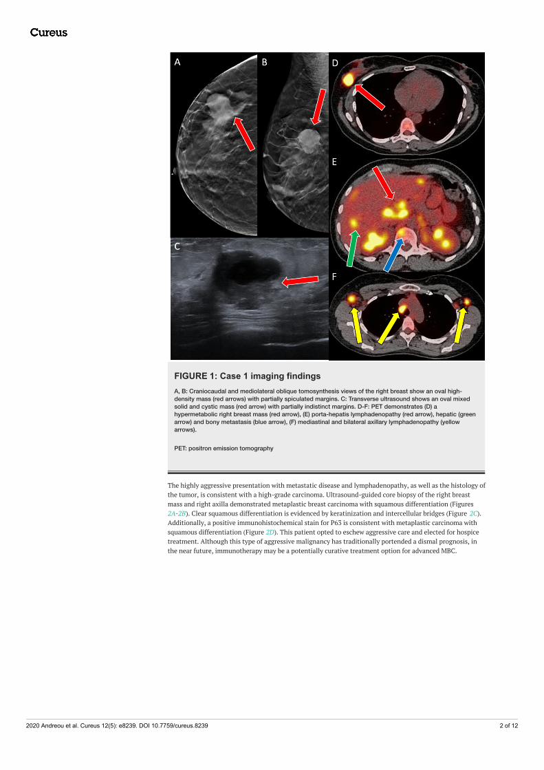
FIGURE 1: Case 1 imaging findingsA, B: Craniocaudal and mediolateral oblique tomosynthesis views of the right breast show an oval high-density mass (red arrows) with partially spiculated margins. C: Transverse ultrasound shows an oval mixedsolid and cystic mass (red arrow) with partially indistinct margins. D-F: PET demonstrates (D) ahypermetabolic right breast mass (red arrow), (E) porta-hepatis lymphadenopathy (red arrow), hepatic (greenarrow) and bony metastasis (blue arrow), (F) mediastinal and bilateral axillary lymphadenopathy (yellowarrows).
PET: positron emission tomography
The highly aggressive presentation with metastatic disease and lymphadenopathy, as well as the histology ofthe tumor, is consistent with a high-grade carcinoma. Ultrasound-guided core biopsy of the right breastmass and right axilla demonstrated metaplastic breast carcinoma with squamous differentiation (Figures2A-2B). Clear squamous differentiation is evidenced by keratinization and intercellular bridges (Figure 2C).Additionally, a positive immunohistochemical stain for P63 is consistent with metaplastic carcinoma withsquamous differentiation (Figure 2D). This patient opted to eschew aggressive care and elected for hospicetreatment. Although this type of aggressive malignancy has traditionally portended a dismal prognosis, inthe near future, immunotherapy may be a potentially curative treatment option for advanced MBC.
2020 Andreou et al. Cureus 12(5): e8239. DOI 10.7759/cureus.8239 2 of 12

FIGURE 2: Case 1 histopathology findingsA: Low power view of metaplastic carcinoma, squamous cell carcinoma subtype. B: Poorly differentiated,large polygonal cells with abundant, eosinophilic cytoplasm (10x magnification). C: Keratinized cells withnumerous atypical mitoses and pyknotic cells present (10x magnification). D: P63 is a nuclear immunostain,which highlights the atypical squamous cells, further supporting the diagnosis of MBC, squamous cell type.
MBC: metaplastic breast cancer
Case 2A 44-Year-Old Female With a Rapidly Enlarging Left Breast Mass
A 44-year-old female presented with an erythematous and painful left breast mass measuring 6.0 cm x 3.1cm on mammography and ultrasound (Figures 3A-3D). A breast MRI revealed diffuse abnormal T2hyperintense edema and skin thickening (Figures 3E-3F).
2020 Andreou et al. Cureus 12(5): e8239. DOI 10.7759/cureus.8239 3 of 12
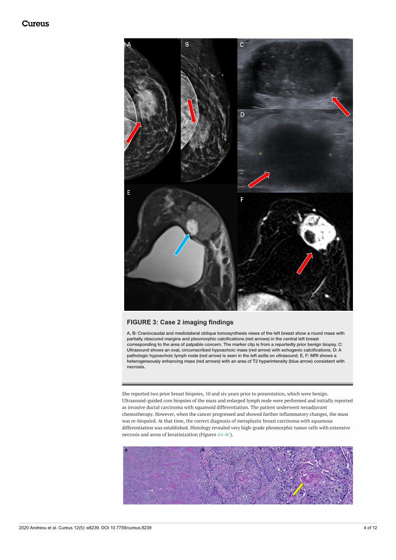
FIGURE 3: Case 2 imaging findingsA, B: Craniocaudal and mediolateral oblique tomosynthesis views of the left breast show a round mass withpartially obscured margins and pleomorphic calcifications (red arrows) in the central left breastcorresponding to the area of palpable concern. The marker clip is from a reportedly prior benign biopsy. C:Ultrasound shows an oval, circumscribed hypoechoic mass (red arrow) with echogenic calcifications. D: Apathologic hypoechoic lymph node (red arrow) is seen in the left axilla on ultrasound. E, F: MRI shows aheterogeneously enhancing mass (red arrows) with an area of T2 hyperintensity (blue arrow) consistent withnecrosis.
She reported two prior breast biopsies, 10 and six years prior to presentation, which were benign.Ultrasound-guided core biopsies of the mass and enlarged lymph node were performed and initially reportedas invasive ductal carcinoma with squamoid differentiation. The patient underwent neoadjuvantchemotherapy. However, when the cancer progressed and showed further inflammatory changes, the masswas re-biopsied. At that time, the correct diagnosis of metaplastic breast carcinoma with squamousdifferentiation was established. Histology revealed very high-grade pleomorphic tumor cells with extensivenecrosis and areas of keratinization (Figures 4A-4C).
2020 Andreou et al. Cureus 12(5): e8239. DOI 10.7759/cureus.8239 4 of 12

FIGURE 4: Case 2 histopathology findingsA: Low magnification image of metaplastic carcinoma with squamous differentiation and extensive necrosis.B: Area of moderately differentiated squamous cell carcinoma (10x). C: High magnification of image B,highlighting distinct keratinization of cells (yellow arrow) with surrounding cells that have a high nuclear tocytoplasmic ratio, abundant cytoplasm, and frequent mitoses (20x).
Case 3A 49-Year-Old Female for a Six-Month Follow-Up of a Benign Left Breast Biopsy
A 49-year-old female presented with an area of palpable concern on her left breast with tenderness. Aprevious biopsy clip was noted on the screening mammogram in the left breast. Ultrasound-guided corebiopsy was performed, which was reported as benign, revealing only stromal sclerosis, skeletal muscle fibers,and mild periductal chronic inflammatory cells. Six months later, a repeat diagnostic mammogram revealeda new, ill-defined mass with very dense coarse calcifications (Figures 5A-5B). Breast ultrasound was thenperformed, which showed a small circumscribed hypoechoic mass with partially obscured andmicrolobulated margins (Figures 5C-5D). Ultrasound-guided core biopsy was performed, yielding thediagnosis of MBC. Breast MRI was also performed at this time, revealing a round T2 hyperintense,homogenously enhancing mass (Figures 5E-5F).
2020 Andreou et al. Cureus 12(5): e8239. DOI 10.7759/cureus.8239 5 of 12

FIGURE 5: Case 3 imaging findingsA, B: Craniocaudal and mediolateral oblique tomosynthesis views of the left breast show a new, round, equaldensity mass (red arrows) with partially obscured margins and coarse calcifications. C, D: Ultrasounddemonstrates a circumscribed round hypoechoic mass (red arrows) with echogenic calcifications andpartially obscured and microlobulated margins. E, F: MRI shows a round, T2, hyperintense, homogeneouslyenhancing mass (red arrows).
MRI: magnetic resonance imaging
Histologically, the first biopsy demonstrated benign findings (Figures 6A-6B). The second biopsy revealed atumor highlighted by clear nests of high-grade carcinoma (Figures 6A-6D). Admixed with the carcinoma,there was also a very discrete sarcomatous component to the tumor, which contained malignant boneformation. The coarse calcifications observed radiologically corresponded to malignant osteoid. Thispresentation of a carcinoma accompanied by sarcomatous elements is diagnostic of metaplastic carcinomawith heterologous differentiation. The tumor profile was triple-negative, and the patient elected for abilateral mastectomy with adjuvant chemotherapy. The mastectomy specimen revealed infiltratingmammary carcinoma, poorly differentiated, metaplastic type with osseous metaplasia. The malignancy washigh-grade (Nottingham grade 3) with pronounced nuclear pleomorphism and moderate mitotic activity.The margins were uninvolved and angiolymphatic invasion was not identified. The previous biopsy sitesdemonstrated sclerosed papilloma and usual ductal hyperplasia. The final pathologic stage was pT1cN0.
FIGURE 6: Case 3 histopathology findingsA: Initial ultrasound-guided core biopsy, which revealed benign mammary tissue with stromal sclerosis andmild periductal inflammation, along with fragments of benign skeletal muscle. B: No areas suspicious formalignancy were present in the biopsy material submitted. C-D: Second ultrasound-guided core biopsyshowing high-grade invasive ductal carcinoma (red arrows) with intermingled areas of osseous differentiation(yellow arrows). E: Representative section from a mastectomy with similar findings of metaplastic carcinomawith osseous metaplasia.
Case 4A 20-Year-Old Female With a Right Breast Palpable Mass
A 20-year-old female with no significant past medical history presented with a palpable mass in her rightbreast. The mammogram revealed an oval equal density mass with obscured margins measuring up to 2.1 cmbut the sensitivity of the mammography was limited due to extremely dense breasts (Figures 7A-7B).Ultrasound revealed an oval, heterogeneously hypoechoic mass with indistinct margins and posterioracoustic enhancement (Figures 7C-7D). The initial ultrasound-guided core biopsy was reported as afibroepithelial neoplasm with abundant stroma. This was thought to represent a benign phyllodes tumor butcomplete excision was recommended.
2020 Andreou et al. Cureus 12(5): e8239. DOI 10.7759/cureus.8239 6 of 12

FIGURE 7: Case 4 imaging findingsA, B: Right diagnostic mammogram shows a round circumscribed mass (red arrows) in the upper outerquadrant corresponding to the area of palpable concern. C, D: The ultrasound shows a round, circumscribedmass (red arrows) with heterogeneous echotexture and internal vascularity.
Three months later, the breast lump continued to grow rapidly, and excision was performed. Evaluation ofthat specimen revealed the correct diagnosis of metaplastic breast carcinoma, spindle cell variant.Histologically, the tumor consisted of bland spindle cells with mild-moderate pleomorphism and severalmitotic figures, concerning for either a low-grade sarcoma versus a metaplastic carcinoma. Extensivecentral necrosis was observed. Staining for P63, as well as focal staining for cytokeratins along with thehistology of the tumor, was consistent with a low-grade fibromatosis-like metaplastic carcinoma (Figures8A-8D).
2020 Andreou et al. Cureus 12(5): e8239. DOI 10.7759/cureus.8239 7 of 12

FIGURE 8: Case 4 histopathology findingsA: Low power magnification of a spindle cell lesion with entrapment of normal mammary glandular structures(yellow arrow), 5x. B: Low magnification demonstrating necrosis and degeneration of the tumor, 5x. C-D: Highpower images emphasizing the sweeping fascicles of spindle cells, in the classic herringbone pattern, alongwith frequent mitotic figures and abnormal nuclei, 20x. E: P63 immunostain showing strong, nuclearpositivity in the spindle cells, 10x
Case 5A 79-Year-Old Female With a Palpable Left Breast Mass
A 79-year-old Asian female presented with a palpable mass in her left breast. Mammography revealed alarge round to oval mass with circumscribed and indistinct margins in the left central, outer breastmeasuring up to 5.8 cm (Figure 9D). The ultrasound revealed a large round complex mass with hypoechoicand echogenic areas and microlobulated margins in the area of palpable concern (Figure 9A).
FIGURE 9: Case 5 initial imaging findingsA, B: Craniocaudal and mediolateral oblique tomosynthesis views of the left breast show an oval, high-
2020 Andreou et al. Cureus 12(5): e8239. DOI 10.7759/cureus.8239 8 of 12

density mass with circumscribed margins in the central outer left breast corresponding to the area ofpalpable concern. C, D: Ultrasound grayscale imaging demonstrates a round heterogeneous mass with smallcentral cystic areas and partially circumscribed margins.
An ultrasound-guided biopsy was performed twice, one month apart, first reporting nonviable highlypleomorphic cells with associated fibrinopurulent exudate (Figure 10A) and then showing fragments ofblood clot and minute, scattered collections of atypical cells (Figure 10B).
FIGURE 10: Case 5 initial histopathology attemptsA: First biopsy, nonviable, highly pleomorphic cells (yellow arrow) with associated fibrinopurulent exudate.The specimen consists mostly of necrotic highly pleomorphic tumor cell debris. B: Fragments of blood clotsand minute scattered collections of atypical cells (yellow arrow). Recommended resampling.
Due to the presence of a mass radiographically with previous biopsies not revealing viable tissue or tumorcells, again resampling was recommended to obtain viable tissue for an accurate diagnosis. Subsequently,the patient underwent an excisional biopsy that yielded useful material for the diagnosis of metaplasticcarcinoma, spindle cell type, with extensive necrosis and hemorrhage (Figures 11A-11D).
FIGURE 11: Case 5 excisional biopsyA, B: Images of high-grade spindle cell component composed of highly pleomorphic cells, atypical mitoseswith myxoid background. Notice the bizarre binucleated cell in image, A (red arrow), 10x. C, D: Focal areas ofmore classic high-grade invasive ductal carcinoma and carcinoma in situ.
A new diagnosis of left breast metaplastic cancer stage III (T3N0M0) triple-negative was subsequently made.
2020 Andreou et al. Cureus 12(5): e8239. DOI 10.7759/cureus.8239 9 of 12

MRI of the left breast revealed a large enhancing mass in the outer to central left breast measuring 6.7 x 6.2x 5cm with necrotic and solid components (Figures 12A-12C).
FIGURE 12: Case 5 breast MRI findingsA: Axial T2-weighted image shows a heterogeneous mass with areas of T2 hyperintensity. B, C: Axial T1-weighted fat-suppressed subtracted without (B) and with (C) color show a heterogeneously enhancingcentrally necrotic mass in the left breast (red arrows).
The PET scan did not show any evidence of metastasis. The patient underwent a modified radicalmastectomy with a sentinel lymph node biopsy. Pathologic findings confirmed the diagnosis of metaplasticcarcinoma, spindle cell variant, along with areas of poorly differentiated invasive ductal carcinoma-nototherwise specified (NOS), as well as high-grade ductal carcinoma in situ. All lymph nodes submitted werenegative for metastatic disease.
DiscussionMetaplastic carcinoma of the breast is a heterogeneous and diverse group of rare, aggressive malignanciesthat are characterized by the coexistence of epithelial and non-epithelial components [8]. Severaldistinctions exist when comparing metaplastic to other histologic subtypes of invasive carcinoma.Metaplastic carcinomas have a more advanced stage at diagnosis. A review of the National Cancer Databasefrom 2006 revealed that patients with MBC had higher rates of stage III and stage IV disease than patientswith non-metaplastic carcinomas [9]. Metaplastic carcinomas have a proclivity for hematogenous metastasisbut are less likely to have lymphatic involvement [10]. They are more likely to recur and have a reduced timeperiod until recurrence when compared to non-MBC [11].
MBC has a broad histological profile, consisting of a heterogeneous group of malignant neoplasms, withboth glandular and non-glandular components, along with mixed epithelial and mesenchymaldifferentiation. MBC is classified into five subtypes: spindle cell, squamous cell, carcinosarcoma, matrix-producing, and metaplastic carcinoma with osteoclastic giant cells [12]. The most common and mostfrequently misdiagnosed subtype is spindle cell. The spindle cell subtype is the most difficult to diagnosebecause of its close resemblance to low-grade sarcomas, phyllodes tumors, or a reactive process such asgranulation tissue [13]. For example, Case 4 was initially misdiagnosed as a benign fibroepithelial neoplasmdiscovered on the ultrasound-guided core biopsy with the correct diagnosis of MBC (spindle call variant)made after surgical excision of the mass. The squamous cell carcinoma subtype displays infiltratingsquamous carcinoma with polygonal cells, eosinophilic cytoplasm, and possible keratin pearl formation [14].The carcinosarcoma subtype is composed of malignant epithelium and malignant stroma. The matrix-producing subtype consists of an apparent carcinoma with a transition to cartilaginous and/or osseousstromal matric lacking a spindle component [15]. Metaplastic carcinoma with the osteoclastic giant cellssubtype displays intraductal or infiltration carcinoma contiguous or mixed with spindle cell or sarcomatousstroma plus osteoclastic cells [16]. Although some studies have mentioned the fibromatosis-like subtype ofMBC has a slightly better outcome than the other, no definitive research has established any significantdifference in overall prognosis solely based on histologic characteristics.
Although metaplastic carcinomas are most commonly negative for estrogen, progesterone, and HER2-likeother invasive carcinomas, unique genetic variations and intratumoral heterogeneity cause this tumor typeto be more chemoresistant than other breast cancers. Neoadjuvant chemotherapy may be insufficient to halttumor progression. Multiple studies have demonstrated results coinciding with a limited effect ofneoadjuvant treatment, including a 2011 study showing a progression rate of 82% [16]. This highlights theimportance of the prompt and accurate diagnosis of MBC. Although immunotherapy is in its infancy,metastatic MBC has been seen to respond completely to checkpoint blockade medications, such as PD-L1inhibitors, even when initially chemo-refractory [17]. This may be due to the propensity of MBC to expressthe PD-1 ligand, perhaps as a mechanism to avoid immunodetection and tumor elimination in theimmunocompetent host [18].
Many patients with metaplastic breast carcinoma are initially misdiagnosed because it is difficult to classifyrare tumors based on heterogeneous imaging characteristics. Similarly, when metaplastic tumors undergocore biopsies, they are histologically difficult to assess because they contain distinct components within the
2020 Andreou et al. Cureus 12(5): e8239. DOI 10.7759/cureus.8239 10 of 12

same tumor. In the cases reviewed above, some patients underwent multiple biopsies before the correctdiagnosis was reached, and only then did they receive appropriate treatment. The delay in diagnosis mayhave provided the tumors time to grow and metastasize. Intratumoral heterogeneity leads to sampling biasand highlights the need to obtain an excisional biopsy in the setting of discordance between histopathologyand imaging findings. An optimal therapeutic approach remains controversial, however, it appears that amultidisciplinary, targeted approach may be the most effective when it can be promptly initiated.
Loco-regional management with surgery followed by chemoradiation is the current standard of care [19].Advancements in histopathology, radiomics, and artificial intelligence may speed up the development ofnew diagnostic and therapeutic strategies to target individual tumors’ unique pathogeneses and amelioratethe need for invasive treatment. Future systemic and intratumoral immunotherapy treatment options maybe guided by the identification of the underlying genetic aberration and tumor mutational burden [20].
ConclusionsThe complex nature of this variant of breast cancer leads to difficulty in diagnosis due to sampling bias. Thetraditional paradigm used to efficiently diagnose high-incidence breast cancers may not account for thischaracteristic of MBC. These biologically aggressive tumors may express heterogeneous imaging andhistologic phenotypes, potentially hampering a timely diagnosis. Red flags include a large, new, or rapidlygrowing mass that may have some benign imaging features on mammography. Complex, mixed, solid, cysticmasses with heterogeneous echogenicity and noncircumscribed margins are some suspicious sonographicfeatures. In these cases where the imaging phenotype is inconsistent with the timeline, it may be useful toobtain breast MRI to look for a high T2 signal consistent with tumor necrosis. Intratumoral heterogeneitymay result in a false-negative biopsy and the propensity to develop into an aggressive metastatic disease ishigh for MBC. If there is any suspicion for MBC based on imaging characteristics, and the initial biopsy isnegative for malignancy, it may be advisable to perform an excisional biopsy of the lesion in order tocircumvent the significant sampling bias that exists from a small core needle biopsy that may or may notobtain adequate tissue for proper diagnosis. These cases exemplify the importance of and the difficulties inachieving an accurate and timely diagnosis of MBC. Treatment considerations may be tailored to give thepatient the best chance of survival, especially if the correct diagnosis of MBC is reached beforehematogenous seeding can occur.
Additional InformationDisclosuresHuman subjects: Consent was obtained by all participants in this study. Conflicts of interest: Incompliance with the ICMJE uniform disclosure form, all authors declare the following: Payment/servicesinfo: All authors have declared that no financial support was received from any organization for thesubmitted work. Financial relationships: All authors have declared that they have no financialrelationships at present or within the previous three years with any organizations that might have aninterest in the submitted work. Other relationships: All authors have declared that there are no otherrelationships or activities that could appear to have influenced the submitted work.
References1. Esbah O, Turkoz FP, Turker I, et al.: Metaplastic breast carcinoma: case series and review of the literature .
Asian Pac J Cancer Prev. 2012, 13:4645-4649. 10.7314/APJCP.2012.13.9.46452. Adams S: Dramatic response of metaplastic breast cancer to chemo-immunotherapy . NPJ Breast Cancer.
2017, 3:8. 10.1038/s41523-017-0011-03. Donato H, Candelária I, Oliveira P, Gonçalo M, Caseiro-Alves F: Imaging Findings of metaplastic carcinoma
of the breast with pathologic correlation. J Belg Soc Radiol. 2018, 102:46. 10.5334/jbsr.13864. Gunhan-Bilgen I, Memis A, Ustun EE, Zekioglu O, Ozdemir N: Metaplastic carcinoma of the breast: clinical,
mammographic, and sonographic findings with histopathologic correlation. AJR Am J Roentgenol. 2002,178:1421-1425. 10.2214/ajr.178.6.1781421
5. Velasco M, Santamaria G, Ganau S, Farrus B, Zanon G, Romagosa C, Fernandez PL: MRI of metaplasticcarcinoma of the breast. AJR Am J Roentgenol. 2005, 184:1274-1278. 10.2214/ajr.184.4.01841274
6. Rungta S, Kleer CG: Metaplastic carcinomas of the breast: diagnostic challenges and new translationalinsights. Arch Pathol Lab Med. 2012, 136:896-900. 10.5858/arpa.2012-0166-CR
7. Lale S, Kure K, Lingamfelter D: Challenges to diagnose metaplastic carcinoma of the breast throughcytologic methods: an eight-case series. Diagn Pathol. 2011, 6:7. 10.1186/1746-1596-6-7
8. McMullen ER, Zoumberos NA, Kleer CG: Metaplastic breast carcinoma: update on histopathology andmolecular alterations. Arch Pathol Lab Med. 2019, 143:1492-1496. 10.5858/arpa.2019-0396-RA
9. Schwartz TL, Mogal H, Papageorgiou C, Veerapong J, Hsueh EC: Metaplastic breast cancer: histologiccharacteristics, prognostic factors and systemic treatment strategies. Exp Hematol Oncol. 2013, 2:31.10.1186/2162-3619-2-31
10. Leddy R, Irshad A, Rumboldt T, Cluver A, Campbell A, Ackerman S: Review of metaplastic carcinoma of thebreast: imaging findings and pathologic features. J Clin Imaging Sci. 2012, 2:21. 10.4103/2156-7514.95435
11. Malmgren JA, Mayer M, Atwood MK, Kaplan HG: Differential presentation and survival of de novo andrecurrent metastatic breast cancer over time: 1990-2010. Breast Cancer Res Treat. 2018, 167:579-590.10.1007/s10549-017-4529-5
12. Okada N, Hasebe T, Iwasaki M, et al.: Metaplastic carcinoma of the breast . Hum Pathol. 2010, 41:960-970.
2020 Andreou et al. Cureus 12(5): e8239. DOI 10.7759/cureus.8239 11 of 12

10.1016/j.humpath.2009.11.01313. Luini A, Aguilar M, Gatti G, et al.: Metaplastic carcinoma of the breast, an unusual disease with worse
prognosis: the experience of the European Institute of Oncology and review of the literature. Breast CancerRes Treat. 2007, 101:349-353. 10.1007/s10549-006-9301-1
14. Tse GM, Tan PH, Putti TC, Lui PC, Chaiwun B, Law BK: Metaplastic carcinoma of the breast: aclinicopathological review. J Clin Pathol. 2006, 59:1079-1083. 10.1136/jcp.2005.030536
15. Ayar S, Dyess DL, Carter E: Matrix-producing carcinoma: a rare variant of metaplastic breast carcinomawith heterologous elements. Breast J. 2010, 16:420-423. 10.1111/j.1524-4741.2010.00925.x
16. Chen IC, Lin CH, Huang CS, et al.: Lack of efficacy to systemic chemotherapy for treatment of metaplasticcarcinoma of the breast in the modern era. Breast Cancer Res Treat. 2011, 130:345-351. 10.1007/s10549-011-1686-9
17. Al Sayed AD, Elshenawy MA, Tulbah A, Al-Tweigeri T, Ghebeh H: Complete response of chemo-refractorymetastatic metaplastic breast cancer to paclitaxel-immunotherapy combination. Am J Case Rep. 2019,20:1630-1635. 10.12659/ajcr.918770
18. Joneja U, Vranic S, Swensen J, et al.: Comprehensive profiling of metaplastic breast carcinomas revealsfrequent overexpression of programmed death-ligand 1. J Clin Pathol. 2017, 70:255-259. 10.1136/jclinpath-2016-203874
19. Rakha EA, Tan PH, Varga Z, et al.: Prognostic factors in metaplastic carcinoma of the breast: a multi-institutional study. Br J Cancer. 2015, 112:283-289. 10.1038/bjc.2014.592
20. Palleschi M, Maltoni R, Sarti S, Melegari E, Bravaccini S, Rocca A: Immunotherapy: the end of the "dark age"for metastatic triple-negative breast cancer?. Breast J. 2019, 26:739-742. 10.1111/tbj.13662
2020 Andreou et al. Cureus 12(5): e8239. DOI 10.7759/cureus.8239 12 of 12