Embed Size (px)
Citation preview
Extracorporeal Life Support What should Cardiologists know?
Samphant Ponvilawan
Bumrungrad International
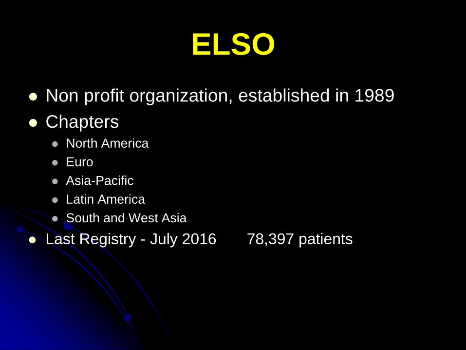
ELSO
Non profit organization, established in 1989
Chapters North America
Euro
Asia-Pacific
Latin America
South and West Asia
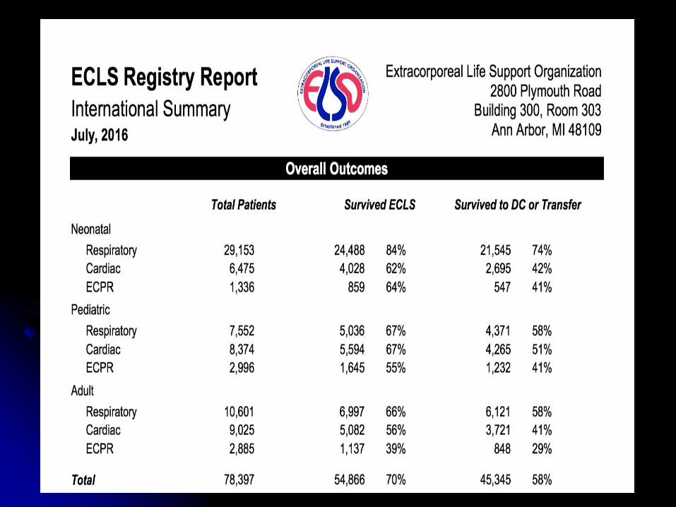
Last Registry - July 2016 78,397 patients
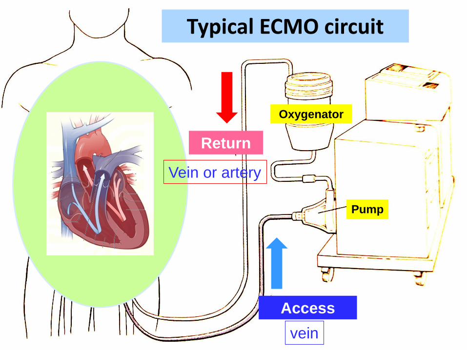
Typical ECMO circuit
Access
Oxygenator
Pump
Return
vein
Vein or artery
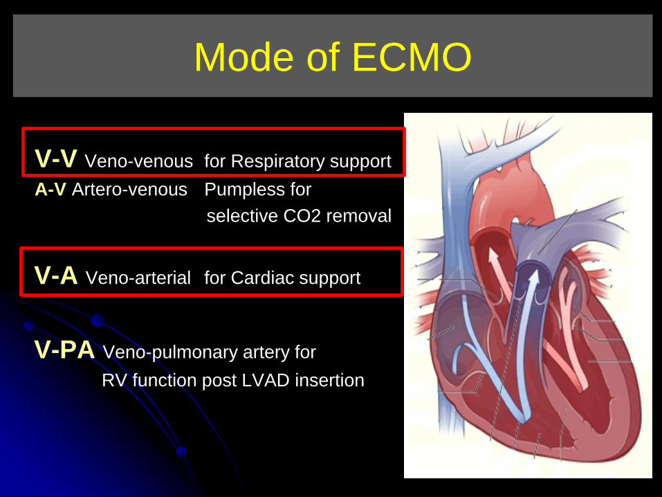
Mode of ECMO
V-V Veno-venous for Respiratory support
A-V Artero-venous Pumpless for
selective CO2 removal
V-A Veno-arterial for Cardiac support
V-PA Veno-pulmonary artery for
RV function post LVAD insertion
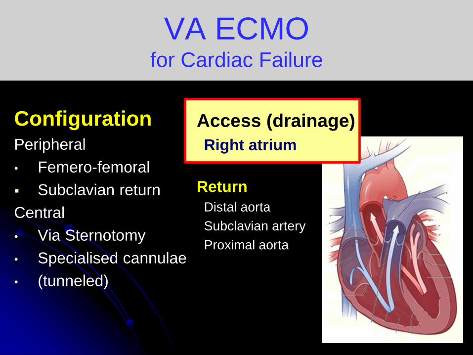
VA ECMO for Cardiac Failure
Configuration Peripheral
• Femero-femoral
Subclavian return
Central
• Via Sternotomy
• Specialised cannulae
• (tunneled)
Access (drainage)
Right atrium
Return
Distal aorta
Subclavian artery
Proximal aorta
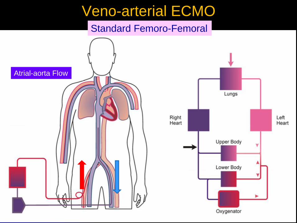
Veno-arterial ECMO Standard Femoro-Femoral
Atrial-aorta Flow
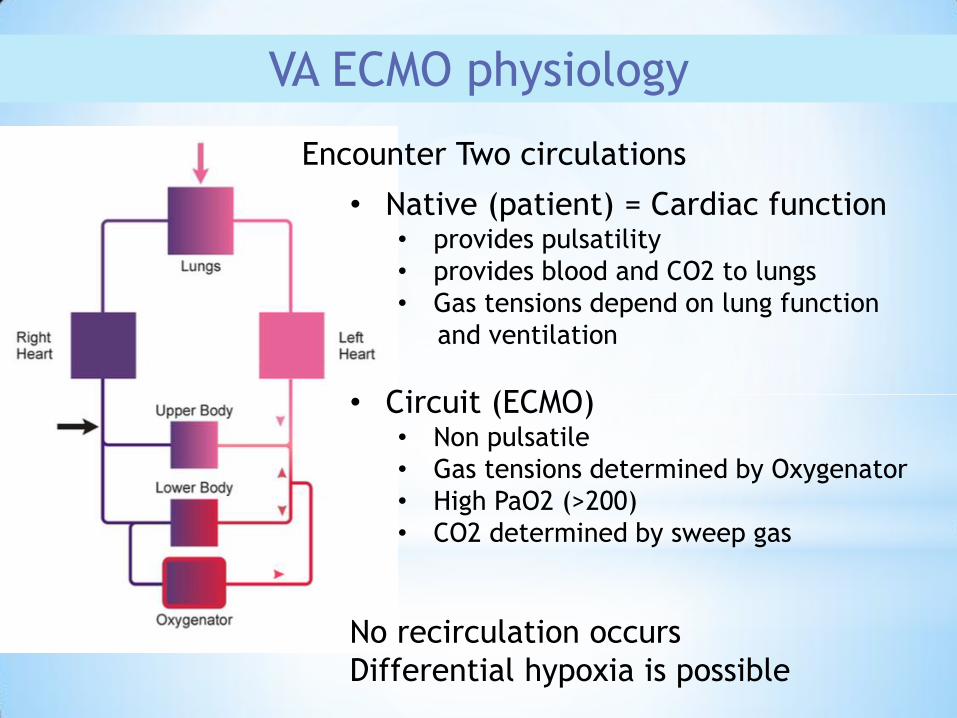
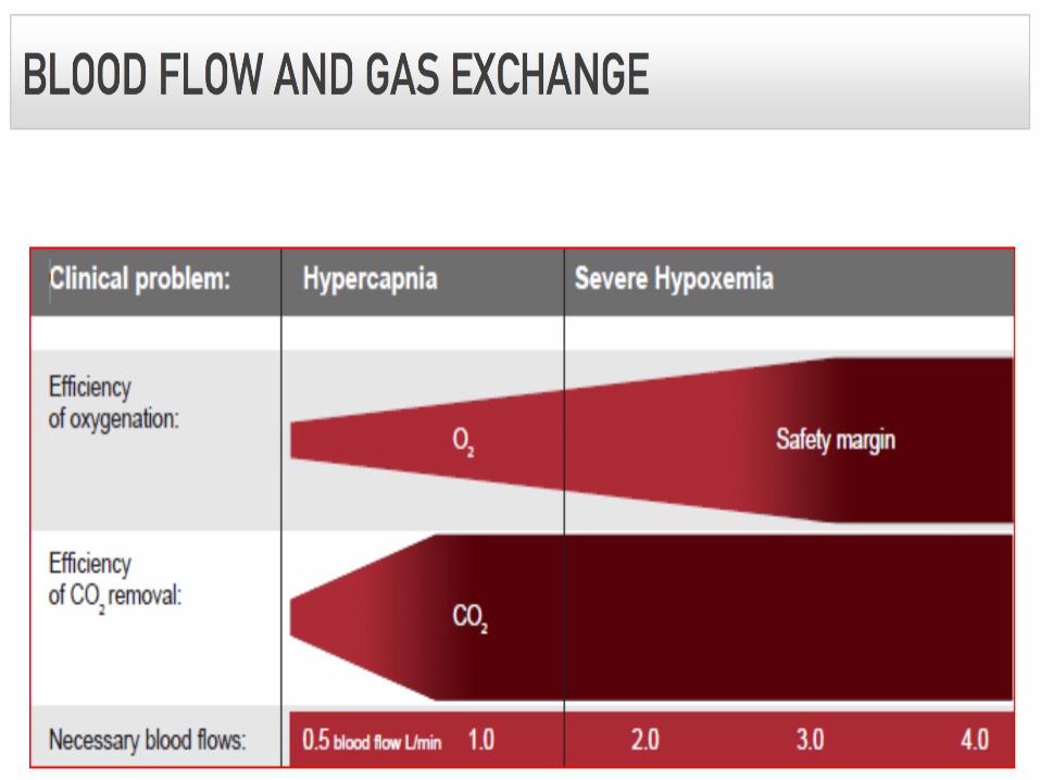
VA ECMO physiology
Encounter Two circulations
• Native (patient) = Cardiac function • provides pulsatility
• provides blood and CO2 to lungs
• Gas tensions depend on lung function
and ventilation
• Circuit (ECMO) • Non pulsatile
• Gas tensions determined by Oxygenator
• High PaO2 (>200)
• CO2 determined by sweep gas
No recirculation occurs
Differential hypoxia is possible
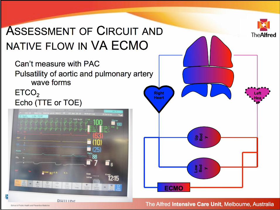

• Target paO2 70-90mmHg
paCO2 40mmHg
ETCO2 20-35mmHg
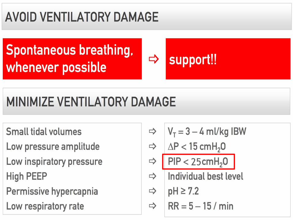
PIP < 25mmHg
• Use TV 6ml/kg, RR 8/min initial setting
Respiratory management (Ventilator setting)
• ETCO2 <20 ; Lung ventilation should be reduced
• High arterial PCO2 ; Gas flow should be increased
• Low arterial PCO2 ; Gas flow should be reduced if V/Q >0.5
• Never turn off gas flow in VA ECMO for low PCO2, will cause hypoxic lower
limbs and abdominal viscera
VA ECMO maintenance
Respiratory setting : TV 6ml/kg, RR 8/min
Cardiac setting : pulsatility, vasodilator
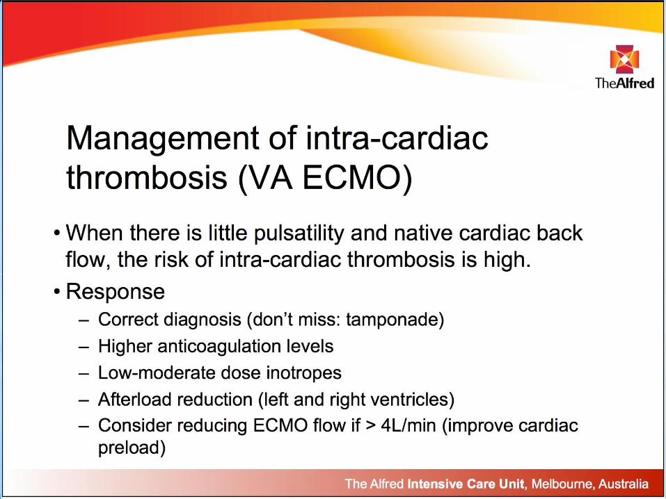
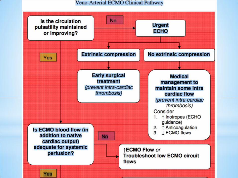
Loss of Pulsatility
Cardiac Tamponade
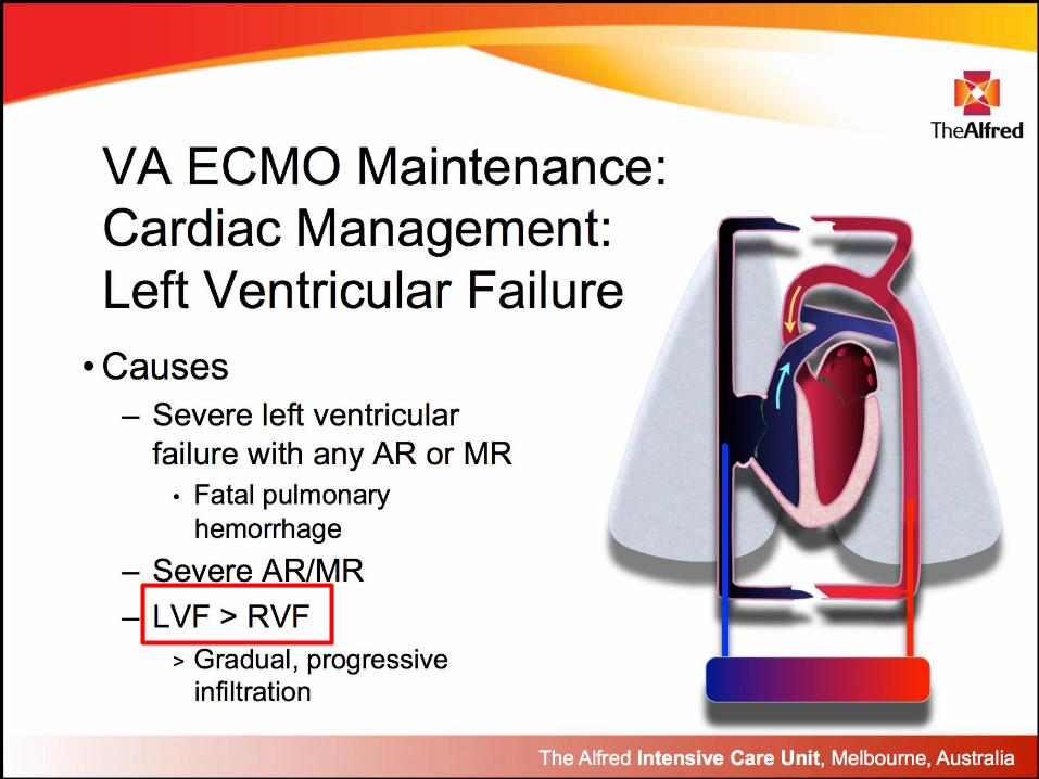
Myocardial failure (w or w/o MR, AR)
LVF>RVF : acute pulmonary edema, hemorrhage
RVF>LVF : unable to wean ECMO
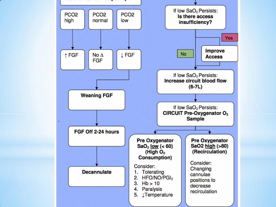
Access insufficiency
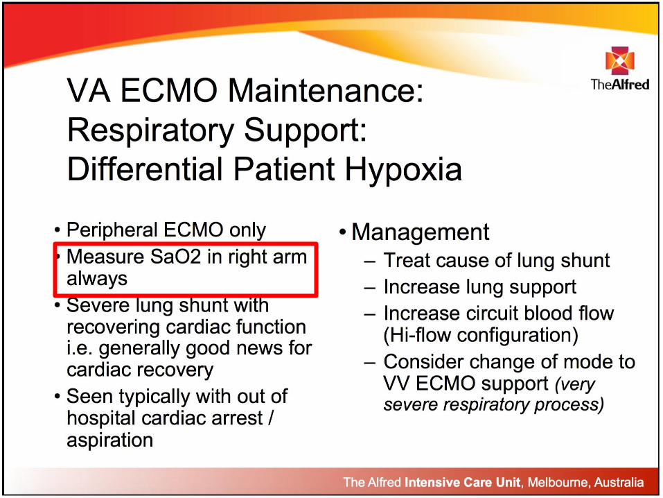
Differential hypoxemia : Bad Lungs
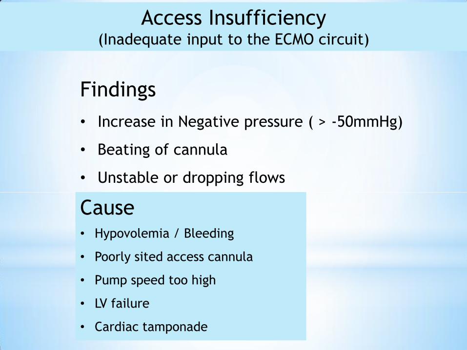
Access Insufficiency (Inadequate input to the ECMO circuit)
Findings
• Increase in Negative pressure ( > -50mmHg)
• Beating of cannula
• Unstable or dropping flows
Cause • Hypovolemia / Bleeding
• Poorly sited access cannula
• Pump speed too high
• LV failure
• Cardiac tamponade
Beautifully supported
with VA ECMO
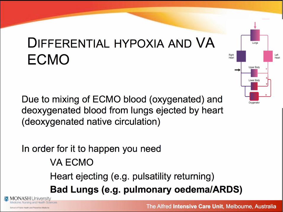
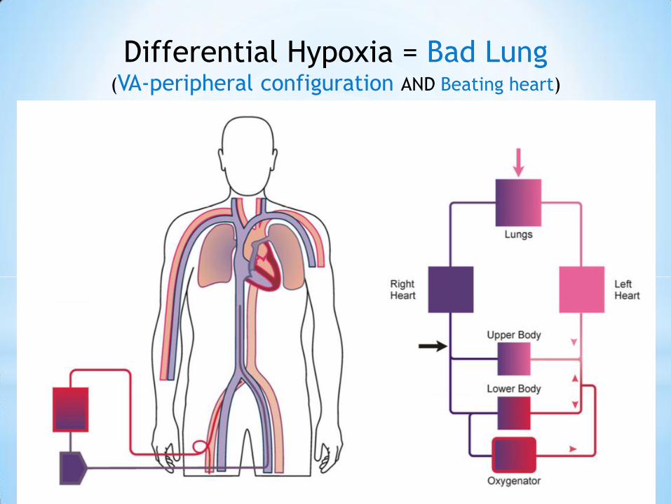
Differential Hypoxia = Bad Lung (VA-peripheral configuration AND Beating heart)
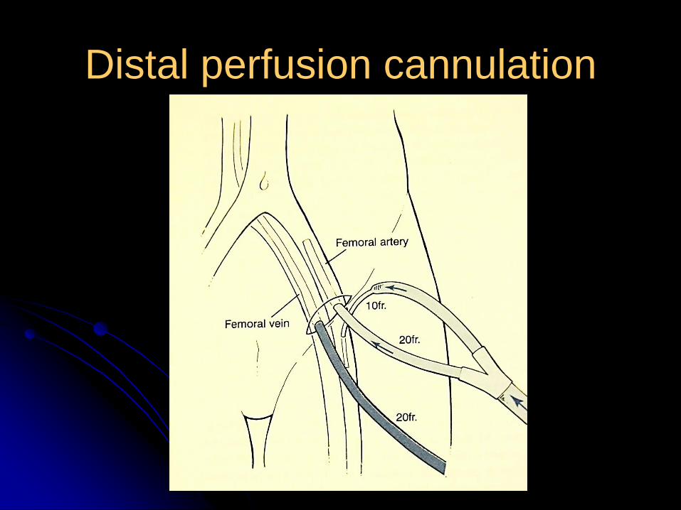
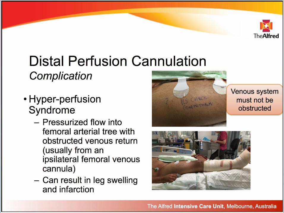
Distal perfusion cannulation
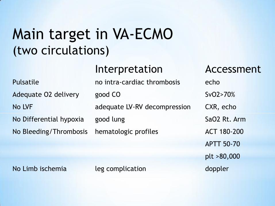
Main target in VA-ECMO (two circulations)
Interpretation Accessment Pulsatile no intra-cardiac thrombosis echo
Adequate O2 delivery good CO SvO2>70%
No LVF adequate LV-RV decompression CXR, echo
No Differential hypoxia good lung SaO2 Rt. Arm
No Bleeding/Thrombosis hematologic profiles ACT 180-200
APTT 50-70
plt >80,000
No Limb ischemia leg complication doppler
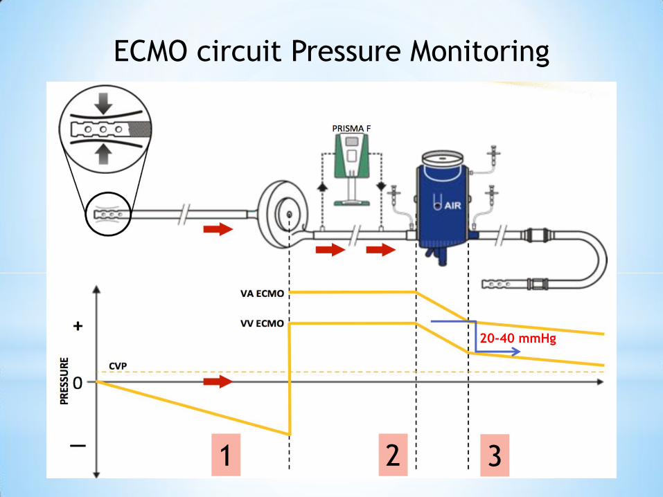
ECMO circuit Pressure Monitoring
20-40 mmHg
1 2 3
Increase Transoxygenator gradient (normal 20-40 mmHg)
• Clot formation within oxygenator
• Return cannula size
• Excessive flow rate
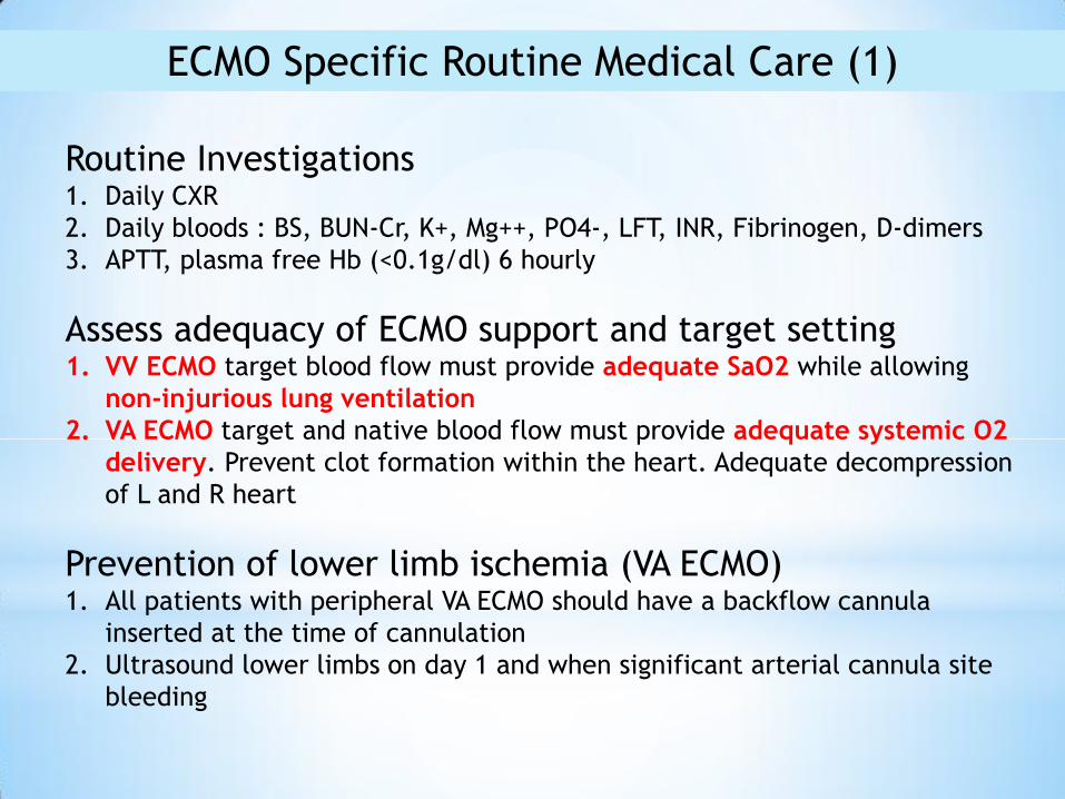
ECMO Specific Routine Medical Care (1)
Routine Investigations 1. Daily CXR
2. Daily bloods : BS, BUN-Cr, K+, Mg++, PO4-, LFT, INR, Fibrinogen, D-dimers
3. APTT, plasma free Hb (<0.1g/dl) 6 hourly
Assess adequacy of ECMO support and target setting 1. VV ECMO target blood flow must provide adequate SaO2 while allowing
non-injurious lung ventilation
2. VA ECMO target and native blood flow must provide adequate systemic O2
delivery. Prevent clot formation within the heart. Adequate decompression
of L and R heart
Prevention of lower limb ischemia (VA ECMO) 1. All patients with peripheral VA ECMO should have a backflow cannula
inserted at the time of cannulation
2. Ultrasound lower limbs on day 1 and when significant arterial cannula site
bleeding
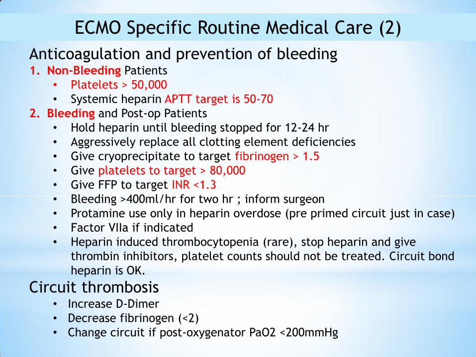
ECMO Specific Routine Medical Care (2)
Anticoagulation and prevention of bleeding 1. Non-Bleeding Patients
• Platelets > 50,000
• Systemic heparin APTT target is 50-70
2. Bleeding and Post-op Patients
• Hold heparin until bleeding stopped for 12-24 hr
• Aggressively replace all clotting element deficiencies
• Give cryoprecipitate to target fibrinogen > 1.5
• Give platelets to target > 80,000
• Give FFP to target INR <1.3
• Bleeding >400ml/hr for two hr ; inform surgeon
• Protamine use only in heparin overdose (pre primed circuit just in case)
• Factor VIIa if indicated
• Heparin induced thrombocytopenia (rare), stop heparin and give
thrombin inhibitors, platelet counts should not be treated. Circuit bond
heparin is OK.
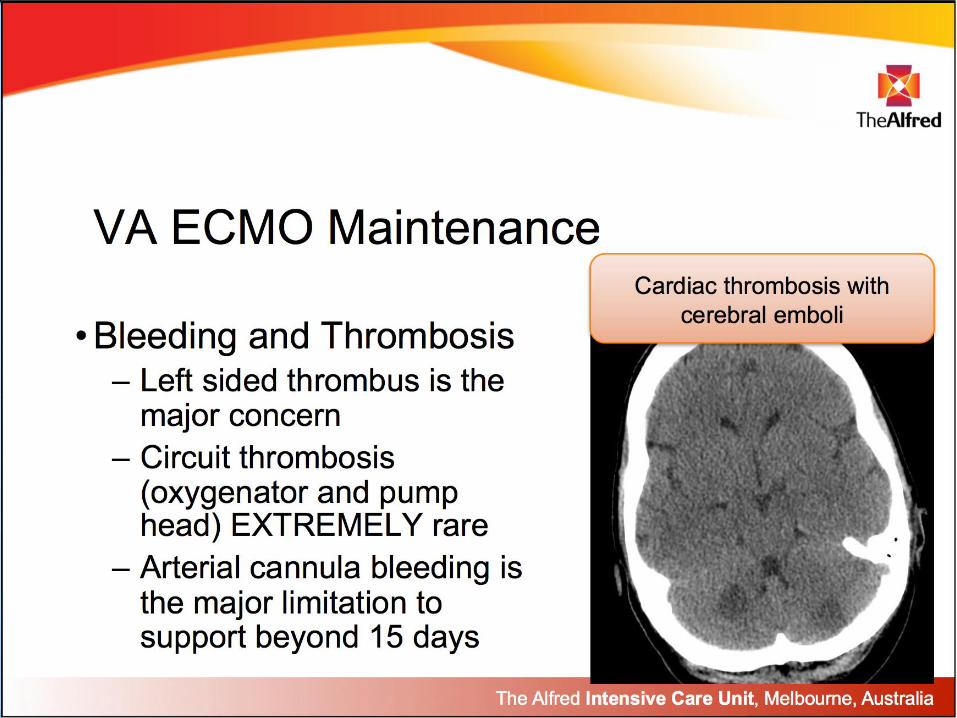
Circuit thrombosis • Increase D-Dimer
• Decrease fibrinogen (<2)
• Change circuit if post-oxygenator PaO2 <200mmHg
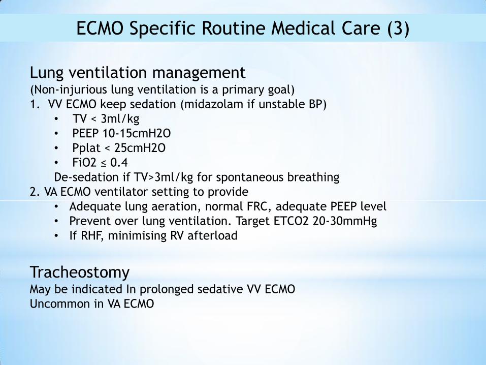
ECMO Specific Routine Medical Care (3)
Lung ventilation management (Non-injurious lung ventilation is a primary goal)
1. VV ECMO keep sedation (midazolam if unstable BP)
• TV < 3ml/kg
• PEEP 10-15cmH2O
• Pplat < 25cmH2O
• FiO2 ≤ 0.4
De-sedation if TV>3ml/kg for spontaneous breathing
2. VA ECMO ventilator setting to provide
• Adequate lung aeration, normal FRC, adequate PEEP level
• Prevent over lung ventilation. Target ETCO2 20-30mmHg
• If RHF, minimising RV afterload
Tracheostomy May be indicated In prolonged sedative VV ECMO
Uncommon in VA ECMO
Sedation
• During cannulation and first 24 hr
to avoid spontaneous breathing, air embolism during cannulation
• After ECLS
stop to allow neurological exam (daily). Then resumed
• Sedation should be minimal but sufficient to avoid increasing native
metabolic rate
• Systemic paralysis and cooling may be necessary if venous drainage
cannot be achieved
2013
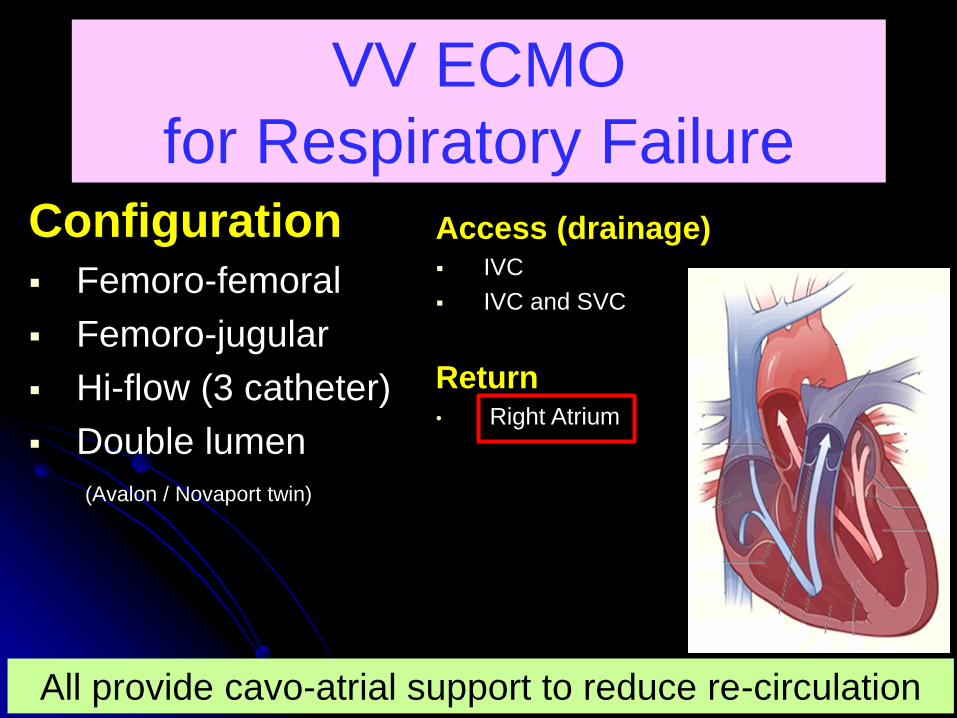
VV ECMO
for Respiratory Failure Configuration Femoro-femoral
Femoro-jugular
Hi-flow (3 catheter)
Double lumen
(Avalon / Novaport twin)
Access (drainage) IVC
IVC and SVC
Return • Right Atrium
All provide cavo-atrial support to reduce re-circulation
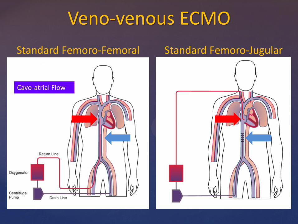
Veno-venous ECMO
Standard Femoro-Femoral Standard Femoro-Jugular
Cavo-atrial Flow
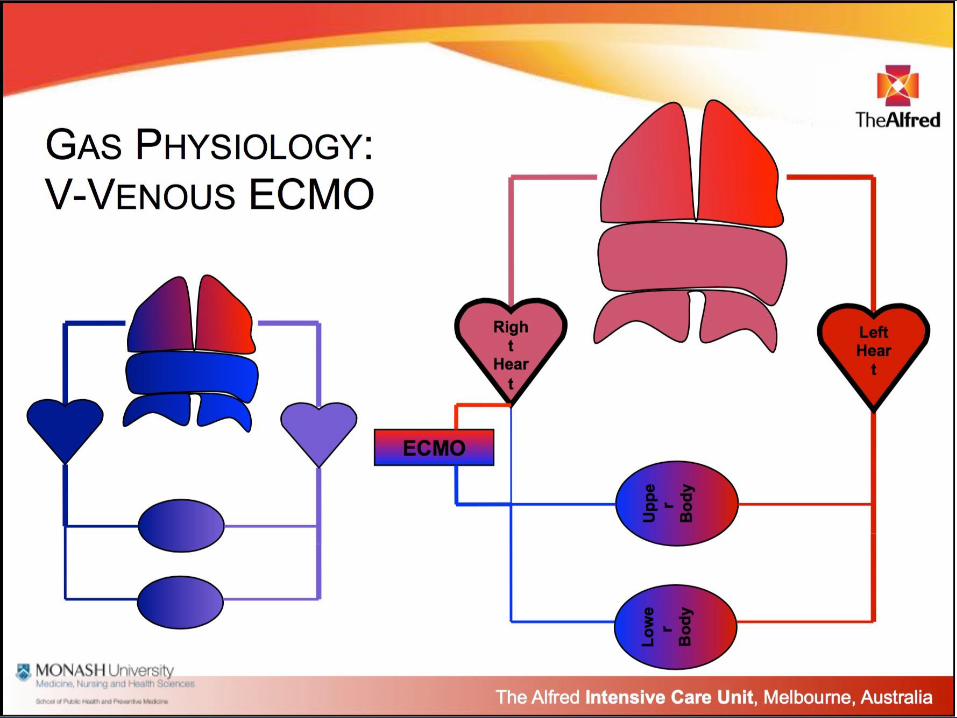
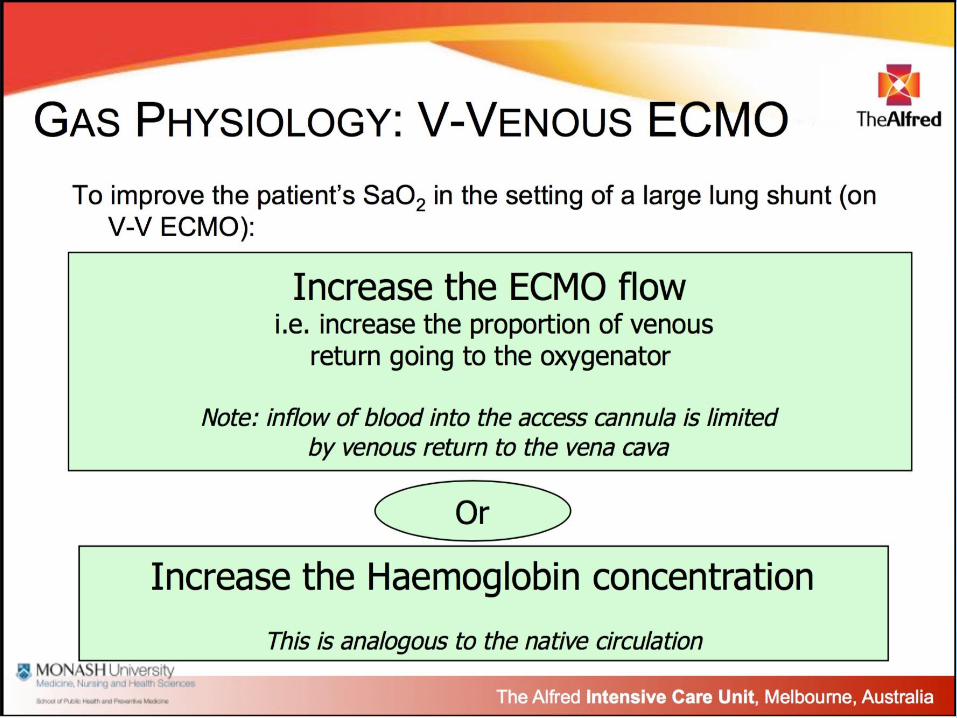
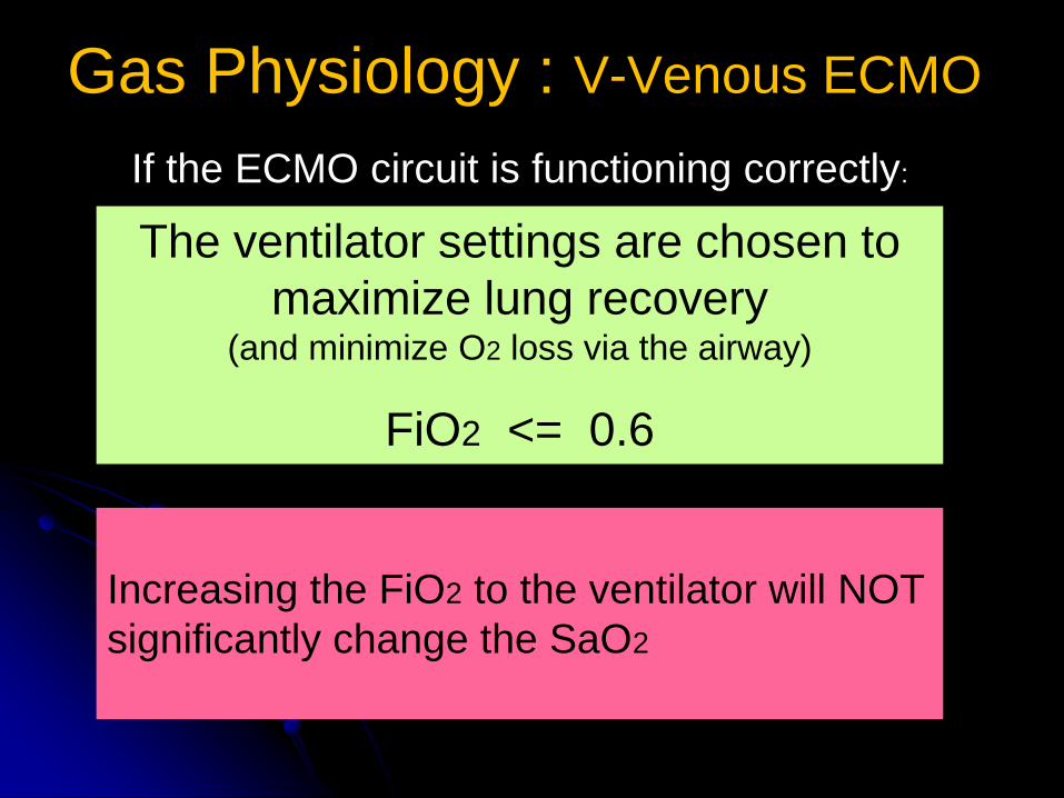
Gas Physiology : V-Venous ECMO
If the ECMO circuit is functioning correctly:
The ventilator settings are chosen to
maximize lung recovery (and minimize O2 loss via the airway)
FiO2 <= 0.6
Increasing the FiO2 to the ventilator will NOT
significantly change the SaO2
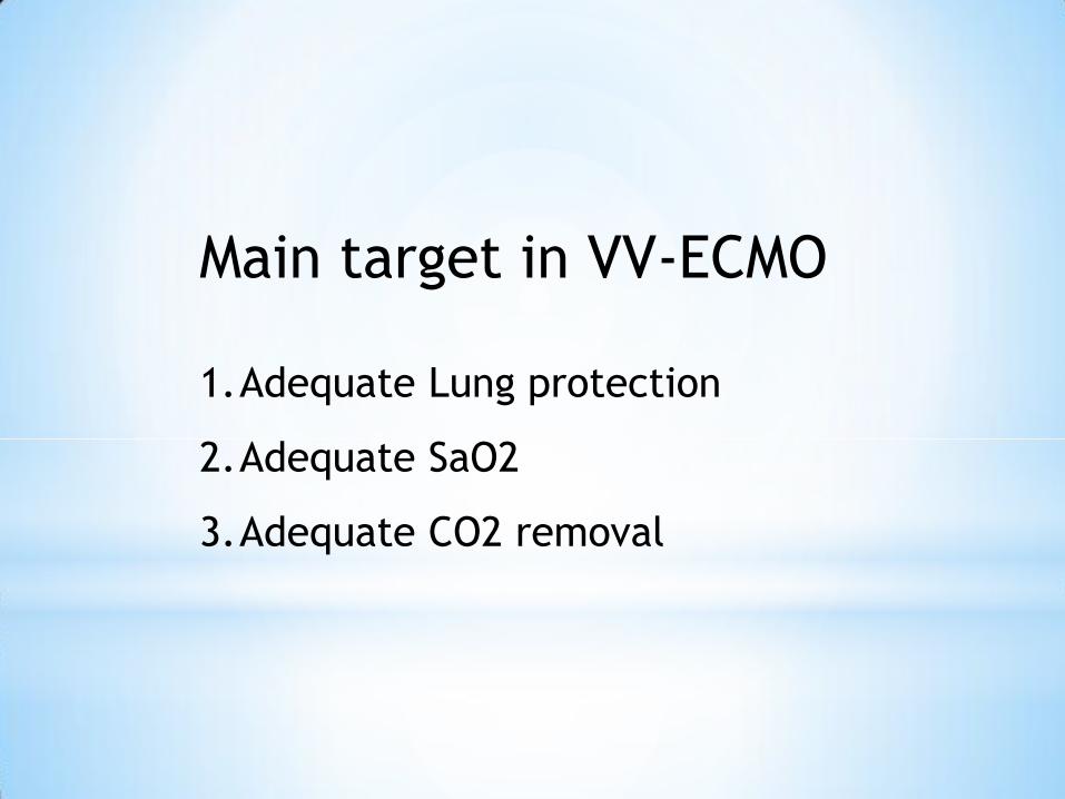
Main target in VV-ECMO
1.Adequate Lung protection
2.Adequate SaO2
3.Adequate CO2 removal
25
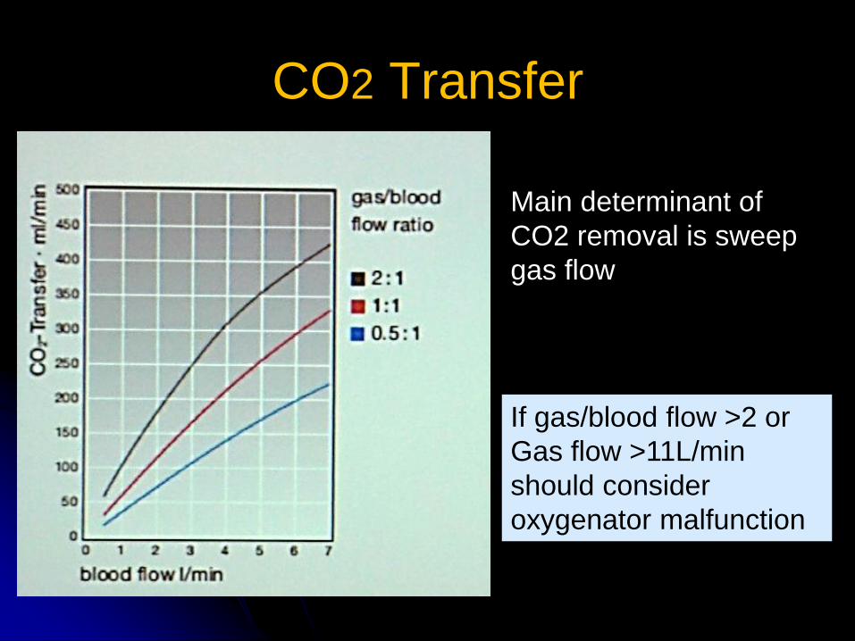
CO2 Transfer
Main determinant of
CO2 removal is sweep
gas flow
If gas/blood flow >2 or
Gas flow >11L/min
should consider
oxygenator malfunction
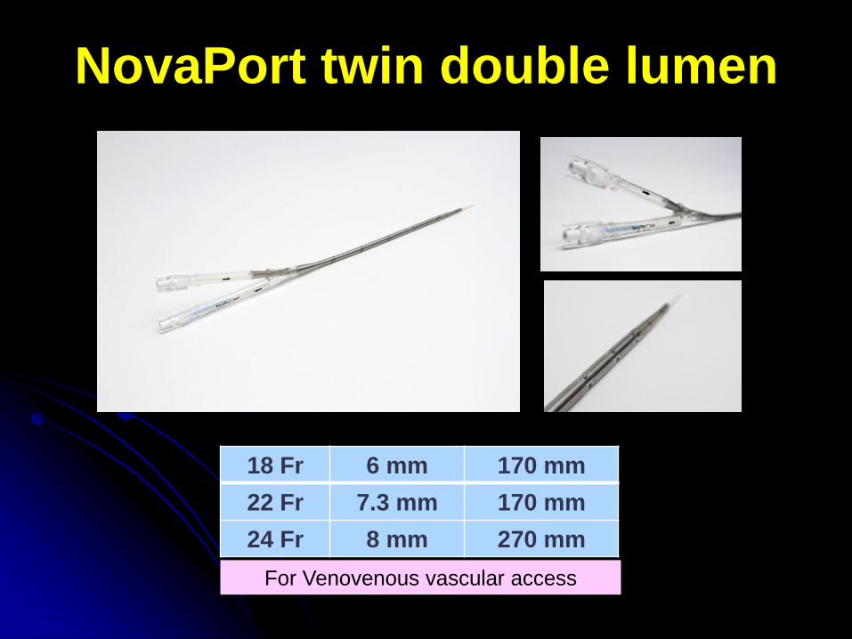
NovaPort twin double lumen
For Venovenous vascular access
18 Fr 6 mm 170 mm
22 Fr 7.3 mm 170 mm
24 Fr 8 mm 270 mm
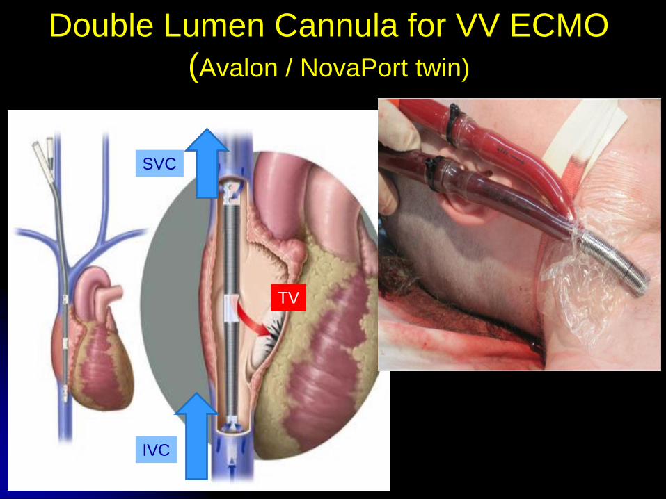
Double Lumen Cannula for VV ECMO
(Avalon / NovaPort twin)
SVC
IVC
TV
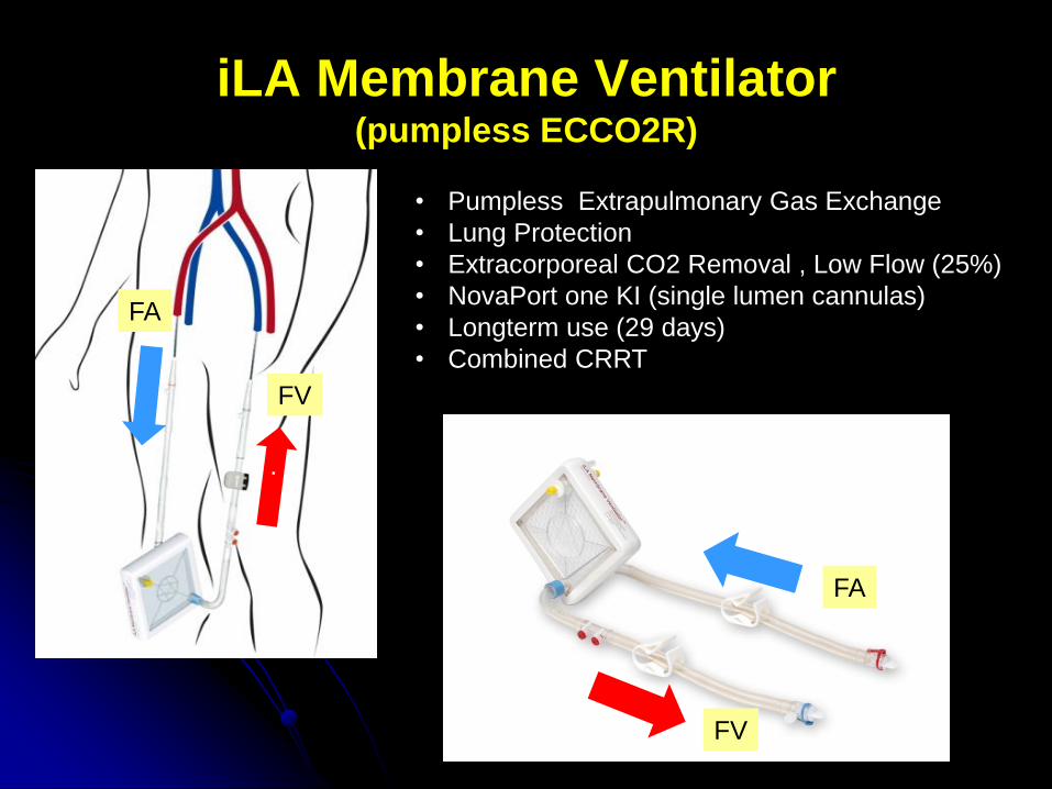
iLA Membrane Ventilator (pumpless ECCO2R)
• Pumpless Extrapulmonary Gas Exchange
• Lung Protection
• Extracorporeal CO2 Removal , Low Flow (25%)
• NovaPort one KI (single lumen cannulas)
• Longterm use (29 days)
• Combined CRRT
FA
FV
FA
FV
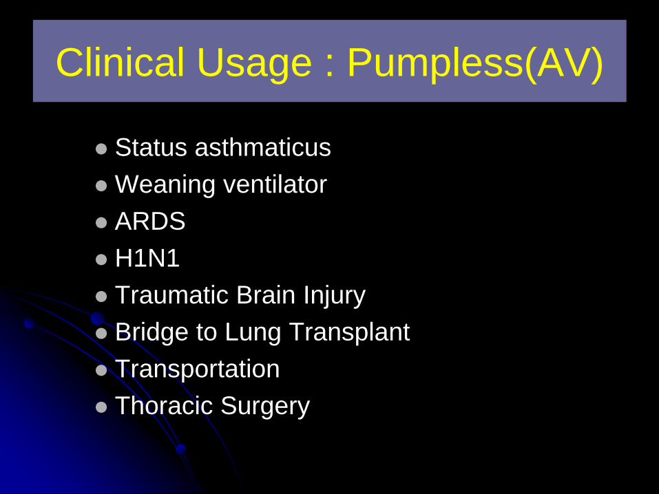
Clinical Usage : Pumpless(AV)
Status asthmaticus
Weaning ventilator
ARDS
H1N1
Traumatic Brain Injury
Bridge to Lung Transplant
Transportation
Thoracic Surgery
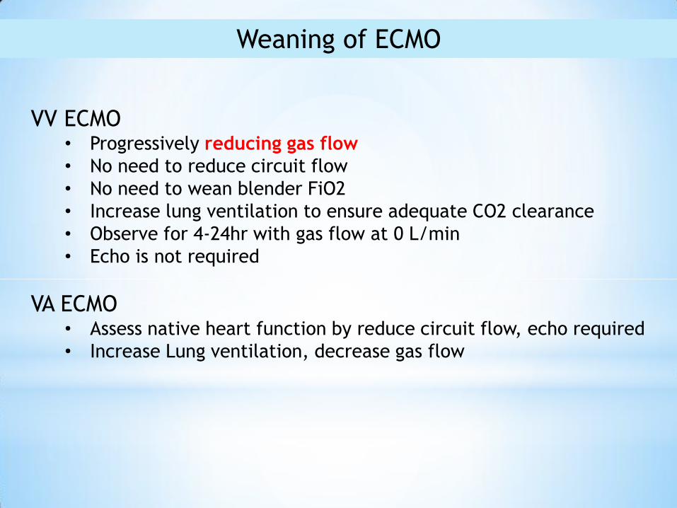
Weaning of ECMO
VV ECMO • Progressively reducing gas flow
• No need to reduce circuit flow
• No need to wean blender FiO2
• Increase lung ventilation to ensure adequate CO2 clearance
• Observe for 4-24hr with gas flow at 0 L/min
• Echo is not required
VA ECMO • Assess native heart function by reduce circuit flow, echo required
• Increase Lung ventilation, decrease gas flow

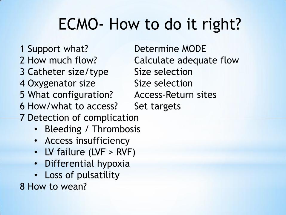
ECMO- How to do it right?
1 Support what? Determine MODE
2 How much flow? Calculate adequate flow
3 Catheter size/type Size selection
4 Oxygenator size Size selection
5 What configuration? Access-Return sites
6 How/what to access? Set targets
7 Detection of complication
• Bleeding / Thrombosis
• Access insufficiency
• LV failure (LVF > RVF)
• Differential hypoxia
• Loss of pulsatility
8 How to wean?