Embed Size (px)
Citation preview
9/8/2017
1
Emory University Physician Assistant Program
Heme Review
Allan Platt, PA-C, MMSc, DFAAPA
Assistant Professor
Emory University School of Medicine
Physician Assistant Program
Atlanta GA
[email protected] www.EmoryPA.org
Disclosure
I have nothing to disclose except
I do work for food
I promote giving Blood
Emory University Physician Assistant Program
Blood Blood has red cells(erythrocyctes)
White cells (leukocytes)
Platelets (thrombocytes)
Emory University Physician Assistant Program
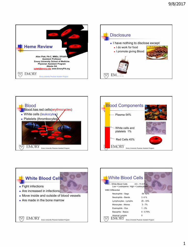
Blood Components
Plasma 54%
White cells and
platelets 1%
Red Cells 45%
Emory University Physician Assistant Program
White Blood Cells
Fight infections
Are increased in infections
Move inside and outside of blood vessels
Are made in the bone marrow
Emory University Physician Assistant Program
White Blood Cells
WBC - White Blood Cells 4.5 - 11.0 K/uL
Low = Leukopenia High = Leukocytosis
WBC Differential
Neutrophils - Segs 54 -62%
Neutrophils - Bands 3 -5 %
Lymphocytes - Lymphs 25 - 33%
Monocytes - Monos 3 - 7%
Eosinophils - Eos 1 - 3%
Basophils - Basos 0 - 0.75%
Atypical Lymphs 0
9/8/2017
2
Emory University Physician Assistant Program
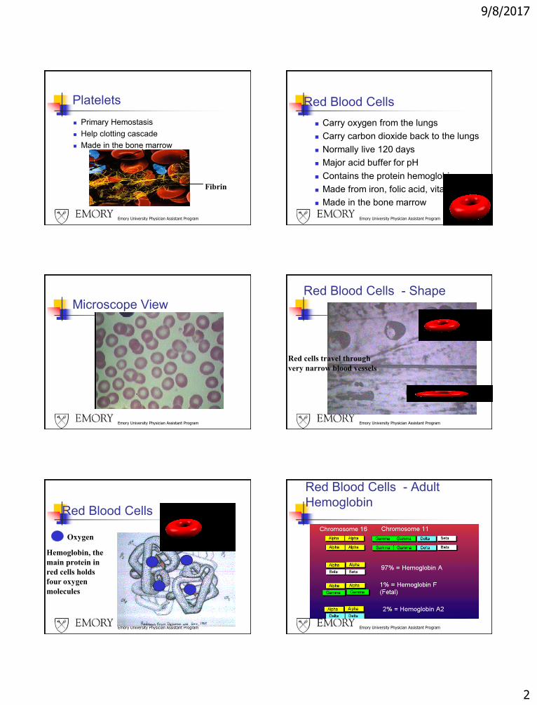
Platelets
Primary Hemostasis
Help clotting cascade
Made in the bone marrow
Fibrin
Emory University Physician Assistant Program
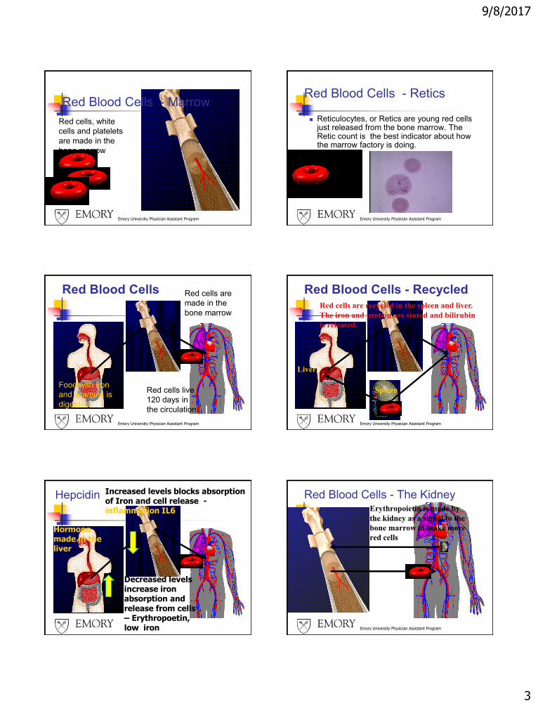
Red Blood Cells
Carry oxygen from the lungs
Carry carbon dioxide back to the lungs
Normally live 120 days
Major acid buffer for pH
Contains the protein hemoglobin
Made from iron, folic acid, vitamin B12
Made in the bone marrow
Emory University Physician Assistant Program
Microscope View
Emory University Physician Assistant Program
Red Blood Cells - Shape
Red cells travel through
very narrow blood vessels
Emory University Physician Assistant Program
Red Blood Cells - Hemoglobin
Oxygen
Hemoglobin, the
main protein in
red cells holds
four oxygen
molecules
Emory University Physician Assistant Program
Red Blood Cells - Adult
Hemoglobin
9/8/2017
3
Emory University Physician Assistant Program
Red Blood Cells - Marrow
Red cells, white
cells and platelets
are made in the
bone marrow
Emory University Physician Assistant Program
Red Blood Cells - Retics
Reticulocytes, or Retics are young red cells just released from the bone marrow. The Retic count is the best indicator about how the marrow factory is doing.
Emory University Physician Assistant Program
Red Blood Cells
Red cells live
120 days in
the circulation
Food with iron
and vitamins is
digested
Red cells are
made in the
bone marrow
Emory University Physician Assistant Program
Red Blood Cells - RecycledRed cells are recycled in the spleen and liver.
The iron and protein are stored and bilirubin
is released.
Spleen
Liver
Hepcidin
Decreased levels increase iron absorption and release from cells – Erythropoetin, low iron
Hormonemade in the liver
Increased levels blocks absorption of Iron and cell release -inflammation IL6
Emory University Physician Assistant Program
Red Blood Cells - The KidneyErythropoietin is made by
the kidney as a signal to the
bone marrow to make more
red cells
9/8/2017
4
Emory University Physician Assistant Program
The History
Weakness
Tiredness - Fatigue
Dyspnea
Dizzy – non vertigo
Palpitations
New angina
Emory University Physician Assistant Program
The History -2
- History of melena, abdominal pain, Aspirin or non-steroidal anti-inflammatory agents (NSAIDs) use, past peptic ulcer disease , then consider GI bleeding, platelet dysfunction.
- In females the menstrual history quantifying the amount of bloodloss ,or possible pregnancy should be obtained.
- History of pica or abnormal craving for ice, clay, starch...; dysphagia then consider iron deficiency.
- Poor diet, then consider iron or folate deficiency, and general
malnutrition
- History of gastric surgery, distal paresthesias, gait problems -consider B12 deficiency
- History of alcohol abuse - consider folate deficiency or liver disease. If moonshine use or lead paint/pipe exposure, consider lead toxicity.
Emory University Physician Assistant Program
The History -3
- Family history of blood cell or bleeding disorder: consider Sickle Cell disease, G6PD,Thalassemia, Hemophilia, von Willebrand
- History of jaundice, transfusion, new medication, infection -consider hemolytic process
- History of weight loss, Cancer, HIV, rheumatoid arthritis, thyroid disease, renal disease -then consider secondary cause
- History of fever and chills, cough, dyspnea, then consider Infection.
Emory University Physician Assistant Program
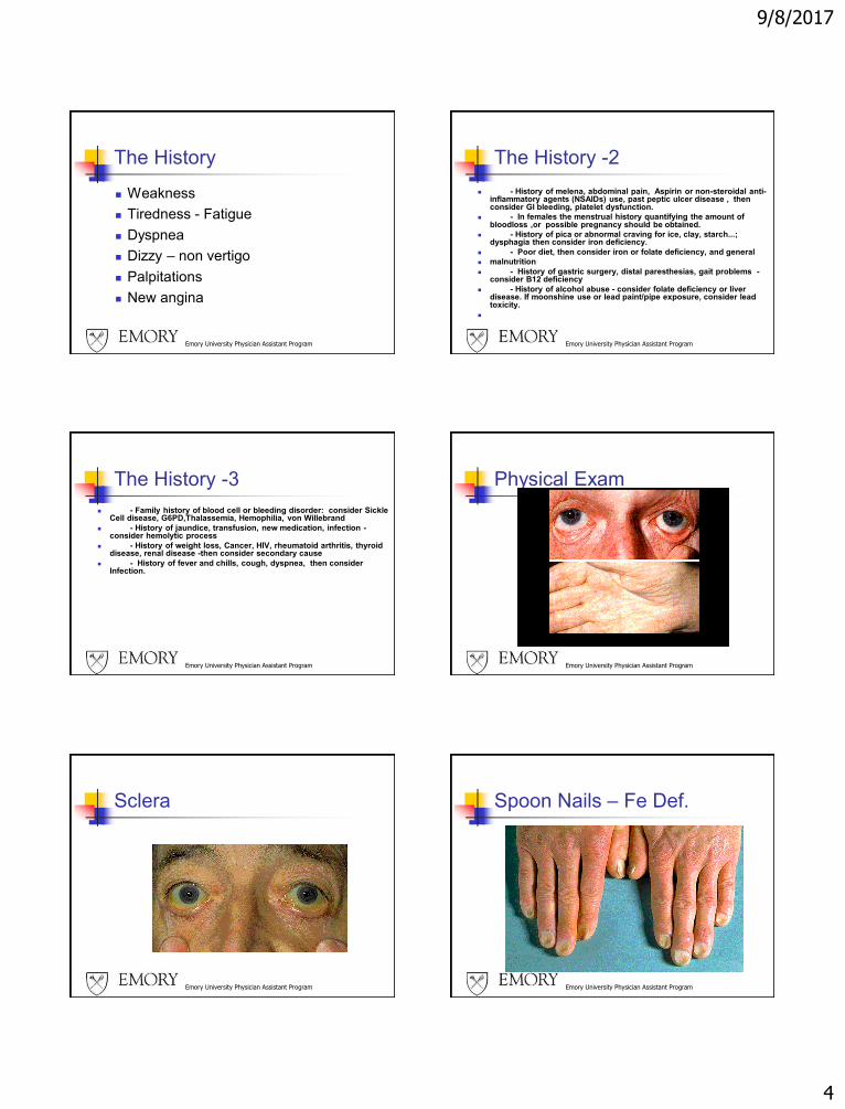
Physical Exam
Emory University Physician Assistant Program
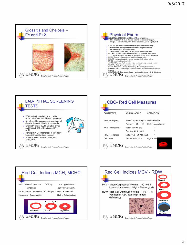
Sclera
Emory University Physician Assistant Program
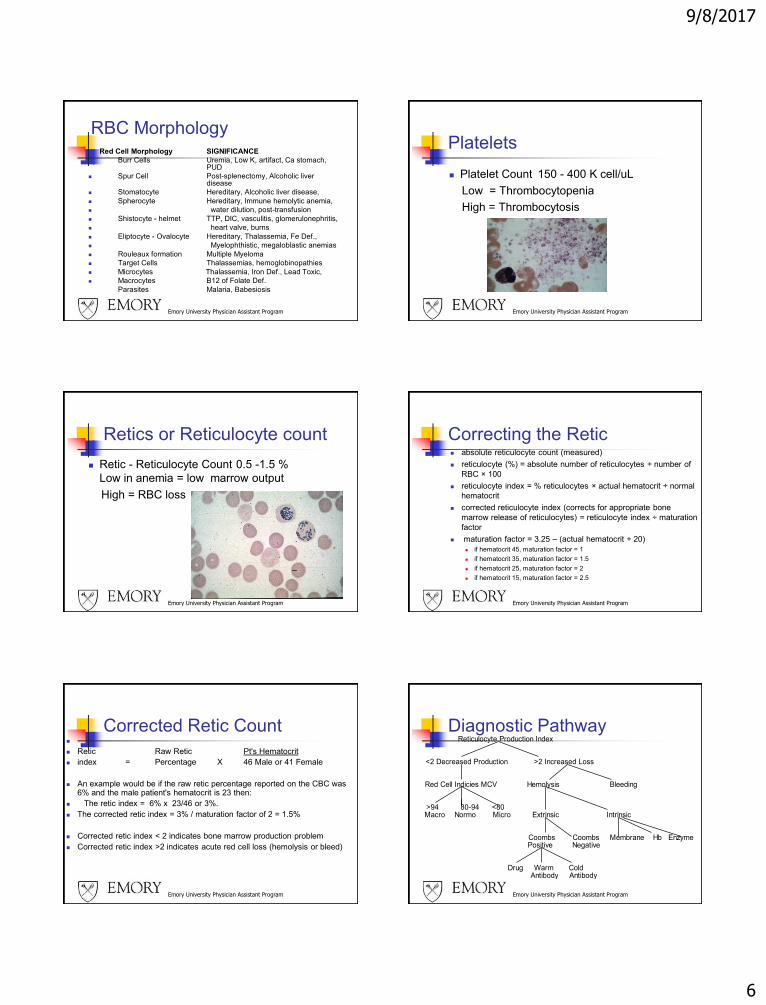
Spoon Nails – Fe Def.
9/8/2017
5
Emory University Physician Assistant Program
Glossitis and Chelosis –
Fe and B12
Emory University Physician Assistant Program
Physical Exam GENERAL INSPECTION- clubbing in TB or lung cancer
Skin- Hypothyroid, SLE, Bruises, lesions, petechiae or purpura.
Weight - Loss in Cancer, HIV, Chronic disease, gain in hypothyroid
VITAL SIGNS- Pulse: Tachycardia from increased cardiac output
Respirations: Tachypnea from decreased oxygen transport
BP: Orthostatic if volume depleted
Temp: Fever in infections and drug or transfusion reactions,
HEENT- Eye: Jaundice if hemolysis, pallor in palpebral conjunctiva
Mouth: Glossitis and angular stomatitis in iron or B12 deficiency
NECK- Thyroid enlargement or nodules, lymph nodes
HEART- Increased output/murmur- consider high output failure
LUNG- consider infection, lesion
ABDOMINAL- Liver/spleen size, masses, tenderness, surgical scars
RECTAL- Stool guaiac, prostate exam in men
PELVIC/BREAST- Uterine abnormality, Pap smear, Breast nodule
LYMPHNODES- consider lymphoma,leukemia,infection,connective tissue Disease
NEUROLOGIC- Decreased vibratory and position sense in B12 deficiency
Emory University Physician Assistant Program
LAB- INITIAL SCREENING
TESTS
CBC, red cell morphology and white blood cell differential, Reticulocyte count
Urinalysis- Hematuria/proteinuria in renal disease ,hemoglobinuria in hemolysis.
Chemistry profile (LDH, Bilirubin- Direct and Indirect, BUN, Creatinine, AST, ALT),
Hemoglobin Electrophoresis if hereditary hemoglobinopathy is suspected
IF BLEEDING - Platelet Count, PT, aPTT, PFA
Emory University Physician Assistant Program
CBC- Red Cell Measures
PARAMETER NORMAL ADULT COMMENTS
HB - Hemoglobin Male= 15.5 +/- 2 mg/dl Low = Anemia
Female = 13.5 +/- 2 High = polycythemia
HCT - Hematocrit Male= 46.0 +/- 6% "
Female= 41.0 +/- 6% "
RBC - Red Blood Male = 4.3 - 5.9 Million/uL "
Cell Count Female = 4.0 - 5.2 “ High in Thalassemia
Emory University Physician Assistant Program
Red Cell Indices MCH, MCHC
MCH - Mean Corpuscular 27 -32 pg Low = Hypochromic
Hemoglobin High = Hyperchromic
MCHC - Mean Corpuscular 30 - 36 gm/dl Low = R/O Fe def.
Hemoglobin Concentration High = Spherocytosis
Emory University Physician Assistant Program
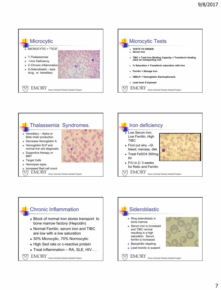
Red Cell Indices MCV - RDW
MCV - Mean Corpuscular Volume 80 - 94 fl
Low = Microcytosis High = Macrocytosis
RDW - Red Cell Distribution Width 11.5 - 14.5
Variation in RBC size (High in Iron
deficiency)
9/8/2017
6
Emory University Physician Assistant Program
RBC Morphology Red Cell Morphology SIGNIFICANCE
Burr Cells Uremia, Low K, artifact, Ca stomach, PUD
Spur Cell Post-splenectomy, Alcoholic liver disease
Stomatocyte Hereditary, Alcoholic liver disease,
Spherocyte Hereditary, Immune hemolytic anemia,
water dilution, post-transfusion
Shistocyte - helmet TTP, DIC, vasculitis, glomerulonephritis,
heart valve, burns
Eliptocyte - Ovalocyte Hereditary, Thalassemia, Fe Def.,
Myelophthistic, megaloblastic anemias
Rouleaux formation Multiple Myeloma
Target Cells Thalassemias, hemoglobinopathies
Microcytes Thalassemia, Iron Def., Lead Toxic,
Macrocytes B12 of Folate Def.
Parasites Malaria, Babesiosis
Emory University Physician Assistant Program
Platelets
Platelet Count 150 - 400 K cell/uL
Low = Thrombocytopenia
High = Thrombocytosis
Emory University Physician Assistant Program
Retics or Reticulocyte count
Retic - Reticulocyte Count 0.5 -1.5 %
Low in anemia = low marrow output
High = RBC loss
Correcting the Retic absolute reticulocyte count (measured)
reticulocyte (%) = absolute number of reticulocytes ÷ number of
RBC × 100
reticulocyte index = % reticulocytes × actual hematocrit ÷ normal
hematocrit
corrected reticulocyte index (corrects for appropriate bone
marrow release of reticulocytes) = reticulocyte index ÷ maturation
factor
maturation factor = 3.25 – (actual hematocrit ÷ 20)
if hematocrit 45, maturation factor = 1
if hematocrit 35, maturation factor = 1.5
if hematocrit 25, maturation factor = 2
if hematocrit 15, maturation factor = 2.5
Emory University Physician Assistant Program
Emory University Physician Assistant Program
Corrected Retic Count
Retic Raw Retic Pt's Hematocrit
index = Percentage X 46 Male or 41 Female
An example would be if the raw retic percentage reported on the CBC was 6% and the male patient's hematocrit is 23 then:
The retic index = 6% x 23/46 or 3%.
The corrected retic index = 3% / maturation factor of 2 = 1.5%
Corrected retic index < 2 indicates bone marrow production problem
Corrected retic index >2 indicates acute red cell loss (hemolysis or bleed)
Emory University Physician Assistant Program
Diagnostic PathwayReticulocyte Production Index
<2 Decreased Production >2 Increased Loss
Red Cell Indicies MCV Hemolysis Bleeding
>94 80-94 <80Macro Normo Micro Extrinsic Intrinsic
Coombs CoombsPositive Negative
Drug Warm ColdAntibody Antibody
Membrane Hb Enzyme
9/8/2017
7
Emory University Physician Assistant Program
Microcytic
MICROCYTIC = "TICS"
T-Thalassemias
I-Iron Deficiency
C-Chronic Inflammation
S-Sideroblastic - lead,
drug, or hereditary
Emory University Physician Assistant Program
Microcytic Tests
TESTS TO ORDER:
Serum Iron
TIBC = Total Iron Binding Capacity = Transferrin binding sites for transporting iron
% Saturation = Transferrin saturation with Iron
Ferritin = Storage Iron
HBELP = Hemoglobin Electrophoresis
Lead level if exposed
Emory University Physician Assistant Program
Thalassemia Syndromes.
Hereditary – Alpha or
Beta chain production
Decrease Hemoglobin A
Hemoglobin ELP and
normal Iron are diagnostic
Supportive therapy or
BMT
Target Cells
Hemolysis signs
Increased Red cell count
Emory University Physician Assistant Program
Iron deficiency
Low Serum iron,
Low Ferritin, High
TIBC
Find out why –GI
bleed, menses, diet
Treat FeSO4 300mg
tid
F/U in 2- 3 weeks
for Retic and Ferritin
Emory University Physician Assistant Program
Chronic Inflammation
Block of normal iron stores transport to
bone marrow factory (Hepcidin)
Normal Ferritin, serum iron and TIBC
are low with a low saturation
30% Microcytic, 70% Normocytic
High Sed rate or c-reactive protein
Treat inflammation – RA, SLE, HIV….
Emory University Physician Assistant Program
Sideroblastic
Ring sideroblasts in
bone marrow
Serum iron is increased
and TIBC normal
resulting in a high
saturation. Serum
ferritin is increased
Basophillic stippling
Lead toxicity is suspect
9/8/2017
8
Emory University Physician Assistant Program
Normocytic Anemia NORMOCYTIC = "NORMAL SIZE“
N-Normal Pregnancy
O-Over hydration
R-Renal Disease
M-Myelophthistic
A-Acute Blood Loss
L-Liver Disease
S - Systemic Infection
I- Inflammatory Block
Z-Zero Production- Aplastic anemia
E-Endocrine:Hypothyroid, hypoadrenal, hypoandrogen
Normocytic Tests
Blood Urea Nitrogen (BUN), Creatinine,
SGOT, Alkaline Phosphatase, Bilirubin,
Erythrocyte Sedimentation Rate (ESR),
Urinalysis, and Thyroid profile
Renal Function tests
Pregnancy Test
Bone Marrow Biopsy
Normocytic - Renal Failure
Anemia caused by decrease
erythropoetin production causing
decreased bone marrow production
Can monitor erythropoetin levels
Treat with epoetin alfa injections
weekly or darbepoetin alpha every other
week or monthly
Aplastic Anemia
idiopathic (78% cases)
hepatitis (5% cases) testing for known
hepatitis viruses usually negative
drugs (2% cases due to gold, 4% due to other
drugs)
Parvo virus B19 (Fifths disease)
Check WBC and Platelet count
May need Bone Marrow Bx and supportive
therapy
Emory University Physician Assistant Program
Emory University Physician Assistant Program
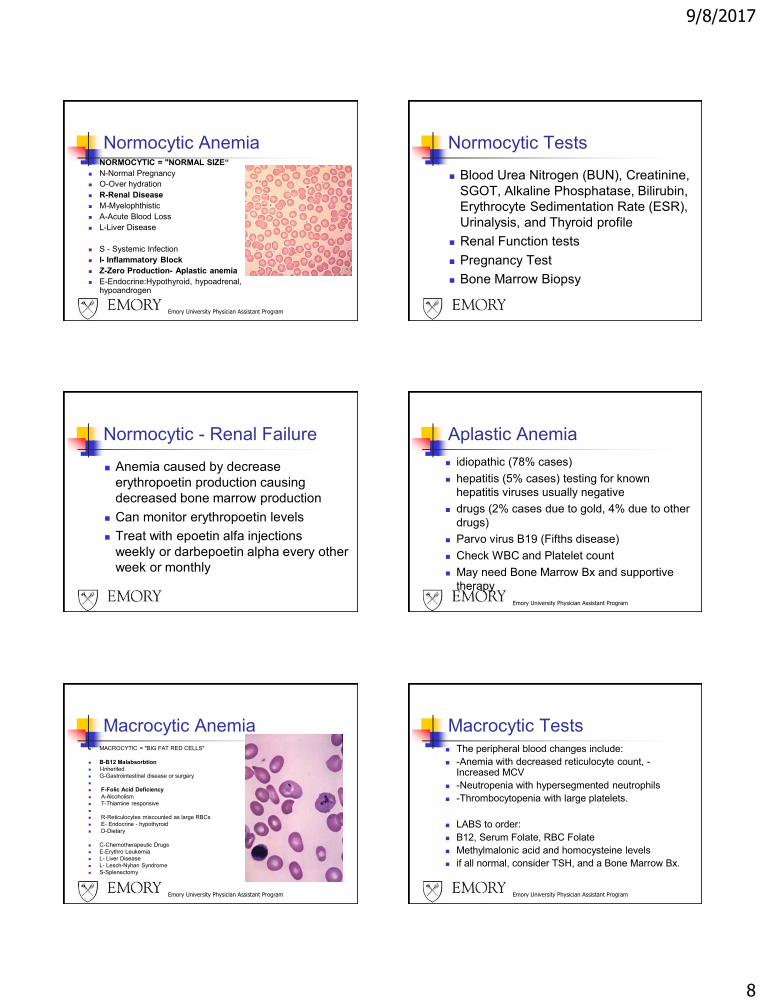
Macrocytic Anemia MACROCYTIC = "BIG FAT RED CELLS"
B-B12 Malabsorbtion
I-Inherited
G-Gastrointestinal disease or surgery
F-Folic Acid Deficiency
A-Alcoholism
T-Thiamine responsive
R-Reticulocytes miscounted as large RBCs
E- Endocrine - hypothyroid
D-Dietary
C-Chemotherapeutic Drugs
E-Erythro Leukemia
L- Liver Disease
L- Lesch-Nyhan Syndrome
S-Splenectomy
Emory University Physician Assistant Program
Macrocytic Tests The peripheral blood changes include:
-Anemia with decreased reticulocyte count, -Increased MCV
-Neutropenia with hypersegmented neutrophils
-Thrombocytopenia with large platelets.
LABS to order:
B12, Serum Folate, RBC Folate
Methylmalonic acid and homocysteine levels
if all normal, consider TSH, and a Bone Marrow Bx.
9/8/2017
9
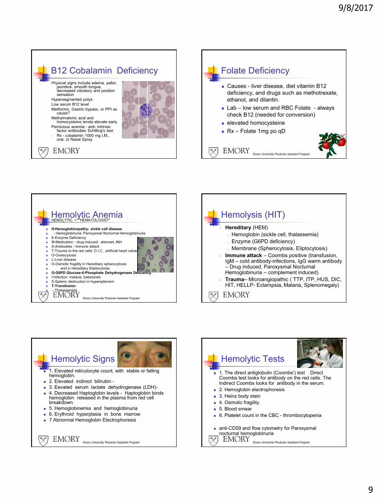
B12 Cobalamin Deficiency
Physical signs include edema, pallor, jaundice, smooth tongue, decreased vibratory and position sensation
Hypersegmented polys
Low serum B12 level
Metformin, Gastric bypass, or PPI as cause?
Methylmalonic acid and homocysteine levels elevate early
Pernicious anemia - anti- intrinsic factor antibodies Schilling's test
• Rx - cobalamin 1000 mg I.M., oral, or Nasal Spray
Emory University Physician Assistant Program
Folate Deficiency
Causes - liver disease, diet vitamin B12
deficiency, and drugs such as methotrexate,
ethanol, and dilantin.
Lab – low serum and RBC Folate - always
check B12 (needed for conversion)
elevated homocysteine
Rx – Folate 1mg po qD
Emory University Physician Assistant Program
Hemolytic Anemia HEMOLYTIC = "HEMATOLOGIST"
H-Hemoglobinopathy: sickle cell disease
- Hemoglobinuria: Paroxysmal Nocturnal Hemoglobinuria
E-Enzyme Deficiency
M-Medication - drug induced: aldomet, INH
A-Antibodies - Immune attack
T-Trauma to the red cells: D.I.C , artificial heart valves
O-Ovalocytosis
L-Liver disease
O-Osmotic fragility in Hereditary spherocytosis
and in Hereditary Eliptocytosis
G-G6PD Glucose-6-Phosphate Dehydrogenase Deficiency
I-Infection: malaria, babesiosis
S-Splenic destruction in hypersplenism
T-Transfusion
- Thalassemias
Hemolysis (HIT)
• Hereditary (HEM)
– Hemoglobin (sickle cell, thalassemia)
– Enzyme (G6PD deficiency)
– Membrane (Spherocytosis, Eliptocytosis)
• Immune attack – Coombs positive (transfusion, IgM – cold antibody-infections, IgG warm antibody – Drug induced, Paroxysmal Nocturnal Hemoglobinuria – complement induced)
• Trauma– Microangiopathic ( TTP, ITP, HUS, DIC, HIT, HELLP- Eclampsia, Malaria, Splenomegaly)
Emory University Physician Assistant Program
Hemolytic Signs
1. Elevated reticulocyte count, with stable or falling hemoglobin.
2. Elevated indirect bilirubin -
3. Eevated serum lactate dehydrogenase (LDH)-
4. Decreased Haptoglobin levels - Haptoglobin binds hemoglobin released in the plasma from red cell breakdown.
5. Hemoglobinemia and hemoglobinuria
6. Erythroid hyperplasia in bone marrow
7 Abnormal Hemoglobin Electrophoresis
Emory University Physician Assistant Program
Hemolytic Tests
1. The direct antiglobulin (Coombs') test Direct Coombs test looks for antibody on the red cells. The Indirect Coombs looks for antibody in the serum.
2. Hemoglobin electrophoresis
3. Heinz body stain
4. Osmotic fragility
5. Blood smear
6. Platelet count in the CBC - thrombocytopenia
anti-CD59 and flow cytometry for Paroxysmal nocturnal hemoglobinuria
9/8/2017
10
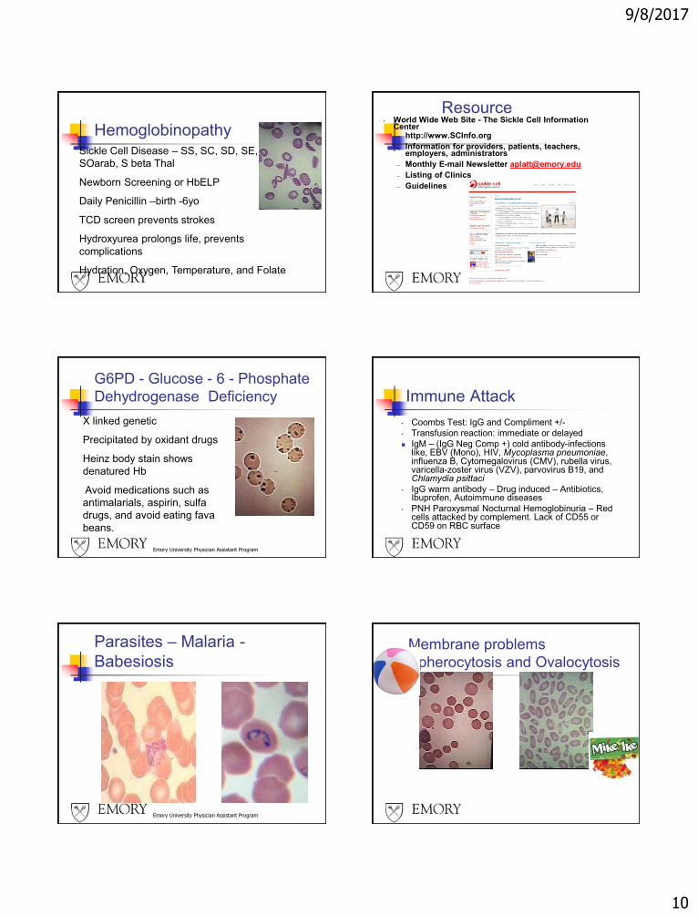
HemoglobinopathySickle Cell Disease – SS, SC, SD, SE,
SOarab, S beta Thal
Newborn Screening or HbELP
Daily Penicillin –birth -6yo
TCD screen prevents strokes
Hydroxyurea prolongs life, prevents
complications
Hydration, Oxygen, Temperature, and Folate
Resource• World Wide Web Site - The Sickle Cell Information
Center– http://www.SCInfo.org
– Information for providers, patients, teachers, employers, administrators
– Monthly E-mail Newsletter [email protected]
– Listing of Clinics
– Guidelines
Emory University Physician Assistant Program
G6PD - Glucose - 6 - Phosphate
Dehydrogenase Deficiency
X linked genetic
Precipitated by oxidant drugs
Heinz body stain shows
denatured Hb
Avoid medications such as
antimalarials, aspirin, sulfa
drugs, and avoid eating fava
beans.
Immune Attack
• Coombs Test: IgG and Compliment +/-
• Transfusion reaction: immediate or delayed
IgM – (IgG Neg Comp +) cold antibody-infections like, EBV (Mono), HIV, Mycoplasma pneumoniae, influenza B, Cytomegalovirus (CMV), rubella virus, varicella-zoster virus (VZV), parvovirus B19, and Chlamydia psittaci
• IgG warm antibody – Drug induced – Antibiotics, Ibuprofen, Autoimmune diseases
• PNH Paroxysmal Nocturnal Hemoglobinuria – Red cells attacked by complement. Lack of CD55 or CD59 on RBC surface
Emory University Physician Assistant Program
Parasites – Malaria -
BabesiosisMembrane problems
Spherocytosis and Ovalocytosis
9/8/2017
11
Emory University Physician Assistant Program
To Clot or Not
Coagulopathies
Allan Platt, PA-C
Faculty, Physician Assistant Program
Emory University School of Medicine
Atlanta, GA
www.EmoryPA.org
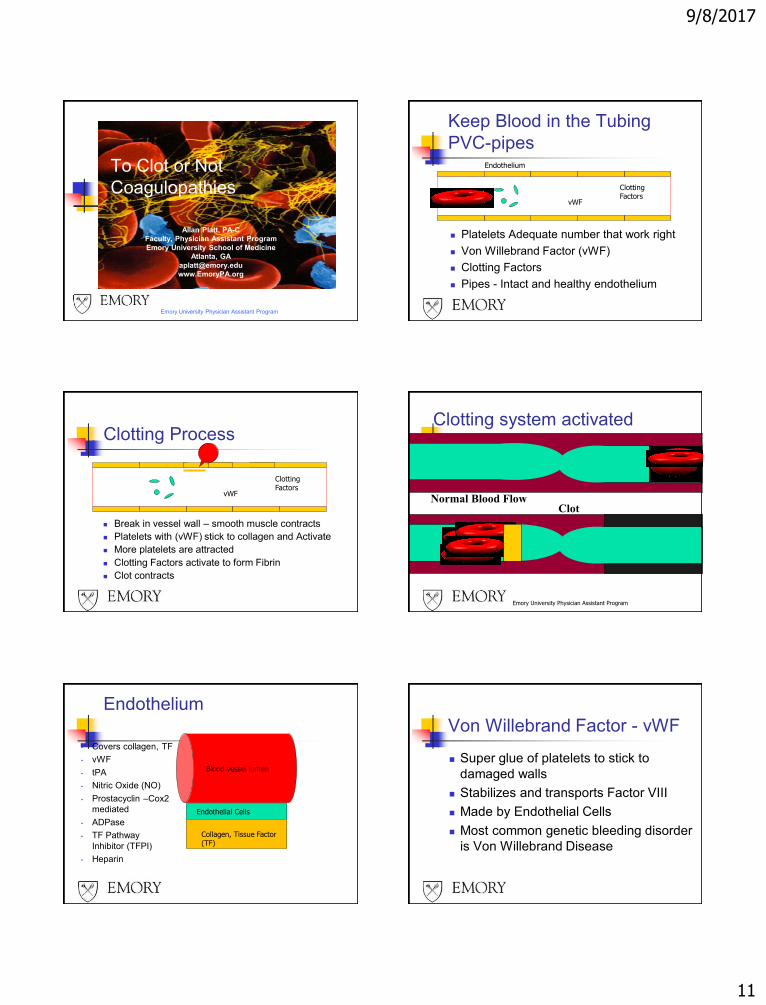
Keep Blood in the Tubing
PVC-pipes
Platelets Adequate number that work right
Von Willebrand Factor (vWF)
Clotting Factors
Pipes - Intact and healthy endothelium
vWF
Clotting Factors
Endothelium
Clotting Process
Break in vessel wall – smooth muscle contracts
Platelets with (vWF) stick to collagen and Activate
More platelets are attracted
Clotting Factors activate to form Fibrin
Clot contracts
vWF
Clotting Factors
Emory University Physician Assistant Program
Clotting system activated
Normal Blood FlowClot
Endothelium
• Covers collagen, TF
• vWF
• tPA
• Nitric Oxide (NO)
• Prostacyclin –Cox2
mediated
• ADPase
• TF Pathway
Inhibitor (TFPI)
• Heparin
Collagen, Tissue Factor (TF)
Endothelial Cells
Blood vessel lumen
Von Willebrand Factor - vWF
Super glue of platelets to stick to
damaged walls
Stabilizes and transports Factor VIII
Made by Endothelial Cells
Most common genetic bleeding disorder
is Von Willebrand Disease
9/8/2017
12
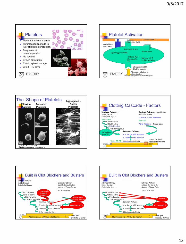
Platelets
Made in the bone marrow
Thrombopoeitin made in
liver stimulates production
Fragments of
megacaryocytes
No nucleus
67% in circulation
33% in spleen storage
Life 8 – 10 days
Emory University Physician Assistant Program
Platelet Activation
glycoprotein (GP) IIb/IIIa receptor
Von Willibron Factor vWF
Aranchidonic acid
Collagen, Thrombin, TXA2
Cyclooxygenase COX
TXA2 and ADP released, also
PF4,
Endothelium
ADP receptor
Fibrinogen attaches to other platelets
Increase cAMP inhibits activation
The Shape of Platelets
Courtesy of Helena Diagnostics
Flowing
Platelets
Activated
Platelets
Aggregated -
Active
Platelets Clotting Cascade - Factors
Intrinsic Pathway –Inside the cut
Endothelial Injury
Test = aPTTXII to XII active
XI to XI activeIX to IX active
VIII to VIIIactive
Common Pathway
X to Xactive with V present
II Prothromin to Thrombin
I Fibrinogen to Fibrin
Extrinsic Pathway – outside the cut in the plasma
Vitamin K - Liver dependant
Test = PT
VII to VIIactive + Tissue factor
XIII to XIIIactive stabilizer to crosslink
fibrin
vWF stabilizes Factor VIII
Test = TT, RT
Built in Clot Blockers and Busters
Intrinsic Pathway –Inside the cut
Endothelial Injury
XII to XII active
XI to XI active
IX to IX active
VIII to VIIIactive
Common Pathway
X to Xactive with V present
II Prothromin to Thrombin
I Fibrinogen to Fibrin
Extrinsic Pathway –outside the cut in the
plasma – Tissue Factor
VII to VIIactive
Plasminogen via t-PA/ PAI-1 to Plasmin
Liver made Protein S
Protein C
Antithrombin III
Fibrin split products, D-Dimer
Tissue Factor Pathway Inhibitor
Built In Clot Blockers and Busters
Intrinsic Pathway –Inside the cut
Endothelial Injury
XII to XII active
XI to XI active
IX to IX active
VIII to VIIIactive
Common Pathway
X to Xactive with V present
II Prothromin to Thrombin
I Fibrinogen to Fibrin
Extrinsic Pathway –outside the cut in the
plasma – Tissue Factor
VII to VIIactive
Plasminogen via t-PA/PAI-1to Plasmin
Antithrombin III
Fibrin split products, D-Dimer
Heparin
9/8/2017
13
Emory University Physician Assistant Program
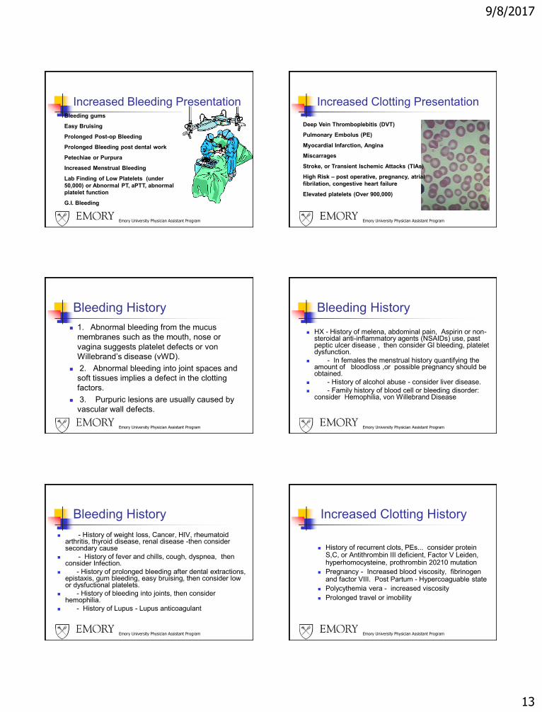
Increased Bleeding PresentationBleeding gums
Easy Bruising
Prolonged Post-op Bleeding
Prolonged Bleeding post dental work
Petechiae or Purpura
Increased Menstrual Bleeding
Lab Finding of Low Platelets (under
50,000) or Abnormal PT, aPTT, abnormal
platelet function
G.I. Bleeding
Emory University Physician Assistant Program
Increased Clotting Presentation
Deep Vein Thromboplebitis (DVT)
Pulmonary Embolus (PE)
Myocardial Infarction, Angina
Miscarrages
Stroke, or Transient Ischemic Attacks (TIAs)
High Risk – post operative, pregnancy, atrial
fibrilation, congestive heart failure
Elevated platelets (Over 900,000)
Emory University Physician Assistant Program
Bleeding History
1. Abnormal bleeding from the mucus
membranes such as the mouth, nose or
vagina suggests platelet defects or von
Willebrand’s disease (vWD).
2. Abnormal bleeding into joint spaces and
soft tissues implies a defect in the clotting
factors.
3. Purpuric lesions are usually caused by
vascular wall defects.
Emory University Physician Assistant Program
Bleeding History
HX - History of melena, abdominal pain, Aspirin or non-steroidal anti-inflammatory agents (NSAIDs) use, past peptic ulcer disease , then consider GI bleeding, platelet dysfunction.
- In females the menstrual history quantifying the amount of bloodloss ,or possible pregnancy should be obtained.
- History of alcohol abuse - consider liver disease.
- Family history of blood cell or bleeding disorder: consider Hemophilia, von Willebrand Disease
Emory University Physician Assistant Program
Bleeding History
- History of weight loss, Cancer, HIV, rheumatoid arthritis, thyroid disease, renal disease -then consider secondary cause
- History of fever and chills, cough, dyspnea, then consider Infection.
- History of prolonged bleeding after dental extractions, epistaxis, gum bleeding, easy bruising, then consider low or dysfuctional platelets.
- History of bleeding into joints, then consider hemophilia.
- History of Lupus - Lupus anticoagulant
Emory University Physician Assistant Program
Increased Clotting History
History of recurrent clots, PEs... consider protein S,C, or Antithrombin III deficient, Factor V Leiden, hyperhomocysteine, prothrombin 20210 mutation
Pregnancy - Increased blood viscosity, fibrinogen and factor VIII. Post Partum - Hypercoaguable state
Polycythemia vera - increased viscosity
Prolonged travel or imobility
9/8/2017
14
Emory University Physician Assistant Program
Increased Clotting History
Smoking, Resent Surgery, Diabetes,
Congestive Heart Failure, Cancer, Atrial
Fibrillation are all high risk
Autoimmune diseases such as systemic lupus
erythematosis, and medications such as
procainamide, chlorpromazine, and quinidine.
Oral contraceptives - Estrogen
Emory University Physician Assistant Program
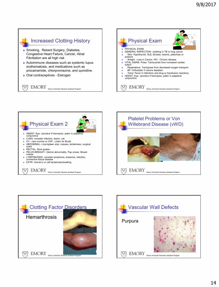
Physical Exam
PHYSICAL EXAM
GENERAL INSPECTION- clubbing in TB or lung cancer
Skin- Hypothyroid, SLE, Bruises, lesions, petechiae or purpura.
Weight - Loss in Cancer, HIV, Chronic disease
VITAL SIGNS- Pulse: Tachycardia from increased cardiac output
Respirations: Tachypnea from decreased oxygen transport
BP: Orthostatic if volume depleted
Temp: Fever in infections and drug or transfusion reactions,
HEENT- Eye: Jaundice if hemolysis, pallor in palpebral conjunctiva
Emory University Physician Assistant Program
Physical Exam 2
HEENT- Eye: Jaundice if hemolysis, pallor in palpebral conjunctiva
LUNG- consider infection, lesion, rub
CV - new murmer or CHF , Listen for Bruits
ABDOMINAL- Liver/spleen size, masses, tenderness, surgical scars
RECTAL- Stool guaiac,
PELVIC/BREAST- Uterine abnormality, Pap smear, Breast nodule
LYMPHNODES- consider lymphoma, leukemia, infection, connective tissue disease
EXTR- Homan’s or calf tenderness/swelling
Emory University Physician Assistant Program
Platelet Problems or Von
Willebrand Disease (vWD)
Emory University Physician Assistant Program
Clotting Factor Disorders
Hemarthrosis
Emory University Physician Assistant Program
Vascular Wall Defects
Purpura
9/8/2017
15
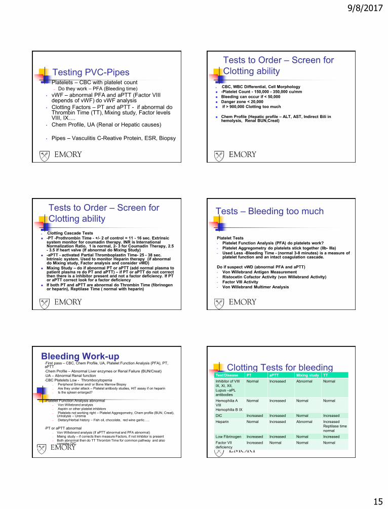
Testing PVC-Pipes• Platelets – CBC with platelet count
– Do they work – PFA (Bleeding time)
• vWF – abnormal PFA and aPTT (Factor VIII depends of vWF) do vWF analysis
• Clotting Factors – PT and aPTT - if abnormal do Thrombin Time (TT), Mixing study, Factor levels VIII, IX….
• Chem Profile, UA (Renal or Hepatic causes)
• Pipes – Vasculitis C-Reative Protein, ESR, Biopsy
Tests to Order – Screen for
Clotting ability
-CBC, WBC Differential, Cell Morphology
-Platelet Count - 150,000 - 350,000 cu/mm
Bleeding can occur if < 50,000
Danger zone < 20,000
if > 900,000 Clotting too much
Chem Profile (Hepatic profile – ALT, AST, Indirect Bili in hemolysis, Renal BUN,Creat)
Tests to Order – Screen for
Clotting ability
-Clotting Cascade Tests
-PT -Prothrombin Time - +/- 2 of control = 11 - 16 sec. Extrinsic system monitor for coumadin therapy. INR is International Normalization Ratio, 1 is normal, 2- 3 for Coumadin Therapy, 2.5 - 3.5 if heart valve (If abnormal do Mixing Study)
-aPTT - activated Partial Thromboplastin Time- 25 - 38 sec. Intrinsic system. Used to monitor Heparin therapy (if abnormal do Mixing study, Factor analysis and consider vWD)
Mixing Study – do if abnormal PT or aPTT (add normal plasma to patient plasma re do PT and aPTT) – if PT or aPTT do not correct then there is a inhibitor present and not a factor deficiency. If PT or aPTT correct look for a factor deficiency
If both PT and aPTT are abnormal do Thrombin Time (fibrinogen or heparin), Reptilase Time ( normal with heparin)
Tests – Bleeding too much
Platelet Tests
• Platelet Function Analysis (PFA) do platelets work?
• Platelet Aggregometry do platelets stick together (IIb- IIIa)
• Used Less -Bleeding Time - (normal 3-8 minutes) is a measure of platelet function and an intact coagulation cascade.
Do if suspect vWD (abnormal PFA and aPTT)
• Von Willebrand Antigen Measurement
• Ristocetin Cofactor Activity (von Willebrand Activity)
• Factor VIII Activity
• Von Willebrand Multimer Analysis
Bleeding Work-up•First pass – CBC, Chem Profile, UA, Platelet Function Analysis (PFA), PT, aPTT
•Chem Profile – Abnormal Liver enzymes or Renal Failure (BUN/Creat)
•UA – Abnormal Renal function
•CBC Platelets Low - Thrombocytopenia– Peripheral Smear and/ or Bone Marrow Biopsy
– Are they under attack – Platelet antibody studies, HIT assay if on heparin
– Is the spleen enlarged?
•Platelet Function Analysis abnormal – Von Willebrand analysis
– Aspirin or other platelet inhibitors
– Platelets not working right – Platelet Aggregometry, Chem profile (BUN, Creat), Urinalysis – Uremia
– Dietary/Herbal history – Fish oil, chocolate, red wine garlic…..
•PT or aPTT abnormal– Von Willebrand analysis (If aPTT abnormal and PFA abnormal)
– Mixing study – if corrects then measure Factors, if not Inhibitor is present
– Both abnormal then do TT Thrombin Time for common pathway and also consider DIC.
Clotting Tests for bleedingTest/Disease PT aPTT Mixing study TT
Inhibitor of VIII
IX, XI, XII,
Lupus –aPL
antibodies
Normal Increased Abnormal Normal
Hemophilia A
VIII
Hemophilia B IX
Normal Increased Normal Normal
DIC Increased Increased Normal Increased
Heparin Normal Increased Abnormal Increased
Reptilase time
normal
Low Fibrinogen Increased Increased Normal Increased
Factor VII
deficiency
Increased Normal Normal Normal
9/8/2017
16
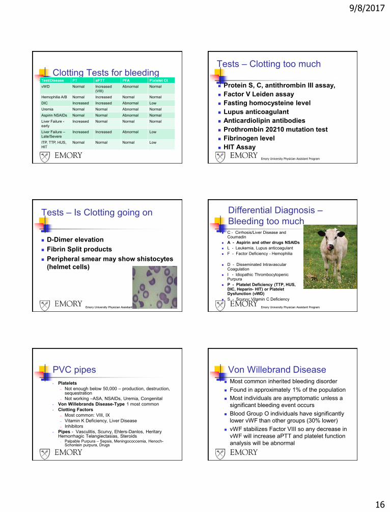
Clotting Tests for bleedingTest/Disease PT aPTT PFA Platelet Ct
vWD Normal Increased
(VIII)
Abnormal Normal
Hemophilia A/B Normal Increased Normal Normal
DIC Increased Increased Abnormal Low
Uremia Normal Normal Abnormal Normal
Aspirin NSAIDs Normal Normal Abnormal Normal
Liver Failure -
early
Increased Normal Normal Normal
Liver Failure –
Late/Severe
Increased Increased Abnormal Low
ITP, TTP, HUS,
HIT
Normal Normal Normal Low
Emory University Physician Assistant Program
Tests – Clotting too much
Protein S, C, antithrombin III assay,
Factor V Leiden assay
Fasting homocysteine level
Lupus anticoagulant
Anticardiolipin antibodies
Prothrombin 20210 mutation test
Fibrinogen level
HIT Assay
Emory University Physician Assistant Program
Tests – Is Clotting going on
D-Dimer elevation
Fibrin Split products
Peripheral smear may show shistocytes
(helmet cells)
Emory University Physician Assistant Program
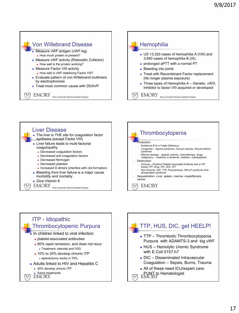
Differential Diagnosis –
Bleeding too much C - Cirrhosis/Liver Disease and
Coumadin
A - Aspirin and other drugs NSAIDs
L - Leukemia, Lupus anticoagulant
F - Factor Deficiency - Hemophilia
D - Disseminated Intravascular Coagulation
I - Idiopathic Thrombocytopenic Purpura
P - Platelet Deficiency (TTP, HUS, DIC, Heparin- HIT) or Platelet Dysfunction (vWD)
S - Scurvy: Vitamin C Deficiency
PVC pipes
• Platelets
– Not enough below 50,000 – production, destruction, sequestration
– Not working –ASA, NSAIDs, Uremia, Congenital
• Von Willebrands Disease-Type 1 most common
• Clotting Factors
– Most common: VIII, IX
– Vitamin K Deficiency, Liver Disease
– Inhibitors
• Pipes - Vasculitis, Scurvy, Ehlers-Danlos, HeritaryHemorrhagic Telangiectasias, Steroids
– Palpable Purpura – Sepsis, Meningococcemia, Henoch-Schonlein purpura, Drugs
Von Willebrand Disease Most common inherited bleeding disorder
Found in approximately 1% of the population
Most individuals are asymptomatic unless a
significant bleeding event occurs
Blood Group O individuals have significantly
lower vWF than other groups (30% lower)
vWF stabilizes Factor VIII so any decrease in
vWF will increase aPTT and platelet function
analysis will be abnormal
9/8/2017
17
Emory University Physician Assistant Program
Von Willebrand Disease Measure vWF antigen (vWF:Ag)
How much protein is present?
Measure vWF activity (Ristocetin Cofactor) How well is the protein working?
Measure Factor VIII activity How well is vWF stabilizing Factor VIII?
Evaluate pattern of von Willebrand multimers by electrophoresis
Treat most common cause with DDAVP
Hemophilia
US 13,320 cases of hemophilia A (VIII) and
3,640 cases of hemophilia B (IX).
prolonged aPTT with a normal PT
Bleeding into joints
Treat with Recombinant Factor replacement
(No longer plasma exposure)
Three types of Hemophilia A – Genetic, vWD,
Inhibitor to factor VIII acquired or developed
Emory University Physician Assistant Program
Emory University Physician Assistant Program
Liver Disease The liver is THE site for coagulation factor
synthesis (except Factor VIII)
Liver failure leads to multi-factorial coagulopathy Decreased coagulation factors
Decreased anti-coagulation factors
Decreased fibrinogen
Decreased platelets
Increased D-dimers (interfere with clot formation)
Bleeding from liver failure is a major cause morbidity and mortality
Give Vitamin K
Thrombocytopenia
• Production
– Nutritional B12 or Folate Deficiency
– Congenital – Alports syndrome, Fanconi anemia, Wiscott-Aldrich syndrome
– Marrow damage – aplastic anemia, chemotherapy, drugs, malignancy – myeloma or leukemia, radiation, mylodysplasia
• Destruction
– Immune – (Positive Platelet Associated Antibody test or HIT assay) ITP, Drug, HIV, SLE, HIT
– Non-Immune- DIC, TTP, Preeclampsia, HELLP syndrome Anti-phospholipid syndrome
• Sequestration- Liver, spleen, marrow -myelofibrosis, cancer
ITP - Idiopathic
Thrombocytopenic Purpura
In children linked to viral infection
platelet-associated antibodies
80% rapid remission, and does not recur
Treatment: steroids and IVIG
10% to 20% develop chronic ITP
splenectomy works in 70%
Adults linked to HIV and Hepatitis C 50% develop chronic ITP
Same treatments
TTP, HUS, DIC, get HEELP!
TTP – Thrombotic Thrombocytopenia
Purpura with ADAMTS-3 and big vWF
HUS – Hemolytic Uremic Syndrome
with E.Coli 0157:h7
DIC – Disseminated Intravascular
Coagulation – Sepsis, Burns, Trauma
All of these need ICU/expert care:
PUNT to Hematologist
9/8/2017
18
HELLP- Pregnancy Hemolysis (high indirect Bilirubin,
LDH)
Elevated Liver Enzymes (AST, ALT)
Low Platelets
severe preeclampsia (BP increased
and proteinuria) increased maternal
and fetal mortality
1 per 1000 pregnancies up to 20%
with preeclampsia/eclampsia at 28 –
36 weeks gestation
Rx Support and Deliver Baby
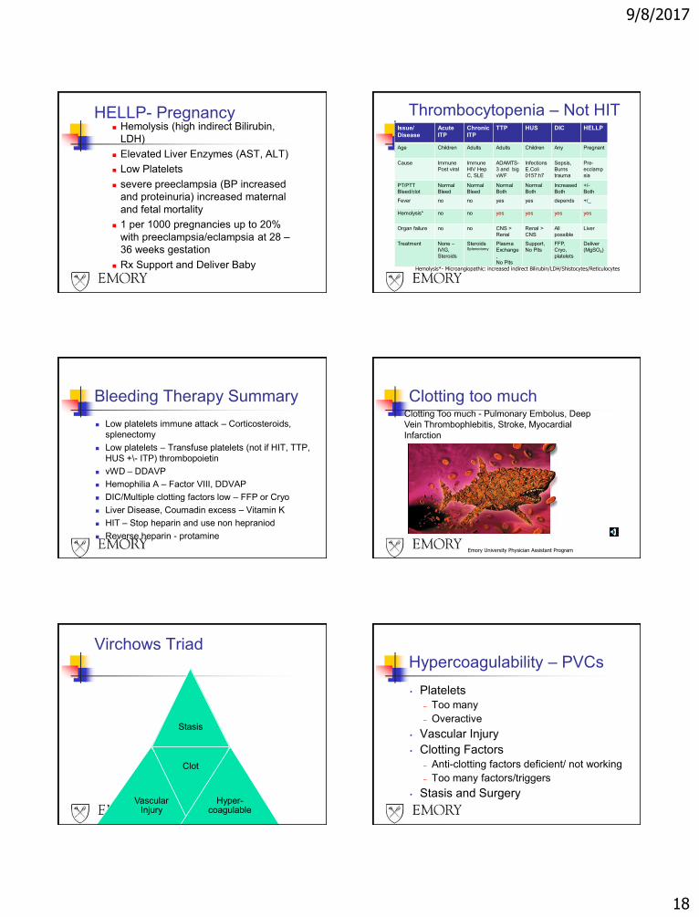
Thrombocytopenia – Not HITIssue/
Disease
Acute
ITP
Chronic
ITP
TTP HUS DIC HELLP
Age Children Adults Adults Children Any Pregnant
Cause Immune
Post viral
Immune
HIV Hep
C, SLE
ADAMTS-
3 and big
vWF
Infections
E.Coli
0157:h7
Sepsis,
Burns
trauma
Pre-
ecclamp
sia
PT/PTT
Bleed/clot
Normal
Bleed
Normal
Bleed
Normal
Both
Normal
Both
Increased
Both
+/-
Both
Fever no no yes yes depends +/_
Hemolysis* no no yes yes yes yes
Organ failure no no CNS >
Renal
Renal >
CNS
All
possible
Liver
Treatment None –
IVIG,
Steroids
SteroidsSplenectomy
Plasma
Exchange
,
No Plts
Support,
No Plts
FFP,
Cryo,
platelets
Deliver
(MgSO4)
Hemolysis*- Microangiopathic: increased indirect Bilirubin/LDH/Shistocytes/Reticulocytes
Bleeding Therapy Summary
Low platelets immune attack – Corticosteroids,
splenectomy
Low platelets – Transfuse platelets (not if HIT, TTP,
HUS +\- ITP) thrombopoietin
vWD – DDAVP
Hemophilia A – Factor VIII, DDVAP
DIC/Multiple clotting factors low – FFP or Cryo
Liver Disease, Coumadin excess – Vitamin K
HIT – Stop heparin and use non hepraniod
Reverse heparin - protamine
Emory University Physician Assistant Program
Clotting too muchClotting Too much - Pulmonary Embolus, Deep
Vein Thrombophlebitis, Stroke, Myocardial
Infarction
Virchows Triad
Stasis
Vascular Injury
Clot
Hyper-coagulable
Hypercoagulability – PVCs
• Platelets
– Too many
– Overactive
• Vascular Injury
• Clotting Factors
– Anti-clotting factors deficient/ not working
– Too many factors/triggers
• Stasis and Surgery
9/8/2017
19
Emory University Physician Assistant Program
Differential Diagnosis -
Hycoagulability
The mnemonic is: 5 Ps HAD CAUSED CLOTs
P - Pregnancy - Increased blood viscosity, fibrinogen and factor VIII. Post Partum - Hypercoaguable state
P – Prothrombin 20210 mutation,
Protien S, C, deficient – Inherited
P - Polycythemia vera - increased viscosity
P – Paroxysmal Nocturnal Hemoglobinuria
S- Smoking
Differential Diagnosis -
Hycoagulability
H – HIT Heparin Induced Thrombocytopenia
H – H Hyperhomocyteinemia
A – Antithrombin III Deficiency
D – Dysfibrinogenemia
C – CHF or Congestive Heart Failure
A – Antiphospholipid Syndrome
U – Uremia – Chronic Renal Failure
S – Surgery – Orthopedic is greatest risk
E – Estrogen – Oral Contraceptives or replacement Rx
D - Diabetes
Emory University Physician Assistant Program
Emory University Physician Assistant Program
Differential Diagnosis
C - Cancer - pro-coagulant effects, Trousseau’s syndrome
L – Leiden Factor V mutation – Activated Protein C resistance
O – Obesity and Cholesterol elevation
T - Trauma, Travel (immobility) - Stasis of blood flow and
release of tissue thromboplastin in trauma
T – Thyroid disease hyper or hypo
S - Sepsis
Emory University Physician Assistant Program
Heparin-induced thrombocytopenia
(HIT)
Due to an antibody against heparin
Occurs in 1-3% of adult patients receiving heparin for 1 week or more. heparin binds to platelet factor 4 (PF4), forming a highly reactive antigenic complex on the surface of platelets
An unexpected fall in platelet count occurring 4-14 days after heparin exposure
Platelet count usually falls by 50%
Mean platelet count 60,000 – 100,000/uL
Platelets become activated and induce clotting
Associated with thrombosis - 10-30% develop arterial or venous thromboses (usually DVTs or PEs)
Of those forming a clot, 30% will die or require amputation
Platelet counts should be monitored while patient is on heparin therapy
HIT Assay
Emory University Physician Assistant Program
Who ya gonna Call?
Clot Busters
tPA (tissue
Plasminogen
Activator)
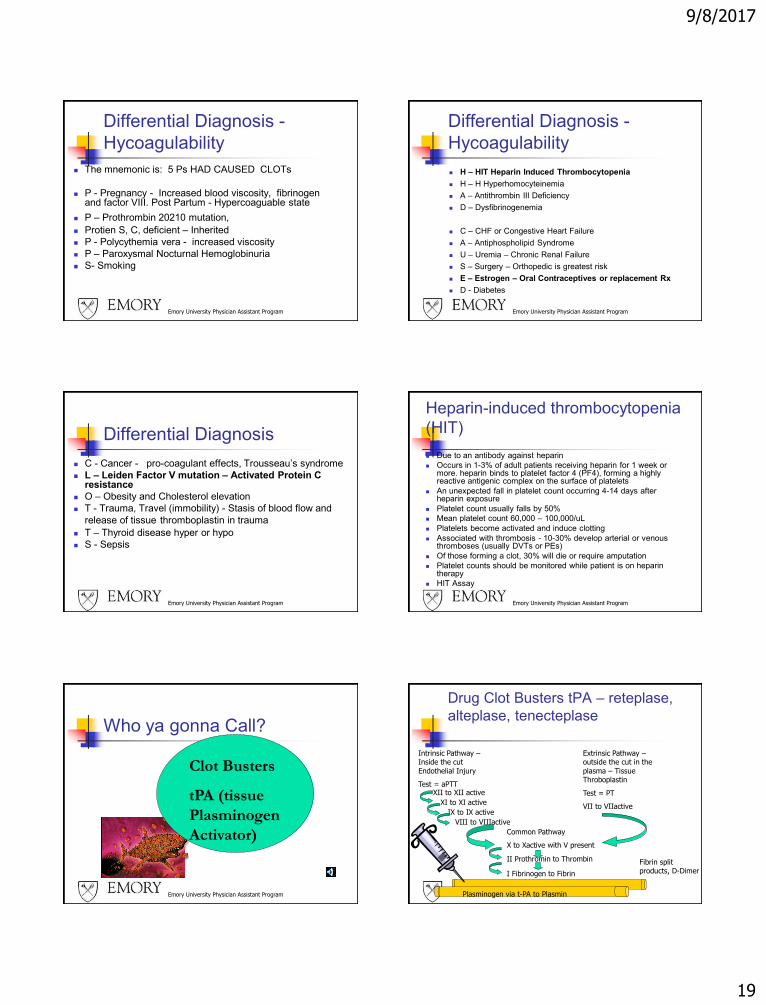
Drug Clot Busters tPA – reteplase,
alteplase, tenecteplase
Intrinsic Pathway –Inside the cut
Endothelial Injury
Test = aPTTXII to XII active
XI to XI active
IX to IX active
VIII to VIIIactive
Common Pathway
X to Xactive with V present
II Prothromin to Thrombin
I Fibrinogen to Fibrin
Extrinsic Pathway –outside the cut in the
plasma – Tissue Throboplastin
Test = PT
VII to VIIactive
Plasminogen via t-PA to Plasmin
Fibrin split products, D-Dimer
9/8/2017
20
Heparin
Intrinsic Pathway –Inside the cut
Endothelial Injury
Test = aPTTXII to XII active
XI to XI active
IX to IX active
VIII to VIIIactive
Common Pathway
X to Xactive with V present
II Prothromin to Thrombin
I Fibrinogen to Fibrin
Extrinsic Pathway –outside the cut in the
plasma – Tissue Factor
Test = PT
VII to VIIactive
Antithrombin III
Heparin
Protamine reverses Heparin
LMW Heparin
Danaparoid,
FondaprinuxIntrinsic Pathway –Inside the cut
Endothelial Injury
Test = aPTTXII to XII active
XI to XI active
IX to IX active
VIII to VIIIactive
Common Pathway
X to Xactive with V present
II Prothromin to Thrombin
I Fibrinogen to Fibrin
Antithrombin III
LMW HeparinDanaparoid - Orgaran
LMWH
dalteparin – (Fragmin)
tinzapain – (Innohep, Logiparin)
enoxaparin (Lovenox, Clexane )
fondaprinux –(Arixtra) direct Xa
blocker, non Heparin
Thrombin Inhibitors
Intrinsic Pathway –Inside the cut
Endothelial Injury
Test = aPTTXII to XII active
XI to XI active
IX to IX active
VIII to VIIIactive
Common Pathway
X to Xactive with V present
II Prothromin to Thrombin
I Fibrinogen to Fibrin
Bivalirudin – AngiomaxLepirudin- Refludan
Argatroban –Antithrombin III - TrombateIII
Coumadin
Intrinsic Pathway –Inside the cut
Endothelial Injury
Test = aPTTXII to XII active
XI to XI active
IX to IX active
VIII to VIIIactive
Common Pathway
X to Xactive with V present
II Prothromin to Thrombin
I Fibrinogen to Fibrin
Extrinsic Pathway – outside the cut in the plasma
Vitamin K - Liver dependant
Test = PT
VII to VIIactive + III Tissue factor
XIII to XIIIactive stabilizer to crosslink
fibrin
Coumadin blocks the liver -Vitamin K
dependent factors
Reverse withVitamin K
Novel Oral Anticoagulants -
Thrombin and Factor Xa inhibiors
NOACsIntrinsic Pathway –Inside the cut
Endothelial Injury
Test = aPPTXII to XII active
XI to XI active
IX to IX active
VIII to VIIIactive
Common Pathway
X to Xactive with V present
II Prothromin to Thrombin
I Fibrinogen to Fibrin
ApixabanRivaroxaban
Edoxaban
May replace Coumadin with fewer side effects. Risk of MI may be increased
Dabigatran (DTI)
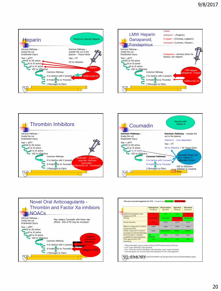
The new oral anticoagulants for VTE – Comparison (Advantages, Disadvantages) Dabiagatran
(Pradaxa) Rivaroxaban
(Xarelto) Apixaban (Eliquis)
Edoxaban (Savaysa)
Started immediately upon diagnosis of VTE
no yes yes no
Dosing twice daily
once daily twice daily once daily
Renal clearance 80 %
33 % 25 % 35 %
Efficacy compared to warfarin (recurrent VTE)
same same same same
Safety compared to warfarin in respect to relevant bleeding
same same/better1 better 2 better 3
Reversal agent / antidote available for major bleeding 4
none none none none
FDA approved for VTE treatment
yes yes yes yes
1 “Major bleeding” same as with warfarin in DVT trial, but less in PE trial 2 Less “major bleeding” with apixaban 3 Less “clinically relevant bleeding” with edoxaban, same “major bleeding” 4 reversal agents are in early clinical development for all 4 anticoagulants
http://professionalsblog.clotconnect.org/2015/01/08/4th-noac-fda-approved-for-dvt-pe-and-atrial-fibrillation-sayasa-edoxaban/
Yes -Praxbind
9/8/2017
21
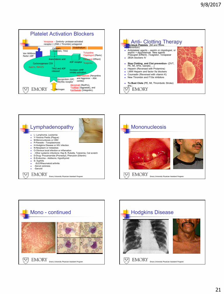
Platelet Activation Blockers
glycoprotein (GP) IIb/IIIa receptor
Abciximab (ReoPro), Tirofiban (Aggrastat), and
Eptifibatide (Integrelin).
Von Willibron Factor vWF
Aranchidonic acid
Collagen, Thrombin, TXA2
Cyclooxygenase COX
TXA2 and ADP released
Endothelium
ADP receptor
Ticlopidine Clopidogrel (Plavix)
Prasugrel (Effient)
Ticagrelor
Aspirin, NSAIDS
Fibrinogen
Increase cAMP inhibits activation
Dipyridamole (Persantine and Aggrenox – ASA
combo)
Vorapaxar – Zontivity- protease-activated receptor-1 (PAR-1 Thrombin) antagonist
Anti- Clotting Therapy To block Platelets (MI and Stoke
prevention)
Antiplatelet agents – aspirin or clopidogrel, or aspirin + dipyridamole New agents Prasugrel (Effient) ,Ticagrelor, Vorapaxar
2B3A blockers IV
Stop Clotting and Clot prevention- (DVT, PE, MI, AFib, Genetic….)
Heparin (Reversed with Protamine)
LMW Heparin and factor Xa blockers
Coumadin (Reversed with vitamin K)
New Thrombin and F10a inhibitors
To Bust Clots (PE, MI, Thrombotic Stroke)
tPA -
Emory University Physician Assistant Program
Lymphadenopathy
L- Lymphoma, Leukemia
Y-Yersinia Pestis (Plague)
M-Mononucleosis or CMV
P-Parasite - Toxoplasmosis
H-Hodgkins Disease or HIV infection
N-Neoplasm or metastisis
O-Obvious local infection or inflamation
-Other systemic infections: Hep B, Rubella, Tularemia, Cat scratch
D-Drug- Procainamide (Pronestyl), Phenytoin (Dilantin)
E-Endocrine - Addisons, Hypothyroid
S- Syphilis
-SLE/Rheumatoid arthritis
-Serum sickness
-Sarcoid
Emory University Physician Assistant Program
Mononucleosis
Emory University Physician Assistant Program
Mono - continued
Emory University Physician Assistant Program
Hodgkins Disease
9/8/2017
22
Emory University Physician Assistant Program
Hodgkins – Reed Sternberg
cells
Emory University Physician Assistant Program
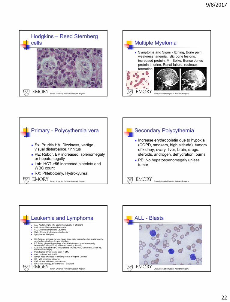
Multiple Myeloma
Symptoms and Signs - Itching, Bone pain,
weakness, anemia, lytic bone lesions,
increased protein, M - Spike, Bence Jones
protein in urine, Renal failure, rouleaux
formation
Emory University Physician Assistant Program
Primary - Polycythemia vera
Sx: Pruritis HA, Dizziness, vertigo, visual disturbance, tinnitus
PE: Rubor, BP increased, splenomegaly or hepatomegally
Lab: HCT >55 lncreased platelets and WBC count
RX: Phlebotomy, Hydroxyurea
Emory University Physician Assistant Program
Secondary Polycythemia
Increase erythropoietin due to hypoxia
(COPD, smokers, high altitude), tumors
of kidney, ovary, liver, brain, drugs:
steroids, androgen, dehydration, burns
PE: No hepatospenomegaly unless
tumor
Emory University Physician Assistant Program
Leukemia and Lymphoma ALL: Acute Lymphocytic Leukemia (Usually in Children)
AML: Acute Myelogenous Leukemia
CLL: Chronic Lymphocytic Leukemia
CML: Chronic Myelogenous Leukemia
Lymphomas, Hodgkins
HX: Fatigue, anorexia, wt loss, fever, bone pain, headaches, lymphadenopathy, non healing infections, thrush, bleeding
PE: Pallor, gingival hyperplasia, Candida infections, lymphadenopathy, hepatosplenomegaly, lung infiltrates, bleeding, bruising
LAB: CBC: elevated WBC/ low platelets, low Hct, WBC Differential, Chem 18, Bone Marrow Biopsy
Philadelphia Chromosome seen in CML
Auer bodies or rods in AML
Lymph node BX: Reed -Sternberg cells in Hodgkins Disease
CT - MRI chest and abdomen
CXR - Chest infiltation, pneumonias
RX: Chemotherapy, Bone Marrow Transplant
Emory University Physician Assistant Program
ALL - Blasts
9/8/2017
23
Emory University Physician Assistant Program
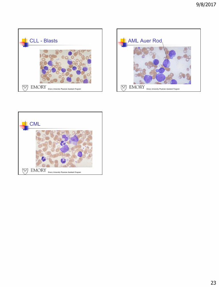
CLL - Blasts
Emory University Physician Assistant Program
AML Auer Rod
Emory University Physician Assistant Program
CML