Embed Size (px)
Citation preview
Carnegie Mellon UniversityResearch Showcase @ CMU
Department of Psychology Dietrich College of Humanities and Social Sciences
2003
What Does Visual Agnosia Tell Us AboutPerceptual Organization and Its Relationship toObject Perception?Marlene BehrmannCarnegie Mellon University
Ruth KimchiUniversity of Haifa
Follow this and additional works at: http://repository.cmu.edu/psychology
This Article is brought to you for free and open access by the Dietrich College of Humanities and Social Sciences at Research Showcase @ CMU. It hasbeen accepted for inclusion in Department of Psychology by an authorized administrator of Research Showcase @ CMU. For more information, pleasecontact [email protected].
What Does Visual Agnosia Tell Us About Perceptual Organizationand Its Relationship to Object Perception?
Marlene BehrmannCarnegie Mellon University
Ruth KimchiUniversity of Haifa
The authors studied 2 patients, S.M. and R.N., to examine perceptual organization and its relationship toobject recognition. Both patients had normal, low-level vision and performed simple grouping operationsnormally but were unable to apprehend a multielement stimulus as a whole. R.N. failed to derive globalstructure even under optimal stimulus conditions, was less sensitive to grouping by closure, and was moreimpaired in object recognition than S.M. These findings suggest that perceptual organization involves amultiplicity of processes, some of which are simpler and are instantiated in lower order areas of visualcortex (e.g., collinearity). Other processes are more complex and rely on higher order visual areas (e.g.,closure and shape formation). The failure to exploit these latter configural processes adversely affectsobject recognition.
The consciously perceived visual world is very different fromthe raw visual information or retinal mosaic of intensities andcolors that arises from external objects. From the chaotic juxtapo-sition of different colors and shapes that stimulate the individualretinal receptors, an object is seen as detached and separable fromadjacent objects and surfaces. This segmentation occurs despite thefact that parts of a single object may be spatially or temporallydiscontinuous, have different colors, or even transect several dif-ferent depth planes. In addition, because most surfaces are opaque,portions of objects are routinely hidden from view and, as onemoves around, surfaces continually undergo occlusion and frag-mentation. As is apparent from this description, the objects ofphenomenal perception are not given in any direct way in theretinal image. Some internal processes of organization must clearlybe responsible, then, for producing a single, coherent percept.Exactly what these processes are remains poorly understood de-spite the roughly 100 years since the Gestalt psychologists firstarticulated the principles of perceptual organization. Although the
Gestalt work on perceptual organization has been widely acceptedas identifying crucial phenomena of perception, there has been,until the last decade or so, relatively little theoretical and empiricalemphasis on perceptual organization, with a few exceptions. And,to the extent that progress has been made, there still remain manyopen questions.
In this article, we address three of these open questions. The firstconcerns the multiplicity of processes involved in deriving struc-ture from a visual image. There is general consensus now thatperceptual organization is not a monolithic entity but, rather, thatseveral different processes exist. What these processes are and howthey differ from one another in terms of their time course, atten-tional demands, and contribution to the ultimate goal of perceptualorganization remains to be specified. A second question concernsthe relationship between the various processes of perceptual orga-nization and object recognition, and a final question relates to thebrain systems that underlie these various perceptual organizationprocesses.
Perceptual Organization: A Multiplicity of Processes
The Gestalt psychologists suggested that perceptual organiza-tion is achieved by grouping elements together by virtue of certainproperties that are present in the image. In the seminal work ofWertheimer (1923/1950) and in the follow-up by Koehler (1928),the different heuristics underlying grouping have been enumerated,and they are now commonly listed in textbooks on perception.These heuristics include grouping by proximity, by closure, bysimilarity, by good continuation, and by common fate. Recently,Palmer (2001; Palmer & Rock, 1994) added two more heuristics:grouping by common region and grouping by connectedness.
Despite the cataloguing and the widespread acceptance of thesedifferent grouping principles, many, although not all, theories ofvisual perception have treated perceptual organization as a unitaryphenomenon that operates at a single, early, preattentive stage, ina bottom-up fashion, to create units which then serve as candidateobjects for later and more elaborated processing, including objectrecognition and identification (Marr, 1982; Neisser, 1967; Treis-
Authorship on this article is assigned alphabetically, and the workreflects an equal contribution by both authors. This research was supportedby Grant MH47566 from the National Institute of Mental Health toMarlene Behrmann, by a United States–Israel Binational Science Founda-tion grant to both authors, and by the Weston Visiting Professorship at theWeizmann Institute of Science (Israel) to Marlene Behrmann. Some of theresearch was conducted while Ruth Kimchi was visiting Carnegie MellonUniversity and while Marlene Behrmann was visiting the WeizmannInstitute.
We thank Thomas McKeeff, who helped with the data collection andstatistical analysis, and Ilona Kovacs, who graciously provided thematerial for the contour interpolation experiment. We are also gratefulto the patients for their good-hearted and continued involvement inthese experiments.
Correspondence concerning this article should be addressed to MarleneBehrmann, Department of Psychology, Carnegie Mellon University, Pitts-burgh, Pennsylvania 15213-3890, or to Ruth Kimchi, Department of Psy-chology, University of Haifa, Mount Carmel, Haifa 31905, Israel. E-mail:[email protected] or [email protected]
Journal of Experimental Psychology: Copyright 2003 by the American Psychological Association, Inc.Human Perception and Performance2003, Vol. 29, No. 1, 19–42
0096-1523/03/$12.00 DOI: 10.1037/0096-1523.29.1.19
19
man, 1982; Treisman, Kahneman, & Burkell, 1983). Several recentstudies, however, have challenged such a view. For example, someresearchers have argued that grouping does not occur as early ashas been widely assumed but instead operates after depth infor-mation has been extracted (Rock & Brosgole, 1964), and afterlightness constancy (Rock, Nijhawan, Palmer, & Tudor, 1992) andperceptual completion (Palmer, Neff, & Beck, 1996; Palmer,2001) have been achieved. Recent studies have also suggested thatgrouping requires attention (Mack, Tang, Tuma, Kahn, & Rock,1992), though other findings demonstrated that grouping can occurunder conditions of inattention, without participants’ consciousawareness (Driver, Davis, Russell, Turatto, & Freeman, 2001;Moore & Egeth, 1997; Razpurker-Apfeld & Kimchi, 2003).
There have also been a host of recent studies that have proposedthat not all grouping principles are created equal in terms of theirtime course, attentional demands, and relative weight in perceptualorganization. For example, Kurylo (1997) obtained evidence sug-gesting that grouping by proximity requires less time than group-ing by good continuity, and Ben-Av and Sagi (1995) and Han,Humphreys, and Chen (1999a) have shown that grouping byproximity is achieved faster than grouping based on similarity ofshape. The features or identity of the visual elements are presum-ably critical for grouping based on similar shape, whereas spatialposition information suffices for grouping by proximity. Kimchi(2000) demonstrated that proximity facilitated grouping by closureor by collinearity, but proximity had less impact when both col-linearity and closure were present in the stimulus, and Donnelly,Humphreys, and Riddoch (1991) showed that a combination ofclosure and collinearity facilitated visual search more than closurealone. Palmer and Rock (1994) argued for an even more basicgrouping heuristic, grouping by uniform connectedness, whichprecedes all other forms of grouping. According to this principle,a connected region of uniform visual property (such as color,texture, and motion) is perceived initially as a single perceptualunit. The claim that uniform connectedness has privileged statushas been challenged, and several recent studies have suggested thatit may not be as powerful as was initially proposed (Han, Hum-phreys, & Chen, 1999b; Kimchi, 1998, 2000).
In addition to noting that grouping involves various principles orheuristics that may differ from each other, it appears that groupingitself may not be a single process. The Gestalt psychologistspreviously suggested that organization involves two distinct pro-cesses that are overlooked by most students of perception: theprocess of unit formation that determines which elements belongtogether (and are segregated from other elements) and the processof shape formation that determines the shape of the groupedelements (Koffka, 1935). Following the Gestaltists, Rock (1986)also suggested that organization has two meanings: grouping in thesense of what goes with what that refers to unit formation andconfiguring that determines the appearance of the grouped ele-ments as a whole based on the interrelationships of the elements.On this account, grouping or element clustering is necessary forshape formation or configuring, but it is not identical to it. Trickand Enns (1997) have recently provided some evidence for thisdistinction. They showed that for neurologically intact partici-pants, the enumeration of hierarchical figures (made of localelements) was as easy as the enumeration of connected line con-figurations, but when the stimuli to be enumerated were presentedamong distractors, the former was more difficult than the latter.Grouping of the local elements presumably suffices for the first
enumeration task but not for the second in which shape discrimi-nation was relevant, indicating that the further operation of shapeformation was required in the second case.
Perceptual Organization and Object Recognition
The different processes of organization may also differ in theirrelative importance for the recognition of different visual objects.That is, specific grouping processes may be necessary and suffi-cient for the recognition of certain objects but not for others. It isinteresting, however, that there has been little concerted effort todifferentiate between the relative contribution of the differentprocesses to object recognition. For example, Donnelly et al.(1991) have suggested that closure and good continuation are bothparticularly important for the derivation of shape descriptions. Onemay also conjecture that the product of grouping (in the sense ofelement clustering) may suffice for some forms of recognition butnot others. For example, grouping collinear elements into a con-tour may be necessary and sufficient for simple line drawings thatcan be recognized by their contours, or the grouping of dots into arow may suffice for the detection of the row. Yet these types ofgrouping may not be sufficient for the recognition of more com-plex objects for which apprehension of the interrelationships andthe configuring of the grouped elements is necessary.
Although not directly germane to our current focus but ofimportance to theories of visual perception more generally, theview that perceptual organization must precede object recognition,espoused by the traditional theories of perception, is also beingchallenged. Several different studies have produced evidenceshowing that knowledge of specific object shapes has an effect ongrouping and figure–ground segregation. These findings are notcompatible with the traditional linear or serial view of perceptualorganization and object recognition, and more dynamic and inter-active accounts have been proposed instead (Kimchi & Hadad,2002; Peterson & Gibson, 1994a, 1994b; Vecera & O’Reilly,1998, 2000).
Neural Mechanisms Underlying Perceptual Organization
In addition to trying to understand the functional processesinvolved in perceptual organization, there is also much work to bedone to understand how these principles are neurally instantiatedand what brain mechanisms might be involved. Considerable neu-rophysiological advances have revealed much about the specific-ities of neuronal responses in visual cortex including their orien-tation selectivity, ocular dominance, wavelength, and directionalselectivity. However, it is not clear how the fragments representedby these local analyzers are assembled to provide a unified percept.
It is worth noting that the Gestaltists did attempt to address theissue of neural implementation and attributed the Gestalt processesto isomorphic brain processes. For example, Kohler (1920/1950)conjectured that electromagnetic fields were the substrate of thebrain’s operation of a physical gestalt system. Although innovativein its time, this view was incorrect and, in fact, was one of thefactors that contributed to the ultimate demise of the Gestaltperspective.
Recently, many studies involving single neuron recording innonhuman primates as well as functional imaging in normal hu-mans have been conducted to explore questions of perceptualorganization. For example, there is a host of research on the
20 BEHRMANN AND KIMCHI
perception of illusory contours (e.g., ffytche & Zeki, 1996; Men-dola, Dale, Fischl, Liu, & Tootell, 1999; von der Heydt & Peter-hans, 1989; von der Heydt, Peterhans, & Baumgartner, 1984) andthe relationship of local fragments to a larger form (e.g., Georgo-poulos et al., 2001; Hasson, Hendler, Ben Bashat, & Malach, 2001;Op de Beeck, Beatse, Wagemans, Sunaert, & Van Hecke, 2000) aswell as several studies on the neural systems involved specificallyin global–local form processing (Fink et al., 1996, 1997; Sasaki etal., 2001). Despite this flurry of recent activity, there remainsmuch to be done to understand the mechanisms whereby lightintensities are translated into meaningful objects by the brain.
Integrative Agnosia
One possible approach to understanding both the psychologicaland neural mechanisms involved in perceptual organization, andthe one adopted here, is to study the performance of individualswho are impaired at the processes of perceptual organizationfollowing brain damage. The logic of this neuropsychologicalapproach is equivalent to backward engineering: As researchers, ifwe can understand the operation of the system once it is unraveled,we might obtain insights into how it functions under normalcircumstances (Coltheart, 2002). In the present article, we explorethe behavior of 2 individuals, S.M. and R.N., whose impairmentprovides an ideal testing ground for investigating processes in-volved in perceptual organization and their relationship to objectperception. The patients have a neuropsychological impairment,referred to as visual object agnosia, in which they are unable torecognize even familiar common objects presented to them in thevisual modality. This object recognition deficit cannot be attrib-uted to a problem in labeling the stimulus per se nor to a loss ofsemantics; presented with the same object in a different modality,either haptically or auditorily, they have no problem in naming itor providing detailed and rich descriptions of it. Visual agnosiarefers to a specific failure to access the meaning of objects fromthe visual modality (Farah, 1990; Humphreys & Riddoch, 2001;Ratcliff & Newcombe, 1982).
Visual agnosia covers a wide spectrum of deficits, including, atone end, patients who are unable to recover even primitive featuresfrom a display (e.g., patients who fail to search in parallel for avertical line among horizontal lines) and, at the other end, patientswho appear able to extract a reasonably intact percept but subse-quently fail to assign meaning to it (Farah, 1990; Humphreys &Riddoch, 2001). The patients we have chosen to study fall midwaybetween these two extremes: Their agnosia does not arise fromimpaired low-level vision nor from the inability to assign meaningto relatively intact visual representations, but rather from problemsin organizational processes. The term applied to this deficit inintermediate vision is integrative agnosia and was coined byRiddoch and Humphreys (1987) on the basis of their studies witha patient called H.J.A. H.J.A. was impaired at search tasks thatrequire the binding of visual elements in a spatially parallel man-ner across a field containing multiple stimuli; for example, he wasdisproportionately slowed, relative to control participants, in de-tecting the presence of an inverted T among upright Ts. In contrast,his search is efficient and rapid for targets that do not require acombination of elements such as a target diagonal among multipleverticals (Humphreys, 1999; Humphreys & Riddoch, 1987; Hum-phreys et al., 1994; Humphreys, Riddoch, Quinlan, Price, & Don-nelly, 1992). When the demands for integration are low, H.J.A.
and other integrative agnosic patients perform significantly abovechance levels: They can make same–different judgments accu-rately on two stimuli that share area and brightness but not shape(aspect ratio changes from square to rectangle; Efron, 1968).
In addition to the impaired ability to integrate all aspects of thedisplay into a whole, several other characteristics now serve as thecore features of integrative agnosia (Behrmann, in press; Behr-mann & Kimchi, in press; Humphreys & Riddoch, 2001). Forexample, the patients are more impaired at identifying items thatoverlap one another compared with the same items presented inisolation. It is interesting and also counterintuitive that, in somepatients, the presence of local information may even reduce theefficiency of visual recognition; in contrast with normal perceiv-ers, both patients H.J.A. (Lawson & Humphreys, 1999; Riddoch &Humphreys, 1987) and S.M. (Butter & Trobe, 1994) identifiedsilhouettes better than line drawings whose internal details appar-ently led to incorrect segmentation. Patients with integrative ag-nosia also fail to identify shapes by subjective contours and do notappreciate occlusion normally. Another key feature of the disorderis the failure to segregate figures from ground effectively; forexample, patient F.G.P. was unable to detect a simple shapeagainst a pattern background (Kartsounis & Warrington, 1991).Finally and critically for our purposes, integrative agnosic patientsare impaired at grouping elements of a display. A clear examplecomes from patient N.M., who was impaired at detecting thepresence of a target letter that was formed by grouping localelements that differed from the background element by line ori-entation (texture), color, luminance, or motion (Ricci, Vaishnavi,& Chatterjee, 1999; see also, Kartsounis & Warrington, 1991;Marstrand, Gerlach, Udesen, & Gade, 2000). Given the paucity ofresearch on integrative agnosia, a definitive definition is not yetestablished, but the critical component is a failure in organizationalprocesses in tandem with a deficit in object recognition.
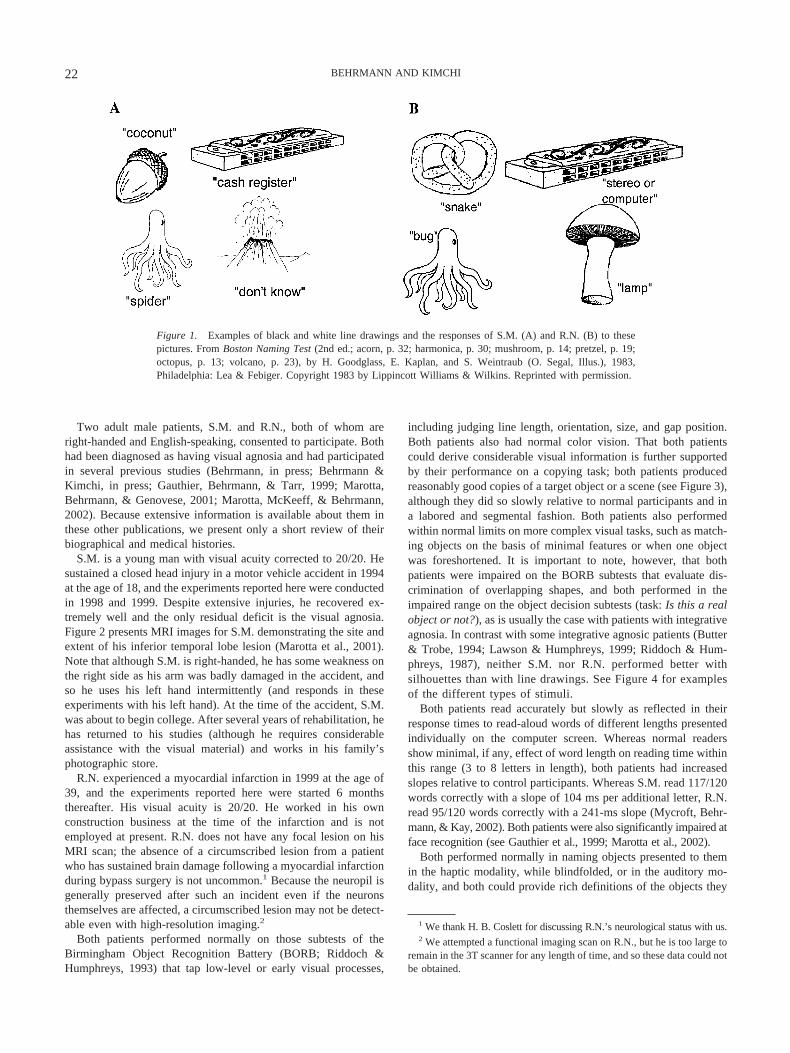
Patients with integrative agnosia are also obviously impaired atobject recognition: As is evident from their responses to black andwhite line drawings (see Figure 1), the 2 patients we studied areclearly able to extract some visual information from the display butapparently are unable to integrate all aspects into a meaningfulwhole. The problem applies equally to the recognition of two- andthree-dimensional stimuli and to black and white and chromaticdisplays although, in some cases, the presence of depth, color, andsurface cues may be of some assistance to the patients in segment-ing the display (Chainay & Humphreys, 2001; Farah, 1990; Hum-phrey, Goodale, Jakobson, & Servos, 1994; Jankowiak, Kins-bourne, Shalev, & Bachman, 1992).
We start off by describing the 2 agnosic patients we havestudied. At the outset, we had some indication that the 2 patientswere differentially impaired in their object recognition ability, andwe exploited this fact in investigating the nature of the groupingimpairment and the way in which various grouping processesmight differentially contribute to object recognition.
Description of Cases
Because the same patients participated in all the experiments,we describe them here at the outset. Control participants werestandardly run in each experiment, but because the exact controlgroup differed from one experiment to another, we describe thecontrol participants separately for each experiment.
21VISUAL AGNOSIA AND PERCEPTUAL ORGANIZATION
Two adult male patients, S.M. and R.N., both of whom areright-handed and English-speaking, consented to participate. Bothhad been diagnosed as having visual agnosia and had participatedin several previous studies (Behrmann, in press; Behrmann &Kimchi, in press; Gauthier, Behrmann, & Tarr, 1999; Marotta,Behrmann, & Genovese, 2001; Marotta, McKeeff, & Behrmann,2002). Because extensive information is available about them inthese other publications, we present only a short review of theirbiographical and medical histories.
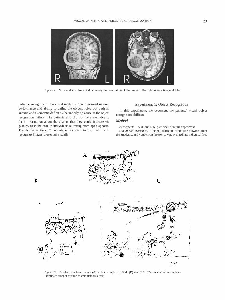
S.M. is a young man with visual acuity corrected to 20/20. Hesustained a closed head injury in a motor vehicle accident in 1994at the age of 18, and the experiments reported here were conductedin 1998 and 1999. Despite extensive injuries, he recovered ex-tremely well and the only residual deficit is the visual agnosia.Figure 2 presents MRI images for S.M. demonstrating the site andextent of his inferior temporal lobe lesion (Marotta et al., 2001).Note that although S.M. is right-handed, he has some weakness onthe right side as his arm was badly damaged in the accident, andso he uses his left hand intermittently (and responds in theseexperiments with his left hand). At the time of the accident, S.M.was about to begin college. After several years of rehabilitation, hehas returned to his studies (although he requires considerableassistance with the visual material) and works in his family’sphotographic store.
R.N. experienced a myocardial infarction in 1999 at the age of39, and the experiments reported here were started 6 monthsthereafter. His visual acuity is 20/20. He worked in his ownconstruction business at the time of the infarction and is notemployed at present. R.N. does not have any focal lesion on hisMRI scan; the absence of a circumscribed lesion from a patientwho has sustained brain damage following a myocardial infarctionduring bypass surgery is not uncommon.1 Because the neuropil isgenerally preserved after such an incident even if the neuronsthemselves are affected, a circumscribed lesion may not be detect-able even with high-resolution imaging.2
Both patients performed normally on those subtests of theBirmingham Object Recognition Battery (BORB; Riddoch &Humphreys, 1993) that tap low-level or early visual processes,
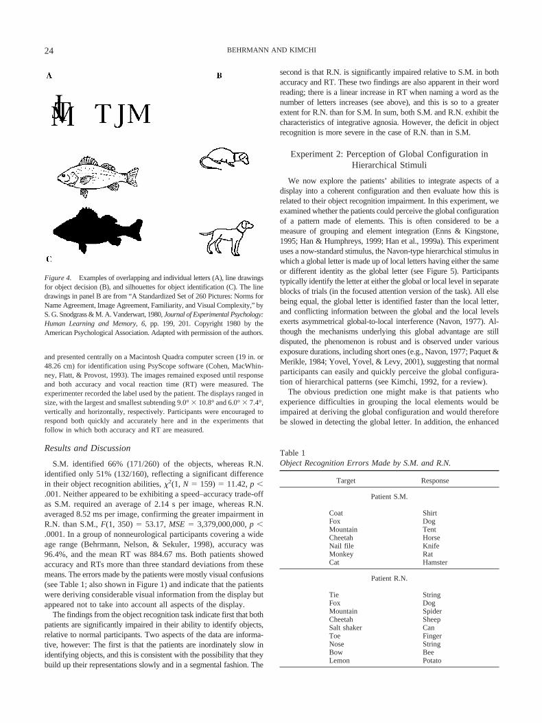
including judging line length, orientation, size, and gap position.Both patients also had normal color vision. That both patientscould derive considerable visual information is further supportedby their performance on a copying task; both patients producedreasonably good copies of a target object or a scene (see Figure 3),although they did so slowly relative to normal participants and ina labored and segmental fashion. Both patients also performedwithin normal limits on more complex visual tasks, such as match-ing objects on the basis of minimal features or when one objectwas foreshortened. It is important to note, however, that bothpatients were impaired on the BORB subtests that evaluate dis-crimination of overlapping shapes, and both performed in theimpaired range on the object decision subtests (task: Is this a realobject or not?), as is usually the case with patients with integrativeagnosia. In contrast with some integrative agnosic patients (Butter& Trobe, 1994; Lawson & Humphreys, 1999; Riddoch & Hum-phreys, 1987), neither S.M. nor R.N. performed better withsilhouettes than with line drawings. See Figure 4 for examplesof the different types of stimuli.
Both patients read accurately but slowly as reflected in theirresponse times to read-aloud words of different lengths presentedindividually on the computer screen. Whereas normal readersshow minimal, if any, effect of word length on reading time withinthis range (3 to 8 letters in length), both patients had increasedslopes relative to control participants. Whereas S.M. read 117/120words correctly with a slope of 104 ms per additional letter, R.N.read 95/120 words correctly with a 241-ms slope (Mycroft, Behr-mann, & Kay, 2002). Both patients were also significantly impaired atface recognition (see Gauthier et al., 1999; Marotta et al., 2002).
Both performed normally in naming objects presented to themin the haptic modality, while blindfolded, or in the auditory mo-dality, and both could provide rich definitions of the objects they
1 We thank H. B. Coslett for discussing R.N.’s neurological status with us.2 We attempted a functional imaging scan on R.N., but he is too large to
remain in the 3T scanner for any length of time, and so these data could notbe obtained.
Figure 1. Examples of black and white line drawings and the responses of S.M. (A) and R.N. (B) to thesepictures. From Boston Naming Test (2nd ed.; acorn, p. 32; harmonica, p. 30; mushroom, p. 14; pretzel, p. 19;octopus, p. 13; volcano, p. 23), by H. Goodglass, E. Kaplan, and S. Weintraub (O. Segal, Illus.), 1983,Philadelphia: Lea & Febiger. Copyright 1983 by Lippincott Williams & Wilkins. Reprinted with permission.
22 BEHRMANN AND KIMCHI
failed to recognize in the visual modality. The preserved namingperformance and ability to define the objects ruled out both ananomia and a semantic deficit as the underlying cause of the objectrecognition failure. The patients also did not have available tothem information about the display that they could indicate viagesture, as is the case in individuals suffering from optic aphasia.The deficit in these 2 patients is restricted to the inability torecognize images presented visually.
Experiment 1: Object Recognition
In this experiment, we document the patients’ visual objectrecognition abilities.
Method
Participants. S.M. and R.N. participated in this experiment.Stimuli and procedure. The 260 black and white line drawings from
the Snodgrass and Vanderwart (1980) set were scanned into individual files
Figure 2. Structural scan from S.M. showing the localization of the lesion to the right inferior temporal lobe.
Figure 3. Display of a beach scene (A) with the copies by S.M. (B) and R.N. (C), both of whom took aninordinate amount of time to complete this task.
23VISUAL AGNOSIA AND PERCEPTUAL ORGANIZATION
and presented centrally on a Macintosh Quadra computer screen (19 in. or48.26 cm) for identification using PsyScope software (Cohen, MacWhin-ney, Flatt, & Provost, 1993). The images remained exposed until responseand both accuracy and vocal reaction time (RT) were measured. Theexperimenter recorded the label used by the patient. The displays ranged insize, with the largest and smallest subtending 9.0° � 10.8° and 6.0° � 7.4°,vertically and horizontally, respectively. Participants were encouraged torespond both quickly and accurately here and in the experiments thatfollow in which both accuracy and RT are measured.
Results and Discussion
S.M. identified 66% (171/260) of the objects, whereas R.N.identified only 51% (132/160), reflecting a significant differencein their object recognition abilities, �2(1, N � 159) � 11.42, p �.001. Neither appeared to be exhibiting a speed–accuracy trade-offas S.M. required an average of 2.14 s per image, whereas R.N.averaged 8.52 ms per image, confirming the greater impairment inR.N. than S.M., F(1, 350) � 53.17, MSE � 3,379,000,000, p �.0001. In a group of nonneurological participants covering a wideage range (Behrmann, Nelson, & Sekuler, 1998), accuracy was96.4%, and the mean RT was 884.67 ms. Both patients showedaccuracy and RTs more than three standard deviations from thesemeans. The errors made by the patients were mostly visual confusions(see Table 1; also shown in Figure 1) and indicate that the patientswere deriving considerable visual information from the display butappeared not to take into account all aspects of the display.
The findings from the object recognition task indicate first that bothpatients are significantly impaired in their ability to identify objects,relative to normal participants. Two aspects of the data are informa-tive, however: The first is that the patients are inordinately slow inidentifying objects, and this is consistent with the possibility that theybuild up their representations slowly and in a segmental fashion. The
second is that R.N. is significantly impaired relative to S.M. in bothaccuracy and RT. These two findings are also apparent in their wordreading; there is a linear increase in RT when naming a word as thenumber of letters increases (see above), and this is so to a greaterextent for R.N. than for S.M. In sum, both S.M. and R.N. exhibit thecharacteristics of integrative agnosia. However, the deficit in objectrecognition is more severe in the case of R.N. than in S.M.
Experiment 2: Perception of Global Configuration inHierarchical Stimuli
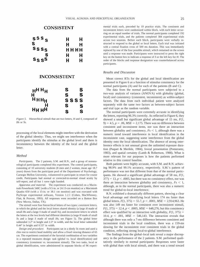
We now explore the patients’ abilities to integrate aspects of adisplay into a coherent configuration and then evaluate how this isrelated to their object recognition impairment. In this experiment, weexamined whether the patients could perceive the global configurationof a pattern made of elements. This is often considered to be ameasure of grouping and element integration (Enns & Kingstone,1995; Han & Humphreys, 1999; Han et al., 1999a). This experimentuses a now-standard stimulus, the Navon-type hierarchical stimulus inwhich a global letter is made up of local letters having either the sameor different identity as the global letter (see Figure 5). Participantstypically identify the letter at either the global or local level in separateblocks of trials (in the focused attention version of the task). All elsebeing equal, the global letter is identified faster than the local letter,and conflicting information between the global and the local levelsexerts asymmetrical global-to-local interference (Navon, 1977). Al-though the mechanisms underlying this global advantage are stilldisputed, the phenomenon is robust and is observed under variousexposure durations, including short ones (e.g., Navon, 1977; Paquet &Merikle, 1984; Yovel, Yovel, & Levy, 2001), suggesting that normalparticipants can easily and quickly perceive the global configura-tion of hierarchical patterns (see Kimchi, 1992, for a review).
The obvious prediction one might make is that patients whoexperience difficulties in grouping the local elements would beimpaired at deriving the global configuration and would thereforebe slowed in detecting the global letter. In addition, the enhanced
Table 1Object Recognition Errors Made by S.M. and R.N.
Target Response
Patient S.M.
Coat ShirtFox DogMountain TentCheetah HorseNail file KnifeMonkey RatCat Hamster
Patient R.N.
Tie StringFox DogMountain SpiderCheetah SheepSalt shaker CanToe FingerNose StringBow BeeLemon Potato
Figure 4. Examples of overlapping and individual letters (A), line drawingsfor object decision (B), and silhouettes for object identification (C). The linedrawings in panel B are from “A Standardized Set of 260 Pictures: Norms forName Agreement, Image Agreement, Familiarity, and Visual Complexity,” byS. G. Snodgrass & M. A. Vanderwart, 1980, Journal of Experimental Psychology:Human Learning and Memory, 6, pp. 199, 201. Copyright 1980 by theAmerican Psychological Association. Adapted with permission of the authors.
24 BEHRMANN AND KIMCHI
processing of the local elements might interfere with the derivationof the global identity: Thus, we might see interference when theparticipants identify the stimulus at the global level and there isinconsistency between the identity of the local and the globalletter.
Method
Participants. The 2 patients, S.M. and R.N., and a group of nonneu-rological participants completed this experiment. The control participants,consisting of 10 university students (8 men and 2 women, mean age 18.7years) drawn from the participant pool of the Department of Psychology,Carnegie Mellon University, volunteered to participate in return for coursecredit. Participants had normal or corrected-to-normal visual acuity byself-report, and all but 1 were right handed.
Apparatus and material. The experiment was conducted on a Macin-tosh Powerbook 540C (with a 9.5-in. or 24.13-cm monitor) or a MacintoshQuadra 650 (with a 15-in. or 38.1 cm monitor) and was executed withPsyScope experimental software, Version 1.2.1 (Cohen, MacWhinney,Flatt, & Provost, 1993). All responses were recorded on a Button Box(New Micros, Dallas, TX).
The stimuli were four hierarchical letters of two types: consistent letters,in which the global and the local letters shared identity (a large H made ofsmall Hs and a large S made of small Ss), and inconsistent letters in whichthe letters at the two levels had different identities (a large H made of smallSs and a large S made of small Hs; see Figure 5). The global lettersubtended 3.2° in height and 2.3° in width, and the local letter subtended0.44° in height and 0.53° in width.
Design and procedure. Participants sat in a dimly lit room and used achin rest to restrict head mobility and allow a fixed viewing distance of 65cm. The experiment consisted of the factorial combination of two variablesin a repeated measures design: task (global vs. local identification) andconsistency (consistent vs. inconsistent stimuli). The two tasks, local orglobal identification, were administered in separate blocks of 96 experi-
mental trials each, preceded by 10 practice trials. The consistent andinconsistent letters were randomized within block, with each letter occur-ring on an equal number of trials. The normal participants completed 192experimental trials, and the patients completed 384 experimental trialsacross two sessions. Before each block, participants were verbally in-structed to respond to the global or local letters. Each trial was initiatedwith a central fixation cross of 500 ms duration. This was immediatelyreplaced by one of the four possible stimuli, which remained on the screenuntil a response was made. Participants were instructed to press the rightkey on the button box to indicate a response of S or the left key for H. Theorder of the blocks and response designation was counterbalanced acrossparticipants.
Results and Discussion
Mean correct RTs for the global and local identification arepresented in Figure 6 as a function of stimulus consistency for thenormal participants (A) and for each of the patients (B and C).
The data from the normal participants were subjected to atwo-way analysis of variance (ANOVA) with globality (global,local) and consistency (consistent, inconsistent) as within-subjectfactors. The data from each individual patient were analyzedseparately with the same two factors as between-subject factorsand trial type as the random variable.
The normal participants were extremely accurate in identifyingthe letters, reporting 96.3% correctly. As reflected in Figure 6, theyshowed a small but significant global advantage of 15 ms, F(1,9) � 4.5, p � .06, MSE � 2,175. There was no difference betweenconsistent and inconsistent items, nor was there an interactionbetween globality and consistency, Fs � 1, although there was anumeric trend toward interference in local identification in theinconsistent case, suggesting some interference from the globalidentity onto the local identification. The absence of strong inter-ference effects is not unusual given the unlimited exposure dura-tion (Paquet & Merikle, 1984), foveal presentation (Pomerantz,1983), and spatial certainty (Lamb & Robertson, 1988). What ismore relevant for our purposes is how the patients performedrelative to this control baseline.
Both patients were highly accurate, with S.M. and R.N. achiev-ing 98.9% and 99.1% accuracy, respectively. S.M.’s pattern ofperformance was not that different from that of the normal partic-ipants. He showed a significant global advantage of 58 ms, F(1,377) � 13, p � .0005, but there was no consistency effect, nor wasthere an interaction between globality and consistency, Fs � 1,although, as in the normal participants, there was also a numerictrend for global-to-local interference.
R.N. exhibited a dramatically different pattern, showing a clearlocal advantage and identifying local letters 174 ms faster thanglobal letters, F(1, 375) � 55.7, p � .0001, MSE � 2,914,963. Hewas also 149 ms faster for consistent over inconsistent stimuli,F(1, 375) � 12.4, p � .0005, MSE � 648,276, but this consistencyeffect was qualified by an interaction with globality, F(1, 375) �10.4, p � .001, MSE � 546,143. The interaction reveals thatalthough there was only a 7-ms difference between consistent andinconsistent trials in the local condition, there was a 159-msslowing for the inconsistent over consistent trials in the globalcondition, reflecting strong local-to-global interference.
The findings from the global–local task reveal a major discrep-ancy in the performance of the 2 patients. S.M. performed quali-tatively similarly to normal participants: Responses were fasterwith global than with local stimuli, and there was a trend toward
Figure 5. Hierarchical stimuli that are two letters, H and S, composed ofHs or Ss.
25VISUAL AGNOSIA AND PERCEPTUAL ORGANIZATION
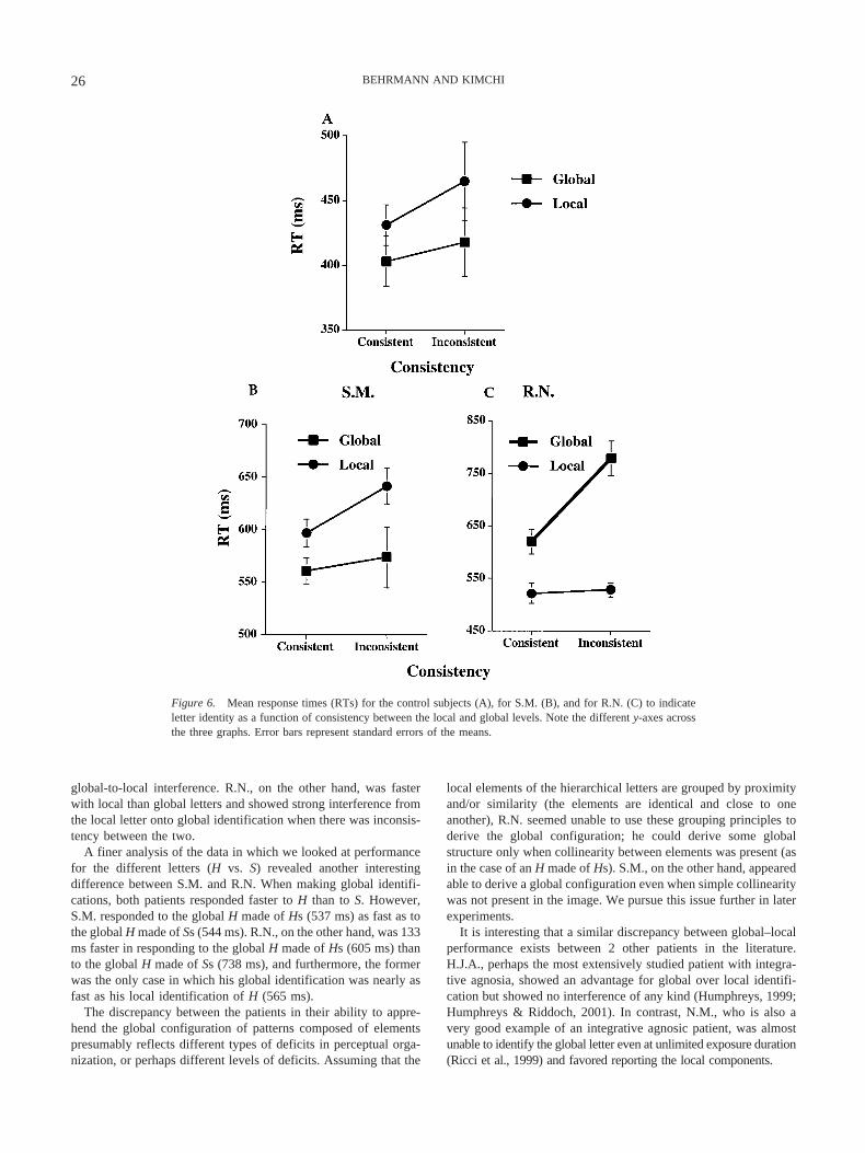
global-to-local interference. R.N., on the other hand, was fasterwith local than global letters and showed strong interference fromthe local letter onto global identification when there was inconsis-tency between the two.
A finer analysis of the data in which we looked at performancefor the different letters (H vs. S) revealed another interestingdifference between S.M. and R.N. When making global identifi-cations, both patients responded faster to H than to S. However,S.M. responded to the global H made of Hs (537 ms) as fast as tothe global H made of Ss (544 ms). R.N., on the other hand, was 133ms faster in responding to the global H made of Hs (605 ms) thanto the global H made of Ss (738 ms), and furthermore, the formerwas the only case in which his global identification was nearly asfast as his local identification of H (565 ms).
The discrepancy between the patients in their ability to appre-hend the global configuration of patterns composed of elementspresumably reflects different types of deficits in perceptual orga-nization, or perhaps different levels of deficits. Assuming that the
local elements of the hierarchical letters are grouped by proximityand/or similarity (the elements are identical and close to oneanother), R.N. seemed unable to use these grouping principles toderive the global configuration; he could derive some globalstructure only when collinearity between elements was present (asin the case of an H made of Hs). S.M., on the other hand, appearedable to derive a global configuration even when simple collinearitywas not present in the image. We pursue this issue further in laterexperiments.
It is interesting that a similar discrepancy between global–localperformance exists between 2 other patients in the literature.H.J.A., perhaps the most extensively studied patient with integra-tive agnosia, showed an advantage for global over local identifi-cation but showed no interference of any kind (Humphreys, 1999;Humphreys & Riddoch, 2001). In contrast, N.M., who is also avery good example of an integrative agnosic patient, was almostunable to identify the global letter even at unlimited exposure duration(Ricci et al., 1999) and favored reporting the local components.
Figure 6. Mean response times (RTs) for the control subjects (A), for S.M. (B), and for R.N. (C) to indicateletter identity as a function of consistency between the local and global levels. Note the different y-axes acrossthe three graphs. Error bars represent standard errors of the means.
26 BEHRMANN AND KIMCHI
The variability observed across patients on this task suggeststhat a problem in deriving global structure might not be a coreelement of integrative agnosia. This conclusion might be prema-ture, however. It is now well known that a variety of stimulus andtask factors affect the balance between global and local processing,including the type of hierarchical stimuli used, the attentional task(divided or focused), and the mode of response (forced choice,go/no-go) (Kimchi, 1992; Yovel et al., 2001). Thus, the variabilityin the pattern of results obtained across patients might be a func-tion of the different testing conditions used with different patients.We note that the experimental conditions used here are favorablefor deriving a global configuration: focused attention (blocked bylocal vs. global response) and global stimulus saliency (in whichmany small elements increase the salience of the global over thelocal letters; e.g., Yovel et al., 2001). Under these conditions andwith unlimited exposure duration, S.M. was able to derive theglobal configuration but, as we will see below, under more strin-gent testing conditions, even S.M. exhibited an impairment inglobal processing. These findings further support the claim thatdifferences in testing conditions may lead to variability in out-come. Alternatively, because perceptual organization refers to amultiplicity of processes, it is possible that patients do vary andthat integrative agnosia might manifest in different ways acrossdifferent individuals. Because such individuals are rare, the op-portunity to systematically analyze all their perceptual skills indepth is not that easy, and so the source of this cross-patientvariability remains to be determined.
In the following experiment, we explore further the question ofperceptual organization and global structure. Before we do so,however, we need to rule out the possibility that the differentialability of the 2 patients in deriving the global configuration is notsimply due to a differential sensitivity to high and low spatialfrequencies, which we do in the next experiment.
Experiment 3: Spatial Frequency Thresholds
Several researchers have suggested an involvement of spatialfilters, based on spatial frequency channels, operating at earlyvisual processing (Ginsburg, 1986) in the perception of global andlocal structures. For example, in a number of these studies, nolatency advantage for global over local processing was found when
low-spatial frequencies were removed from hierarchical stimuli(Badcock, Whitworth, Badcock, & Lovegrove, 1990; Hughes,Fendrich, & Reuter-Lorenz, 1990; Lamb & Yund, 1993; Shulman,Sullivan, Gish, & Sakoda, 1986; Shulman & Wilson, 1987), sug-gesting that the global advantage effect is mediated by low-spatial-frequency channels. Thus, one possible explanation for the pa-tients’ differential inability to perceive the global form of ahierarchical stimulus might concern a fundamental limitation inprocessing low-spatial-frequency information. The obvious pre-diction from this in relation to the patients is that R.N., whoappears to process stimuli almost entirely at the local level, shouldbe impaired at processing low-frequency displays, resulting in anincreased low-spatial-frequency threshold, relative to control par-ticipants, whereas S.M., who shows some global form processing,should not show as much of an increase in this threshold. InExperiment 3, we establish thresholds for both patients across awide range of spatial frequencies and compare them to those ofcontrol participants.
Method
Participants. In addition to S.M. and R.N., 10 normal male partici-pants, with a mean age of 27.7 years (range � 19 to 51 years) participatedin this experiment. Nine of the participants were right-handed, and all hadnormal or corrected-to-normal vision.
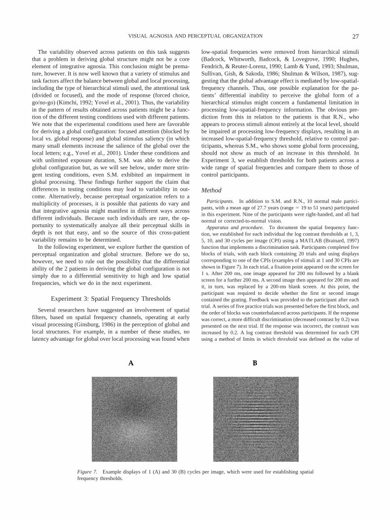
Apparatus and procedure. To document the spatial frequency func-tion, we established for each individual the log contrast thresholds at 1, 3,5, 10, and 30 cycles per image (CPI) using a MATLAB (Brainard, 1997)function that implements a discrimination task. Participants completed fiveblocks of trials, with each block containing 20 trials and using displayscorresponding to one of the CPIs (examples of stimuli at 1 and 30 CPIs areshown in Figure 7). In each trial, a fixation point appeared on the screen for1 s. After 200 ms, one image appeared for 200 ms followed by a blankscreen for a further 200 ms. A second image then appeared for 200 ms andit, in turn, was replaced by a 200-ms blank screen. At this point, theparticipant was required to decide whether the first or second imagecontained the grating. Feedback was provided to the participant after eachtrial. A series of five practice trials was presented before the first block, andthe order of blocks was counterbalanced across participants. If the responsewas correct, a more difficult discrimination (decreased contrast by 0.2) waspresented on the next trial. If the response was incorrect, the contrast wasincreased by 0.2. A log contrast threshold was determined for each CPIusing a method of limits in which threshold was defined as the value of
Figure 7. Example displays of 1 (A) and 30 (B) cycles per image, which were used for establishing spatialfrequency thresholds.
27VISUAL AGNOSIA AND PERCEPTUAL ORGANIZATION
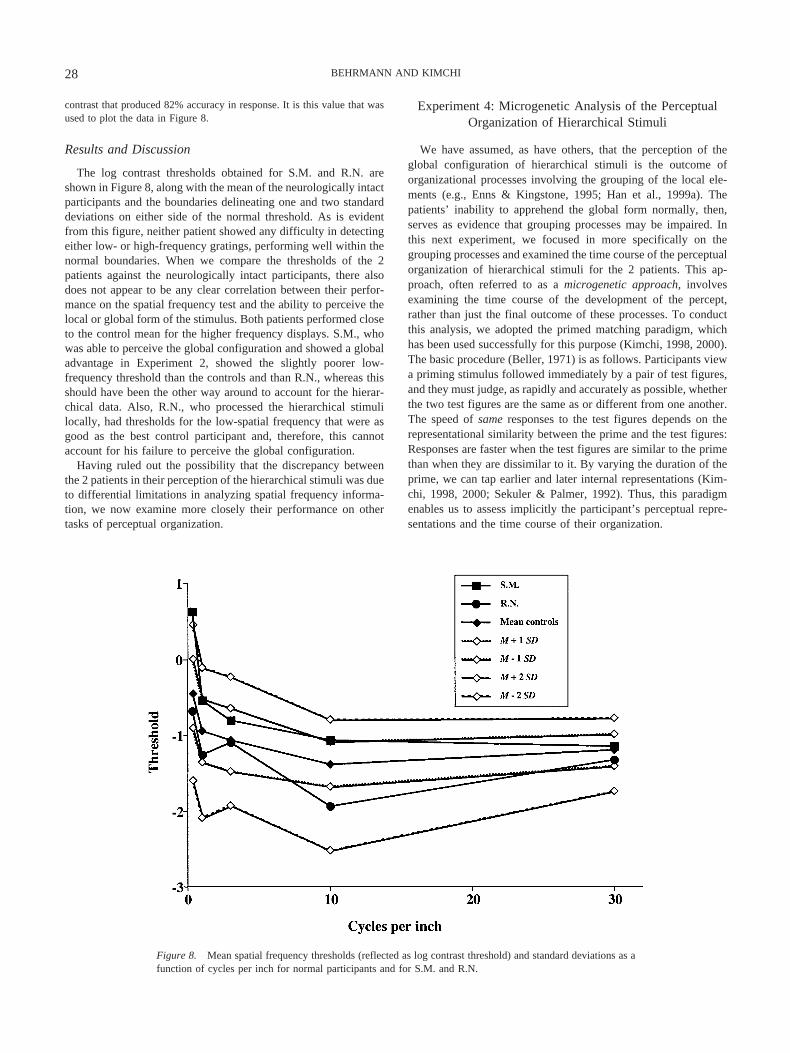
contrast that produced 82% accuracy in response. It is this value that wasused to plot the data in Figure 8.
Results and Discussion
The log contrast thresholds obtained for S.M. and R.N. areshown in Figure 8, along with the mean of the neurologically intactparticipants and the boundaries delineating one and two standarddeviations on either side of the normal threshold. As is evidentfrom this figure, neither patient showed any difficulty in detectingeither low- or high-frequency gratings, performing well within thenormal boundaries. When we compare the thresholds of the 2patients against the neurologically intact participants, there alsodoes not appear to be any clear correlation between their perfor-mance on the spatial frequency test and the ability to perceive thelocal or global form of the stimulus. Both patients performed closeto the control mean for the higher frequency displays. S.M., whowas able to perceive the global configuration and showed a globaladvantage in Experiment 2, showed the slightly poorer low-frequency threshold than the controls and than R.N., whereas thisshould have been the other way around to account for the hierar-chical data. Also, R.N., who processed the hierarchical stimulilocally, had thresholds for the low-spatial frequency that were asgood as the best control participant and, therefore, this cannotaccount for his failure to perceive the global configuration.
Having ruled out the possibility that the discrepancy betweenthe 2 patients in their perception of the hierarchical stimuli was dueto differential limitations in analyzing spatial frequency informa-tion, we now examine more closely their performance on othertasks of perceptual organization.
Experiment 4: Microgenetic Analysis of the PerceptualOrganization of Hierarchical Stimuli
We have assumed, as have others, that the perception of theglobal configuration of hierarchical stimuli is the outcome oforganizational processes involving the grouping of the local ele-ments (e.g., Enns & Kingstone, 1995; Han et al., 1999a). Thepatients’ inability to apprehend the global form normally, then,serves as evidence that grouping processes may be impaired. Inthis next experiment, we focused in more specifically on thegrouping processes and examined the time course of the perceptualorganization of hierarchical stimuli for the 2 patients. This ap-proach, often referred to as a microgenetic approach, involvesexamining the time course of the development of the percept,rather than just the final outcome of these processes. To conductthis analysis, we adopted the primed matching paradigm, whichhas been used successfully for this purpose (Kimchi, 1998, 2000).The basic procedure (Beller, 1971) is as follows. Participants viewa priming stimulus followed immediately by a pair of test figures,and they must judge, as rapidly and accurately as possible, whetherthe two test figures are the same as or different from one another.The speed of same responses to the test figures depends on therepresentational similarity between the prime and the test figures:Responses are faster when the test figures are similar to the primethan when they are dissimilar to it. By varying the duration of theprime, we can tap earlier and later internal representations (Kim-chi, 1998, 2000; Sekuler & Palmer, 1992). Thus, this paradigmenables us to assess implicitly the participant’s perceptual repre-sentations and the time course of their organization.
Figure 8. Mean spatial frequency thresholds (reflected as log contrast threshold) and standard deviations as afunction of cycles per inch for normal participants and for S.M. and R.N.
28 BEHRMANN AND KIMCHI
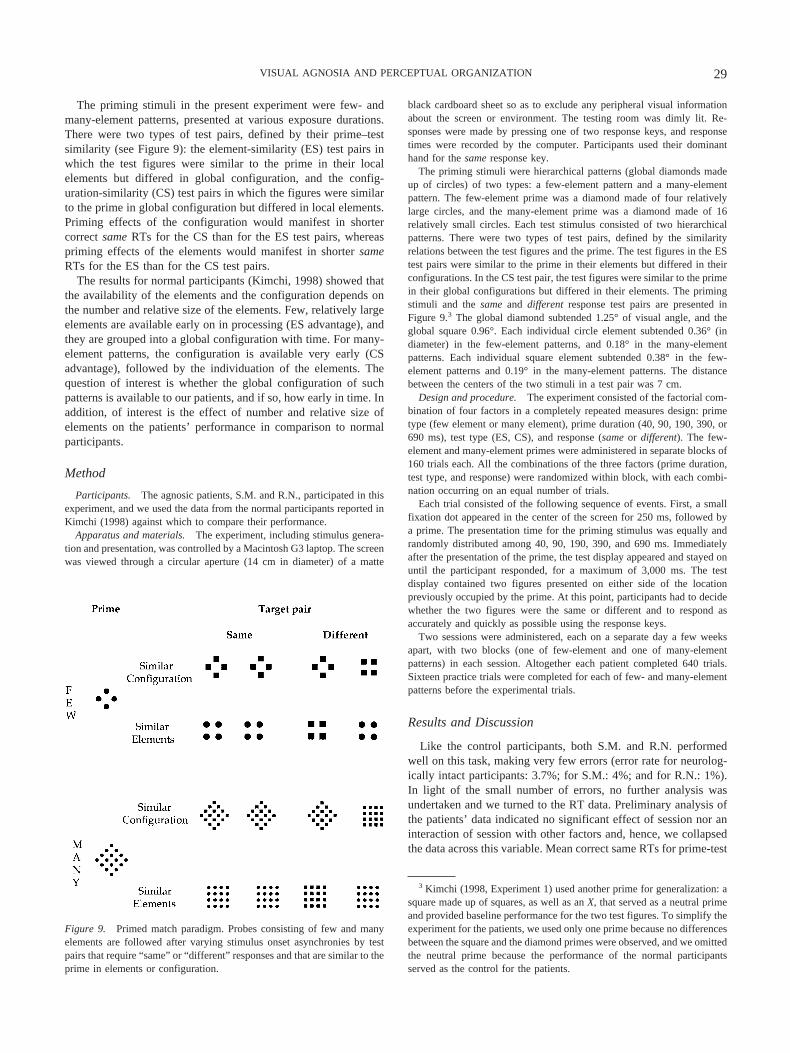
The priming stimuli in the present experiment were few- andmany-element patterns, presented at various exposure durations.There were two types of test pairs, defined by their prime–testsimilarity (see Figure 9): the element-similarity (ES) test pairs inwhich the test figures were similar to the prime in their localelements but differed in global configuration, and the config-uration-similarity (CS) test pairs in which the figures were similarto the prime in global configuration but differed in local elements.Priming effects of the configuration would manifest in shortercorrect same RTs for the CS than for the ES test pairs, whereaspriming effects of the elements would manifest in shorter sameRTs for the ES than for the CS test pairs.
The results for normal participants (Kimchi, 1998) showed thatthe availability of the elements and the configuration depends onthe number and relative size of the elements. Few, relatively largeelements are available early on in processing (ES advantage), andthey are grouped into a global configuration with time. For many-element patterns, the configuration is available very early (CSadvantage), followed by the individuation of the elements. Thequestion of interest is whether the global configuration of suchpatterns is available to our patients, and if so, how early in time. Inaddition, of interest is the effect of number and relative size ofelements on the patients’ performance in comparison to normalparticipants.
Method
Participants. The agnosic patients, S.M. and R.N., participated in thisexperiment, and we used the data from the normal participants reported inKimchi (1998) against which to compare their performance.
Apparatus and materials. The experiment, including stimulus genera-tion and presentation, was controlled by a Macintosh G3 laptop. The screenwas viewed through a circular aperture (14 cm in diameter) of a matte
black cardboard sheet so as to exclude any peripheral visual informationabout the screen or environment. The testing room was dimly lit. Re-sponses were made by pressing one of two response keys, and responsetimes were recorded by the computer. Participants used their dominanthand for the same response key.
The priming stimuli were hierarchical patterns (global diamonds madeup of circles) of two types: a few-element pattern and a many-elementpattern. The few-element prime was a diamond made of four relativelylarge circles, and the many-element prime was a diamond made of 16relatively small circles. Each test stimulus consisted of two hierarchicalpatterns. There were two types of test pairs, defined by the similarityrelations between the test figures and the prime. The test figures in the EStest pairs were similar to the prime in their elements but differed in theirconfigurations. In the CS test pair, the test figures were similar to the primein their global configurations but differed in their elements. The primingstimuli and the same and different response test pairs are presented inFigure 9.3 The global diamond subtended 1.25° of visual angle, and theglobal square 0.96°. Each individual circle element subtended 0.36° (indiameter) in the few-element patterns, and 0.18° in the many-elementpatterns. Each individual square element subtended 0.38° in the few-element patterns and 0.19° in the many-element patterns. The distancebetween the centers of the two stimuli in a test pair was 7 cm.
Design and procedure. The experiment consisted of the factorial com-bination of four factors in a completely repeated measures design: primetype (few element or many element), prime duration (40, 90, 190, 390, or690 ms), test type (ES, CS), and response (same or different). The few-element and many-element primes were administered in separate blocks of160 trials each. All the combinations of the three factors (prime duration,test type, and response) were randomized within block, with each combi-nation occurring on an equal number of trials.
Each trial consisted of the following sequence of events. First, a smallfixation dot appeared in the center of the screen for 250 ms, followed bya prime. The presentation time for the priming stimulus was equally andrandomly distributed among 40, 90, 190, 390, and 690 ms. Immediatelyafter the presentation of the prime, the test display appeared and stayed onuntil the participant responded, for a maximum of 3,000 ms. The testdisplay contained two figures presented on either side of the locationpreviously occupied by the prime. At this point, participants had to decidewhether the two figures were the same or different and to respond asaccurately and quickly as possible using the response keys.
Two sessions were administered, each on a separate day a few weeksapart, with two blocks (one of few-element and one of many-elementpatterns) in each session. Altogether each patient completed 640 trials.Sixteen practice trials were completed for each of few- and many-elementpatterns before the experimental trials.
Results and Discussion
Like the control participants, both S.M. and R.N. performedwell on this task, making very few errors (error rate for neurolog-ically intact participants: 3.7%; for S.M.: 4%; and for R.N.: 1%).In light of the small number of errors, no further analysis wasundertaken and we turned to the RT data. Preliminary analysis ofthe patients’ data indicated no significant effect of session nor aninteraction of session with other factors and, hence, we collapsedthe data across this variable. Mean correct same RTs for prime-test
3 Kimchi (1998, Experiment 1) used another prime for generalization: asquare made up of squares, as well as an X, that served as a neutral primeand provided baseline performance for the two test figures. To simplify theexperiment for the patients, we used only one prime because no differencesbetween the square and the diamond primes were observed, and we omittedthe neutral prime because the performance of the normal participantsserved as the control for the patients.
Figure 9. Primed match paradigm. Probes consisting of few and manyelements are followed after varying stimulus onset asynchronies by testpairs that require “same” or “different” responses and that are similar to theprime in elements or configuration.
29VISUAL AGNOSIA AND PERCEPTUAL ORGANIZATION
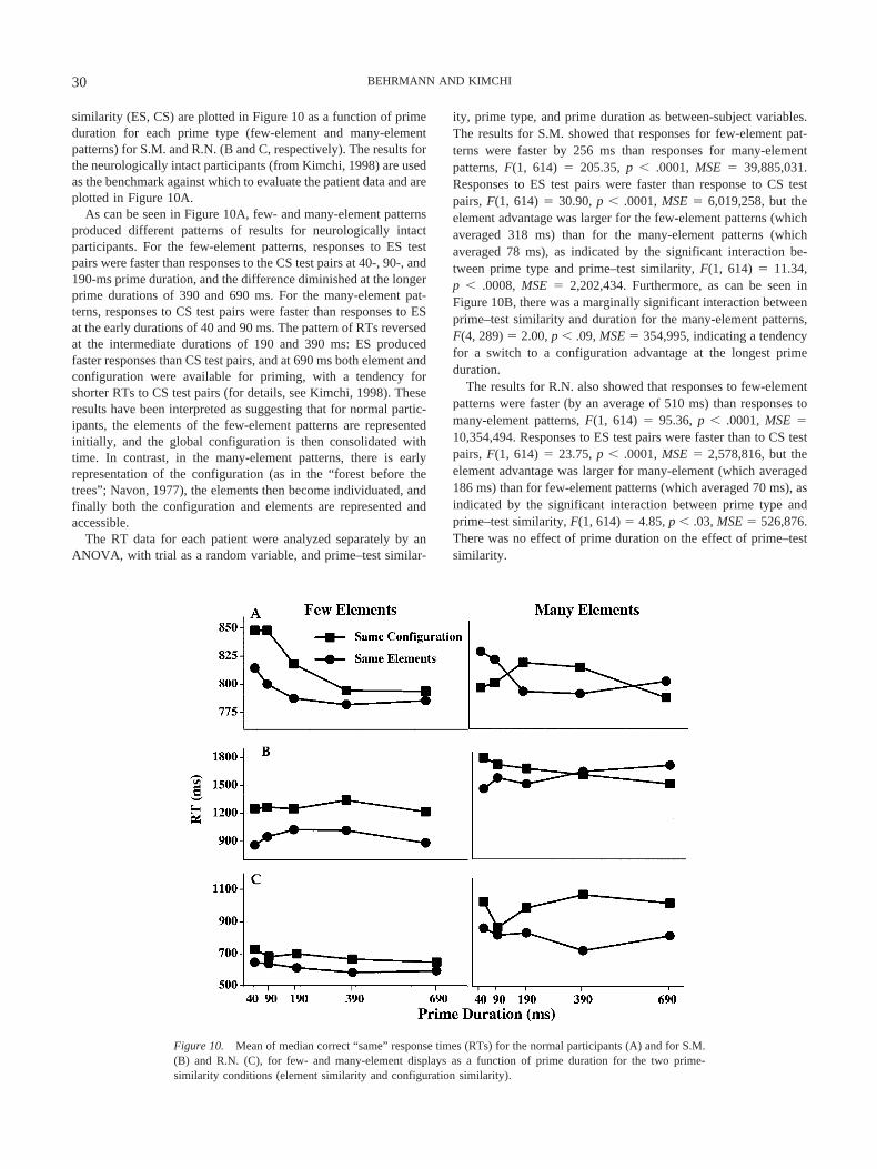
similarity (ES, CS) are plotted in Figure 10 as a function of primeduration for each prime type (few-element and many-elementpatterns) for S.M. and R.N. (B and C, respectively). The results forthe neurologically intact participants (from Kimchi, 1998) are usedas the benchmark against which to evaluate the patient data and areplotted in Figure 10A.
As can be seen in Figure 10A, few- and many-element patternsproduced different patterns of results for neurologically intactparticipants. For the few-element patterns, responses to ES testpairs were faster than responses to the CS test pairs at 40-, 90-, and190-ms prime duration, and the difference diminished at the longerprime durations of 390 and 690 ms. For the many-element pat-terns, responses to CS test pairs were faster than responses to ESat the early durations of 40 and 90 ms. The pattern of RTs reversedat the intermediate durations of 190 and 390 ms: ES producedfaster responses than CS test pairs, and at 690 ms both element andconfiguration were available for priming, with a tendency forshorter RTs to CS test pairs (for details, see Kimchi, 1998). Theseresults have been interpreted as suggesting that for normal partic-ipants, the elements of the few-element patterns are representedinitially, and the global configuration is then consolidated withtime. In contrast, in the many-element patterns, there is earlyrepresentation of the configuration (as in the “forest before thetrees”; Navon, 1977), the elements then become individuated, andfinally both the configuration and elements are represented andaccessible.
The RT data for each patient were analyzed separately by anANOVA, with trial as a random variable, and prime–test similar-
ity, prime type, and prime duration as between-subject variables.The results for S.M. showed that responses for few-element pat-terns were faster by 256 ms than responses for many-elementpatterns, F(1, 614) � 205.35, p � .0001, MSE � 39,885,031.Responses to ES test pairs were faster than response to CS testpairs, F(1, 614) � 30.90, p � .0001, MSE � 6,019,258, but theelement advantage was larger for the few-element patterns (whichaveraged 318 ms) than for the many-element patterns (whichaveraged 78 ms), as indicated by the significant interaction be-tween prime type and prime–test similarity, F(1, 614) � 11.34,p � .0008, MSE � 2,202,434. Furthermore, as can be seen inFigure 10B, there was a marginally significant interaction betweenprime–test similarity and duration for the many-element patterns,F(4, 289) � 2.00, p � .09, MSE � 354,995, indicating a tendencyfor a switch to a configuration advantage at the longest primeduration.
The results for R.N. also showed that responses to few-elementpatterns were faster (by an average of 510 ms) than responses tomany-element patterns, F(1, 614) � 95.36, p � .0001, MSE �10,354,494. Responses to ES test pairs were faster than to CS testpairs, F(1, 614) � 23.75, p � .0001, MSE � 2,578,816, but theelement advantage was larger for many-element (which averaged186 ms) than for few-element patterns (which averaged 70 ms), asindicated by the significant interaction between prime type andprime–test similarity, F(1, 614) � 4.85, p � .03, MSE � 526,876.There was no effect of prime duration on the effect of prime–testsimilarity.
Figure 10. Mean of median correct “same” response times (RTs) for the normal participants (A) and for S.M.(B) and R.N. (C), for few- and many-element displays as a function of prime duration for the two prime-similarity conditions (element similarity and configuration similarity).
30 BEHRMANN AND KIMCHI
The patients were slower than the normal participants, except inthe case of R.N. with the few-element patterns (note the differenceof y-axis between patients and normal participants in Figure 10). Incontrast to the normal participants who responded about equallyfast (on average) to few- and many-element patterns, both patientswere slower in their responses to many-element than to few-element patterns, suggesting that it was more difficult for them tohandle patterns when the number of elements was increased.
For the few-element patterns, in contrast with the normal par-ticipants who seemed to derive the global configuration over time,neither S.M. nor R.N. appeared to be able to derive a globalconfiguration, even at the longest exposure duration of 690 ms.Both patients showed shorter RTs to the ES test pairs, and therewas no effect of prime duration on this element advantage. Previ-ous research has suggested that for patterns composed of few,relatively large elements, the local elements are perceived bynormal individuals as figural parts of the overall form (Goldmeier,1936/1972; Kimchi & Palmer, 1982), and the local elements andthe global form are perceptually integral (Kimchi, 1988; Kimchi &Palmer, 1985). The 2 patients, however, seemed unable to inte-grate the local elements into a global entity, so that they failed toperceive the local elements as figural parts of an overall form and,rather, perceived them as discrete, unrelated entities.
For the many-element patterns, again in contrast with the normalparticipants, neither patient exhibited an early advantage for theconfiguration. Rather, R.N. showed an advantage for the ES testpairs as early as 40 ms, and this element advantage remained fairlyunchanged over the entire time course, so that the global config-uration was not available to him even at the longest duration of 690ms. S.M. also did not show any early advantage for the CS testpairs, although he eventually showed a tendency for shorter RTsfor CS than ES test pairs at the longest duration of 690 ms, as didnormal participants.
It has been suggested (Kimchi, 1998) that for normal partici-pants, many relatively small elements (as in the many-elementpatterns) are rapidly grouped without the individuation of theelements that occurs later in time. It has also been suggested thatsuch early grouping is functional for a system whose goal is objectidentification and recognition, because many small elements inclose proximity to one another are more likely to be textureelements of a single object rather than discrete objects. No rapidgrouping of the many relatively small elements was observed foreither patient. Rather, it seems that for both patients, the many-element patterns were processed initially as discrete elements,hence the longer RTs for many-element relative to few-elementpatterns.
In addition to the differences between the patients and thenormal participants, there were also some differences betweenR.N. and S.M. First, the difference in RTs for many- and few-element patterns was larger for R.N. (510 ms) than for S.M. (256ms), reflecting the greater difficulty in processing the many-element patterns for R.N. than for S.M.4 Second, for R.N., the ESadvantage for the many-element patterns was larger than for few-element patterns, whereas the opposite was true for S.M. Third,whereas no effect whatsoever of prime duration on prime–testsimilarity was observed for R.N., a tendency for a reversal in therelative advantage of ES and CS was observed for S.M. at thelongest duration for the many-element patterns.
Taken together, these differences between the patients suggestthat in the case of S.M., although there is no evidence for the early
rapid grouping of many elements that characterizes normal per-ception, grouping processes do operate with many elements. Even-tually these grouping processes can lead to the perception of theglobal configuration. This finding is consistent with the results ofExperiment 2, in which, with unlimited exposure duration, S.M.showed a global advantage for hierarchical letters, similar tonormal participants. Note that previous findings with neurologi-cally intact individuals have shown that grouping is more efficientwith an increased number of elements (Bacon & Egeth, 1991).R.N., on the other hand, seemed unable to group the elements intoa global configuration even when conditions and time favor group-ing. This clear local advantage in the perception of hierarchicalstimuli is consistent with his performance in Experiment 2.
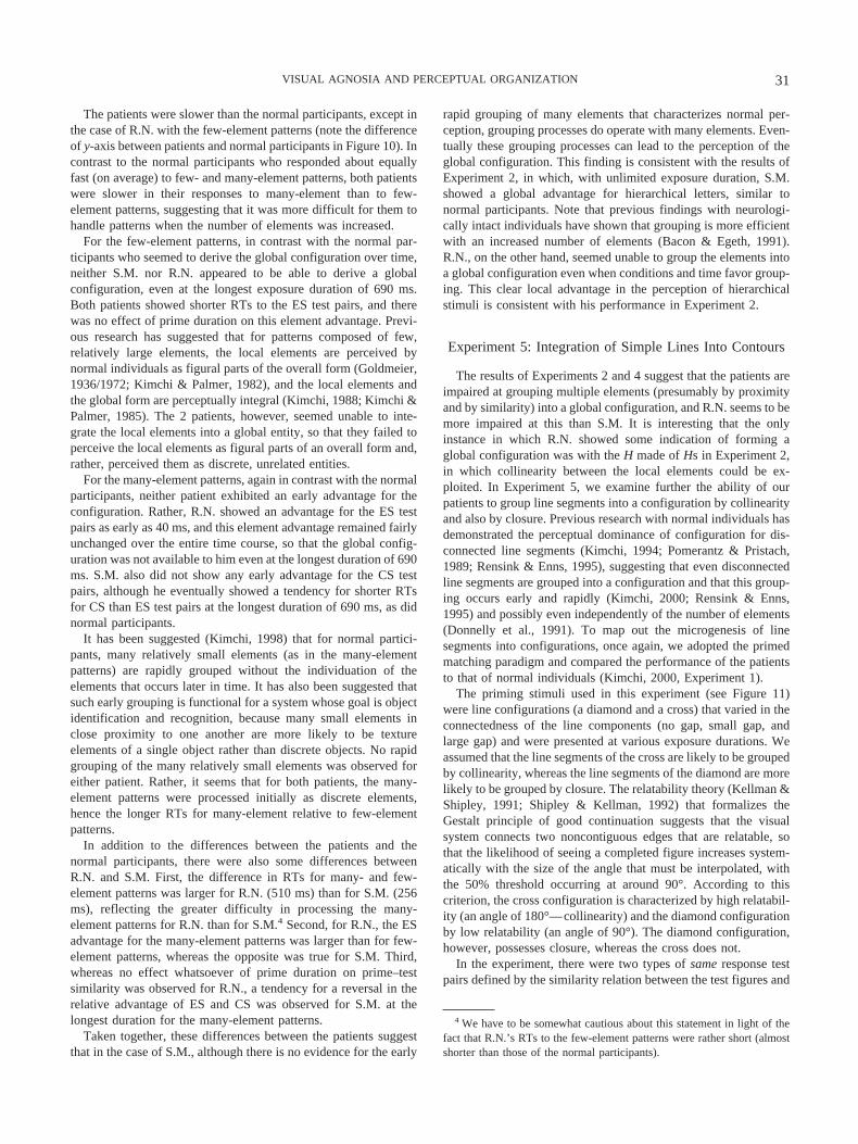
Experiment 5: Integration of Simple Lines Into Contours
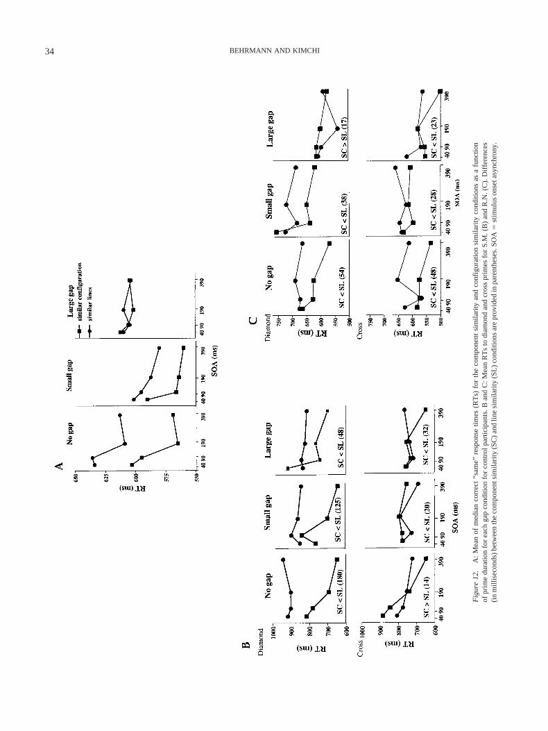
The results of Experiments 2 and 4 suggest that the patients areimpaired at grouping multiple elements (presumably by proximityand by similarity) into a global configuration, and R.N. seems to bemore impaired at this than S.M. It is interesting that the onlyinstance in which R.N. showed some indication of forming aglobal configuration was with the H made of Hs in Experiment 2,in which collinearity between the local elements could be ex-ploited. In Experiment 5, we examine further the ability of ourpatients to group line segments into a configuration by collinearityand also by closure. Previous research with normal individuals hasdemonstrated the perceptual dominance of configuration for dis-connected line segments (Kimchi, 1994; Pomerantz & Pristach,1989; Rensink & Enns, 1995), suggesting that even disconnectedline segments are grouped into a configuration and that this group-ing occurs early and rapidly (Kimchi, 2000; Rensink & Enns,1995) and possibly even independently of the number of elements(Donnelly et al., 1991). To map out the microgenesis of linesegments into configurations, once again, we adopted the primedmatching paradigm and compared the performance of the patientsto that of normal individuals (Kimchi, 2000, Experiment 1).
The priming stimuli used in this experiment (see Figure 11)were line configurations (a diamond and a cross) that varied in theconnectedness of the line components (no gap, small gap, andlarge gap) and were presented at various exposure durations. Weassumed that the line segments of the cross are likely to be groupedby collinearity, whereas the line segments of the diamond are morelikely to be grouped by closure. The relatability theory (Kellman &Shipley, 1991; Shipley & Kellman, 1992) that formalizes theGestalt principle of good continuation suggests that the visualsystem connects two noncontiguous edges that are relatable, sothat the likelihood of seeing a completed figure increases system-atically with the size of the angle that must be interpolated, withthe 50% threshold occurring at around 90°. According to thiscriterion, the cross configuration is characterized by high relatabil-ity (an angle of 180°—collinearity) and the diamond configurationby low relatability (an angle of 90°). The diamond configuration,however, possesses closure, whereas the cross does not.
In the experiment, there were two types of same response testpairs defined by the similarity relation between the test figures and
4 We have to be somewhat cautious about this statement in light of thefact that R.N.’s RTs to the few-element patterns were rather short (almostshorter than those of the normal participants).
31VISUAL AGNOSIA AND PERCEPTUAL ORGANIZATION
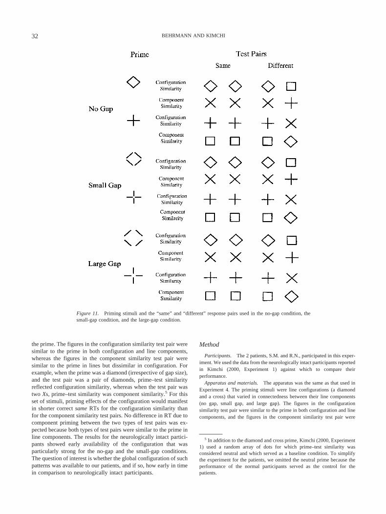
the prime. The figures in the configuration similarity test pair weresimilar to the prime in both configuration and line components,whereas the figures in the component similarity test pair weresimilar to the prime in lines but dissimilar in configuration. Forexample, when the prime was a diamond (irrespective of gap size),and the test pair was a pair of diamonds, prime–test similarityreflected configuration similarity, whereas when the test pair wastwo Xs, prime–test similarity was component similarity.5 For thisset of stimuli, priming effects of the configuration would manifestin shorter correct same RTs for the configuration similarity thanfor the component similarity test pairs. No difference in RT due tocomponent priming between the two types of test pairs was ex-pected because both types of test pairs were similar to the prime inline components. The results for the neurologically intact partici-pants showed early availability of the configuration that wasparticularly strong for the no-gap and the small-gap conditions.The question of interest is whether the global configuration of suchpatterns was available to our patients, and if so, how early in timein comparison to neurologically intact participants.
Method
Participants. The 2 patients, S.M. and R.N., participated in this exper-iment. We used the data from the neurologically intact participants reportedin Kimchi (2000, Experiment 1) against which to compare theirperformance.
Apparatus and materials. The apparatus was the same as that used inExperiment 4. The priming stimuli were line configurations (a diamondand a cross) that varied in connectedness between their line components(no gap, small gap, and large gap). The figures in the configurationsimilarity test pair were similar to the prime in both configuration and linecomponents, and the figures in the component similarity test pair were
5 In addition to the diamond and cross prime, Kimchi (2000, Experiment1) used a random array of dots for which prime–test similarity wasconsidered neutral and which served as a baseline condition. To simplifythe experiment for the patients, we omitted the neutral prime because theperformance of the normal participants served as the control for thepatients.
Figure 11. Priming stimuli and the “same” and “different” response pairs used in the no-gap condition, thesmall-gap condition, and the large-gap condition.
32 BEHRMANN AND KIMCHI
similar to the prime in lines but dissimilar in configuration. All test figureswere connected configurations (see Figure 11).
Each individual line subtended 1.43° in length for the diamond and thecross primes for all primes and test figures. The connected diamondsubtended about 2.02° � 2.02°, and the connected cross subtended 2.86° �2.86°. The gaps between the lines subtended 0.29° each in the small-gapcondition and 1° each in the large-gap condition. The size of the test figureswas identical in all gap conditions. The distance between the centers of thetwo stimuli in a test pair was 7 cm. The figures in the different response testpairs appeared equally often in each of the two possible locations.
Design and procedure. The experiment consisted of the factorial com-bination of five factors in a completely repeated measures design: gap (nogap, small gap, large gap), prime type (diamond or cross), prime duration(40, 90, 190, or 390 ms), prime–test similarity (component similarity orconfiguration similarity), and response (same or different). The threedifferent gap conditions constituted a between-blocks manipulation for theneurologically intact participants. All the combinations of the four otherfactors (prime type, prime duration, test type, and response) were random-ized within block, with each combination occurring on an equal number oftrials. For each gap condition, there were six blocks of 160 experimentaltrials each, preceded by a block of 15 practice trials. The patients com-pleted two sessions, a few weeks apart, with three blocks in each session.The procedure was the same as in Experiment 4.
Results and Discussion
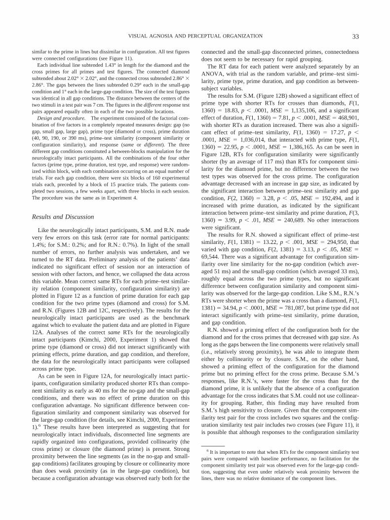
Like the neurologically intact participants, S.M. and R.N. madevery few errors on this task (error rate for normal participants:1.4%; for S.M.: 0.2%; and for R.N.: 0.7%). In light of the smallnumber of errors, no further analysis was undertaken, and weturned to the RT data. Preliminary analysis of the patients’ dataindicated no significant effect of session nor an interaction ofsession with other factors, and hence, we collapsed the data acrossthis variable. Mean correct same RTs for each prime–test similar-ity relation (component similarity, configuration similarity) areplotted in Figure 12 as a function of prime duration for each gapcondition for the two prime types (diamond and cross) for S.M.and R.N. (Figures 12B and 12C, respectively). The results for theneurologically intact participants are used as the benchmarkagainst which to evaluate the patient data and are plotted in Figure12A. Analyses of the correct same RTs for the neurologicallyintact participants (Kimchi, 2000, Experiment 1) showed thatprime type (diamond or cross) did not interact significantly withpriming effects, prime duration, and gap condition, and therefore,the data for the neurologically intact participants were collapsedacross prime type.
As can be seen in Figure 12A, for neurologically intact partic-ipants, configuration similarity produced shorter RTs than compo-nent similarity as early as 40 ms for the no-gap and the small-gapconditions, and there was no effect of prime duration on thisconfiguration advantage. No significant difference between con-figuration similarity and component similarity was observed forthe large-gap condition (for details, see Kimchi, 2000, Experiment1).6 These results have been interpreted as suggesting that forneurologically intact individuals, disconnected line segments arerapidly organized into configurations, provided collinearity (thecross prime) or closure (the diamond prime) is present. Strongproximity between the line segments (as in the no-gap and small-gap conditions) facilitates grouping by closure or collinearity morethan does weak proximity (as in the large-gap condition), butbecause a configuration advantage was observed early both for the
connected and the small-gap disconnected primes, connectednessdoes not seem to be necessary for rapid grouping.
The RT data for each patient were analyzed separately by anANOVA, with trial as the random variable, and prime–test simi-larity, prime type, prime duration, and gap condition as between-subject variables.
The results for S.M. (Figure 12B) showed a significant effect ofprime type with shorter RTs for crosses than diamonds, F(1,1360) � 18.83, p � .0001, MSE � 1,135,106, and a significanteffect of duration, F(1, 1360) � 7.81, p � .0001, MSE � 468,901,with shorter RTs as duration increased. There was also a signifi-cant effect of prime–test similarity, F(1, 1360) � 17.27, p �.0001, MSE � 1,036,014, that interacted with prime type, F(1,1360) � 22.95, p � .0001, MSE � 1,386,165. As can be seen inFigure 12B, RTs for configuration similarity were significantlyshorter (by an average of 117 ms) than RTs for component simi-larity for the diamond prime, but no difference between the twotest types was observed for the cross prime. The configurationadvantage decreased with an increase in gap size, as indicated bythe significant interaction between prime–test similarity and gapcondition, F(2, 1360) � 3.28, p � .05, MSE � 192,494, and itincreased with prime duration, as indicated by the significantinteraction between prime–test similarity and prime duration, F(3,1360) � 3.99, p � .01, MSE � 240,689. No other interactionswere significant.
The results for R.N. showed a significant effect of prime–testsimilarity, F(1, 1381) � 13.22, p � .001, MSE � 294,950, thatvaried with gap condition, F(2, 1381) � 3.13, p � .05, MSE �69,544. There was a significant advantage for configuration sim-ilarity over line similarity for the no-gap condition (which aver-aged 51 ms) and the small-gap condition (which averaged 33 ms),roughly equal across the two prime types, but no significantdifference between configuration similarity and component simi-larity was observed for the large-gap condition. Like S.M., R.N.’sRTs were shorter when the prime was a cross than a diamond, F(1,1381) � 34.94, p � .0001, MSE � 781,087, but prime type did notinteract significantly with prime–test similarity, prime duration,and gap condition.
R.N. showed a priming effect of the configuration both for thediamond and for the cross primes that decreased with gap size. Aslong as the gaps between the line components were relatively small(i.e., relatively strong proximity), he was able to integrate themeither by collinearity or by closure. S.M., on the other hand,showed a priming effect of the configuration for the diamondprime but no priming effect for the cross prime. Because S.M.’sresponses, like R.N.’s, were faster for the cross than for thediamond prime, it is unlikely that the absence of a configurationadvantage for the cross indicates that S.M. could not use collinear-ity for grouping. Rather, this finding may have resulted fromS.M.’s high sensitivity to closure. Given that the component sim-ilarity test pair for the cross includes two squares and the config-uration similarity test pair includes two crosses (see Figure 11), itis possible that although responses to the configuration similarity
6 It is important to note that when RTs for the component similarity testpairs were compared with baseline performance, no facilitation for thecomponent similarity test pair was observed even for the large-gap condi-tion, suggesting that even under relatively weak proximity between thelines, there was no relative dominance of the component lines.
33VISUAL AGNOSIA AND PERCEPTUAL ORGANIZATION
Fig
ure
12.
A:
Mea
nof
med
ian
corr
ect
“sam
e”re
spon
setim
es(R
Ts)
for
the
com
pone
ntsi
mila
rity
and
conf
igur
atio
nsi
mila
rity
cond
ition
sas
afu
nctio
nof
prim
edu
ratio
nfo
rea
chga
pco
nditi
onfo
rco
ntro
lpar
ticip
ants
.Ban
dC
:Mea
nR
Ts
todi
amon
dan
dcr
oss
prim
esfo
rS.
M.(
B)
and
R.N
.(C
).D
iffe
renc
es(i
nm
illis
econ
ds)
betw
een
the
com
pone
ntsi
mila
rity
(SC
)an
dlin
esi
mila
rity
(SL
)co
nditi
ons
are
prov
ided
inpa
rent
hese
s.SO
A�
stim
ulus
onse
tasy
nchr
ony.
34 BEHRMANN AND KIMCHI
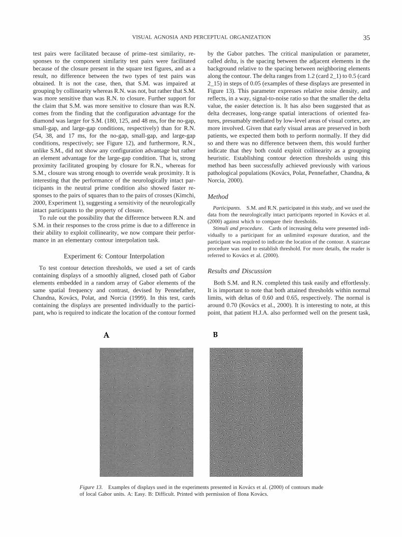
test pairs were facilitated because of prime–test similarity, re-sponses to the component similarity test pairs were facilitatedbecause of the closure present in the square test figures, and as aresult, no difference between the two types of test pairs wasobtained. It is not the case, then, that S.M. was impaired atgrouping by collinearity whereas R.N. was not, but rather that S.M.was more sensitive than was R.N. to closure. Further support forthe claim that S.M. was more sensitive to closure than was R.N.comes from the finding that the configuration advantage for thediamond was larger for S.M. (180, 125, and 48 ms, for the no-gap,small-gap, and large-gap conditions, respectively) than for R.N.(54, 38, and 17 ms, for the no-gap, small-gap, and large-gapconditions, respectively; see Figure 12), and furthermore, R.N.,unlike S.M., did not show any configuration advantage but ratheran element advantage for the large-gap condition. That is, strongproximity facilitated grouping by closure for R.N., whereas forS.M., closure was strong enough to override weak proximity. It isinteresting that the performance of the neurologically intact par-ticipants in the neutral prime condition also showed faster re-sponses to the pairs of squares than to the pairs of crosses (Kimchi,2000, Experiment 1), suggesting a sensitivity of the neurologicallyintact participants to the property of closure.
To rule out the possibility that the difference between R.N. andS.M. in their responses to the cross prime is due to a difference intheir ability to exploit collinearity, we now compare their perfor-mance in an elementary contour interpolation task.
Experiment 6: Contour Interpolation
To test contour detection thresholds, we used a set of cardscontaining displays of a smoothly aligned, closed path of Gaborelements embedded in a random array of Gabor elements of thesame spatial frequency and contrast, devised by Pennefather,Chandna, Kovacs, Polat, and Norcia (1999). In this test, cardscontaining the displays are presented individually to the partici-pant, who is required to indicate the location of the contour formed
by the Gabor patches. The critical manipulation or parameter,called delta, is the spacing between the adjacent elements in thebackground relative to the spacing between neighboring elementsalong the contour. The delta ranges from 1.2 (card 2_1) to 0.5 (card2_15) in steps of 0.05 (examples of these displays are presented inFigure 13). This parameter expresses relative noise density, andreflects, in a way, signal-to-noise ratio so that the smaller the deltavalue, the easier detection is. It has also been suggested that asdelta decreases, long-range spatial interactions of oriented fea-tures, presumably mediated by low-level areas of visual cortex, aremore involved. Given that early visual areas are preserved in bothpatients, we expected them both to perform normally. If they didso and there was no difference between them, this would furtherindicate that they both could exploit collinearity as a groupingheuristic. Establishing contour detection thresholds using thismethod has been successfully achieved previously with variouspathological populations (Kovacs, Polat, Pennefather, Chandna, &Norcia, 2000).
Method
Participants. S.M. and R.N. participated in this study, and we used thedata from the neurologically intact participants reported in Kovacs et al.(2000) against which to compare their thresholds.
Stimuli and procedure. Cards of increasing delta were presented indi-vidually to a participant for an unlimited exposure duration, and theparticipant was required to indicate the location of the contour. A staircaseprocedure was used to establish threshold. For more details, the reader isreferred to Kovacs et al. (2000).
Results and Discussion
Both S.M. and R.N. completed this task easily and effortlessly.It is important to note that both attained thresholds within normallimits, with deltas of 0.60 and 0.65, respectively. The normal isaround 0.70 (Kovacs et al., 2000). It is interesting to note, at thispoint, that patient H.J.A. also performed well on the present task,
Figure 13. Examples of displays used in the experiments presented in Kovacs et al. (2000) of contours madeof local Gabor units. A: Easy. B: Difficult. Printed with permission of Ilona Kovacs.
35VISUAL AGNOSIA AND PERCEPTUAL ORGANIZATION
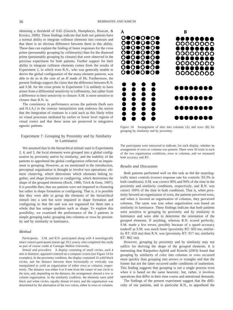
obtaining a threshold of 0.65 (Giersch, Humphreys, Boucart, &Kovacs, 2000). These findings indicate that both our patients havea normal ability to integrate collinear elements into contours andthat there is no obvious difference between them in this ability.These data can explain the finding of faster responses for the crossprime (presumably grouping by collinearity) than for the diamondprime (presumably grouping by closure) that were observed in theprevious experiment for both patients. Further support for theirability to integrate collinear elements comes from the results ofExperiment 2, in which even R.N., who was generally unable toderive the global configuration of the many-element patterns, wasable to do so in the case of an H made of Hs. Furthermore, thepresent findings support the claim that the difference between R.N.and S.M. for the cross prime in Experiment 5 is unlikely to havearisen from a differential sensitivity to collinearity, but rather froma difference in their sensitivity to closure: S.M. is more sensitive toclosure than R.N. is.
The consistency in performance across the patients (both oursand H.J.A.) in the contour interpolation task endorses the notionthat the integration of contours in a task such as this likely relieson visual processes mediated by earlier or lower level regions ofvisual cortex and that these areas are preserved in integrativeagnosic patients.
Experiment 7: Grouping by Proximity and by Similarityin Luminance
We assumed that in the hierarchical stimuli used in Experiments2, 4, and 5, the local elements were grouped into a global config-uration by proximity and/or by similarity, and the inability of thepatients to apprehend the global configuration reflected an impair-ment in grouping. However, as we mentioned in the introduction,perceptual organization is thought to involve two operations: ele-ment clustering, which determines which elements belong to-gether, and shape formation or configuring, which determines theshape of the grouped elements (Rock, 1986; Trick & Enns, 1997).It is possible then, that our patients were not impaired in clusteringbut rather in shape formation or configuring. That is, it is possiblethat they were able to group the elements of the hierarchicalstimuli into a unit but were impaired in shape formation andconfiguring so that the unit was not organized for them into awhole that has unique qualities such as shape. To explore thispossibility, we examined the performance of the 2 patients insimple grouping tasks: grouping into columns or rows by proxim-ity and by similarity in luminance.
Method
Participants. S.M. and R.N. participated along with 4 neurologicallyintact control participants (mean age 19.2 years), who completed this studyas part of course credit at Carnegie Mellon University.
Stimuli and procedure. A display consisting of small circles, each 4mm in diameter, appeared centered on a computer screen (see Figure 14 forexamples). In the proximity condition, the display contained 32 solid blackcircles, and the distance between them horizontally or vertically wasmanipulated to yield an organization of either rows or columns, respec-tively. The distance was either 4 or 8 mm from the center of one circle tothe next, and, depending on the distance, the arrangement obeyed a row orcolumn organization. In the similarity condition, the elements were solidblack and white circles, equally distant (4 mm), and the organization wasdetermined by the alternation of the two colors, either in rows or columns.
The participants were instructed to indicate, for each display, whether anarrangement of rows or columns was present. There were 50 trials in eachof the two organization conditions, rows or columns, and we measuredboth accuracy and RT.
Results and Discussion
Both patients performed well on this task as did the neurolog-ically intact controls (correct response rate for controls: 93.3% inboth conditions). S.M. was correct 90% and 94% of the time in theproximity and similarity conditions, respectively, and R.N. wascorrect 100% of the time in both conditions. That is, when prox-imity favored an organization of rows, the patients perceived rows,and when it favored an organization of columns, they perceivedcolumns. The same was true when organization was based onsimilarity in luminance. These findings indicate that both patientswere sensitive to grouping by proximity and by similarity inluminance and were able to determine the orientation of thegrouped elements. If anything, whereas R.N. scored perfectly,S.M. made a few errors, possibly because of a speed–accuracytradeoff as S.M. was much faster (proximity RT: 603 ms; similar-ity RT: 659 ms) than R.N. was (proximity RT: 917 ms; similarityRT: 862 ms).
However, grouping by proximity and by similarity may notsuffice for deriving the shape of the grouped elements. It isinteresting that Razpurker-Apfeld and Kimchi (2003) found thatgrouping by similarity of color into columns or rows occurredmore quickly than grouping into arrows or triangles and that theformer but not the latter occurred under conditions of inattention.This finding suggests that grouping is not a single process evenwhen it is based on the same heuristic, but, rather, it involvesoperations that differ in their time course and attentional demands.
The findings of the present experiment suggest that the diffi-culty of our patients, and in particular R.N., to apprehend the
Figure 14. Arrangement of dots into columns (A) and rows (B) forgrouping by similarity and by proximity.
36 BEHRMANN AND KIMCHI
global configuration of hierarchical stimuli was not due to animpairment in simple grouping (i.e., in the sense of what goes withwhat) but presumably to an impairment in the ability to apprehendthe interrelationships of the elements and to derive the emergentstructure or shape.
General Discussion
The goal of this article has been to explore the psychologicaland neural processes involved in deriving structure and coherencefrom visual input. Many traditional theories of visual perceptionhave assumed that perceptual organization processes operate earlyand preattentively to deliver candidate units for further processing,such as object identification. These theories make no attempt todistinguish in detail between the different perceptual organizationprocesses nor do they evaluate the relative contribution of thedifferent processes to object recognition. To address these issues,we have investigated the behavior of 2 individuals with acquiredintegrative visual object agnosia on tasks of object recognition andperceptual organization, with a specific emphasis on groupingelements into global forms. By understanding how the systembreaks down and how a perceptual organization deficit is relatedto impaired object recognition, we hoped to obtain insight intothe normal processes of perceptual organization and objectidentification.
When required to integrate many small elements into a globalconfiguration, S.M. but not R.N. was able to derive the global formalthough he required more time to do so than did the normalparticipants. A microgenetic analysis of this integration processconfirmed that given enough time and sufficient data-driven sup-port, S.M. was eventually able to derive the global form. It isimportant to note that neurologically intact perceivers unify amultielement stimulus early and quickly, reflecting their sponta-neous bias to deal with such a stimulus as a unit rather than asdisparate components, and only later do they individuate the ele-ments. S.M. did not show this early and fast grouping of the manyelements, and only with time was he able to laboriously derive aglobal form. Even under optimal circumstances, R.N. failed toderive the global form from the image. When the stimulus wascomposed of only a few relatively large elements, neither patientwas able to extract a global structure. Under these conditions,neurologically intact participants could apprehend the global struc-ture despite the relative salience of the individual elements, and theglobal configuration became further consolidated with time. Notethat the differences between the 2 patients and the differencebetween them and the neurologically intact participants in appre-hending the global configuration of hierarchical stimuli cannot beattributed to a differential sensitivity to low-spatial-frequency in-formation, as both patients displayed spatial frequency thresholdfunctions within normal limits.
In an investigation of the time course of the ability to groupsimple line segments into configurations by collinearity and byclosure, we found that both patients were able to exploit theseproperties early on, as is also true of neurologically intact partic-ipants. However, S.M. was more sensitive to closure than was R.N.Strong proximity facilitated grouping by closure for R.N., but S.M.was able to group by closure even when proximity was weak. Weshould note that R.N.’s performance here might even be mediatedby collinearity at its limits (relatability at 90°), indicating that hemight be even less sensitive to closure than we have suggested. In
a task that was designed specifically to evaluate the ability tointegrate collinear elements into simple contours, both S.M. andR.N. performed like normal perceivers and did not differ from oneanother.
The final result was that when presented with classic, simplegestalt displays requiring grouping into rows or columns, bothpatients were able to group by proximity and by similarity ofluminance and did not differ from one another nor from theneurologically intact participants.
In sum, both patients were able to group collinear elements intoa contour, to group elements into simple rows or columns byproximity and by similarity in luminance or color, and to groupsimple line segments into simple configurations by closure. It isimportant to note, however, that although the basic groupingabilities of both patients seemed intact under simple conditions,they nevertheless encountered difficulties under more difficultconditions as in segmenting overlapping shapes. In contrast withthe seemingly intact basic grouping, there was a significant im-pairment in both patients in deriving global structure and appre-hending a multielement stimulus as a whole with a specific shape,and the impairment was more marked in R.N. than in S.M. R.N.failed to derive a global structure even under the most favorableconditions and unlimited time and was also less sensitive toclosure than was S.M. A critical point here is that the patientsdiffered from each other in the severity of their object recognitiondeficit, with R.N. performing significantly more poorly than S.M.in both accuracy and latency.
We now discuss these findings in light of our original questions,namely, the differences between various processes of perceptualorganization, the relative contribution of these different processesto object recognition, and the neural systems subserving thesemechanisms.
The first important conclusion is that not all organizationalprocesses are created equal. Clearly, grouping by collinearity,proximity, and similarity by luminance or color was easily andequally well achieved by the patients, whereas this was not thecase for grouping by closure. The relative preservation of groupingby collinearity is also evident in two other agnosic patients: H.J.A.(Giersch et al., 2000) and N.M. (Ricci et al., 1999), who wereimpaired at integrating low-level elements into a whole, but,nevertheless, were able to extract contours from an image (see alsopatient F.G.P., Kartsounis & Warrington, 1991). The present dataalso clearly show that although the patients were sensitive to basicgrouping, they were not equally able to derive a global structureand shape, suggesting that they might be impaired (and to differentdegrees) in configuring or shape formation.
The differential sensitivity to different forms of grouping isconsistent with the idea that some perceptual organization pro-cesses may precede others; for example, some processes operateon fairly local components, such as edges, and map onto basicneurophysiological interactions quite early in the visual pathway(Kellman, 2000; Kovacs, Kozma, Feher, & Benedek, 1999; Ship-ley & Kellman, 1992). This also fits well with recent neuroimagingand neurophysiological work (Lamme & Roelfsema, 2000; Lee, inpress; Sugita, 1999; Westheimer, 1999) suggesting that the abilityto interpolate across discrete collinear elements arises from thelateral connections and long-range interactions in early (V1 andV2) visual cortex. Time constants associated with the V1 and V2operations have been estimated at 45 to 50 ms and 70 to 90 ms inV1 and V2, respectively (Doniger et al., 2000; von der Heydt &
37VISUAL AGNOSIA AND PERCEPTUAL ORGANIZATION
Peterhans, 1989). Unfortunately, as is often the case in neuropsy-chological investigations and as is true in our case too, the lesionlocalization in our patients is not precise enough to yield definitiveevidence for the neural structures that are involved, but our resultsare consistent with the neurophysiological and imaging findings.We know, for instance, that the early visual areas were preservedin S.M. and although not definitely established, were likely intactin R.N., too, given the absence of low-level deficits. It is thesepreserved visual regions that probably mediated the patients’ abil-ity to exploit collinearity and grouping by proximity and similarityin luminance. An obvious future question to be addressed concernsthe neural substrate for these organization processes. Ideally, con-trasting a group of integrative agnosia patients with lesions in aparticular area with a group of control patients with lesions in adifferent area would present stronger evidence for the brain–behavior correspondences in which we are interested.
In contrast with these rather simple forms of grouping, otherforms of organization had more global influences, as in the case ofclosure and deriving a structured whole, and these more complexforms were probably mediated by more anterior regions of thevisual system. Some evidence to support this claim, for example,comes from a recent high-density, event-related potential study(Doniger et al., 2000) in which the amount of visual information inthe image was incrementally increased with each subsequent pre-sentation. The critical result was the existence of a bilateral oc-cipitotemporal negative potential that tracked the amount of clo-sure in the image; activation did not manifest in an all-or-nonefashion at the point of closure but rather built incrementally overa number of preidentification levels. This finding suggests that thisregion is involved in the computation of closure rather than justregistering its presence. It is important to note that the peak ofactivation in this region occurred at approximately 290 ms, muchlater than the estimated onset of V1 or V2 activity. That perceptualclosure is subserved by ventral occipitotemporal areas is alsosupported by recent hemodynamic and metabolic data (Gerlach etal., 2002) showing that the inferior occipital gyri (perhaps evenincluding area V2) are involved in the integration of visual ele-ments into perceptual wholes, irrespective of whether the wholeswere familiar objects or not (see Georgopoulos et al., 2001, forsimilar evidence; and Gauthier & Tarr, 2002, for behavioral evi-dence on different forms of configuring and associated neuralsubstrates).
The hypothesis that we have put forward entails that someorganization processes precede others, and we have linked these tobrain structures on a continuum from more posterior to anteriorregions. We do not, however, propose that the system operates ina purely serial and feedforward fashion. There is now ampleevidence for bidirectional connectivity and mutual bottom-up andtop-down reciprocity (Bullier & Nowak, 1995; Lee, in press; Zhou,Friedman, & von der Heydt, 2000) and the fact that processingoperates in a cascaded and interactive fashion in the visual system.We do suggest, however, that there is a temporal advantage forsome processes over others, and the order in which these processestake place follows a posterior–anterior brain organization.
The finding that S.M. and R.N. are similar to one another inmore basic, presumably low-level grouping operations but showmarked differences in their ability to derive a global form from amultielement display strongly suggests that perceptual organiza-tion involves not only grouping in the sense of element clustering,but also presumably higher level configuring and shape formation.
It is in these more configural forms of grouping that the 2 patientsdiffered from one another and in which R.N. was more impairedthan was S.M. This distinction we have made between groupingand shape formation or configuring may also help to clarify someconfusion found in the literature on perceptual organization. Forexample, understanding the attentional demands of perceptualorganization may depend on whether we refer to grouping (i.e.,element clustering) or to configuring (shape formation). Theformer is more likely to occur under conditions of inattention thanthe latter, and a failure to distinguish between the two organiza-tional processes may lead to seemingly conflicting results. Also,when a task that is designed to assess grouping performance alsorequires shape formation, caution is necessary in interpretation.For example, Ricci et al. (1999) reported that patient N.M. wasimpaired in grouping (by luminance, by color, and by line orien-tation). However, the test that was administered to examine N.M.’sgrouping ability required her to identify a hierarchical letter em-bedded in a background of elements. Clearly, grouping alone (i.e.,determining which elements belong together) is not sufficient forderiving the structure or the shape of the grouped elements in thiscase, and shape formation is also necessary. It is unclear, then,whether N.M. was impaired in grouping or in shape formation orin both.
The differences between the patients in their sensitivity toclosure and their ability in configuring and shape formation par-allels the difference between their object recognition performancein that R.N. was more impaired than S.M. was in both accuracyand speed of object identification. Clearly both patients were ableto group collinear elements into a contour, but there is more to anobject than a contour, and it appears that it is the higher levelprocesses that are critical for object recognition. This is supportedby R.N.’s object recognition errors, which reflect his inability toderive form with extent and surfaces, and his reliance on simplecontours to extract the outline of the shape. For example, whenshown a black and white line drawing of a tie, he identified it as“string” and on another occasion referred to a drawing of a nose as“string.” In contrast to simple contours, objects are considered tobe complex wholes: They have contours, but they also have extent,closure, and internal structure (Feldman, 1999, 2000; Sanocki &Sellers, 2001). Indeed, some studies have shown that under somecircumstances, the global shape (and perhaps some surface prop-erties, as revealed, for example, when silhouettes are used asstimuli) may automatically induce activation of object representa-tions (Boucart & Humphreys, 1992b; Boucart, Humphreys, &Lorenceau, 1995; Dell’Acqua, Job, & Grainger, 2001).
At the same time, it is conceivable that there are circumstancesin which certain lower level grouping may suffice for objectrecognition; for example, when grouping by collinearity providescontours, and the object is easily recognizable from the contours.It may also be the case that object recognition may occur withouta full apprehension of the whole (Davidoff & Warrington, 1999).For example, a real, familiar object may be recognized by adistinctive feature or part that is uniquely diagnostic of the object’sidentity. Thus, we do not claim that all forms of grouping andconfiguring (or shape formation) are always necessary for objectrecognition, but rather that simple grouping is not sufficient forobject recognition, whereas shape formation and configuring arecritical for it.
Before concluding, we need to consider a final issue thatemerges from the present findings and that concerns the relation-
38 BEHRMANN AND KIMCHI
ship between spatial frequency analysis, performance on tasksevaluating global or local processing, and the relationship betweenthese and the two cerebral hemispheres. With regard to spatialfrequency and global or local processing, one view has assumed adirect relationship between spatial frequency filters and global orlocal bias: High-spatial-frequency information supports the localanalysis of the image, and low-spatial-frequency information sup-ports the global analysis of the image (Hughes et al., 1990;Shulman et al., 1986; Shulman & Wilson, 1987). The data fromour 2 patients challenge this assumption. Both patients exhibitednormal spatial frequency thresholds in both the high- and low-frequency range, yet both were impaired (and differentially so) atderiving the global shape from multielement displays.
A rather different view on this matter has focused on relativespatial frequency. According to Ivry and Robertson (1998; Rob-ertson & Ivry, 2000), there is a secondary stage of processing thatis sensitive to the relative rather than absolute spatial frequenciesin the image, and this stage is functionally asymmetric and asso-ciated with more anterior cortical regions than those that registerthe absolute frequencies. According to this account, the differencebetween global and local information is a difference along acontinuum of spatial frequency. With respect to the hemispheres,the claim is that the two hemispheres are biased toward differentinformation along the same dimension of spatial frequency (Ivry &Robertson, 1998; Lamb, Robertson, & Knight, 1990; Robertson &Ivry, 2000; Robertson & Lamb, 1991), with the result that the righthemisphere is preferentially biased to process global informationand the left hemisphere local information. Although our findingthat the patients have normal spatial frequency thresholds is notincompatible with Ivry and Robertson’s approach, within theirperspective, there does not appear to be a clear way to accommo-date the finding that the few- and many-element displays areprocessed differently by normal participants and by 1 of ourpatients (S.M.) given that the spatial frequency of the elements isrelatively higher than that of the configuration for both displays. Apotential further complication is that S.M., who has a clearlydefined right-hemisphere lesion, is still able to derive the globalform. In light of these findings, we suggest that the processing ofglobal and local components is tied more to organizational pro-cesses than to differences along a continuum of spatial frequencyand its relation to hemispheric biases.
This is not to say that the two hemispheres play equivalent rolesin perceptual organization, as they apparently do not, but it sug-gests that the means whereby organization occurs is not primarilydependent on hemispheric-tuned spatial frequency filters. Al-though the neuroimaging studies have obtained bilateral activationin posterior cortex in many integration tasks (Gerlach et al., 2002),this does not necessarily imply that there is an equal contributionof both hemispheres to this process. As revealed by patient H.J.A.,a lesion to the right hemisphere alone can impair the ability toderive closure (Boucart & Humphreys, 1992a, 1992b). Moreover,the relatively greater contribution of the right hemisphere to per-ceptual organization is also observed in split-brain patients: Cor-ballis, Fendrich, Shapley, and Gazzaniga (1999) showed thatwhereas both hemispheres seemed to be equally capable of per-ceiving illusory contours, amodal completion is more readilyachieved by the right hemisphere.
In conclusion, we have examined the perceptual organizationand object recognition abilities of 2 visual agnosic patients to shedlight on the nature of these psychological processes, how they
relate to one another, and the possible underlying neural substrates.Our findings indicate that perceptual organization is not a unitaryphenomenon but rather a multiplicity of processes, some of whichare simpler, operate earlier, and are instantiated in lower levelareas of visual cortex, such as grouping by collinearity. In contrast,other processes are more complex, operate later, and rely on higherorder visual areas, such as grouping by closure and shape forma-tion. It is these latter processes that are critical for object recog-nition. The failure to exploit these more complex, configuralprocesses, despite the preserved ability to do basic grouping, givesrise to a deficit in object recognition. The implication of thesefindings is that the ability to organize elements into visual units isnecessary but not sufficient for object identification and recogni-tion. To appreciate the identity of an object, one must necessarilyapprehend the internal structure and its emergent global form.
References
Bacon, W. F., & Egeth, H. E. (1991). Local processes in pre-attentivefeature detection. Journal of Experimental Psychology: Human Percep-tion and Performance, 17, 77–90.
Badcock, C. J., Whitworth, F. A., Badcock, D. R., & Lovegrove, W. J.(1990). Low-frequency filtering and processing of local–global stimuli.Perception, 19, 617–629.
Behrmann, M. (in press). Neuropsychological approaches to perceptualorganization: Evidence from visual agnosia. In G. Rhodes & M. Peterson(Eds.), Analytic and holistic processes in the perception of faces, objectsand scenes. New York: Oxford University Press.
Behrmann, M., & Kimchi, R. (in press). Perceptual organization in visualagnosia. In R. Kimchi, M. Behrmann, & C. Olson (Eds.), Perceptualorganization in vision: Behavioral and neural perspectives. Mahwah,NJ: Erlbaum.
Behrmann, M., Nelson, J., & Sekuler, E. (1998). Visual complexity inletter-by-letter reading: “Pure” alexia is not so pure. Neuropsychologia,36, 1115–1132.
Beller, H. K. (1971). Priming: Effects of advance information on matching.Journal of Experimental Psychology, 87, 176–182.
Ben-Av, M. B., & Sagi, D. (1995). Perceptual grouping by similarity andproximity: Experimental results can be predicted by intensity auto-correlations. Vision Research, 35, 853–866.
Boucart, M., & Humphreys, G. W. (1992a). The computation of perceptualstructure from collinearity and closure: Normality and pathology. Neu-ropsychologia, 30, 527–546.
Boucart, M., & Humphreys, G. W. (1992b). Global shape cannot beattended without object identification. Journal of Experimental Psychol-ogy: Human Perception and Performance, 18, 785–806.
Boucart, M., Humphreys, G. W., & Lorenceau, J. (1995). Automatic accessto object identity: Global information, not particular physical dimen-sions, is important. Journal of Experimental Psychology: Human Per-ception and Performance, 21, 584–601.
Brainard, D. H. (1997). The Psychophysics Toolbox. Spatial Vision, 10,433–436.
Bullier, J., & Nowak, L. G. (1995). Parallel versus serial processing: Newvistas on the distributed organization of the visual system. CurrentOpinion in Neurobiology, 5, 497–503.
Butter, C. M., & Trobe, J. D. (1994). Integrative agnosia following pro-gressive multifocal leukoencephalopathy. Cortex, 30, 145–158.
Chainay, H., & Humphreys, G. W. (2001). The real-object advantage inagnosia: Evidence for a role of surface and depth information in objectrecognition. Cognitive Neuropsychology, 18, 175–191.
Cohen, J. D., MacWhinney, B., Flatt, M., & Provost, J. (1993). PsyScope:A new graphic interactive environment for designing psychology exper-iments. Behavioral Research Methods, Instruments and Computers, 25,257–271.
39VISUAL AGNOSIA AND PERCEPTUAL ORGANIZATION
Coltheart, M. (2002). Cognitive neuropsychology. In J. Wixted (Ed.),Steven’s handbook of experimental psychology: Vol. 4. Methodology(3rd ed., pp. 139–174). New York: Wiley.
Corballis, P. M., Fendrich, R., Shapley, R. M., & Gazzaniga, M. S. (1999).Illusory contour perception and amodal boundary completion: Evidenceof a dissociation following callosotomy. Journal of Cognitive Neuro-science, 11, 459–466.
Davidoff, J., & Warrington, E. K. (1999). The bare bones of objectrecognition: Implications from a case of object recognition impairment.Neuropsychologia, 37, 279–292.
Dell’Acqua, R., Job, R., & Grainger, J. (2001). Is global shape sufficientfor automatic object identification? Visual Cognition, 8, 801–822.
Doniger, G., Foxe, J. J., Murray, M. M., Higgins, B. A., Snodgrass, J. G.,Schroeder, C. E., & Javitt, D. C. (2000). Activation timecourse of ventralvisual stream object-recognition areas: High density electrical mappingof perceptual closure processes. Journal of Cognitive Neuroscience, 12,615–621.
Donnelly, N., Humphreys, G. W., & Riddoch, M. J. (1991). Parallelcomputations of primitive shape descriptions. Journal of ExperimentalPsychology: Human Perception and Performance, 17, 561–570.
Driver, J., Davis, G., Russell, C., Turatto, M., & Freeman, E. (2001).Segmentation, attention and phenomenal visual objects. Cognition, 80,61–95.
Efron, R. (1968). What is perception? Boston Studies in Philosophy ofScience, 4, 137–173.
Enns, J. T., & Kingstone, A. (1995). Access to global and local propertiesin visual search for compound stimuli. Psychological Science, 6, 283–291.
Farah, M. J. (1990). Visual agnosia: Disorders of object recognition andwhat they tell us about normal vision. Cambridge, MA: MIT Press.
Feldman, J. (1999). The role of objects in perceptual grouping. ActaPsychologica, 102, 137–163.
Feldman, J. (2000). Bias toward regular form in mental shapes. Journal ofExperimental Psychology: Human Perception and Performance, 26,152–165.
ffytche, D. H., & Zeki, S. (1996). Brain activity related to the perceptionof illusory contours. NeuroImage, 3, 104–108.
Fink, G. R., Halligan, P. W., Marshall, J. C., Frith, C. D., Frackowiak,R. S. J., & Dolan, R. J. (1996, August 15). Where in the brain does visualattention select the forest and the trees? Nature, 382, 626–628.
Fink, G. R., Halligan, P. W., Marshall, J. C., Frith, C. D., Frackowiak,R. S. J., & Dolan, R. J. (1997). Neural mechanisms involved in theprocessing of global and local aspects of hierarchically organized visualstimuli. Brain, 120, 1779–1791.
Gauthier, I., Behrmann, M., & Tarr, M. J. (1999). Can face recognitionreally be dissociated from object recognition? Journal of CognitiveNeuroscience, 11, 349–370.
Gauthier, I., & Tarr, M. J. (2002). Unraveling mechanisms for expert objectrecognition: Bridging brain activity and behavior. Journal of Experi-mental Psychology: Human Perception and Performance, 28, 431–446.
Georgopoulos, A. P., Wang, K., Georgopoulos, M. A., Tagaris, G. A.,Amirikian, B., Richter, W., et al. (2001). Functional magnetic resonanceimaging of visual object construction and shape discrimination: Rela-tions among task, hemispheric lateralization, and gender. Journal ofCognitive Neuroscience, 13, 72–89.
Gerlach, C., Aaside, C. T., Humphreys, G. W., Gade, A., Paulson, O. B.,& Law, I. (2002). Brain activity related to integrative processes in visualobject recognition: Bottom-up integration and the modulatory influenceof stored knowledge. Neuropsychologia, 40, 1254–1267.
Giersch, A., Humphreys, G., Boucart, M., & Kovacs, I. (2000). Thecomputation of occluded contours in visual agnosia: Evidence for earlycomputation prior to shape binding and figure–ground coding. CognitiveNeuropsychology, 17, 731–759.
Ginsburg, A. P. (1986). Spatial filtering and visual form information. In
K. R. Boff, L. Kaufman, & J. P. Thomas (Eds.), Handbook of humanperception and performance (pp. 1–41). New York: Wiley.
Goldmeier, E. (1972). Similarity in visually perceived forms. Psycholog-ical Issues, 8(Suppl. 1, Whole No. 29). (Original work published 1936)
Goodglass, H., Kaplan, E., & Weintraub, S. (O. Segal, Illus.) (1983).Boston Naming Test (2nd ed.). Philadelphia: Lea & Febiger.
Han, S., & Humphreys, G. W. (1999). Interactions between perceptualorganization based on Gestalt laws and those based on hierarchicalprocessing. Perception & Psychophysics, 61, 1287–1298.
Han, S., Humphreys, G. W., & Chen, L. (1999a). Parallel and competitiveprocesses in hierarchical analysis: Perceptual grouping and encoding ofclosure. Journal of Experimental Psychology: Human Perception andPerformance, 25, 1411–1432.
Han, S., Humphreys, G. W., & Chen, L. (1999b). Uniform connectednessand classical Gestalt principles of perceptual grouping. Perception &Psychophysics, 61, 661–674.
Hasson, U., Hendler, T., Ben Bashat, D., & Malach, R. (2001). Vase orface? A neural correlate of shape-selective grouping processes in thehuman brain. Journal of Cognitive Neuroscience, 13, 744–753.
Hughes, H. C., Fendrich, R., & Reuter-Lorenz, P. (1990). Global versuslocal processing in the absence of low spatial frequencies. Journal ofCognitive Neuroscience, 2, 272–282.
Humphrey, G. K., Goodale, M. A., Jakobson, L. S., & Servos, P. (1994).The role of surface information in object recognition: Studies of a visualform agnosic and normal subjects. Perception, 23, 1457–1481.
Humphreys, G. W. (1999). Integrative agnosia. In G. W. Humphreys (Ed.),Case studies in vision (pp. 41–58). London: Psychology Press.
Humphreys, G. W., & Riddoch, M. J. (1987). To see but not to see: A casestudy of visual agnosia. Hillsdale, NJ: Erlbaum.
Humphreys, G. W., & Riddoch, M. J. (2001). Neuropsychological disor-ders of visual object recognition and naming. In F. Boller & J. Grafman(Series Eds.) & M. Behrmann (Vol. Ed.), Handbook of neuropsychol-ogy: Vol. 4. Disorders of visual behavior (2nd ed., pp. 159–180).Amsterdam: Elsevier Science.
Humphreys, G. W., Riddoch, M. J., Donnelly, N., Freeman, T., Boucart,M., & Muller, H. M. (1994). Intermediate visual processing and visualagnosia. In M. J. Farah & G. Ratcliff (Eds.), The neuropsychology ofhigh-level vision (pp. 63–101). Hillsdale, NJ: Erlbaum.
Humphreys, G. W., Riddoch, M. J., Quinlan, P. T., Price, C. J., &Donnelly, N. (1992). Parallel pattern processing and visual agnosia.Canadian Journal of Psychology, 46, 377–416.
Ivry, R., & Robertson, L. C. (1998). The two sides of perception. Cam-bridge, MA: MIT Press.
Jankowiak, J., Kinsbourne, M., Shalev, R. S., & Bachman, D. L. (1992).Preserved visual imagery and categorization in a case of associativevisual agnosia. Journal of Cognitive Neuroscience, 4, 119–131.
Kartsounis, L., & Warrington, E. K. (1991). Failure of object recognitiondue to a breakdown in figure–ground discrimination in a patient withnormal acuity. Neuropsychologia, 29, 969–980.
Kellman, P. J. (2000). An update on Gestalt psychology. In B. Landau, J.Sabini, E. Newport, & J. Jonides (Eds.), Essays in honor of Henry andLila Gleitman (pp. 157–190). Cambridge, MA: MIT Press.
Kellman, P. J., & Shipley, T. F. (1991). A theory of visual interpolation inobject perception. Cognitive Psychology, 23, 141–221.
Kimchi, R. (1988). Selective attention to global and local levels in thecomparison of hierarchical patterns. Perception & Psychophysics, 43,189–198.
Kimchi, R. (1992). Primacy of wholistic processing and global/local par-adigm: A critical review. Psychological Bulletin, 112, 24–38.
Kimchi, R. (1994). The role of wholistic/configural properties versusglobal properties in visual form perception. Perception, 23, 489–504.
Kimchi, R. (1998). Uniform connectedness and grouping in the perceptualorganization of hierarchical patterns. Journal of Experimental Psychol-ogy: Human Perception and Performance, 24, 1105–1118.
40 BEHRMANN AND KIMCHI
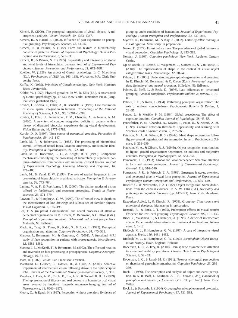
Kimchi, R. (2000). The perceptual organization of visual objects: A mi-crogenetic analysis. Vision Research, 40, 1333–1347.
Kimchi, R., & Hadad, R. (2002). Influence of past experience on percep-tual grouping. Psychological Science, 13, 41–47.
Kimchi, R., & Palmer, S. (1982). Form and texture in hierarchicallyconstructed patterns. Journal of Experimental Psychology: Human Per-ception and Performance, 8, 521–535.
Kimchi, R., & Palmer, S. E. (1985). Separability and integrality of globaland local levels of hierarchical patterns. Journal of Experimental Psy-chology: Human Perception and Performance, 11, 673–688.
Koehler, W. (1928). An aspect of Gestalt psychology. In C. Murchison(Ed.), Psychologies of 1925 (pp. 163–195). Worcester, MA: Clark Uni-versity Press.
Koffka, K. (1935). Principles of Gestalt psychology. New York: HarcourtBrace Jovanovich.
Kohler, W. (1950). Physical gestalten. In W. D. Ellis (Ed.), A sourcebookof Gestalt psychology (pp. 17–54). New York: Humanities Press. (Orig-inal work published 1920)
Kovacs, I., Kozma, P., Feher, A., & Benedek, G. (1999). Late maturationof visual spatial integration in humans. Proceedings of the NationalAcademy of Sciences, U.S.A., 96, 12204–12209.
Kovacs, I., Polat, U., Pennefather, P. M., Chandna, A., & Norcia, A. M.(2000). A new test of contour integration deficits in patients with ahistory of disrupted binocular experience during visual development.Vision Research, 40, 1775–1783.
Kurylo, D. D. (1997). Time course of perceptual grouping. Perception &Psychophysics, 59, 142–147.
Lamb, M. R., & Robertson, L. (1988). The processing of hierarchicalstimuli: Effects of retinal locus, location uncertainty, and stimulus iden-tity. Perception & Psychophysics, 44, 172–181.
Lamb, M. R., Robertson, L. C., & Knight, R. T. (1990). Componentmechanisms underlying the processing of hierarchically organized pat-terns—Inferences from patients with unilateral cortical lesions. Journalof Experimental Psychology: Learning, Memory, and Cognition, 16,471–483.
Lamb, M., & Yund, E. W. (1993). The role of spatial frequency in theprocessing of hierarchically organized structure. Perception & Psycho-physics, 54, 773–784.
Lamme, V. A. F., & Roelfsema, P. R. (2000). The distinct modes of visionoffered by feedforward and recurrent processing. Trends in Neuro-sciences, 23, 571–579.
Lawson, R., & Humphreys, G. W. (1999). The effects of view in depth onthe identification of line drawings and silhouettes of familiar objects.Visual Cognition, 6, 165–195.
Lee, T. S. (in press). Computational and neural processes of attentiveperceptual organization. In R. Kimchi, M. Behrmann, & C. Olson (Eds.),Perceptual organization in vision: Behavioral and neural perspectives.Mahwah, NJ: Erlbaum.
Mack, A., Tang, B., Tuma, R., Kahn, S., & Rock, I. (1992). Perceptualorganization and attention. Cognitive Psychology, 24, 475–501.
Marotta, J., Behrmann, M., & Genovese, C. (2001). A functional MRIstudy of face recognition in patients with prosopagnosia. NeuroReport,12, 1581–1582.
Marotta, J. J., McKeeff, T., & Behrmann, M. (2002). The effects of rotationand inversion on face processing in prosopagnosia. Cognitive Neuropsy-chology, 19, 31–47.
Marr, D. (1982). Vision. San Francisco: Freeman.Marstrand, L., Gerlach, C., Udesen, H., & Gade, A. (2000). Selective
impairment of intermediate vision following stroke in the right occipitallobe. Journal of the International Neuropsychological Society, 6, 381.
Mendola, J., Dale, A. M., Fischl, B., Liu, A. K., & Tootell, R. B. H. (1999).The representation of illusory and real contours in human cortical visualareas revealed by functional magnetic resonance imaging. Journal ofNeuroscience, 19, 8560–8572.
Moore, C., & Egeth, H. (1997). Perception without attention: Evidence of
grouping under conditions of inattention. Journal of Experimental Psy-chology: Human Perception and Performance, 23, 339–352.
Mycroft, R., Behrmann, M., & Kay, J. (2002). Letter-by-letter reading invisual agnosia. Manuscript in preparation.
Navon, D. (1977). Forest before trees: The precedence of global features invisual perception. Cognitive Psychology, 9, 353–383.
Neisser, U. (1967). Cognitive psychology. New York: Appleton CenturyCrofts.
Op de Beeck, H., Beatse, E., Wagemans, J., Sunaert, S., & Van Hecke, P.(2000). The representation of shape in the context of visual objectcategorization tasks. NeuroImage, 12, 28–40.
Palmer, S. E. (2001). Understanding perceptual organization and grouping.In R. Kimchi, M. Behrmann, & C. Olson (Eds.), Perceptual organiza-tion: Behavioral and neural processes. Hillsdale, NJ: Erlbaum.
Palmer, S., Neff, J., & Beck, D. (1996). Late influences on perceptualgrouping: Amodal completion. Psychonomic Bulletin & Review, 3, 75–80.
Palmer, S. E., & Rock, I. (1994). Rethinking perceptual organization: Therole of uniform connectedness. Psychonomic Bulletin & Review, 1,29–55.
Paquet, L., & Merikle, P. M. (1984). Global precedence: The effect ofexposure duration. Canadian Journal of Psychology, 38, 45–53.
Pennefather, P. M., Chandna, A., Kovacs, I., Polat, U., & Norcia, A. M.(1999). Contour detection threshold: Repeatability and learning with“contour cards.” Spatial Vision, 2, 257–266.
Peterson, M. A., & Gibson, B. S. (1994a). Must shape recognition followfigure–ground organization? An assumption in peril. Psychological Sci-ence, 9, 253–259.
Peterson, M. A., & Gibson, B. S. (1994b). Object recognition contributionsto figure–ground organization: Operations on outlines and subjectivecontours. Perception & Psychophysics, 56, 551–554.
Pomerantz, J. R. (1983). Global and local precedence: Selective attentionin form and motion perception. Journal of Experimental Psychology:General, 112, 516–540.
Pomerantz, J. R., & Pristach, E. A. (1989). Emergent features, attention,and perceptual glue in visual form perception. Journal of ExperimentalPsychology: Human Perception and Performance, 15, 635–649.
Ratcliff, G., & Newcombe, F. A. (1982). Object recognition: Some deduc-tions from the clinical evidence. In A. W. Ellis (Ed.), Normality andpathology in cognitive functions (pp. 147–171). New York: AcademicPress.
Razpurker-Apfeld, I., & Kimchi, R. (2003). Grouping: Time course andattentional demands. Manuscript in preparation.
Rensink, R., & Enns, J. T. (1995). Preemption effects in visual search:Evidence for low-level grouping. Psychological Review, 102, 101–130.
Ricci, R., Vaishnavi, S., & Chatterjee, A. (1999). A deficit of intermediatevision: Experimental observations and theoretical implications. Neuro-case, 5, 1–12.
Riddoch, M. J., & Humphreys, G. W. (1987). A case of integrative visualagnosia. Brain, 110, 1431–1462.
Riddoch, M. J., & Humphreys, G. W. (1993). Birmingham Object Recog-nition Battery. Hove, England: Erlbaum.
Robertson, L. C., & Ivry, R. (2000). Hemispheric asymmetries: Attentionto visual and auditory primitives. Current Directions in PsychologicalScience, 9, 59–63.
Robertson, L. C., & Lamb, M. R. (1991). Neuropsychological perspectiveson theories of part/whole organization. Cognitive Psychology, 23, 299–330.
Rock, I. (1986). The description and analysis of object and event percep-tion. In K. R. Boff, L. Kaufman, & J. P. Thomas (Eds.), Handbook ofperception and human performance (Vol. 33, pp. 1–71). New York:Wiley.
Rock, I., & Brosgole, L. (1964). Grouping based on phenomenal proximity.Journal of Experimental Psychology, 67, 531–538.
41VISUAL AGNOSIA AND PERCEPTUAL ORGANIZATION
Rock, I., Nijhawan, R., Palmer, S. E., & Tudor, L. (1992). Grouping basedon phenomenal similarity of achromatic color. Perception, 21, 779–789.
Sanocki, T., & Sellers, E. (2001). Shifting resources to recognize a formingobject: Dependencies involving object properties. Visual Cognition, 8,197–235.
Sasaki, Y., Hadjikani, N., Fischl, B., Liu, A. K., Marret, S., Dale, A. M.,& Tootell, R. B. H. (2001). Local and global attention are mappedretinotopically in human occipital cortex. Proceedings of the NationalAcademy of Sciences, U.S.A., 98, 2077–2082.
Sekuler, A. B., & Palmer, S. E. (1992). Perception of partly occludedobjects: A microgenetic analysis. Journal of Experimental Psychology:General, 121, 95–111.
Shipley, T. F., & Kellman, P. (1992). Perception of occluded objects andillusory figures: Evidence for an identity hypothesis. Journal of Exper-imental Psychology: Human Perception and Performance, 18, 106–120.
Shulman, G. L., Sullivan, M. A., Gish, K., & Sakoda, W. J. (1986). Therole of spatial-frequency channels in the perception of local and globalstructure. Perception, 15, 259–273.
Shulman, G. L., & Wilson, J. (1987). Spatial frequency and selectiveattention to local and global information. Neuropsychologia, 18, 89–101.
Snodgrass, S. G., & Vanderwart, M. A. (1980). A standardized set of 260pictures: Norms for name agreement, image agreement, familiarity, andvisual complexity. Journal of Experimental Psychology: Human Learn-ing and Memory, 6, 174–215.
Sugita, Y. (1999, September 16). Grouping of image fragments in primaryvisual cortex. Nature, 401, 269–272.
Treisman, A. (1982). Perceptual grouping and attention in visual search forfeatures and for objects. Journal of Experimental Psychology: HumanPerception and Performance, 8, 194–214.
Treisman, A., Kahneman, D., & Burkell, J. (1983). Perceptual objects andthe cost of filtering. Perception & Psychophysics, 33, 527–532.
Trick, L. M., & Enns, J. M. (1997). Clusters precede shapes in perceptualorganization. Psychological Science, 8, 124–129.
Vecera, S. P., & O’Reilly, R. (1998). Figure–ground organization andobject recognition processes: An interactive account. Journal of Exper-imental Psychology: Human Perception and Performance, 24, 441–462.
Vecera, S. P., & O’Reilly, R. C. (2000). Graded effects in hierarchicalfigure–ground organization: Reply to Peterson (1999). Journal of Ex-perimental Psychology: Human Perception and Performance, 26, 1221–1231.
von der Heydt, R., & Peterhans, E. (1989). Mechanisms of contour per-ception in monkey visual cortex. Journal of Neuroscience, 9, 1731–1748.
von der Heydt, R., Peterhans, E., & Baumgartner, G. (1984, June 15).Illusory contours and cortical neuron responses. Science, 224, 1260–1262.
Wertheimer, M. (1950). Untersuchungen zur Lehre von der Gestalt, II[Laws of organization in perceptual forms]. Psychologische Forschung,4, 301–350. (Original work published 1923)
Westheimer, G. (1999). Gestalt theory reconfigured: Max Wertheimer’santicipation of recent developments in visual neuroscience. Perception,18, 5–15.
Yovel, G., Yovel, I., & Levy, J. (2001). Hemispheric asymmetries forglobal and local visual perception: Effects of stimulus and task factors.Journal of Experimental Psychology: Human Perception and Perfor-mance, 27, 1369–1385.
Zhou, H., Friedman, H. S., & von der Heydt, R. (2000). Coding of borderownership in monkey visual cortex. Journal of Neuroscience, 20, 6594–6611.
Received July 3, 2001Revision received January 18, 2002
Accepted May 16, 2002 �
42 BEHRMANN AND KIMCHI