Embed Size (px)
Citation preview
15
Wernicke’s Encephalopathy
Radu Tanasescu1,2 et al.*
1Carol Davila University of Medicine and Pharmacy, Bucharest, 2Department of Neurology, Colentina Clinical Hospital, Bucharest
Romania
1. Introduction
Wernicke’s Encephalopathy (WE) is an underdiagnosed, potentially fatal, acute or subacute
neurologic disorder caused by the impairment of thiamine (vitamin B1) -dependent
enzymatic activity in susceptible brain cells. The biologically active form of thiamine (TH),
thiamine diphosphate (THDP), serves as a cofactor for several apoenzymes involved mainly
in the carbohydrate metabolism. Except for very rare cases, WE occurs in the presence of TH
deficiency, which is directly related to at least two other clinical entities: neurological
beriberi and cardiovascular beriberi. The preferential expression of one (or more) of these
entities may be the consequence of genetic polymorphism of genes encoding TH
transporters. The topography of WE brain lesions is highly specific, typically the
periventricular and periaqueductal grey areas being symmetrically involved. In the majority
of cases the early so-called ‘biochemical lesions’ are completely reversed if TH is promptly
supplied. However, if the THDP dependent enzymatic activity is not restored, irreversible
structural damage and eventually exitus may occur (Donnino, Vega et al. 2007; Hazell and
Butterworth 2009; Thorarinsson, Olafsson et al. 2011).
Carl Wernicke was the first to describe the clinical and neuropathological characteristics of
the encephalopathy that currently, according to the ICD-10 (WHO 2010), bears his name.
The first case compatible with WE was reported in 1822 by James Jackson. In the following
years Samuel Wilks (1868) and Charles Gayet (1875) encountered other similar cases. In
1881, Carl Wernicke published the cases of three patients - two alcohol abusing men, and a
young woman with persistent vomiting - that died within two weeks after the acute onset of
the nowadays considered classical triad – i.e. stance and/or gait ataxia, ocular motility sings
and mental-status changes - accompanied by similar fundoscopic modifications.
Neuropathological examinations were conducted. Wernicke considered that they all had the
same disease he named ‘polioencephalitis haemorrhagica superioris’ (Pearce 2008;
*Laura Dumitrescu2, Carmen Dragos3, Dimela Luca2, Alexandra Oprisan1,2, Catalina Coclitu4, Oana Simionescu2, Lorena Cojocaru2, Marius Stan2, Andreea Carasca2, Andreea Gitman2, Adela Chiru2 and Marina Ticmeanu1,2 1Carol Davila University of Medicine and Pharmacy, Bucharest, Romania 2Department of Neurology, Colentina Clinical Hospital, Bucharest Romania 3Department of Radiology, Coltea Clinical Hospital, Bucharest, Romania 4Department of Neurology, University Emergency Hospital, Bucharest, Romania
www.intechopen.com
Miscellanea on Encephalopathies – A Second Look
328
Thorarinsson, Olafsson et al. 2011). During the late 1880’s, Korsakoff published three
comprehensive series of cases presenting the characteristic features of the amnestic
syndrome that currently bears his name (i.e. Korsakoff syndrome/’psychosis’, KS),
commonly preceded or accompanied by a clinical picture highly suggestive of WE. He
considered that all these patients had the same disease he named ‘polyneuritic psychosis’.
The KS results from the bilateral dysfunction of the limbic system. As expected considering
the distribution of WE lesions, KS frequently accompanies WE. The association between WE
and KS was noticed and in this respect acute or sequelar WE with prominent KS and/or
post-WE KS are denominated by some Wernicke-Korsakoff syndrome (WKS). The etiology
and treatment of WE remained unknown until up to almost the half of the XXth century.
Beginning with Strauss (1935), Campbell (1940), Russell (1940) and Phillips (1952), who
established that TH deficiency is directly related to the development of WE, and with
Williams and Cline (1936), who published the first correct TH chemical formula and
synthesis pathway, remarkable progress has been made in understanding the spectrum of
diseases caused by TH deficiency (Phillips, Victor et al. 1952; Donnino, Vega et al. 2007).
Currently, WE is a highly and easily treatable disease frequently associated with alcoholism
and/or malnutrition related TH deficiency. The diagnosis remains mainly clinical as none of
the modern diagnostic tools has adequate sensitivity and/or specificity. The classical triad is
encountered in less than a third of cases and may be completely absent. The clinical picture
may vary from the classical signs to hypotension or coma. Unfortunately, the misconception
that WE is a rare and stereotypic disease occurring only in malnourished alcoholics is still
found in clinical practice, frequently leading to the delay or even failure of diagnosis
(Tanasescu 2009).
The following chapter provides a thorough overview of WE, with updates on the recent epidemiological and etiophysiopathological data. A brief reference to the possibility of effective prophylaxis is made. The diagnosis and treatment are discussed. The relationship with other TH deficiency diseases, the particularities of alcohol versus non-alcohol related cases and the correlations with KS are underlined. Considering the deleterious consequences of untreated WE, the importance of maintaining a high index of suspicion for diagnosis and a low threshold for parenteral TH administration - as recommended by the 2010 EFNS guideline - is advocated (Galvin, Brathen et al. 2010).
2. Epidemiology
WE epidemiological data are scarce and mostly based on necroptic class IV studies
conducted in the developed countries. However the literature is abundant in case reports
and to a lesser extent in small retrospective clinical studies. WE occurs throughout the
world, but, even if considering the probable report bias, appears to have uneven geographic
distribution (Galvin, Brathen et al. 2010).
According to the retrospective clinical studies available, the prevalence of WE is lower than
0.13%. Nevertheless, necropsies of the general population reveal brain lesions consistent
with WE in 0.4 to 2.8% of the cases (average 1.3%), the highest rates being reported in West
Australia during the 1970’s (Harper 1979; Harper 1983; Harper, Giles et al. 1986). In prior
alcohol abusers the prevalence of neuropathology confirmed WE is reported to be around
12% and raises up to 30% in those associating cerebellar atrophy. If considering only those
www.intechopen.com
Wernicke’s Encephalopathy
329
with alcohol related deaths, WE prevalence may reach 59% (Victor, Adams et al. 1971;
Thomson and Marshall 2006). Other populations with significantly higher WE prevalence
were identified, necroptic changes compatible with WE being found in approximately 10%
of the AIDS patients and in 6% of those that underwent bone marrow transplant
(Butterworth, Gaudreau et al. 1991; Boldorini, Vago et al. 1992; Donnino, Vega et al. 2007).
The correct diagnosis of WE is made prior to necropsy in only up to 25% of the adult cases
(values as low as 1% being reported!), and in approximately 40% of the pediatric cases). Up
to 70% of the alcohol related cases and almost 95% of the non alcohol related cases are not
diagnosed prior to death (Victor, Adams et al. 1971; Harper, Giles et al. 1986; Harper, Gold
et al. 1989; Torvik 1991; Vasconcelos, Silva et al. 1999). AIDS patients seem to be the category
most often misdiagnosed (Butterworth, Gaudreau et al. 1991). Considering the discrepancy
between the prevalence of the clinic versus necroptic diagnosis (0.4-2.8% versus 0.04-0.13%),
WE appears to be underdiagnosed during lifetime (Victor, Adams et al. 1971). Since
necropsy studies may be biased towards preferentially identifying the more severe cases,
the prevalence of WE may be even higher than the one predicted by the necroptic studies
(Galvin, Brathen et al. 2010). However, some have suggested that the histopathological
changes may precede the clinical onset of disease, and thus the necropsies of the general
population may also identify mild subclinical cases (Caine, Halliday et al. 1997). It was
estimated that 13 to 35% of alcoholics and up to 1.5% of the non-alcoholics develop WE
(Harper, Rodriguez et al. 1988). The incidence and prevalence of WE are considered to be at
least 10 times higher in alcoholics than in non-alcoholics (Harper 2006). In order to have a
clearer picture, the 2010 EFNS guideline recommends performing necropsies with detailed
neuropathological examination in all patients dying from diseases suggestive of WE
(Galvin, Brathen et al. 2010).
Besides exceptionally rare cases, all WE patients have TH deficiency. According to the
available data, TH deficiency is not uncommon in the developed countries. In the UK,
approximately 20% of the patients admitted to the emergency departments had TH
deficiency (Jamieson, Obeid et al. 1999). In the USA, 8 to 31% of the elderly living at home
and 23 to 40% of those in nursing homes had TH deficiency (Harper 2006). In Canada, TH
deficiency was found in almost 13% of the critically ill children (Fattal-Valevski 2011).
During the 1992-1993 Cuban neuropathy epidemic the local prevalence of TH deficiency
ranged from 30% in some regions to 70% in others (Macias-Matos, Rodriguez-Ojea et al.
1996). Among chronic alcoholics, 25 to 80% may have a certain degree of TH deficiency
(Caine, Halliday et al. 1997). Several studies on AIDS patients found TH deficiency in 10 to
23% of the cases (Davtyan and Vinters 1987; Foresti and Confalonieri 1987; Hutchin 1987).
According to the WHO’s report on TH (WHO 1999), in South-East Asia TH deficiency has a
high prevalence, while in Africa and Central and North America it has a low prevalence.
Chronic ethanol consumption and/or malnutrition have a significant impact on the
incidence and prevalence of WE, up to 90% of the WE patients having TH deficiency in this
context (Antunez, Estruch et al. 1998). Several important TH deficiency epidemics have been
recorded by the modern history. At the beginning of the XXth century the introduction of
the large scale use of cheap polished rice in urban South-East Asia led to several great
outbreaks of TH deficiency associated with beriberi. During the last two decades of the XXth
century TH deficiency epidemics were recorded in Thailand, Guinea, Djibouti, East Ethiopia
and Nepal, especially among political refugees (WHO 1999). In 2003 a TH deficiency
www.intechopen.com
Miscellanea on Encephalopathies – A Second Look
330
outbreak affecting infants fed with a soy milk formula with no detectible TH content
emerged in Israel (Fattal-Valevski, Kesler et al. 2005). To the best of our knowledge, WE-
related epidemiologic data from the developing or underdeveloped countries are not
available. Approximately 90% of the WE cases occurring in the developed countries are
alcohol related (Thomson 2000). A correlation between the per capita alcohol consumption
and the prevalence of WE could not be established (Torvik 1991). The male to female ratios
range from 1.7:1 to 3:1 in necroptic studies, and were reported to be 5:1 in a large clinical
study (Victor, Adams et al. 1971; Harper 1979; Victor 1989; Rolland and Truswell 1998). WE
may affect individuals of any age but appears to have a higher prevalence during the fifth
decade of life (Vasconcelos, Silva et al. 1999). A trend towards an increased incidence and
prevalence of WE has recently been observed in USA, UK and Japan, possibly related to the
increased number of bariatric surgery interventions and the persistent shortages in
intravenous vitamins, to the restriction of parenteral TH administration due to fear of
anaphylaxis (leading to the routine prophylaxis in hospitalized alcoholics with per os
instead of parenteral thiamine) and respectively to the restriction of parenteral vitamins
supplementation by a healthcare policy (Ramayya and Jauhar 1997; Hahn, Berquist et al.
1998; Shikata, Mizutani et al. 2000; Feeney and Connor 2008). Several regional socio-
economic and cultural particularities along with local health care related factors appear to
have important influences on the prevalence and prognosis of WE. Considering the
important burden that untreated WE puts on health care systems worldwide (Galvin,
Brathen et al. 2010), further epidemiological studies are needed in order to better define the
populations at risk and to identify efficient prophylactic approaches.
3. Etiology
WE is caused by the disruption of the THDP dependent enzymatic activity in susceptible
brain cells, commonly secondary to TH deficiency. This correlates directly with three
pathogenic entities: WE, cardiovascular beriberi, and neuropathic beriberi. Recently, two
other conditions that seem to be directly related to TH deficiency have been described:
African (Nigerian) seasonal ataxia and gastrointestinal beriberi (Adamolekun and Ndububa
1994; Nishimune, Watanabe et al. 2000; Donnino 2004). TH deficiency seems also to be
involved in other diseases like Strachan syndrome (i.e. polyneuropathy, optic neuropathy,
orogenital ulcerations), ‘tobacco-alcohol amblyopia’, tropical ataxic neuropathy (i.e. sensory
neuropathy, optic neuropathy, sensoneural deafness), Marchiafava-Bignami disease,
subacute ‘alcoholic’ cerebellar degeneration and epidemic spastic paraparesis (konzo). TH
competitive antagonists and/or impaired TH to THDP conversion may lead to WE even in
the presence of normal TH blood levels. Impaired apoenzyme activation due to magnesium
(Mg) deficiency, and/or decreased activity of the THDP dependent enzymes may condition
the degree of susceptibility to borderline low levels of THDP. Due to physiologic
particularities not all tissues are equally susceptible, the nervous system and the cardiac
muscle being the most vulnerable. The pattern of cellular susceptibility may be influenced
by genetic and environmental factors, including the nutritional and hormonal status, and
seems not to be homogenously represented among individuals, thus possibly explaining the
preferential expression of one (or more) of the potential pathologic entities related to TH
deficiency (Zhao, Gao et al. 2002).
www.intechopen.com
Wernicke’s Encephalopathy
331
3.1 Thiamine
TH is a water soluble heat labile quaternary ammonia compound, containing an aminopyrimidine ring linked by a methylene bridge to a thiazole ring. It is synthesized by different biochemical reactions in fungi, bacteria, plants and some protozoa, but not in humans (Fattal-Valevski 2011). In the human body it is found as unphosporilated TH (i.e. free TH) and as phosphorilated derivates: TH monophospate (THMP), TH diphospate (THDP, aka TH pyrophosphate), and TH triphospate (THTP). Intracellularly, free TH is converted by thiaminpyrophosphokinase into THDP, in a process requiring Mg as cofactor. Three plasma membrane bidirectional transporters for TH and TH derivates have been described: TH transporter 1 (THTR1) encoded by the SCL19A2 gene (location 1q23.3), TH transporter 2 (THTR2) encoded by the SCL19A3 gene (location 2q37) and reduced folate carrier transporter 1 (RFC1) encoded by the SCL19A1 gene (location 21q22.3). A mitochondria membrane transporter for THDP (i.e. mitochondria membrane THDP transporter) encoded by the SCL25A19 has also been described. THTR1 and THTR2 transport free TH. THTR1 seems to be highly expressed in skeletal and heart muscle and to lesser degrees in placenta, liver, kidneys, small intestine and lungs. THTR2 seems to be highly express in the placenta, kidneys, liver and thalamus and also in the small intestine. RFC1 is mainly a folate transporter, but also transports THDP and THMP. Considering that THDP is found exclusively intracellularly, RFC1 transports THDP only from the intracellular to the extracellular space (where THDP is rapidly converted to free TH). In the presence of low free TH and THMP plasma levels the cells that highly express RFC1 on their membranes have an overall negative TH balance, exporting THDP without importing free TH or THMP. RFC1 is highly expressed on the apical brush border of the choroid plexus. The pattern of distribution of the TH and TH derivates transporters may play a significant role in establishing and maintaining the tissue distribution of TH (Boulware, Subramanian et al. 2003; Subramanian, Marchant et al. 2003; Said, Balamurugan et al. 2004; Miyajima and Kono 2010). The intestinal absorption of TH occurs mainly in the proximal small intestine by an active saturable mechanism and probably also by passive diffusion. At the intestinal brush border TH is mainly found in its free form. TH absorption is enhanced by TH deficiency and reduced by thyroid hormones, ethanol exposure, low temperature and TH analogs. TH absorption may also be reduced in those with diabetes mellitus or advanced aged. At low concentration (<2 microM/liter) TH absorption is an active, rate-limited process, involving the high affinity THTR2 and, to a lesser extent, THTR1. At high intestinal TH concentration (5-50 microM/liter) TH seems to be absorbed through passive diffusion. It has been reported that under physiological circumstances, even when large TH quantities are administered, no more than 4.5 to 5 mg can be absorbed from a single oral dose. TH has restricted distribution. Up to 90% of the circulating TH is found in the red cells (mostly as THDP), the rest being found in the other blood cells and in plasma, mainly bound by proteins, as free TH or THMP (Dudeja, Tyagi et al. 2001; Martin, Singleton et al. 2003). Under physiologic circumstances, TH is excreted renally. In the presence of high plasmatic TH concentration rapid renal excretion as free TH occurs. After the intravenous administration of 50 mg of TH hydrochloride the plasma half-time is about 96 minutes. Thus, for correcting TH deficiency the administration of parenteral TH in many smaller doses rather than in an equivalent single dose seems justified (Donnino; Boulware, Subramanian et al. 2003). The blood brain barrier (BBB) allows the passage of free TH and THMP through both active and passive mechanisms. Active passage occurs at low TH
www.intechopen.com
Miscellanea on Encephalopathies – A Second Look
332
serum concentrations. At high serum concentrations, free TH passes the BBB passively, driven by the existing concentration gradient. The intravenous administration of TH provides a superior concentration gradient that facilitates passive diffusion (Thomson, Cook et al. 2002). Several natural and synthetic TH structural analogues having different pharmacological profiles exist. Pyrithiamine and oxythiamine act as competitive antagonists. Pyrithiamine passes the BBB and thus is useful for inducing TH deficiency encephalopathy, the experimental model of WE. TH hydrochloride and TH mononitrate are water soluble TH salts that act as TH agonists and have similar bioavailability, distribution and excretion with TH. Allithiamine is a naturally occurring lipophylic TH analogue resulting from the enzymatic conversion of TH in the freshly crushed bulbs of garlic and other alli plants. Thiaminetetrahydrofufuryl disulfide is a synthetic analogue of allithiamine. Prosultiamine and sulbutiamine are lipid-soluble synthetic TH analogues. All these lipophylic TH derivates have better bioavailability and BBB penetrability than TH. Benfotiamine is a synthetic TH analogue that has better bioavailability and cellular penetrability than TH, but does not pass the BBB (Baker and Frank 1976; Kitamori and Itokawa 1993). TH may have a structural role as part of the cellular membranes, and may be involved in the synaptic transmission, cellular differentiation, axonal growth, myelinogenesis and regulation of brain development during fetal and early postnatal life. To the best of our knowledge THMP and THTP have no clearly identified metabolic or structural roles (Makarchikov, Lakaye et al. 2003). THDP serves as a cofactor for several apoenzymes involved in the carbohydrate metabolism: apo-alpha-ketoglutarate dehydrogenase (aKGDH), apo-pyruvate dehydrogenase (PDH) and apo-transketolase (TK). Mg is the second cofactor required by these apoenzymes, especially by apo-TK. aKGDH and PDH are mitochondrial enzymes important for the tricarboxylic acid cycle (TAC, i.e. Krebs cycle), though the latter is not part of it. TK is a cytosolic enzyme involved in the non-oxidative phase of the pentose-phosphate pathway (PPP or hexose monophosphat shunt). It has been shown that TH deficiency inhibits the expression of the genes encoding TK and PDH (Pekovich, Martin et al. 1998; Donnino, Vega et al. 2007). Under physiologic circumstances, almost 30% of the brain glucose is metabolized to pyruvate. In the absence of a functional PDH complex and Krebs cycle pyruvate is reduced to lactate (Ishii, Sarai et al. 1979). The human body has TH deposits ranging from 25 to 50 mg, commonly corresponding to the amount of TH required for 18 to 42 days. Most of the TH is stored in the liver as THDP. Food sources of TH are cereals, beans, nuts, brown (unpolished) rice and meat. Polished (white) rice, highly purified cereals and excessively cooked food may contain no TH. The daily TH requirements for a healthy adult may range from 1 to 2 mg and depend on the carbohydrate intake and on several metabolic factors. According to the current literature, the TH intake should be of at least 0.33 mg per 1000 kcal, ideally 0.5 mg per 1000 kcal, but no less than 1 mg per day. Some recommend daily intakes above 1.1 mg for adult women and 1.2 mg for adult men, even if the corresponding caloric intake is lower. A balanced diet usually contains the recommended quantity. No upper tolerability limit intake has been established for TH and to the best of our knowledge no cases of oral TH toxicity have been described (WHO 1999; Thomson and Marshall 2006; Sechi and Serra 2007; Fattal-Valevski 2011).
The TH content of pharmacological or biological samples may be measured directly by several methods including spectrophotometry, spectrofluorometry, various techniques of high performance liquid chromatography (HPLC), capillary electrophoresis and voltametry.
www.intechopen.com
Wernicke’s Encephalopathy
333
The classical method used to assess the human THDP status is the estimation of the effect of THDP on the erythrocyte TK activity (ETKA). A low ETKA with more than 25% increase after THDP adding confirms THDP erythrocyte deficit, and though indirect, has good sensibility, specificity and reproducibility in estimating the whole blood total TH and the erythrocyte THDP levels. However, since it is laborious, it has been replaced by the direct measurement of the total TH (free TH and its phosphatesters) or of THDP in the whole blood using various techniques of HPLC. The whole blood THDP levels correlate well with erythrocyte THDP levels providing the correction with the haemoglobin level is made. The measurement of whole blood THDP was suggested to be the most suitable method for use in clinical practice. (Lee, Ong et al. 1991; Tallaksen, Bell et al. 1993; Herve, Beyne et al. 1994). In the apparently healthy human adults the THDP and total TH blood levels range within nanomoles per liter levels, specific values depending upon the technique used. In animal studies the lethal blood TH level ranges from 7.2 to 10 mg/dl. Death occurs due to respiratory failure. If respiratory support is provided blood levels as high as 36.9 mg/dl are tolerated (Smith, Foa et al. 1947; Galvin, Brathen et al. 2010).
3.2 Predisposing factors
TH deficiency is the predisposing factor most frequently associated with WE. TH deficiency is the consequence of one or more of the following mechanisms: inadequate dietary intake (absolute or relative), impaired intestinal absorption, impaired storage, excessive elimination and/or increased metabolic requirements. Impaired TH intestinal absorption may occur due to gastrointestinal diseases, protein-caloric malnutrition (decrease in the active TH absorption) and/or ingestion of certain substances (e.g. ‘anti-TH factors’, antacids, phenytoin, cephalosporins, tetracycline). Impaired TH storage may occur due to chronic liver disease. Excessive renal elimination may occur due to renal disease, use of certain drugs and/or impaired TH storage. In those already marginally deficient WE may be precipitated by an event that rapidly increases the metabolic requirements of TH. In most of the cases, TH deficiency may be traced back to improper diet. Regardless of the cause, unbalanced nutrition persisting for more than 14 to 21 days, or even less in those already marginally deficient or with higher demands, may lead to TH deficiency. In healthy adults, intakes of less than 0.2 mg per 1000 kcal or of less than 0.66 mg per day lasting for several weeks lead to clinically manifest TH deficiency. Diets rich in the so-called ‘anti-TH factors’ (i.e. ‘thiaminases’ and dietary ‘TH antagonists’) may result in TH deficiency. The thiaminases are heat labile enzymes that disintegrate TH (found in raw or fermented fish, shellfish, ferns and certain bacteria) or reduce its intrinsic activity (found in certain bacteria). The dietary ‘TH antagonists’ are heat stable non-enzymatic substances that interfere with the intestinal absorption of TH, including polyphenols (e.g. caffeic acid, chlorogenic acid, tannic acid, tartaric acid, citric acid, ascorbic acid which are found in tea, coffee, betel nuts, red currants), flavonoids (quercetin and rutin, found mainly in fruits), haemin (found in animal tissues) and sulphites in high amounts (WHO 1999; Thomson and Marshall 2006; Fattal-Valevski 2011). Gender may influence the risk of developing WE, possibly because of genetic differences but also because of gender-related environmental factors. No definite race predisposition has been described, but a population-specific susceptibility has been reported: it seems that Asians with TH deficiency are prone to cardiovascular beriberi, while Europeans with TH deficiency are more likely develop neurological beriberi and/or WE (Sechi and Serra 2007).
www.intechopen.com
Miscellanea on Encephalopathies – A Second Look
334
Chronic ethanol abuse is the condition most frequently associated to WE. Alcoholics may have higher TH demands, TH being necessary for the metabolism of ethanol. They frequently have TH intake below 0.29 mg per 1000 kcal and associate Mg depletion (Thomson 2000). They may have impaired TH absorption secondary to ethanol-induced intestinal mucosa damage, impaired transmembrane transport due to folate or other B vitamins deficiency, decreased intestinal ATP-ase activity and reduced expression of the THTR1 and THTR2 encoding genes (Hoyumpa 1980; Subramanya, Subramanian et al. 2010). The type of alcoholic beverage consumed may have an influence (Lemos, Azevedo et al. 2005). TH malabsorption seems to be reversible providing ethanol consumption stops (Bujanda 2000). Considering that not all alcoholics with similar nutritional status develop WE, it may be speculated that other environmental and/or genetic factors may interfere (Mukherjee, Svoronos et al. 1987). Physiological hypercatabolic states like infancy, pregnancy and lactation may predispose to TH deficiency. A particular situation is that of hyperemesis gravidarium. It has been reported that vomiting persisting for more than three weeks and elevated transaminase levels highly correlate with the occurrence of WE in pregnant women (Rotman, Hassin et al. 1994 ). The infants fed by TH deficient mother or by TH deficient milk formula develop TH deficiency. Pathological hypercatabolic states may predispose to TH deficiency not only because of increase TH requirements but also because they are frequently associated with improper nutrition, impaired intestinal absorption, persistent vomiting and use of drugs that may interfere with TH utilization (Otsuka, Tada et al. 1997; Sechi and Serra 2007). In children, malignancy has been reported to be the condition most frequently associated to WE (Vasconcelos, Silva et al. 1999). Gastrointestinal surgery that removes or by-passes the parts of the gastrointestinal system involved in TH absorption is an important predisposing factor for TH deficiency. Bariatric surgery has been identified as predisposing factor in a significant number of the recently reported WE cases. Persistent vomiting may lead to TH deficiency if adequate parenteral supplementation is not provided (Singh and Kumar 2007). Chronic liver disease (occurring in up to half of the alcoholics developing WE) may lead to TH deficiency due to impaired storage. Hemodialysis and peritoneal dialysis have been reported to increase TH elimination (Sun, Yang et al. 2006; Ueda, Utsunomiya et al. 2007). Uremic encephalopathy may cause impaired cerebral TK activity and thus may predispose to WE (Brouns and De Deyn 2004). High doses of intravenous glucose may precipitate WE in marginally TH deficient individuals. Refeeding, hyperalimentation and total parenteral nutrition without adequate TH supplementation may also precipitate iatrogenic WE (Watson, Walker et al. 1981). Drugs such as nitroglycerin and tolazamide may may play a role in the development of WE in susceptible individuals (Sechi and Serra 2007). The chronic use of metronidazole may predispose to WE due to its conversion into a TH analogue that acts as a TH competitive antagonist (Alston and Abeles 1987). The chemotherapeutic drugs 5-fluorouracil, cisplatin, erbulozole and ifosfamide seem to interfere with TH pharmacokinetics, predisposing to WE (Van Belle, Distelmans et al. 1993; Kondo, Fujiwara et al. 1996; Hamadani and Awan 2006; Cho, Chang et al. 2009). WE occurring during the chronic use of tolazamide, a sulfonylurea blood glucose lowering drug that might increase the intracellular demands of TH, has been reported (Kwee and Nakada 1983).
Several genetic factors seem to predispose to the development of WE. The occurrence of WE
is more frequently encountered in both monozygotic twins than in both heterozygote twins
(Martin, Singleton et al. 2003), but to the best of our knowledge, no significant family
www.intechopen.com
Wernicke’s Encephalopathy
335
aggregation has been reported. The function and/or upregulation of the receptors
responsible for the intestinal and renal uptake of TH may be genetically impaired in some
individuals who develop WE. Some reported that the presence of a genetically conditioned
low affinity TK may predispose to WE (Mukherjee, Svoronos et al. 1987). Since no
differences in the nucleotide sequence of the encoding gene or in the amino acid sequence
were identified, it has been proposed that the biochemical difference in the activity of TK
may be caused by posttranscriptional changes or by differences in the three-dimensional
conformation (McCool, Plonk et al. 1993). Another possible genetic factor predisposing to
WE is the mutation of the X-linked transketolase-like 1 gene (Coy, Dubel et al. 1996). Genetic
variants of the enzymes involved in the metabolism of ethanol may also predispose to WE
(Sechi and Serra 2007). Mutations in an untranslated regulatory region of SLC19A2 gene
(also involved in TH-responsive megaloblastic anemia) seem to be involved in the genetic
predisposition to WE (Guerrini, Thomson et al. 2005). These genetic defects might explain
the inability of certain individuals to cope with borderline-low TH deficiency. A WE-like
phenotype caused by defects of the SLC19A3 gene (typically involved in childhood onset
biotin-responsive basal ganglia disease) has been reported in two Japanese brothers. Both of
them were compound heterozygote for the K44E and E320Q mutations. These mutations
were not found in 192 ethnically matched controls (Kono, Miyajima et al. 2009).
In conclusion, one or more genetic mutations occurring in the same individual probably cause subtle alterations in the neuroglial and/or neuronal TH transporter systems and/or in the activity of THDP dependent enzymes, that in the presence of absolute or relative THDP deficiency and/or Mg deficit lead to the development of WE (Thomson and Marshall 2006).
4. Neuropathology
The anatomical pathology of WE is well described, mostly due to the large number of necroptic studies performed. The macroscopic and microscopic characteristics depend on the stage and severity of the disease (Sechi and Serra 2007). WE may coexist with typical hepatic encephalopathy findings, the neuropathologic differential diagnosis being sometimes difficult (Caine, Halliday et al. 1997). To the best of our knowledge, WE electronic microscopy data are not available. Gross findings consist of bilateral symmetrical grayish discoloration, congestion and recent petechial hemorrhages involving the periaqueductal grey matter, mamillary bodies, and medial thalamus. The most frequent lesion observed (up to 75% of the cases) is spongy or granular brown-grayish discoloration of the thalamus (Victor, Adams et al. 1971). The presence of punctuate hemorrhages in the mamillary bodies is highly specific (Thorarinsson, Olafsson et al. 2011). Larger hemorrhages (up to 8 mm in diameter) found in the vicinity of the third and the fourth ventricle have been reported in at least two cases with otherwise typical histopathological and clinical presentation (Rosenblum and Feigin 1965; Vortmeyer, Hagel et al. 1992). Rarely, discoloration may be observed in the reticular formation of the midbrain, corpora quadrigemina and in the cortex. The cerebellum may show atrophy of the vermis. Typically the brain has normal weight, though in chronic alcoholics significant atrophy may exist. Its surface has normal appearance. The lateral ventricles may sometimes be dilated, most likely secondary to chronic alcohol abuse-related atrophy (Victor, Adams et al. 1971; Harper 1979). Atrophy of the corpus callosum has been reported in both alcohol and non alcohol
www.intechopen.com
Miscellanea on Encephalopathies – A Second Look
336
related WE. The extent and the location of callosal atrophy seems to vary in relationship with alcohol consumption (Lee, Jung et al. 2005). In a significant number of cases gross examination alone does not reveal any lesions (Harper 1979; Donnino, Vega et al. 2007).
Microscopically, the typical aspect of the WE lesions consists of symmetric microhemorrhagic and/or necrotic lesions and microglia proliferation affecting symmetrically the cerebral midline regions, mainly the thalamus, mamillary bodies, periaqueductal region, hypothalamus, cerebellar vermis, proximity of the third ventricle and the floor of the fourth ventricle (Victor, Adams et al. 1971; Fattal-Valevski 2011). The distribution of the lesions is highly localized. The medial dorsal thalamic nucleus and the mamillary bodies are affected in virtually all patients (Victor, Adams et al. 1971). The locus ceruleus, oculomotor and vestibular nuclei and the medial aspect of the thalamus are also frequently involved. In the most severe cases extensive necrosis of the affected areas is observed. In the mild cases only loss of myelin and to a lesser degree of neuronal bodies is noticed. The number of astrocytes and macrophages is commonly increased. Focal microhemorrhages are sometimes found. Macrophages containing hemosiderin (thus indicating previous hemorrhage) may be encountered. Sometimes lesions consisting of patchy or diffuse neuronal loss and Alzheimer type II astrocytic proliferation (typically seen in hepatic encephalopathy) are found in the hippocampus, fornix, septal regions and cerebral cortex. The acute WE lesions are characterized by vascular congestion, petechial hemorrhages and astrocytes swelling affecting mainly the brainstem and the thalamus. The older lesions are characterized by demyelination, gliosis, edema and loss of neuropils in spite of the relative preservation of neurons (Sechi and Serra 2007). The capillaries are dilated and are surrounded by edema and microhemorrhages. Some have observed capillary proliferation, while others did not. Cerebellar vermis lesions compatible with those found in the alcoholic cerebellar degeneration – i.e. selective loss of Purkinje cells - are found in about half of the cases (Thorarinsson, Olafsson et al. 2011). Edema, microhemorrhages and possible necrosis involving the optic nerve may rarely be found (Li and Rucker 2010). The chronic lesions usually affect the mamillary bodies and the dorsomedial thalamic nuclei. Atrophy of the mamillary bodies is highly specific for the sequelae of WE, being found even by macroscopic examination in the majority of the cases. Widening of the third and fourth ventricle and of the aqueduct is also observed in the late and sequelar stages. Microscopically there is proliferation of astrocytes, tissue destruction and gliosis, while the capillary endothelium is normal and microhemorrhages are absent (Donnino, Vega et al. 2007; Thorarinsson, Olafsson et al. 2011). Swelling, disruption and hyperplasia of the choroid plexus has been reported in AIDS patients with WE (Boldorini, Vago et al. 1992). Frequently the peripheral nerves have identical aspect with that seen in beriberi, i.e. distal demyelination. The spinal cord may be affected, a decrease in the anterior horn cells and sometimes involvement of the anterior and posterior roots being encountered (Sechi and Serra 2007).
5. Physiopatogeny
In spite of the fact that an animal model is easily designable the physiopathological
pathways that lead to WE are incompletely understood. The disruption of the THDP
dependent enzymatic activity in WE susceptible individuals results in highly localized
specific metabolic dysfunction corresponding to the so-called reversible ‘biochemical
www.intechopen.com
Wernicke’s Encephalopathy
337
lesions’. Providing the disruption is not promptly restored, the ‘biochemical lesions’ are
replaced by irreversible structural damage (i.e. necrosis). The typical lesions are located
symmetrically in the periventricular and periaqueductal grey areas. The clinical picture is
highly correlated with the topography of the lesions (Hazell and Butterworth 2009). If the
THDP dependent enzymatic activity is resumed WE is cured (with or without sequelae).
Otherwise, exitus commonly occurs (Sechi and Serra 2007). WE develops rapidly, being
usually induced by severe short-term TH deficiency. Persistent or recurring mild THDP
deficiency may lead to a chronic evolution (Thomson and Marshall 2006). The reason for the
specific selective topographic distribution of the WE lesions is still a matter of debate. A
high cellular specificity seems to exist, the astrocytes being the most susceptible to THDP
deficiency. The degree of activity reduction seems to be different for each THDP-dependent
enzyme and strongly related to the cell’s type. Intuitively, one may assume that the most
affected brain regions are those with higher metabolic demands, and thus with higher TH
requirements. Nevertheless, the cortex is most often spared (Butterworth, Kril et al. 1993;
Hazell 2009). Some have proposed that the periventricular areas are affected to a greater
degree due to the parenchyma consequences of the high CSF glutamate levels (Nixon 2008).
Others have observed that the occurrence of adult neurogenesis may be one of the main
differences between the affected regions and the cortical areas, rendering the former more
susceptible (Zhao, Pan et al. 2009). Pre and post-transcriptional regulation of the genes
encoding the THDP-dependent apoenzymes may possibly be involved (Hazell 2009; Hazell
and Butterworth 2009).
A chronologic sequence of the physiopathological changes encountered in WE has been proposed. Accordingly, after about 4 days of THDP deficiency the activity of the astrocytic aKGDH decreases resulting in cytotoxic edema. After 7 to 10 days a decrease in the activity of the astrocytic TK occurs. The astrocytic dysfunction leads to the increase in extracellular glutamate levels (resulting in excitocytotoxicity), accumulation of free radicals and cytokines and loss of the osmotic gradients. Endothelial cell dysfunction resulting in increase nitric oxide (NO) production occurs. The BBB is disrupted and glial and neuronal vasogenic edema appears. After about 14 days focal lactic acidosis, neuronal DNA fragmentation and neuronal necrosis occur (Sechi and Serra 2007). According to a recent in vivo animal imagistic study the first observable consequence of TH deficiency may be the dysfunction the choroid plexus leading to blood-CSF barrier alteration (Nixon, Jordan et al. 2008). The earliest biochemical change reported in experimental WE animal models consists of decreased astrocyte aKGDH activity and the first histopathological finding observed is exclusive neuroglial damage (Butterworth 1986). The THDP deficiency has a profound effect on the functional integrity of the astrocytes. One of the consequences of the decreased aKGDH activity is the impairment of the Krebs cycle leading to cellular energetic failure. Increase oxidative stress and lactate production occur, the latter leading to focal acidosis. Increased lactic acid levels are observed in the areas which subsequently develop histological lesions and the magnetic resonance spectroscopy studies demonstrate a characteristic lesional lactate peak (Butterworth 1989; Pannunzio, Hazell et al. 2000; Donnino, Vega et al. 2007; Sullivan and Pfefferbaum 2009). The disturbed function of the astrocytic membrane results in the alterations of the ionic and osmotic gradients, and thus, in cytotoxic edema. The impairment of the astrocyte function along with the subsequent endothelial dysfunction causes BBB dysfunction that leads to vasogenic edema (Hazell and Butterworth 2009). The endothelial dysfunction causes increased production of NO and
www.intechopen.com
Miscellanea on Encephalopathies – A Second Look
338
cytokines, the former exacerbating the BBB dysfunction (Sechi and Serra 2007). The astrocyte dysfunction leads to increased extracellular glutamate levels mainly due to the suboptimal astrocyte uptake, the oxidative stress leading to the downregulation of the astrocytic glutamate 1 (GLT1) and glutamate-aspartate transporters (Langlais and Zhang 1993; Danbolt 2001; Hazell, Rao et al. 2001; Nixon 2008; Hazell and Butterworth 2009). The accumulating extracellular glutamate leads to N-methyl D-aspartate receptor (NMDA-R) mediated excitocytotoxicity resulting in neuronal loss (Todd and Butterworth 1998). Several studies have provided strong evidence for the presence of excitotoxic mediated cell death in TH deficient brains (Hazell, Butterworth et al. 1993; Langlais and Zhang 1993). The glutamate neuronal overstimulation leads to the accumulation of high extracellular potassium (K) levels. The elevated K is uptaked from the extracellular space by the astrocytes which consequently swell even more via osmosis. This leads to the further impairment of the astrocytes function which become unable to adequately buffer the accumulating extracellular glutamate and K, with deleterious consequences on the surrounding neurons (Kimelberg, Goderie et al. 1990; Kimelberg, Rutledge et al. 1995). The excess of glutamate is also removed from the interstitial fluid by passive diffusion in the CSF followed by choroid plexus active clearance. Decreased aKGDH activity in the ependymal and endothelial cells of the choroid plexus may lead to impairment of the energy dependent processes, and thus impairment of the CSF glutamate clearance. Therefore, as already mentioned, the glutamate levels may be additionally increased in the periventricular areas. The excessive presence of CSF glutamate may lead to further impairment of the choroid plexus activity (Nixon 2008). Increased aquaporin 4 (AQP4) gene expression has also been reported to occur in astrocytes, possibly being induced by the local lactic acidosis. This causes upregulation of the AQP4 membrane water channels which facilitate the astrocytic edema (Morishima, Aoyama et al. 2008; Hazell 2009; Hazell and Butterworth 2009). The loss of the aKGDH activity may also lead to a decrease in the GABA levels. The decreased GABA levels may exacerbate the glutamate mediated excitotoxic brain injury (Heroux and Butterworth 1988). It has been reported that WE patients may have increased neuronal peroxidase activity and decreased superoxide dismutase activity (Slekar, Kosman et al. 1996). The presence of oxidative stress appears to be associated with selective neurodegeneration (Calingasan, Chun et al. 1999). The pathogenic role of the oxidative stress in WE is supported by the neuroprotective effects of selegiline on the THDP deficiency induced brain injury (Slekar, Kosman et al. 1996; Hazell and Butterworth 2009). The production of NO increases rapidly in the TH deprived brain, especially in the medial thalamus, possibly as a consequence of aKGDH activity impairment. Elevated NO levels may have physiopathological significance exacerbating the oxidative stress (Fattal-Valevski 2011). The decreased activity of the global nitric oxide synthase (NOS), reported by some to selectively affect the thalamus and cerebellum of TH deficient animals, may be a marker of the neuronal loss (Rao, Mousseau et al. 1996). Due to the impairment of the PPP secondary to THDP deficiency the local reducing activity decreases and ribose production diminishes. This results in increased oxidative stress and respectively in impaired nucleotide, nucleic acids, coenzymes and polysaccharides synthesis (Slekar, Kosman et al. 1996). In the absence of adequate PDH activity, pyruvate cannot be converted to acetyl-CoA, thus rendering oxidative phosphorylation inefficient. This exacerbates the already present lactic acidosis. It has been reported that in the presence of TH deficiency the susceptible and non-susceptible brain regions exhibit a significant upregulation in inflammatory genes transcription (Hazell and Butterworth 2009). The expression of the cyclooxigenase-2 (cox-2) seems to be
www.intechopen.com
Wernicke’s Encephalopathy
339
selectively increased in the susceptible brain areas of the animals with symptomatic TH deficiency. The increased expression of cox-2 is accompanied by an increase in prostaglandin E2 (PGE2) levels which is not observed in the presimptomatic stages. The administration of the cox-2 inhibitor nimensulide decreases PGE2 levels but leads to the exacerbation of the neuronal injury, suggesting that PGE2 may exert a neuroprotective role. The differences in the expression of the inflammatory related genes in the different brain regions may be one of the factors leading to the selective brain vulnerability (Gu, Desjardins et al. 2008). If the TH deficiency persists for more than two weeks DNA fragmentation triggering apoptosis occurs in the thalamic neurons (Pannunzio, Hazell et al. 2000). In a study on cultured neuroblastoma cells TH deficiency resulted in the accumulation of glutamate due to aKGDH reduced activity. Overt signs of necrosis (i.e. condensed chromatin, decreased oxygen consumption, and uncoupled mitochondria with disorganized cristae) were observed. The normalization of the TH levels resulted in the reversal of all changes, mitochondrial morphology being recovered within an hour (Bettendorff, Goessens et al. 1997). This suggests that the slowing of the Krebs cycle is the main cause of the biochemical lesions induced by TH deficiency. In vivo, the clinical improvement following TH administration corresponds to the improvement in the PDH activity (Thomson and Marshall 2006). This suggests that in vivo the impairment in PDH activity has great consequences, possibly by its effect on the Krebs cycle. It was proposed that the impairment of the PDH activity may lead to the impairment of acetylcholine synthesis, though several studies failed to confirm this hypothesis (Heinrich, Stadler et al. 1973). However, three case reports suggest that WE patients may benefit from the administration of the acetylcholinesterase inhibitor donepezil (Thomson and Marshall 2006). The possible role of other neurotransmitters in the physiopathology of WE has been speculated. Some have reported that changes in GABA, glutamate, and aspartate levels may occur, as their production requires proper glucose metabolism (Hazell, Butterworth et al. 1993). Others reported no impairment in these neurotransmitters levels (Fattal-Valevski 2011). Decreased levels of the serotonin metabolite 5-hydroxyindoleacetic acid where found in the CSF of WE patients, while some studies found that cerebellar extracellular serotonin levels were increased due to decreased cellular uptake (Plaitakis, Van Woert, et al. 1978).
It has been observed that alcohol related WE is more frequently associated with lesions with typical topography, irreversible brain damage and KS. Several possible physiopathological explanations have been proposed. The alcoholics may have recurrent episodes of mild TH deficiency that may render the brain’s affected areas more vulnerable to future injury and/or may have persistent subclinical TH deficiency leading to a chronic evolution. In a study on rhesus monkeys the recurrent stereotypic rapid variation of the TH levels ranging from very low to very high led to the progressive worsening of the TH deficiency-induced symptoms, in spite of their complete resolution after TH administration (Thomson and Marshall 2006). It seems that the length of a single TH deprivation period and not their number determines the severity of the induced structural brain damage. The effects are cumulative, the symptoms of TH deprivation appearing sooner with each episode (Witt 1985; Ciccia and Langlais 2000). Some suggested that chronic alcohol exposure and/or TH deficiency may render certain brain areas (that are not typically affected in alcoholics but are often affected in non alcoholics) more resistant to TH deficiency. It has been proposed that large and repeated fluctuations in TH levels may impair the capacity of the brain to cope with low TH levels and in this respect some have suggested that the administration of high
www.intechopen.com
Miscellanea on Encephalopathies – A Second Look
340
TH doses in alcoholic patients with asymptomatic TH deficiency may be deleterious providing a lifestyle change does not occur. Chronic ethanol exposure blocks the NMDA-R at the glutamate site. This results in the upregulation of the glutamate receptors. Alcohol withdrawal may increase TH requirements and may exacerbate the glutamate mediated excitocytotoxic injury, the abundant extracellular glutamate acting on an upredgulated NMDA-R population which is no longer blocked by ethanol. A preliminary clinical study suggests that the NMDA antagonist memantine might be beneficial in WE (Harper 2006). It has been suggested that ethanol may accelerate the cerebral metabolism of TH and that chronic alcohol exposure may reduce the TK’s affinity for THDP (Laforenza, Patrini et al. 1990). Alcoholics may frequently have other vitamin and mineral deficiencies that may contribute to the WE particularities (Ihara, Ito et al. 1999). Since pyridoxine and riboflavin are necessary for the conversion of glutamate to GABA, WE alcoholics may have an additional increase of the extracellular glutamate levels (Thomson, Cook et al. 2002). Recurrent seizures and/or head trauma are more frequent in alcoholics and may subtlety alter the brain’s capacity to withstand additional metabolic injury. Previous episodes of hepatic encephalopathy may render the brain more susceptible to TH deficiency (Caine, Halliday et al. 1997).
6. Clinical presentation and anatomo-clinical correlations
The classical hallmark of WE consists of ocular motility signs, stance and/or gait ataxia and
mental status changes having acute or subacute onset. The absence of all these signs may
occur in approximately 16% of the cases, though usually at least one of them appears at
some point in the course of the disease. When present, the stance and/or gait ataxia usually
precedes the onset of the other symptoms. The ocular motility signs have been reported in
29 to 90% of the cases, ataxia has been reported 23 to 70% and mental status changes have
been reported in 82 to 90% (Harper, Giles et al. 1986; Victor 1989; Ogershok, Rahman et al.
2002). The symptoms may develop simultaneously or in a succeeding manner, over a
shorter or longer period of time. In a significant number of patients, typical fundoscopic
findings are present (Doss, Mahad et al. 2003). Other less specific WE manifestations include
anorexia, vomiting, hypotension, orthostatic hypotension, hypothermia, hyperthermia,
miosis, urine retention, tachycardia, dyspnea, visual disturbances and sensitive ataxia
(Victor 1989; Donnino, Vega et al. 2007). Except for the motor ocular and vestibular nerves
nuclei, WE is not commonly responsible of cranial nerve involvement. However, bilateral
peripheral facial paresis has been reported. Also, dysphagia and dysphonia in association
with vagus nerve degeneration have been reported in severely malnourished WE patients
(Novak and Victor 1974). The presence of the Babinsky reflex has been very rarely observed.
Signs and symptoms consistent with polyneuropathy are present in up to 82% of WE
patients, and usually have acute or subacute onset, closely preceding the onset of WE.
Commonly the clinical signs are typical for peripheral motor and sensitive involvement, the
lower extremities being significantly more frequently and severely affected than the upper
extremities. Concomitant autonomic neuropathy is rare; it leads to orthostatic hypotension if
the sympathetic system is affected and to urinary retention if the parasympathetic system is
affected. Overt cardiovascular beriberi only rarely coexists, but mild forms have been
reported. The cardiovascular involvement caused by TH deficiency is responsible of either
high output cardiac failure, with pulmonary and peripheral edema, or, less often, of low
www.intechopen.com

Wernicke’s Encephalopathy
341
output cardiac failure with concomitant hypotension and acidosis. The latter may have
dramatic hyperacute evolution, i.e. the so-called shoshin beriberi (Thorarinsson, Olafsson et
al. 2011). Gastrointestinal beriberi, resulting in abdominal pain and other digestive
symptoms, may rarely coexist with WE, especially in children (Donnino 2004). The so-called
subclinical TH deficiency states may be responsible of nonspecific symptoms such as
recurrent headaches, irritability, abdominal discomfort and, in children, decline in growth
rate. Late manifestations of untreated WE include KS (mamillo-thalamic involvement),
spastic paresis (frontal cortex and/or pyramidal tracts involvement), hyperthermia
unresponsive to antipyretics (anterior hypothalamus involvement), and increased muscular
tone with nuchal rigidity and chorea (basal ganglia and/or mesopontine tegmental
involvement) (Ogershok, Rahman et al. 2002; Donnino 2004; Harper 2006; Sechi and Serra
2007). Myoclonus has been reported in several neuropathologically confirmed WE cases,
clinically diagnosed as Creutzfeldt-Jakob disease (Bertrand, Brandel et al. 2009).
The ocular motility signs encountered in WE are the consequence of lesions involving the
oculomotor, abducens and vestibular nuclei, the pontine tegmentum and the internuclear
fibers (Victor 1989; Ogershok, Rahman et al. 2002; Harper 2006; Sechi and Serra 2007).
Typically they consist of nystagmus, bilateral lateral rectus muscle palsy and impairment of
conjugate gaze. However, these three ocular signs are only rarely seen together. The
nystagmus is the most common ocular finding, being reported in 10 to 85% of the patients
(Harper, Giles et al. 1986; Ogershok, Rahman et al. 2002). Gaze evoked horizontal bilateral
nystagmus is the type of nystagmus most frequently observed. Vertical nystagmus evoked
by upward gaze may coexist, but the presence of isolated vertical or rotator nystagmus is
unusual. Lateral rectus palsy is the most frequent ophtalmoplegia, occurring in 4 to 54% of
WE cases. It is virtually always bilateral, but not necessarily symmetric. It is associated with
diplopia and internal strabismus (Ogershok, Rahman et al. 2002). Other ocular muscles
palsies may rarely occur. Conjugate gaze palsy of variable intensity may be found in up to
44% of the patients. Vertical gaze palsy occurs only exceptionally, isolated paralysis of
downward gaze being seldom reported (Harper 2006). The ocular motility signs show the
most rapid reversibility, ophtalmoplegia disappearing even as soon as six hours after
intravenous TH administration. Commonly, complete recovery occurs in less than 7 days. In
up to 60% of the cases the resolution of the ophtalmoplegia was reported to occur in less
than a day. This may be used as clinical diagnostic test, supporting the WE diagnosis (Victor
1989; Sechi and Serra 2007). The nystagmus recovers more slowly and was reported to
persist for at least 2 years in 60 % of the treated patients (Thorarinsson, Olafsson et al. 2011).
Pupillary dysfunction, ptosis, impaired convergence and internuclear ophtalmoplegia are
very rarely found. The pupils are often spared except in the presence of hypothermia or in
the final stages when they may be anisocoric, miotic and non-reacting (Victor 1989;
Ogershok, Rahman et al. 2002; Harper 2006; Sechi and Serra 2007). Fundoscopic findings
comprising of retinal hemorrhages, papillary edema or disc pallor are commonly seen in WE
patients and have been reported even by Wernicke himself. Some have proposed that these
are caused by a TH deficiency optic neuropathy associated with WE. Usually these
fundoscopic changes are not associated with symptoms. Rarely disturbances of vision
consisting of decreased visual acuity, scotomas and even bilateral blindness have been
reported (Doss, Mahad et al. 2003; Surges, Beck et al. 2007).
www.intechopen.com
Miscellanea on Encephalopathies – A Second Look
342
The ataxic syndrome encountered in WE is the consequence of propriocerebellar and vestibulocerebellar dysfunction. Most frequently, the WE cerebellar lesions are located in the superior vermis. However, central vestibular ataxia and disequilibrium due to central vestibular dysfunction and peripheral proprioceptive ataxia due to polyneuropathy may coexist. Typically variable degrees of ataxia of stance and gait, ranging from barely noticeable tandem walking impairment to inability to walk or stand, are found. Since TH deficiency usually does not lead to cerebellar hemispheric lesions, limb ataxia and other cerebellar signs are only exceptionally seen (Victor 1989; Ogershok, Rahman et al. 2002; Harper 2006). However, in acute settings mild limb ataxia might be present. WE patients may associate alcoholic cerebellar degeneration that frequently causes limb ataxia (Antunez, Estruch et al. 1998). Ataxic speech has rarely been reported in WE patients. The vestibular dysfunction is very common in acute WE, the ice-water caloric testing and the vestibulo-ocular reflex showing impairment in virtually all cases. It has been proposed that the vestibular dysfunction may be responsible of the postural imbalance of WE patients. Usually vertigo and/or hearing loss are not present (Doss, Mahad et al. 2003). Commonly the cerebellar and vestibular dysfunctions begin to improve after 2 to 6 days. In a significant number of cases the gait disturbances persist indefinitely (Sechi and Serra 2007).
The mental status changes encountered in WE patients are probably the consequence of the bilateral thalamic, mamillary bodies and rarely head of the caudate nuclei lesions. The connections between the left-sided anterior thalamus and the ipsilateral mamillary body, documented by functional MRI (fMRI) studies, have been reported to be impaired in those with WE, and to a lesser degree in alcoholics without WE (Kim, Ku et al. 2009). The critical pattern of the lesions that results in KS has not been identified, but is agreed upon that bilateral involvement of the limbic circuits is necessary. Some proposed that the concomitant bilateral involvement of the hippocampal (medial limbic) and amygdaloid (basolateral limbic) circuits results in KS. According to the necroptic and imagistic studies, KS is associated with lesions involving the anterior, mediodorsal and/or midline thalamus, mamillary bodies and/or the mamillo-thalamic tracts. All these may result in the disruption of the above mentioned circuits (Mishkin 1978; Mair, Warrington et al. 1979; Kopelman 1995). The fMRI based studies on patients with acute or persistent anterograde amnesia due to WE revealed absence of hippocampal activation during encoding or retrieving tasks in spite of lack of MRI evidence for medial temporal structural damage, and in the presence of fMRI proof of intact perception, attention and judgment (Caulo, Van Hecke et al. 2005) and positron emission tomography (PET) studies reported the presence of significant glucose hypometabolism in the limbic circuits, bilateral thalamic nuclei, mesial prefrontal cortices and fronto-temporo-parietal cortices (Joyce, Rio et al. 1994; Aupee, Desgranges et al. 2001; Fellgiebel, Scheurich et al. 2003). The mental status changes may range from apathy, hypoprosexia, disorientation, confusion, mild hypomnesia, anterograde and retrograde amnesia and confabulations (typically spontaneous) to hallucinations and behavioral disturbances. Altered states of consciousness ranging from somnolence and stupor to coma also occur, the latter usually being related to bilateral thalamic or head of caudate nuclei lesions. Stupor and coma may be the presenting symptoms more frequently than expected, being reported in up to 10% of the cases (Lana-Peixoto, Dos Santos et al. 1992; Thomson, Cook et al. 2008). Untreated comatose patients usually die after several days (Harper, Giles et al. 1986; Victor 1989). Confusion seems to be the most frequent symptom, being reported in up to 82% of the cases (Harper, Giles et al. 1986; Victor 1989; Antunez, Estruch et al. 1998). KS
www.intechopen.com
Wernicke’s Encephalopathy
343
comprises of anterograde amnesia affecting the episodic memory and in variable degree the semantic memory (the working memory is intact and so is the secondary implicit memory!), retrograde temporally graded amnesia and confabulations (spontaneous in the early stages, latterly provoked by memory challenges) occurring in the absence of other significant cognitive impairments (language, visuo-perceptual functioning, problem solving, and judgment intact!), usually in the absence of insight (Yoneoka, Takeda et al. 2004; Kopelman, Thomson et al. 2009). Some of the mental status changes may be caused by the comorbid conditions (e.g. delirium tremens). In WE alone, agitation may rarely be seen. Seizures may occur, most likely in relationship with the comorbid conditions (e.g. alcohol withdrawal), though cortical lesion may rarely be encountered. Some have proposed that the seizure threshold may be lowered due to the presence of excessive glutamatergic stimulation. In a significant number of cases the mental status changes are only partially reversible, KS being a frequent sequel, especially in alcoholics. The mental status changes begin to improve after a few days to a week of treatment, the confusional syndrome being the first to recover. Sequels may occur, especially in those with KS. WE patients have increased risk of sudden death. The risk of sudden death is not necessarily related to WE, the cardiovascular dysfunction due to TH deficiency or comorbid conditions (e.g. alcoholic cardiomyopathy) possibly being responsible (Harper 2006; Thomson and Marshall 2006; Sechi and Serra 2007).
Particularities of the clinical presentations of WE in alcoholics, non alcoholics and other selected sub-groups have been reported. However, no typical syndromes have been identified and part of the differences may be related with the associated conditions. The classical triad and KS occur more frequently in alcoholics. The polyneuropathy, seizures and sudden death are also encountered more often in alcoholics. Typically alcoholics with WE are malnourished and underweight, though overweight beer drinkers are also prone to WE. The onset of WE in alcoholics is usually subacute and the evolution might be chronic in those with recurrent mild, subclinical TH deficiency episodes (Harper, Giles et al. 1986; Victor 1989; Harper 2006). Most of the cases of necropsy confirmed WE presenting with coma were reported in alcoholic patients (Thomson, Cook et al. 2008). The non alcohol-related WE usually has acute onset. Neurologic disturbances reversible after TH administration have been reported to occur in obese patients after 14 to 34 days of therapeutic fasting. The first symptoms where atypical and consisted of drowsiness, dizziness, apathy, psychosis and visual disturbances (Ogershok, Rahman et al. 2002). Individuals that underwent bariatric surgery have been reported to develop WE in 2 to 4 weeks after the procedure, those with persistent vomiting appearing to be more susceptible. Cases occurring as late as 12 to 24 weeks have been reported. The clinical picture was atypical. In those with non-bariatric gastrointestinal surgery WE develops in 2 to 8 weeks after the procedure, but cases occurring as early as 2 weeks or as late as 20 years have been reported. The risk of developing WE was reported to be higher in those with weight loss greater than 7 kilograms per month (Foster, Falah et al. 2005; Dallal 2006; Serra, Sechi et al. 2007; Aasheim 2008). Patients with severe hyperemesis gravidarium were reported to develop WE after 7.7 +/- 2.8 weeks of persistent vomiting. The clinical triad was present in half of the cases. The resolution of symptoms was slow in spite of adequate treatment and sequelae commonly occurred. In almost half of the cases the pregnancy was loss (Chiossi, Neri et al. 2006). Hunger strikers were reported to develop symptoms consistent with WE (nystagmus and axial ataxia) after about 6 weeks of fasting. The clinical triad occurred in almost a quarter of patients. Peripheral nervous system involvement occurred, but was less
www.intechopen.com
Miscellanea on Encephalopathies – A Second Look
344
prominent than the central nervous system involvement (Basoglu, Yetimalar et al. 2006). War prisoners on a TH deficient diet developed anorexia, nausea, vomiting, diplopia, insomnia, anxiety, hypoprosexia, memory impairment, confusion, confabulation, hallucination and eventually coma (De and Lennox 1947). In infants the onset is usually hyperacute and exitus may rapidly occur. The first noted symptoms are constipation, vomiting, restlessness, followed by aphonia, absence of deep tendon reflexes, metabolic acidosis, meningeal irritation, convulsions and heart failure (Fattal-Valevski, Kesler et al. 2005).
7. Imagistic findings
Brain imagistic investigations are required in virtually all patients presenting with a clinical
picture suggestive of WE. The imagistic evaluation methods used in the current clinical
practice are the brain X-ray computed axial tomography (CT) and the magnetic resonance
imaging (MRI), the latter providing more information. Other structural and functional
imagistic modalities have minute relevance for the management of WE patients, are not
widely available and are expensive, therefore being mainly reserved as research tools (Sechi
and Serra 2007). They will not be discussed. The MRI techniques commonly used for the
assessment of WE are T1, T2 and T2 based modalities (e.g. late echo T2 and FLAIR),
accompanied or not by contrast administration (i.e. gadolinium). Less frequently used, but
very informative providing the correlation with the apparent diffusion coefficient (ADC) is
made, is the diffusion weighted imaging (DWI). The typical WE lesions have high T2 and
FLAIR signal (hyperintensity) and normal or low T1 signal (iso- or hypointensity). The
FLAIR acquisition technique eliminates the hyperintense T2 signal of the CSF and therefore
even small periventricular and periaqueductal high signal lesions become obvious. The
contrast enhancement is the imagistic expression of BBB disruption (Zuccoli, Gallucci et al.
2007; Sullivan and Pfefferbaum 2009). The DWI is an acquisition technique in which the
signal from unbound water is suppressed, thus areas with high concentrations of unbound
water appearing as hypointense, while areas with low concentration of unbound water
appear as hyperintense (i.e. restricted diffusion areas). The echo-planar DWI technique
allows very short examination times, making its use practical even in acute settings. The
typical WE lesions appear hyperintense on DWI, as if diffusion were restricted. When the
DWI hyperintensity corresponds to a T2 hyperintensity, the DWI finding may be the
consequence of the so-called ‘T2 hyperintensity shine through effect’ and may not truly
represent restricted diffusion. The presence of diffusion restriction is confirmed by
confronting the correspondent values of the ADC, which should be low, and the ADC map,
which should show hypointensity. In the case of ‘T2 hyperintensity shine through effect’ the
ADC value is normal or high and the ADC map shows iso- or hyperintensity. In the very
early stages the WE lesions have true restricted diffusion, translating the presence of
cytotoxic edema in the absence of vasogenic edema. Considering that the DWI is very
sensitive for cytotoxic edema, this MRI technique might be of great use for the very early
identification of WE lesions. Late, atrophic WE lesions also show restricted diffusion
(Halavaara, Brander et al. 2003; Lapergue, Klein et al. 2006; Unlu, Cakir et al. 2006; Zuccoli,
Santa Cruz et al. 2009). The most frequent conventional MRI findings in active WE are
bilateral, symmetric, contrast enhancing or not, T2, late echo T2 and/or FLAIR
hyperintensity of the mamillary bodies, anterior and medial nuclei of the thalamus,
dorsomedial thalamus, periaqueductal grey matter periventricular grey matter, inferior and
www.intechopen.com
Wernicke’s Encephalopathy
345
superior colliculi, caudate nuclei, midbrain and cerebellum (Schroth, Wichmann et al. 1991;
Chu, Kang et al. 2002; Zhong, Jin et al. 2005; Unlu, Cakir et al. 2006; Zuccoli, Gallucci et al.
2007; Zuccoli, Cravo et al. 2011). The characteristics of the MRI lesions change along the
course of the disease. The earliest imagistic changes are probably those reflecting cytotoxic
edema, followed by contrast enhancement reflecting BBB disruption, and byT2 and FLAIR
signal changes reflecting the presence of vasogenic edema. In most of the cases the
mamillary bodies appear to be the first structure involved. Bilateral mamillary body
restricted diffusion followed by contrast enhancement and by FLAIR and T2 signal changes
might be the chronology of the earliest MRI findings, though, as recently argued by one
author, choroid plexus contrast enhancement may precede the other MRI changes.
Providing TH is supplied, the MRI findings may disappear as early as two days. In the early
stages the typically injured areas show restricted diffusion. Restricted diffusion of the
corpus callosum’s splenium has also been reported (Loh, Watson et al. 2005). The atrophy of
the mamillary bodies, thalamus and cerebellar vermis may appear as soon as one week. The
regions that frequently show contrast enhancement are the mamillary bodies, followed by
the tectal plate, thalamus and periaqueductal grey matter. MRI signal changes involving the
fornix, dorsal medulla oblongata, central pons, globus pallidus, putamen, frontal and/or
parietal cortex, splenium, dentate nuclei, red nuclei and cranial nerve nuclei have rarely
been reported (Park, Kim et al. 2001; Weidauer, Nichtweiss et al. 2003; Zuccoli, Gallucci et al.
2007; Nixon, Jordan et al. 2008; Sullivan and Pfefferbaum 2009; Zuccoli; Zuccoli, Cravo et al.
2011). WE cortical lesions comprising of linear more or less symmetric FLAIR and/or T2
hyperintensities have also been described. A case of WE related obstructive hydrocephalus
in the presence of otherwise typical MRI findings has been reported (Doss, Mahad et al.
2003). To the best of our knowledge, the imagistic characteristics of WE-related optic
neuropathy have not been presented. The imagistic sequelae of WE are scarcely described.
Atrophy of the mamillary bodies, cerebellar vermis and less often of the thalamus
(corresponding with third ventricular enlargement) has been reported. These are
frequently found in alcoholics and correlate with the persistence of clinical findings. MRI
evidence of mamillary bodies, pons, thalami, cerebellar hemisphere, superior vermis and
hippocampal atrophy has been documented in KS patients with prior alcohol related WE.
Except for hippocampal atrophy which, as reported by some, seems to occur only in KS patients, the other brain volume changes are also found in different degrees in alcoholics without KS and/or history of WE (Park, Kim et al. 2001). The typical conventional MRI findings described above are more commonly found in alcohol related WE. The bilateral mamillary body contrast enhancement, in association or not with incidental or subsequent mamillary body atrophy, shows a positive statistical correlation with alcohol consumption. Cortical, frontal lobe and cerebellar (especially vermian) atrophy are frequent CT and MRI findings in chronic alcohol abusers. These changes have higher incidence in alcoholics with concomitant WE than in alcoholics without WE. The MRI findings that may be related to chronic alcohol consumption and/or to prior WE episodes (e.g. mamillary body atrophy and possibly cerebellar atrophy) are also more frequently encountered in alcoholics. Besides the enlargement of the lateral ventricles, probably secondary to alcohol related cerebral atrophy, the enlargement of the third ventricle may be seen on the CT and MRI of alcoholic WE patients (Gallucci, Bozzao et al. 1990; Antunez, Estruch et al. 1998). Atypical MRI findings comprising of characteristic signal changes in the cerebellum, cranial nerves nuclei (especially abducens, facial, vestibular
www.intechopen.com
Miscellanea on Encephalopathies – A Second Look
346
and hypoglossal), red nuclei, dentate nuclei, caudate nuclei, splenium and cerebral cortex, that coexist or not with the above mentioned typical MRI findings are more frequently encountered in non alcohol related WE (Zhong, Jin et al. 2005; Liu, Fuh et al. 2006; Fei, Zhong et al. 2008; Zuccoli and Motti 2008). Commonly the pediatric WE patients have similar MRI findings as adult patients, but symmetric basal ganglia alterations with bilateral involvement of the putamen has been reported (Fattal-Valevski, Kesler et al. 2005; Zuccoli, Siddiqui et al. 2010).
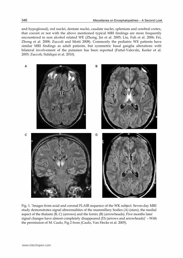
Fig. 1. ‘Images from axial and coronal FLAIR sequence of the WK subject. Seven-day MRI study demonstrates signal abnormalities of the mammillary bodies (A) (stars), the medial aspect of the thalami (B, C) (arrows) and the fornix (B) (arrowheads). Five months later signal changes have almost completely disappeared (D) (arrows and arrowheads)’ – With the permission of M. Caulo, Fig 2 from (Caulo, Van Hecke et al. 2005).
www.intechopen.com
Wernicke’s Encephalopathy
347
8. Diagnosis
In spite of the significant advances in imagistic and laboratory assessments WE remains
mainly a clinical diagnosis. The paraclinical workup, comprising of brain MRI, blood TH
assessments, routine blood tests and sometimes ancillary investigations, improves the
accuracy of the diagnosis and is mandatory for the identification of potential comorbidities.
The diagnosis is highly supported by the favorable response to parenteral TH
administration, but is not excluded by the lack of it (Harper, Giles et al. 1986; Thomson,
Cook et al. 2008). WE may present with a wide spectrum of nonspecific findings. Classically,
the diagnosis requires the presence of the clinical triad consisting of ocular motility signs,
stance and/or gait ataxia and mental status changes, having acute or subacute onset.
However, the classic triad has been reported in less than a third of the adult WE cases (up to
16% in necroptic retrospective studies!) and in 20% of the pediatric cases (Harper, Giles et al.
1986; Victor 1989; Vasconcelos, Silva et al. 1999). Based on the data available in the literature
regarding neuropathological proven WE cases, the prevalence of the classical triad is
estimated to be 8.2% (Galvin, Brathen et al. 2010). The WE diagnosis may easily be
overlooked, the 2010 EFNS guideline recommending the maintenance of a high index of
suspicion for WE. ‘Operational clinical criteria for the classification of the chronic alcoholics
and the identification of WE’ that take into account dietary deficiencies as predisposing
factor for WE were proposed in 1997 by Caine. The intended purpose of these criteria was to
accurately differentiate between alcoholics with WE (active and sequelae) and alcoholics
without WE and between alcoholics with WE and alcoholics with hepatic encephalopathy.
Since their intended applicability concerned only the alcoholic population they were not
tested for reproducibility and variability in non alcoholics. According to these criteria the
accurate ante-mortem identification of WE patients requires the presence of two of the
following: dietary deficiencies (defined as: ‘undernutrition, vitamin deficiency’), eye signs
(defined as: ‘ophtalmoplegia, nystagmus, gaze palsy’), cerebellar signs (defined as:
‘unsteady, ataxia, cerebellar dysfunction’) and either altered mental status (defined as:
‘disoriented, confuse, comatose, digit span abnormal’) or mild memory impairment (defined
as: ‘mild-moderate memory problems, confabulating’). These criteria accurately differentiate
between alcoholics with and without WE (though they do not differentiate between active
WE and sequelae!) and also between alcoholics with WE and hepatic encephalopathy. Their
use was estimated to increase the sensitivity (but not the specificity!) of the diagnosis of WE
from 31% when using the classic triad to 100% (Caine, Halliday et al. 1997). Criteria for
accurately identifying WE patients in the ‘accident and emergency department’ were
proposed in 2001 by the UK Royal College of Physicians. According to their guideline, all
patient presenting with any evidence of chronic alcohol use and any of the following: acute
confusion, decreased consciousness level, ataxia, ophtalmoplegia, memory disturbance,
hypothermia and hypotension and all patients presenting with acute alcohol intoxication or
with delirium tremens should be presumed of having WE (Thomson, Cook et al. 2002).
Though these criteria may accurately identify alcoholic patients with WE they are not
applicable in the non alcoholic population. Since no better alternative exists, the 2010 EFNS
guideline recommends the use of the ‘operational clinical criteria for the classification of the
chronic alcoholics and the identification of WE’ proposed by Caine in 1997 for help in the
diagnosis of WE in both alcoholics and non alcoholics (Galvin, Brathen et al. 2010).
According to the 2010 EFNS guideline, brain imagistic investigations and measurement of
www.intechopen.com
Miscellanea on Encephalopathies – A Second Look
348
the total blood TH level should be included in the paraclinical workup of all those in which
WE is suspected. The brain CT has low sensitivity (i.e. 13%) for the detection of WE lesions,
but nevertheless it is of great utility in acute settings, the purpose of the imagistic assessments being not only to offer additional information supporting the diagnosis of WE, but to exclude other pathologies that might be responsible for the clinical presentation. The MRI offers better sensitivity than the CT for both acute and sequelar WE lesions. The sensitivity varies accordingly to the power of the magnet and to the technique of acquisition used. The estimated sensitivity for the conventional MRI techniques (i.e. T1, T2, FLAIR, paramagnetic contrast) is just above 50%, values of 53% and 58% being reported. Though it does not rule out WE, the MRI is probably the best method of confirming the diagnosis of WE, its specificity being estimated to be as high as 93% (Antunez, Estruch et al. 1998; Weidauer, Nichtweiss et al. 2003). The presence of low TH blood levels has low specificity for WE. The venous blood necessary for the laboratory assessments should be collected prior to TH administration (since is highly influenced by the recent TH intake) and kept away from light. The presence of low total TH or THDP levels is neither necessary nor sufficient for the diagnosis of WE. The recommended laboratory assays are the measure the total TH or THDP in the whole blood through various methods of HPLC (Talwar, Davidson et al. 2000; Thomson and Marshall 2006) The measurement of the 24 hours urine TH levels has little utility in the diagnosis of WE. The level of TH in the urine collected in the following 4 hours after the administration of 1 mg intravenous TH may be used to estimate the tissue THDP state, but due to practical difficulties is not commonly used in clinical practice (Tallaksen, Bohmer et al. 1991; Tallaksen, Bell et al. 1993). Routine laboratory investigations are mandatory for the differential diagnosis of WE and for the identification of associated disorders. The routine laboratory investigations include the assessment of blood glucose levels, serum electrolytes, acute-phase reactants, liver, renal and hematologic status. Toxicology panels may be required in those presenting with acute confusion syndromes. The performance of additional testes (e.g. blood ammonia, thyroid tests, blood lipase, blood B12 levels, viral markers, immune tests) should be adapted to each patient. WE patients may show laboratory signs of dehydration and may have abnormal liver tests, hypomagnesaemia and anaemia (Traviesa 1974; Sechi and Serra 2007). The blood levels of lactic acid and pyruvate may be increased due to TH deficiency. Their measurement may be used for estimating the activity status of the THDP dependent enzymes, but they are neither specific nor sensible for TH deficiency or WE. Some have suggested that WE patients may associate lactic acidosis with or without respiratory alkalosis, leading to a characteristic acid-base pattern. In infants and in other selected susceptible patients the assessment of the acid-base equilibrium is required (Donnino, Miller et al. 2007). Lumbar puncture may be required especially in those associating hyperthermia. Commonly the routine analysis of the CSF of WE patients shows no particularities. In the later stages of WE mild protein elevation may occur (Victor 1989). The presence of protein 14-3-3 has been reported in several patients with WE (Michowitz, Copel et al. 2005; Bertrand, Brandel et al. 2009). CSF pleocytosis or protein levels above 100 mg/dl should suggest an alternative or a coexistent disorder. The assessment of the electroencephalogram (EEG) may be required especially in those in which epileptic partial complex status is suspected. The EEG activity may be dominated by nonspecific diffuse slow waves in about half of WE patients (Victor 1989; Sechi and Serra 2007). Considering that the WE patients may associate cardiac pathology related or not to TH deficiency (e.g. alcoholic dilatated cardiomyopathy) and considering that infections may precipitate WE,
www.intechopen.com
Wernicke’s Encephalopathy
349
an electrocardiogram and chest radiography should be routinely included in the paraclinical workup. WE patients may associate other encephalopathies (hepatic encephalopathy!), as well as brain unrelated diseases. The required workup should be adapted to each patient. It should be kept in mind that these comorbidities may predispose to WE and may correlate with the atypical presentations. Considering that WE patients commonly associate polyneuropathy and that WE may mimic Miller Fischer syndrome, nerve conduction studies may be required (Harper, Giles et al. 1986; Sechi and Serra 2007).
9. Differentials
The diferential diagnosis of WE is vast, comprising mainly of acute or subacute toxic, nutritional and metabolic encephalopaties, encephalitis and meningoencephalitis, and particular strokes. In certain cases diferentiating betwen acute or chronic WE and WE sequelae (including KS!) may be difficult. KS may have other etiologies than WE, and these shoud also be considered for the differential diagnosis. Among the acute or subacute toxic, nutritional and metabolic encephalopaties we emphasize acute alcohol intoxication, delirium tremens, acute Marchiafava-Bignami disease, subacute alcoholic cerebellar degeneration, acute alcoholic pellagra, hepatic encephalopaty, metronidazole encephalopaty. All of these may coexist with WE (Arbelaez, Pajon et al. 2003; Zuccoli, Pipitone et al. 2008; Thorarinsson, Olafsson et al. 2011). In certain clinical settings hypoglicemic coma, hypoxic or hypercapnic encephalopaty, hypertensive encephalopaty, severe hypophosphatemia, methyl bromide intoxication and sepsis asociated encephalopaty should be taken into account. Though it appears earlier and does not asociate the ocular motility signs characteristic for WE, pancreatic encephalopaty may be considered as differential diagnosis in those with severe acute pancreatitis (Sun, Yang et al. 2006; Thorarinsson, Olafsson et al. 2011). Variant Creuzefeld-Jakob disease and Fabry’s disease may resemble WE. Inflamatory diseases of the brain (infectious, autoimmune or disimmune), especially those with perimidline tropism, may present with clinical pictures compatible with WE. Among these we emphasize viral encephalitis and meningoencephalitis (influenza A, West Nile, Japanes, Murray, citomegalovirus, Herpes simplex), tuberculose meningoencephalitis, paraneoplastic cerebelitis, ventriculoencephalitis, acute disseminated encephalomyelitis, limbic encephalitis, brainstem encephalitis and primary or secondary cerebral vasculitides (Brechtelsbauer, Urbach et al. 1997; Torgovnick, Arsura et al. 2000; Chung, Kim et al. 2003; Bertrand, Brandel et al. 2009). Pontine or extrapontine mielinolysis, adult onset Leigh disease and other mitochondrial encephalopaties may be included in the diferential diagnosis of WE. In certain selected cases, the recently described ‘chronic lymphocitic inflamation with pontine perivascular enhancement responsive to steroids’ syndrome (CLIPPERS) (Pittock, Debruyne et al. 2010) and ‘cerebellar ataxia, neuropathy and vestibular areflexia syndrome’ (CANVAS) (Szmulewicz, Waterston et al. 2011) may also be taken into account. The clinical picture of Miller-Fischer syndrome, African seasonal ataxia and tropical ataxic polineuropathy may resemble WE. Percheron artery thrombosis or embolia, basilar artery trombosis and deep cerebral veins thrombosis may also be included in the differential diagnosis of WE. Thalamic and mamillothalamic tract infarcts occuring in individuals with previous strategicaly localised lesions may mimic WE (Bogousslavsky, Regli et al. 1988; Ghika-Schmid and Bogousslavsky 2000). Brain neoplasy involving the perimidline regions (e.g. primary cerebral lymphoma, bilateral thalamic glioma) may
www.intechopen.com
Miscellanea on Encephalopathies – A Second Look
350
sometimes be considered. In children WE may mimik meningitis. Regarding atypical WE presentations, the differential diagnosis should be adapted for each situation. Optic neuritis should be considered in those presentig with optic nerve inolvement. Leber hereditary optic neuropathy precipitated by TH deficiency that also resulted in WE has been reported, and should be considered in WE patients with optic nerve involvement unresponsive to TH administration (Li and Rucker 2010).
10. Treatment
WE is a medical emergency. Its etiopathogenic treatment implies the restoration of adequate brain TH levels. As detailed further on, this is optimally accomplished by high dose parenteral TH administration. The correction of coexisting hypomagnesaemia (by intravenous Mg sulphate administration in doses titrated against Mg serum levels) is mandatory. The reinstitution of a balanced diet and the elimination of the associated modifiable predisposing factors are also required. Scientific evidences regarding the optimal dose, frequency, route and duration of TH administration for WE treatment are currently lacking. The management of the WE patients is commonly conducted according to the empirical recommendations provided by the local guidelines. Since any delay in TH administration may result in permanent neurologic sequelae or death, parenteral TH administration should be started as soon as possible. The decision of initiating parenteral TH administration should be taken on clinical grounds and should never be delayed until the paraclinical workup is completed and/or the results are available (Thomson and Marshall 2006; Sechi and Serra 2007; Galvin, Brathen et al. 2010; Thorarinsson, Olafsson et al. 2011). The identification and treatment of WE comorbidities is mandatory. Supportive and symptomatic therapy should be provided. These should be adapted to each patient, and will not be detailed in the present chapter. Considering that carbohydrate load may precipitate or aggravate WE, glucose should never be administered prior to TH (! hypoglycaemic coma or seizures). To the best of our knowledge, scientifically rigorous studies assessing the optimal TH posology for the treatment of WE have not been conducted. However, the results of several uncontrolled studies on adults are available. It is commonly accepted that a rapid restoration of brain TH levels is more beneficial than a slower restoration, therefore frequent high dose intravenous administration being required. Low parenteral TH doses and the oral route of administration are inefficient for the treatment of WE (Thomson, Cook et al. 2002; Galvin, Brathen et al. 2010). It was reported that WE patients treated with parenteral TH doses of 50 or 100 mg per day fully recovered in only up to 16% of the cases, while death occurred in up to 20% (Victor 1989; Thomson, Cook et al. 2002). The recommended TH doses range from 100 to 1500 mg per day (Thomson, Cook et al. 2002; Sechi and Serra 2007). Within this range, the administration of higher rather than lower TH doses is favoured. The therapeutic scheme should be constantly adapted to the patient’s evolution according to the best clinical judgement of the treating physician. Differences in the therapeutic response of alcoholics and non alcoholics have been reported, alcoholics requiring higher doses and for extended periods of time, some advising 500 mg thrice a day as the minimum dose. Non alcoholics with WE may have adequate therapeutic response providing only 100 to 200 mg of parenteral TH are supplied. According to the 2010 EFNS recommendations both alcoholics and non alcoholics should receive a minimum of 200 mg thrice a day. In hyperemesis gravidarium related WE cases, patients requiring significantly higher TH doses have been reported (Thomson, Cook et al. 2002; Galvin, Brathen et al.
www.intechopen.com
Wernicke’s Encephalopathy
351
2010). To the best of our knowledge, the pediatric parenteral TH doses have not been established. According to the current literature the TH deficient infants were treated with 50 mg of intramuscular TH hydrochloride for 14 days (Fattal-Valevski, Kesler et al. 2005). The opinions regarding the adequate treatment duration vary from a few days of intravenous high dose TH (followed by another few days of intramuscular lower doses) to the continuation of high dose intravenous TH administration until there is no further clinical improvement. The latter appears to be the most reasonable and is recommended by the 2010 EFNS guideline. Secondary prophylaxis with oral TH (30 mg twice a day) is advocated by some. Some suggest that in the absence of clinical improvement TH administration should be stopped after 2 or 3 days. Since certain patients may require prolonged treatment with doses above 500 mg thrice a day, the decision to stop TH treatment in those that seem not to respond should be taken with caution (Sechi and Serra 2007; Paparrigopoulos, Tzavellas et al. 2010). Concomitant parenteral administration of other vitamins, especially of B complex and ascorbic acid, is advocated by some. Niacin should be administered whenever signs suggestive of pellagra exist. The available pharmaceutical preparates for parenteral administration contain variable quantities of TH hydrochloride alone or in combination with other vitamins, especially B complex. Parenteral TH hydrochloride is available for subcutaneous, intramuscular and intravenous injections. Lipid-soluble TH analogues preparates are not widely available (except for benfotiamine, which does not pass the BBB). Oral TH hydrochloride administration has very good tolerability, side effects being extremely rarely reported, and overdoses being virtually impossible (Baker and Frank 1976; Wrenn, Murphy et al. 1989; Kitamori and Itokawa 1993). Intravenous thiamine is not contraindicated in pregnant or lactating women. According to the current literature, the only serious and potentially life threatening side effects of TH administration are anaphylactic and anaphylactoid reactions (Thomson, Cook et al. 2002; Harper 2006). These have been reported to occur during or in the few hours following TH administration. They are only exceptionally encountered in relation with oral TH administration and most frequently encountered in relation with bolus intravenous TH administration (Thomson, Cook et al. 2002; Galvin, Brathen et al. 2010), but even so, parenteral TH administration has a very high safety profile. The global estimated incidence of the serious adverse reactions following parenteral administration of multivitamin pharmacological products containing TH was reported to be 1 in every 5 million intramuscular injections and 1 in every million intravenous injections, therefore lower than that of penicillin or streptokinase. Arguments for the existence of TH induced anaphylaxis have been provided in a few case reports, but it may be speculated that anaphylaxis occurs less frequently than anaphylactoid reactions. Sensitivity skin test prior to TH administration is not necessary. In order to minimise the risk of anaphylactoid reactions without minimising the benefits of intravenous administration, it is recommended that TH is administered over at least 10 minutes by intravenous infusion. Ideally, when administering parenteral TH, rapid access to cardio-pulmonary resuscitation facilities should be available (Luskin and Luskin 1996; Harper 2006; Galvin, Brathen et al. 2010). According to the 2010 EFNS guideline TH should be given to all patients with suspected or manifest WE, before any carbohydrate, intravenously (diluted in 100 ml of normal saline or glucose solution; 30 minutes infusion) or intramuscularly, in doses of at least 200 mg thrice a day, even in the absence of resuscitation facilities, until there is no further clinical improvement (level C evidence). The early restoration of an adequate diet is advised (Galvin, Brathen et al. 2010).
www.intechopen.com
Miscellanea on Encephalopathies – A Second Look
352
11. Red flags for WE, red flags for TH
In order to prevent irreversible brain damage and death, TH should be administered as soon
as possible to all WE patients. However, even when best clinical management is provided,
the diagnosis of WE may be overlooked. Evidence based recommendations for the
identification of patients that should receive parenteral TH are not available, but, as
discussed, criteria for the accurate identification of WE in alcoholics have been proposed in
1997 by Cain and in 2002 by the UK Royal College of Physicians. Since parenteral TH
administration is reasonably cheap and safe, overtreatment is preferred, some advising that
parenteral TH should be routinely administered until neurologic assessments are made and
paraclinical data are available to all patients prone to TH deficiency presenting in the
emergencies departments (even in the absence of other reasonable arguments for WE), and
the 2010 EFNS guideline recommending that a low threshold for parenteral TH
administration is maintained (Ogershok, Rahman et al. 2002; Thomson and Marshall 2006;
Galvin, Brathen et al. 2010; Thorarinsson, Olafsson et al. 2011). Red flags for WE and for TH
administration include prior history of WE, presence of any of the classical triad signs,
chronic ethanol abuse, acute alcohol intoxication, delirium tremens, altered mental status,
altered consciousness, hypothermia, hyperthermia, edema, tachycardia, hypotension,
cardiac failure and lactic acidosis, persistent emesis, impaired nutrition (prolonged fasting,
malnourishment, eating disorders and other psychiatric disorders, exclusive parenteral
nutrition, infants fed by nutritionally impaired women, neglected pediatric, geriatric and
disabled persons), recent significant weight loss, low body mass index (BMI), history of
gastrointestinal surgery, consumptive diseases (including malignancies –especially digestive
tract and in children acute leukemia, AIDS, malaria, tuberculosis, meningoencephalitis,
thyreotoxicosis), chronic liver disease, chronic dialysis, hepatic and uremic encephalopathy,
pregnancy and lactation (Lana-Peixoto, Dos Santos et al. 1992; Antunez, Estruch et al. 1998;
Ihara, Ito et al. 1999; Vasconcelos, Silva et al. 1999; Bleggi-Torres, de Medeiros et al. 2000;
Donnino, Vega et al. 2007; Sechi and Serra 2007). Special consideration regarding parenteral
TH administration should receive all those on chronic diuretic drugs presenting for cardiac
failure (Khan and Garg 2011).
12. Prognosis
The majority of the WE cases are cured (with or without sequelae) providing prompt and
adequate treatment is administered. In the absence of adequate therapy, WE has high
mortality and morbidity, resulting in death in up to 24% of the cases, and in permanent
disability in 75 to 85% of the survivors (Thomson, Cook et al. 2002; Donnino, Vega et al.
2007; Thorarinsson, Olafsson et al. 2011). Alcoholics have worst prognosis than non
alcoholics. Death or persistent sequelae have also been reported to occur in non alcoholic
patients, especially in those with hyperemesis gravidarium (Sechi and Serra 2007). Those
with fewer MRI lesion, and with lesions restricted to the periaqueductal grey area and
thalami seem to have a good prognosis, while those with cortical MRI lesions seem to have a
worse prognosis (Varnet, De Seze et al. 2002; Zuccoli, Santa Cruz et al. 2009). The most
serious sequelae are gait disturbances and KS. Gait disturbances were reported to persist in
61% of the survivors. When present, KS was reported to persist for at least two years in 56 to
84% of the cases. Up to 26% of those with KS are severely disabled (Victor 1989; Caine,
www.intechopen.com
Wernicke’s Encephalopathy
353
Halliday et al. 1997). In certain cases, KS related to alcohol consumption and TH deficiency
may appear in the absence of an overt episode of WE or after an interval of time of apparent
health, probably due to a precipitating factor that alters the equilibrium of a susceptible
brain (Harper, Giles et al. 1986; Galvin, Brathen et al. 2010). Several authors denominate the
sequelae of WE ‘chronic WE’, though they not the consequence of persistent TH deficiency
(Harper, Giles et al. 1986; Kopelman, Thomson et al. 2009; Thorarinsson, Olafsson et al.
2011). A true chronic form of WE, consequence of the cumulative effects of several mild,
subclinical, more or less reversible episodes of TH deficiency, might exist, especially in
alcoholics (Galvin, Brathen et al. 2010).
13. Prophylaxis
Currently there are no evidence based recommendations for the primary or secondary
prophylaxis of WE, but attempts for TH deficiency prophylaxis have been made either by
fortifying food or alcoholic beverages with TH or by administering oral or parenteral TH
to selected populations considered at risk. Their efficacy has not been thoroughly
evaluated in controlled studies. The fortification of food with TH (commonly flour and
rice, but also margarine, soft drinks and other products) is inexpensive and technically
feasible. Empirical primary prophylactic approaches are currently implemented in several
regions. The fortification of food with TH is legally mandatory in certain areas (i.e.
Australia, UK, Denmark, Canada, Chile) and optional but reglemented by law in others
(i.e. Switzerland, Brazil, Yemen). Several countries have the minimum content of flour TH
established by law (WHO 1999). Commonly, TH mononitrate is used for the fortification
of flower, rice and soft drinks. The supplementation of alcoholic beverages (beer and vine)
is feasible, and has been proposed as it better targets those that take most of their caloric
intake from alcoholic beverages. However, experimental data suggests that alcohol
consumption and/or malnourishment lead to impaired TH and hydro-soluble TH
analogues absorption and/or utilization, and therefore these prophylactic approaches
may result in limited benefits. The clinical retrospective studies conducted in Australia 5
years after the legally imposed bread fortification with TH reported a 39% decrease in the
incidence of WE and a 15% decrease in the prevalence of WE (acute and chronic cases
reported as WE, Korsakoff psychosis, WKS) that maintained at similar levels since the first
years, while the necroptic studies reported a decrease in the prevalence of WE from 2.1%
to 1.1% (Rolland and Truswell 1998). The oral prophylactic administration of high doses
of TH hydrochloride was shown not to prevent WE in malnourished obese patients on
therapeutic fasting and in hunger strikers. In this respect it is recommended that in certain
selected population the prophylaxis of WE should be made by parenteral TH
administration. All alcoholics and malnourished individuals admitted to hospitals should
receive 200 to 250 mg of parenteral TH hydrochloride per day for 3 to 5 days. 200 mg of
parenteral TH should be administered prior to glucose infusion to all those prone to TH
deficiency and in comatose and status epilepticus patients. Per os TH supplementation (at
least 15 mg per day, recommended 30 mg twice a day) is advised for the secondary
prophylaxis of WE. Patients receiving drugs that may deplete the TH stores or
undergoing dialysis should also receive per os TH supplementation.(Thomson, Cook et al.
2002; Galvin, Brathen et al. 2010).
www.intechopen.com
Miscellanea on Encephalopathies – A Second Look
354
14. Summary
WE Is a potentially fatal but highly and easily treatable disease occurring throughout the
world. It is commonly (but not exclusively!) encountered in TH deficient alcoholic and/or
malnourished susceptible individuals. The topography of the lesions is highly specific,
commonly the perimidline regions being symmetrically involved. The classical clinical
hallmark (sometimes completely absent!) consists of ocular motility signs, stance and/or
gait ataxia and mental status changes having acute or subacute onset. No paraclinical
investigation can accurately exclude the diagnosis of WE, however, the imagistic and
laboratory workup is useful for supporting the diagnosis and excluding comorbidities.
Though the necessity of prompt parenteral TH administration in all WE patients is
unanimously accepted, evidence based recommendations for its therapeutic management
are not available. The administration of high dose parenteral TH (recommended at least 200
mg thrice a day; NB! correction of hypomagnesaemia) should be initiated as soon as
possible in all patients with suspected WE. Considering the deleterious medical and
economical consequences of untreated WE, and that parenteral TH is reasonably safe and
cheap and has no contraindication except prior allergic reaction, high dose parenteral TH
should be administered to all those presenting to the emergency departments with TH
deficiency predisposing factors, ethanol intoxication or delirium tremens. The maintenance
of a high index of suspicion for WE and a low threshold for parenteral TH administration is
advocated (Thomson, Cook et al. 2002; Sechi and Serra 2007; Galvin, Brathen et al. 2010).
15. References
Aasheim, E. T. (2008). "Wernicke encephalopathy after bariatric surgery: a systematic review." Ann Surg 248(5): 714-720.
Adamolekun, B. (2010). "Etiology of Konzo, epidemic spastic paraparesis associated with cyanogenic glycosides in cassava: role of thiamine deficiency?" J Neurol Sci 296(1-2): 30-33.
Adamolekun, B. and F. R. Ibikunle (1994). "Investigation of an epidemic of seasonal ataxia in Ikare, western Nigeria." Acta Neurol Scand 90(5): 309-311.
Adamolekun, B. and D. A. Ndububa (1994). "Epidemiology and clinical presentation of a seasonal ataxia in western Nigeria." J Neurol Sci 124(1): 95-98.
Alston, T. A. and R. H. Abeles (1987). "Enzymatic conversion of the antibiotic metronidazole to an analog of thiamine." Arch Biochem Biophys 257(2): 357-362.
Antunez, E., R. Estruch, et al. (1998). "Usefulness of CT and MR imaging in the diagnosis of acute Wernicke's encephalopathy." AJR Am J Roentgenol 171(4): 1131-1137.
Arbelaez, A., A. Pajon, et al. (2003). "Acute Marchiafava-Bignami disease: MR findings in two patients." AJNR Am J Neuroradiol 24(10): 1955-1957.
Aupee, A. M., B. Desgranges, et al. (2001). "Voxel-based mapping of brain hypometabolism in permanent amnesia with PET." Neuroimage 13(6 Pt 1): 1164-1173.
Baker, H. and O. Frank (1976). "Absorption, utilization and clinical effectiveness of allithiamines compared to water-soluble thiamines." J Nutr Sci Vitaminol (Tokyo) 22 SUPPL: 63-68.
Basoglu, M., Y. Yetimalar, et al. (2006). "Neurological complications of prolonged hunger strike." Eur J Neurol 13(10): 1089-1097.
www.intechopen.com
Wernicke’s Encephalopathy
355
Bertrand, A., J. P. Brandel, et al. (2009). "Wernicke encephalopathy and Creutzfeldt-Jakob disease." J Neurol 256(6): 904-909.
Bettendorff, L., G. Goessens, et al. (1997). "Reversibility of thiamine deficiency-induced partial necrosis and mitochondrial uncoupling by addition of thiamine to neuroblastoma cell suspensions." Mol Cell Biochem 174(1-2): 121-124.
Bleggi-Torres, L. F., B. C. de Medeiros, et al. (2000). "Neuropathological findings after bone marrow transplantation: an autopsy study of 180 cases." Bone Marrow Transplant 25(3): 301-307.
Bogousslavsky, J., F. Regli, et al. (1988). "Thalamic infarcts: clinical syndromes, etiology, and prognosis." Neurology 38(6): 837-848.
Boldorini, R., L. Vago, et al. (1992). "Wernicke's encephalopathy: occurrence and pathological aspects in a series of 400 AIDS patients." Acta Biomed Ateneo Parmense 63(1-2): 43-49.
Boulware, M. J., V. S. Subramanian, et al. (2003). "Polarized expression of members of the solute carrier SLC19A gene family of water-soluble multivitamin transporters: implications for physiological function." Biochem J 376(Pt 1): 43-48.
Brechtelsbauer, D. L., H. Urbach, et al. (1997). "Cytomegalovirus encephalitis and primary cerebral lymphoma mimicking Wernicke's encephalopathy." Neuroradiology 39(1): 19-22.
Brouns, R. and P. P. De Deyn (2004). "Neurological complications in renal failure: a review." Clin Neurol Neurosurg 107(1): 1-16.
Bujanda, L. (2000). "The effects of alcohol consumption upon the gastrointestinal tract." Am J Gastroenterol 95(12): 3374-3382.
Butterworth, R. F. (1986). "Cerebral thiamine-dependent enzyme changes in experimental Wernicke's encephalopathy." Metab Brain Dis 1(3): 165-175.
Butterworth, R. F. (1989). "Effects of thiamine deficiency on brain metabolism: implications for the pathogenesis of the Wernicke-Korsakoff syndrome." Alcohol Alcohol 24(4): 271-279.
Butterworth, R. F. (1993). "Pathophysiologic mechanisms responsible for the reversible (thiamine-responsive) and irreversible (thiamine non-responsive) neurological symptoms of Wernicke's encephalopathy." Drug Alcohol Rev 12(3): 315-322.
Butterworth, R. F., C. Gaudreau, et al. (1991). "Thiamine deficiency and Wernicke's encephalopathy in AIDS." Metab Brain Dis 6(4): 207-212.
Butterworth, R. F., J. J. Kril, et al. (1993). "Thiamine-dependent enzyme changes in the brains of alcoholics: relationship to the Wernicke-Korsakoff syndrome." Alcohol Clin Exp Res 17(5): 1084-1088.
Caine, D., G. M. Halliday, et al. (1997). "Operational criteria for the classification of chronic alcoholics: identification of Wernicke's encephalopathy." J Neurol Neurosurg Psychiatry 62(1): 51-60.
Calingasan, N. Y., W. J. Chun, et al. (1999). "Oxidative stress is associated with region-specific neuronal death during thiamine deficiency." J Neuropathol Exp Neurol 58(9): 946-958.
Calingasan, N. Y. and G. E. Gibson (2000). "Vascular endothelium is a site of free radical production and inflammation in areas of neuronal loss in thiamine-deficient brain." Ann N Y Acad Sci 903: 353-356.
www.intechopen.com
Miscellanea on Encephalopathies – A Second Look
356
Caulo, M., J. Van Hecke, et al. (2005). "Functional MRI study of diencephalic amnesia in Wernicke-Korsakoff syndrome." Brain 128(Pt 7): 1584-1594.
Chiossi, G., I. Neri, et al. (2006). "Hyperemesis gravidarum complicated by Wernicke encephalopathy: background, case report, and review of the literature." Obstet Gynecol Surv 61(4): 255-268.
Cho, I. J., H. J. Chang, et al. (2009). "A case of Wernicke's encephalopathy following fluorouracil-based chemotherapy." J Korean Med Sci 24(4): 747-750.
Chu, K., D. W. Kang, et al. (2002). "Diffusion-weighted imaging abnormalities in wernicke encephalopathy: reversible cytotoxic edema?" Arch Neurol 59(1): 123-127.
Chung, S. P., S. W. Kim, et al. (2003). "Magnetic resonance imaging as a diagnostic adjunct to Wernicke encephalopathy in the ED." Am J Emerg Med 21(6): 497-502.
Ciccia, R. M. and P. J. Langlais (2000). "An examination of the synergistic interaction of ethanol and thiamine deficiency in the development of neurological signs and long-term cognitive and memory impairments." Alcohol Clin Exp Res 24(5): 622-634.
Coy, J. F., S. Dubel, et al. (1996). "Molecular cloning of tissue-specific transcripts of a transketolase-related gene: implications for the evolution of new vertebrate genes." Genomics 32(3): 309-316.
Dallal, R. M. (2006). "Wernicke encephalopathy after bariatric surgery: losing more than just weight." Neurology 66(11): 1786.
Danbolt, N. C. (2001). "Glutamate uptake." Prog Neurobiol 65(1): 1-105. Davtyan, D. G. and H. V. Vinters (1987). "Wernicke's encephalopathy in AIDS patient
treated with zidovudine." Lancet 1(8538): 919-920. De, W. H. and B. Lennox (1947). "Cerebral beriberi (Wernicke's encephalopathy); review of
52 cases in a Singapore prisoner-of-war hospital." Lancet 1(6436): 11-17. Donnino, M. (2004). "Gastrointestinal beriberi: a previously unrecognized syndrome." Ann
Intern Med 141(11): 898-899. Donnino, M. W., J. Miller, et al. (2007). "Distinctive acid-base pattern in Wernicke's
encephalopathy." Ann Emerg Med 50(6): 722-725. Donnino, M. W., J. Vega, et al. (2007). "Myths and misconceptions of Wernicke's
encephalopathy: what every emergency physician should know." Ann Emerg Med 50(6): 715-721.
Doss, A., D. Mahad, et al. (2003). "Wernicke encephalopathy: unusual findings in nonalcoholic patients." J Comput Assist Tomogr 27(2): 235-240.
Dudeja, P. K., S. Tyagi, et al. (2001). "Mechanism of thiamine uptake by human jejunal brush-border membrane vesicles." Am J Physiol Cell Physiol 281(3): C786-792.
Fattal-Valevski, A. (2011). "Thiamine (Vitamin B1)." Journal of Evidence-Based Complementary&Alternative Medicine I(16): 12-20.
Fattal-Valevski, A., A. Kesler, et al. (2005). "Outbreak of life-threatening thiamine deficiency in infants in Israel caused by a defective soy-based formula." Pediatrics 115(2): e233-238.
Feeney, G. F. and J. P. Connor (2008). "Wernicke-Korsakoff syndrome (WKS) in Australia: no room for complacency." Drug Alcohol Rev 27(4): 388-392.
Fehily, L. (1944). "Human-milk Intoxication due to B1 Avitaminosis." Br Med J 2(4374): 590-592.
Fei, G. Q., C. Zhong, et al. (2008). "Clinical characteristics and MR imaging features of nonalcoholic Wernicke encephalopathy." AJNR Am J Neuroradiol 29(1): 164-169.
www.intechopen.com
Wernicke’s Encephalopathy
357
Fellgiebel, A., A. Scheurich, et al. (2003). "Persistence of disturbed thalamic glucose metabolism in a case of Wernicke-Korsakoff syndrome." Psychiatry Res 124(2): 105-112.
Flink, E. B. (1978). "Role of magnesium depletion in Wernicke-Korsakoff syndrome." N Engl J Med 298(13): 743-744.
Foresti, V. and F. Confalonieri (1987). "Wernicke's encephalopathy in AIDS." Lancet 1(8548): 1499.
Foster, D., M. Falah, et al. (2005). "Wernicke encephalopathy after bariatric surgery: losing more than just weight." Neurology 65(12): 1987; discussion 1847.
Fournier, H. and R. F. Butterworth (1989). "Effects of maternal thiamine deficiency on the development of thiamine-dependent enzymes in regions of the rat brain." Neurochem Int 15(4): 439-444.
Gallucci, M., A. Bozzao, et al. (1990). "Wernicke encephalopathy: MR findings in five patients." AJNR Am J Neuroradiol 11(5): 887-892.
Galvin, R., G. Brathen, et al. (2010). "EFNS guidelines for diagnosis, therapy and prevention of Wernicke encephalopathy." Eur J Neurol 17(12): 1408-1418.
Ghika-Schmid, F. and J. Bogousslavsky (2000). "The acute behavioral syndrome of anterior thalamic infarction: a prospective study of 12 cases." Ann Neurol 48(2): 220-227.
Guerrini, I., A. D. Thomson, et al. (2005). "Direct genomic PCR sequencing of the high affinity thiamine transporter (SLC19A2) gene identifies three genetic variants in Wernicke Korsakoff syndrome (WKS)." Am J Med Genet B Neuropsychiatr Genet 137B(1): 17-19.
Hahn, J. S., W. Berquist, et al. (1998). "Wernicke encephalopathy and beriberi during total parenteral nutrition attributable to multivitamin infusion shortage." Pediatrics 101(1): E10.
Halavaara, J., A. Brander, et al. (2003). "Wernicke's encephalopathy: is diffusion-weighted MRI useful?" Neuroradiology 45(8): 519-523.
Hamadani, M. and F. Awan (2006). "Role of thiamine in managing ifosfamide-induced encephalopathy." J Oncol Pharm Pract 12(4): 237-239.
Harata, N. and Y. Iwasaki (1995). "Evidence for early blood-brain barrier breakdown in experimental thiamine deficiency in the mouse." Metab Brain Dis 10(2): 159-174.
Harper, C. (1979). "Wernicke's encephalopathy: a more common disease than realised. A neuropathological study of 51 cases." J Neurol Neurosurg Psychiatry 42(3): 226-231.
Harper, C. (1983). "The incidence of Wernicke's encephalopathy in Australia--a neuropathological study of 131 cases." J Neurol Neurosurg Psychiatry 46(7): 593-598.
Harper, C. (2006). "Thiamine (vitamin B1) deficiency and associated brain damage is still common throughout the world and prevention is simple and safe!" Eur J Neurol 13(10): 1078-1082.
Harper, C. (2009). "The neuropathology of alcohol-related brain damage." Alcohol Alcohol 44(2): 136-140.
Harper, C., J. Gold, et al. (1989). "The prevalence of the Wernicke-Korsakoff syndrome in Sydney, Australia: a prospective necropsy study." J Neurol Neurosurg Psychiatry 52(2): 282-285.
Harper, C., M. Rodriguez, et al. (1988). "The Wernicke-Korsakoff syndrome in Sydney--a prospective necropsy study." Med J Aust 149(11-12): 718, 720.
www.intechopen.com
Miscellanea on Encephalopathies – A Second Look
358
Harper, C. G., M. Giles, et al. (1986). "Clinical signs in the Wernicke-Korsakoff complex: a retrospective analysis of 131 cases diagnosed at necropsy." J Neurol Neurosurg Psychiatry 49(4): 341-345.
Harper, C. G. and J. J. Kril (1990). "Neuropathology of alcoholism." Alcohol Alcohol 25(2-3): 207-216.
Hazell, A. S. (2009). "Astrocytes are a major target in thiamine deficiency and Wernicke's encephalopathy." Neurochem Int 55(1-3): 129-135.
Hazell, A. S. and R. F. Butterworth (2009). "Update of cell damage mechanisms in thiamine deficiency: focus on oxidative stress, excitotoxicity and inflammation." Alcohol Alcohol 44(2): 141-147.
Hazell, A. S., R. F. Butterworth, et al. (1993). "Cerebral vulnerability is associated with selective increase in extracellular glutamate concentration in experimental thiamine deficiency." J Neurochem 61(3): 1155.
Hazell, A. S., K. V. Rao, et al. (2001). "Selective down-regulation of the astrocyte glutamate transporters GLT-1 and GLAST within the medial thalamus in experimental Wernicke's encephalopathy." J Neurochem 78(3): 560-568.
Heinrich, C. P., H. Stadler, et al. (1973). "The effect of thiamine deficiency on the acetylcoenzyme A and acetylcholine levels in the rat brain." J Neurochem 21(5): 1273-1281.
Heroux, M. and R. F. Butterworth (1988). "Reversible alterations of cerebral gamma-aminobutyric acid in pyrithiamine-treated rats: implications for the pathogenesis of Wernicke's encephalopathy." J Neurochem 51(4): 1221-1226.
Herve, C., P. Beyne, et al. (1994). "Determination of thiamine and its phosphate esters in human erythrocytes by high-performance liquid chromatography with isocratic elution." J Chromatogr B Biomed Appl 653(2): 217-220.
Hoyumpa, A. M., Jr. (1980). "Mechanisms of thiamin deficiency in chronic alcoholism." Am J Clin Nutr 33(12): 2750-2761.
Hutchin, K. C. (1987). "Thiamine deficiency, Wernicke's encephalopathy, and AIDS." Lancet 1(8543): 1200.
Ihara, M., T. Ito, et al. (1999). "Wernicke's encephalopathy associated with hemodialysis: report of two cases and review of the literature." Clin Neurol Neurosurg 101(2): 118-121.
Indraccolo, U., G. Gentile, et al. (2005). "Thiamine deficiency and beriberi features in a patient with hyperemesis gravidarum." Nutrition 21(9): 967-968.
Ishii, K., K. Sarai, et al. (1979). "Concentrations of thiamine and its phosphate esters in rat tissues determined by high-performance liquid chromatography." J Nutr Sci Vitaminol (Tokyo) 25(6): 517-523.
Jamieson, C. P., O. A. Obeid, et al. (1999). "The thiamin, riboflavin and pyridoxine status of patients on emergency admission to hospital." Clin Nutr 18(2): 87-91.
Joyce, E. M., D. E. Rio, et al. (1994). "Decreased cingulate and precuneate glucose utilization in alcoholic Korsakoff's syndrome." Psychiatry Res 54(3): 225-239.
Khan, A. and P. Garg (2011). "Acute decompensated heart failure secondary to thiamine deficiency: often a missed diagnosis." Clin Med 11(2): 203; author reply 203-204.
Kim, E., J. Ku, et al. (2009). "Mammillothalamic functional connectivity and memory function in Wernicke's encephalopathy." Brain 132(Pt 2): 369-376.
www.intechopen.com
Wernicke’s Encephalopathy
359
Kimelberg, H. K., S. K. Goderie, et al. (1990). "Swelling-induced release of glutamate, aspartate, and taurine from astrocyte cultures." J Neurosci 10(5): 1583-1591.
Kimelberg, H. K., E. Rutledge, et al. (1995). "Astrocytic swelling due to hypotonic or high K+ medium causes inhibition of glutamate and aspartate uptake and increases their release." J Cereb Blood Flow Metab 15(3): 409-416.
Kitamori, N. and Y. Itokawa (1993). "Pharmacokinetics of thiamin after oral administration of thiamin tetrahydrofurfuryl disulfide to humans." J Nutr Sci Vitaminol (Tokyo) 39(5): 465-472.
Kondo, K., M. Fujiwara, et al. (1996). "Severe acute metabolic acidosis and Wernicke's encephalopathy following chemotherapy with 5-fluorouracil and cisplatin: case report and review of the literature." Jpn J Clin Oncol 26(4): 234-236.
Kono, S., H. Miyajima, et al. (2009). "Mutations in a thiamine-transporter gene and Wernicke's-like encephalopathy." N Engl J Med 360(17): 1792-1794.
Kopelman, M. D. (1995). "The Korsakoff syndrome." Br J Psychiatry 166(2): 154-173. Kopelman, M. D., A. D. Thomson, et al. (2009). "The Korsakoff syndrome: clinical aspects,
psychology and treatment." Alcohol Alcohol 44(2): 148-154. Laforenza, U., C. Patrini, et al. (1990). "Effects of acute and chronic ethanol administration on
thiamine metabolizing enzymes in some brain areas and in other organs of the rat." Alcohol Alcohol 25(6): 591-603.
Lana-Peixoto, M. A., E. C. Dos Santos, et al. (1992). "Coma and death in unrecognized Wernicke's encephalopathy. An autopsy study." Arq Neuropsiquiatr 50(3): 329-333.
Langlais, P. J. and S. X. Zhang (1993). "Extracellular glutamate is increased in thalamus during thiamine deficiency-induced lesions and is blocked by MK-801." J Neurochem 61(6): 2175-2182.
Lapergue, B., I. Klein, et al. (2006). "Diffusion weighted imaging of cerebellar lesions in Wernicke's encephalopathy." J Neuroradiol 33(2): 126-128.
Lee, B. L., H. Y. Ong, et al. (1991). "Determination of thiamine and its phosphate esters by gradient-elution high-performance liquid chromatography." J Chromatogr 567(1): 71-80.
Lee, S. T., Y. M. Jung, et al. (2005). "Corpus callosum atrophy in Wernicke's encephalopathy." J Neuroimaging 15(4): 367-372.
Leevy, C. M., L. Cardi, et al. (1965). "Incidence and significance of hypovitaminemia in a randomly selected municipal hospital population." Am J Clin Nutr 17(4): 259-271.
Lemos, C., I. Azevedo, et al. (2005). "Effect of red wine on the intestinal absorption of thiamine and folate in the rat: comparison with the effect of ethanol alone." Alcohol Clin Exp Res 29(4): 664-671.
Li, J. M. and J. C. Rucker (2010). "Irreversible optic neuropathy in wernicke encephalopathy and leber hereditary optic neuropathy." J Neuroophthalmol 30(1): 49-53.
Liu, Y. T., J. L. Fuh, et al. (2006). "Correlation of magnetic resonance images with neuropathology in acute Wernicke's encephalopathy." Clin Neurol Neurosurg 108(7): 682-687.
Loh, Y., W. D. Watson, et al. (2005). "Restricted diffusion of the splenium in acute Wernicke's encephalopathy." J Neuroimaging 15(4): 373-375.
Luskin, A. T. and S. S. Luskin (1996). "Anaphylaxis and Anaphylactoid Reactions: Diagnosis and Management." Am J Ther 3(7): 515-520.
www.intechopen.com
Miscellanea on Encephalopathies – A Second Look
360
Macias-Matos, C., A. Rodriguez-Ojea, et al. (1996). "Biochemical evidence of thiamine depletion during the Cuban neuropathy epidemic, 1992-1993." Am J Clin Nutr 64(3): 347-353.
Mair, W. G., E. K. Warrington, et al. (1979). "Memory disorder in Korsakoff's psychosis: a neuropathological and neuropsychological investigation of two cases." Brain 102(4): 749-783.
Makarchikov, A. F., B. Lakaye, et al. (2003). "Thiamine triphosphate and thiamine triphosphatase activities: from bacteria to mammals." Cell Mol Life Sci 60(7): 1477-1488.
Martin, P. R., C. K. Singleton, et al. (2003). "The role of thiamine deficiency in alcoholic brain disease." Alcohol Res Health 27(2): 134-142.
McCool, B. A., S. G. Plonk, et al. (1993). "Cloning of human transketolase cDNAs and comparison of the nucleotide sequence of the coding region in Wernicke-Korsakoff and non-Wernicke-Korsakoff individuals." J Biol Chem 268(2): 1397-1404.
Michowitz, Y., L. Copel, et al. (2005). "Non-alcoholic Wernicke's encephalopathy - unusual clinical findings." Eur J Intern Med 16(6): 443-444.
Miyajima, H. and S. Kono (2010). "Familial Wernicke's-like encephalopathy." Rinsho Shinkeigaku 50(11): 855-857.
Morishima, T., M. Aoyama, et al. (2008). "Lactic acid increases aquaporin 4 expression on the cell membrane of cultured rat astrocytes." Neurosci Res 61(1): 18-26.
Mukherjee, A. B., S. Svoronos, et al. (1987). "Transketolase abnormality in cultured fibroblasts from familial chronic alcoholic men and their male offspring." J Clin Invest 79(4): 1039-1043.
Nishimune, T., Y. Watanabe, et al. (2000). "Thiamin is decomposed due to Anaphe spp. entomophagy in seasonal ataxia patients in Nigeria." J Nutr 130(6): 1625-1628.
Nixon, P. F. (2008). "Glutamate export at the choroid plexus in health, thiamin deficiency, and ethanol intoxication: review and hypothesis." Alcohol Clin Exp Res 32(8): 1339-1349.
Nixon, P. F., L. Jordan, et al. (2008). "Choroid plexus dysfunction: the initial event in the pathogenesis of Wernicke's encephalopathy and ethanol intoxication." Alcohol Clin Exp Res 32(8): 1513-1523.
Novak, D. J. and M. Victor (1974). "The vagus and sympathetic nerves in alcoholic polyneuropathy." Arch Neurol 30(4): 273-284.
Ogershok, P. R., A. Rahman, et al. (2002). "Wernicke encephalopathy in nonalcoholic patients." Am J Med Sci 323(2): 107-111.
Osuntokun, B. O., A. Aladetoyinbo, et al. (1985). "Vitamin B nutrition in the Nigerian tropical ataxic neuropathy." J Neurol Neurosurg Psychiatry 48(2): 154-156.
Otsuka, F., K. Tada, et al. (1997). "Gestational thyrotoxicosis manifesting as wernicke encephalopathy: a case report." Endocr J 44(3): 447-452.
Pacal, L., J. Tomandl, et al. (2011). "Role of thiamine status and genetic variability in transketolase and other pentose phosphate cycle enzymes in the progression of diabetic nephropathy." Nephrol Dial Transplant 26(4): 1229-1236.
Pannunzio, P., A. S. Hazell, et al. (2000). "Thiamine deficiency results in metabolic acidosis and energy failure in cerebellar granule cells: an in vitro model for the study of cell death mechanisms in Wernicke's encephalopathy." J Neurosci Res 62(2): 286-292.
www.intechopen.com
Wernicke’s Encephalopathy
361
Paparrigopoulos, T., E. Tzavellas, et al. (2010). "Complete recovery from undertreated Wernicke-Korsakoff syndrome following aggressive thiamine treatment." In Vivo 24(2): 231-233.
Park, S. H., M. Kim, et al. (2001). "Magnetic resonance reflects the pathological evolution of Wernicke encephalopathy." J Neuroimaging 11(4): 406-411.
Pearce, J. M. (2008). "Wernicke-Korsakoff encephalopathy." Eur Neurol 59(1-2): 101-104. Pekovich, S. R., P. R. Martin, et al. (1998). "Thiamine deficiency decreases steady-state
transketolase and pyruvate dehydrogenase but not alpha-ketoglutarate dehydrogenase mRNA levels in three human cell types." J Nutr 128(4): 683-687.
Phillips, G. B., M. Victor, et al. (1952). "A study of the nutritional defect in Wernicke's syndrome; the effect of a purified diet, thiamine, and other vitamins on the clinical manifestations." J Clin Invest 31(10): 859.
Pittock, S. J., J. Debruyne, et al. (2010). "Chronic lymphocytic inflammation with pontine perivascular enhancement responsive to steroids (CLIPPERS)." Brain 133(9): 2626-2634.
Ramayya, A. and P. Jauhar (1997). "Increasing incidence of Korsakoff's psychosis in the east end of Glasgow." Alcohol Alcohol 32(3): 281-285.
Rao, V. L., D. D. Mousseau, et al. (1996). "Nitric oxide synthase activities are selectively decreased in vulnerable brain regions in thiamine deficiency." Neurosci Lett 208(1): 17-20.
Rolland, S. and A. S. Truswell (1998). "Wernicke-Korsakoff syndrome in Sydney hospitals after 6 years of thiamin enrichment of bread." Public Health Nutr 1(2): 117-122.
Rosenblum, W. I. and I. Feigin (1965). "The hemorrhagic component of Wernicke's encephalopathy." Arch Neurol 13(6): 627-632.
Rotman, P., D. Hassin, et al. (1994). "Wernicke's encephalopathy in hyperemesis gravidarum: association with abnormal liver function." Isr J Med Sci 30(3): 225-228.
Said, H. M., K. Balamurugan, et al. (2004). "Expression and functional contribution of hTHTR-2 in thiamin absorption in human intestine." Am J Physiol Gastrointest Liver Physiol 286(3): G491-498.
Schroth, G., W. Wichmann, et al. (1991). "Blood-brain-barrier disruption in acute Wernicke encephalopathy: MR findings." J Comput Assist Tomogr 15(6): 1059-1061.
Sechi, G. and A. Serra (2007). "Wernicke's encephalopathy: new clinical settings and recent advances in diagnosis and management." Lancet Neurol 6(5): 442-455.
Serra, A., G. Sechi, et al. (2007). "Wernicke encephalopathy after obesity surgery: a systematic review." Neurology 69(6): 615; author reply 615-616.
Shikata, E., T. Mizutani, et al. (2000). "'Iatrogenic' Wernicke's encephalopathy in Japan." Eur Neurol 44(3): 156-161.
Singh, S. and A. Kumar (2007). "Wernicke encephalopathy after obesity surgery: a systematic review." Neurology 68(11): 807-811.
Slekar, K. H., D. J. Kosman, et al. (1996). "The yeast copper/zinc superoxide dismutase and the pentose phosphate pathway play overlapping roles in oxidative stress protection." J Biol Chem 271(46): 28831-28836.
Smith, J. A., P. P. Foa, et al. (1947). "Some toxic effects of thiamine." Fed Proc 6(1 Pt 2): 204. Subramanian, V. S., J. S. Marchant, et al. (2003). "Cell biology of the human thiamine
transporter-1 (hTHTR1). Intracellular trafficking and membrane targeting mechanisms." J Biol Chem 278(6): 3976-3984.
www.intechopen.com
Miscellanea on Encephalopathies – A Second Look
362
Subramanian, V. S., J. S. Marchant, et al. (2006). "Targeting and trafficking of the human thiamine transporter-2 in epithelial cells." J Biol Chem 281(8): 5233-5245.
Subramanya, S. B., V. S. Subramanian, et al. (2010). "Chronic alcohol consumption and intestinal thiamin absorption: effects on physiological and molecular parameters of the uptake process." Am J Physiol Gastrointest Liver Physiol 299(1): G23-31.
Sullivan, E. V. and A. Pfefferbaum (2009). "Neuroimaging of the Wernicke-Korsakoff syndrome." Alcohol Alcohol 44(2): 155-165.
Sun, G. H., Y. S. Yang, et al. (2006). "Pancreatic encephalopathy and Wernicke encephalopathy in association with acute pancreatitis: a clinical study." World J Gastroenterol 12(26): 4224-4227.
Surges, R., S. Beck, et al. (2007). "Sudden bilateral blindness in Wernicke's encephalopathy: case report and review of the literature." J Neurol Sci 260(1-2): 261-264.
Szmulewicz, D. J., J. A. Waterston, et al. (2011). "Sensory neuropathy as part of the cerebellar ataxia neuropathy vestibular areflexia syndrome." Neurology 76(22): 1903-1910
Tallaksen, C. M., H. Bell, et al. (1993). "Thiamin and thiamin phosphate ester deficiency assessed by high performance liquid chromatography in four clinical cases of Wernicke encephalopathy." Alcohol Clin Exp Res 17(3): 712-716.
Tallaksen, C. M., T. Bohmer, et al. (1991). "Concomitant determination of thiamin and its phosphate esters in human blood and serum by high-performance liquid chromatography." J Chromatogr 564(1): 127-136.
Tallaksen, C. M., A. Sande, et al. (1993). "Kinetics of thiamin and thiamin phosphate esters in human blood, plasma and urine after 50 mg intravenously or orally." Eur J Clin Pharmacol 44(1): 73-78.
Talwar, D., H. Davidson, et al. (2000). "Vitamin B(1) status assessed by direct measurement of thiamin pyrophosphate in erythrocytes or whole blood by HPLC: comparison with erythrocyte transketolase activation assay." Clin Chem 46(5): 704-710.
Tanasescu, R. (2009). "Wernicke’s Encephalopathy In General Neurological Practice: Short Considerations On The Need For Revision (I)." Romanian Journal of Neurology VIII(3): 3.
Thomson, A. D. (2000). "Mechanisms of vitamin deficiency in chronic alcohol misusers and the development of the Wernicke-Korsakoff syndrome." Alcohol Alcohol Suppl 35(1): 2-7.
Thomson, A. D., C. C. Cook, et al. (2008). "Wernicke's encephalopathy: 'Plus ca change, plus c'est la meme chose'." Alcohol Alcohol 43(2): 180-186.
Thomson, A. D., C. C. Cook, et al. (2002). "The Royal College of Physicians report on alcohol: guidelines for managing Wernicke's encephalopathy in the accident and Emergency Department." Alcohol Alcohol 37(6): 513-521.
Thomson, A. D., C. C. Cook, et al. (2008). "Wernicke's encephalopathy revisited. Translation of the case history section of the original manuscript by Carl Wernicke 'Lehrbuch der Gehirnkrankheiten fur Aerzte and Studirende' (1881) with a commentary." Alcohol Alcohol 43(2): 174-179.
Thomson, A. D. and E. J. Marshall (2006). "The natural history and pathophysiology of Wernicke's Encephalopathy and Korsakoff's Psychosis." Alcohol Alcohol 41(2): 151-158.
Thomson, A. D. and E. J. Marshall (2006). "The treatment of patients at risk of developing Wernicke's encephalopathy in the community." Alcohol Alcohol 41(2): 159-167.
www.intechopen.com
Wernicke’s Encephalopathy
363
Thorarinsson, B. L., E. Olafsson, et al. (2011). "[Wernicke's encephalopathy in chronic alcoholics]." Laeknabladid 97(1): 21-29.
Todd, K. G. and R. F. Butterworth (1998). "Evaluation of the role of NMDA-mediated excitotoxicity in the selective neuronal loss in experimental Wernicke encephalopathy." Exp Neurol 149(1): 130-138.
Todd, K. G., A. S. Hazell, et al. (1999). "Alcohol-thiamine interactions: an update on the pathogenesis of Wernicke encephalopathy." Addict Biol 4(3): 261-272.
Torgovnick, J., E. L. Arsura, et al. (2000). "Cytomegalovirus ventriculoencephalitis presenting as a Wernicke's encephalopathy-like syndrome." Neurology 55(12): 1910-1913.
Torvik, A. (1991). "Wernicke's encephalopathy--prevalence and clinical spectrum." Alcohol Alcohol Suppl 1: 381-384.
Traviesa, D. C. (1974). "Magnesium deficiency: a possible cause of thiamine refractoriness in Wernicke-Korsakoff encephalopathy." J Neurol Neurosurg Psychiatry 37(8): 959-962.
Ueda, Y., H. Utsunomiya, et al. (2007). "[Wernicke encephalopathy in a chronic peritoneal dialysis patient--correlation between diffusion MR and pathological findings]." No To Hattatsu 39(3): 210-213.
Unlu, E., B. Cakir, et al. (2006). "MRI findings of Wernicke encephalopathy revisited due to hunger strike." Eur J Radiol 57(1): 43-53.
Van Belle, S. J., W. Distelmans, et al. (1993). "Phase I trial of erbulozole (R55104)." Anticancer Res 13(6B): 2389-2391.
Varnet, O., J. De Seze, et al. (2002). "[Wernicke-Korsakoff syndrome: diagnostic contribution of magnetic resonance imaging]." Rev Neurol (Paris) 158(12): 1181-1185.
Vasconcelos, M. M., K. P. Silva, et al. (1999). "Early diagnosis of pediatric Wernicke's encephalopathy." Pediatr Neurol 20(4): 289-294.
Victor, M., R. D. Adams, et al. (1971). "The Wernicke-Korsakoff syndrome. A clinical and pathological study of 245 patients, 82 with post-mortem examinations." Contemp Neurol Ser 7: 1-206.
Victor, M., Adams, K. M., and Collins, G. H. (1989). "The Wernicke-Korsakoff Syndrome and Related Disorders due to Alcoholism and Malnutrition."
Vortmeyer, A. O., C. Hagel, et al. (1992). "Haemorrhagic thiamine deficient encephalopathy following prolonged parenteral nutrition." J Neurol Neurosurg Psychiatry 55(9): 826-829.
Watanabe, I. (1978). "Pyrithiamine-induced acute thiamine-deficient encephalopathy in the mouse." Exp Mol Pathol 28(3): 381-394.
Watson, A. J., J. F. Walker, et al. (1981). "Acute Wernickes encephalopathy precipitated by glucose loading." Ir J Med Sci 150(10): 301-303.
Weidauer, S., M. Nichtweiss, et al. (2003). "Wernicke encephalopathy: MR findings and clinical presentation." Eur Radiol 13(5): 1001-1009.
WHO (1999). "Thiamine deficiency and its prevention and control in major emergencies". http://www.who.int/nutrition/publications/emergencies/WHO_NHD_99.13/en/index.html
WHO (version 2010). "International Statistical Classification of Diseases and Related Health Problems 10th Revision".
http://apps.who.int/classifications/icd10/browse/2010/en
www.intechopen.com
Miscellanea on Encephalopathies – A Second Look
364
Winston, A. P., C. P. Jamieson, et al. (2000). "Prevalence of thiamin deficiency in anorexia nervosa." Int J Eat Disord 28(4): 451-454.
Witt, E. D. (1985). "Neuroanatomical consequences of thiamine deficiency: a comparative analysis." Alcohol Alcohol 20(2): 201-221.
Wood, B., J. Currie, et al. (1986). "Wernicke's encephalopathy in a metropolitan hospital. A prospective study of incidence, characteristics and outcome." Med J Aust 144(1): 12-16.
Wrenn, K. D., F. Murphy, et al. (1989). "A toxicity study of parenteral thiamine hydrochloride." Ann Emerg Med 18(8): 867-870.
Yoneoka, Y., N. Takeda, et al. (2004). "Acute Korsakoff syndrome following mammillothalamic tract infarction." AJNR Am J Neuroradiol 25(6): 964-968.
Zhao, R., F. Gao, et al. (2002). "Reduced folate carrier transports thiamine monophosphate: an alternative route for thiamine delivery into mammalian cells." Am J Physiol Cell Physiol 282(6): C1512-1517.
Zhao, Y., X. Pan, et al. (2009). "Decreased transketolase activity contributes to impaired hippocampal neurogenesis induced by thiamine deficiency." J Neurochem 111(2): 537-546.
Zhong, C., L. Jin, et al. (2005). "MR Imaging of nonalcoholic Wernicke encephalopathy: a follow-up study." AJNR Am J Neuroradiol 26(9): 2301-2305.
Zuccoli, G., I. Cravo, et al. (2011). "Basal Ganglia involvement in wernicke encephalopathy: report of 2 cases." AJNR Am J Neuroradiol 32(7): E129-131.
Zuccoli, G., M. Gallucci, et al. (2007). "Wernicke encephalopathy: MR findings at clinical presentation in twenty-six alcoholic and nonalcoholic patients." AJNR Am J Neuroradiol 28(7): 1328-1331.
Zuccoli, G. and L. Motti (2008). "Atypical Wernicke's encephalopathy showing lesions in the cranial nerve nuclei and cerebellum." J Neuroimaging 18(2): 194-197.
Zuccoli, G., N. Pipitone, et al. (2008). "Metronidazole-induced and Wernicke encephalopathy: two different entities sharing the same metabolic pathway?" AJNR Am J Neuroradiol 29(9): E84; author reply.
Zuccoli, G., D. Santa Cruz, et al. (2009). "MR imaging findings in 56 patients with Wernicke encephalopathy: nonalcoholics may differ from alcoholics." AJNR Am J Neuroradiol 30(1): 171-176.
Zuccoli, G., N. Siddiqui, et al. (2010). "Neuroimaging findings in pediatric Wernicke encephalopathy: a review." Neuroradiology 52(6): 523-529.
www.intechopen.com
Miscellanea on Encephalopathies - A Second LookEdited by Dr. Radu Tanasescu
ISBN 978-953-51-0558-9Hard cover, 390 pagesPublisher InTechPublished online 25, April, 2012Published in print edition April, 2012
InTech EuropeUniversity Campus STeP Ri Slavka Krautzeka 83/A 51000 Rijeka, Croatia Phone: +385 (51) 770 447 Fax: +385 (51) 686 166www.intechopen.com
InTech ChinaUnit 405, Office Block, Hotel Equatorial Shanghai No.65, Yan An Road (West), Shanghai, 200040, China
Phone: +86-21-62489820 Fax: +86-21-62489821
The book project “Miscellanea on Encephalopathies-a second look” aims to cover some of the importantaspects regarding metabolic, hypoxic, neoplasm- and drug-related encephalopathies, by transmitting valuableinformation filtered through the real life clinical and research experience of the authors.
How to referenceIn order to correctly reference this scholarly work, feel free to copy and paste the following:
Radu Tanasescu, Laura Dumitrescu, Carmen Dragos, Dimela Luca, Alexandra Oprisan, Catalina Coclitu,Oana Simionescu, Lorena Cojocaru, Marius Stan, Andreea Carasca, Andreea Gitman, Adela Chiru and MarinaTicmeanu (2012). Wernicke's Encephalopathy, Miscellanea on Encephalopathies - A Second Look, Dr. RaduTanasescu (Ed.), ISBN: 978-953-51-0558-9, InTech, Available from:http://www.intechopen.com/books/miscellanea-on-encephalopathies-a-second-look/wernicke-s-encephalopathy