Embed Size (px)
Citation preview

Welcome to Our
Microbial Genetics Class
College of Bioengineering
Tianjin University of Science and Technology
Lesson Three
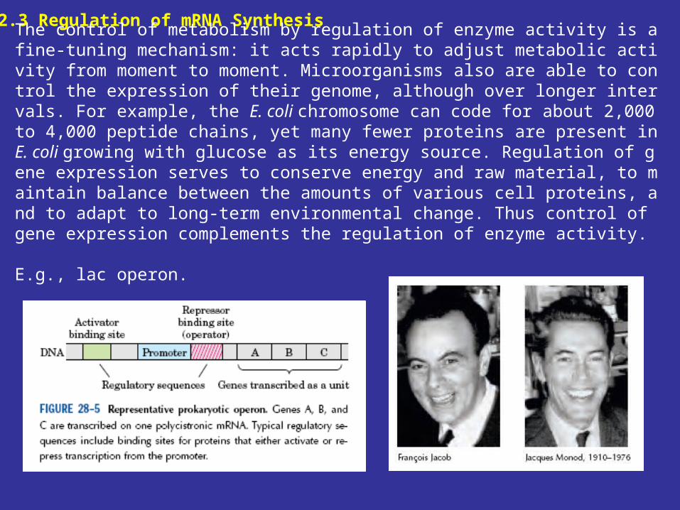
12.3 Regulation of mRNA Synthesis
The control of metabolism by regulation of enzyme activity is a fine-tuning mechanism: it acts rapidly to adjust metabolic activity from moment to moment. Microorganisms also are able to control the expression of their genome, although over longer intervals. For example, the E. coli chromosome can code for about 2,000 to 4,000 peptide chains, yet many fewer proteins are present in E. coli growing with glucose as its energy source. Regulation of gene expression serves to conserve energy and raw material, to maintain balance between the amounts of various cell proteins, and to adapt to long-term environmental change. Thus control of gene expression complements the regulation of enzyme activity.
E.g., lac operon.
Induction and Repression
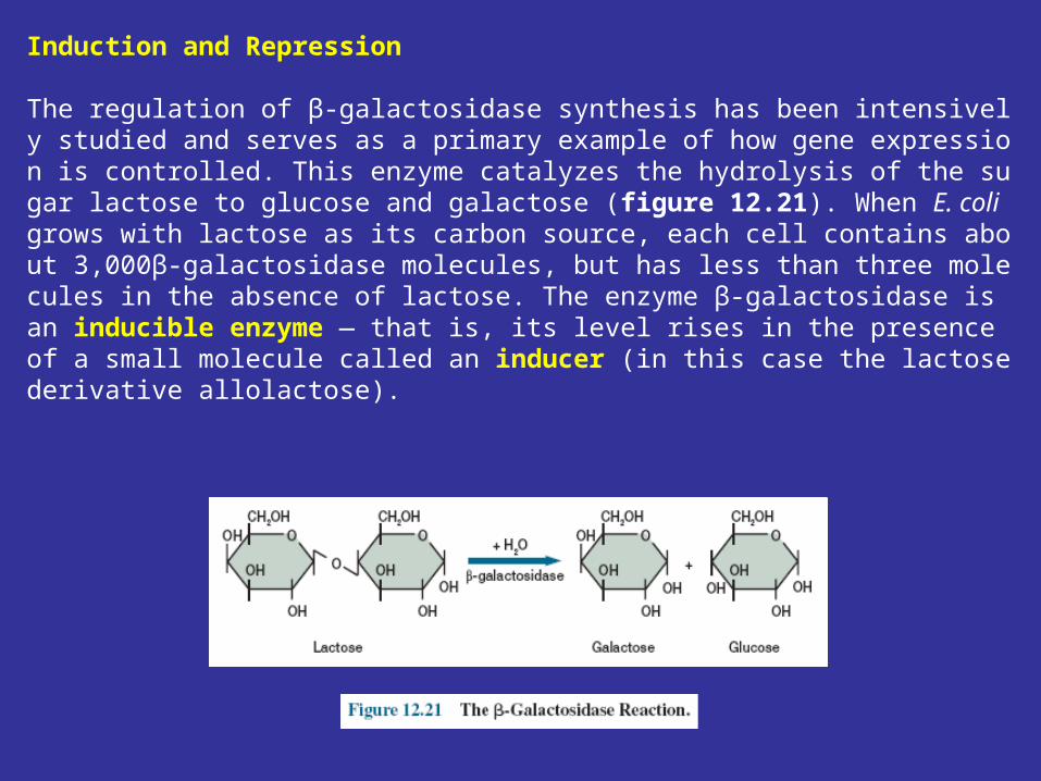
The regulation of β-galactosidase synthesis has been intensively studied and serves as a primary example of how gene expression is controlled. This enzyme catalyzes the hydrolysis of the sugar lactose to glucose and galactose (figure 12.21). When E. coli grows with lactose as its carbon source, each cell contains about 3,000β-galactosidase molecules, but has less than three molecules in the absence of lactose. The enzyme β-galactosidase is an inducible enzyme — that is, its level rises in the presence of a small molecule called an inducer (in this case the lactose derivative allolactose).
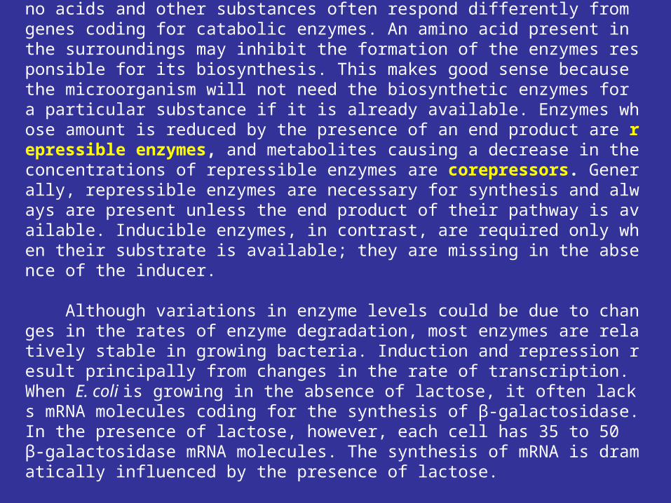
The genes for enzymes involved in the biosynthesis of amino acids and other substances often respond differently from genes coding for catabolic enzymes. An amino acid present in the surroundings may inhibit the formation of the enzymes responsible for its biosynthesis. This makes good sense because the microorganism will not need the biosynthetic enzymes for a particular substance if it is already available. Enzymes whose amount is reduced by the presence of an end product are repressible enzymes, and metabolites causing a decrease in the concentrations of repressible enzymes are corepressors. Generally, repressible enzymes are necessary for synthesis and always are present unless the end product of their pathway is available. Inducible enzymes, in contrast, are required only when their substrate is available; they are missing in the absence of the inducer.
Although variations in enzyme levels could be due to changes in the rates of enzyme degradation, most enzymes are relatively stable in growing bacteria. Induction and repression result principally from changes in the rate of transcription. When E. coli is growing in the absence of lactose, it often lacks mRNA molecules coding for the synthesis of β-galactosidase. In the presence of lactose, however, each cell has 35 to 50 β-galactosidase mRNA molecules. The synthesis of mRNA is dramatically influenced by the presence of lactose.
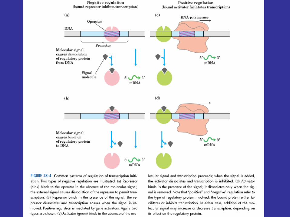
Negative Control
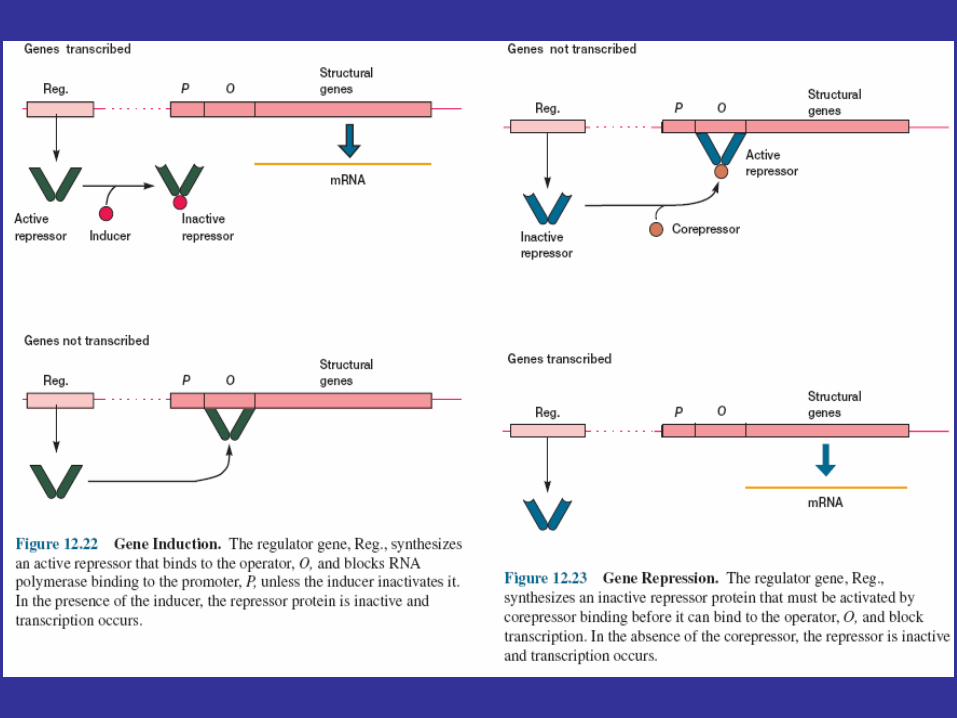
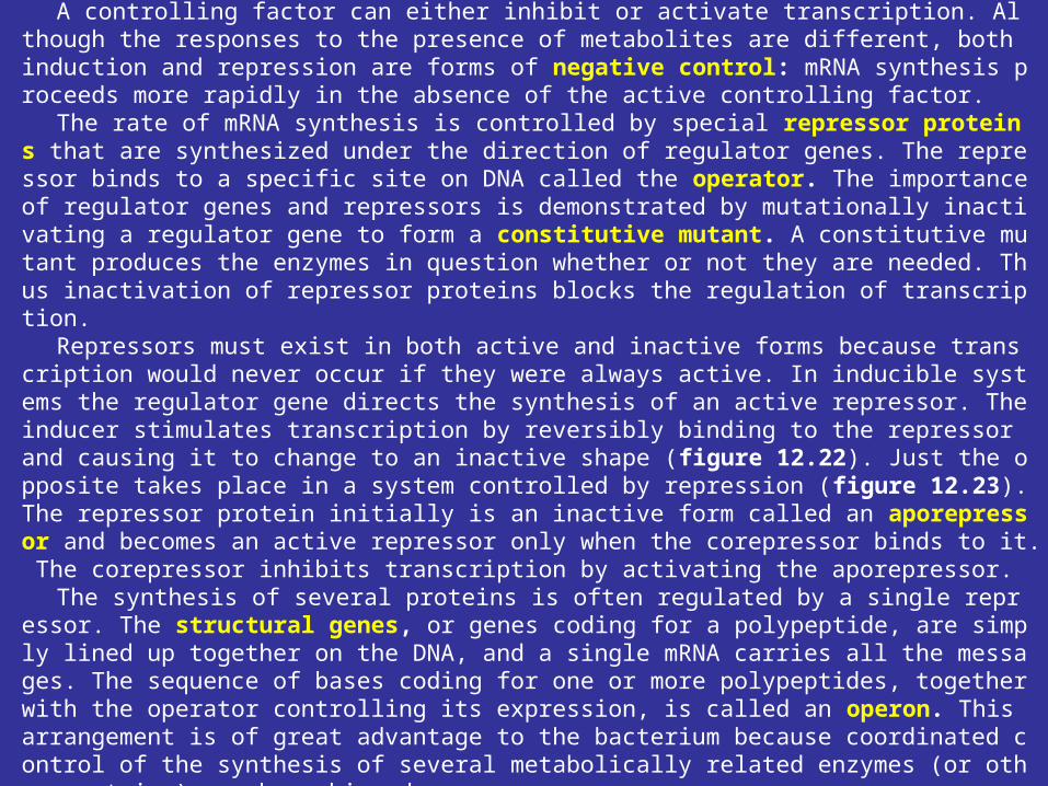
A controlling factor can either inhibit or activate transcription. Although the responses to the presence of metabolites are different, both induction and repression are forms of negative control: mRNA synthesis proceeds more rapidly in the absence of the active controlling factor.
The rate of mRNA synthesis is controlled by special repressor proteins that are synthesized under the direction of regulator genes. The repressor binds to a specific site on DNA called the operator. The importance of regulator genes and repressors is demonstrated by mutationally inactivating a regulator gene to form a constitutive mutant. A constitutive mutant produces the enzymes in question whether or not they are needed. Thus inactivation of repressor proteins blocks the regulation of transcription.
Repressors must exist in both active and inactive forms because transcription would never occur if they were always active. In inducible systems the regulator gene directs the synthesis of an active repressor. The inducer stimulates transcription by reversibly binding to the repressor and causing it to change to an inactive shape (figure 12.22). Just the opposite takes place in a system controlled by repression (figure 12.23). The repressor protein initially is an inactive form called an aporepressor and becomes an active repressor only when the corepressor binds to it. The corepressor inhibits transcription by activating the aporepressor.
The synthesis of several proteins is often regulated by a single repressor. The structural genes, or genes coding for a polypeptide, are simply lined up together on the DNA, and a single mRNA carries all the messages. The sequence of bases coding for one or more polypeptides, together with the operator controlling its expression, is called an operon. This arrangement is of great advantage to the bacterium because coordinated control of the synthesis of several metabolically related enzymes (or other proteins) can be achieved.
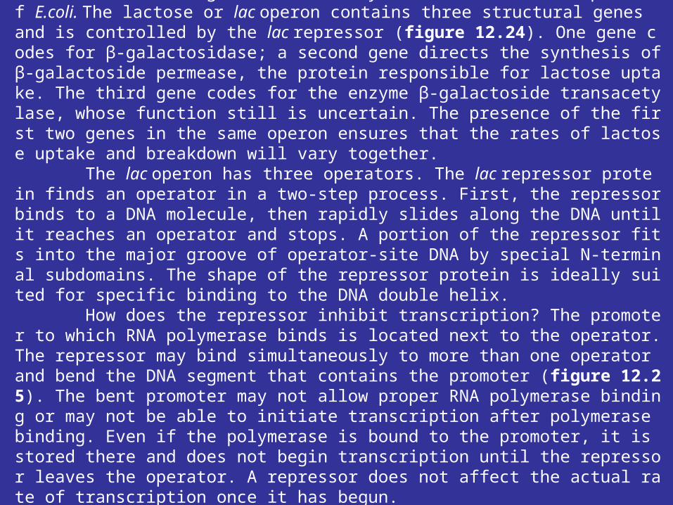
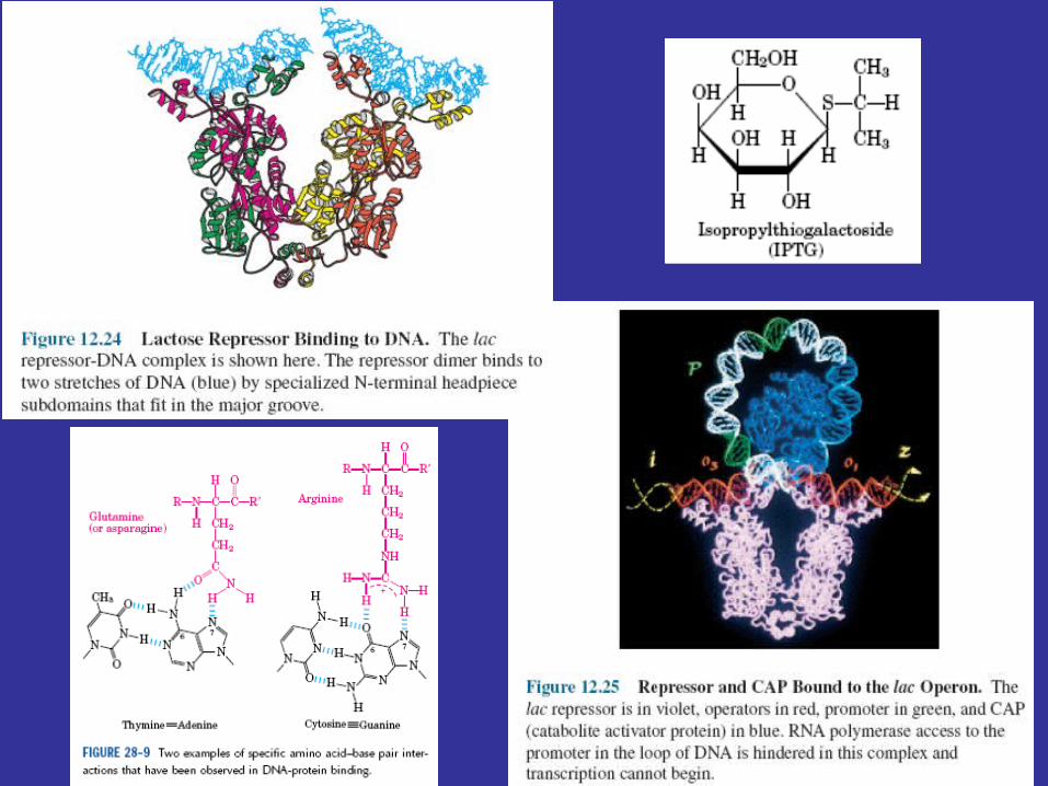
The Lactose OperonThe best-studied negative control system is the lactose operon of E.coli. The lactose or lac operon contains three structural genes and is controlled by the lac repressor (figure 12.24). One gene codes for β-galactosidase; a second gene directs the synthesis ofβ-galactoside permease, the protein responsible for lactose uptake. The third gene codes for the enzyme β-galactoside transacetylase, whose function still is uncertain. The presence of the first two genes in the same operon ensures that the rates of lactose uptake and breakdown will vary together. The lac operon has three operators. The lac repressor protein finds an operator in a two-step process. First, the repressor binds to a DNA molecule, then rapidly slides along the DNA until it reaches an operator and stops. A portion of the repressor fits into the major groove of operator-site DNA by special N-terminal subdomains. The shape of the repressor protein is ideally suited for specific binding to the DNA double helix. How does the repressor inhibit transcription? The promoter to which RNA polymerase binds is located next to the operator. The repressor may bind simultaneously to more than one operator and bend the DNA segment that contains the promoter (figure 12.25). The bent promoter may not allow proper RNA polymerase binding or may not be able to initiate transcription after polymerase binding. Even if the polymerase is bound to the promoter, it is stored there and does not begin transcription until the repressor leaves the operator. A repressor does not affect the actual rate of transcription once it has begun.
Positive Control
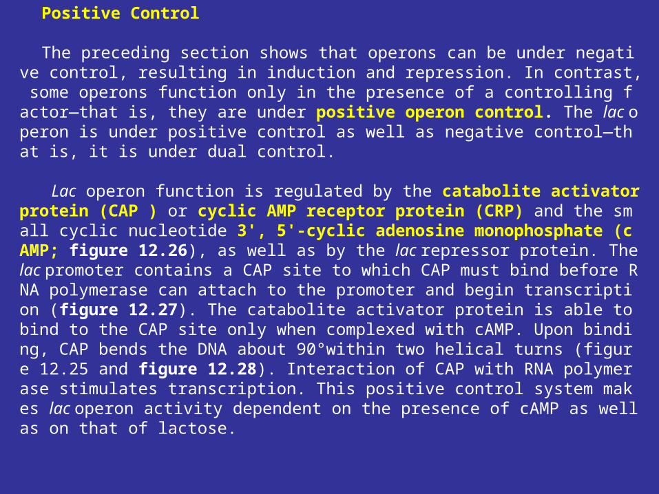
The preceding section shows that operons can be under negative control, resulting in induction and repression. In contrast, some operons function only in the presence of a controlling factor—that is, they are under positive operon control. The lac operon is under positive control as well as negative control—that is, it is under dual control.
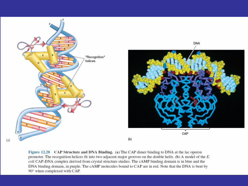
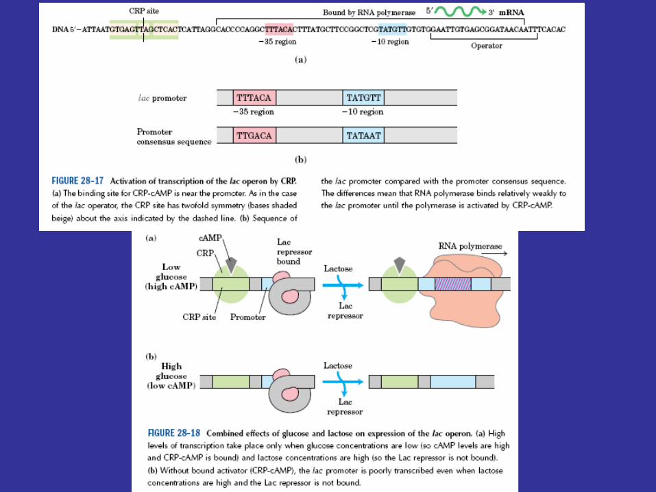
Lac operon function is regulated by the catabolite activator protein (CAP ) or cyclic AMP receptor protein (CRP) and the small cyclic nucleotide 3', 5'-cyclic adenosine monophosphate (cAMP; figure 12.26), as well as by the lac repressor protein. The lac promoter contains a CAP site to which CAP must bind before RNA polymerase can attach to the promoter and begin transcription (figure 12.27). The catabolite activator protein is able to bind to the CAP site only when complexed with cAMP. Upon binding, CAP bends the DNA about 90°within two helical turns (figure 12.25 and figure 12.28). Interaction of CAP with RNA polymerase stimulates transcription. This positive control system makes lac operon activity dependent on the presence of cAMP as well as on that of lactose.
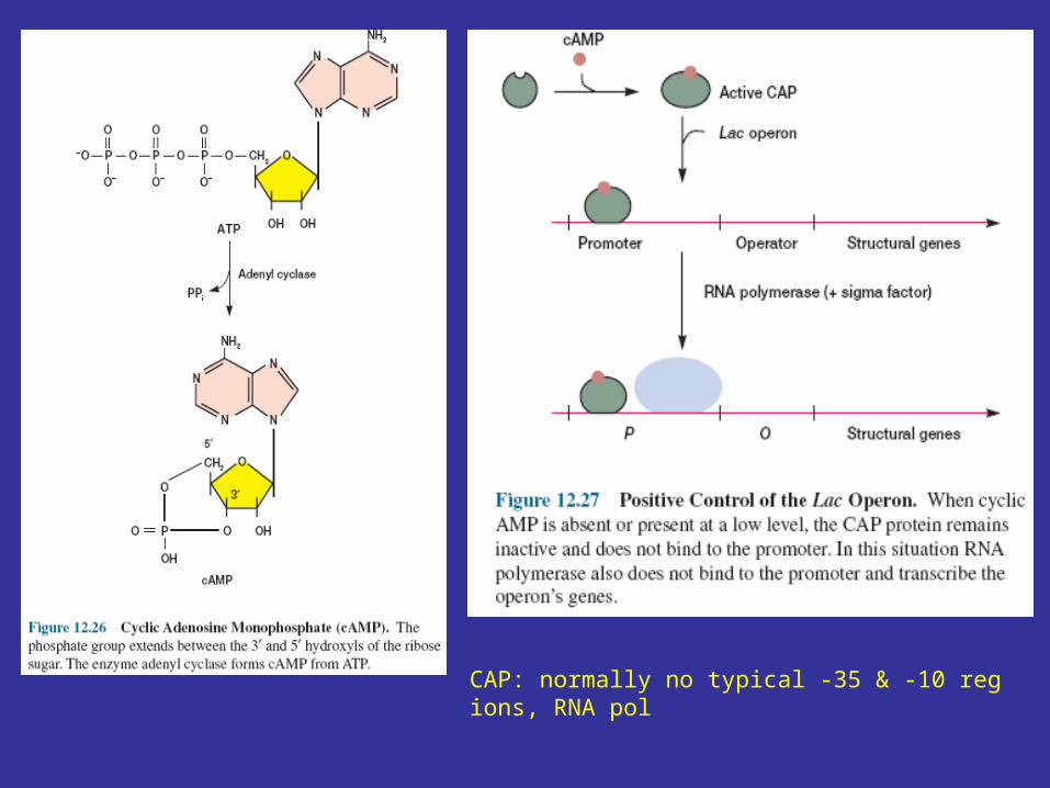
CAP: normally no typical -35 & -10 regions, RNA pol
12.4 Attenuation
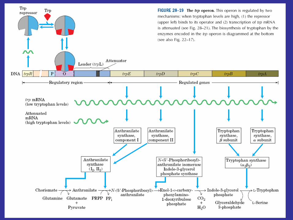
Bacteria can regulate transcription in other ways, as may be seen in the tryptophan operon of E. coli. The tryptophan operon contains structural genes for five enzymes in this amino acid’s biosynthetic pathway. As might be expected, the operon is under the control of a repressor protein coded for by the trpR gene (trp stands for tryptophan), and excess tryptophan inhibits transcription of operon genes by acting as a corepressor and activating the repressor protein. Although the operon is regulated mainly by repression, the continuation of transcription also is controlled. That is, there are two decision points involved in transcriptional control, the initiation of transcription and the continuation of transcription past the attenuator region.
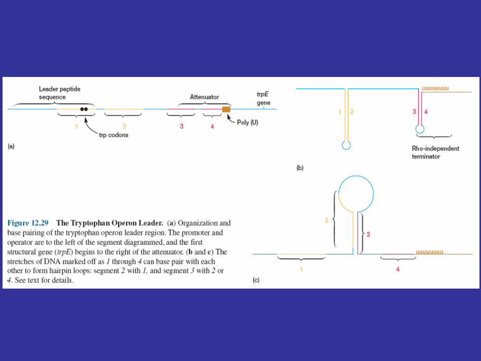
A leader region lies between the operator and the first structural gene in the operon, the trpE gene, and is responsible for controlling the continuation of transcription after the RNA polymerase has bound to the promoter (figure 12.29a). The leader region contains an attenuator and a sequence that codes for the synthesis of a leader peptide. The attenuator is a rho-independent termination site with a short GC-rich segment followed by a sequence of eight U residues. The four stretches marked off in figure 12.29a have complementary base sequences and can base pair with each other to form hairpin loops. In the absence of a ribosome, mRNA segments one and two pair to form a hairpin, while segments three and four generate a second loop next to the poly(U) sequence (figure 12.29b). The hairpin formed by segments three and four plus the poly(U) sequence will terminate transcription. If segment one is prevented from base pairing with segment two, segment two is free to associate with segment three. As a result segment four remains single stranded (figure 12.29c) and cannot serve as a terminator for transcription. It is important to note that the sequence coding for the leader peptide contains two adjacent codons that code for the amino acid tryptophan. Thus the complete peptide can be made only when there is an adequate supply of tryptophan. Since the leader peptide has not been detected, it must be degraded immediately after synthesis.
Ribosome behavior during translation of the mRNA regulates RNA polymerase activity as it transcribes the leader region. This is possible because translation and transcription are tightly coupled. When the active repressor is absent, RNA polymerase binds to the promoter and moves down the leader synthesizing mRNA. If there is no translation of the mRNA after the RNA polymerase has begun copying the leader region, segments three and four form a hairpin loop, and transcription terminates before the polymerase reaches the trpE gene (figure 12.30a). When tryptophan is present, there is sufficient tryptophanyl-tRNA for protein synthesis. Therefore the ribosome will synthesize the leader peptide and continue moving along the mRNA until it reaches a UGA stop codon (see section 12.2) lying between segments one and two. The ribosome halts at this codon and projects into segment two far enough to prevent it from pairing properly with segment three (figure 12.30b). Segments three and four form a hairpin loop, and the RNA polymerase terminates at the attenuator just as if no translation had taken place. If tryptophan is lacking, the ribosome will stop at the two adjacent tryptophan codons in the leader peptide sequence and prevent segment one from base pairing with segment two, because the tryptophan codons are located within segment one (figures 12.29a and12.30c). If this happens while the RNA polymerase is still transcribing the leader region, segments two and three associate before segment four has been synthesized. Therefore segment four will remain single stranded and the terminator hairpin will not form. Consequently, when tryptophan is absent, the RNA polymerase continues on and transcribes tryptophan operon genes. Control of the continuation of transcription by a specific aminoacyl-tRNA is called attenuation.
Attenuation’s usefulness is apparent. If the bacterium is deficient in an amino acid other than tryptophan, protein synthesis will slow and tryptophanyl-tRNA will accumulate. Transcription of thetryptophan operon will be inhibited by attenuation. When the bacterium begins to synthesize protein rapidly, tryptophan may be scarce and the concentration of tryptophanyl-tRNA may be low. This would reduce attenuation activity and stimulate operon transcription, resulting in larger quantities of the tryptophan biosynthetic enzymes. Acting together, repression and attenuation can coordinate the rate of synthesis of amino acid biosynthetic enzymes with the availability of amino acid end products and with the overall rate of protein synthesis. When tryptophan is present at high concentrations, any RNA polymerases not blocked by the activated repressor protein probably will not get past the attenuator sequence. Repression decreases transcription about seventy fold and attenuation slows it another eight- to ten fold; when both mechanisms operate together, transcription can be slowed about 600-fold.
Attenuation seems important in the regulation of several amino acid biosynthetic pathways. At least five other operons have leader peptide sequences that resemble the tryptophan system in organization. For example, the leader peptide sequence of the histidine operon codes for seven histidines in a row and is followed by an attenuator that is a terminator sequence.

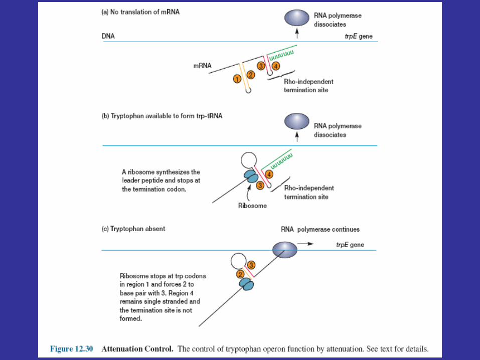
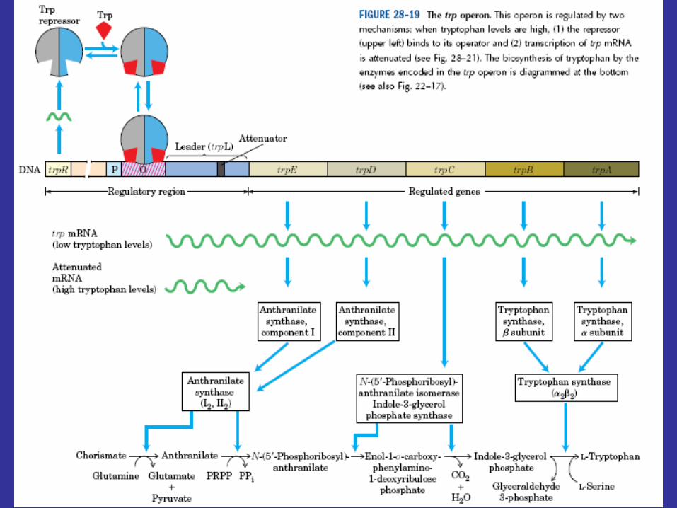
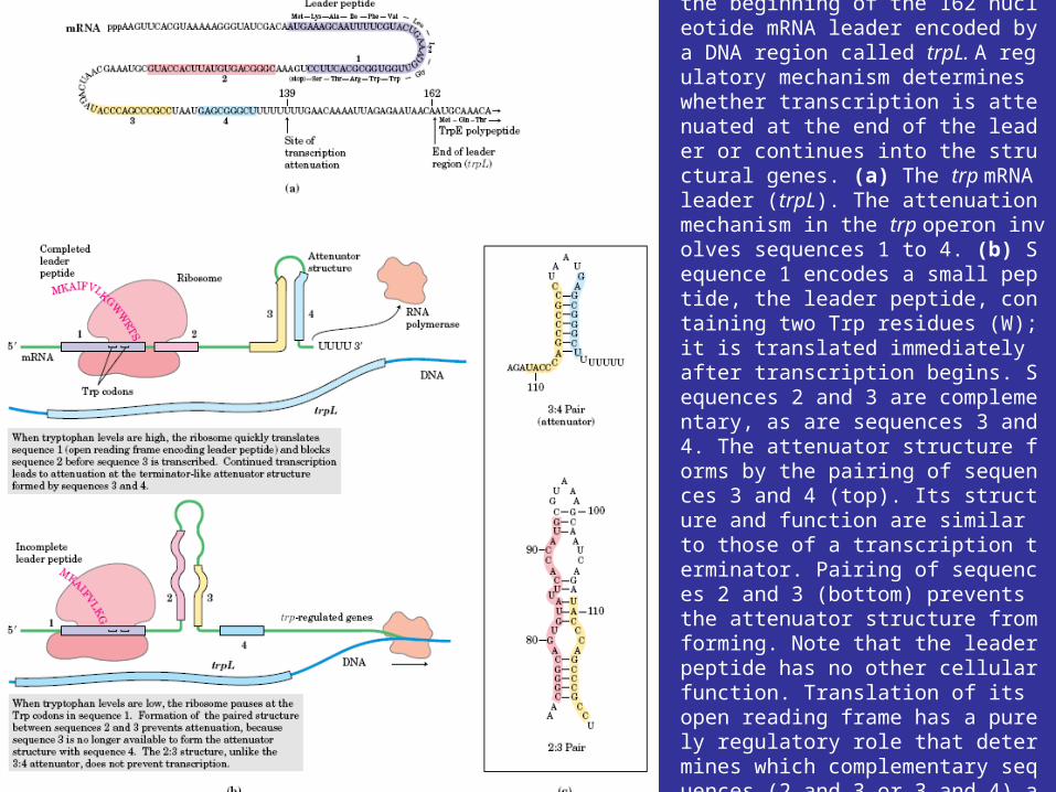
FIGURE 28–21 Transcriptional attenuation in the trp operon. Transcription is initiated at the beginning of the 162 nucleotide mRNA leader encoded by a DNA region called trpL. A regulatory mechanism determines whether transcription is attenuated at the end of the leader or continues into the structural genes. (a) The trp mRNA leader (trpL). The attenuation mechanism in the trp operon involves sequences 1 to 4. (b) Sequence 1 encodes a small peptide, the leader peptide, containing two Trp residues (W); it is translated immediately after transcription begins. Sequences 2 and 3 are complementary, as are sequences 3 and 4. The attenuator structure forms by the pairing of sequences 3 and 4 (top). Its structure and function are similar to those of a transcription terminator. Pairing of sequences 2 and 3 (bottom) prevents the attenuator structure from forming. Note that the leader peptide has no other cellular function. Translation of its open reading frame has a purely regulatory role that determines which complementary sequences (2 and 3 or 3 and 4) are paired. (c) Base-pairing schemes for the complementary regions of the trp mRNA leader.
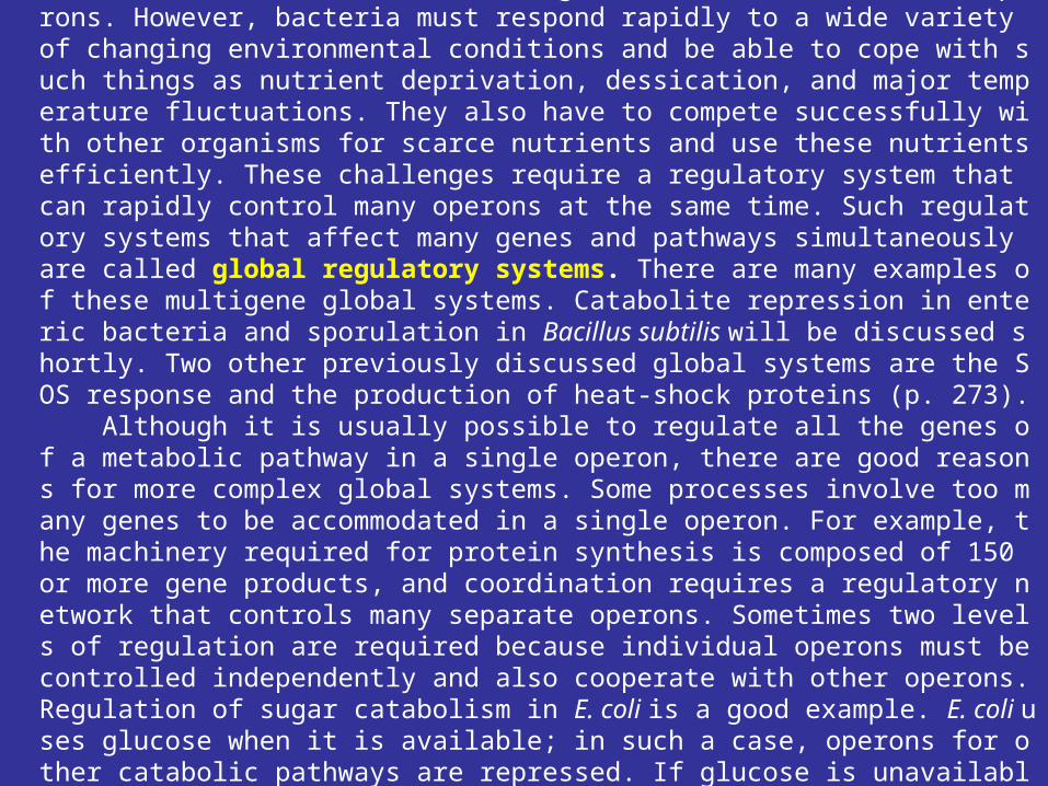
12.5 Global Regulatory SystemsThus far, we have been considering the function of isolated operons. However, bacteria must respond rapidly to a wide variety of changing environmental conditions and be able to cope with such things as nutrient deprivation, dessication, and major temperature fluctuations. They also have to compete successfully with other organisms for scarce nutrients and use these nutrients efficiently. These challenges require a regulatory system that can rapidly control many operons at the same time. Such regulatory systems that affect many genes and pathways simultaneously are called global regulatory systems. There are many examples of these multigene global systems. Catabolite repression in enteric bacteria and sporulation in Bacillus subtilis will be discussed shortly. Two other previously discussed global systems are the SOS response and the production of heat-shock proteins (p. 273). Although it is usually possible to regulate all the genes of a metabolic pathway in a single operon, there are good reasons for more complex global systems. Some processes involve too many genes to be accommodated in a single operon. For example, the machinery required for protein synthesis is composed of 150 or more gene products, and coordination requires a regulatory network that controls many separate operons. Sometimes two levels of regulation are required because individual operons must be controlled independently and also cooperate with other operons. Regulation of sugar catabolism in E. coli is a good example. E. coli uses glucose when it is available; in such a case, operons for other catabolic pathways are repressed. If glucose is unavailable and another nutrient is present, the appropriate operon is activated.
Global regulatory systems are so complex that a specialized nomenclature is used to describe the various kinds. Perhaps the most basic type is the regulon. A regulon is a collection of genes or operons that is controlled by a common regulatory protein. Usually the operons are associated with a single pathway or function (e.g., the production of heat-shock proteins or the catabolism of glycerol). A somewhat more complex situation is seen with a modulon. This is an operon network under the control of a common global regulatory protein, but whose constituent operons also are controlled separately by their own regulators. A good example of a modulon is catabolite repression. The most complex global systems are referred to as stimulons. A stimulon is a regulatory system in which all operons respond together in a coordinated way to an environmental stimulus. It may contain several regulons and modulons, and some of these may not share regulatory proteins. The genes involved in a response to phosphate limitation are scattered among several regulons and are part of one stimulon.
We will now briefly consider three examples of global regulation. First we will discuss catabolite repression and the use of positive operon control. Then an introduction to regulation by sigma factors and the induction of sporulation will follow. Finally, the regulation of porin protein synthesis by antisense RNA will be described.
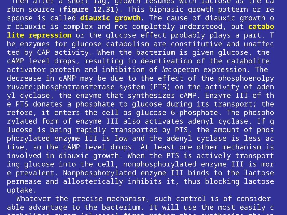
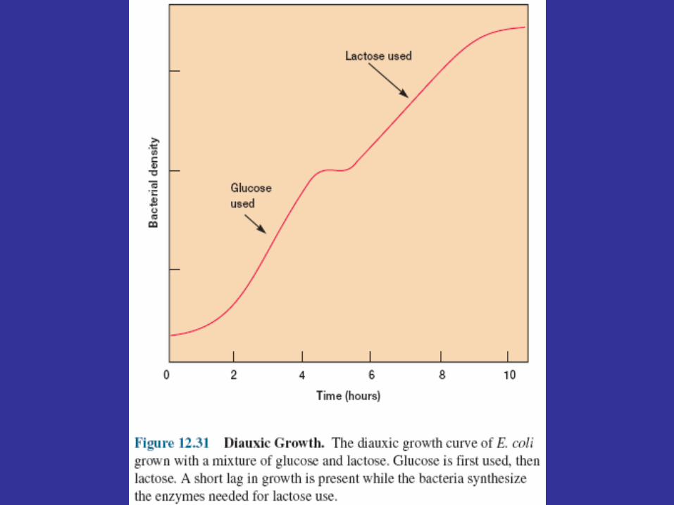
Catabolite Repression If E. coli grows in a medium containing both glucose and lactose, it uses glucose
preferentially until the sugar is exhausted. Then after a short lag, growth resumes with lactose as the carbon source (figure 12.31). This biphasic growth pattern or response is called diauxic growth. The cause of diauxic growth or diauxie is complex and not completely understood, but catabolite repression or the glucose effect probably plays a part. The enzymes for glucose catabolism are constitutive and unaffected by CAP activity. When the bacterium is given glucose, the cAMP level drops, resulting in deactivation of the catabolite activator protein and inhibition of lac operon expression. The decrease in cAMP may be due to the effect of the phosphoenolpyruvate:phosphotransferase system (PTS) on the activity of adenyl cyclase, the enzyme that synthesizes cAMP. Enzyme III of the PTS donates a phosphate to glucose during its transport; therefore, it enters the cell as glucose 6-phosphate. The phosphorylated form of enzyme III also activates adenyl cyclase. If glucose is being rapidly transported by PTS, the amount of phosphorylated enzyme III is low and the adenyl cyclase is less active, so the cAMP level drops. At least one other mechanism is involved in diauxic growth. When the PTS is actively transporting glucose into the cell, nonphosphorylated enzyme III is more prevalent. Nonphosphorylated enzyme III binds to the lactose permease and allosterically inhibits it, thus blocking lactose uptake.
Whatever the precise mechanism, such control is of considerable advantage to the bacterium. It will use the most easily catabolized sugar (glucose) first rather than synthesize the enzymes necessary for another carbon and energy source. These control mechanisms are present in a variety of bacteria and metabolic pathways.
Regulation by Sigma Factors and Control of Sporulation
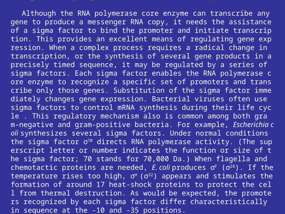
Although the RNA polymerase core enzyme can transcribe any gene to produce a messenger RNA copy, it needs the assistance of a sigma factor to bind the promoter and initiate transcription. This provides an excellent means of regulating gene expression. When a complex process requires a radical change in transcription, or the synthesis of several gene products in a precisely timed sequence, it may be regulated by a series of sigma factors. Each sigma factor enables the RNA polymerase core enzyme to recognize a specific set of promoters and transcribe only those genes. Substitution of the sigma factor immediately changes gene expression. Bacterial viruses often use sigma factors to control mRNA synthesis during their life cycle . This regulatory mechanism also is common among both gram-negative and gram-positive bacteria. For example, Escherichia coli synthesizes several sigma factors. Under normal conditions the sigma factor σ70 directs RNA polymerase activity. (The superscript letter or number indicates the function or size of the sigma factor; 70 stands for 70,000 Da.) When flagella and chemotactic proteins are needed, E. coli produces σF (σ28). If the temperature rises too high, σH (σ32) appears and stimulates the formation of around 17 heat-shock proteins to protect the cell from thermal destruction. As would be expected, the promoters recognized by each sigma factor differ characteristically in sequence at the -10 and -35 positions.
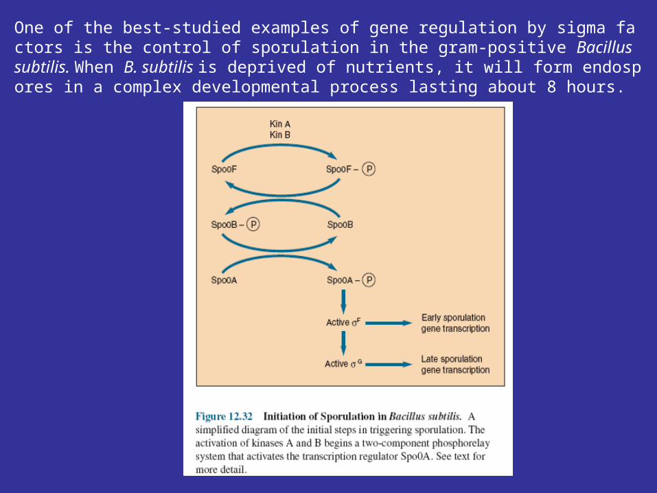
One of the best-studied examples of gene regulation by sigma factors is the control of sporulation in the gram-positive Bacillus subtilis. When B. subtilis is deprived of nutrients, it will form endospores in a complex developmental process lasting about 8 hours.
Antisense RNA and the Control of Porin Proteins
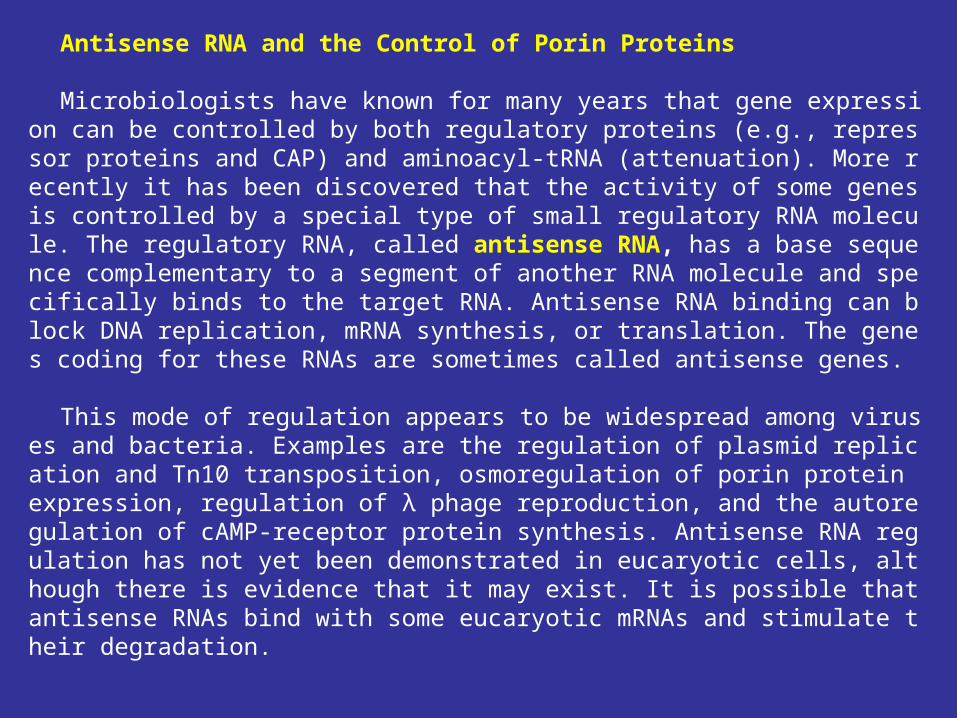
Microbiologists have known for many years that gene expression can be controlled by both regulatory proteins (e.g., repressor proteins and CAP) and aminoacyl-tRNA (attenuation). More recently it has been discovered that the activity of some genes is controlled by a special type of small regulatory RNA molecule. The regulatory RNA, called antisense RNA, has a base sequence complementary to a segment of another RNA molecule and specifically binds to the target RNA. Antisense RNA binding can block DNA replication, mRNA synthesis, or translation. The genes coding for these RNAs are sometimes called antisense genes.
This mode of regulation appears to be widespread among viruses and bacteria. Examples are the regulation of plasmid replication and Tn10 transposition, osmoregulation of porin protein expression, regulation of λ phage reproduction, and the autoregulation of cAMP-receptor protein synthesis. Antisense RNA regulation has not yet been demonstrated in eucaryotic cells, although there is evidence that it may exist. It is possible that antisense RNAs bind with some eucaryotic mRNAs and stimulate their degradation.
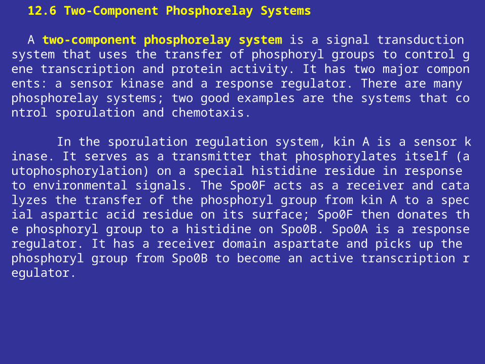
12.6 Two-Component Phosphorelay Systems A two-component phosphorelay system is a signal transduction system that use
s the transfer of phosphoryl groups to control gene transcription and protein activity. It has two major components: a sensor kinase and a response regulator. There are many phosphorelay systems; two good examples are the systems that control sporulation and chemotaxis.
In the sporulation regulation system, kin A is a sensor kinase. It serves as a transmitter that phosphorylates itself (autophosphorylation) on a special histidine residue in response to environmental signals. The Spo0F acts as a receiver and catalyzes the transfer of the phosphoryl group from kin A to a special aspartic acid residue on its surface; Spo0F then donates the phosphoryl group to a histidine on Spo0B. Spo0A is a response regulator. It has a receiver domain aspartate and picks up the phosphoryl group from Spo0B to become an active transcription regulator.
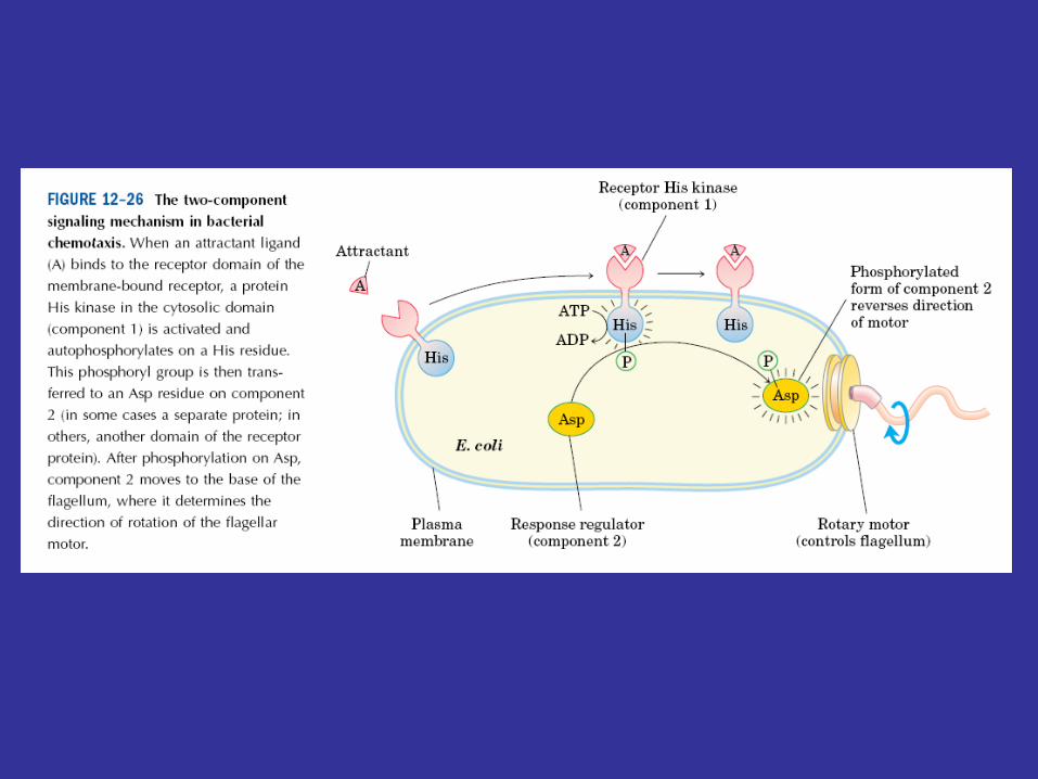
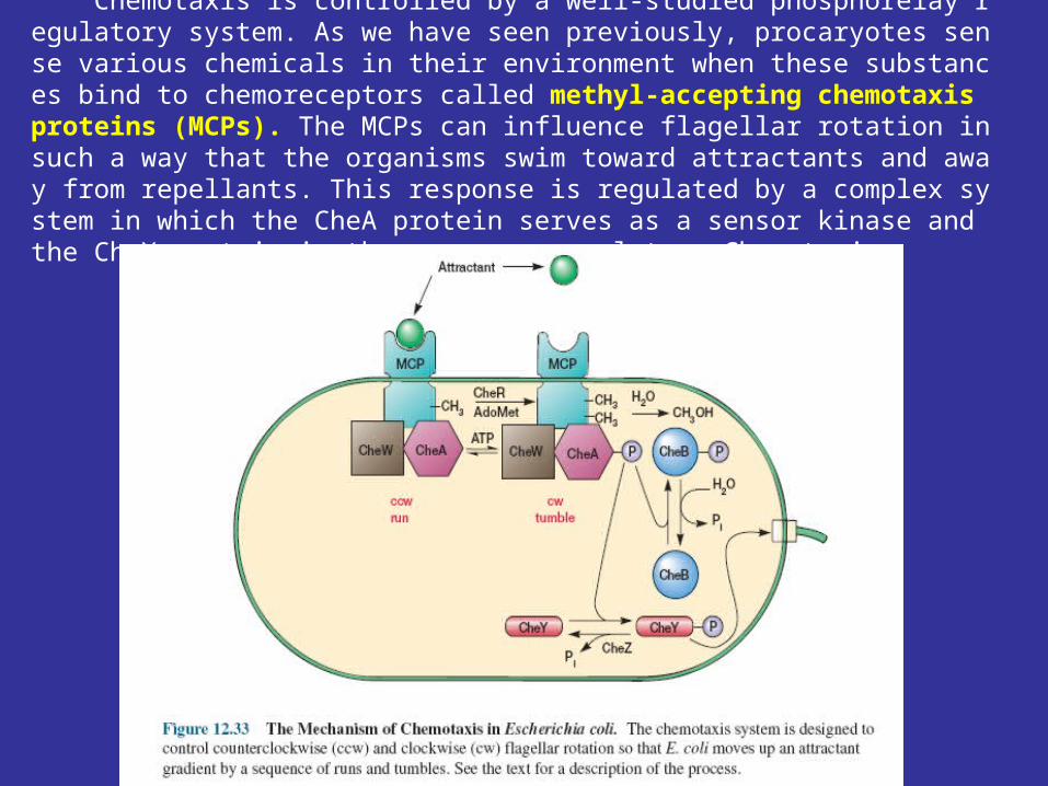
Chemotaxis is controlled by a well-studied phosphorelay regulatory system. As we have seen previously, procaryotes sense various chemicals in their environment when these substances bind to chemoreceptors called methyl-accepting chemotaxis proteins (MCPs). The MCPs can influence flagellar rotation in such a way that the organisms swim toward attractants and away from repellants. This response is regulated by a complex system in which the CheA protein serves as a sensor kinase and the CheY protein is the response regulator. Chemotaxis.
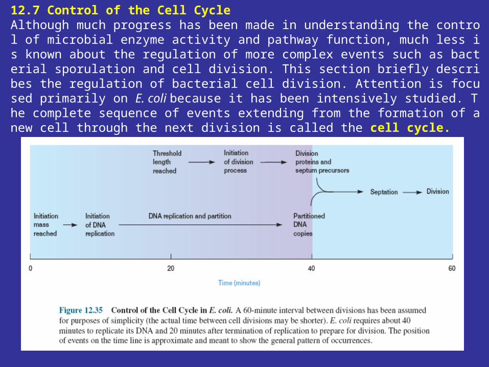
12.7 Control of the Cell Cycle Although much progress has been made in understanding the control of microbial enzyme activity and pathway function, much less is known about the regulation of more complex events such as bacterial sporulation and cell division. This section briefly describes the regulation of bacterial cell division. Attention is focused primarily on E. coli because it has been intensively studied. The complete sequence of events extending from the formation of a new cell through the next division is called the cell cycle.

Vielen dank!