Embed Size (px)
Citation preview

Welcome to MICROnesia
4 “Bug” Case Studies
“Life of a Blood Culture” Slide Show
Questions welcomed!

Case #1 UTI BUG
Ambulatory 26 year old female with 101° temperature and painful urination
Physician orders a urine culture with gram stain

Gram Stain Results
Gram stain morphology shows many gram-positive cocci in pairs and chains

Urine Culture Setup
Urine plated to agar plates
1/1000 ml inoculating loop used
One big drop of urine is enough for a culture!

Urine Culture Results
Culture grows >100,000 colonies of bacteria on a blood agar plate
Patient’s UTI caused by a strep-like organism called Enterococcus

Identifying Enterococcus
Produces an enzyme called PYRase
Detectable in a two minute test

Normal sites for Enterococcus
Upper respiratory tract Gastrointestinal tract Genitourinary tract

Enterococcus Infections
UTI’s Nosocomial UTI’s Wound infections

Emerging Resistance
Emerging strains showing resistance to Vancomycin
Resistant strains called Vancomycin Resistant Enterococcus or VRE
Bone marrow transplant and other immunocompromised patients at risk

Identifying VRE
Identify VRE as an Enterococcus faecalis or faecium using biochemical tests interpreted by an automated instrument

Phoenix Automated Instrument
Performs both biochemical tests and susceptibilities
100 organisms can be tested at a time

VRE on the rise
Enterococcus showing resistance to Vancomycin E-strip
VRE strains account for 6% of all Enterococcus
Patients placed in isolation
Reported to RN and Infection Control

Case #2 Wound Bug
65 year old male with 101° temperature after hip replacement surgery
Develops redness, tenderness and drainage at incision site
Physician orders a culture and gram stain on incision site

Incision site Gram Stain
Gram stain shows few gram-positive cocci in clusters with few wbc’s

Bacterial culture results
Staph aureus isolated on culture
White colonies on blood agar

Identifying Staph aureus
Latex agglutination test can identify an organism as Staph aureus in 10 seconds
Staph aureus infections
Skin infections Scalded Skin Syndrome Toxic Shock Syndrome Osteomyelitis Food poisoning

Staph aureus reservoirs
Carried in nose of 20-40% of adults Higher % in hospital personnel Transferred from nose to skin Passed to others by direct contact or droplets Primary way nosocomial infections occur

Staph aureus treatment
Penicillin discovered in 1920 – worked great on Staph!
More difficult to treat the last 50 years Some SA now showing resistant to methicillin,
a commonly used drug

Identifying MRSA
Strains resistant to methicillin are called MRSA
Extraction test can identify SA as an MRSA strain in 15 minutes

Lots of MRSA
Up to 50% of SA isolated are MRSA strains Carriage rate for MRSA higher in hospitals MRSA often found on health club gym
equipment Pets can get MRSA from their owners
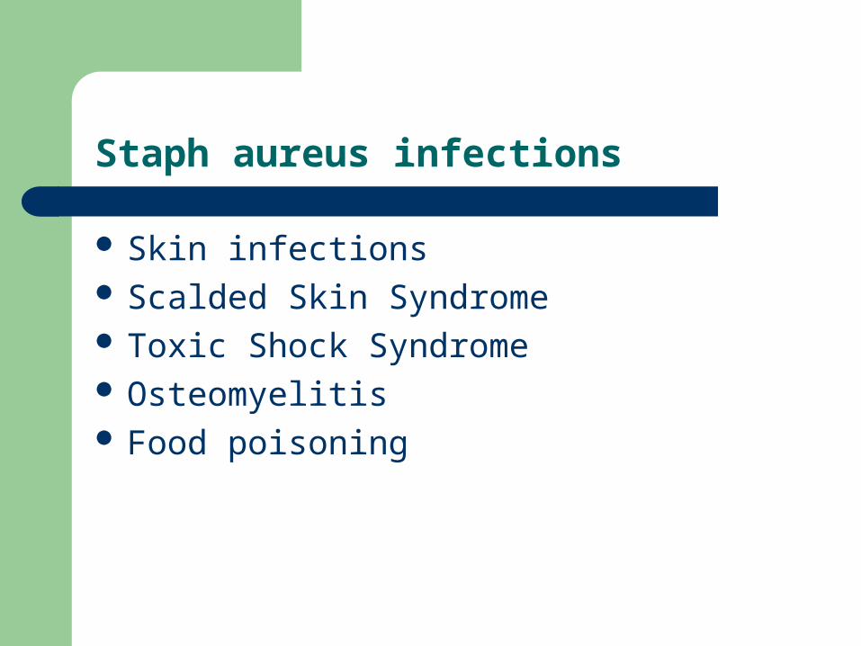
Wash Your Hands
Good handwashing essential!
Careful wound dressing technique
Patients with MRSA placed in isolation
Reported to RN and Infection Control

Case #3 GI BUG
38 year-old HIV positive male Several previous hospital admissions Taking AZT & Bactrim antibiotic therapy 3 day history of severe diarrhea with 10 pound
weight loss and profound dehydration

Lab Results Stat
Leukotest = negative (test for fecal wbc’s) Occult blood exam = negative Both tests usually positive with diarrhea
caused by Salmonella or Shigella Negative Leukotest and Occult blood =
noninflammatory diarrhea

Lab Results not Stat
Ova & Parasite exam negative Stool culture negative for enteric pathogens Campylobacter EIA assay negative Shiga Toxin EIA assay negative

Other Findings
No recent travel history Patient has not recently eaten shellfish

Clues from Patient History
Severe diarrhea consistent with enterotoxigenic E.coli or Vibrio cholerae
Endemic in limited regions Raw or undercooked shellfish may contain
Vibrio cholerae Patient had not consumed shellfish

Suppressive Antibiotic Therapy
Normal gut flora protects the bowel from invasive pathogens
Antibiotics destroy large part normal flora Allows overgrowth of organisms usually
suppressed
Responsible Bug
Clostridium difficile frequently causes antibiotic-associated diarrhea
Disrupted normal flora allows C. difficile to multiply
Produces two different exotoxins

Patient’s Diagnosis
Patient suffering from Clostridium difficile colitis “Pseudomembranous colitis”

More about Clostridium difficile
C. difficile is an anaerobe
Gram-positive rods on Gram Stain

Diagnosing C. difficile colitis
Detect exotoxins in stool using EIA assay
Performed twice daily in Microbiology
Takes about 3 hours Pea-size amount of stool
needed for testing Positive results called to
patient’s RN

Important to Establish Cause of Diarrhea
Many causes of diarrhea in AIDS patients untreatable
C. difficile treatable with oral antibiotics Patient placed in isolation to avoid hospital
outbreaks

Life of a BLOOD CULTURE Slide Show
Drawn in yellow-top SPS tubes
Full size & pedi-tube

Life of a BLOOD CULTURE
4 Kinds of Blood Culture Bottles
Aerobic Anaerobic Pediatric ARD
(Antimicrobial Removal Device)

Life of a BLOOD CULTURE
Chlorhexidine preps or swabs disinfect venipuncture site
Scrub arm for 30 seconds, not to exceed a 2 inch square surface
Let arm air dry

Life of a BLOOD CULTURE
Use of Chlorhexidine preps has decreased blood culture contamination rate by 50%
Blood culture considered “contaminated” if common skin flora grows from one or both bottles in a set

Life of a BLOOD CULTURE
Clean SPS tubes with alcohol and let air dry
Draw 2 SPS tubes for each set of cultures
10 ml in each tube One tube –> aerobic One tube –> anaerobic Record collection site on label
(peripheral, art line, etc.)

Life of a BLOOD CULTURE
Recommended draw times: Two sets drawn at least 30 minutes apart in a
24 hour period Bacterial recovery rate increases by 57% when
2 sets are drawn

Life of a BLOOD CULTURE
Bottles placed in an automated Bactec instrument
Incubate for 5 days Monitored every 15
minutes for bacterial growth

Life of a BLOOD CULTURE
Loud alarm sounds when growth is detected!
Positive blood culture considered a STAT
Subcultured to agar plates
Plates incubate for 18 hours

Life of a BLOOD CULTURE
Gram stain slide made from “positive” bottle

Life of a BLOOD CULTURE
Gram Stain takes about two minutes
Look for bacteria on slide under the microscope
Gram stain results called to patient’s RN

Case #4 BLOOD BUG
37 year old man with sickle cell disease and numerous hospitalizations
Porta-cath placed in right subclavian vein Patient admitted to ED two weeks after porta-
cath placement

Emergency Department findings
Patient has right arm discomfort and swelling Physician orders two sets of blood cultures One drawn through porta-cath One drawn through peripheral vein

Blood culture results
Both sets of blood cultures show gram positive cocci in clusters on smear
Both cultures grow the same organism

Responsible Bug
Two positive blood cultures + porta-cath = probable line-related sepsis
Most common bug causing line-related infection is Coagulase Negative Staph or CNS
CNS important cause of nosocomial bacteremia
Foreign body devices act as source

Identifying CNS
Grow as white colonies on blood agar plate
Nonreactive in rapid latex tests

Sources of CNS
Normal inhabitants of skin, mucous membranes and nares
About 20 species of CNS Most common is Staph epidermidis

Slime Producers
CNS secrete a virulence factor called slime Makes them “sticky” Stick to plastic surfaces like catheter tips Slime-producing strains more difficult to treat
with antibiotics Indwelling catheters place patient at risk for
infection

Diagnosing Line-Related Sepsis
Draw 2 sets of blood cultures from a patient with fever or signs of infection at the IV site
One set from catheter line One set from peripheral site

Two sites important
CNS on skin can be a blood culture “contaminant” if blood not collected properly
Single positive blood culture with CNS may be skin contamination and not true infection

Two sites important
Negative peripheral culture and positive line culture with CNS may just show local infection of the catheter site
Two Blood cultures with CNS from two different sites more likely represents true infection

Confirming line-related sepsis
Confirm by performing a catheter tip culture Catheter is removed and sent to Micro Lab

Culturing the Catheter tip
Catheter tip cut to 50mm Roll on surface of blood
agar plate

Interpreting Catheter tip cultures
Culture “positive” if 15 colonies grow from a 50 mm tip
CNS growing on plate

Diagnosis confirmed
If Catheter tip culture has CNS and blood cultures from both the line and peripheral draws have CNS
Patient has a confirmed line-related sepsis

Microbiology Art
Thank you! Please call Microbiology
with any questions: 2-2422 2-2435