Embed Size (px)
Citation preview
Welcome To Journal Club
Presented by:
Dr. Aminul Islam
Lecturer of Microbiology, MMC
Molecular Diagnosis of Infective Endocarditis by PCR Amplification and
Direct Sequencing of DNA from Valve Tissue
Journal:
Journal of Clinical Microbiology 2003 February; 41(2): 763–766.
Author: Valérie Gauduchon, Lara Chalabreysse, Jerome
Etienne, Marie Célard, Yvonne Benito,
Abstract
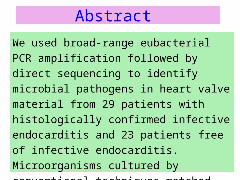
We used broad-range eubacterial PCR
amplification followed by direct sequencing to
identify microbial pathogens in heart valve
material from 29 patients with histologically
confirmed infective endocarditis and 23
patients free of infective endocarditis.
Microorganisms cultured by conventional
techniques matched those identified by PCR
in 21 cases.
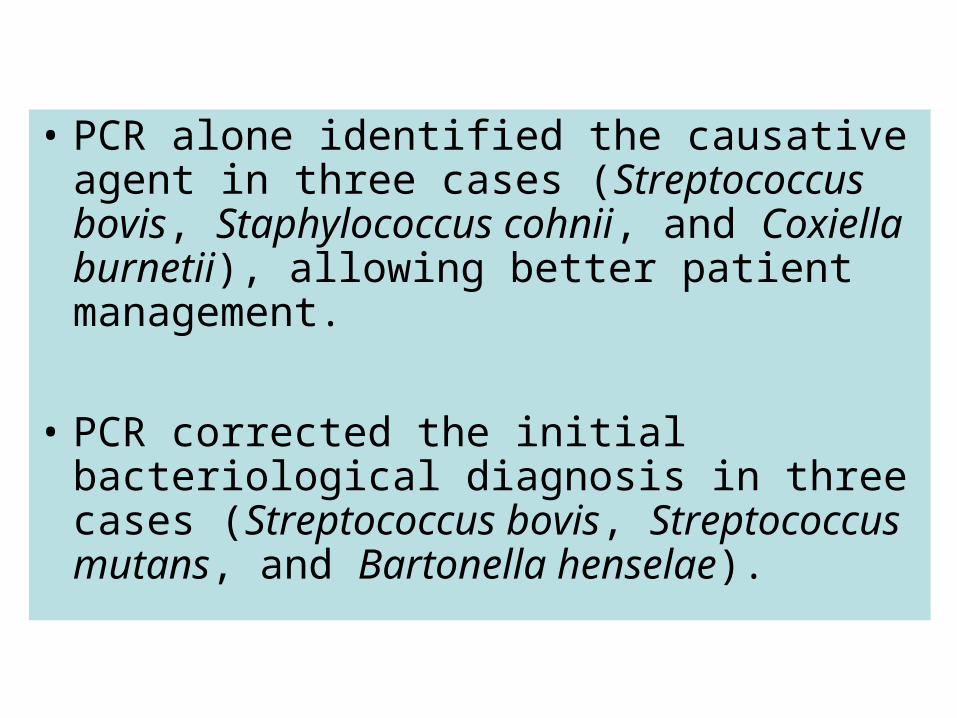
• PCR alone identified the causative agent in three cases (Streptococcus bovis, Staphylococcus cohnii, and Coxiella burnetii), allowing better patient management.
• PCR corrected the initial bacteriological diagnosis in three cases (Streptococcus bovis, Streptococcus mutans, and Bartonella henselae).
Introduction
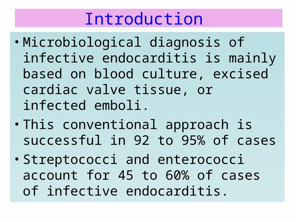
• Microbiological diagnosis of infective endocarditis is mainly based on blood culture, excised cardiac valve tissue, or infected emboli.
• This conventional approach is successful in 92 to 95% of cases
• Streptococci and enterococci account for 45 to 60% of cases of infective endocarditis.
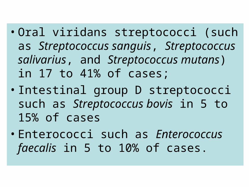
• Oral viridans streptococci (such as Streptococcus sanguis, Streptococcus salivarius, and Streptococcus mutans) in 17 to 41% of cases;
• Intestinal group D streptococci such as Streptococcus bovis in 5 to 15% of cases
• Enterococci such as Enterococcus faecalis in 5 to 10% of cases.
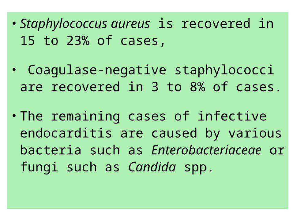
• Staphylococcus aureus is recovered in 15 to 23% of cases,
• Coagulase-negative staphylococci are recovered in 3 to 8% of cases.
• The remaining cases of infective endocarditis are caused by various bacteria such as Enterobacteriaceae or fungi such as Candida spp.
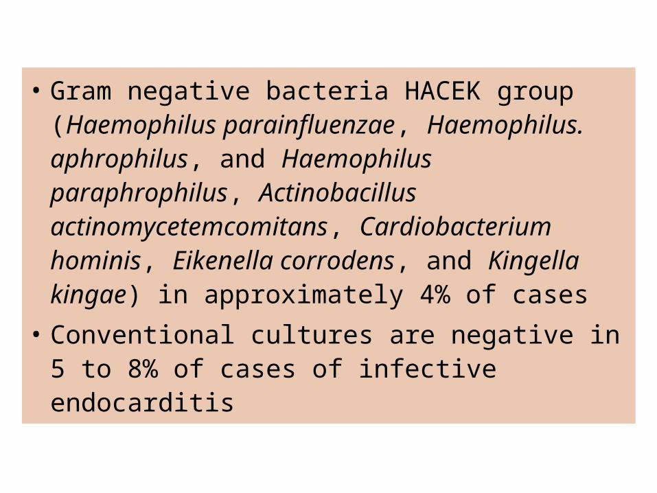
• Gram negative bacteria HACEK group (Haemophilus parainfluenzae, Haemophilus. aphrophilus, and Haemophilus paraphrophilus, Actinobacillus actinomycetemcomitans, Cardiobacterium hominis, Eikenella corrodens, and Kingella kingae) in approximately 4% of cases
• Conventional cultures are negative in 5 to 8% of cases of infective endocarditis
MATERIALS AND METHODS
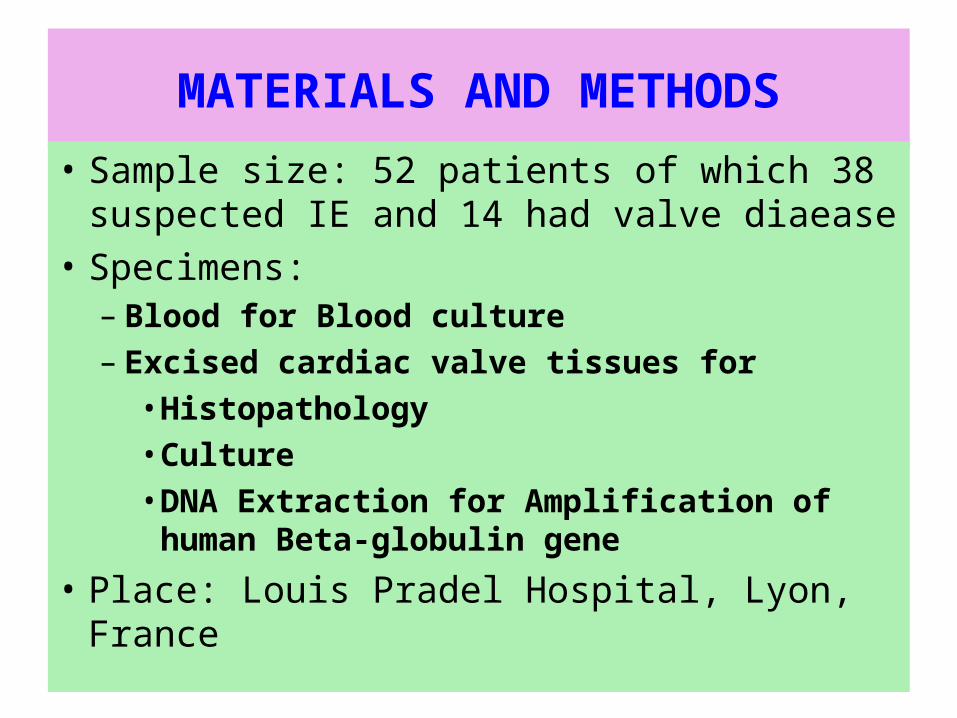
• Sample size: 52 patients of which 38 suspected IE and 14 had valve diaease
• Specimens: – Blood for Blood culture– Excised cardiac valve tissues for
• Histopathology• Culture• DNA Extraction for Amplification of
human Beta-globulin gene
• Place: Louis Pradel Hospital, Lyon, France
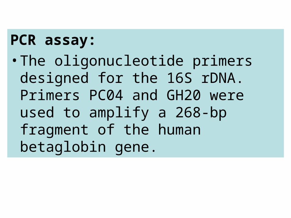
PCR assay:
• The oligonucleotide primers designed for the 16S rDNA. Primers PC04 and GH20 were used to amplify a 268-bp fragment of the human betaglobin gene.
RESULTS
1. First group of 38 patients with suspected IE:
• Definite infective endocarditis was diagnosed in 28 patients, on the basis of vegetations or intracardiac abscesses found at surgery and histopathologic confirmation of active infective endocarditis.
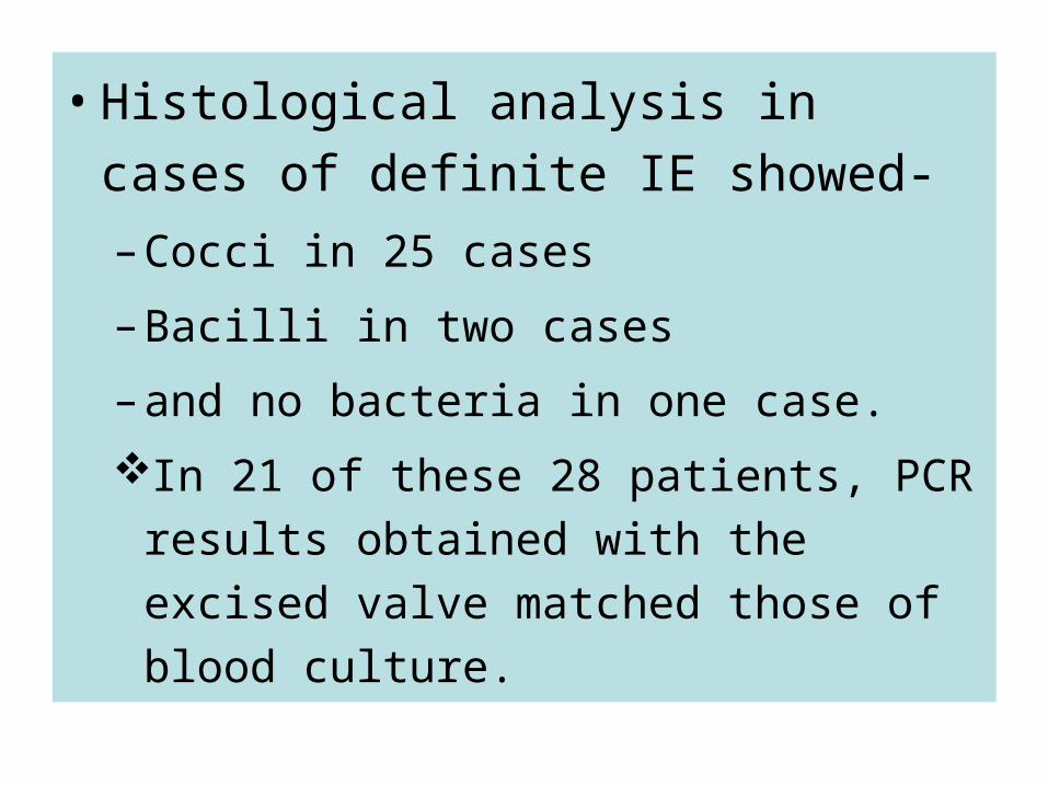
• Histological analysis in cases of
definite IE showed-
– Cocci in 25 cases
– Bacilli in two cases
– and no bacteria in one case.
In 21 of these 28 patients, PCR results
obtained with the excised valve matched
those of blood culture.
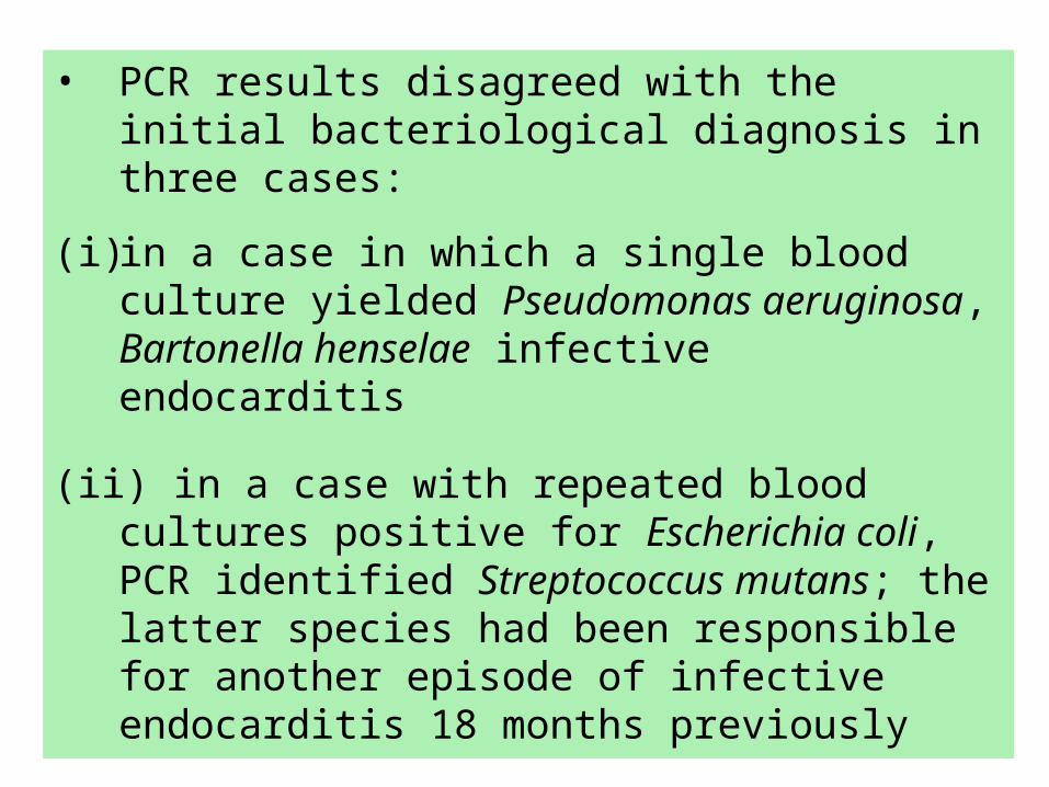
• PCR results disagreed with the initial bacteriological diagnosis in three cases:
(i) in a case in which a single blood culture yielded Pseudomonas aeruginosa, Bartonella henselae infective endocarditis
(ii) in a case with repeated blood cultures positive for Escherichia coli, PCR identified Streptococcus mutans; the latter species had been responsible for another episode of infective endocarditis 18 months previously
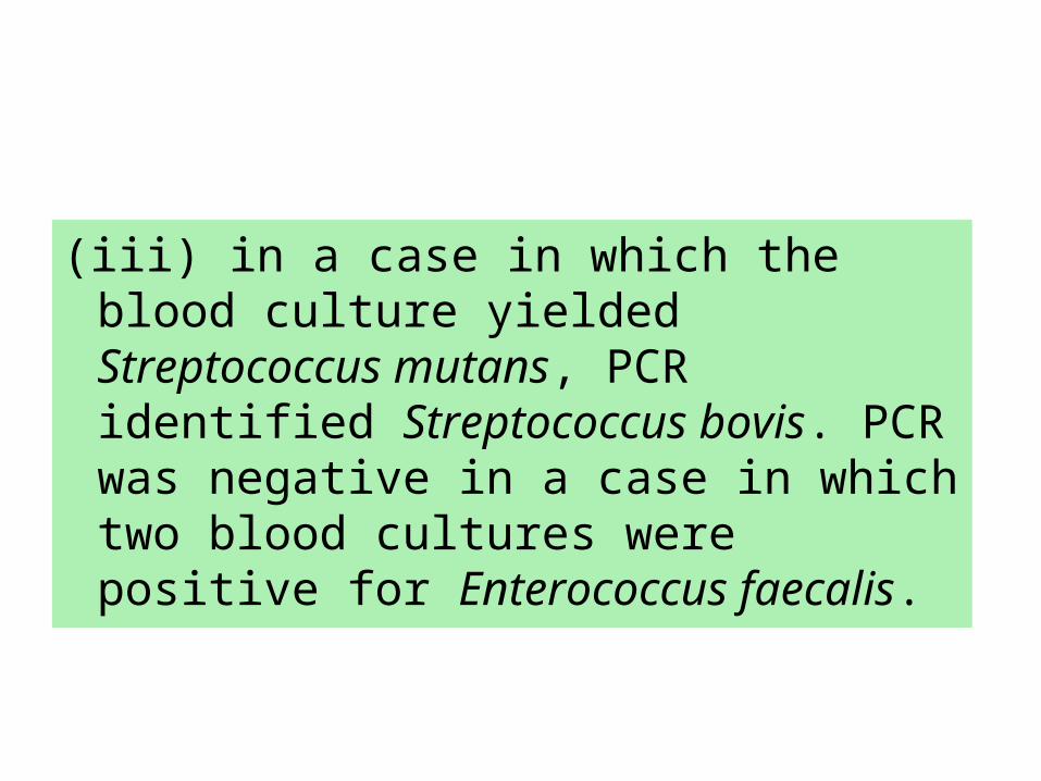
(iii) in a case in which the blood culture yielded Streptococcus mutans, PCR identified Streptococcus bovis. PCR was negative in a case in which two blood cultures were positive for Enterococcus faecalis.
• In two patients with a negative blood culture, the etiological agent of infective endocarditis was identified by PCR on excised valve tissue:
• The diagnosis of infective endocarditis was rejected in all but one of the 14 negative control patients. PCR identified Coxiella burnetii infective endocarditis
DISCUSSION
• PCR findings usually agreed with the results of conventional bacteriological and histopathologic diagnosis.
• Bacteria were visualized in valve tissue for up to 150 days after initial antibiotic treatment and were always associated with histopathological evidence of active infective endocarditis. PCR was positive even when the valve tissue culture was negative.
• In our study, PCR modified the initial
bacteriological diagnosis in three cases. In
the first, Streptococcus bovis was identified
by PCR (instead of Streptococcus mutans);.

• In the second, Streptococcus mutans
was identified by PCR in a patient
who had had Streptococcus mutans
infective endocarditis 18 months
previously and currently had
interfering Escherichia coli sepsis.
• In the third, Bartonella henselae was
identified in a patient with interfering
Pseudomonas aeruginosa sepsis
secondary to needle biopsy of the kidney.
• Our results suggest that this molecular approach may prove useful in other cases:
• (i) when bacteria rarely associated with infective endocarditis are recovered by blood culture (e.g., Enterobacteriaceae);
• (ii) when blood culture is positive only once
• (iii) when strain identification is unsure (e.g.,
• (iv) when valve replacement for noninfectious indications leads to histologic diagnosis of infective endocarditis.
• In conclusion, we confirm that broad-
range PCR amplification followed by
direct sequencing is a reliable and
accurate method when applied to
resected heart valves.


![(e)MMC/SD/SDIO State of Affairsconnect.linaro.org.s3.amazonaws.com/sfo17... · First MMC stack by Russell King in kernel 2.6.9 [MMC] Add MMC core](https://img.pdfslide.us/doc/110x75/5f5ee69463a1e67f0c5f43dc/emmcsdsdio-state-of-first-mmc-stack-by-russell-king-in-kernel-269-mmc-add.jpg)