Embed Size (px)
Citation preview
Wegener Granulomatosis with Meningeal Involvement
Steven Tishler, 1 Todd Williamson ,2 Suzanne S. Mirra,3 Jeffrey B. Lichtman,4 •7 Pedro Gismondi ,5 and Michael B. Kibble6
Summary: The cl inical , radiographic, and neuropathologic features of the case of a 41-year-old man with Wegener granulomatosis presenting with neurologic symptoms are correlated.
CT and MR scans of the head demonstrated extensive meningeal thickening and enhancement. The importance of considering this diagnosis, confirmed by antineutrophil cytoplasmic autoantibodies , is emphasized.
Index terms: Granuloma; Meninges, computed tomography;
Meninges, m agnetic resonance
We report a case of Wegener granulomatosis (WG) with computed tomography (CT) and magnetic resonance (MR) imaging findings of thick , nodular meningeal involvement.
Case Report
A 41 -year-old construction worker noted the onset of severe headaches four years prior to admission. With no specific treatment, his headaches improved. Three years later, he noted blurry vision in his left eye and subsequently had a generalized tonic-clonic seizure.
Physical examination revealed a well-appearing man with a temperature of 99.2°F and otherwise normal vital signs. An infero-nasal field cut was present in the left eye. Fundoscopic examination revealed pallor of the left optic disc. Sensation to pin prick was decreased in the V 1 distribution of the left trigemina l nerve.
Routine laboratory evaluation including liver and renal function profiles, complete blood count, and urinalysis was normal. The sedimentation rate was 54 U (normal range: 0-20 U) and the following serological tests were negative: antinuclear antibody, rheumatoid factor, rapid plasm a reagin, fluorescent treponema! antibody, and human immunodeficiency virus. A skin test for tuberculosis was normal. Chest roentgenograms and chest CT were negat ive. CT scan of the head demonstrated an extraaxial enhancing mass near the left cavernous sinus, thick meningeal enhancement, and a nonenhancing left frontal lobe hypodensity adjacent to the thickened meninges (Figs. 1 A-1 C).
In addition, partial opacification of the sphenoid , ethmoid, and both maxillary sinuses was demonstrated. There was no bone destruction identified (Figs. 1 D and 1 E). MR scans confirmed the CT findings (Fig. 2) . Two biopsies were subsequently performed .
Biopsy 1
Multiple tissue fragments were removed from the dura overlying the left frontal cortex . Microscopic examination revealed numerous necrotizing granulomas composed of a rim of palisading histiocytes and giant cells with central necrosis. Multiple foci of lymphoplasmacytic infiltrates were also present. A battery of special stains for acid-fast bacilli , fungi , bacteria , and spirochetes were all negative. Similarly, electron microscopic examination failed to reveal any microorganisms.
The patient was discharged on antituberculous medication (as a therapeutic trial , in view of necrotizing . granulomas on biopsy) and on phenytoin. He developed progressive generalized fatigue, nausea , and vomiting, and continued to have seizures. Two months after his initial admission, a second brain biopsy was performed with similar results.
Biopsy 2 (3 months later)
Tissue was received from the dura as well as the underly ing frontal cortex. Microscopic examination again revealed numerous necrotizing granulomas with scattered multinucleated giant cells at their periphery and lymphoplasm acytic inflammation involving the dura . Foreign body granulomata surrounding suture material were also noted , reflecting the previous surgery at this site. In addition, a fragment of cerebral cortex showed striking changes: the pia-arachnoid was thickened and fibrotic with numerous granulom ata , often surrounding and involving blood vessels, along with sheets of lymphocytes in the subarachnoid space. The underly ing cortex showed lymphocytic inflammation in vessel walls as well as reactive astrocytosis . Again, a battery of special stains and electron microscopy fa iled to reveal any microorganisms.
Received June 30, 1992; revision requested August 26; revision received October 20 and accepted October 27. 1 Department of Radiology, 2 Department of Neurology, 3 Department of Pathology and Labora tory of Medicine, 4
·6 Department of Radiology , and
5 Department of Medicine (Rheumatology), Emory University , Atlanta, Georgia 30322. 3
_.·6 V.A. Medical Center (Atlanta), 1364 Clairmont Road, Decatur, GA 30033.
5 Current address: COMG, 3330 NW 56th Street, Suite 400, Oklahoma City, OK 73 112. 7 Address requests for reprints to Jeffrey B. Lichtman, MD, Department of Rad iology (11 4), V.A. Medical Center (Atlanta), 1670 Cla irmont Road,
Decatur, GA 30033.
AJNR 14:1248-1252, Sep/ Oct 1993 0195-6 108/ 93/ 1405-1248 © American Society of Neuroradiology
1248

AJNR: 14, September /October 1993 WEGENER GRANULOMATOSIS 1249
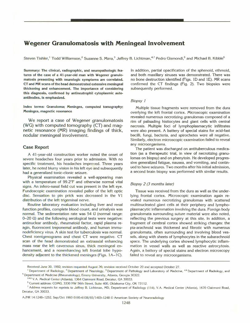
A 8 c
D E F Fig. 1. A-E, Axial and coronal CT images obtained immediately following intravenous contrast. F, Axial contrast-enhanced CT
image obtained after a 16-month interval, following therapy. A, Enhancing mass lateral to left cavernous sinus, contiguous with meninges, extending along medial left temporal lobe and inferior
left frontal lobe (arrows). B, Enhancement of optic chiasm (straight arrow). Nonenhancing deep left frontal lobe hypodensity (curved arrow). C, Thick , nodular meningeal enhancement adjacent to left frontal lobe, extending along the anterior fa lx (arrows). There is mass
effect upon the left frontal horn. D, Partial opacification of the sphenoid sinus (arrow). The sphenoid roof is intact. E, Nodular opacification of right maxillary sinus. Bilateral partial ethmoid opacification . F , Partial resolution of enhancing thickened meninges, following therapy (arrows). There was similar partial resolution at all other
levels.
At the time of the second biopsy , a neutrophil cytoplasmic antibody test was positive at 1 :80 with . a cytoplasmic pattern; a diagnosis of WG was made. The patient refused to permit a biopsy of his maxillary sinuses.
The patient was treated with 125 mg cyclophosphamide by mouth daily, with improvement of all symptoms and marked resolution of the lesions on CT scan (Fig. 1 F). Serum creatinine, urinalysis, and chest roentgenograms subsequently have remained normal.
Discussion
WG is a rare disorder of unknown etiology characterized by acute necrotizing granulomata
of the nose, sinuses, and lung, multifocal necrotizing vasculitis affecting the small arteries and veins of the respiratory tract and other sites, and focal or diffuse glomulonephritis (1). Although the paranasal sinuses, lungs, and kidneys are most commonly affected , many other organs can be involved. Presentation with only neurologic signs and symptoms is infrequent but has been described by several authors. In a review of 1 04 cases, Drachman described three nervous system presentations of this disorder (2) : 1) necrotizing vasculitis involving the cerebral, spinal , and radicular vasculature (28% of total cases); 2) contig-

1250 TISHLER AJNR: 14, September / October 1993
A 8
D E Fig. 2. A-D, Axial and coronal Tl-weighted MR images (800/20/1 (repetition time/echo time/excitations)) following administration
of gadolin ium-DTPA. £ , Unenhanced axial T2-weighted MR image (2000/ 80/ 2). A, Lobulated, enhancing extraax ial mass lateral to the left cavernous sinus with meningeal enhancement extending amund the left
temporal lobe (straight arrows). Ethmoid sinus enhancement (curved arrow). B, Bilateral meningeal enhancement (arrows). Mass effect on the left frontal horn. Small area of increased signal in right frontal sinus
(curved arrow) . C, Thick left frontoparieta l meningeal enhancement with effacement of underlying brain (arrows). D, Enhancing extraaxial mass adjacent to the left cavernous sinus with thick meningeal enhancem ent extending over the left
convexity (straight arrows). Abnormal signal in left lateral extension of the sphenoid sinus and right inferior sphenoid sinus (curved arrows).
E , T hickened meninges displaying predominantly high signal intensity (straight arrows) but also som e areas of intermediate and low signal intensity (open curved arrows). High signal intensity in left frontal lobe (curved arrow) and in frontal sinus (open straight arrow).
uous extension from extravascular granulomas in the paranasal sinuses, nasal cavities, or orbits (26 %); and 3) primary necrotizing granulomas in the skull , meninges, cranial nerves, or brain (4% ). Fauci and coworkers followed 85 patients with WG over a 21-year period (3) . Men were affected more than women in a ratio of approximately 3:2 , the mean age at onset of disease was 40.6
years, and the most common presentations were related to the sinuses or lungs. After evaluation, all patients were found to have involvement of the upper and/or lower respiratory tract. Although involvement of the kidneys occurred in 85% , renal failure was the presenting illness in only 11 % of cases. In Fauci et at's report, neurologic involvement was present in 22% as com-

AJNR: 14, September/October 1993
pared with 54% of the cases reviewed by Drachman. This difference probably reflects more disease prior to the introduction of effective treatment in Drachman's series (2).
The most common neurologic manifestation in Fauci et at's patients was mononeuritis multiplex (3). Central nervous system involvement most often took the form of cranial nerve palsies with the seventh nerve being affected most often (3). Multiple other neurologic presentations have been described including diabetes insipidus, subacute meningitis, syncope, and large vessel occlusion resulting in infarction (3-5).
The laboratory evaluation in WG frequently reveals anemia, an elevated sedimentation rate, and negative antinuclear antibody and lupus erythematosus preparations (3). Antineutrophil cytoplasmic autoantibodies (ANCA) have been shown to be very helpful in making the diagnosis. These antibodies were first reported in 1982 in association with necrotizing glomerulonephritis (6). Since then, ANCA have been associated with systemic vasculitis, polyarteritis nodosa, and crescentic glomerulonephritis (7). Two patterns of immunofluorescence have been described: perinuclear (P-ANCA), which may in some cases show affinity for myeloperoxidase, and diffuse cytoplasmic (C-ANCA), which may have affinity for proteinase 3 (8). Nolle and coworkers (9) studied 277 patients with active WG along with 1657 controls and found that, by immunofluorescence, C-ANCA has a sensitivity of 67% and a specificity of 99%. The sensitivity was 96% in patients with active generalized disease and 41% for patients in full remission after active generalized disease. Thus, disease activity may be followed with C-ANCA (9) and may, in fact , play a role in its pathogenesis by stimulating leukocytes and leading to inflammation and necrosis. Speeks and colleagues arrived at similar conclusions, using a large control group including patients with sarcoidosis, malignancies, non-Wegener vasculitis, and other rheumatologic disease (10).
The radiographic findings of paranasal involvement in WG are nonspecific. Mucoperiosteal thickening and destruction of sinus walls or the nasal septum may be present (11). Imaging studies in the present case do not reveal contiguous invasion of the skull base from paranasal disease, although the basilar involvement is in close proximity to the sphenoid sinus. It is postulated that these necrotizing granulomata represent primary involvement of the brain.
WEGENER GRANULOMATOSIS 1251
Meningeal enhancement (CT or MR) may be associated with neoplastic or inflammatory diseases. Infectious meningitis generally results in a diffuse linear pattern of enhancement. A thick nodular pattern, as seen in this case, is more often associated with meningeal tumor (particularly lymphoma or metastatic disease) or noninfectious inflammatory disease (particularly sarcoidosis) (12); the thick pattern of meningeal involvement of these entities may be indistinguishable from that seen with this case of WG (13). Rarer entities that may display a thick nodular pattern include hypertrophic cranial pachymeningitis (14) and Erdheim-Chester disease (lipid granulomatosis) . This latter entity favors a parasagittal location and may display prolonged enhancement with gadolinium-DTPA (up to 6 days) (15).
In general, the use of T1-weighted images with gadolinium-DTP A improves the detection of meningeal involvement as compared with unenhanced T2-weighted images (the high signal of cerebrospinal fluid with unenhanced T2 weighting may obscure a similarly high signal from abnormal meninges) (16). This case of WG follows these generalizations, as the abnormal meninges display predominantly high signal intensity on the T2-weighted images (Fig. 2E), and the abnormal meninges are much better distinguished on the enhanced T 1-weighted images.
Although primary central nervous system involvement by WG is rare, the success of cyclophosphamide in treating this potentially devastating disease (3) has heightened the importance of its recognition. In the appropriate clinical setting, WG should be considered in the differential diagnosis of meningeal enhancement or of a mass lesion in the central nervous system. However, the constellation of clinical symptoms and imaging patterns described in this case is not specific for the diagnosis of WG.
References
1. Cotran RS. Kumar V, Robbins SL. Pathological basis of disease. 4th
ed. Philadelphia: W. B. Saunders, 1989:553-595
2. Drachman DA. Neurologica l complications of Wegener 's granuloma
tosis. Arch !'leurol1963;8:141-155
3. Fauci AS, Haynes BF. Katz P. et al. Wegener's granulomatosis:
prospective clinica l and therapeutic experience with 85 patien ts for
21 years. A nn Intern Med 1983;98: 76-85
4. Atcheson SG, Horn GV. Subacute meningitis heralding a diffuse
granulomatous angi itis: (Wegener's granulomatosis?). Neurology
1977;27:262-264 5. Satoh J , Miyaska N. Yamacha T , et al. Extensive cerebral infarction

1252 TISHLER
due to involvement of both anterior cerebra l arteries by Wegener's
granulomatosis._Ann Rheum Dis 1988;47:606-611
6. Davies DJ, Moran JE, Niall JF, et al. Segmental necrotizing glomer
ulonephritis with antineutrophil antibody: possible arbovirus a etiol
ogy. Br Med J 1982;285:606
7. Jennette JC, Wilkman AS, Falk RJ. Anti-neutrophil cytoplasmic
autoantibody-associated glomerulonephritis and vasculitis. Am J Pa
thol1989;135:921-930
8. Jennett JC, Falk RJ. Antineutrophil cytoplasmic autoantibodies with
associated disease: a review. Am J Kidney Dis 1990;15:517-529
9. Nolle B, Specks U, Ludemann J, et al. Anticytoplasmic autoantibod
ies: their immunodiagnositic value in Wegener's granulomatosis. Ann
Intern Med 1989; 111:28-40
10. Specks U, Wheatley CL, McDonald T J , et al. Anticytoplasmic auto
antibodies in the diagnosis and follow up of Wegener's granuloma
tosis . Mayo C/in Proc 1989;84:28-36
11. Som PM. The paranasal sinuses. In: Bergeron RT, Osburn AG, Som
AJNR: 14, September/October 1993
PM, eds. Head and neck imaging. St. Louis: C. V. Mosby, 1984:1-
142
12. Phillips ME, Ryals T J, Karnbhu SA, et al. Neoplastic vs. inflammatory
meningeal enhancement with GD-DTPA. J Comput Assist Tomogr
1990;14:536-541
13. Sherman JL, Stern BJ. Sarcoidosis of the CNS: comparison of
unenhanced and enhanced MR images. AJNR: Am J Neuroradiol
1990;11:915-923
14. Martin N, Masson C, Henin D, et al. Hypertrophic cranial pachymen
ingitis: assessment with CT and MR imaging. AJNR: Am J Neuroradiol
1989; 10:4 77-483
15. Tien RD, Brasch RC, Jackson DE, et al. Cerebral Erdheim-Chester
disease: persistent enhancement with Gd-DTPA on MR images. Ra
diology 1989;172:791-792
16. Modic MT. Case 20. In: Siegel BA, sect ed. Magnetic resonance test
and syllabus. Reston , Va: American College of Radiology, 1991:348-
363