Embed Size (px)
Citation preview

Week 5: Neonatal Neurology
Pediatric Neurology
Friday, July 31 4:30-5:45 pm EDT
Moderators Christopher Smyser Tomo Tarui
EDT Abstract Title Presenting Author 4:30 pm Introduction & General Information
4:35 pm 3379333 Disruption in Expression of Circadian Rhythm Genes Following Traumatic Brain Injury in Children Catherine Stacey
4:45 pm 3376625
In Infants with Neonatal Abstinence Syndrome, MRI-Derived Cerebral Perfusion Reflects Hammersmith Neonatal Neurological Examination Findings Kristen Benninger
4:55 pm 3376615 Effect of obesity on levetiracetam pharmacokinetics in children Kanecia Zimmerman
5:05 pm 3377496 The Natural History of Lyme-Related Facial Nerve Palsy and Bell’s Palsy, and Effects of Steroid Use in Children Danielle Barber
5:15 pm 3380985 New Daily Persistent Headache in a Pediatric Population Marc DiSabella
5:25 pm 3383318 White matter differences in children with NF1 compared to controls. Lisa Bruckert
5:35 pm Wrap Up
Note: Schedule subject to change based on presenter availability.
< Return to Abstract Search< Return to Abstract Search PrintPrint
CONTROL ID: 3379333TITLE: Disruption in Expression of Circadian Rhythm Genes Following Traumatic Brain Injury in ChildrenPRESENTER: Catherine Stacey
AUTHORS (LAST NAME, FIRST NAME): Stacey, Catherine1; Ryan, Emer1; Kelly, Lynne A.2; Duff, Eimear1;FitzGerald, Michael3; Doherty, Dermot R.3; Molloy, Eleanor1
AUTHORS/INSTITUTIONS: C. Stacey, E. Ryan, E. Duff, E. Molloy, Trinity College Dublin, Department of Paediatrics, Dubiln, IRELAND;L.A. Kelly, Trinity Translational Medicine Institute, Dublin, IRELAND;M. FitzGerald, D.R. Doherty, Department of ICU Medicine, CHI Temple St, Dublin, IRELAND;CURRENT CATEGORY: NeurologyCURRENT SUBCATEGORY: Pediatric NeurologyKEYWORDS: Traumatic Brain Injury , Circadian Rhythm, Disruption.SESSION TITLE: Pediatric Neurology |Pediatric NeurologySESSION TYPE: Webinar|Oral Poster SymposiaABSTRACT BODY: Background: Traumatic brain injury (TBI) is a common occurrence in childhood. 30-70% of patients recovering from TBI are prone to sleep-wake cycle disturbances, which indicates disruption of the mechanisms that regulates sleep. Circadian rhythms are generated by the feedback loops of the core circadian genes, which regulate the control of sleep, feeding patterns & immune function. It is crucially important to understand the interactions between sleep & TBI, from mild to severe, & its impact on the developing brains of children.
Objective: To measure sleep disturbance subjectively by patient & parent report, & objectively by circadian gene analysis to evaluate sleep disturbance following both mild & severe TBI.
Design/Methods: Patients with mTBI (n=53) & their parents completed the Post-Concussion Symptom Inventory (PCSI) Questionnaires at two weeks post TBI. Whole blood was analysed from children with TBI within 24 hours of injury & at 2 weeks post injury (n=24 {mTBI n=20, sTBI n=4}) & compared to healthy paediatric controls at baseline (n=20). Whole blood RNA was isolated, cDNA synthesized & analysed by quantitative PCR for the expression of circadian rhythm genes CLOCK, CRY, BMAL & REVERBa.
Results: The mean (SD) age of Controls and TBI cohorts was 7.9 (+/- 4.6), and 11.81 (+/-2.44) years, respectively. PCSI scores showed statistically significant sleep disturbance in the Total Sleep Domain compared to pre injury functioning in both child (p= 0.005) & parental reporting (p <0.0001). There was significantly decreased expression in all Circadian rhythm genes: CLOCK, CRY, BMAL, REV-ERBa in severe TBI compared to healthy controls (p=0.0001, p=0.0001, p=0.01, p=0.003 respectively). At presentation with mTBI, CRY & REV-ERBa expression was decreased p=0.04 &p=0.007 respectively. At two weeks from injury in the mTBI cohort CLOCK, CRY, BMAL & REV-ERBa expression was decreased relative to controls (p=0.003, p =0.04, p=0.02, p=0.0005 respectively).
Conclusion(s): There are marked changes in the expression of CLOCK, CRY, BMAL & REVERBa severe TBI with lesser but significant change in mild TBI. This disruption of circadian rhythm and sleep is also sustained on PCSI subjectively and objectively by decreased circadian gene transcription at two weeks from injury. This disruption is a key component of the long-term cognitive effects of TBI. The impact of a chronological regulator such as melatonin for TBI may have future treatment potential.
(No Image Selected)
CONTROL ID: 3376625TITLE: In Infants with Neonatal Abstinence Syndrome, MRI-Derived Cerebral Perfusion Reflects Hammersmith

Neonatal Neurological Examination FindingsPRESENTER: Kristen L Benninger
AUTHORS (LAST NAME, FIRST NAME): Benninger, Kristen L.1; Hu, Houchun H.3; Ho, Mai-Lan3; Less, Julia2;Smith, Mark3; Rusin, Jerome A.3; Wang, Danny J.4; Maitre, Nathalie L.1AUTHORS/INSTITUTIONS: K.L. Benninger, N.L. Maitre, Neonatology, Nationwide Children's Hospital, Columbus, Ohio, UNITED STATES;J. Less, Center for Perinatal Research, Nationwide Children's Hospital, Columbus, Ohio, UNITED STATES;H.H. Hu, M. Ho, M. Smith, J.A. Rusin, Radiology, Nationwide Childrens Hospital, Columbus, Ohio, UNITED STATES; D.J. Wang, University of Southern California (USC), Los Angeles, California, UNITED STATES;CURRENT CATEGORY: NeonatologyCURRENT SUBCATEGORY: Neurology: ClinicalKEYWORDS:SESSION TITLE: Pediatric Neurology |Pediatric NeurologySESSION TYPE: Webinar|Oral Poster SymposiaABSTRACT BODY: Background: Neuroimaging of opioid-exposed patients reveals common areas of neuroanatomic vulnerability. However, there are currently no neuroimaging correlates for the altered perinatal neurological and behavioral findings of infants with neonatal abstinence syndrome (NAS). A possible approach, 3D multi-delay arterial spin labeling (ASL) perfusion MRI, has been used in pediatric and adult populations to quantify whole-brain and regional cerebral blood flow (CBF).
Objective: Investigate the comparative differences in ASL perfusion MRI of infants with NAS to those without in relation to their neurologic findings.
Design/Methods: We used a prospective study of 60 infants (30 with NAS; 30 gestational age (GA)-matched controls). Infants in the NAS group were ≥36 weeks GA at birth with confirmed in-utero opioid exposure and required pharmacologic treatment for NAS. Controls had no drug exposure and no CNS abnormalities. Serial videotaped Hammersmith Neonatal Neurologic Examinations (HNNE) were performed after reliability training. Non-sedated brain MRI on a Siemens 3T Magnetom PRISMA scanner using a multi-array head coil was performed within 14 days of birth. Multi-delay 3D ASL used delay times from 300-2300ms in 500ms increments. Quantitative CBF maps were generated by CereFlow software (TransMRI, LLC) (Figure 1). Analyses used 2-tailed group-level ANOVAs or paired comparisons controlling for multiple comparisons.
Results: Average whole-brain CBF (mL/100g/min) was 14.2±5.5 in the NAS group and 11.2±4.2 in the control group (p = .02) (Figure 2). Multiple scores for individual HNNE items were significantly different between groups (p <.001 to .04) (Table 1). Regional CBF analysis revealed increased CBF, especially in the deep grey matter brain structures (insula, caudate, lentiform nucleus) with a positive correlation with HNNE items indicating poor autonomic regulation (tremor, startle) and increased neurologic tone (R .269 to .405, P .01 to.05) (Table 2).
Conclusion(s): Quantitative multi-delay ASL perfusion MRI demonstrates significant brain perfusion differences between infants with NAS and matched controls. Regional CBF correlated with neurological exam findings characteristic of infants with prolonged opioid exposure and withdrawal, suggesting altered cerebral activation patterns, particularly in deep brain nuclei. Future analyses will investigate functional neural correlates of MRI via concurrent time-locked electrical neuroimaging and connectivity.
Figure 1. Sample quantitative CBF maps generated by CereFlow software (TransMRI, LLC).Sample regional CBF maps generated for each patient following 3D muti-delay ASL perfusionMRI. Value for each region is CBF in mL/100g/min.
ACA, anterior cerebral artery; ACHA, anterior choroidal artery; ASL, arterial spin labeling; CBF,cerebral blood flow; LeptoACA, leptomeningeal anterior cerebral artery; LeptoMCA,

leptomeningeal middle cerebral artery; leptoPCA, leptomeningeal posterior cerebral artery; MCA,middle cerebral artery; PCA, posterior cerebral artery
Figure 2. Representative CBF maps from a control patient (left) and NAS patient (right). Imagesshown on scale of 0-60 ml/100g/min for CBF. Data acquired at 3T, using multi-delay ASL. Thecontrol patient (left) has low whole-brain CBF (11.25 mL/100g/min) while the NAS patient (right)has significantly higher whole-brain CBF (22.91 mL/100g/min).
ASL, arterial spin labeling; CBF, cerebral blood flow; NAS, neonatal abstinence syndrome
IMAGE CAPTION:Figure 1. Sample quantitative CBF maps generated by CereFlow software (TransMRI, LLC). Sample regional CBF mapsgenerated for each patient following 3D muti-delay ASL perfusion MRI. Value for each region is CBF in mL/100g/min.
ACA, anterior cerebral artery; ACHA, anterior choroidal artery; ASL, arterial spin labeling; CBF, cerebral blood flow;LeptoACA, leptomeningeal anterior cerebral artery; LeptoMCA, leptomeningeal middle cerebral artery; leptoPCA,leptomeningeal posterior cerebral artery; MCA, middle cerebral artery; PCA, posterior cerebral artery
Figure 2. Representative CBF maps from a control patient (left) and NAS patient (right). Images shown on scale of 0-60ml/100g/min for CBF. Data acquired at 3T, using multi-delay ASL. The control patient (left) has low whole-brain CBF(11.25 mL/100g/min) while the NAS patient (right) has significantly higher whole-brain CBF (22.91 mL/100g/min).
ASL, arterial spin labeling; CBF, cerebral blood flow; NAS, neonatal abstinence syndrome

CONTROL ID: 3376615TITLE: Effect of obesity on levetiracetam pharmacokinetics in childrenPRESENTER: Kanecia Zimmerman
AUTHORS (LAST NAME, FIRST NAME): Zimmerman, Kanecia12; Wu, Huali1; Chen, Jia-Yuh3; Hornik, Chi2;Arnold, Susan T.4; Muller, William J.5; Al-Uzri, Amira6; Meyer, Marisa7; Shiloh-Malawsky, Yael8; Taravath,Sasidharan9; Lakhotia, Arpita10; Joshi, Charuta11; Hornik, Christoph P.12
AUTHORS/INSTITUTIONS: H. Wu, Duke Clinical Research Institute, Durham, North Carolina, UNITED STATES; C. Hornik, Pediatrics and Pharmacy, Duke University Medical Center, Durham, North Carolina, UNITED STATES;J. Chen, The EMMES Corporation, Rockville, Maryland, UNITED STATES;S.T. Arnold, University of Texas Southwestern Medical School, Dallas, Texas, UNITED STATES;W.J. Muller, Ann & Robert H. Lurie Children’s Hospital of Chicago, Chicago, Illinois, UNITED STATES;A. Al-Uzri, Pediatrics, Oregon Health & Science University, Portland, Oregon, UNITED STATES;M. Meyer, Alfred I. DuPont Hospital for Children, Wilmington, Delaware, UNITED STATES;Y. Shiloh-Malawsky, University of North Carolina at Chapel Hill, Chapel Hill, North Carolina, UNITED STATES;S. Taravath, Coastal Children’s Services, Wilmington, North Carolina, UNITED STATES;A. Lakhotia, Norton Children’s Hospital and University of Louisville, Louisville, Kentucky, UNITED STATES;C. Joshi, The Children’s Hospital Colorado, Aurora, Colorado, UNITED STATES;K. Zimmerman, C.P. Hornik, Pediatrics, Duke University Medical Center, Durham, North Carolina, UNITED STATES;CURRENT CATEGORY: NeurologyCURRENT SUBCATEGORY: Pediatric NeurologyKEYWORDS: Obesity, Pharmacokinetics, Levetiracetam.SESSION TITLE: Pediatric Neurology |Pediatric NeurologySESSION TYPE: Webinar|Oral Poster SymposiaABSTRACT BODY: Background: Obesity may contribute to altered pharmacokinetics (PK) in children, increasing the risk for drug toxicity or therapeutic failure. Toxicity or therapeutic failure of anti-epileptics may be life-threatening. The effect of obesity on one of the most commonly-prescribed anti-epileptics, levetiracetam, is currently unknown.
Objective: To characterize the PK of levetiracetam in children with and without obesity.
Design/Methods: We collected opportunistic blood samples under protocol NICHD-2011-POP01 (POP01) from children 0-<21 years of age who were receiving oral or intravenous levetiracetam per standard of care. We identified participants
as non-obese or having Class 1 (body mass index (BMI) ≥95th percentile and <120% of the 95th percentile), Class II(BMI ≥120% and <140% of the 95th percentile or BMI ≥35 and <40kg/m2) or Class III obesity (BMI ≥ 140% of the 95th
percentile or BMI ≥40 kg/m2). We also collected timed blood samples under protocol NICHD-2015-AED01 (AED01) from children 2-<18 years of age who met criteria for class I, II, or III obesity and received oral levetiracetam per standard of care. We analyzed plasma samples using a validated HPLC/MS/MS assay (limits of quantification: 1ng/mL-112.5mcg/mL). We conducted a population PK analysis using NONMEM and screened postnatal age, postmenstrual age, gestational age, BMI, BMI percentile, serum creatinine, estimated creatinine clearance, obesity, morbid obesity, obesity severity, race, and sex as potential covariates. Based on the final model, we derived empirical Bayesian estimates of PK parameters and compared their distributions across obesity classes using the Wilcoxon rank-sum test.
Results: We collected 365 plasma concentrations from 191 participants. Half of the plasma concentrations were collected after oral administration (Table 1). A 1-compartment model-best fit the data. In the final model, weight, postmenstrual age, creatinine clearance, and obesity severity were significant covariates for clearance (CL) (Table 2). Weight-adjusted clearance was significantly lower and half-life was significantly longer among those with Class I (0.06 L/h/kg [0.02-0.19], 6.41 h [0.80-22.42]), Class II 0.05 L/h/kg (0.03-0.18), 8.90 h [2.72-17.57]), and Class III obesity (0.04 L/h/kg[0.02-0.07], 12.35 h [3.45-18.72]) compared to those without obesity (0.09 L/h/kg [0.03-0.15], 4.87 h [1.91-17.05] (Table 3).
Conclusion(s): Obesity severity has substantial effect on levetiracetam PK in children and may have important

implications for dosing.
IMAGE CAPTION:
CONTROL ID: 3377496TITLE: The Natural History of Lyme-Related Facial Nerve Palsy and Bell’s Palsy, and Effects of Steroid Use in ChildrenPRESENTER: Danielle Guez Barber
AUTHORS (LAST NAME, FIRST NAME): Barber, Danielle G.1; Swami, Sanjeev2; Harrison, Jacqueline B.3; McGuire, Jennifer L.4
AUTHORS/INSTITUTIONS: D.G. Barber, Pediatrics, Division of Neurology, Children's Hospital of Philadelphia, Penn Valley, Pennsylvania, UNITED STATES;S. Swami, Department of Pediatrics, The Children's Hospital of Philadelphia, Philadelphia, Pennsylvania, UNITED STATES;J.B. Harrison, Children's Hospital of Philadelphia, Ardmore, Pennsylvania, UNITED STATES;J.L. McGuire, Neurology, University of Pennsylvania / Children's Hospital of Philadelphia, Philadelphia, Pennsylvania, UNITED STATES;CURRENT CATEGORY: NeurologyCURRENT SUBCATEGORY: Pediatric NeurologyKEYWORDS: Facial Nerve Palsy, Lyme Disease, Bell's Palsy.SESSION TITLE: Pediatric Neurology |Pediatric NeurologySESSION TYPE: Webinar|Oral Poster SymposiaABSTRACT BODY: Background: Unilateral acute-onset peripheral facial nerve palsy is common in children. The most common causes are early disseminated Lyme disease in Borrelia burgdorferi endemic areas and Bell’s palsy. There are few data on the
natural history and optimal treatment of either Lyme-related facial palsy (LRFP) or Bell’s palsy in children.
Objective: 1) Define the prevalence and natural history of LRFP and Bell’s palsy in a large cohort of children withunilateral acute-onset peripheral facial palsy (FP) at a tertiary care children’s hospital.2) Compare the proportion of children with LRFP and Bell’s palsy who fully recover facial strength between those whodid and did not receive corticosteroids in the first week of symptoms.
Design/Methods: Retrospective cohort study of children 1.0-18.0 years old who received clinical care at our institutionover a five year period for unilateral acute-onset peripheral FP. Potentially eligible children were identified by electronicmedical record query for FP or cranial neuritis ICD-10 codes; cases were confirmed by chart review. Demographics andclinical data were abstracted.
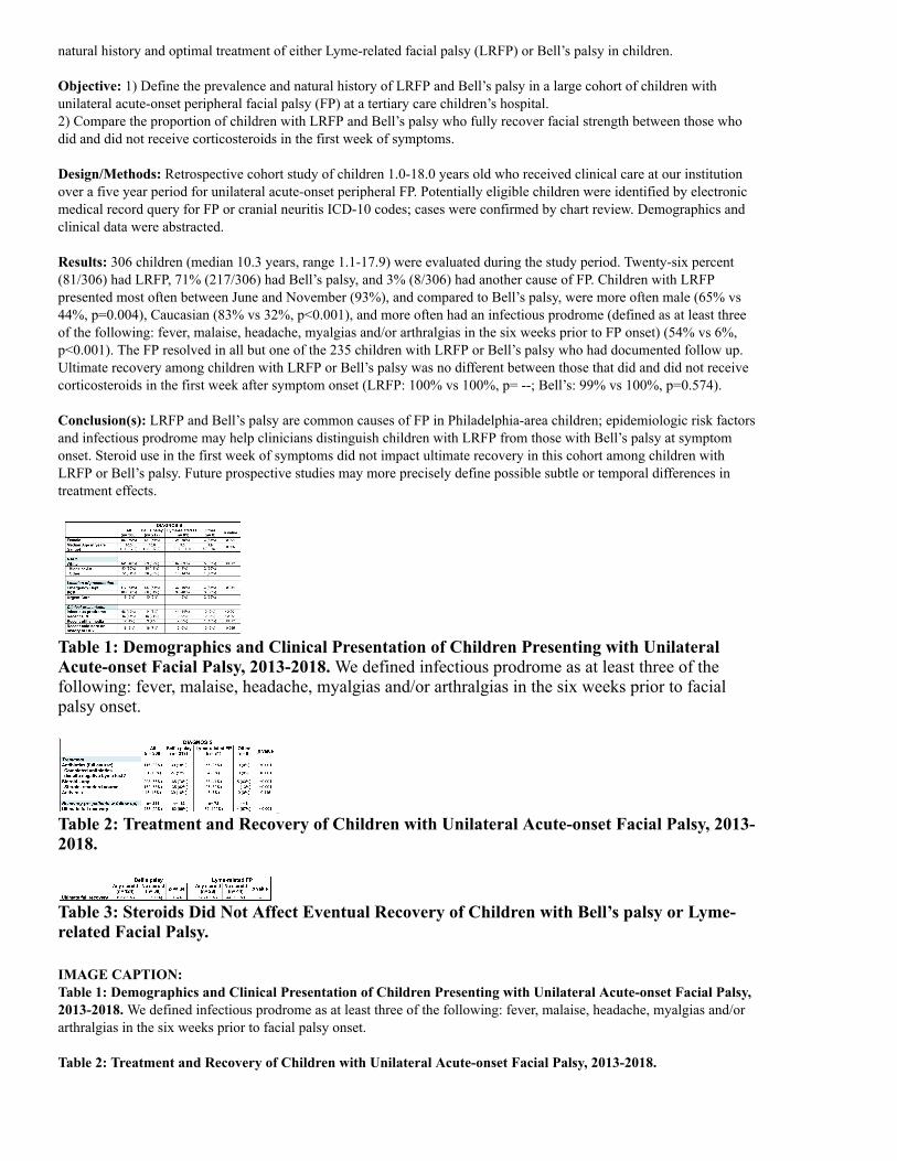
Results: 306 children (median 10.3 years, range 1.1-17.9) were evaluated during the study period. Twenty-six percent(81/306) had LRFP, 71% (217/306) had Bell’s palsy, and 3% (8/306) had another cause of FP. Children with LRFPpresented most often between June and November (93%), and compared to Bell’s palsy, were more often male (65% vs44%, p=0.004), Caucasian (83% vs 32%, p<0.001), and more often had an infectious prodrome (defined as at least threeof the following: fever, malaise, headache, myalgias and/or arthralgias in the six weeks prior to FP onset) (54% vs 6%,p<0.001). The FP resolved in all but one of the 235 children with LRFP or Bell’s palsy who had documented follow up.Ultimate recovery among children with LRFP or Bell’s palsy was no different between those that did and did not receivecorticosteroids in the first week after symptom onset (LRFP: 100% vs 100%, p= --; Bell’s: 99% vs 100%, p=0.574).
Conclusion(s): LRFP and Bell’s palsy are common causes of FP in Philadelphia-area children; epidemiologic risk factorsand infectious prodrome may help clinicians distinguish children with LRFP from those with Bell’s palsy at symptomonset. Steroid use in the first week of symptoms did not impact ultimate recovery in this cohort among children withLRFP or Bell’s palsy. Future prospective studies may more precisely define possible subtle or temporal differences intreatment effects.
Table 1: Demographics and Clinical Presentation of Children Presenting with UnilateralAcute-onset Facial Palsy, 2013-2018. We defined infectious prodrome as at least three of thefollowing: fever, malaise, headache, myalgias and/or arthralgias in the six weeks prior to facialpalsy onset.
Table 2: Treatment and Recovery of Children with Unilateral Acute-onset Facial Palsy, 2013-2018.
Table 3: Steroids Did Not Affect Eventual Recovery of Children with Bell’s palsy or Lyme-related Facial Palsy.
IMAGE CAPTION:Table 1: Demographics and Clinical Presentation of Children Presenting with Unilateral Acute-onset Facial Palsy,2013-2018. We defined infectious prodrome as at least three of the following: fever, malaise, headache, myalgias and/orarthralgias in the six weeks prior to facial palsy onset.
Table 2: Treatment and Recovery of Children with Unilateral Acute-onset Facial Palsy, 2013-2018.

Table 3: Steroids Did Not Affect Eventual Recovery of Children with Bell’s palsy or Lyme-related Facial Palsy.
CONTROL ID: 3380985TITLE: New Daily Persistent Headache in a Pediatric PopulationPRESENTER: Marc DiSabella
AUTHORS (LAST NAME, FIRST NAME): Pierce, Emily L.1; Langdon, Raquel L.1; Strelzik, Jeffrey A.1;McClintock, William M.2; Cameron, Mark1; DiSabella, Marc3
AUTHORS/INSTITUTIONS: E.L. Pierce, R.L. Langdon, J.A. Strelzik, M. Cameron, Neurology, Children's National Medical Center, Washington, District of Columbia, UNITED STATES;W.M. McClintock, Neurology, Chidren's National Medical Center, Washington, District of Columbia, UNITED STATES; M. DiSabella, Neurology, Childrens National Medical Center, Washington, District of Columbia, UNITED STATES;CURRENT CATEGORY: NeurologyCURRENT SUBCATEGORY: Pediatric NeurologyKEYWORDS: Chronic Headache, New Daily Persistent Headache, Pediatric Headache.SESSION TITLE: Pediatric Neurology |Pediatric NeurologySESSION TYPE: Webinar|Oral Poster SymposiaABSTRACT BODY: Background: New Daily Persistent Headache (NDPH), a subtype of Chronic Daily Headache, is characterized by an intractable, daily, and unremitting headache, lasting for at least 3 months. Currently there are limited studies in the pediatric population describing the characteristics of NDPH.
Objective: The objective of the current study is to describe the characteristics of NDPH in pediatric patients presenting to a headache program at a tertiary referral center.
Design/Methods: The participants in the current study were pediatric patients who attended the Headache Clinic at Children’s National Medical Center between 2016 and 2018. All patients seen in the Headache Clinic were enrolled in an IRB-approved patient registry. The registry was queried for NDPH (ICD-10 G44.52) and these records were reviewed to examine their clinical presentation.
Results: Between 2016 and 2018 there were a total of 3,260 patient encounters. 454 of the encounters were identified as having NDPH, representing 13.9% of the total. NDPH patients were predominantly female (78%) and white (72%). The median age was 14.8 years (SD=1.58). The median pain intensity was 6 of 10 (SD= 1.52). Females were more likely than males to report migrainous features including photophobia, phonophobia, nausea, and orthostatic lightheadedness. 72%of NDPH patients were using abortive medications at the time of the visit. 55% of NDPH patients failed at least one abortive medication (SD = 1.35), of which ibuprofen was the most frequent failure. 36% of patients were additionally diagnosed with Medication Overuse Headache (MOH), most often with ibuprofen. 78% of patients were diagnosed with additional comorbidities including head trauma (18%), anxiety (14%), and depression (8%).
Conclusion(s): The findings of this study suggest that NDPH is a relatively frequent disorder among pediatric patients presenting to a tertiary pediatric headache program. Females report more migrainous features than males, and approximately one-third of patients have co-existing MOH. Further studies are needed in order to better understand NDPH in the pediatric population.
(No Image Selected)
CONTROL ID: 3383318TITLE: White matter differences in children with NF1 compared to controls.PRESENTER: Lisa Bruckert
AUTHORS (LAST NAME, FIRST NAME): Bruckert, Lisa1; Travis, Katherine E.2; McKenna, Emily S.1; Yeom, Kristen1; Campen, Cynthia J.1

AUTHORS/INSTITUTIONS: L. Bruckert, E.S. McKenna, K. Yeom, C.J. Campen, Department of Pediatrcis, StanfordSchool of Medicine, Stanford, California, UNITED STATES;K.E. Travis, Stanford School of Medicine, Stanford, California, UNITED STATES;CURRENT CATEGORY: NeurologyCURRENT SUBCATEGORY: Pediatric NeurologyKEYWORDS: Neurofibromatosis type 1, Diffusion MRI, White matter tracts.SESSION TITLE: Pediatric Neurology |Pediatric NeurologySESSION TYPE: Webinar|Oral Poster SymposiaABSTRACT BODY: Background: Neurofibromatosis type 1 (NF1) is a common genetic condition in which 50% of children experiencelearning challenges including deficits in attention, executive function and reading. White matter tracts have beenimplicated in these cognitive functions. Whole-brain analyses have also shown diffuse white matter abnormalities inchildren with NF1. However, specific white matter tracts have not been well characterized in NF1.
Objective: Here, we employed diffusion MRI tractography to examine the microstructural characteristics of multiplewhite matter tracts in children with NF1.
Design/Methods: Participants were 20 children with NF1 aged 1.4 to 17.6 years (M = 9.5 years, 24 male) and 20 age andsex-matched controls who underwent diffusion MRI at 3T (25 directions, b=1000 s/mm2). Children with NF1 had nomedical history of moyamoya and no optic pathway glioma or other intracranial mass. We used an automated approach tosegment and extract white matter metrics including fractional anisotropy (FA) and mean diffusivity (MD) of 18 majorwhite matter tracts. First, we conducted analyses of covariance to examine the effect of group (NF1, controls) on whitematter metrics after controlling for intracranial volume. Second, we conducted regression analyses for each tract todetermine whether the association of white matter metrics with age was different in children with NF1 compared tocontrols. Group and age were entered in the first and their interaction in the second step of the regression analyses.Statistical significance was set at p < 0.05 after correcting for multiple comparisons using false discovery rate.
Results: Compared to controls, children with NF1 had significantly decreased FA in 8 and significantly increased MD in12 out of 18 tracts (Table 1). Differences in FA and MD remained significant after controlling for intracranial volume. Inaddition, the interaction term between group and age accounted for a significant proportion of the variance in FA in 9 andin MD in 16 out of 18 tracts. The interaction plots showed that FA and MD differences between children with NF1 andcontrols were greater at younger than older ages (Figure 1).
Conclusion(s): Microstructural differences were observed in multiple white matter tracts in children with NF1 comparedto controls. Novel findings were that these differences were not explained by intracranial volume, and that they weremost pronounced in younger children with NF1 compared to controls. These findings have implications forunderstanding neurocognitive deficits observed in children with NF1.

IMAGE CAPTION:
< Return to Abstract Search< Return to Abstract Search PrintPrint