Embed Size (px)
Citation preview
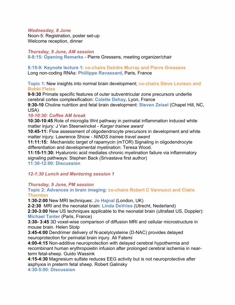
Wednesday, 8 June Noon-5: Registration, poster set-up Welcome reception, dinner Thursday, 9 June, AM session 8-8:15: Opening Remarks - Pierre Gressens, meeting organizer/chair 8:15-9: Keynote lecture 1: co-chairs Deirdre Murray and Pierre Gressens Long non-coding RNAs: Phillippe Ravassard, Paris, France Topic 1: New insights into normal brain development; co-chairs Steve Levison and Bobbi Fleiss 9-9:30 Primate specific features of outer subventricular zone precursors underlie cerebral cortex complexification: Colette Dehay, Lyon, France 9:30-10 Choline nutrition and fetal brain development: Steven Zeisel (Chapel Hill, NC, USA) 10-10:30: Coffee AM break 10:30-10:45 Role of microglia Wnt pathway in perinatal inflammation induced white matter injury: J Van Steenwinckel - Karger trainee award 10:45-11: Flow assessment of oligodendrocyte precursors in development and white matter injury: Lawrence Shiow - NINDS trainee travel award 11:11:15: Mechanistic target of rapamycin (mTOR) Signaling in oligodendrocyte differentiation and developmental myelination: Teresa Wood. 11:15-11:30: Hyaluronic acid mediates chronic myelination failure via inflammatory signaling pathways: Stephen Back (Srivastava first author) 11:30-12:00: Discussion 12-1:30 Lunch and Mentoring session 1 Thursday, 9 June, PM session Topic 2: Advances in brain imaging: co-chairs Robert C Vannucci and Claire Thornton 1:30-2:00 New MRI techniques: Jo Hajnal (London, UK) 2-2:30 MRI and the neonatal brain: Linda DeVries (Utrecht, Nederland) 2:30-3:00 New US techniques applicable to the neonatal brain (ultrafast US, Doppler): Michael Tanter (Paris, France) 3:30- 3:45 3D voxel-wise comparison of diffusion MRI and cellular microstructure in mouse brain. Helen Stolp 3:45-4:00 Dendrimer delivery of N-acetylcysteine (D-NAC) provides delayed neuroprotection for perinatal brain injury. Ali Fatemi 4:00-4:15 Non-additive neuroprotection with delayed cerebral hypothermia and recombinant human erythropoietin infusion after prolonged cerebral ischemia in near-term fetal-sheep. Guido Wassink 4:15-4:30 Magnesium sulfate reduces EEG activity but is not neuroprotective after asphyxia in preterm fetal sheep. Robert Galinsky 4:30-5:00: Discussion
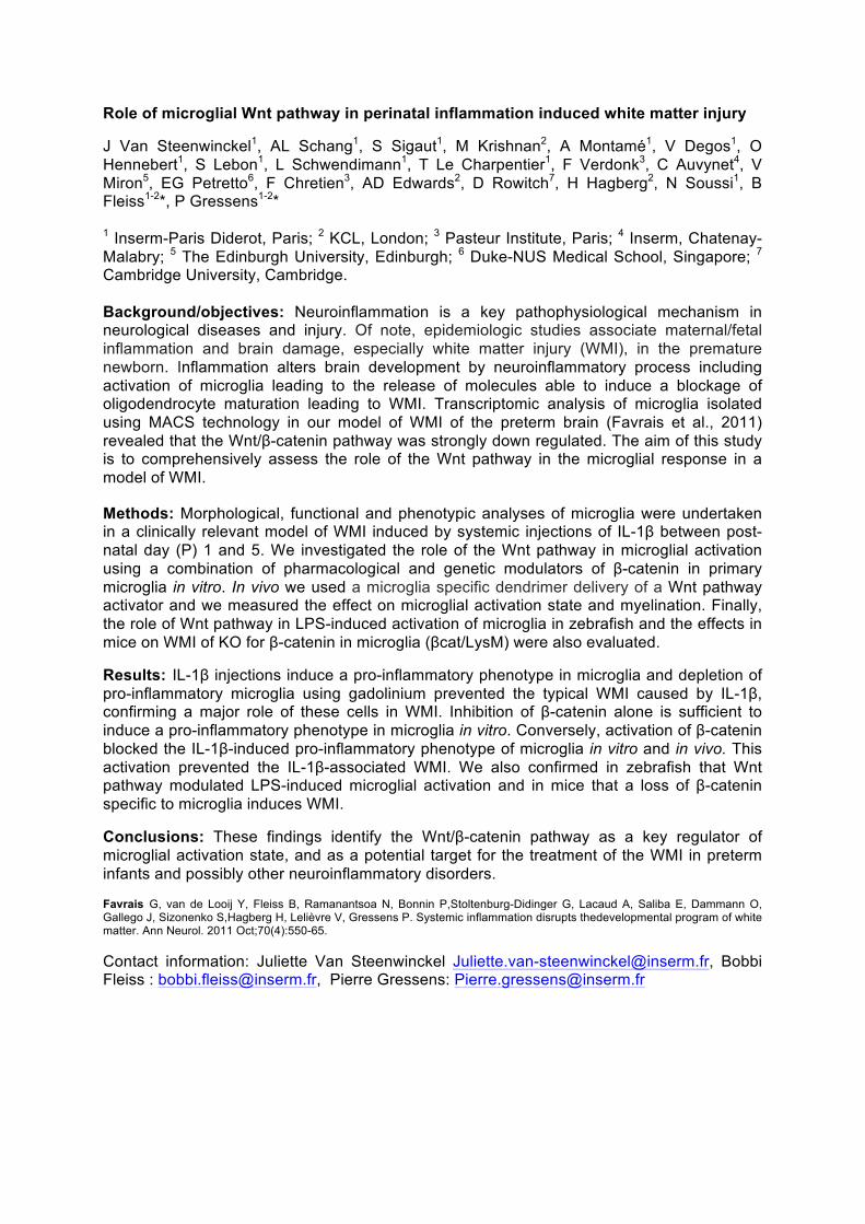
Role of microglial Wnt pathway in perinatal inflammation induced white matter injury
J Van Steenwinckel1, AL Schang1, S Sigaut1, M Krishnan2, A Montamé1, V Degos1, O Hennebert1, S Lebon1, L Schwendimann1, T Le Charpentier1, F Verdonk3, C Auvynet4, V Miron5, EG Petretto6, F Chretien3, AD Edwards2, D Rowitch7, H Hagberg2, N Soussi1, B Fleiss1-2*, P Gressens1-2* 1 Inserm-Paris Diderot, Paris; 2 KCL, London; 3 Pasteur Institute, Paris; 4 Inserm, Chatenay-Malabry; 5 The Edinburgh University, Edinburgh; 6 Duke-NUS Medical School, Singapore; 7 Cambridge University, Cambridge. Background/objectives: Neuroinflammation is a key pathophysiological mechanism in neurological diseases and injury. Of note, epidemiologic studies associate maternal/fetal inflammation and brain damage, especially white matter injury (WMI), in the premature newborn. Inflammation alters brain development by neuroinflammatory process including activation of microglia leading to the release of molecules able to induce a blockage of oligodendrocyte maturation leading to WMI. Transcriptomic analysis of microglia isolated using MACS technology in our model of WMI of the preterm brain (Favrais et al., 2011) revealed that the Wnt/β-catenin pathway was strongly down regulated. The aim of this study is to comprehensively assess the role of the Wnt pathway in the microglial response in a model of WMI.
Methods: Morphological, functional and phenotypic analyses of microglia were undertaken in a clinically relevant model of WMI induced by systemic injections of IL-1β between post-natal day (P) 1 and 5. We investigated the role of the Wnt pathway in microglial activation using a combination of pharmacological and genetic modulators of β-catenin in primary microglia in vitro. In vivo we used a microglia specific dendrimer delivery of a Wnt pathway activator and we measured the effect on microglial activation state and myelination. Finally, the role of Wnt pathway in LPS-induced activation of microglia in zebrafish and the effects in mice on WMI of KO for β-catenin in microglia (βcat/LysM) were also evaluated.
Results: IL-1β injections induce a pro-inflammatory phenotype in microglia and depletion of pro-inflammatory microglia using gadolinium prevented the typical WMI caused by IL-1β, confirming a major role of these cells in WMI. Inhibition of β-catenin alone is sufficient to induce a pro-inflammatory phenotype in microglia in vitro. Conversely, activation of β-catenin blocked the IL-1β-induced pro-inflammatory phenotype of microglia in vitro and in vivo. This activation prevented the IL-1β-associated WMI. We also confirmed in zebrafish that Wnt pathway modulated LPS-induced microglial activation and in mice that a loss of β-catenin specific to microglia induces WMI.
Conclusions: These findings identify the Wnt/β-catenin pathway as a key regulator of microglial activation state, and as a potential target for the treatment of the WMI in preterm infants and possibly other neuroinflammatory disorders.
Favrais G, van de Looij Y, Fleiss B, Ramanantsoa N, Bonnin P,Stoltenburg-Didinger G, Lacaud A, Saliba E, Dammann O, Gallego J, Sizonenko S,Hagberg H, Lelièvre V, Gressens P. Systemic inflammation disrupts thedevelopmental program of white matter. Ann Neurol. 2011 Oct;70(4):550-65.
Contact information: Juliette Van Steenwinckel [email protected], Bobbi Fleiss : [email protected], Pierre Gressens: [email protected]
Title: Flow assessment of oligodendrocyte precursors in development and white matter injury
Authors: Lawrence Shiow MD PhD ([email protected]) and David Rowitch MD PhD Institution: Department of Pediatrics, University of California San Francisco, CA
Background/objective: White matter injury in the premature infant brain, a reliable predictor for development of cerebral palsy and other neurodevelopmental deficits, is a pathologic disturbance in myelination with maturation arrested oligodendrocyte precursor cells (OPCs). Mechanisms that promote OPC maturation arrest in premature infants are still poorly understood. There is evidence for cellular diversity within OPCs based on brain region (white vs. gray matter), neurotransmitter responsiveness, or capacity to differentiate. To explore oligodendrocyte lineage diversity, we combined multiple surface markers and intracellular reporters by flow cytometry to detect functionally different subsets.
Methods: We utilize the Olig2-GFP mouse that contains a bicistronic GFP reporter in the endogenous OLIG2 locus. Dissociated cortical samples from different postnatal ages were surfaced stained with multiple markers (PDGFRα, O4, O1) or loaded with a calcium dye, and profiled by flow cytometry.
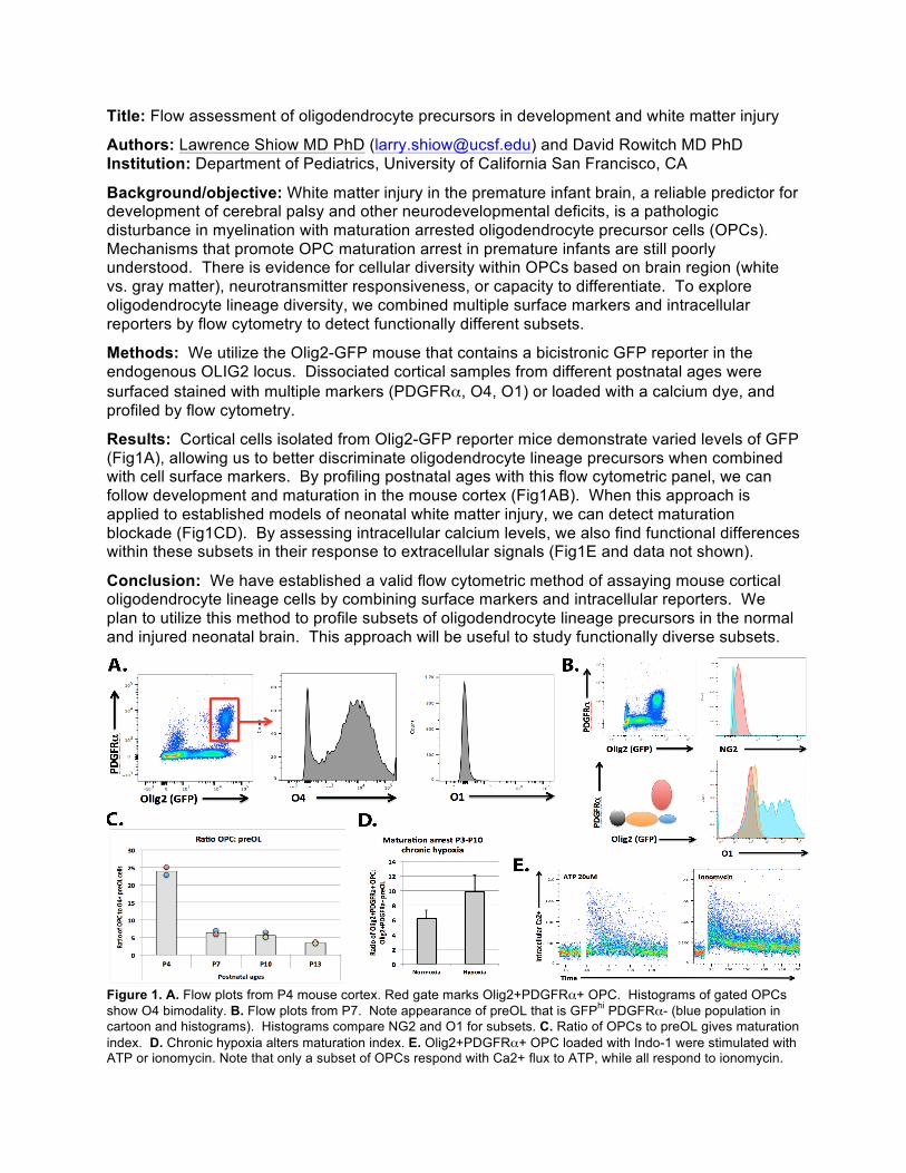
Results: Cortical cells isolated from Olig2-GFP reporter mice demonstrate varied levels of GFP (Fig1A), allowing us to better discriminate oligodendrocyte lineage precursors when combined with cell surface markers. By profiling postnatal ages with this flow cytometric panel, we can follow development and maturation in the mouse cortex (Fig1AB). When this approach is applied to established models of neonatal white matter injury, we can detect maturation blockade (Fig1CD). By assessing intracellular calcium levels, we also find functional differences within these subsets in their response to extracellular signals (Fig1E and data not shown).
Conclusion: We have established a valid flow cytometric method of assaying mouse cortical oligodendrocyte lineage cells by combining surface markers and intracellular reporters. We plan to utilize this method to profile subsets of oligodendrocyte lineage precursors in the normal and injured neonatal brain. This approach will be useful to study functionally diverse subsets.
Figure 1. A. Flow plots from P4 mouse cortex. Red gate marks Olig2+PDGFRα+ OPC. Histograms of gated OPCs show O4 bimodality. B. Flow plots from P7. Note appearance of preOL that is GFPhi PDGFRα- (blue population in cartoon and histograms). Histograms compare NG2 and O1 for subsets. C. Ratio of OPCs to preOL gives maturation index. D. Chronic hypoxia alters maturation index. E. Olig2+PDGFRα+ OPC loaded with Indo-1 were stimulated with ATP or ionomycin. Note that only a subset of OPCs respond with Ca2+ flux to ATP, while all respond to ionomycin.
Hyaluronic Acid mediates Chronic Myelination Failure via Inflammatory Signaling Pathways Taasin Srivastava, PhD,1 Justin Dean, PhD,1 Daniel Shaver, BA,1 Matthew Hagen, BS,1 Weiping Su, PhD,2 Larry S. Sherman, PhD, 2 and Stephen A. Back, MD, PhD1 1Oregon Health and Science University and 2Oregon National Primate Research Center Correspondence: [email protected] Background: Hyaluronic acid (HA) is a non-sulfated, protein-free glycosaminoglycan that accumulates in the extracellular matrix. In adult models of multiple sclerosis, reactive astrocytes synthesize high molecular weight HA (hHA; i.e., >106 Da) that reversibly inhibits pre-oligodendrocyte (preOL) maturation. hHA inhibits re-myelination via digestion products of hHA (~64 kDa) specifically generated by PH20, a GPI-anchored, secreted neutral hyaluronidase, which is the major CNS neutral hyaluronidase. We hypothesized that preOL arrest and myelination failure in neonatal WMI are mediated via HA digestion products generated by PH20. Methods: We employed a preterm equivalent model of WMI mediated by hypoxia-ischemia (H-I) in P3 rats. After H-I, expression of PH20, GFAP, CD44 and MBP were analyzed between P3 and P21 by image analysis, Western blot or qRT-PCR. An in vitro model of chronic WMI employed neonatal rat or mouse slice cultures to define HA-mediated signaling pathways. In rat, slice cultures show progressive accumulation of reactive astrocytes and HA by day 9 in vitro (DIV9). A robust block in preOL maturation provides a reproducible in vitro system to define mechanisms relevant to human neonatal myelination failure. Results: After H-I, an early rise in PH20 levels coincided with elevated expression of GFAP, CD44 and low levels of MBP. Chronic lesions between P3 and P14 displayed extensive astrogliosis with low levels of staining for total HA. By P21, WMI displayed extensive but incomplete myelination and higher levels of total HA that coincided with a decline in PH20, GFAP and CD44. Protein lysates prepared from white matter lesions 2 days after H-I, were analyzed by agarose gel electrophoresis, which showed that hHA digestion products were generated in vivo that ranged between ~200-500 kDa. To define mechanisms of hHA-mediated preOL arrest in vitro, we treated slice cultures until DIV9 in the presence of a novel selective PH20 inhibitor identified in a screen of synthetically modified derivatives of apigenin, a natural flavonoid inhibitor of hyaluronidases. Inhibition of PH20 reversed preOL arrest and restored the density of mature MBP+ OLs to control. We confirmed that the effects of hHA and PH20 inhibition were cell autonomous in primary preOL cultures. To define size ranges of bioactive HA digestion products, we analyzed HA preparations of different size ranges. When tested in vitro, 175-300 kDa HA fragments (fHA) were as potent as hHA (>1800 kDa) to block preOL maturation in rat and mouse slices. In primary preOL cultures, 175-300 kDa fragments blocked preOL maturation, but 5-20 and 40-75 kDa fHA had no activity. To define the receptor(s) required to mediate the effects of HA, we tested the effects fHA in slice cultures from TLR2/TLR4 double null mice or CD44 null mice. fHA blocked OL maturation in CD44 null mice, whereas in TLR2/4 null mice, fHA did not block OL maturation. Since the actions of TLR2/4 are typically mediated via a canonical pathway that involves MyD88, we evaluated hHA and fHA activity in MyD88 null mice. Despite deletion of MyD88, both hHA and fHA blocked OL maturation in slice cultures and OPCs. We thus evaluated whether TLRs might signal via an alternative pathway involving RhoA. We co-treated wild type rat slice cultures with hHA or hHA and rhosin (a specific small molecule inhibitor of RhoA). Rhosin completely reversed the negative effects of hHA and significantly promoted preOL maturation. Conclusions: A distinct size range of HA fragments mediates neonatal myelination failure via a receptor complex that involves TLR2 and TLR4 but not CD44 to activate a downstream MyD88-independent signaling pathway that blocks preOL maturation via activation of RhoA.
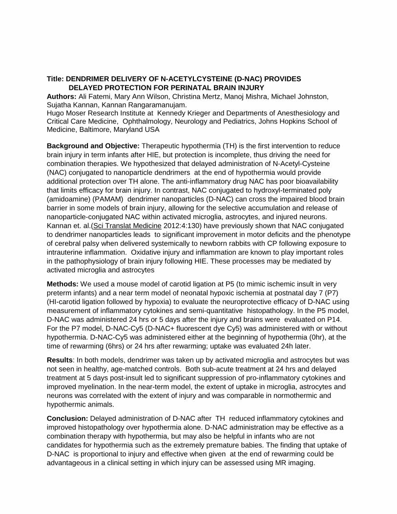
Title: DENDRIMER DELIVERY OF N-ACETYLCYSTEINE (D-NAC) PROVIDES DELAYED PROTECTION FOR PERINATAL BRAIN INJURY Authors: Ali Fatemi, Mary Ann Wilson, Christina Mertz, Manoj Mishra, Michael Johnston, Sujatha Kannan, Kannan Rangaramanujam. Hugo Moser Research Institute at Kennedy Krieger and Departments of Anesthesiology and Critical Care Medicine, Ophthalmology, Neurology and Pediatrics, Johns Hopkins School of Medicine, Baltimore, Maryland USA Background and Objective: Therapeutic hypothermia (TH) is the first intervention to reduce brain injury in term infants after HIE, but protection is incomplete, thus driving the need for combination therapies. We hypothesized that delayed administration of N-Acetyl-Cysteine (NAC) conjugated to nanoparticle dendrimers at the end of hypothermia would provide additional protection over TH alone. The anti-inflammatory drug NAC has poor bioavailability that limits efficacy for brain injury. In contrast, NAC conjugated to hydroxyl-terminated poly (amidoamine) (PAMAM) dendrimer nanoparticles (D-NAC) can cross the impaired blood brain barrier in some models of brain injury, allowing for the selective accumulation and release of nanoparticle-conjugated NAC within activated microglia, astrocytes, and injured neurons. Kannan et. al.(Sci Translat Medicine 2012:4:130) have previously shown that NAC conjugated to dendrimer nanoparticles leads to significant improvement in motor deficits and the phenotype of cerebral palsy when delivered systemically to newborn rabbits with CP following exposure to intrauterine inflammation. Oxidative injury and inflammation are known to play important roles in the pathophysiology of brain injury following HIE. These processes may be mediated by activated microglia and astrocytes
Methods: We used a mouse model of carotid ligation at P5 (to mimic ischemic insult in very preterm infants) and a near term model of neonatal hypoxic ischemia at postnatal day 7 (P7) (HI-carotid ligation followed by hypoxia) to evaluate the neuroprotective efficacy of D-NAC using measurement of inflammatory cytokines and semi-quantitative histopathology. In the P5 model, D-NAC was administered 24 hrs or 5 days after the injury and brains were evaluated on P14. For the P7 model, D-NAC-Cy5 (D-NAC+ fluorescent dye Cy5) was administered with or without hypothermia. D-NAC-Cy5 was administered either at the beginning of hypothermia (0hr), at the time of rewarming (6hrs) or 24 hrs after rewarming; uptake was evaluated 24h later.
Results: In both models, dendrimer was taken up by activated microglia and astrocytes but was not seen in healthy, age-matched controls. Both sub-acute treatment at 24 hrs and delayed treatment at 5 days post-insult led to significant suppression of pro-inflammatory cytokines and improved myelination. In the near-term model, the extent of uptake in microglia, astrocytes and neurons was correlated with the extent of injury and was comparable in normothermic and hypothermic animals.
Conclusion: Delayed administration of D-NAC after TH reduced inflammatory cytokines and improved histopathology over hypothermia alone. D-NAC administration may be effective as a combination therapy with hypothermia, but may also be helpful in infants who are not candidates for hypothermia such as the extremely premature babies. The finding that uptake of D-NAC is proportional to injury and effective when given at the end of rewarming could be advantageous in a clinical setting in which injury can be assessed using MR imaging.
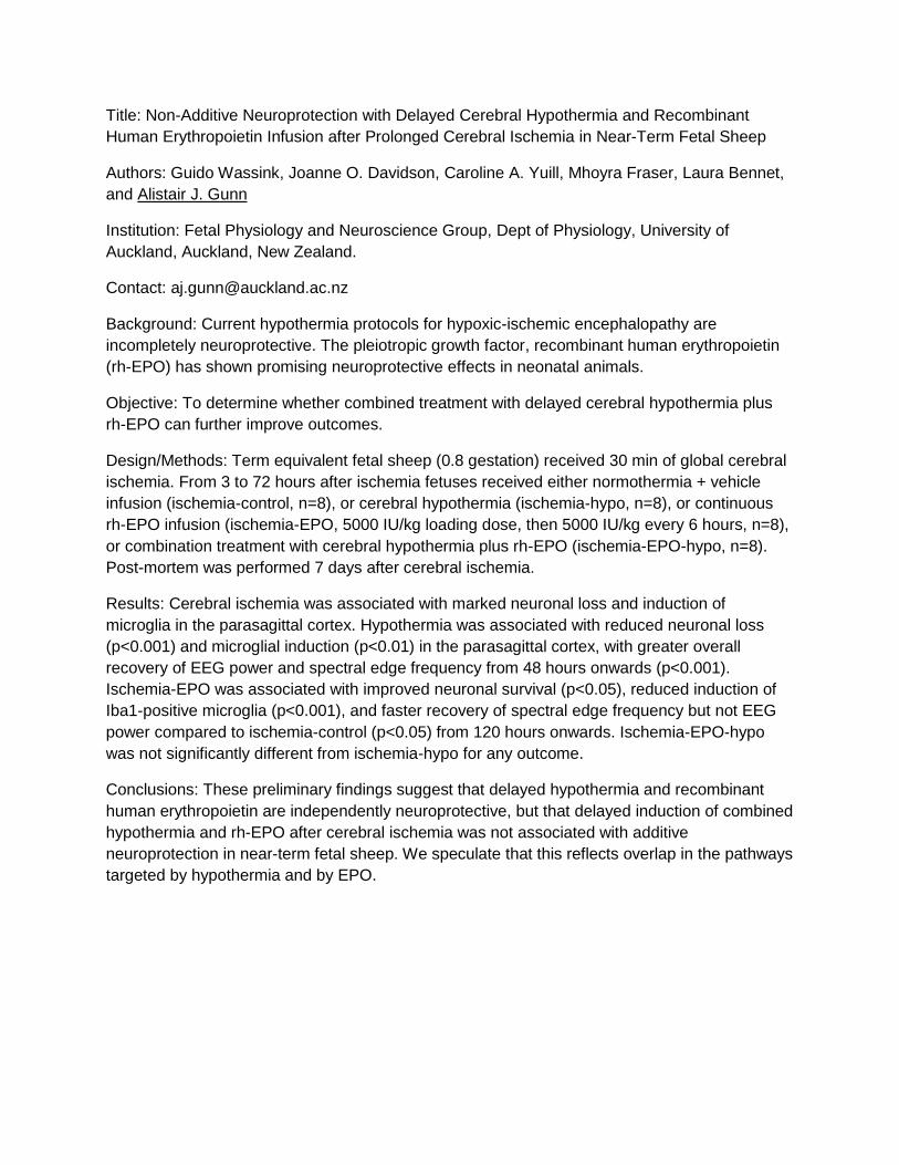
Title: Non-Additive Neuroprotection with Delayed Cerebral Hypothermia and Recombinant
Human Erythropoietin Infusion after Prolonged Cerebral Ischemia in Near-Term Fetal Sheep
Authors: Guido Wassink, Joanne O. Davidson, Caroline A. Yuill, Mhoyra Fraser, Laura Bennet,
and Alistair J. Gunn
Institution: Fetal Physiology and Neuroscience Group, Dept of Physiology, University of
Auckland, Auckland, New Zealand.
Contact: [email protected]
Background: Current hypothermia protocols for hypoxic-ischemic encephalopathy are
incompletely neuroprotective. The pleiotropic growth factor, recombinant human erythropoietin
(rh-EPO) has shown promising neuroprotective effects in neonatal animals.
Objective: To determine whether combined treatment with delayed cerebral hypothermia plus
rh-EPO can further improve outcomes.
Design/Methods: Term equivalent fetal sheep (0.8 gestation) received 30 min of global cerebral
ischemia. From 3 to 72 hours after ischemia fetuses received either normothermia + vehicle
infusion (ischemia-control, n=8), or cerebral hypothermia (ischemia-hypo, n=8), or continuous
rh-EPO infusion (ischemia-EPO, 5000 IU/kg loading dose, then 5000 IU/kg every 6 hours, n=8),
or combination treatment with cerebral hypothermia plus rh-EPO (ischemia-EPO-hypo, n=8).
Post-mortem was performed 7 days after cerebral ischemia.
Results: Cerebral ischemia was associated with marked neuronal loss and induction of
microglia in the parasagittal cortex. Hypothermia was associated with reduced neuronal loss
(p<0.001) and microglial induction (p<0.01) in the parasagittal cortex, with greater overall
recovery of EEG power and spectral edge frequency from 48 hours onwards (p<0.001).
Ischemia-EPO was associated with improved neuronal survival (p<0.05), reduced induction of
Iba1-positive microglia (p<0.001), and faster recovery of spectral edge frequency but not EEG
power compared to ischemia-control (p<0.05) from 120 hours onwards. Ischemia-EPO-hypo
was not significantly different from ischemia-hypo for any outcome.
Conclusions: These preliminary findings suggest that delayed hypothermia and recombinant
human erythropoietin are independently neuroprotective, but that delayed induction of combined
hypothermia and rh-EPO after cerebral ischemia was not associated with additive
neuroprotection in near-term fetal sheep. We speculate that this reflects overlap in the pathways
targeted by hypothermia and by EPO.
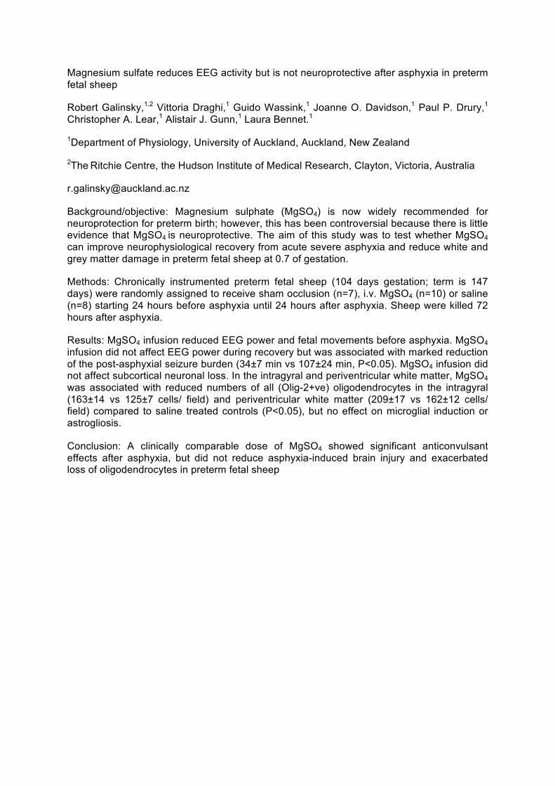
Magnesium sulfate reduces EEG activity but is not neuroprotective after asphyxia in preterm fetal sheep
Robert Galinsky,1,2 Vittoria Draghi,1 Guido Wassink,1 Joanne O. Davidson,1 Paul P. Drury,1 Christopher A. Lear,1 Alistair J. Gunn,1 Laura Bennet.1
1Department of Physiology, University of Auckland, Auckland, New Zealand
2The Ritchie Centre, the Hudson Institute of Medical Research, Clayton, Victoria, Australia
Background/objective: Magnesium sulphate (MgSO4) is now widely recommended for neuroprotection for preterm birth; however, this has been controversial because there is little evidence that MgSO4 is neuroprotective. The aim of this study was to test whether MgSO4 can improve neurophysiological recovery from acute severe asphyxia and reduce white and grey matter damage in preterm fetal sheep at 0.7 of gestation.
Methods: Chronically instrumented preterm fetal sheep (104 days gestation; term is 147 days) were randomly assigned to receive sham occlusion (n=7), i.v. MgSO4 (n=10) or saline (n=8) starting 24 hours before asphyxia until 24 hours after asphyxia. Sheep were killed 72 hours after asphyxia.
Results: MgSO4 infusion reduced EEG power and fetal movements before asphyxia. MgSO4 infusion did not affect EEG power during recovery but was associated with marked reduction of the post-asphyxial seizure burden (34±7 min vs 107±24 min, P<0.05). MgSO4 infusion did not affect subcortical neuronal loss. In the intragyral and periventricular white matter, MgSO4 was associated with reduced numbers of all (Olig-2+ve) oligodendrocytes in the intragyral (163±14 vs 125±7 cells/ field) and periventricular white matter (209±17 vs 162±12 cells/ field) compared to saline treated controls (P<0.05), but no effect on microglial induction or astrogliosis.
Conclusion: A clinically comparable dose of MgSO4 showed significant anticonvulsant effects after asphyxia, but did not reduce asphyxia-induced brain injury and exacerbated loss of oligodendrocytes in preterm fetal sheep
Dinner, after dinner poster-session Friday, 10 June, AM session 8:30-9:15 Keynote lecture 2: co-chairs Susan Vannucci and Ivo Bendix The glio-vascular unit and the role of Hypoxia inducible factor: David Rowitch (San Francisco, CA, USA) Topic 3: Extracellular vesicles and lipodomics signatures- hallmarks for developmental disorders; co-chairs Carina Mallard and Justin Dean 9:15-9:45 Transfer of exosomes from glia to neurons: “goodies” for neuronal fitness?: Eva-Maria Kramer-Albers (Mainz, Germany) 9:45-10-15 Lipidomics and lipid markers in the study of infant development: Albert Koulman (Cambridge, UK) 10:15-10:45 Coffee/AM Break 10:45-11:00 MSC-derived extracellular vesicles for treatment of inflammation-induced preterm brain injury. K. Drommelschmidt – Karger trainee award 11:11:15 Dedifferentiated fat cells as a treatment for perinatal hypoxic-ischemic injury. Alkisti Mikrogeorgiou - NINDS travel awardee 11:15-11:30 In vivo investigation of cerebral polyunsaturated fatty acids and related lipid metabolites in neonates with hypoxic-ischemic encephalopathy. Jessica Wisnowski 11:30-12:00: Discussion 12-1:30 Lunch and mentoring session 2 Friday, 10 June, PM session Topic 4: Perinatal origin of childhood and adult brain disorders; co-chairs Sid Tan and Mary Tolcos 1:30-2:00 Persisting effects of preterm birth; autism, schizophrenia and adult neurodegeneration: Bobbi Fleiss (London, UK) 2:00-2:30 Early life gut-microbiome-brain interactions: Implications for neurodevelopmental disorders: Rochellys Diaz-Heijtz (Stockholm, Sweden) 2:30-3:00 PM Break 3:00-3:15 GluN2B tyrosine phosphorylation at Y1472 modifies synaptic GluN2B complex and downstream signaling following neonatal brain hypoxia-ischemia. Renatta Knox - trainee and Diversity Outreach Award NINDS 3:15-3:30 Targeting insulin-like growth factor-1 signaling for treatment of preterm brain injury following neonatal inflammation. J Prasad 3:30-3:45 Cord-blood IL-6 predicts neurodevelopmental outcome at 3 years in perinatal asphyxia and neonatal hypoxic-ischemic encephalopathy. Caroline Ahearne - Karger trainee award 3:45-4:00 Origin and dynamics of lineage-specific progenitor’s recruitment following perinatal hypoxia. Diane Angonin 4:00-5:00: Discussion Dinner, after dinner party with dancing
MSC-derived extracellular vesicles for treatment of inflammation-induced preterm
brain injury 1 Drommelschmidt K, 1 Serdar M, 1 Bendix I, 1 Bertling F, 1 Herz J, 5 Duhan V, 2 Ludwig AK, 2
Radtke S, 2 Miroschedji K, 2 Horn P,2,3 van de Looji Y, 2 Giebel B, 1 Felderhoff-Mueser U
1 Dpt. of Neonatology, 2 Dpt. of Transfusion Medicine, University Hospital Essen, University
Duisburg- Essen, Germany; 2 Department of Pediatrics, University of Geneva, Genève,
Switzerland; 3 Laboratory of Functional and Metabolic Imaging, Ecole Polytechnique
Fédérale de Lausanne, Lausanne, Switzerland; 4Department of Immunology, University
Hospital Essen, University Duisburg-Essen, Essen, Germany
Background: Preterm brain injury is considered as multi-factorial with strong evidence that
the underlying key player is fetal and/or postnatal inflammation resulting in white and gray
matter injury. Experimental studies revealed neuroprotective properties of mesenchymal
stem cells (MSCs). However the lack of correlation between functional improvement and cell
engraftment indicate a paracrine mechanism via MSC-derived extracellular vesicles (MSC-
EVs). Here, we investigated the therapeutic short- and long-term potential of MSC-EVs in a
rodent model of inflammatory preterm brain injury.
Methods: To evaluate immunomodulatory and neuroprotective capacities of MSC-EVs 3-day
old Wistar rats (P3) were intraperitoneally (i.p.) injected with 0.25 mg/kg Lipopolysaccharide
(LPS) or saline (0.9% NaCl) and treated with two repetitive doses of 1x10⁷ cell equivalents of
MSC-EV. Cellular degeneration and reactive gliosis at P5 as well as myelination at P11 were
determined by immunohistochemistry and western blot analysis. Long-term cognitive and
motor function was assessed by Barnes Maze, Novel Object Recognition and Open Field at
P30 and P90. Diffusion tensor imaging (DTI) was carried out at P125 to evaluate long-term
microstructural white matter alterations.
Results: LPS-induced inflammation led to increased cellular degeneration which was
significantly ameliorated by MSC-EV treatment. MSC-EVs reduced microglia activation and
prevented reactive astrogliosis caused by systemic inflammation. Short-term myelination
deficits and long-term microstructural abnormalities of white matter structures were also
restored. Furthermore, MSC EV-treatment improved long-lasting cognitive deficits induced by
perinatal LPS-injection.
Conclusion: MSC-EVs might serve as a powerful therapeutic option to treat neonatal brain
injury by restoration of white matter microstructure and reduction of gliosis and long-term
functional improvement.
Dedifferentiated fat cells as a treatment for perinatal hypoxic ischemic brain injury Alkisti Mikrogeorgiou1,2, Yoshiaki Sato1, Taiki Kondo1,2, Yuichiro Sugiyama1,2, Keiko Nakanishi3, Masahiro Tsuji4, Tomohiko Kazama5, Taro Matsumoto5, Koichiro Kano6, Masahiro Hayakawa1 1. Division of Neonatology, Center for Maternal-Neonatal Care, Nagoya University Hospital, Department of Pediatrics 2. Department of Pediatrics, Nagoya University Graduate School of Medicine, Nagoya, Japan 3. Department of Perinatology, Institute for Developmental Research, Aichi Human Service Center, Kasugai, Aichi, Japan 4. Department of Regenerative Medicine and Tissue Engineering, National Cerebral and Cardiovascular Center, Osaka, Japan 5. Division of Cell Regeneration and Transplantation, Department of Functional Morphology, Nihon University, Tokyo, Japan 6. Department of Applied Biological Science, College of Bioresource Sciences. Nihon University, Tokyo, Japan *Correspondence: Yoshiaki Sato, M.D., Ph.D. ([email protected])
Background: Neonatal hypoxic ischemic encephalopathy is associated with high mortality and persistent neurological handicaps in the affected individuals. With hypothermia being the only legitimate treatment option, scientific interest has shifted towards cell based therapy. Among the various stem cell types researched, dedifferentiated fat (DFAT) cells, derived from mature adipocytes via ceiling culture constitute an abundant and easily accessible source that may offer many advantages as a neuroprotective therapeutic means. Objective: To determine whether treatment with DFAT cells improves perinatal brain injury and investigate the mechanism through which such effect might be mediated. Methods: Hypoxic ischemic brain injury was generated in 7 days old rat pups by permanent ligation of the left common carotid artery and exposure to an atmosphere of 8% O2 for 60min. Diffusion weighted MRI was used to assess the severity of damage and to assign animals into equivalent groups. Twenty-four hours after hypoxia, the treatment group received GFP-labeled DFAT cells through an intravenous route (105 cells/ pup). Forty-eight hours after the insult, brain sections were evaluated immunohistochemically with anti-caspase 3 and anti-ED-1 antibodies as apoptosis and activated microglia markers respectively. Additionally, motor functioning was assessed 2 and 5 weeks after the insult using rotarod and cylinder test. In an in vitro approach, oxygen glucose deprivation was induced on primary E16 rat cortical neurons, that had previously been treated for 24 or 48 hours with DFAT conditioned medium (DFAT-CM) at a concentration range of 2.5% to 10%. Cell death was subsequently analyzed by measuring the released LDH levels through absorption spectroscopy. Results: The number of cells, stained positive for active-caspase-3 in the hippocampus and cortex areas of the DFAT group was reduced by 73% and 48%, respectively, while activated microglia (ED-1 positive cells) was found 66% and 40% lower in the same areas, when compared with the control group. We were able to track the administered GFP labeled DFAT cells immunohistologically in liver and lung sections of the treated animals; however, the cells were hardly detectable in the brain sections of the same animals. In both rotarod and cylinder test the control group showed profound motor impairment when compared with sham operated animals, whilst such difference was not evident between the treatment group and the sham group. In in vitro experiments, the rate of neuronal death in the cells treated with DFAT-CM forty- eight hours prior to exposure was significantly lower compared to the controls (10.8% - 15.5% versus 21%). Cell death rates slightly increased when DFAT-CM was added 24 hours before exposure but the treatment effect remained significant. Conclusion: These results indicate that intravenous injection of DFAT cells is effective in ameliorating hypoxic-ischemic brain injury, possibly through paracrine effects.
In vivo investigation of cerebral polyunsaturated fatty acids and related lipid metabolites in neonates with hypoxic-ischemic encephalopathy Jessica L. Wisnowski1,2,3,4, Tai-Wei Wu1,2, Aaron J. Reitman1,2, Eugenia Ho1,2, Claire McLean1,2, Douglas Vanderbilt1,2, Thomas Chavez1,2, Marvin D. Nelson1,2, Ashok Panigrahy3, Philippe Friedlich1,2, and Stefan Blüml1,2,4. 1Children’s Hospital Los Angeles, 2University of Southern California, 3Children's Hospital of Pittsburgh of UPMC; 4Rudi Schulte Research Institute, Santa Barbara, CA.
Background/Objective: Proton MR Spectroscopy (1H-MRS) has been used to non-invasively demonstrate cell death-dependent changes in phospholipid metabolism in both in vitro and in vivo models of human carcinoma (1). These studies have established a direct correlation between the number of apoptotic cells and the magnitude of the lipid signals at 1.3 ppm and 2.8 ppm, which arise from the methylene protons -(CH2)n and bisallylic methylene protons, (=CH-CH2-HC=) of mobile lipids. (Note: the latter signifies the presence of polyunsaturated fatty acids [PUFA]). The aim of this study was to determine the specificity and time course of these and other lipid biomarkers in neonates with hypoxic-ischemic encephalopathy (HIE).
Methods: 41 neonates with HIE (19 male; mean GA: 38.8 ± 1.8 wks) underwent MRI during therapeutic hypothermia (TH) and after rewarming. MRI/MRS were acquired on a 3T MR system (Philips, Best, The Netherlands) and included conventional T1-, T2- and diffusion weighted (DWI) imaging as well as single-voxel point resolved spectroscopy (PRESS) localized to the left thalamus (TE=35ms). MRS data were processed using a modified LCModel (Ver. 6.3-1c, Provencher Inc) pipeline, which included simulated peaks at 5.3 and 2.8 ppm corresponding to the methine (-CH ═ CH-) and bisallylic methylene (=CH-CH2-HC=) protons of the unsaturated acyl chain. Conventional MRI was used to identify neonates with moderate to severe brain injury (2). Mann-Whitney U was used to compare metabolite concentrations across outcome groups.
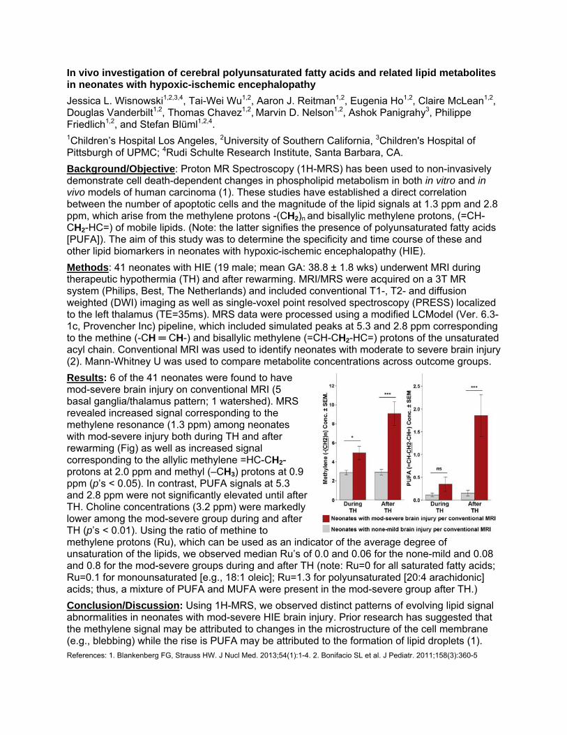
Results: 6 of the 41 neonates were found to have mod-severe brain injury on conventional MRI (5 basal ganglia/thalamus pattern; 1 watershed). MRS revealed increased signal corresponding to the methylene resonance (1.3 ppm) among neonates with mod-severe injury both during TH and after rewarming (Fig) as well as increased signal corresponding to the allylic methylene =HC-CH2- protons at 2.0 ppm and methyl (–CH3) protons at 0.9 ppm (p’s < 0.05). In contrast, PUFA signals at 5.3 and 2.8 ppm were not significantly elevated until after TH. Choline concentrations (3.2 ppm) were markedly lower among the mod-severe group during and after TH (p’s < 0.01). Using the ratio of methine to methylene protons (Ru), which can be used as an indicator of the average degree of unsaturation of the lipids, we observed median Ru’s of 0.0 and 0.06 for the none-mild and 0.08 and 0.8 for the mod-severe groups during and after TH (note: Ru=0 for all saturated fatty acids; Ru=0.1 for monounsaturated [e.g., 18:1 oleic]; Ru=1.3 for polyunsaturated [20:4 arachidonic] acids; thus, a mixture of PUFA and MUFA were present in the mod-severe group after TH.)
Conclusion/Discussion: Using 1H-MRS, we observed distinct patterns of evolving lipid signal abnormalities in neonates with mod-severe HIE brain injury. Prior research has suggested that the methylene signal may be attributed to changes in the microstructure of the cell membrane (e.g., blebbing) while the rise is PUFA may be attributed to the formation of lipid droplets (1). References: 1. Blankenberg FG, Strauss HW. J Nucl Med. 2013;54(1):1-4. 2. Bonifacio SL et al. J Pediatr. 2011;158(3):360-5
GluN2B tyrosine phosphorylation at Y1472 modifies synaptic GluN2B complex and downstream signaling following neonatal brain hypoxia-ischemia
Guo Shao1, Yongqiang Wang2, Shenheng Guan3, Fuxin Lu4, Renatta Knox5,
Donna M Ferriero4,6 and Xiangning Jiang4
1Department of Biochemistry, Baotou Medical College, Inner Mongolia, China Departments of 2Cellular & Molecular Pharmacology, 3Pharmaceutical Chemistry, 4Pediatrics,
6Neurology, University of California San Francisco, CA, USA 94143 5Department of Pediatrics, Weill Cornell Medical College, New York, NY, USA 10065 Background and Objective: Overactivation of NMDA receptors (NMDAR) is implicated in excitotoxic cell death following neonatal brain hypoxia-ischemia (HI). Our previous work has shown that protein kinase Fyn-mediated phosphorylation at tyrosine (Y) 1472 of NMDAR subunit GluN2B contributes to neonatal HI brain injury, partly through up-regulation of superoxide-mediated oxidative stress. Since GluN2B Y1472 phosphorylation plays a role in receptor trafficking at synapses, as well as in GluN2B interactions with downstream proteins, we aimed to investigate whether this modification could modulate the composition of synaptic GluN2B complex and associated signaling networks leading to neuronal death/survival. Methods: Neonatal mice with a knock-in mutation of Y1472 to phenyalanine (YF-KI) and their wildtype (WT) littermates were used for HI at postnatal day 7 with the Vannucci model. Purified synaptic membrane fractions from sham- and HI-injured cortices were subjected to immunoprecipitation (IP) with a GluN2B antibody followed by PAGE gel electrophoreses. After silver staining, protein bands were sliced and underwent reduction, alkylation and digestion with trypsin and lysyl endopeptidase. The mixture of tryptic peptides were dried and sent for LC/MS/MS proteomic analysis. The MS/MS data were searched against the SwissProt database using the in-house Protein Prospector search engine. Protein interactions were validated by co-IP experiments. Results: Proteomic characterization of synaptic GluN2B complexes has revealed numerous proteins that can be classified into glutamate receptors; scaffolds/adaptors; kinases/phosphatases and associated proteins; small G-proteins and modulators; cell-adhesion and cytoskeletal proteins, and others. Many of them are implicated in GluN2B downstream signaling pathways. We have verified that nNOS, Src, Fyn, Src kinase signaling inhibitor 1 (SRCN1), PI3K subunit p85, CaMKIIα, PKCs, MAP kinase p38 and ERK1/2 were recruited to GluN2B early after HI (at 15min). We found a significant decreased CaMKIIα activation in the YF-KI animals at 15min and 1hour after HI compared to the WT animals. The expression of PSD95, as well as its interaction with GluN2B was lower in the YF-KI mice compared to their WT littermates. Conclusion: Neonatal HI activates specific signaling pathways downstream of GluN2B. Y1472 phosphorylation may modify synaptic GluN2B interactions with other postsynaptic density proteins (for example, PSD95) and thereby contribute to HI brain injury. Supported by RO1NS084057 (Dr. Jiang) and RO1NS33997 (Dr Ferriero)
Targeting Insulin-Like Growth Factor-1 Signaling for Treatment of Preterm Brain Injury Following Neonatal Inflammation J. Prasad, S Ranchhod, K Gunn, T Fowke, J Bai, J Guan, J Dean* Department of Physiology, Faculty of Medical and Health Sciences, the University of Auckland, New Zealand *Tel: +64 9 923 6201; Email: [email protected] Background/objective: Preterm infants have high rates of life-long disability, which are strongly associated with exposure to infection and resulting inflammation around birth. The major of pattern of brain injury in preterm infants involves white matter damage, with selective death and impaired maturation of oligodendrocytes, the cells responsible for myelination. Insulin-like growth factor-1 (IGF-1) is a neurotropic factor critical for oligodendrocyte survival and maturation. A naturally occurring stable IGF-1 metabolite, cyclic-glycine-proline (cGP), can cross the blood-brain-barrier and promote IGF-1 release. In this study, we hypothesized that early postnatal inflammation would cause deficits in IGF-1 signaling, and that restoring normal IGF-1 signaling with cGP would promote normal white matter development. Methods: Sprague-Dawley rat pups were randomly assigned to daily injection of saline (control), lipopolysaccharide (LPS; 0.3 mg/kg i.p.), or LPS + cGP (0.1 mg/kg s.c.; acute treatment) groups on postnatal day 1 (PND1) only or PND1–3. Animals were recovered to PND2, 4, and 14. In further experiments, pups received LPS at PND1–3, followed by repeated cGP from PND7–14 (delayed treatment), and animals collected at PND14. Blood plasma and brain tissues were collected at all time-points for analysis of (i) cytokine/chemokine protein levels by ELISA, (ii) total IGF-1 and IGF-binding protein-3 (IGFBP-3) protein levels by ELISA, (iii) IGF-1 receptor signaling by Western blot, and (iv) oligodendrocyte survival in the white matter by immunohistochemistry. Results: LPS animals showed an overall reduction in body weight and brain weight compared to controls. Levels of IL-1β and TNF-α were acutely elevated in the blood plasma and brain in LPS animals compared to controls. LPS animals showed a reduction in total plasma IGF-1, and a biphasic increase then decrease in plasma IGFBP-3, compared to controls. There was also an overall reduction in IGF-1 receptor signaling in the brains of LPS animals. In the white matter, LPS was associated with acute increase in caspase-3-mediated oligodendrocyte cell death at PND4, and reduced numbers of mature oligodendrocytes at PND14. Acute cGP treatment increased total plasma IGF-1 levels and prevented oligodendrocyte cell death in the white matter at PND4, but had no effect on body weight, brain weight, or total plasma IGF-1 levels at PND14. By contrast, delayed cGP treatment improved body weight, brain weight, total plasma IGF-1 levels, and numbers of mature oligodendrocytes at PND14. Conclusion: These data suggest that inflammation in newborn rats can cause white matter injury with deficits in peripheral and central IGF-1 signaling. Further, restoring IGF-1 signaling with cGP is a potential therapeutic strategy to promote normal white matter development.
Cord%Blood%IL*16%predicts%neurodevelopmental%outcome%at%3%years%in%perinatal%asphyxia%and%neonatal%hypoxic*ischaemic%encephalopathy%%Caroline%E%Ahearne%1,2,%Ruby%Y%Chang2,%Brian%H%Walsh3,%Geraldine%B%Boylan1,2,%Deirdre%M%Murray1,2%1The%Irish%Centre%for%Fetal%and%Neonatal%Translational%Research%(INFANT)%%2Dept%of%Paediatrics%and%Child%Health,%University%College%Cork,%Ireland%%3Division%of%Newborn%Medicine,%Boston%Children’s%Hospital%and%the%Department%of%Pediatric%Newborn%Medicine%Brigham%and%Women’s%Hospital,%Boston,%MA,%USA!!!!Objective:!Activation!of!the!inflammatory!pathway!appears!to!be!increasing!important!in!determining!poor!outcome!despite!therapeutic!hypothermia!in!neonatal!encephalopathy.!We!have!previously!found!that!raised!cord!interleukin<6!and!interleukin<16!predict!the!grade!of!hypoxic<ischaemic!encephalopathy(HIE)!and!wished!to!examine!their!ability!to!predict!neurodevelopmental!outcome!at!3!years.!!Methods:!A!prospective!longitudinal!cohort!study!set!in!a!single,!tertiary!maternity!unit.!Term!infants!with!clinical!and!biochemical!signs!of!perinatal!asphyxia!and!those!who!then!proceeded!to!develop!clinical!and!electrographic!signs!of!hypoxic<ischaemic!encephalopathy!were!recruited!at!birth.!Therapeutic!hypothermia!was!administered!in!those!meeting!the!current!TOBY!registry!cooling!criteria.!Umbilical!cord!serum!was!collected!at!birth!and!interleukin<6!and!interleukin<16!was!measured!using!a!Luminex!assay.!Neurodevelopmental!outcome!of!these!infants!was!assessed!at!3!years!using!the!Bayley!Scales!of!Infant!and!Toddler!Development!(Edition!3).!!Results:!Early!cord!blood!measurement!of!IL<16!and!long!term!outcome!was!available!in!33!infants.!Median!(IQR)!IL<16!differentiated!infants!with!a!severely!abnormal!outcome!(n=6)!compared!to!all!others,![646!(466<1085)pg/mL!vs.!383.5!(284<494)pg/mL,!p=0.012].!IL<16!levels!were!able!to!predict!a!severe!outcome!with!an!AUROC!(CI)!of!0.827!(0.628<1.000),!p=0.014.!Levels!≥514pg/mL!predicted!a!severe!outcome!with!a!sensitivity!of!83%!and!a!specificity!of!81%.!Interleukin<6!did!not!show!any!association!with!3–year!outcome.!!Conclusion:!Raised!interleukin<16!levels!in!umbilical!cord!blood!predict!a!severe!neurodevelopmental!outcome!at!3!years!despite!therapeutic!hypothermia!and!may!help!to!identify!those!infants!where!alternative!interventions!are!required.!!%
Origin and dynamics of lineage-specific progenitor’s recruitment following perinatal
hypoxia
Diane Angonin, Vanessa Donega, Elodie Gaborieau, Laurent Bezin, Olivier Raineteau
Inserm U1208, Stem Cell and Brain Research Institute, 18 Avenue Doyen Lépine, 69500
Bron, France; Université de Lyon, Université Lyon 1, 69500, Bron, France.
Background/objective: Perinatal hypoxia leads to degeneration, atrophy and delayed
maturation of oligodendrocytes and cortical glutamatergic neurons. Previous studies support
regeneration of those cell types after hypoxia. The source of this regenerative attempt, as well
as its amplitude and the appropriate specification of newly generated neurons remain however
elusive. We have shown that oligodendrocytes precursor cells (OPCs) and glutamatergic
neuron progenitors (Glu progenitors) are still present at birth in the dorsal most region of the
subventricular zone (SVZ). Here, we used targeted electroporation to investigate the dorsal
SVZ as a cellular source for postnatal cortical regeneration.
Results: Dorsal radial glial cells, the neural stem cells of the postnatal SVZ, were permanently
labelled before chronic hypoxia, and their fate analysed at the end of the hypoxic period, as
well as 1 week post-hypoxia. In addition, responses of defined progenitor populations (i.e.
OPCs and Glu progenitors) were assessed at the same timepoints. While OPCs survived the
period of hypoxia, the number of Glu progenitors decreased to eventually recover at the later
timepoint. This was paralleled by an increased proliferation, observed for the two progenitor
populations, which occurred immediately after the period of hypoxia. Fate mapping of dorsal
neural stem cell progenies further revealed an increased migration of new-born cells to the
overlying corpus callosum and cortex following hypoxia, and confirmed generation of both OLs
and glutamatergic neurons. Finally, expression of cortical layer specific markers supports the
appropriate specification of the newly born cortical neurons.
Conclusions: Our results highlight a dynamic and lineage-specific response of dorsal neural
stem cells to hypoxia and identify the early postnatal dorsal SVZ as a malleable source of stem
cells for forebrain repair following trauma that occur early in life.
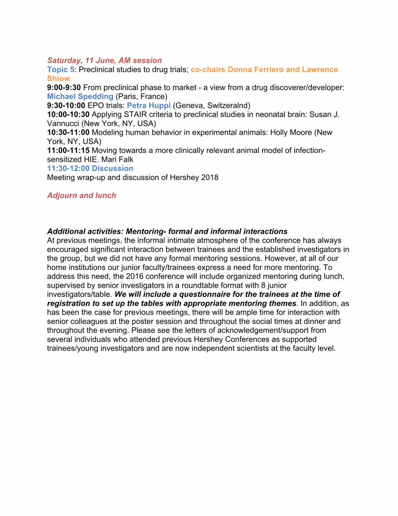
Saturday, 11 June, AM session Topic 5: Preclinical studies to drug trials; co-chairs Donna Ferriero and Lawrence Shiow 9:00-9:30 From preclinical phase to market - a view from a drug discoverer/developer: Michael Spedding (Paris, France) 9:30-10:00 EPO trials: Petra Huppi (Geneva, Switzeralnd) 10:00-10:30 Applying STAIR criteria to preclinical studies in neonatal brain: Susan J. Vannucci (New York, NY, USA) 10:30-11:00 Modeling human behavior in experimental animals: Holly Moore (New York, NY, USA) 11:00-11:15 Moving towards a more clinically relevant animal model of infection-sensitized HIE. Mari Falk 11:30-12:00 Discussion Meeting wrap-up and discussion of Hershey 2018 Adjourn and lunch Additional activities: Mentoring- formal and informal interactions At previous meetings, the informal intimate atmosphere of the conference has always encouraged significant interaction between trainees and the established investigators in the group, but we did not have any formal mentoring sessions. However, at all of our home institutions our junior faculty/trainees express a need for more mentoring. To address this need, the 2016 conference will include organized mentoring during lunch, supervised by senior investigators in a roundtable format with 8 junior investigators/table. We will include a questionnaire for the trainees at the time of registration to set up the tables with appropriate mentoring themes. In addition, as has been the case for previous meetings, there will be ample time for interaction with senior colleagues at the poster session and throughout the social times at dinner and throughout the evening. Please see the letters of acknowledgement/support from several individuals who attended previous Hershey Conferences as supported trainees/young investigators and are now independent scientists at the faculty level.
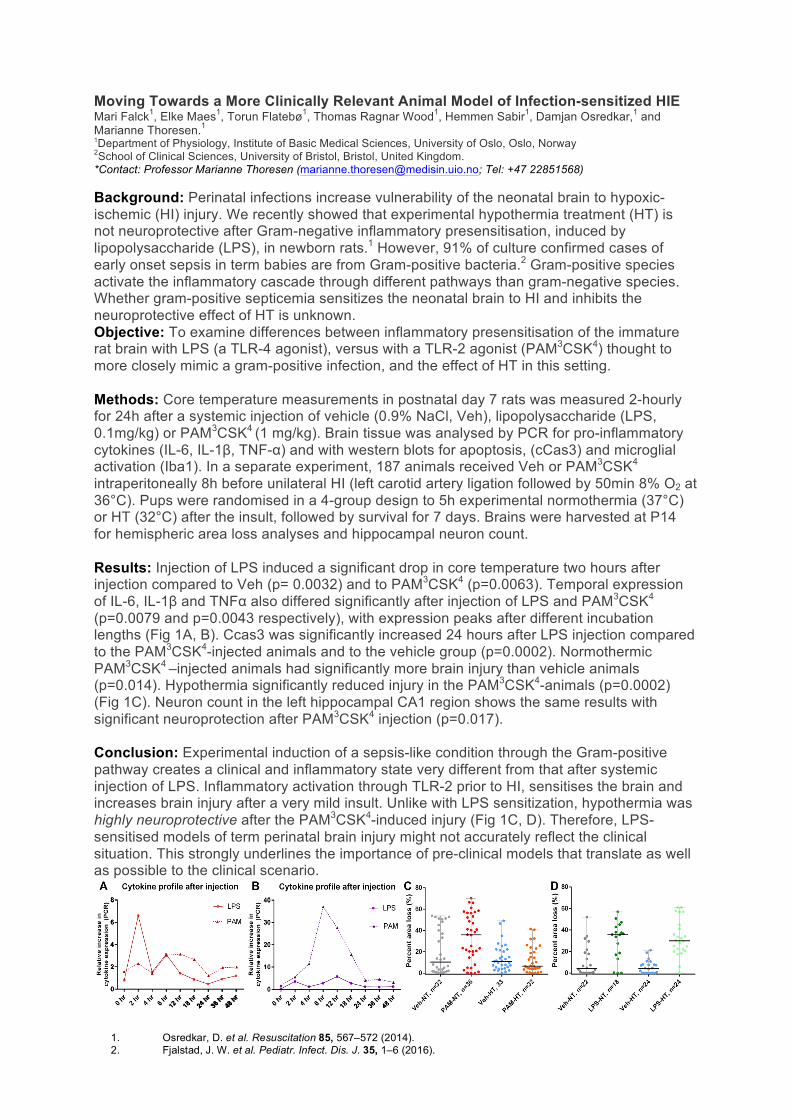
Moving Towards a More Clinically Relevant Animal Model of Infection-sensitized HIE Mari Falck1, Elke Maes1, Torun Flatebø1, Thomas Ragnar Wood1, Hemmen Sabir1, Damjan Osredkar,1 and Marianne Thoresen.1 1Department of Physiology, Institute of Basic Medical Sciences, University of Oslo, Oslo, Norway 2School of Clinical Sciences, University of Bristol, Bristol, United Kingdom. *Contact: Professor Marianne Thoresen ([email protected]; Tel: +47 22851568) Background: Perinatal infections increase vulnerability of the neonatal brain to hypoxic-ischemic (HI) injury. We recently showed that experimental hypothermia treatment (HT) is not neuroprotective after Gram-negative inflammatory presensitisation, induced by lipopolysaccharide (LPS), in newborn rats.1 However, 91% of culture confirmed cases of early onset sepsis in term babies are from Gram-positive bacteria.2 Gram-positive species activate the inflammatory cascade through different pathways than gram-negative species. Whether gram-positive septicemia sensitizes the neonatal brain to HI and inhibits the neuroprotective effect of HT is unknown. Objective: To examine differences between inflammatory presensitisation of the immature rat brain with LPS (a TLR-4 agonist), versus with a TLR-2 agonist (PAM3CSK4) thought to more closely mimic a gram-positive infection, and the effect of HT in this setting. Methods: Core temperature measurements in postnatal day 7 rats was measured 2-hourly for 24h after a systemic injection of vehicle (0.9% NaCl, Veh), lipopolysaccharide (LPS, 0.1mg/kg) or PAM3CSK4 (1 mg/kg). Brain tissue was analysed by PCR for pro-inflammatory cytokines (IL-6, IL-1β, TNF-α) and with western blots for apoptosis, (cCas3) and microglial activation (Iba1). In a separate experiment, 187 animals received Veh or PAM3CSK4 intraperitoneally 8h before unilateral HI (left carotid artery ligation followed by 50min 8% O2 at 36°C). Pups were randomised in a 4-group design to 5h experimental normothermia (37°C) or HT (32°C) after the insult, followed by survival for 7 days. Brains were harvested at P14 for hemispheric area loss analyses and hippocampal neuron count. Results: Injection of LPS induced a significant drop in core temperature two hours after injection compared to Veh (p= 0.0032) and to PAM3CSK4 (p=0.0063). Temporal expression of IL-6, IL-1β and TNFα also differed significantly after injection of LPS and PAM3CSK4
(p=0.0079 and p=0.0043 respectively), with expression peaks after different incubation lengths (Fig 1A, B). Ccas3 was significantly increased 24 hours after LPS injection compared to the PAM3CSK4-injected animals and to the vehicle group (p=0.0002). Normothermic PAM3CSK4 –injected animals had significantly more brain injury than vehicle animals (p=0.014). Hypothermia significantly reduced injury in the PAM3CSK4-animals (p=0.0002) (Fig 1C). Neuron count in the left hippocampal CA1 region shows the same results with significant neuroprotection after PAM3CSK4 injection (p=0.017). Conclusion: Experimental induction of a sepsis-like condition through the Gram-positive pathway creates a clinical and inflammatory state very different from that after systemic injection of LPS. Inflammatory activation through TLR-2 prior to HI, sensitises the brain and increases brain injury after a very mild insult. Unlike with LPS sensitization, hypothermia was highly neuroprotective after the PAM3CSK4-induced injury (Fig 1C, D). Therefore, LPS-sensitised models of term perinatal brain injury might not accurately reflect the clinical situation. This strongly underlines the importance of pre-clinical models that translate as well as possible to the clinical scenario.
1. Osredkar, D. et al. Resuscitation 85, 567–572 (2014). 2. Fjalstad, J. W. et al. Pediatr. Infect. Dis. J. 35, 1–6 (2016).