Embed Size (px)
Citation preview

GRAFT RECOVERY MEDIATED BY DIALLYL DISULFIDE
DURING COLD STORAGE IN A NON-HEART BEATING RAT
LIVER DONATION MODEL.
Cecilia L. BALABAN, Joaquín V. RODRIGUEZ, Edgardo E. GUIBERT.Centro Binacional (Argentina-Italia) de Criobiología Clínica y Aplicada (CAIC). Universidad Nacional
de Rosario, Rosario, Argentina.E-mail: [email protected]
Abstract. Liver transplantation is currently the preferred treatment option for end-stage liver disease, reaching that status because of the continuous improvement in the organ transplantation field. Non-heart beating donation (NHBD) was employed in the early years of organ donation, before brain death criteria were established. These organs are subjected to variable periods of warm ischemia that may intensify cold ischemia/reperfusion injuries. However, due to shortage of brain death donors, the use of NHB grafts is being reconsidered. The present work deals with the setting of a rat model of liver donation after cardiac death and the assessment of a preservation solution additive, diallyl disulfide (DADS, garlic derived,) thought to have cytoprotectant properties mediated by H2S.Cardiopulmonary arrest was induced in a sedated animal by an I.V. injection of KCl/Heparin and warm ischemia period extended up to 45 minutes. Livers were surgically removed and subsequently stored in Custodiol® HTK Solution (Histidine-tryptophan-ketoglutarate) at 0-4 °C. Experimental groups were the following: I- NHBD preserved 24 hs in HTK; II- NHBD preserved 24 hs in HTK + 5 µM DADS and III- Heart Beating Donated (HBD) preserved 24 hs in HTK. After cold storage, livers were evaluated through ex vivo reperfusion. Intrahepatic Resistance (IR60’: mmHg.min.gliv/mL) and Perfusion Flow (PF60’: mL/min.gliv) values showed liver microvasculature damage in NHBD grafts (group I*: IR 2.88±0.55 PF 2.41±0.71) when compare to livers from HBD (group III: IR 2.17±0.11 PF 3.39±0.17) (*p<0.05, vs. group III). Interestingly, when NHBD livers where cold stored in HTK complemented with H 2S donor (group II: IR 2.01±0.31 PF 3.73±0.51), microvasculature state reverted to that of group III livers.Bile production and Oxygen uptake are very helpful tools in order to judge liver physiology since a complex machinery is involved in both procedures. Again, the presence of DADS in the preservation media could be related with an augmented bile secretion and Oxygen consumption.This data suggest that rat liver donation after 45 minutes of warm ischemia post cardiac arrest, constitutes an appropriate model to test different approaches to the rescuing of marginal organs. Also, commercial HTK supplementation with DADS represents a potential treatment to recover non-heart beating harvested organs.
Key words: hypothermic preservation, gasotransmitters, isolated reperfused rat liver, non-heart beating donation.
1. INTRODUCTION
Over the last five decades improvement in donor management, organ preservation, transplantation surgery as well as intensive care and immunosuppression
have turned liver transplantation into the preferred treatment option for end-stage liver failure.The persisting gap between patients in waiting lists and the number of available grafts, has led to an increased interest in using ‘extended criteria’, previously called marginal, donor livers for orthotopic liver transplantation (OLT). These grafts, which in the main comprise steatotic organs, advanced age donation, prolonged cold ischemia and non-hearth beating donation (NHBD) are known to be more susceptible to ischemia reperfusion injury, translated as primary graft non-function, early graft dysfunction or severe biliary tract damage. Nevertheless, several transplant units around the world are currently re-evaluating non-heart beating donation as a strategy to enlarge the pool of organs available for liver transplantation. Clearly, new strategies must be developed for the safe inclusion of such donors [Monbaliu D, 2012].In the past 20 years, hydrogen sulfide (H2S) has changed its reputation from just a toxic gas to potentially a therapeutic drug for the treatment of ischemia/reperfusion (I/R) injury. Recent research has revealed many beneficial roles for H2S in ischemia for a variety of tissues. For example, H2S has been shown to be cardioprotective through both endogenous and exogenous applications at or prior to ischemia, neuroprotective via H2S-induced hypothermia, hepatoprotective by decreasing lipid peroxidation, increasing antioxidant and anti-apoptotic signaling, renal protective through the actions of cystathionine β-synthase (CBS), and protective against pulmonary ischemia injury by the activity of cystathionine γ-lyase (CSE) [Nicholson CK, 2010]. Benavides et al [Benavides G, 2007] demonstrated that the major beneficial effects of garlic rich diets, specifically on cardiovascular disease and more broadly on overall health, are mediated by the biological production of H2S from garlic-derived organic polysulfides (diallyl disulfide, diallyl trisulfide). Substances like Diallyl disulfide act as stable H2S donors when they react with biological thiols, such as glutathione, emerging as potential natural pharmacological agents to confront I/R insults. Therefore, we wondered if DADS may exert cytoprotective effects when added to standard cold storage solution in a model of rat liver donation after cardiac death.In a few words, the present work deals with the setting of a rat model of liver donation after cardiac death and the assessment of a preservation solution additive, diallyl disulfide (DADS, garlic derived) thought to act as a cytoprotectant through H2S.
2. MATERIALS AND METHODS
2.1 Animals
Adult male Wistar rats (weighing 300–320 g) used in these studies were obtained from the Central Animal Building of the School of Biochemistry and Pharmaceutical Sciences, National University of Rosario. Rats were maintained on standard food pellets and water ad libitum and the protocols used were approved by the National Council of Research in Argentina and a Local Ethics Committee from the School of Biochemistry and Pharmaceutical Sciences from the National University of Rosario and they are in concordance with International regulations.
2.2 Non-heart beating donation rat model
Male Wistar rats, weighing between 300 and 320 g, were anaesthetized by intraperitoneal injection of chloral hydrate (Parafarm, Argentina) (500 mg/Kg). The abdomen was opened by midline incision and the liver freed from all ligamentous
attachments. In brief, the bile duct was cannulated with a PE-50 catheter (Intramedic, USA) and the portal vein was cannulated with a 14 G catheter (Abbocath) but perfusion was stalled until later. Cardiac arrest was induced by intravenous injection of concentrated potassium chloride solution (2 M) [García Valdecasas JC, 1998]. Also, 150 UI of heparin were injected into the femoral vein. 45 minutes later, perfusion with buffer Krebs-Henseleit started and the suprahepatic inferior vena cava was cannulated with steel tubing (internal diameter 3 mm). Finally, the liver was removed, flushed out with 20 mL of cold storage fluid and submerged in a reservoir containing the remaining 80 mL of hypothermic preservation solution (Custodiol). Static cold storage extended up to 24 hr at 0-4°C.
2.3 Experimental groups
Rat livers were divided into three experimental groups: Group I/HBD, (n=5): Heart beating donated, these grafts did not underwent a
warm ischemic period. Group II/NHBD, (n=5): Non-hearth beating donated (45 min of warm
ischemia), followed by 24 hr of static cold storage in standard HTK solution. Group III/NHBD-DADS, (n=5): Non-hearth beating donated (45 min of warm
ischemia), followed by 24 hr of static cold storage in HTK solution supplemented with diallyl disulfide (5 µM).
2.4 Isolated perfused rat liver (IPRL)
This in vitro model (fig.1) provided the opportunity to assess cellular injury and liver function in an isolated setting [Bessems M, 2006], [Balaban CL, 2011].After hypothermic storage, livers were fixed in a glass support and perfusion flow was increased gradually over 10 min to allow stabilization of the system. Next, 150 mL of Krebs-Henseleit complemented with 2 % dextran were perfused at constant pressure of 10.3 mmHg in a recirculating system modified from Perfusion media was constantly oxygenated through a thin wall silicone tubing curled in a carbogen (5%CO2/95%O2) ventilated chamber, yielding a perfusate with a pO2 > 450 mmHg. The buffer was subsequently passed through a water heater bath (37°C) and bubble trap prior to entering the liver. pH of perfusate was constantly adjusted to 7.38–7.42 by fractionated addition of HCl (1 N). Portal venous pressure was measured during perfusion by means of a water column connected to the portal inflow line. Hepatic effluent drained into the reservoir through a steel catheter placed firmly into the suprahepatic inferior vena cava.Portal flow rate was measured every 15 min and intrahepatic resistance (IR) was calculated considering perfusion pressure (10.3 mmHg). A 300 µM Taurocholate solution was infused into the perfusion medium at 0.2 mL/min throughout the experiment to sustain bile production. The bile was collected in pre-weighted tubes every 15 min and the bile flow was estimated gravimetrically and expressed as μL/min.g.liver. Perfusate samples were taken from the reservoir in order to determine lactate dehydrogenase (LDH) and Alanine transaminase (ALT) leakage and Urea production. Oxygen consumption was determined at 30 min intervals from inflow and outflow perfusate samples using an YSI model 5300 Biological Oxygen Monitor (Yellow Springs, Ohio, USA), equipped with a Clark–type sensor (YSI 5331 oxygen probe, Yellow Springs, Ohio, USA).Finally, livers were weighed and cut into small blocks that were fixed in 10 % formaldehyde for subsequent histological studies.
Fig. 1. IPRL system
2.5 Perfusate studiesLDH and ALT activities in perfusate samples were determined by kinetic
spectrophotometry, following the decrease in absorbance of nicotinamide adenine dinucleotide (NADH).
2.6 Histology
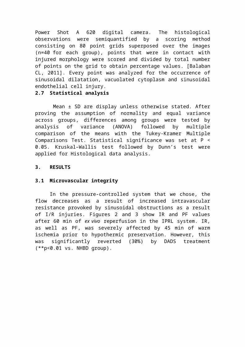
Hematoxilin and Eosin (H/E) staining followed by light microscopy was performed to judge the morphological integrity of the parenchyma. We analyzed 40 microscopic images of three different biopsies from each one of the experimental groups. Fields were chosen randomly within the liver parenchyma and the images were captured by a Cannon Power Shot A 620 digital camera. The histological observations were semiquantified by a scoring method consisting on 80 point grids superposed over the images (n=40 for each group), points that were in contact with injured morphology were scored and divided by total number of points on the grid to obtain percentage values. [Balaban CL, 2011]. Every point was analyzed for the occurrence of sinusoidal dilatation, vacuolated cytoplasm and sinusoidal endothelial cell injury.
2.7 Statistical analysis
Mean ± SD are display unless otherwise stated. After proving the assumption of normality and equal variance across groups, differences among groups were tested by analysis of variance (ANOVA) followed by multiple comparison of the means with the Tukey-Kramer Multiple Comparisons Test. Statistical significance was set at P < 0.05. Kruskal–Wallis test followed by Dunn’s test were applied for Histological data analysis.
3. RESULTS
3.1 Microvascular integrity
In the pressure-controlled system that we chose, the flow decreases as a result of increased intravascular resistance provoked by sinusoidal obstructions as a result of I/R injuries. Figures 2 and 3 show IR and PF values after 60 min of ex vivo reperfusion in the IPRL system. IR, as well as PF, was severely affected by 45 min of warm ischemia prior to hypothermic preservation. However, this was significantly reverted (30%) by DADS treatment (**p<0.01 vs. NHBD group).
Fig. 2. Intrahepatic resistance after 60 min of ex vivo reperfusion. Calculation: IR=10.3mmHg/PF60’. Mean ± SD are presented from 5 different experiments. *p<0.05 vs NHBD group, **p<0.01 vs. NHBD group.

Fig. 3. Perfusion flow after 60 min of ex vivo reperfusion. Mean ± SD are presented from 5 different experiments. *p<0.05 vs. NHBD group, **p<0.01 vs. NHBD group.
3.2 Liver function
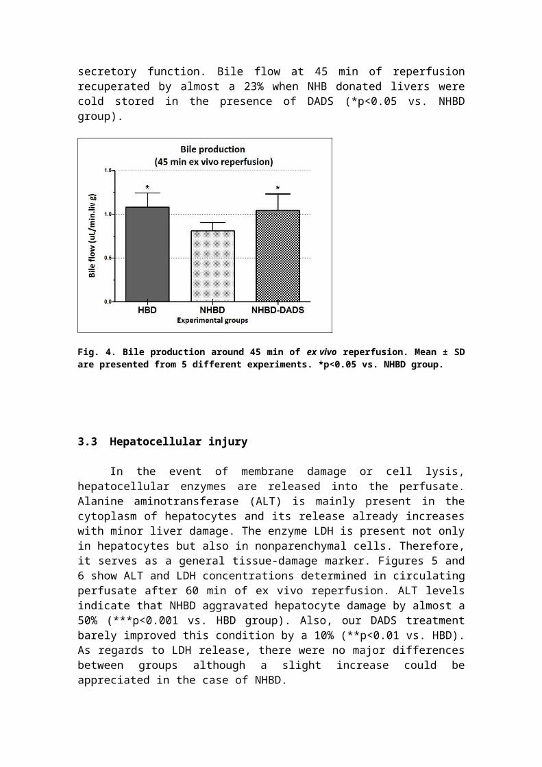
Bile production has long been considered as one of the most important parameters of liver function. Bile flow was calculated from bile samples collected in 15 min periods. Figure 4 depicts bile flow around 45 min of ex vivo reperfusion. Warm ischemia exerted negative effects on bile secretory function. Bile flow at 45 min of reperfusion recuperated by almost a 23% when NHB donated livers were cold stored in the presence of DADS (*p<0.05 vs. NHBD group).
Fig. 4. Bile production around 45 min of ex vivo reperfusion. Mean ± SD are presented from 5 different experiments. *p<0.05 vs. NHBD group.
3.3 Hepatocellular injury
In the event of membrane damage or cell lysis, hepatocellular enzymes are released into the perfusate. Alanine aminotransferase (ALT) is mainly present in the cytoplasm of hepatocytes and its release already increases with minor liver damage. The enzyme LDH is present not only in hepatocytes but also in nonparenchymal cells. Therefore, it serves as a general tissue-damage marker. Figures 5 and 6 show ALT and LDH concentrations determined in circulating perfusate after 60 min of ex vivo reperfusion. ALT levels indicate that NHBD aggravated hepatocyte damage by almost a 50% (***p<0.001 vs. HBD group). Also, our DADS treatment barely improved this condition by a 10% (**p<0.01 vs. HBD). As regards to LDH release, there were no major differences between groups although a slight increase could be appreciated in the case of NHBD.
Fig. 5. ALT release after 60 min of ex vivo reperfusion. Mean ± SD are presented from 5 different experiments. ***p<0.001 vs. HBD group, **p<0.01 vs. HBD group.
Fig. 6. LDH release after 60 min of ex vivo reperfusion. Mean ± SD are presented from 5 different experiments. No significant differences were detected.

3.4 Oxygen consumptionOxygen uptake from perfusate was a valuable marker to obtain further
information about livers physiology. Oxygen consumption was calculated with eq. 1.
Eq. 1. (µmol O2 / min. g liver) = (Cin–Cout) / perfusion flow (mL / min. g liver)
where Cin and Cout are the oxygen concentration in the inflow and outflow, respectively. CO2 (µmol O2 / mL) = pO2 (kPa). SO2
37°C (µmol O2 / mL.kPa). Where SO237°C is the oxygen
solubility in water at 37°C. SO237°C = 0.01056 µmol O2 / mL.kPa [Gnaiger E. 2004].
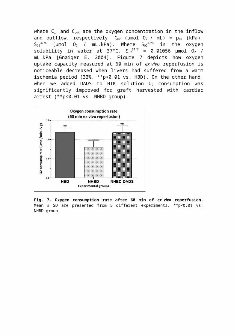
Figure 7 depicts how oxygen uptake capacity measured at 60 min of ex vivo reperfusion is noticeable decreased when livers had suffered from a warm ischemia period (33%, **p<0.01 vs. HBD). On the other hand, when we added DADS to HTK solution O2
consumption was significantly improved for graft harvested with cardiac arrest (**p<0.01 vs. NHBD group).
Fig. 7. Oxygen consumption rate after 60 min of ex vivo reperfusion. Mean ± SD are presented from 5 different experiments. **p<0.01 vs. NHBD group.
3.4 Histological studies
Our experimental protocol did not generate severe alterations to livers morphology for any of the three experimental groups, as it can be seen in fig. 8 where H/E staining of liver sections shows relatively conserved tissue architecture. Nevertheless, a deeper analysis shown in fig. 9 revealed differences between livers submitted to our model of cardiac arrested donors and control hearth beating donation (sinusoid and endothelial cell parameters). In addition, we detected that DADS supplementation reduced significantly the presence of vacuoles and endothelial cell injuries.
Fig. 8. Hematoxilyn / Eosin stained liver sections post-IPRL.

Fig. 9. Histology analysis: Percentage values account for frequency of injured morphology event in each microscopic field. Sinusoidal injury was defined as widening of sinusoids, and endothelial cell injury was defined as cell shrinkage with cellular rounding, retraction and detachment. Mean ± SEM are presented from 40 different microscopic fields. *p<0.05 vs. NHBD.
4. DISCUSSION
The success of solid organ transplantation has considerably expanded waiting lists with insufficient donation rates and substantial waiting list mortality. This has lead to re-examination of the term “marginal” donors, and particularly focused on Non-Heart Beating Donation (NHBD). Organs from these donors inevitably sustain warm ischemic
damage which varies in its extent and affects early graft function as well as graft survival [Monbaliu D, 2012]. Interruption of blood supply to the warm liver produces ischemia, which results in deprivation of energy supply of the metabolically active organ and severe damage. This warm ischemic deterioration prior to liver harvesting in cardiac arrested donors will enhance hypothermic preservation injuries and worsen reperfusion insults. Therefore, it is important to devise strategies to ameliorate I/R related damage during extended criteria organ procurement for transplantation.H2S-related pharmacological research is a rapidly emerging field, which is likely to yield a number of therapeutic possibilities. A number of independent groups have reported the beneficial effects of H2S or sulphide-donor compounds in animal models of I/R [Kang K, 2009; Elrod JW, 2007; Shaik IH, 2008]. Organic polysulfides, molecules with chains of sulfur atoms between two functional groups, are being investigated as natural H2S donor compounds. Diallyl disulfide and diallyl trisulfide (DATS) are two garlic-derived organic polysulfides that can act as stable H2S donors when they react with biological thiols, such as glutathione [Benavides G, 2007].In this work, a model of ex vivo reperfusion was successfully applied to assess physiology of cold preserved livers under different conditions. One of our goals was to set up a rat model of liver Non-heart beating donation (NHBD) that would allow us to evaluate our proposal for the rescuing of marginal livers. We were able to develop a suitable protocol for NHBD grafts, inducing cardiac arrest with a high concentration of KCl and 45 min of warm ischemia. Indeed, this grafts had poorer outcomes when compared to livers of hearth beating donor rats.Diallyl disulfide was added to the commercial preservation solution (Custodiol®) in a final concentration of 5 µM to judge its potential therapeutic power to restore NHBD suboptimal grafts. The final concentration of DADS was selected in our laboratory by screening assays of 4 different doses (1,5,50 and 500 µM) Data not shown.Hepatic microcirculatory system was evaluated through intrahepatic resistance along with perfusion flow. Data suggested that DADS treatment enhanced vascular protection as livers presented lower resistance to perfusion. In accordance with these results, histological studies showed a rather conserved morphology with lower rates of endothelial cell injury. This is not a surprising result, since vasorelaxant properties of garlic are well described in the bibliography [Banerjee SK, 2002]. Moreover, Benavides demonstrated that H2S mediates vasoactivity of garlic. Although we expected to find a marked decrease on hepatocellular damage for DADS treated group, differences with NHBD group were not statistically significant for ALT and LDH release in perfusate. Bile production reflected that livers remained functional in the conditions evaluated, at least up to 45 min of ex vivo reperfusion. Still, higher rates of bile flow were detected for NHBD-DADS group. With regards to oxygen consumption during IPRL, we determined that O2 uptake of NHBD grafts reverted to control condition (no-warm ischemia) when they were stored with DADS compound.
Undoubtedly, mechanisms of action of DADS through H2S delivery to the preservation fluid still need to be elucidated. In summary, our results support DADS tissue-protective roll when added to hypothermic preservation solution which makes it a promising candidate for improvement of classic static cold storage and subsequent expansion of transplantable organ criteria.

CONCLUSION
The persisting gap between patients in waiting lists and the number of available grafts, has led to an increased interest in using ‘extended criteria’ donors. These grafts, which include non-hearth beating donation (NHBD) are known to be more susceptible to ischemia reperfusion injury, translated as primary graft non-function and early graft dysfunction. We have presented data supporting that rat liver donation after 45 minutes of warm ischemia post cardiac arrest, constitutes an appropriate model to test different approaches to the rescuing of marginal organs. Also, commercial HTK supplementation with DADS arises as a promising alternative for the optimization of cold storage of livers in order to recover non-heart beating harvested organs.
ACKNOWLEDGMENTS
This work was supported by grant PIP-1208 from CONICET, grant - Prot 19096/PT Regione Autonoma Friuli-Venezia Giulia, Italy and grant 1BIO176 from UNR. C.L. Balaban, J.V. Rodríguez, and E.E. Guibert are members of CONICET.
BIBLIOGRAPHY
Balaban CL, Rodriguez JV, and Guibert EE. Delivery of the Bioactive Gas Hydrogen Sulfide During Cold Preservation of Rat Liver: Effects on Hepatic Function in an Ex vivo Model. Artif Organs. 2011, 358(5):508-515.Banerjee SK and Maulik SK. Effect of garlic on cardiovascular disorders: a review. Nutrition Journal 2002, article available from: http://www.nutritionj.com/content/1/1/4.Benavides G, Squadrito GL, Mills RW, Patel HD, Isbell TS, Patel RP, Darley-Usmar VM, Doeller JE, and Kraus DW. Hydrogen sulfide mediates the vasoactivity of garlic. PNAS. 2007, 104(46), 17977-82.Bessems M, ‘t Hart NA, Tolba R, Doorschodt BM, Leuvenink HGD, Ploeg RJ, Minor T and van Gulik TM. The isolated perfused rat liver: standardization of a time-honoured model. Laboratory Animals. 2006, 40:236–246.Elrod JW, Calvert JW, Morrison J, Doeller JE, Kraus DW, Tao L, Jiao X, Scalia R, Kiss L, Szabo C, Kimura H, Chow C-W, and. Lefer DJ. Hydrogen sulfide attenuates myocardial ischemia-reperfusion injury by preservation of mitochondrial function. PNAS. 2007,104(39):15560-65.García Valdecasas JC, Tabet J, Valero R, Taurá P, Rull R, García F, Montserrat E, González F, Ordi J, Beltran J, López Boado M, Deulofeu R, Angás J, Cifuentes A y Visa J. Liver conditioning after cardiac arrest: the use of normothermic recirculation in an experimental animal model. Transpl Int. 1998,11:424-432.Gnaiger E. Oxygen solubility in experimental media. Mitochondrial Physiology Network. 2001–2004;6:1–6. Available at: http://www.oroboros.at/index.php?oxygen-solubility.Kang K, Zhao M, Jiang H, Tan G, Pan S, and Sun X. Role of Hydrogen Sulfide in Hepatic Ischemia- Reperfusion–Induced Injury In Rats. Liver Transpl. 2009,15:1306-14.Monbaliu D, Pirenne J, Talbot D. Liver transplantation using Donation after Cardiac Death donors. Journal of Hepatology. 2012, 56:474–485.Nicholson CK, Calvert JW. Hydrogen sulfide and ischemia–reperfusion injury. PharmacologicalResearch. 2010, 62:289–297.Shaik IH, George JM, Thekkumkara TJ, and R Mehvar. Protective Effects of Diallyl Sulfide, a Garlic Constituent, on the Warm Hepatic Ischemia–Reperfusion Injury in a Rat Model. Pharmaceutical Research. 2008, 25(10):2231-42.