Embed Size (px)
Citation preview
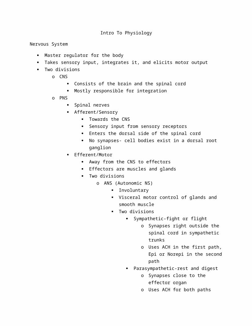
Intro To Physiology
Nervous System
Master regulator for the body Takes sensory input, integrates it, and elicits motor output Two divisions
o CNS Consists of the brain and the spinal cord Mostly responsible for integration
o PNS Spinal nerves Afferent/Sensory
Towards the CNS Sensory input from sensory receptors Enters the dorsal side of the spinal cord No synapses- cell bodies exist in a dorsal root ganglion
Efferent/Motor Away from the CNS to effectors Effectors are muscles and glands Two divisions
o ANS (Autonomic NS) Involuntary Visceral motor control of glands and smooth muscle Two divisions
Sympathetic-fight or flighto Synapses right outside the spinal cord in
sympathetic trunkso Uses ACH in the first path, Epi or Norepi
in the second path Parasympathetic-rest and digest
o Synapses close to the effector organo Uses ACH for both paths
Both divisions have their first path myelinated while the second path is unmyelinated
o Somatic NS Voluntary motor control From the CNS to skeletal muscles Single neuron all myelinated Uses ACH as the neurotransmitter
Glial cells-non neuronal cells in the nervous systemo Many more glial cells than neurons
o Insulation (myelin) Oligodendrocytes in the CNS
Can wrap more than 1 neuron Schwann Cells in the PNS
Can only wrap 1o Protection
Astrocytes The majority of glial cells Provide nourishment to the cells Create the Blood Brain Barrier
o Circulation Ependymal cells
Ciliated Circulate (do not produce) CSF
o CSF is produced by a choroid plexuso Immunity
Microglia Macrophages Immune system isn’t allowed into nervous system
Neurono Dendrites
The receptive region of the neuron Contains Na and K ion channels to start depolarization Receives neurotransmitter via receptors Post synapse
The signal goes electrical, chemical, electricalo Membrane potential
Resting cells are at -70 mv Action potential brings them to +30 Opening of Na channels brings + charges into the cell
Causes the climb in charge Depolarization
Repolarization is caused by the delayed opening of the K gated ion channel (activated at the same time but delayed) allowing K ions out of the cell
Also slow to close, causing hyperpolarization This has the added benefit of making sure the neuron cant fire too much
too fast Na/K pump restores ion concentrations after The anions in the cell are mostly responsible for the negative charge Charges balance out

An electrical equilibrium is created based on the regular charge of the cell, or based on an outside charge (experimentally)
Charges flow to make the charges equal Add + charge to cell, it wants to decrease the potential so it flows out Add – charge to cell, it wants to increase the potential so ions flow in
Threshold potential of the neuron is reached at -55 mv Reached at the axon hillock
-58 mv is the K ion potential equilibriumo Graded potentials
Depolarizes one region of the cell Can be any level
-34 -24 -85 (so it can even be hyperpolarization) Etc
The cell region is depolarized yet does not reach threshold at -55 mv Graded potentials can lead to Action potentials Can be summed in time or in space
Temporal summationo One after another, they add up
Spatial summationo In the same space, add together
o Action potential Reaches threshold Generated at the axon hillock Depolarization due to Na channels opening Reaches about +30 Then K ion channels open up and there is repolarization Then hyperpolarization as they are slow to shut Then the gradient is reestablished by the Na/K pump Another one cannot be fired for 4-5 ms Needs to be strong to overcome hyperpolarization
Absolute refractory- nothing can be fired Relative refractory-only a very strong graded potential will lead to an AP
The number of AP’s determines the stimulus strengtho Variables
Size Increase diameter, the speed increases
Myelination Myelination increases speed by a lot Saltatory conduction

o In jumps only to the nodes of ranviero Makes it so that only at the nodes of ranvier does the AP have
to occuro Three types of neurons
Multipolar Most common
Bipolar Found only in special senses
Unipolar Sensory receptors
o Three functions Sensory (afferent) Motor (efferent) Interneurons
CNS Most common (99%)
Neurotransmitterso Diffuse from high to low gradient across the synaptic clefto IPSP
Inhibitory Hyperpolarizes EX: Cl- or GABA
o EPSP Excitatory Depolarizes EX: glutamate
o EPSP and IPSP can fight each other to try and create (or not create) an APo The distance from the axon hillock makes a difference, as the depolarization decreases
as the distance increases Terminal bulbs
o Ca ion channels (voltage gated) open when the AP reaches, and the influx of Ca pushes vesicles containing NTs to the wall of the terminal bulb
o This causes their release into the synapse Ionotropic receptors
o Everything discussed hereo Fast
Metabotropic receptorso Use second messengerso Usually G-protein linkedo And takes longer due to the second messengers

Endocrine System
Method of communication in the bodyo Use the blood streamo Hormones are long distance
Hypothalamus- top of the pituitary gland- master control Pituitary-second in command Lipid soluble hormones can go directly through the bilayer and attach to a receptor inside the
cell Hormones are usually controlled by metabotropic receptors
o Their effect takes longer than neurotransmitters Due to amplification
Target cell activation depends on 3 factorso Number of hormones in the bloodo Number of receptorso Affinity for receptors (how tightly they bind)
cAMP second messengero Gs on a G-protein receptoro Response is amplifiedo Activates PKAo Hormones that use this cAMP
Cattecholamines Acth FSH LH Glucagon PTH TSH Calcitonin
Pip second messengero DAG and IP3 activated Calcium ion release and therefore PKCo Hormones
Catecholamines TRH ADH GnRH Ocytocin
o Gq subuinits Hormone interactions
o Antagonism

o Permissivenesso Synergism
Endocrine gland stimulationso Humoral (nutrients in blood)o Hormoneso Neuronal
Faster than hormonal Pituitary gland portal system
o Regular portal system is arteriole-capillary-venuleo Pituitary portal system is capillary-vein-capillary
Goes quickly from hypothalamus to pituitary More concentrated hormones in a shorter distance
Speed and concentration Pituitary
o Posterior pituitary Oxytocin
Made in the paraventricular nuclei of the hypothalamus and sent to the posterior pituitary to be released
ADH Supraoptic nuclei in the hypothalamus make it
o Anterior pituitary TSH, GH, ACTH, prolactin, LH, FSH
Aneurysmo Saccular-bulb at risk of rupturingo Fusiform-not as dangerous-> longer elongationo Ruptured-broken open
Kidneyso Adh production
Mechanism? Triggers kidneys to reabsorb water, therefore increasing blood volume
and pressure Based on osmolality
Thyroid hormoneo Major metabolic hormoneo T3 and T4 (needs IODINE)o Path
Hypothalamus-TSH (ant pituitary)-thyroid-thyroid hormones PTH vs Calcitonin
o PTH increases the blood Ca level and breaks the bone downo Calcitonin decreases blood Ca level by building the bone up
Insulin vs Glucagon

o Insulin decreases blood glucose levelo Glucagon increases glucose level
Aldosteroneo Directly brings in Na and water into the blood in the kidneyso Controlled
Increased K+ level in blood Decreased blood volume or blood pressure Acth released by anterior pituitary Increased blood pressure or volume
Inhibitoryo Targets kidney tubuleso Antagonized by ANP release by the heart
Stress responseo CRH released by hypothalamus (corticotropin releasing hormone)o Causes the adrenal medulla to release catecholamines (epi/norepi)o Catecholamines release ACTH to raise the blood pressureo Catecholamines short term stress responseo Mineralo and glucocorticoids=long term stress response
Different behavior?o Due to different receptors
Muscles
Smooth, cardiac, and skeletal Skeletal is under conscious control Smooth and cardiac are unconscious Four functional characteristics of muscle tissue
o Contractiono Extensiono Elastico Excitability
Skeletalo Striatedo Multinucleatedo Cylindricalo ACH binds to it and is always excitatory
Cardiaco Striatedo Multibranchedo Pacemakero Cells connected by calated discs

Smootho No striationso Spindle shapedo Shortens with calmodulin
Skeletal muscleso Contract small to largeo Smaller motor units first
Each bundle separated by perimysiumo Bundle=fascicle
Muscle fiber (cell)o Surrounded by the endomysium
Myofibrilso Contains two filaments (myofilaments)
Actin and myosin Each muscle fiber contains many myofibrils Motor unit
o The motor neuron and all of the muscle fibers it innervateso Vary in size
Myosin and actino Work together to contract the muscle
T tubuleso Allow the AP to go down into cell and release Ca from the sarcoplasmic reticulum
Sarcoplasmic reticulumo Calcium ion storage
Glycosomeso Storage of glycogen for quick energy
Sarcomereo The smallest unit of the muscleo Z disc to z disco H zone is based on thick filaments onlyo I bands only thino A band length of myosin
Calciumo The final trigger for the muscle contraction, what is the initial trigger for the muscle cell
Muscle contraction stepso Na enters and AP propagated along the cell and down the t tubuleo AP triggers Ca channel in the SR to openo Ca binds to troponin
Changes the shape of tropomyosin and actin active sites are exposedo Contraction
Myosin heads bind to active sites on actin and use energy by ATP to push

o Removal of calcium from the cytosolo Tropomyosin blockage restored
Thin filamentso Actino Two actin molecules helix shape o Actin associated with troponin and tropomyosino Troponin binds to Ca ionso Tropomyosin covers active sites
Myosino Thick filamentso 2 lobed heads
Cross bridgeo Myosin head attaches and forms a cross bridgeo ATP is released to push the cross bridge
Power strokeo ATP attaches to dislodge crossbridgeo The ATP to ADP charges it for its next powerstroke
Sliding filament theoryo Myosin pulls actin closero The H band gets smaller
Rigor mortiso Crossbridge remain engaged after death
High tensiono Muscles need ATP to relax
Motor unit recruitment and wave summationo Increasely strong muscle contractionso Motor unit recruitment recruits more motor unitso Wave summation work for one motor unit increasing in force (not calling more)
Size principle for motor unitso Small-meduim-large
Muscle contactiono Isometric
Same length No shortening of the sarcomere Develop tension in the muscle
o Isotonic Shortening Same tension Tension is at or above the load so the muscle shortens
Energyo Creatine phosphate

Directly converts ADP to ATP for quick energyo Anaerobic mechanism
Hlycolysis and lactic acid formso Aerobic mechanism
Glucose and fatty acids from fat tissue Provides hours of energy
3 types of fiberso Slow oxidative
Endurance activities First recruited
o Fast oxidative Sprinting/walking
o Fast glycolytic Short term intense movement
Oxygen debto Spend more energy than can be providedo Lactic acid build up needs to be corrected
Blood Vessels
Deoxygenated blood comes from a capillary into the right atrium from the vena cava Pulmonary circuit is lower pressure Systemic circuit is high pressure Arterioles go away from the heart Veins go towards the heart Pulmonary veins-left atrium-left ventricle-aorta-arterioles-capillary-venules-veins-vena cava-
right atrium-right ventricle-pulmonary arteries Cardiac output
o HR X (EDV-ESV)o EDV- amount of blood left in ventricle when the heart is relaxedo ESV- amount of blood left in ventricle during contraction
The highest pressure of anywhere in the circulatory system is in the left ventricle (to get it into the aorta)
Average of 5.25L of blood per minute Preload
o Greater preload leads to a higher stroke volumeo Unless they are too high, which decreases stroke volume
Stretches the sarcomere too much Contractility
o The contractile force of the hearto Increased by the amount of Ca ions released
More calcium, more crossbridges

Afterloado The amount left overo Greater left over, need more pressure to push the blood out
Sympathetic nervous system increases the heart rate Parasymp decreases the heart rate ADH and aldosterone can affect the volume of the blood CHF caused by MI, high Bp, etc
o Pulmonary CHF Left side fails first and the blood backs up in the lungs
o Peripheral CHF Right side fails first and the blood pools in extremities
Norepi or epio Released in adrenal medullao Activate cAMP mechanismso Allows more Ca into the cell and cause a stronger contraction
Vesselso Capillaries are responsible for a gas exchange o Types
Fenestrated Holes in the walls (window) Intestines Filtration or absorption is their function SI is the site of most absorption Endocrine glands also use fenestrated capillaries
Continuous No holes/spaces Lungs-gasses are small enough you just need PM Specialized continuous capillaries are in the brain
Sinusoidal Allows cells to go through WBC’s move around
Arterieso Elastic-conductingo Muscular-distributingo More elastic tissue
Withstand higher pressureo Thicker tunica media
Veinso Valveso Lower pressure
Capillary beds

o Perfusion depends on the sphincter of arterioleo Can control which capillary beds are open or closed at a time
Mean arteriole pressure pushes blood through the hearto Diastolic pressure + 1/3 (pulse pressure (difference between systole and diastole))
o Pressure through blood vessels is steady-does not vary up and down How do the veins return blood to the heart
o Skeletal muscle contractionso Breathingo Valves in the veins preventing backflow
Contractility is defined by a sarcomere lengtho At a stable sarcomere length still get increased contraction due to higher calcium levels
Kidney compensationo Multiple ways the kidneys can affect BPo Renin Angiotensin aldosterone system
Angiotensin II causes an increase in BP Constricts blood vessels by impairing NO synthesis
o Increases aldosterone production Retains Na and water increasing blood volume and blood pressure
Intrinsic mechanismso Localo Distribute blood flow to individual organs as neededo Metabolic controls and myogenic controls
Extrinsic mechanismso Global

o Nerves and hormoneso Control levels all over the body, not just to specific places
Factors determine fluid in and outo Hydrostatic pressure-force fluid outo Osmotic pressure-water moving ino Net filtration pressure
Respiratory System
4 processes of respirationo Inspirationo Expirationo External respirationo Internal respiration
Continuous capillaries Breathing
o Air passes to alveoli where gas exchange occurso Inspiration
O2 into the bodyo Exhalation
CO2 out Conducting zone
o Filter and warm (nasal cavity)o Moisten the airo Rigid tube with cartilageo Cilia/structures that help filter
Bronchi split until they reach the terminal bronchioles Alveoli pop up in the respiratory bronchioles Dividing line is between the terminal bronchioles and respiratory bronchioles Respiratory zone
o Less rigido Some smooth muscleo Smooth muscle wrapped around the respiratory bronchioles
What controls the dilation of the airwayo Smooth muscle controlled by
Gas Sympathetic
Dilates!!!!!!!!! α receptors constrict (found in the vessels) β receptors dilate (found in the lungs)
also found in coronary arteries and skeletal muscle arteries

external respiration membraneo 3 or 4 structureso Alveolio Continuous capillarieso 2 types of cells that make up an alveolus
Type 1-shared respiratory membrane Type 2-surfacant secreting
Detergent that breaks surface tension Basement membrane (fused)
o Immune cells in our lungs Macrophages constantly hunt for bacteria and pathogens Macrophage is stuck in alveoli
Negative pressure breathingo Generate a negative pressure to pull air in o Inhalation is active
Contract the muscleso Exhalation is passive
Relax muscles and push air outo Breathing muscles
Diaphragm and intercostal muscles Lungs automatically want to collapse, chest pushing out Creates an interplural pressure that is always negative relative to the atmospheric pressure Visceral pleura lines lungs Parietal pleura lines the inside of the chest wall Relative to ATM
o Interpulmonary pressure negative on inspiration, zero at break, and positive on expiration
Transpulmonary pressure at 0? o Pneumothorax
Pp=PT X Co Partial pressure= total pressure X concentration
Gasses diffuse from high to lowo Tissues are in need of oxygen, oxygen diffuses into them
Dead air spaceo Mixes oxygeno 150 mL o Conducting structures
Dead air space causes expired O2 to be higher than alveolar O2o The alveolar oxygen is low due to it perfusing into the capillaries
High altitudeo Breathing rate increases

o More Red Blood cell production Respiratory groups
o Ventral respiratory group Generate normal breathing GABA
o Dorsal Respiratory Group Modulates the VRG Can sense changes in parameters, to then effect
o Pontine respiratory center Can change rate
Respiratory stimulanto CO2 levels drive breathingo CO2 produced from tissueso Decreases the PH of the blood by being converted to carbonic acid which lowers the PH
Hyperventilationo Usually a V/Q matcho Ventilationo Perfusiono Need to match
Oxygen carried in the blood by hemoglobin (Hb) 90% of Oxygen attached to Hb
o Gives it more solubilityo 4 bonds pere Heme
The affinity of hemoglobin for oxygen changes with the extent of oxygen saturation More oxygen there is, the more affinity there is for O2 High levels of O2 sat
o Usually unladed fast to the tissues, and difficult to load to the Hb Decreased CO2 pressure leads it to be harder to unload Increased CO2 pressure? Easier to unload Lower body temperature triggers decreased co2 levels High metabolic activity leads to increased CO2 CO2
o Typically carried in the bloodo Bicarbonate iono 70% is bicarbonate iono 20% is dissolved directly in the plasmao 10% bound to Hb
Carbonic anhydraseo Converts CO2 to carbonic acido In the Red Blood Cell
Cl- comes into the RBC as the HCO3- ion leaves to balance the charges

Haldane effecto Lower O2 levels?
Higher PCO2o Higher O2 levels?
Lower PCO2 If oxygen is saturated, harder for CO2 to bind to Hb BPG
o Helps push O2 off of the Hb so high levels in systemic capillaries causes O2 unloading HAPE
o High altitude pulmonary edemao Fluid in the lungso Causes a decrease in alveolar ventilation
Digestive system
Nutrient competition in the gut between good and bad bacteria Leptin is made by bacteria and signals body to feed Many organs have dual functions
o Livero Pancreaso Pharynx
Accessory organso Support the digestive system (not a part of the alimentary canal)o The alimentary canal is the path from the mouth to the anus
The “tube within the tube”o Pancreaso Gallbladdero Livero Stomach cells
Mechanical digestion o Occurs via the teeth
Chemical digestiono Chemically breaks down foodo First is salivary amylase
Breaks down carbohydrateso HCl degrades food in the stomach
Creates chime Acid also kills many bacteria
o SI Major place of chemical digestion Proteins, fats, carbs are all degraded and absorbed

Gallbladdero Stores concentrated bile from the livero Shunts it to the small intestine via the common bile duct (with the pancreas)
Large intestineo Major site of water reabsorptiono Also produces the feces
The goal of digestion is to get the components of food broken down into usable parts Long reflexes
o Go all the way to the CNS and get integratedo Prepare the body to receive food
Short reflexeso Local (enteric) nerve plexus
Reflexes are stimulated by food Antacids work by blocking H+ ion production and making the stomach acid less acidic
o Has the effect of not killing some of the bacteriao Acid is usually 2-3 pH
Stomach digestiono Breaks down food mechanically and chemically
Mechanical is churning Chemical is acid and peptidases
o Gastrin Stimulates the secretion of HCl and pepsinogen which works to chemically
digest proteins (in its active pepsin form)o Pepsinogen
Activated by HCl Into pepsin Pepsin then works to break down proteins
o Parietal cells Make the HCl Secreted by goblet cells
o Chief cells Make the pepsinogen
o Enteroendocrine cells Hormone producing cells
How does the brain control gastric juice productiono Brain senses food and secretes additional gastric juice
Reflect of food…salivao Symp-lesso Parasymp-moreo Ph based
Low pH-less gastric juice

Vagus nerve of the parasymp NS stimulates the digestive system Which 3 chemicals are necessary for maximum HCl production?
o Gastrino ACH
From the parasymp (vagus)o Histamine
Chyme gets sent to the duodenumo The duodenum produces many hormones
Secretin CCK (Cholecystokinin) VIP (vasoactive intestinal peptide)
o All of these hormones are known under the collective name enterogastrones SI
o Nutrient absorption Microvilli Folds (gyri) Increased surface area
o Microvilli contain brush border enzyme Bring things to the monomer form
Nutrients absorbed go to the livero Hepatic portal systemo Liver creates bileo Bile salts
Emulsifiers Help break down fats
o Lacteals take the fats to the liver Triglycerides pass into lacteals Called chylomicrons
Livero Processes SI bloodo Liver stores the glycogeno Detoxifies compoundso Arterioles and venules send blood the same way
Arterioles supply oxygen Venules supply nutriets
o Kupffer cells WBC (macrophages)
o Liver can regenerate o Bile production
CCK-singals the gallbladder to release Secretin

Tells the liver to make bile H+ is neutralized in the SI by bicarbonate ion
o Comes from the pancreatic juice CCK induces the pancreas to produce pancreatic juice Secretin binds and causes copious release of bicarbonate ions in pancreatic juice Pancreas hormones (in juice)
o Trypsinogen trypsino Chemotrypsinogenchemotrypsino Procarboxypeptidasecarboxypeptidaseo Activated by brush boarder enzymes
Large intestineo Water reabsorptiono Some absorption of ions occurs with the watero A lot of bacteria in the LI, some good others bad
Immune System
Adaptive vs innate immune systemo Innate
First line of defense Skin barrier Mucus membranes Skin secretions
Doesn’t require previous exposure Present at birth Enzymes that can directly kill bacteria Faster and more immediate Has internal nonspecific defenses
Phagocytes Fever Natural killer cells Antimicrobial proteins Inflammation
NK cells recognize antigen (just that it has an antigen) and releases chemicals called granzymes to perforate the membrane
Macrophages Derived from monocytes Stem cell is the hemocytoblast General macrophages eat bacteria Search for food and engulf them through a process known as
phagocytosis Arrive after neutrophils (part of innate defense)

Phagocytosis Success rate increased (opsonization) Antibodies specific to the antigen are produces and coat the bacteria
o Neutralizationo Agglutinationo Precipitation
Inflammatory responseo Neutrophils enter blood from the bone marrowo Margination
They stick to the sides of the blood vessel wall via CAM (cell adhesion molecules)
o Diapediesis Walk to the site
o Positive chemotaxis Call more WBC
o Four cardinal signs Swelling
Edema Heat
Increased blood flow Pain
Leaked protein rich fluid Histamine released
Redness Increased blood flow
Increased temperature short term helps fight the infection Long term fever can be dangerous for the organism Interferons and complement proteins enhance the innate defenses by attacking microbes
directlyo Interferons are localo Complement proteins are more whole body
Adative immune systemo Specifico Made to attack very specific microbeso Vascularo Antibodieso Lymphocytes vs leukeocytes
Lymphocyte is one of three specific types of WBC Leukeocytes are WBC overall (larger category)
o Immunocompetance (need to get educated) Happens in the T gland for T cells

Bone marrow for B cells Have not been exposed to antigen? Naïve
o APC (antigen presenting cells) Big 3?
Dendritic cells B cells Macrophages
o T cells Recognize our own MHC proteins If they recognize it? They are killed off to avoid an autoimmune reaction Surviving cells are self tolerant
o Activated immune cells are “selected” and begin to divideo B cells
Plasma B cells-antibody secreted Memory B cells-stay around for next reaction for less of a lag time
o Humoral immunity-antibody mediated (B-cell) Active-make antibodies
Natural-antibodies/reaction Artificial-vaccine
Passive-pass antibodies Natural-passed from mom Artificial-serum (antibodies)
o Cell mediated immunity T cell response
Helper T cells recognize and help mount an attacko CD4o Respond to class II MHC’s via the APC’s
Cytotoxic T cells kill directlyo Induce apoptosis with granzymes and perforino CD8o Respond directly to class I MHC proteins
MHCII Only present in APC Taken in by APC cells (endocytosed) and combined with a MHCII
receptor Only an antigen fragment is displayed (antigenic determinant)
MHCI All cells carry it Display any antigens that invade the cell
Urinary System

Kidney is at the center of the urinary systemo Major functions
Filter blood Excrete urine pH balance is important
o blood comes from the aorta and feeds the kidneyso pair of ureters collects the urine
nephrons-functional unit of the kidneyo also the urine forming units
Proximal convoluted tubule (PCT)o Cells have lots of mitochondria and microvilli
Absorption function Kidneys modulate the systemic BP
o Catcholamineso Angiotensino Renino ADH
Glomerulus is where renin is releasedo Can sense the BPo Renin raises the BP (vasoconstrict)
Proteins cannot pass throught he glomerular filtration membrane because it is too selective three forces determine the filtrate formation
o glomerular hydrostatic pressure out
o osmotic pressure in
o capsular hydrostatic pressure in
o net outward pressure is 10mm hg three mechanisms help maintain glomerular filtration rate when systemic bp is altered
o myogenico tubuloglomerularo hormonal
afferent arterioles enter the glomerular capsul efferent arterioles leave the macula densa cells in the ascending loop of henle sense alteration of the filtrate flow and
the NaCl concentration The water in kidneys follows solutes out passively Proximal Convoluted Tubule
o Contains the Na/K pump that establishes the gradient to maintain ion balance Primary and secondary active transport for absorption

Transport maximum exists for everything but Na iono Maximum number of carriers are operating
DCTo Primary control of tubular secretion/reabsorption
Sends things back IN to the Tube if there is too much in the bloodo Aldosterone-sends Na ino Parathyroid hormone (PTH)-sends Ca in
Loop of henleo Descending limb-water reabsorption passiveo Ascending limb-solute reabsorption (mostly active)
Collecting ducto Water reuptake
Concentrates the urineo ADH affects it
Higher levels, more aquaporins, more water reabsorptiono Water is reabsorbed into the vasa recta as to not affect the ion concentrations
Reproduction
Meiosis evolved o Mechanism to correct for error without cell divisiono Ploidy cycle-sometimes benefit from being haploid
Genes required for mitosis were repurposed for meiosis Sex started-evolution occurred rapidly Meiosis
o Two stages Meiosis I-hapliod but still replicated Meiosis II-haploid and only one copy
o Genetic variability Random assortment in metaphase I Crossing over in prophase I Random fertilization
o 4 gametes out of the male processo 1 viable gamete for females (3 polar bodies)
Male gametes made in the seminiferous tubuleso Edge cells move towards the lumen and become sperm
Spermatogoniao The male stem cellso Type A and B daughter cells
Sustentacular cells (sertoli cells)o Surround and protect the spermatogonia

o Keep the immune system out Tight junctions
o Produce ABP (andreogen binding proteins) Respond to FSH and LH
o APB and testosterone combine to form the complex needed to form mature sperm FSH and LH
o Stimulated by GnRH Females
o Meiosis produces polar bodies These polar bodies are not viable gametes Nonfunctional
o 1 function oocyte is producedo a woman has 2 million primary oocytes present at birtho only 10% make it to primary follicle stageo Always more than one oocyte going through stages
Only one responds best to the hormoneso Follicular phase and luteal phase
Follicular phase Initial follicle to the thick follicle Primary to secondary oocyte 14 days average
Lueteal phase Almost always 14 days Antrum, secondary oocyte, and its release
o Thecal cells Release male hormones (androgens)
o Granulosa cells Convert androgens to estrogens
o Sometimes more than one oocyte is released Fertilized by 2 different sperm?
Fraternal twinso Fertilized
Once it is fertilized, the blastocyst is implanted into the uterine wall to allow it to grow
o Sperm cell is only good for 1-2 days because the mitochondria die out and use up all the energy
o 3-4 days for an oocyte Has all of the cytoplasm and organelles needed to survive
o Increasing LH/FSH surge Induces the follicle growth
o LH

Receptors on the thecal cells create androgenso FSH
Receptors on the follicle cellso Estrogen release in the blood causes a burst of FSH/LH release (not production)
Positive feedback mechanismo Follicle gets larger, produces more estrogen, this triggers a shut offo FSH and LH surge occurs around 14 days
Estrogen dependento Surge causes ovulationo After ovulation, the follicle ruptures and this leads to declining estrogen levels in the
bloodo Estrogen levels decrease, progesterone increaseso Follicular cells become the corpus luteum
Under control of LH Degraded if no fertilization
o Estrogen and progesterone Estrogen=ovulation
Also allows sperm into the uterus by making the mucus slimy Progesterone=changing in the uterus to prepare for ovulation
Blocks the uterus againo No fertilization?
The uterine lining is shedo Regular bleeding and shedding (monthly)
The purpose is to make sure the uterine wall is responding to the hormones Also helps disinfect the uterus
o Uterine cycle Menstrual phase (shed lining) Proliferative phase (builds up) Secretory phase (postovulatory)
The endometrium prepares for the implantation of the embryoo Implantation occurs
Now the uterus needs to keep the nutrients upo Inner cell mass develops into the blastocyteo The lungs of the baby develop lasto Human chorionic gonadotropic (HCG)
Spike helps initially develop the placenta Develops the corpus luteum
That secretes estrogen and progesteroneo Hormones from the corpus luteum maintain the uterus structureo The HCG comes from the oocyte itself if fertilizedo HCG drops off?

This leads to estrogen and progesterone getting released from the placenta This occurs after the embryo is implanted
o Fetus is not rejected by the mother because the mother recognizes it