Embed Size (px)
Citation preview
Anaesthesia&
Medical DiseaseSeminar
Mongolia 2015
TABLE OF CONTENTS
CARDIAC FAILURE............................................................................................................................. 3EBRAHIM BHAM
HYPERTENSIVE DISEASE.............................................................................................................. 11JAKE GEERTSEMA
ISCHAEMIC HEART DISEASE........................................................................................................19JUN KEAT CHAN
ANAESTHESIA FOR PATIENTS WITH LIVER DISEASE..........................................................28SIAN GRIFFITHS
CHRONIC KIDNEY DISEASE AND ANAESTHESIA....................................................................40MICHELLE CHAN
THE ALCOHOLIC PATIENT............................................................................................................50MICHELLE CHAN
CHRONIC OBSTRUCTIVE PULMONARY DISEASE...................................................................58JAKE GEERTSEMA
SMOKING............................................................................................................................................ 64MICHELLE CHAN
PATIENT WITH A RENAL TRANSPLANT..................................................................................71EBRAHIM BHAM
MYASTHENIA GRAVIS & MULTIPLE SCLEROSIS....................................................................75SIAN GRIFFITHS
STROKE............................................................................................................................................... 85AMANDA BARIC
SEPSIS.................................................................................................................................................. 93GWENDOLYN STEWART
The views expressed in this publication are those of the authors alone. Every effort has been made to trace and acknowledge copyright. However should any infringement have occurred, the authors tender their apologies and invite copyright owners to contact them.
Creative commons license developing nations 2.O
2

CARDIAC FAILURE
DEFINITION OF HEART FAILURE
Heart Failure (HF) is a complex clinical syndrome that results from any structural or functional impairment of ventricular filling or ejection of blood.
The clinical syndrome of HF may result from disorders of the pericardium, myocardium, endocardium, heart valves, or great vessels, or from certain metabolic abnormalities. Most patients with HF have symptoms due to impaired left ventricular (LV) myocardial function that impairs the ability of the ventricle of the heart to fill with or eject blood (particularly during physical activity).
STAGES OF HEART FAILURE
Classification of Heart Failure (ACCF/AHA 2013)
The ACCF/AHA stages of HF emphasize the development and progression of disease, whereas the NYHA classes focus on exercise capacity and the symptomatic status of the disease.
3
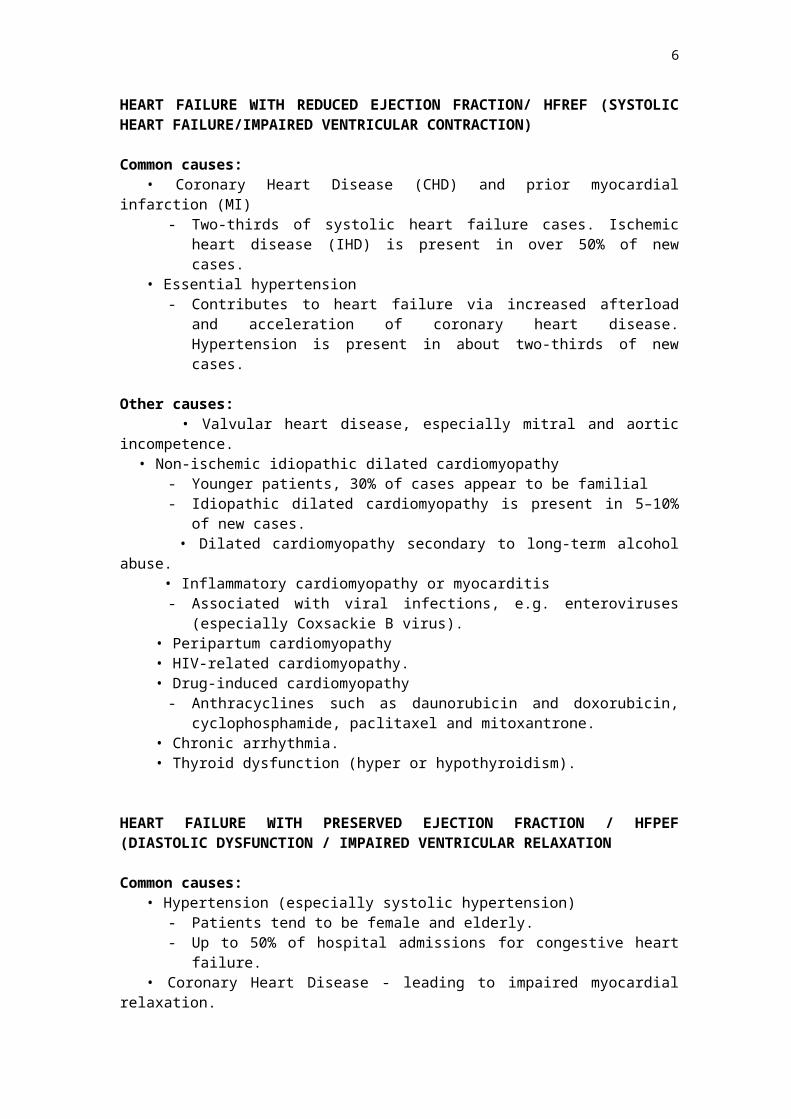
HEART FAILURE WITH REDUCED EJECTION FRACTION/ HFREF (SYSTOLIC HEART FAILURE/IMPAIRED VENTRICULAR CONTRACTION)
Common causes:• Coronary Heart Disease (CHD) and prior myocardial infarction (MI)
- Two-thirds of systolic heart failure cases. Ischemic heart disease (IHD) is present in over 50% of new cases.
• Essential hypertension - Contributes to heart failure via increased afterload and acceleration of
coronary heart disease. Hypertension is present in about two-thirds of new cases.
Other causes: • Valvular heart disease, especially mitral and aortic incompetence.
• Non-ischemic idiopathic dilated cardiomyopathy- Younger patients, 30% of cases appear to be familial- Idiopathic dilated cardiomyopathy is present in 5–10% of new cases.
• Dilated cardiomyopathy secondary to long-term alcohol abuse. • Inflammatory cardiomyopathy or myocarditis
- Associated with viral infections, e.g. enteroviruses (especially Coxsackie B virus).
• Peripartum cardiomyopathy • HIV-related cardiomyopathy. • Drug-induced cardiomyopathy
- Anthracyclines such as daunorubicin and doxorubicin, cyclophosphamide, paclitaxel and mitoxantrone.
• Chronic arrhythmia. • Thyroid dysfunction (hyper or hypothyroidism).
HEART FAILURE WITH PRESERVED EJECTION FRACTION / HFPEF (DIASTOLIC DYSFUNCTION / IMPAIRED VENTRICULAR RELAXATION
Common causes:• Hypertension (especially systolic hypertension)
- Patients tend to be female and elderly. - Up to 50% of hospital admissions for congestive heart failure.
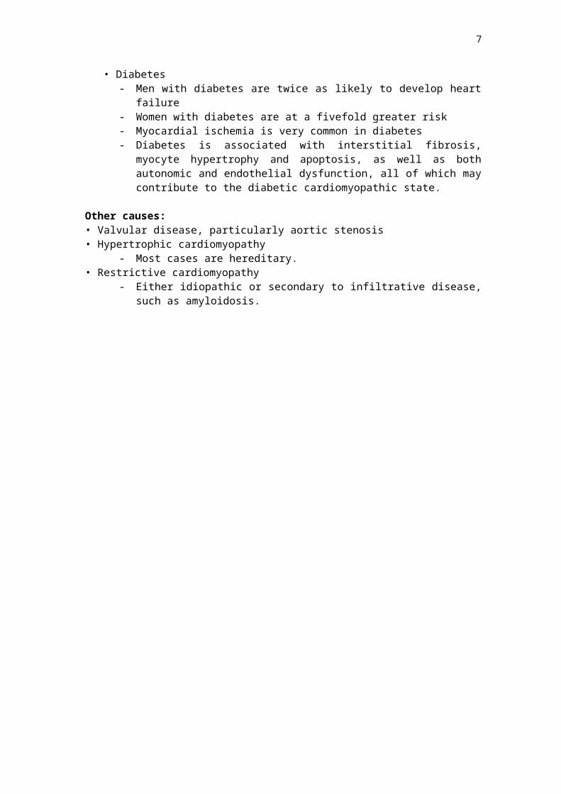
• Coronary Heart Disease - leading to impaired myocardial relaxation.• Diabetes
- Men with diabetes are twice as likely to develop heart failure - Women with diabetes are at a fivefold greater risk - Myocardial ischemia is very common in diabetes - Diabetes is associated with interstitial fibrosis, myocyte hypertrophy and
apoptosis, as well as both autonomic and endothelial dysfunction, all of which may contribute to the diabetic cardiomyopathic state.
Other causes:• Valvular disease, particularly aortic stenosis• Hypertrophic cardiomyopathy
- Most cases are hereditary.• Restrictive cardiomyopathy
- Either idiopathic or secondary to infiltrative disease, such as amyloidosis.
4

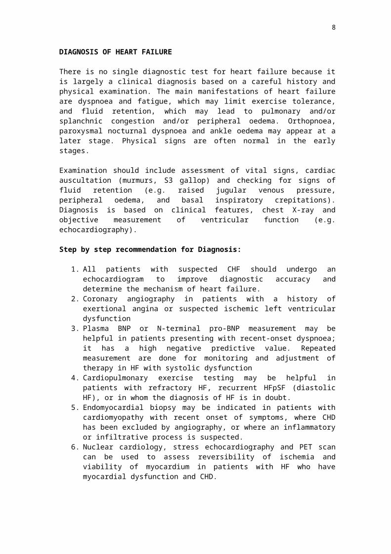
DIAGNOSIS OF HEART FAILURE
There is no single diagnostic test for heart failure because it is largely a clinical diagnosis based on a careful history and physical examination. The main manifestations of heart failure are dyspnoea and fatigue, which may limit exercise tolerance, and fluid retention, which may lead to pulmonary and/or splanchnic congestion and/or peripheral oedema. Orthopnoea, paroxysmal nocturnal dyspnoea and ankle oedema may appear at a later stage. Physical signs are often normal in the early stages.
Examination should include assessment of vital signs, cardiac auscultation (murmurs, S3 gallop) and checking for signs of fluid retention (e.g. raised jugular venous pressure, peripheral oedema, and basal inspiratory crepitations). Diagnosis is based on clinical features, chest X-ray and objective measurement of ventricular function (e.g. echocardiography).
Step by step recommendation for Diagnosis:
1. All patients with suspected CHF should undergo an echocardiogram to improve diagnostic accuracy and determine the mechanism of heart failure.
2. Coronary angiography in patients with a history of exertional angina or suspected ischemic left ventricular dysfunction
3. Plasma BNP or N-terminal pro-BNP measurement may be helpful in patients presenting with recent-onset dyspnoea; it has a high negative predictive value. Repeated measurement are done for monitoring and adjustment of therapy in HF with systolic dysfunction
4. Cardiopulmonary exercise testing may be helpful in patients with refractory HF, recurrent HFpSF (diastolic HF), or in whom the diagnosis of HF is in doubt.
5. Endomyocardial biopsy may be indicated in patients with cardiomyopathy with recent onset of symptoms, where CHD has been excluded by angiography, or where an inflammatory or infiltrative process is suspected.
6. Nuclear cardiology, stress echocardiography and PET scan can be used to assess reversibility of ischemia and viability of myocardium in patients with HF who have myocardial dysfunction and CHD.
7. Thyroid function tests should be considered, especially in older patients without pre-existing CHD who develop atrial fibrillation, or in whom no other cause of HF is evident.
8. MRI to assess and diagnose infiltrative disorders. However, MRI is not widely available.
MANAGEMENT OF HEART FAILURE
Management involves prevention, early detection, slowing of disease progression, relief of symptoms, minimisation of exacerbations, and prolongation of survival.
Non-Pharmacological Management
Regular physical activity with a specially designed program for all HF patients is recommended except for those who have an acute exacerbation, or are clinically unstable. They should undergo a period of bed rest until their condition improves.
Patient to follow up with a doctor or home visit by a nurse to prevent clinical deterioration.
Patients with coexisting sleep apnoea to consult with sleep physician as they may benefit from nasal CPAP
5
Fluid intake should generally be limited to 1.5 L /day with mild to moderate symptoms, and 1 L /day in severe cases, especially if there is coexistent hyponatraemia. Dietary sodium should be limited to below 2 g/day.
Alcohol intake should preferably be nil, but should not exceed 10–20 g a day (one to two standard drinks). Smoking should be strongly discouraged.
Patients should be advised to weigh themselves daily and to consult their doctor if weight increases by more than 2 kg in a two-day period, or if they experience dyspnoea, oedema or abdominal bloating.
Patients should be vaccinated against influenza and pneumococcal disease. High-altitude destinations should be avoided. Travel to very humid or hot climates
should be undertaken with caution, and fluid status should be carefully monitored.
Obese patients should be advised to lose weight. Pregnancy should be avoided in patients with moderate to severe HF.
Pharmacological Management
First Line Treatment
Angiotensin Converting Enzyme Inhibitors (ACEIs) Recommended for all patients with systolic heart failure (LVEF < 40%), whether symptoms are mild, moderate or severe. (Unless not tolerated or contraindicated)
Angiotensin II receptor antagonists (ARB’s)Used as an alternative in patients who do not tolerate ACEIs due to kinin-
mediated adverse effects (e.g. cough). They should also be considered for reducing morbidity and mortality in patients with systolic CHF who remain symptomatic despite receiving ACEIs.
Diuretics Used to achieve euvolaemia in fluid-overloaded patients. In patients with systolic LV dysfunction, diuretics should never be used as monotherapy, but combined with an ACEI to maintain euvolaemia.
Beta-blockers Recommended for all patients with systolic CHF who remain mild to moderately symptomatic despite appropriate doses of an ACEI. (Unless not tolerated or contra-indicated). Beta-blockers are also indicated for patients with symptoms of advanced CHF.
Aldosterone receptor blockade with spironolactone Recommended for patients who remain severely symptomatic, despite appropriate doses of ACEIs and diuretics.
Aldosterone blockade with eplerenone In systolic heart failure patients who still have mild (NYHA Class II) symptoms despite receiving standard therapies (ACEI, beta- blocker).
Direct sinus node inhibition with ivabradine CHF patients with i m p a i r e d systolic function and a recent heart failure hospitalisation who are in sinus rhythm where their heart rate remains > 70 bpm despite efforts to maximise dosage of background beta- blockade.
6
Second Line treatment
Digoxin Considered for symptom relief and to reduce hospitalisation in patients with
advanced CHF. It remains a valuable therapy in CHF patients with AF. Hydralazine-isosorbide dinitrate combination
Reserved for patients who a r e truly intolerant of ACEIs and Angiotensin II receptor antagonists, or are contraindicated and no other therapeutic option exists.
Other Treatments
Amlodipine and felodipine Can be used to treat co-morbidities such as hypertension and coronary heart disease in patients with systolic CHF. They have been shown to neither increase nor decrease mortality.
Iron deficiency Should be looked for and treated in CHF patients to improve symptoms, exercise tolerance and quality of life
Fish oil (Omega-3 polyunsaturated fatty acids) Second-line agent for patients with CHF who remain symptomatic despite standard therapy, which should include ACEIs or ARBs and beta-blockers if tolerated.
Device-based treatment of symptomatic CHF Biventricular pacing (cardiac resynchronisation therapy, with or without
implantable cardiac defibrillator {ICD}) Should be considered in patients with CHF who fulfil each of the following criteria:
1. NYHA symptoms Class III/IV on treatment2. Dilated heart failure with LVEF ≤ 35%3. QRS duration ≥ 120 ms4. Sinus rhythm.5. In patients in whom implantation of an ICD is planned and LVEF is ≤
30% and the QRS duration is ≥ 150 ms (left bundle branch block morphology)
ICD implantation Should be considered in patients with CHF who fulfil any of the following criteria:
1. Survived cardiac arrest resulting from ventricular fibrillation or ventricular tachycardia not due to a transient or reversible cause
2. Spontaneous sustained ventricular tachycardia in association with structural CHD
3. LVEF ≤ 30% measured at least 1 month after acute MI, or 3 months after coronary artery revascularisation surgery
4. Symptomatic CHF (i.e. NYHA functional class II/III) and LVEF ≤ 35%
5. Together with surgical approaches in myocardial revascularisation, insertion of devices and cardiac transplantation.
Left Ventricular Assist Devices (LVAD)Most often used as a temporary bridge to cardiac transplantation, or for recovery of the heart post- cardiac surgery
7
SURGICAL MANAGEMENT OF HEART FAILURE
Coronary revascularisation
Surgical repair or replacement of affected valve
Ventricular reconstruction
Cardiac transplant
Indications:i. Persistent NYHA Class IV symptoms
ii. Volume of oxygen consumed per minute at maximal exercise (VO2 max) < 10 mL/kg/min
iii. Severe ischaemia not amenable to revascularisationiv. Recurrent uncontrollable ventricular arrhythmia
ANAESTHETIC MANAGEMENT OF PATIENT WITH HEART FAILURE
1. Pre-operative Management
Pre-op Assessment
Patients with HF are at increased risk for morbidity and mortality after all surgical procedures compared to those with a diagnosis of coronary artery disease only. The goal is to assess severity and stability of symptoms and stratify risk of cardiac morbidity and mortality.
In patients with decompensated HF (NYHA IV, worsening or new-onset HF), surgery is postponed if non-urgent to allow treatment and stabilization of HF symptoms.
Preoperative tests
I. Twelve-lead electrocardiogram - to exclude and treat life-threatening arrhythmias
II. Blood investigation FBC - Treatment of anaemia, infectionU & E - Check electrolyte level especially K+ levelsMetabolic panel - To exclude other metabolic disorders
III. Chest radiograph In patients with signs or symptoms of acute decompensated HF.
IV. Echocardiography +/- Dobutamine stress test Indicated for patients with unstable chronic HF
Management of Medications
1. Beta blockers – Continue but do not initiate pre operatively.2. ACEI and ARB – Continue unless hypotensive.
3. Aldosterone antagonists – Check potassium level for possible hyperkalemia.
8
4. Diuretics – Check potassium level for possible hypokalemia. Perioperative use is based upon assessment of volume status.
5. Digoxin – Monitor for Digoxin level and induced arrhythmias
6. Anticoagulation – Timing of interruption and use of bridging should balance thromboembolic risk and bleeding risk.
2. Intra-operative management
Monitoring
Use of an intra-arterial catheter, central venous catheter, pulmonary artery catheter and/or transoesophageal echocardiography is based on specific patient needs.
Regional Anaesthesia
Primarily guided by the surgical site and procedure. Modifications of techniques (e.g., a low-dose combined spinal-epidural with or without intrathecal opioids, or a very slowly titrated epidural anaesthetic) are a reasonable optionBenefits:
Pre-emptive postoperative analgesia Decreased risk of pneumonia
Reduction in 30-day mortality in high cardiac risk
General Anaesthesia
1. Induction a. Short-acting hypnotic (e.g., etomidate [0.2 to 0.3 mg/kg], ketamine [1 to
2.5 mg/kg], or low dose of propofol [1 mg/kg])
b. Moderate dose of an opioid (e.g., fentanyl 1 to 2 mcg/kg) +/- lignocaine (50 to 100 mg)
c. Muscle relaxant with rapid onset.
2. Maintenance
a. Intravenous or volatile anaesthetic agents.
b. Doses are reduced in HF patients to avoid significant myocardial depression.
3. Fluid management
a. Maintain adequate tissue perfusion by optimizing intravascular volume status and stroke volume.
b. Administration of smaller than usual (1 to 2 mL/kg) crystalloid boluses, as needed.
4. Transfusion of RBC (pack cells) and other blood components
9
a. In patients with borderline haemoglobin levels (<8 g/dL) who have ongoing bleeding and for coagulopathy and evidence of inadequate perfusion of vital organs.
5. Intra-operative decompensated HF
a. In patients with severe systolic dysfunction and low cardiac output syndrome use an inodilator agent e.g. Milrinone (0.375 to a maximum of 0.750 mcg/kg per min).
b. In patients with systemic hypertension or severely symptomatic fluid overload, use of a vasodilator e.g. Glyceryl Trinitrate (GTN 10 to 500 ug/min titrating to adequate systolic blood pressure)
c. In decompensated HF patients with hypotension and evidence of end-organ hypo perfusion, use of an agent with inotropic and vasopressor properties (e.g. norepinephrine 0.01 to 0.3 ug/kg/min)
d. In severe or refractory vasodilatory shock, the potent direct peripheral vasoconstrictor Vasopressin (1 to 4 Units per hour) may be necessary.
3. Postoperative management
Patients with HF are more likely to develop postoperative complications such as pulmonary edema, new or unstable myocardial ischemia, arrhythmias and sudden cardiac death.
There is a proven role for Non Invasive Ventilation or NIV (using CPAP or BIPAP mode) in patients with acute pulmonary oedema secondary to heart failure.
Patients with HF who remain hypotensive or in pulmonary oedema despite oxygen, diuresis, and vasodilators may benefit from short-term intravenous inotropic support in Intensive Care Unit.
Careful discharge plan with review of medication should be carried out in a multi-disciplinary team approach involving anaesthetist, surgeon, cardiologist and patient’s family physician/GP.
References
1. 2013 ACCF/AHA Guideline for the Management of Heart Failure, Clyde W. Yancy et al 2. Guidelines for the prevention, detection and management of chronic heart failure in
Australia, October 20113. Anesthesia for noncardiac surgery in patients with heart failure, Up to date, Dec 2014
10
HYPERTENSIVE DISEASE
Hypertension remains one of the most important preventable contributors to disease and death. It is extremely common, affecting more than a billion people worldwide and increases the risk of myocardial infarction, heart failure, renal failure and stroke. It is responsible for the deaths of over seven million people annually. As it also associated with an ageing population and this patient group is more likely to require surgery, the perioperative management of hypertension is of significant importance to anaesthetists. Definitions and classification
Most hypertension is referred to as “essential hypertension”, implying that the underlying cause is unknown. This encompasses approximately 95% of all hypertensive patients.
The remaining five per cent of hypertension is “secondary”, in which the underlying cause of the high blood pressure is a known medical condition. These can be categorised into:
Vascular pathologyo Such as coarctation of the aorta or renal artery stenosis
Endocrine conditionso Particularly phaeochromocytoma, Cushing’s syndrome and primary
hyperaldosteronism Renal disease Obstetric causes Misuse of drugs
o Either prescribed or for recreational purposes
Secondary hypertension must always be considered in young patients with severe hypertension, in any patient with resistant hypertension, or in patients with atypical presentations, as the consequences of a failure of diagnosis in this group may be catastrophic, especially in the perioperative period. The management of these conditions will not be discussed here.
The severity of hypertension is categorised bands, as illustrated:
Aronson et al. (2006)
11

It is important that the diagnosis and classification of hypertension is based on the average of two or more stable readings, taken at two or more visits after initial screening, and not on a single isolated recording. It is increasingly recognised that office, or in-hospital blood pressure measurement is the least reliable assessment of blood pressure, and that home blood pressure monitors are better equipped to assess blood pressure control as they avoid the problems of “white coat hypertension”.
Pathogenesis
Systolic blood pressure (SBP) rises continuously with age, while diastolic blood pressure reaches a plateau in the fifth or sixth decade, and may then decrease. Thus, systolic hypertension is more common in the elderly, and an increase in pulse pressure is seen in older patients. Hypertension of all types increases the risk of mortality, with severe hypertension (SBP greater than 180 mmHg) increasing the relative risk of death by six times. Of importance here is that low diastolic pressure may limit myocardial perfusion, which occurs in diastole.
End-organ damage, predominantly of the heart, the kidney and brain, is common in hypertension. Even asymptomatic hypertension presents a risk of end-organ damage, and as the disease advances, renal injury may progress to end-stage renal failure. The heart will suffer from hypertrophy, coronary artery disease, and systolic and diastolic dysfunction, all ultimately increasing the risk of myocardial infarction, cardiac failure and death. The brain is also at substantial risk, with hypertension increasing the incidence of dementia and ischaemic or haemorrhagic cerebral events, leading to stroke.
In the heart, a vicious cycle is established by the increased myocardial oxygen demand caused by increased myocardial wall tension, which is amplified by left ventricular hypertrophy. Both of these changes lead to coronary insufficiency, infarction and heart failure. Diastolic dysfunction is seen increasingly frequently with advancing age, occurring in up to 70% of patients with heart failure.
Loss of autoregulation predisposes the kidney to damage from hypotensive events, and there is an increasing incidence of glomerular sclerosis, and a reduction in glomerular filtration rate, with advancing hypertension.
The shift of autoregulatory thresholds in the brain in hypertensives is well known, and hypertension increases the perioperative risk of both ischaemic and haemorrhagic strokes. Carotid stenosis is more common in hypertensive patients, adding to the ischaemic risk.
Treatment
There is differing opinion as to the threshold value where treatment needs to commence. The latest Joint National Committee on prevention, detection, evaluation and treatment of high blood pressure (JNC8) published guidelines in December 2013 in the Journal of the American Medical Association (JAMA), which have relaxed treatment goals in the older population groups.
12
These treatment goals are now summarised as follows in the JNC8 document: • In patients 60 years or older, start treatment in blood pressures >150 mmHg
systolic or >90 mmHg diastolic and treat to under those thresholds. • In patients <60 years, treatment initiation and goals should be 140/90 mmHg, the
same threshold used in patients >18 years with either chronic kidney disease (CKD) or diabetes.
• In non-black patients with hypertension, initial treatment can be a thiazide-type diuretic, calcium channel blocker (CCB), or angiotensin converting enzyme inhibitor (ACE), or angiotensin receptor blocker (ARB).
• In patients >18 years with CKD, initial or add-on therapy should be an ACE inhibitor or ARB, regardless of race or diabetes status.
The American Heart Association (AHA) and American College of Cardiology (ACC) do not support these guidelines. One of the major areas of disagreement is the raising of the target blood pressure in the 60 plus age group from 140 to 150 mmHg. Without this consensus, the management of the elderly hypertensive patient may not be clear. A further area of controversy relates to the trigger level where blood pressure treatment needs to commence. There is a body of opinion that suggests that healthy patients with stage 1 hypertension are unnecessarily started on treatment and exposed to the side effects of these drugs without any evidence of benefit. While diastolic blood pressure used to be the main target of antihypertensive therapy, it has now become evident that systolic blood pressure and an increase in pulse pressure above 65 mmHg is more important in terms of outcome, especially in the patient population above 50 years of age. The benefits of treating isolated systolic hypertension are now clearly established and carry a greater risk to the patient of developing a cardiac or neurological incident.
Treatment of hypertension has a significant effect on complications, reducing the incidence of stroke and cardiac failure, and improves five-year morbidity and mortality.
Lifestyle modifications of proven value in lowering blood pressure include weight reduction or prevention of weight gain, moderation of alcohol intake, increase in physical activity, adherence to recommendations for dietary calcium and potassium intake, and moderation in dietary salt intake. Smoking cessation is critical, because smoking is an independent risk factor for cardiovascular disease.
Weight loss may be the most efficacious of all non-pharmacologic interventions in the treatment of hypertension. A 10-kg weight loss decreases the systolic and diastolic blood pressure by an average of 6.0 mm Hg and 4.6 mm Hg, respectively. Weight loss also enhances the efficacy of antihypertensive drug therapy. Alcohol consumption is associated with an increase in blood pressure, and excessive use of alcohol may cause resistance to antihypertensive drugs. However, moderate alcohol ingestion has been shown to decrease overall cardiovascular risk in the general population. At least 30 minutes of moderate-intensity physical activity, such as brisk walking or bicycling, can lower blood pressure in both normotensive and hypertensive individuals.
A variety of drugs have been used to manage hypertension. Of these, the thiazide diuretics have the longest and best-established track record in improving outcome. This is partly because these drugs have been the most widely used agents, but they should also be the usual first-line treatment in essential hypertension.
13
Five major classes of drugs are recognised for the treatment of essential hypertension:
Diuretics Angiotensin-converting enzyme (ACE) inhibitors Angiotensin-receptor blockers Calcium-channel blockers Beta blockers
The choice of agent depends on conditions that favour or contraindicate the use of individual drugs, for example beta-blockers for patients with ischaemic heart disease (IHD). Increasing evidence suggests that an ACE inhibitor plus a calcium channel blocker offer better cardiovascular protection than an ACE inhibitor plus a diuretic, or a diuretic plus a beta-blocker.
Perioperative risk
For a long time, the contribution of hypertension to anaesthetic risk has been controversial. The current weight of evidence suggests that hypertension alone carries minimal anaesthetic risk.
Several studies have shown that systolic blood pressures < 180 mmHg and diastolic blood pressures of < 110 mmHg are not an independent risk factor for perioperative cardiovascular events. It seems that established treatment of hypertension with a good response may reduce the perioperative risks of hypertension, and that resistant hypertension may represent a higher risk group, but at present, data are inadequate to make this a definitive conclusion.
Hypertension is most certainly associated with increased haemodynamic instability in the perioperative period, and this is associated with an increased risk of myocardial injury.
As stated, hypertension is a major risk factor for coronary artery disease, cardiac failure, and renal and cerebral disease, all of which increase the perioperative risk. Hypertension is also associated with dyslipidaemia, diabetes and obesity, and the side-effects of the drugs required to treat these conditions. Hypertension is therefore associated with an increased risk of end-organ disease, which would certainly increase the perioperative risk.
Perioperative approach
Careful preoperative evaluation of the hypertensive patient is important. This should include a review of blood pressure control, and the agents needed to achieve that. It is important to detect end-organ dysfunction that may have resulted from hypertension, as this is much more likely to predict perioperative adverse events than hypertension alone. Such end-organ damage includes cardiac injury, cerebral vascular disease, renal dysfunction and peripheral vascular disease. All patients found to have preoperative hypertension require appropriate investigation. This should include a preoperative electrocardiogram (ECG), blood sugar analysis, electrolytes and creatinine measurement (with calculation of glomerular filtration rate), together with a urinary dipstick. Where the situation is urgent these investigations must still be performed even if postoperatively.
14
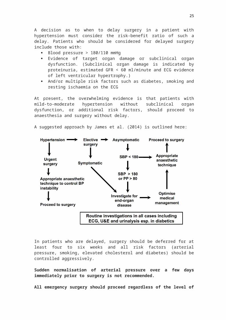
A decision as to when to delay surgery in a patient with hypertension must consider the risk-benefit ratio of such a delay. Patients who should be considered for delayed surgery include those with:
Blood pressure > 180/110 mmHg Evidence of target organ damage or subclinical organ dysfunction. (Subclinical
organ damage is indicated by proteinuria, estimated GFR < 60 ml/minute and ECG evidence of left ventricular hypertrophy.)
And/or multiple risk factors such as diabetes, smoking and resting ischaemia on the ECG
At present, the overwhelming evidence is that patients with mild-to-moderate hypertension without subclinical organ dysfunction, or additional risk factors, should proceed to anaesthesia and surgery without delay.
A suggested approach by James et al. (2014) is outlined here:
In patients who are delayed, surgery should be deferred for at least four to six weeks and all risk factors (arterial pressure, smoking, elevated cholesterol and diabetes) should be controlled aggressively.
Sudden normalisation of arterial pressure over a few days immediately prior to surgery is not recommended.
All emergency surgery should proceed regardless of the level of blood pressure, and urgent surgery should also not be delayed for uncomplicated hypertension.
The American College of Cardiology (ACC)/ American Heart Association (AHA) guidelines suggest that a systolic arterial pressure greater than 180 mmHg, and/ or a diastolic arterial pressure greater than 110 mmHg, is justification for the establishment of control of hypertension prior to surgery. Furthermore, these guidelines state that the use of rapid-acting agents to control blood pressure in the hours prior to surgery may be considered, but advance no evidence for this view. This practice may, in fact, be dangerous in patients who have lost cerebral, renal or cardiac autoregulation.
15
Consideration of the use of perioperative beta blockers should consider the evidence brought to light by the Perioperative Ischemic Evaluation Study (POISE). In this study, beta blockade was given acutely to at-risk patients two to four hours prior to major non-cardiac surgery. Although there was a reduction in the rate of myocardial infarction, all-cause mortality, particularly from stroke, was increased. Immediate preoperative beta blockade should therefore be used with caution. The addition of a statin is probably justified in any patient with a high risk of myocardial injury. In high-risk patients, acute hypotension is more dangerous than modest elevations in blood pressure.
Where urgent surgery is required in a patient with very severe hypertension (SBP > 210 / DBP > 120), it may be helpful to attempt to decrease arterial pressure with vasodilator agents by approximately 25%, prior to induction of anaesthesia. Short-acting agents such as nitroprusside or glyceryl trinitrate may be helpful, as they can be discontinued rapidly should hypotension become a problem, but there is no conclusive evidence to support this practice.
An isolated recording of a very high blood pressure immediately prior to surgery is not necessarily a reason to postpone the surgery. In such cases, where prior information is available, such as ward blood pressures, or information from the patient’s family practitioner that suggests that the resting arterial pressure is well controlled, it may be reasonable to proceed with anaesthesia. However, such an episode identifies a patient as a potentially unstable hypertensive. There is growing evidence that such patients deserve increased medical surveillance.
Very severe hypertension (SBP >210 or DBP > 120) does appear to present a significant perioperative risk, and such patients should be deferred for treatment if at all possible.
If the decision is made to delay surgery, the anaesthetist is obliged to advise the surgical team on how to best manage the patient. If the patient is already on antihypertensive medication, but is poorly responsive, simply increasing the dosage of currently prescribed drugs would seem to be an inadequate approach. In consultation with a physician, a change in therapy is probably more appropriate. However, such a change in therapy will require re-evaluation of end-organ status.
Anaesthesia
Once a hypertensive patient is accepted for anaesthesia, consideration must be given to the effects that the prescribed drugs may have on the anaesthesia. Withdrawal of antihypertensive medication is generally considered inadvisable, as many of these drugs may produce rebound effects when withdrawn. This applies to alpha-methyldopa and clonidine particularly, where severe hypertension may follow withdrawal. Withdrawal of beta blockers may precipitate angina. In the case of diuretics, the effects on renal function and electrolyte abnormalities must always be considered. The ACE inhibitors/ARBs represent a special case, and withdrawal of these agents 10 hours prior to surgery appears to be associated with a reduction in the risk of immediate post-induction hypotension.
Intraoperatively, haemodynamic stability appears to be more important than absolute values of the blood pressure.
16
Controlling the intubation response is important, and a variety of agents (including short-acting opiates, glyceryl trinitrate and magnesium sulphate) can be considered for this purpose, but the risks of post-induction hypotension must be remembered. Intraoperatively, esmolol (if and where available), may be very valuable for controlling tachycardia and hypertension. (It must be remembered that beta blockade is contraindicated if the hypertensive event is due to an excess of catecholamines, caused either by administration of adrenaline or cocaine by the surgeon, or by endogenous secretion e.g. a phaeochromocytoma. In these circumstances, magnesium sulphate (MgSO4), as a 4-g bolus, is probably the safest and most effective first-line therapy.) MgSO4 should constitute routine initial treatment of any intraoperative hypertensive crisis. Alpha2-agonists, as part of the anaesthetic technique, may provide useful blood pressure control, together with enhanced analgesia, and some protection against myocardial ischaemia. Postoperatively, care must be taken to ensure that a hypertensive crisis does not ensue, and that good analgesia is instituted. A strategy must be developed for blood pressure control, including a planned early return to standard preoperative therapy.
Intraoperative Hypertensive Emergency1
Consider: Adequate oxygenation and ventilation Correct measurement Adequate depth of anaesthesia Adequate analgesia Correct drugs given – recheck running infusions, ampoules Pre-eclampsia? ? Intracranial hypertension Thyroid storm Phaeochromocytoma
Depending on the severity of the blood pressure, consider asking the surgeon to stop surgery until the blood pressure is under control. Increase the inspired oxygen concentration and ventilation if required. In addition to increasing the anaesthetic depth and providing more analgesia options include:
Vasodilatorso E.g. hydralazine 5mg IV every 15 minuteso Glyceryl Trinitrate (GTN) 3mg per HOUR and titrate to effecto Sodium nitroprusside 0.5 – 1.5 mcg/kg /minute
Beta blockade (especially if an accompanying tachycardia is present)o Metoprolol 1-2 mg in incrementso Labetalol 5-10 mg incrementso Esmolol 0.5 mg/kg bolus followed by 50-200 mcg/kg/minute
Conclusion
Hypertensive disease in its different manifestations is a common occurrence and the anaesthetist should be well equipped to deal with this. Modern anaesthesia provided by a well-trained, experienced anaesthetist offers sufficient perioperative cardiac protection to make cancellation of surgery for the sole purpose of controlling hypertension unnecessary under most circumstances.
References
1 Non-anaesthetic related hypertensive crises are not covered in this document.
17

1. Allman, K. and McIndoe, A. (2009). Emergencies in Anaesthesia. Oxford University Press, USA.
2. Aronson, Solomon, and Manuel L Fontes. “Hypertension: a New Look at an Old Problem.” Current Opinion in Anaesthesiology 19, no. 1 (February 2006): 59–64. doi:10.1097/01.aco.0000192784.47645.7b.
3. Beyer, K, P Taffé, P Halfon, V Pittet, S Pichard, G Haller, B Burnand, for the ADS study Group. “Hypertension and Intra-Operative Incidents: a Multicentre Study of 125 000 Surgical Procedures in Swiss Hospitals.” Anaesthesia 64, no. 5 (May 2009): 494–502. doi:10.1111/j.1365-2044.2008.05821.x.
4. Jackson, M., Mookherjee, S., Hamlin, N. and Berger, G. (2013). The perioperative medicine consult handbook.
5. Jaffer, A. and Grant, P. (2012). Perioperative medicine. Hoboken, N.J.: Wiley-Blackwell.6. James, Michael FM, Robert A Dyer, and B L Rayner. “A Modern Look at Hypertension
and Anaesthesia.” How Important Is Peri-Operative Hypertension? Journal of Endocrinology, Metabolism and Diabetes of South Africa 19, no. 2 (2014): 56–61.
7. James, Paul A, Suzanne Oparil, Barry L Carter, William C Cushman, Cheryl Dennison-Himmelfarb, Joel Handler, Daniel T Lackland, et al. “2014 Evidence-Based Guideline for the Management of High Blood Pressure in Adults.” JAMA : the Journal of the American Medical Association 311, no. 5 (February 5, 2014): 507. doi:10.1001/jama.2013.284427.
8. Stoelting, R., Hines, R. and Marschall, K. (2012). Stoelting's anesthesia and co-existing disease. Philadelphia: Saunders/Elsevier.
9. Varon, Joseph, and Paul E Marik. “Perioperative Hypertension Management..” Vascular Health and Risk Management 4, no. 3 (2008): 615–27.
18
ISCHAEMIC HEART DISEASE
Ischaemic heart disease is present when clinical features indicate an inadequate supply of blood to the myocardium. This is commonly caused by atherosclerosis.
Angina pectoris, or angina for short, occurs when the myocardial oxygen demand exceeds the oxygen supply, and this usually manifests as chest discomfort.
Ischaemic heart disease can be further classified into stable and unstable varieties.
Unstable ischaemic heart disease can be further divided into unstable angina, non-ST elevation myocardial infarction and ST-elevation myocardial infarction. 1
PATHOPHYSIOLOGY
There has been an evolution in the thought processes involving the understanding of the pathophysiology concerning ischaemic heart disease. Previously thought to be a cholesterol storage disorder, atherosclerosis is currently viewed as an inflammatory disorder. There is remodeling of the arterial wall due to a combination of haemodynamic stresses and inflammatory processes.
Inflammation is mediated by components of the immune system including platelets, T cells and macrophages. Pro-inflammatory factors include cytokines, matrix metalloproteinases, platelet-derived growth factor, insulin-like growth factor, transforming growth factors alpha and beta, thrombin, and angiotensin II (A-II).
There is progression from the fatty streak, through to formation of fibroatheromatous plaques. The fibrous cap of these plaques may weaken with time. Rupture of these plaques exposes the thrombogenic contents of these plaques to the circulating blood. This then leads to thrombus formation, partial or complete occlusion of the blood vessel, and progression of the atherosclerotic lesion due to organisation of the thrombus and incorporation within the plaque. 2
DIAGNOSIS
Stable ischaemic heart disease is diagnosed based on a classic history of angina pectoris, together with one or more risk factors for atherosclerotic cardiovascular disease.
Differential diagnosis
- Other cardiac- Non-cardiac
o Gastrointestinal diseaseo Pulmonary disease
EvaluationA good history and examination of the patient should be undertaken, assessing for the likelihood of chest pain matching classical angina pectoris. Assessment of risk factors for ischaemic heart disease should also be undertaken at the same time – these would include modifiable risk factors such as diabetes mellitus, smoking, and hypercholesterolaemia.
19
A family history may reveal family members who develop premature cardiovascular disease or hypertrophic obstructive cardiomyopathy.
Physical examination is often normal, but may reveal an increased heart rate, increased blood pressure, pathological heart sounds and murmurs.
Electrocardiogram may show features of cardiac ischaemia.
Stress testing
This provides a means to provide accurate diagnostic and prognostic information with patients who have known or suspected ischaemic heart disease. Stress testing can be done via exercise or pharmacological means. The most common way is exercise ECG testing.
There are also other stress imaging modalities such as:
Radionuclide stress myocardial perfusion Stress echocardiogram Stress cardiac MRI
Coronary angiography
Gold standard test to identify stenosis and suitability for angioplasty/ coronary stenting or coronary artery bypass surgery.
RISK FACTORS
There are both fixed and modifiable risk factors for ischaemic heart disease.
Fixed risk factors include: Age Gender Family history
Modifiable risk factors include: Diabetes mellitus Smoking Hypercholesterolaemia
PREVENTION
Risk factor reduction is the primary means of prevention of ischaemic heart disease. This includes: the control of hypertension, smoking cessation, weight reduction, adequate glycaemic control in the diabetic patient and reduction in stress.
MEDICAL THERAPY OF ANGINA
Antianginal therapy is divided into 3 classes: beta-blockers, calcium channel blockers and nitrates.
Beta-blockers are first line therapy. They relieve angina by reducing the heart rate and myocardial contractility. They are also the only antianginal drugs proven to prevent infarction and to improve survival in patients who have sustained a myocardial infarction.
20
Calcium channel blockers are used in combination with beta-blockers when they are not effective or as a substitute for beta-blockers when they are contraindicated or cause intolerable side effects. Calcium channel blockers work by causing coronary and peripheral vasodilatation and reducing contractility. The degree to which these changes occur depends on the type of calcium channel blocker given. Common examples would include long-acting diltiazem or verapamil or a second-generation dihydropyridine (amlodipine or felodipine).
Nitrates, usually in the form of a sublingual preparation, are first-line therapy for the treatment of acute angina symptoms. Long acting nitrates are added to beta-blockers (with or without calcium channel blockers) to control stable angina.
In addition to antianginal agents, other preventative therapies are initiated at the same time. In the absence of a contraindication, patients should be commenced on antiplatelet therapy. Low dose aspirin (75-325 mg daily) is associated with the best risk/benefit ratio.
Statins are also proven to reduce cholesterol and reduce reinfarction in patients with coronary artery disease. They reduce coronary calcium, which may be a predictor of perioperative cardiac events in vascular surgery patients.
ACUTE CORONARY SYNDROMES (ACS)
There are 3 types of presentations:1. Unstable angina2. Non ST segment elevation myocardial infarction (NSTEMI)3. ST segment elevation myocardial infarction (STEMI)
In a patient presenting with chest pain suspicious of acute coronary syndrome to the emergency department, the diagnosis of myocardial infarction can be confirmed by the electrocardiogram and serum cardiac biomarker elevation; the history is relied upon heavily to make the diagnosis of unstable angina.
The types of angina that suggest ACS: Rest angina, usually more than 20 minutes duration New onset angina that limits physical activity Increasing angina that is more frequent, longer in duration, or occurs with
less exertion than previous angina
Initial therapy should be rapidly instituted, within 20 minutes of presentation would be ideal.
Supplemental oxygen should be given to patients who are hypoxaemic or in respiratory distress.
Sublingual GTN is administered, up to 3 doses, followed by intravenous GTN. IV morphine is given to relieve chest pain. Aspirin therapy is initiated
Early use of beta-blocker is recommended, unless patient is haemodynamically compromised. A cardioselective beta-blocker such as metoprolol or atenolol is preferred.
Statin therapy should be initiated prior to hospital discharge, with some evidence supporting initiation at the time of diagnosis.
Antithrombotic therapy should be a combination of antiplatelet agents and anticoagulant therapy. Antiplatelet therapy is usually dual therapy with aspirin and a P2Y12 receptor
21

blocker. Anticoagulant therapy that is typically initiated includes unfractionated heparin, enoxaparin, fondaparinux and bivalirudin. In very high-risk scenarios, inhibitors of the platelet glycoprotein IIb/ 3aα β receptor such as eptifibatide or tirofiban may be used.
When there is actual myocardial damage, this type of acute coronary syndrome is then called acute myocardial infarction. This can be further subclassified as non-ST elevation acute myocardial infarction or ST-elevation acute myocardial infarction.
The most common triggering event is the disruption of atherosclerotic plaque in an epicardial coronary artery, which leads to a clotting cascade, sometimes resulting in total occlusion of the artery. When a severe enough plaque rupture occurs in the coronary vasculature, it leads to a myocardial infarction.
WHO criteria formulated in 1979 have classically been used to diagnose MI; a patient is diagnosed with MI if two (probable) or three (definite) of the following criteria are satisfied:
Clinical history of ischemic type chest pain lasting for more than 20 minutes Changes in serial ECG tracings Rise and fall of serum cardiac biomarkers
Electrocardiogram
For a person to qualify as having a STEMI, in addition to reported angina, the ECG must show new ST elevation in two or more adjacent ECG leads. This must be ST segment elevation greater than 2 mm (0.2 mV) for males and greater than 1.5 mm (0.15mV) in females if in leads V2 and V3 or greater than 1 mm (0.1 mV) if it is in other ECG leads. A left bundle branch block that is believed to be new used to be considered the same as ST elevation; however, this is no longer the case. In early STEMIs there may just be peaked T waves with ST elevation developing later.
Cardiac biomarkers
While there are a number of different biomarkers, troponins are considered to be the best and reliance on older tests (such as CK-MB) or myoglobin is discouraged. Copeptin may be useful to rule out MI rapidly when used along with troponin.
Troponins are the most specific and sensitive test for myocardial damage. It is released during MI from the cytosolic pool of myocytes. Its subsequent release is prolonged with degradation of actin and myosin filaments. Isoforms of the protein, T and I, are specific to myocardium. Differential diagnosis of troponin elevation includes acute infarction, severe pulmonary embolism causing acute right heart overload, heart failure, and myocarditis. Troponins can also calculate infarct size but the peak must be measured in the 3rd day. After myocyte injury, troponin is released in 2–4 hours, peaks at 12 hours and persists for up to 7 days.
22
Imaging
A chest radiograph and routine blood tests may indicate complications or precipitating causes, and are often performed upon arrival to an emergency department. New regional wall motion abnormalities on an echocardiogram are also suggestive of a MI. Echo may be performed in equivocal cases by the on-call cardiologist. In stable patients whose symptoms have resolved by the time of evaluation, technetium (99mTc) sestamibi (i.e. a "MIBI scan") or thallium-201 chloride can be used in nuclear medicine to visualize areas of reduced blood flow in conjunction with physiological or pharmacological stress. Thallium may also be used to determine viability of tissue, distinguishing whether nonfunctional myocardium is actually dead or merely in a state of hibernation or of being stunned.
NSTEMI management
Essentially the management is similar to unstable angina, consideration to early percutaneous coronary intervention (PCI) is considered but the basic steps are the same.
STEMI management
The current definitive treatment modalities for MI with ECG evidence of ST elevation (STEMI) include thrombolysis and percutaneous coronary intervention.
Thrombolysis involves the administration of medication that activates the enzymes that normally destroy blood clots. The first thrombolysis agent was streptokinase, but most thrombolysis agents used currently are artificial forms of the human enzyme tissue plasminogen activator (tPA): reteplase, alteplase and tenecteplase. They are administered intravenously.
Once a STEMI has been diagnosed, and no contraindications are present (such as a high risk of bleeding), thrombolysis can be administered in the pre-hospital or in-hospital setting. There is inconclusive evidence whether pre-hospital thrombolysis reduces death in people with STEMI compared to in-hospital thrombolysis. Pre-hospital thrombolysis reduces time to receipt of thrombolytic treatment, based on studies conducted in higher income countries. If despite thrombolysis there is significant cardiogenic shock, continued severe chest pain, or less than a 50% improvement in ST elevation on the ECG recording after 90 minutes, then rescue PCI is indicated emergently.
Primary percutaneous coronary intervention (PCI) is the treatment of choice for STEMI if it can be performed in a hospital in a timely manner.
If PCI cannot be performed within 90 to 120 minutes then thrombolysis, preferably within 30 minutes of arrival to hospital, is recommended. After PCI, people are generally placed on dual antiplatelet therapy for at least a year (which is generally aspirin and clopidogrel)
Percutaneous Coronary Intervention
This can be done through the radial or femoral approach. There is less bleeding with the radial approach. The vessel is stented directly in most cases, after aspiration thrombectomy.
Drug-eluting stents (DES) are used in preference to bare-metal stents (BMS) in many PCI procedures because they reduce the incidence of restenosis and target vessel revascularization without causing a significant increase in the cumulative rate of adverse outcomes in many patient groups.
23
In patients with STEMI who are able to comply with a recommendation for a minimum of one year of dual antiplatelet therapy, we prefer second generation DES to BMS. Among the commercially available second-generation stents, we prefer everolimus-eluting stents in these patients. 3
Coronary Artery Bypass Graft Surgery
In patients with multivessel coronary disease, compared with PCI, CABG leads to an unequivocal reduction in long-term mortality and myocardial infarctions and to reductions in repeat revascularizations, regardless of whether patients are diabetic or not. These findings have implications for management of such patients. 4
PERIOPERATIVE SETTING
Preoperative cardiac risk stratification
Risk stratification starts with simple bedside evaluation that integrates clinical risk factors, functional capacity and type of surgery. Patients at low risk could be offered early surgery after assessment by their general practitioner, while complex patients may need more detailed assessment by a perioperative physician or cardiologist, in liaison with anaesthetists, surgeons and general practitioners.
Functional status remains a reliable predictor of perioperative and long-term cardiac events. Functional status is often expressed in terms of metabolic equivalents (METs), where 1 MET is the resting or basal oxygen consumption of a 40 year old, 70-kg man. Perioperative cardiac and long-term risks are increased in patients unable to perform 4 METs of work during daily activities.
Evaluating of clinical risk factors can be done via an index such as the revised cardiac risk index (RCRI). The Revised Cardiac Risk Index (RCRI) is a multivariable predictive index for major perioperative cardiac complications. All clinical variables contribute equally to the index (1 point each), with scores of 0, 1, 2 and 3 points corresponding to estimated risks of major cardiac complications of 0.4%, 0.9%, 7% and 11%, respectively. Low-risk patients have an RCRI score of 0, intermediate-risk patients have a score of 1 or 2, and high-risk patients have a score of 3 or more.
Type of surgery performed can influence risk as well. Surgically induced stress can predispose to coronary thrombosis and myocardial ischaemia. Surgical interventions can be divided into low-, intermediate- and high-risk groups, with estimated 30-day death or MI rates of < 1%, 1%–5%, and > 5%, respectively. While laparoscopic surgery and regional anaesthesia confer better pain relief and earlier functional recovery than open surgery and general anaesthesia, it remains unclear whether they significantly reduce cardiac risk.
Preoperative cardiac investigations
Investigations should only be performed if:
1. The results are expected to accurately and significantly change clinical estimates of risk
2. These altered risk estimates consistently lead to changed management decisions3. The resultant management changes have been shown in clinical trials to
improve clinical outcomes.
24
As situations that satisfy all three of these criteria are rare in perioperative medicine, the value of investigations, apart from a routine 12-lead electrocardiogram (ECG), is limited in preoperative cardiac management. The most useful applications may be in reclassifying intermediate-risk patients to either low-risk (surgery can safely proceed without further intervention) or high-risk (needing more detailed evaluation and use of prophylaxis), or in determining unacceptable surgical risk in high-risk patients undergoing high-risk surgery.
Resting echocardiograms are of no value.
Non-invasive stress testing should only be done for high-risk patients or those undergoing intermediate or high-risk surgery if results are likely to change management.
Cardiopulmonary exercise testing assesses functional capacity more accurately than self-report, and measures of total oxygen consumption and anaerobic threshold (if above certain threshold values) seem to identify individuals at very low surgical risk. While cardiopulmonary exercise testing may provide additional prognostic information in older patients with cardiopulmonary disease or patients undergoing major thoracic or abdominal operations, there are currently insufficient data to show its routine use alters perioperative care or outcomes compared with bedside risk stratification methods.
Newer tests such as specific cardiac biomarkers (high sensitive troponin and B-type natriuretic peptide) and computed tomography coronary angiography can add prognostic information to the RCRI, but their impact on decision making and clinical outcomes remains unclear.
Perioperative cardiac prophylaxis
Several preventive strategies may be considered in intermediate- and high-risk patients undergoing intermediate or high-risk surgery. Medications patients are already receiving for known coronary artery disease (CAD) should be continued throughout the perioperative period unless specific contraindications supervene.
Beta-blockers are potentially useful in lowering cardiac risk by antagonising the effects of adrenaline and other stress hormones and exerting negative chronotropic and inotropic actions. The large Perioperative Ischemic Evaluation Study (POISE) showed a 31% reduction in the risk of non-fatal MI with beta-blockers, at the expense of a 34% increased risk of all cause mortality and 89% increased risk of non-fatal stroke. However, two large retrospective observational studies using propensity-based risk adjustment suggest that beta-blockers reduce all-cause in-hospital deaths proportionally to increasing cardiac risk, as measured by an RCRI score ≥ 2, while increasing deaths in those with an RCRI score < 2. It is unclear which patients will benefit from beta-blockers, with suggestions that they should be commenced early and haemodynamically titrated to a tolerable dose that lowers heart rate to 70 beats/min.
Perioperative statin therapy should be continued for patients currently taking statins and scheduled for non-cardiac surgery. Perioperative initiation of statins is reasonable in patients undergoing vascular surgery.
Alpha-2 agonists for prevention of cardiac events are not recommended in patients undergoing non-cardiac surgery. Agents such as clonidine did not reduce the rate of death or nonfatal MI, but did increase the rate of nonfatal cardiac arrest and clinically important hypotension.
25
Calcium channel blockers significantly reduced myocardial ischaemia and supraventricular tachycardia. There was also a trend with calcium channel blockers toward reduced death and MI. Most of these benefits were with diltiazem.
Angiotensin-converting enzyme (ACE) inhibitors and angiotensin receptor blockers (ARBs) have not been shown to improve outcomes in the absence of left ventricular systolic dysfunction.
Aspirin interacts with the cyclo-oxygenase enzyme system and irreversibly inhibits platelet aggregation, theoretically lessening risk of coronary thrombosis but increasing the risk of perioperative bleeding. In patients with known coronary artery disease, excluding those with recent coronary artery stent insertion (discussed below), the risk of subsequent death or MI is increased two- to threefold if aspirin is ceased before surgery.
Coronary artery revascularisation via PCI or CABG is only indicated before non-cardiac surgery in clinically unstable patients (those with unstable angina, recent MI or ventricular arrhythmias) with significant left main or three-vessel (or two-vessel if this includes the proximal left anterior descending artery) coronary artery disease.
Myocardial Infarction (MI) and surgery
The recommendation is a minimum of 60 days between time of MI and non-cardiac surgery in the absence of a coronary intervention. 5
Recent PCI with stent insertion and dual antiplatelet therapy
Australian guidelines (Cardiac Society of Australia and New Zealand) recommend that elective surgery requiring cessation of dual antiplatelet therapy should be postponed for at least 6 weeks after insertion of bare-metal stents and 12 months after insertion of drug-eluting stents (DES). More recent experience and development of newer generation DES have suggested a minimum period of 6 months.
Most minor surgery can safely be continued with dual antiplatelet therapy without elevated risk of major bleeding.
The question of whether or not antiplatelet therapy should be continued, and whether or not bridging anticoagulation should be used can only be answered after careful multidisciplinary consultation between anaesthetists, surgeons and cardiologists. There needs to be a consideration towards the risk of in-stent thrombosis and risk of major bleeding from surgery. 6, 7
ANAESTHETIC CONSIDERATION AND PERIOPERATIVE MANAGEMENT
In considering neuraxial versus general anaesthesia in patients with ischaemic heart disease, there is no evidence to suggest a cardioprotective benefit from the use or addition of neuraxial anaesthesia for intraoperative anaesthetic management.
In considering volatile general anaesthesia versus total intravenous anaesthesia in patients with ischaemic heart disease, there is no evidence to suggest a difference in myocardial ischaemia or myocardial infarction rates between the 2 different methods of anaesthesia.
For pain management, neuraxial anaesthesia techniques in selected procedures can decrease the incidence of perioperative cardiac events. Examples are such as epidural anaesthesia in patients undergoing abdominal aortic surgery, and epidural analgesia in patients with a hip fracture.
26
The use of transoesophageal echocardiography is reasonable in patients with haemodynamic instability undergoing non-cardiac surgery to determine the cause of haemodynamic instability when it persists despite attempted corrective therapy, if expertise is readily available.
Maintenance of normothermia may be reasonable to reduce perioperative cardiac events in patients undergoing noncardiac surgery.
Use of pulmonary artery catheters should only be reserved for patients when medical conditions significantly alter haemodynamics and cannot be corrected before surgery.
With anaemia and ischaemic heart disease, a restrictive transfusion strategy is employed and a symptom-guided approach is employed to evaluating a haemoglobin level when determining whether to transfuse a patient with anaemia.
In perioperative surveillance for myocardial infarction, measurement of troponin levels and obtaining an ECG with features of myocardial ischaemia/ infarction is recommended.5
References
27
ANAESTHESIA FOR PATIENTS WITH LIVER DISEASE
Introduction
Patients with liver disease may present for elective or emergency surgery that may, or may not be related to their liver pathology. It is important to have a clear understanding of the potential complications as this group of patients represent a high-risk population for anaesthesia. The purpose of this chapter is to give you the background information to liver disease so that you are better prepared to manage your patients with liver disease presenting for an anaesthetic.
Liver Disease
Liver disease is a multi-system disorder as summarised in Figure 1. A typical patient with advanced liver disease will be jaundiced, have en enlarged abdomen full of ascites, may demonstrate muscle wasting, gynaecomastia and often have spider naevi.
Figure 1. Clinical manifestations of liver disease. (Diagram from McPhee SJ, Hammer GD. Pathophysiology of Disease: An Introduction to Clinical Medicine).
28
EpidemiologyLiver disease is remarkably common and in the western world it is the only major disease group from which the mortality is continuing to rise (Figures 2&3). In the UK, liver disease is the fifth biggest cause of death. The increasing mortality rate in the UK, compared to Europe, may be related to increasing alcohol intake.
Figure 2. Mortality secondary to liver disease in the UK is rising. Deaths are reported per million population. British Liver Trust, Office of National Statistics.
Figure 3. Trend in mortality rate for liver cirrhosis for men aged 15-44years over time (1950 – 2002), Leon et al 2006.
29
Mongolia has the world’s highest rate of mortality from liver cancer, and the number is increasing. By the time the diagnosis of hepatocellular carcinoma (HCC) is made, the disease is often inoperable with only about 10% of patients being identified early enough for surgery. One in every ten deaths in the country is due to HCC or its precursor, cirrhosis. Furthermore, nearly all Mongolians with liver disease are carriers of hepatitis B (HBV) or hepatitis C (HCV).
The high incidence of HCC in Mongolia is shown in Figure 4. It has been reported by the World Health Organisation that Mongolia has the highest annual mortality from HCC worldwide (Figure 5).
Figure 4. Incidence of hepatocellular carcinoma per 100,000 population as of 2009 (Ministry of Health, Mongolia). Blue area indicates areas of high prevalence, light blue
moderate prevalence, and white-blue low prevalence.
Figure 5. Countries with the highest annual mortality of hepatocellular carcinoma.
30

Cirrhosis and its aetiology
The end result of liver disease is cirrhosis, characterised by fibrosis, necrosis and nodular regeneration. The consequences of cirrhosis include portal hypertension and splenomegaly. Massively dilated vessels, varices, may develop in patients with portal hypertension.
The commonest cause of abnormal liver function tests, not cirrhosis, is probably Non-Alcoholic Fatty Liver Disease (NAFLD). It is believed to be the hepatic manifestation of the metabolic syndrome encompassing central obesity, hypertension and diabetes. A proportion of patients with fatty livers develop non-alcoholic steatohepatitis and a proportion of these patients will develop cirrhosis requiring a transplant for survival.
Over the past thirty years, mortality from alcoholic liver disease has risen 450% in the UK that has been ascribed to a change in drinking habits (Figure 3).
Viral hepatitis is another common cause of cirrhosis globally. HBV affects 2 billion people worldwide and, although HCV is less common, immunisation can protect individuals from HBV infection but not HCV. All healthcare workers having direct contact with patients should be provided with the HBV vaccine.
Viral hepatitis, predominantly HBV and HCV, are the commonest cause of liver disease in Mongolia. More than a quarter of Mongolians are chronic carriers of at least one of these viruses, and almost none are aware of their status. The epidemic of HCV is further compounded by the high alcohol use that hastens progression to cirrhosis.
Cirrhosis, and any inflammatory disease, predisposes to HCC so it is not surprising to see rates of HCC increasing.
Associated co-morbidities
The associated co-morbidities with liver disease are summarised in Table 1.
Renal Dysfunction Hepato-renal syndromeAcute tubular necrosisAscitesHigh mortality
Pulmonary vascular abnormalities Hepato-pulmonary syndromePort-pulmonary hypertension
Cirrhotic cardiomyopathy Dilated cardiomyopathyDiastolic dysfunctionSystolic dysfunctionConduction defectsFibrosis
Table 1. Common co-morbidities associated with liver disease.
31
Renal dysfunction
Renal dysfunction commonly occurs as a complication of cirrhosis and is associated with a high mortality. Hepatorenal syndrome develops as a result of poor renal perfusion leading to a pre-renal renal failure type picture (Figure 6). Patients with cirrhosis are massively vasodilated due to an imbalance of vasodilators and vasoconstrictors. The dilated splanchnic circulation reduces the effective circulating volume causing reflex vasoconstriction mainly by activation of the renin-angiotensin system. Urine becomes very concentrated with maximal salt and water reabsorption similar to that seen with pre-renal failure. The mechanism is worsened if the patient has a reduction in circulating volume e.g. through hypovolaemia or sepsis and as such, this must be recognised early in order to prevent the development of hepatorenal syndrome.
Figure 6. Mechanism of hepatorenal syndrome secondary to cirrhosis.
Portal hypertension and a dilated splanchnic vascular bed cause sodium and water retention due to low renal perfusion. Patients become overloaded with fluid with a high total body sodium content. Sodium, water and protein then leaks from the splanchnic circulation into the peritoneal cavity causing ascites. This fluid must be replaced intravascularly if it is drained in order to prevent the patient becoming hypovolaemic. This is also the case if the fluid is drained at the time of surgery e.g. laparotomy. Large collections of ascitic fluid may compromise respiratory function when the patient lies flat and this increase in intra-abdominal pressure may predispose the patient to reflux necessitating a rapid sequence induction for general anaesthesia. Pleural effusions may also develop and as such oxygen saturations should be checked pre-operatively and consideration made to performing a chest x-ray.
32
Pulmonary vascular abnormalities
The hepato-pulmonary syndrome is a triad of advanced liver disease, arterial deoxygenation and intra-pulmonary vascular dilation (Figure 7). Patients complain of breathlessness when standing because more blood goes through the shunt. There are two different types. In type 1, there is massive dilation of the vessels so red blood cells are not maximally oxygenated as they flow through the lungs. The patient will respond by increasing the inspired oxygen that increases the diffusion gradient. In type 2, there are discrete vascular channels bypassing the vascular endothelium. These patients do not respond to additional oxygen.
Pulmonary hypertension complicates 1-2% of patients with cirrhosis. It is associated with portal hypertension and is known as porto-pulmonary hypertension. There is no association with severity of liver disease. Patients may develop right heart failure due to the increase in right-sided afterload. It is reversible only in the early stages with standard pulmonary vasodilators.
33
Type 2 HPS
Figure 7. Types 1&2 hepato-pulmonary syndrome – dilated capillaries causing unequal blood flow and the development of ventilation – perfusion mismatch.
Cirrhotic cardiomyopathy
Patients present with dilated cardiomyopathy, diastolic dysfunction, poor contractility and down regulation of -receptors with insensitivity to -agonists. This can beβ β particularly problematic if patients are bleeding intra-operatively as they may be resistant to inotropic support. Conduction defects may also occur with, for example, prolonged QT. The cardiac muscle may become fibrotic with development of hypertrophic obstructive cardiomyopathy.
Risks from anaesthesia and surgery
Hepatic decompensation
The most important risk from anaesthesia is decompensation of liver disease. In patients with long-term liver disease, their physiology will be in a state of equilibrium that may be disturbed by anaesthesia or surgery with catastrophic results. Almost any form of anaesthesia will reduce hepatic blood flow thereby damaging hepatocytes with a resultant effect of causing further vasodilation, hypotension and compromising splanchnic blood flow (Figure 8).
34
Once decompensation occurs, patients are at risk of renal failure, cardiovascular collapse and multi-organ failure.
Figure 8. Mechanism of anaesthesia induced hepatic decompensation.
Coagulopathy
There are a number of different causes for coagulopathy secondary to liver disease. Primarily, vitamin K deficiency is often evident due to poor absorption, causing a prolonged international normalised ratio (INR). Vitamin K is required for several clotting factors (II,VII, IX,X) and can be replaced parenterally. Insufficient clotting factors may be produced if the patient has hepatocyte failure, which is a poor prognostic sign. These can be replaced with fresh frozen plasma, cryoprecipitate and factor concentrates.
Patients with severe liver failure often don’t bleed because coagulation is a balance of pro-coagulant and anti-coagulant factors and if both these factors are reduced equally there is no imbalance. However, reduced clotting factors makes this equilibrium easier to disturb and thus hyperfibrinolyisis often develops.
Abdominal varices are another recognised cause for bleeding.
Encephalopathy
Patients with cirrhosis are at risk of developing encephalopathy, even after minor surgery. Patients are more sensitive to the central nervous system depressant drugs such as opioids and benzodiazepines. The condition can be made worse by constipation or an ileus such as that which may occur after surgery. For those patients at particular risk, consideration should be cared for in high-dependency or intensive care units post-operatively.
Sepsis
The liver has an important role in immunological function. A significant cause of death in patients with end stage liver disease is sepsis. Recurrent infections are common and can cause multi-organ failure. Patients are susceptible to sepsis because of neutrophil dysfunction possibly due to reduced phagocytic capacity and reduced ability to produce reactive oxygen species, which are essential functions required to destroy invading bacteria.
35
Which patients are at risk?
It is important to understand which patients are at risk of decompensation when making your pre-operative assessment. There are two risk-scoring methods that can be used to help determine the risk:
Child Turcott Pugh
This is a useful scoring system that can be performed at the bedside using five categories (clinical and laboratory values)(Figure 9). The score was initially designed to predict mortality in patients having surgical portosystemic shunts but it gives a useful indication of survival regardless of whether the patient has had surgery. Surgical mortality is estimated at 10% for patients scoring ‘Child’s A’ but increases to 75% for ‘Child’s C’.
Figure 9. Child Turcott Pugh scoring system to predict survival in patients with liver disease.
Model for End-stage Liver Disease (MELD)
An alternative, less subjective, scoring system is the Model for end-stage liver disease. It was designed to predict outcome after TIPS (transjugular intrahepatic portosystemic shunt) procedures but is also useful to predict outcome or determine which patients are most likely to benefit from a liver transplant. It is the sum of the natural logs of bilirubin, INR and creatinine (Figure 10). Patients with a MELD score of 16 or greater are more likely to benefit form a transplant whereas those scoring 14 will do better without a transplant.
36
Figure 10. Model for end-stage liver disease scoring system.
Anaesthetic considerations
The issues to consider when formulating a perioperative anaesthetic plan are outlined in Table 2. The pharmacological implications are commonly discussed but in practice often do not affect the management as drugs are usually administered according to effect. However, it is prudent to avoid muscle relaxants that require hepatic function for their metabolism and clearance. Opioids should be used cautiously, in particular for post operative management when accumulation may occur. Paracetamol should also be avoided in patients with moderate to severe liver dysfunction.
Coagulopathy & bleeding
Ascites
Encephalopathy
Renal dysfunction
Pulmonary function
Drug handling
Infectivity
Table 2. Aspects to consider when planning an anaesthetic for a patient with liver disease.
Infectivity
It is essential for all healthcare workers to practice safe infection control precautions when they have contact with patients. This includes using personal protective equipment such as gloves or glasses to protect against eye splashes as well as safe disposal of needles in sharps bins. The risk of transmission of HBV is 40-60% with a needlestick injury compared to 2% for HCV.
37
Summary
Patients acutely unwell with hepatitis are more likely to suffer complications and will benefit from a delay in their surgery. Patients presenting with ‘Child’s C’ or a high MELD score are at high risk of mortality from surgery. Their liver disease should be optimised or an alterative treatment to surgery should be considered. Patients with ‘Child’s A or B’ should also be optimised as far as possible. This includes management of ascites, encephalopathy, and coagulopathy (Figure 11). Ensure patients are not hypovolaemic and consider treatment for sepsis.
Liver disease is very common in Mongolia. Take precautions to avoid liver decompensation that can cause multi-organ failure and death. Renal and pulmonary dysfunction are common. Bleeding, ascites and encephalopathy can usually be managed.
Figure 11. Schematic outline of anaesthetic management of patients with liver disease.
38
References
1. Alcorn T. Mongolia’s struggle with liver cancer. Lancet 2011; 377(9772): 1139-40.2. Jazag A, Puntsagdulam N, Chinburen J. Status Quo of Chronic Liver Disease, Including
Hepatocellular Carcinoma, In Mongolia. Korean J Intern Med 2012; 27(2): 121-7.3. Hoetzel A, Ryan H, Schmidt R. Anesthetic considerations for the patient with liver
disease. Curr Opinion Anaesthesiol 2012; 25(3): 340-7.4. Vaja R, McNicol L, Sisley I. Anaesthesia for patients with liver disease. Contin Educ in
Anaesth, Crit Care Pain 2010; 10(1): 15-9.5. Bayly P. Anaesthesia for Incidental Surgery in Patients with Liver Disease.
Learn@AAGBI Association of Anaesthetists of Great Britain and Ireland Winter Scientific Meeting 2012, http://learnataagbi.org/video/play/79 accessed February 2015.
6. McPhee SJ, Hammer GD. Pathophysiology of Disease: An Introduction to Clinical Medicine. 6th Edition http//www.accessmedicine.com.
7. Facts about liver disease, British Liver Trust http://www.britishlivertrust.org.uk/about-us/media-centre/facts-about-liver-disease/ accessed February 2015.
8. Leon D, McCambridge J. Liver cirrhosis mortality rates in Britain from 1950 to 2002: an analysis of routine data. Lancet 2006; 367(9504):52-6.
39
CHRONIC KIDNEY DISEASE AND ANAESTHESIA
Each year, approximately 200 Mongolians are diagnosed with end-stage renal failure, in a population of 2.7 million (in 2011).11 The number of patients with less severe chronic kidney disease is likely to be 50 times that.6 The overall mortality in patients with chronic renal disease in the United States is about 7 times that of the general population. The mortality of dialysis patients undergoing surgery is about 4%.10 Chronic renal disease is a multi-system problem that requires careful perioperative management.
WHAT IS CHRONIC KIDNEY DISEASE?
Chronic kidney disease is either structural or functional kidney tissue damage lasting for over 3 months. Functional damage is measured by a drop in glomerular filtration rate (GFR) to less than 60 ml/kg/1.73m2. A drop below this level represents a loss of more than 50% of the renal function in an adult.
The term chronic renal disease covers a spectrum of severity. It has been classified into 5 stages according to the reduction in GFR. GFR measures the ability of the kidney to filter blood. It is estimated by using a calculation based on the patient’s serum creatinine, age, sex and ethnicity.10
TABLE 1 – Classification of Chronic Kidney Disease
STAGE DESCRIPTION GFR (ml/min/1.73m2)1 Kidney dysfunction with normal GFR >902 Kidney dysfunction with mild decrease in
GFR60-89
3 Moderate decrease GFR 30-594 Severe decrease GFR 15-295 End Stage Renal Failure (ESRF) <15 (or on dialysis)
CAUSES OF CHRONIC KIDNEY DISEASE
The top causes of chronic kidney disease in the United Kingdom are listed in the table below.6
TABLE 2 – Causes of Chronic Kidney Disease
CAUSE %Diabetes Mellitus 19.8Glomerulonephritis 10.3Pyelonephritis 8.2Renovascular disease 7.6Polycystic Kidneys 6.1Hypertension 4.8Unknown 28Other 15.2
In Mongolia in 2009, the main causes of renal failure in renal transplant recipients were similar – glomerulonephritis, pyelonephritis, and diabetes mellitus.11
40
SYSTEMIC EFFECTS OF CHRONIC KIDNEY DISEASE
Chronic kidney disease does not only affect the kidneys; it is a multi-system disease often associated with multiple co-morbidities. Renal disease can be caused by pre-existing disease, such as diabetes mellitus or hypertension, but once established it will exert its own effects on other systems. It is important to seek out and assess the severity of these co-morbidities prior to surgery.
Cardiovascular System
The risk of cardiovascular disease is 10 to 30 times higher in dialysis patients than in the normal population. Over 50% of deaths in dialysis patients result from cardiovascular disease, namely myocardial infarction, heart failure and stroke.2
Coronary artery disease is common and tends to follow an accelerated course in renal failure patients due to endothelial dysfunction, low-grade inflammation and dyslipidaemia.2 Vascular calcification also tends to occur, with calcification of atherosclerotic plaques, heart valves and the heart conducting system. This contributes to cardiac ischaemia, valvular heart disease and increased prevalence of 2nd and 3rd degree AV block requiring permanent pacing.2
One study of end stage renal failure (ESRF) patients found that on echocardiography 32% had ventricular dilatation and 74% had ventricular hypertrophy. Hypertrophy occurs from pressure overload due to hypertension and arteriosclerosis, and dilatation from volume overload due to impaired ability to excrete a sodium load, leading to water retention. As a result, systolic and diastolic dysfunction and arrhythmias are common in ESRF patients. Other factors contributing to heart failure include uraemia, chronic anaemia, and the presence of an AV fistula. One-third of ESRF patients have clinical heart failure.2
Hypertension can either be the cause of renal disease, or can be secondary to renal parenchymal or renovascular disease. Blood pressure control and blocking the renin-angiotensin system by using ACE inhibitors or angiotensin receptor blockers is an important part of preserving remaining renal function. The long-term prognostic benefits typically outweigh the short-term deterioration in renal function they may cause.6
Anaemia is common and is usually normochromic, normocytic. Anemia has been linked to cardiovascular morbidity, and the relative risk of mortality in dialysis patients is slightly raised for each 1.0g/dl decrease in haemoglobin level. Anemia occurs as a result of impaired erythropoiesis from decreased erythropoietin synthesis by the kidneys. Additional factors include increased haemolysis and repeated blood loss during haemodialysis, uraemia induced bone marrow suppression, and iron, folate and vitamin B6/B12 deficiencies.12
Coagulation and Haemostasis
Uraemia in chronic kidney disease causes impaired platelet aggregation and adhesiveness and increases bleeding time, thereby increasing the risk of perioperative hemorrhage. A low haematocrit also appears to contribute to prolonged bleeding time, and patients who receive a transfusion so that haematocrit is >6% have a normal bleeding time.6 To decrease the level of uraemia, dialysis should be performed 12-24 hours prior to surgery. Desmopressin (DDAVP) may shorten the bleeding time, but the prior use of recombinant erythropoietin has limited the use of desmopressin.10
Paradoxically, a prothrombotic state also exists concurrently in chronic renal failure, which is a problem with thrombosis of arterio-venous fistulas.
41
Autonomic Neuropathy
Autonomic neuropathy is common in chronic kidney disease (CKD) patients, with an incidence between 40 and 87%. Contributing factors include diabetes mellitus, uraemia and hyperparathyroidism. An association between the radiological signs of osteodystrophy and autonomic neuropathy has been shown. Symptoms of peripheral sensory and motor neuropathy correlate with cardiovascular autonomic neuropathy. There is reduced baroreceptor sensitivity, sympathetic overactivity and parasympathetic dysfunction. This can manifest perioperatively as labile blood pressure, reduced heart rate variability, reduced response to atropine, and arrhythmias. Delayed gastric emptying may be present in up to 69% of patients.6
Metabolic and electrolytes
The decrease in ammonia synthesis and the reduced ability to excrete hydrogen ions causes metabolic acidosis in renal failure. Deleterious effects of metabolic acidosis include myocardial depression; reduced cardiac output and reduced renal blood flow. It also potentiates hyperkalemia.
Plasma potassium concentration usually stays within normal range until the late stages of renal failure, as there is increased excretion of potassium per functioning nephron and increased output of potassium in the stool. However hyperkalemia is still a risk if the patient is challenged with excessive exogenous potassium or transcellular potassium shifts. Drugs such as beta-adrenergic receptor blockers, non-steroidal anti-inflammatory medications, ACE inhibitors, potassium-sparing diuretics (spironolactone, amiloride), and heparin can all cause hyperkalemia, as well as the depolarizing muscle relaxant succinylcholine. Metabolic acidosis also contributes to extracellular shift of potassium and hyperkalemia.6
Hyperphosphataemia results from the decreased ability of the kidney to excrete phosphate, and still remains a problem on dialysis. Phosphate ions bind with calcium, leading to hypocalcaemia, which then causes an increase in parathyroid hormone to increase calcium levels (secondary hyperparathyroidism). If this is left untreated, renal osteodystrophy results, as the resorption of bone occurs to increase calcium levels. Treatment is by giving phosphate binders.
The lack of active vitamin D3, which requires good renal function, results in hypocalcaemia with the resultant secondary hyperparathyroidism and renal osteodystrophy. Administering active Vitamin D3 treats this.
Diabetes mellitus is the most common co-morbidity in patients with end stage renal disease (ESRD). Patients with diabetic nephropathy are known to have higher mortality rates than patients with other causes of ESRD. 2
Vascular access
Patients with ESRF on haemodialysis require vascular access, which may be temporary or permanent. Permanent options include native arteriovenous fistula, and synthetic arteriovenous grafts. Temporary vascular access includes (short-term) non-cuffed and (longer term) cuffed vascular catheters.
Native AV fistulas have the best long-term patency rates, require fewer interventions and are associated with fewer infections than other options. The most common sites are in the arm – radiocephalic and brachiocephalic. However, sites in the leg may also be used.
42
Temporary vascular catheters are best placed in the right internal jugular vein, as the risk of stenosis of the vein is less. The left internal jugular vein is associated with a poorer blood flow rate, and a greater risk of stenosis and thrombosis. The subclavian vein has a 40-50% risk of stenosis after catheterization, limiting subsequent AV fistula formation. The femoral vein can be used but is associated with the higher risk of infection, and stenosis of the external iliac vein, which may affect a future kidney transplant.6
MANAGING PATIENTS WITH RENAL FAILURE FOR ANAESTHESIA
Renal patients come to theatre for a range of surgical procedures, either related (e.g. creation of AV fistula, kidney transplant) or unrelated to their CKD. They can be elective or emergency procedures. Emergency procedures carry a higher perioperative risk for CKD patients, as there is less or no time for optimisation.10 The principles of assessment and management of the patient with chronic renal disease is the same regardless of the type of surgical procedure being done.
Perioperative mortality
Numerous studies have found that renal disease, with creatinine >2mg/dl, is an independent risk factor for perioperative cardiovascular morbidity and mortality – acute myocardial infarction, arrhythmia, stroke, heart failure and death.10 This is regardless of treatment and is for both non-cardiac and cardiac surgery. The risk of morbidity and mortality increases with severity of renal disease.
Pre-operative Assessment and Optimization
HistoryApart from a standard anaesthetic history, features one should seek out specific to ESRD patients include:
- Cause of renal disease- Comorbidites - Symptoms of coronary artery disease
- Symptoms of heart failure- Diabetes mellitus- Hypertension- Delayed gastric emptying
- Functional status of patient, expressed in metabolic equivalents (METS), according to the classification of the American Heart Association (AHA)
- Vascular access – site, patency- Haemodialysis – frequency, last dialysis (ideally 12-24 hours prior to surgery),
dry body weight- Fluid restriction- Urine output – some patients still pass urine
Examination
Apart from standard examination, one should specifically assess for:- Signs of fluid overload and left and right heart failure- Heart murmurs
o (Note that physiological flow murmurs and transmitted bruits are common when a patient has an AV fistula.)
- Arrhythmias- Anaemia- AV fistula site – patency- Presence of a peritoneal dialysis catheter / “Tenckhoff catheter”
43
Investigation
Based on severity of renal disease, and the risk of surgery being performed, it is recommended that patients with Stage 1 and 2 renal disease, with a functional capacity of >4 METS, undergoing low or intermediate risk surgery, need no additional studies.
Stage 3, 4 or 5 chronic renal patients or patients undergoing high risk surgery should receive –
- Electrocardiogram – arrhythmias, cardiac ischaemia, LV hypertrophy, hyperkalaemia
- Chest X-ray- Blood tests for urea, creatinine, albumin and electrolytes such as sodium,
potassium, chlorine and calcium - Full blood examination for haemoglobin/haematocrit and platelet count - Coagulation profile
In patients on haemodialysis, the recommendation is to monitor electrolytes and clotting times after 2 h of the last dialysis session.
According to AHA perioperative guidelines, performing noninvasive stress testing of the heart can be considered in patients with functional capacity <4 METS who have 1 or more risk factors associated with coronary artery diseases (CAD).10 These risk factors include diabetes mellitus, prior cardiovascular disease, duration of dialysis greater than 1 year, left ventricular hypertrophy, age greater than 60 years, smoking, hypertension, or dyslipidemia.12
TABLE 3 – Metabolic Equivalents
1 TO 4 METS Can get dressed independentlyCan walk at home on level groundCan climb 2 stepsHousehold chores – sweep floor, wash dishes
>4 METS Climb a flight of stairsWalk more than two blocksHeavy household choresPlays doubles tennis
Optimisation
Optimisation of the renal patient will include –
- Fluid status- Blood pressure- Electrolytes- Anaemia- Coagulation status- Preservation of renal function
These tasks should be completed with the full involvement of the renal physician.
Haemodialysis should optimally take place 12 to 24 hours prior to surgery. This is so that the patient has ample opportunity recover from intravascular fluid shifts, electrolyte changes and residual anticoagulation from heparinisation of the haemodialysis circuit.2
44
Serum electrolytes, especially potassium, need to be measured after dialysis, but not before enough time has passed for equilibration of electrolyte levels to take place. The patient should ideally be back to dry weight.
Potassium levels may be high, or conversely low if there has been excessive removal during haemodialysis. Renal patients are usually tolerant of large variations in serum potassium, so it is best to check with a renal physician prior to correction. ECG changes of hyperkalemia, however, require immediate emergency treatment. These changes include peaked T waves, bradycardia, PR prolongation, QRS widening and AV block. 2
FIGURE 1: ECG of hyperkalaemia3
Treatment of hyperkalemia includes instituting basic and advanced life support measures as necessary. ECG monitoring and IV access should be established. Cardiac membrane stabilization, to prevent arrhythmias, is achieved by giving 10 mL of 10% calcium gluconate over 2-3 minutes. This works rapidly in minutes, but only lasts for 30 to 60 minutes.3 To promote intracellular shift of potassium, short-acting insulin with dextrose is given eg.10 units actrapid IV, along with 50mL of 50% dextrose to counteract hypoglycaemia. Other methods of shifting potassium into cells include use of bicarbonate (care with fluid load in ESRD), and salbutamol (increased risk of arrhythmia). These are all temporary solutions, with the only permanent solution being to remove excess potassium from the body. This is achieved according to the patient’s renal function, by either promoting renal excretion via diuresis, or by using cation exchange resins (oral or rectal), or by renal replacement therapy using haemodialysis or haemofiltration.2
Take into consideration the acceptable perioperative haemoglobin level. Haemoglobin may be boosted by iron supplementation or erythropoietin pre-operatively. Remember that blood transfusion is a sensitizing event and may preclude, or severely hinder future transplantation.
45
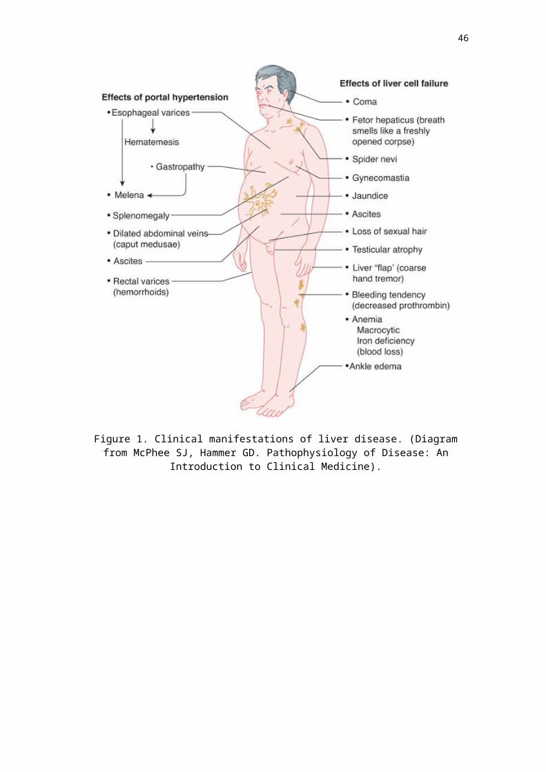
Consider which modality of anaesthesia suits the patient - neuraxial/regional/ local/sedation/general anaesthesia. There is no evidence that one anaesthetic technique has improved outcomes over another (in terms of mortality, cardiac morbidity or fistula failure).2
Ensure intravenous access and blood pressure devices (non-invasive or invasive) are on the non-fistula limb. Take special care to avoid pressure on the fistula. Take normal precautions against delayed gastric emptying/reflux.
Intra-operative management
In terms of optimal renal outcome, there is no evidence to support one anaesthetic technique over another in a general surgery population. Regardless of whether a general or regional anaesthetic is given, the main aim is to avoid renal insults and nephrotoxic drugs, maintain cardiac output, normovolaemia and normotension, avoid decreases in renal perfusion, avoid electrolyte and acid-base disturbances, and understand how the drugs given are affected by renal impairment.1
Intravenous fluid management
Patients should be kept normovolaemic and normotensive so that existing renal function is preserved. In stage 5 CKD (end-stage renal disease) be mindful that the patient may not have urine output and is dependent on dialysis to remove fluid – therefore observation of fluid restriction is important.
The choice of crystalloid fluid to use remains controversial, but is leaning towards a balanced-salt solution (e.g. Hartmann’s), rather than normal saline. Normal saline has a high chloride load, which can cause a hyperchloraemic metabolic acidosis. As well as contributing to hyperkalemia, it can also decrease renal blood flow and have adverse effects on kidney function.
The concern with Hartmann’s solution is hyperkalemia, because it contains potassium. However the amount of potassium in 1 litre of Hartmann’s solution is 4 mmol, which is minute compared to total body stores. Some studies comparing administration of normal saline to Hartmann’s solution showed that hyperkalemia occurred more often in patients receiving normal saline.13
Drug management
Induction agents
The pharmacokinetics of propofol are unaltered in renal failure, and it is metabolized to inactive metabolites. The induction dose of propofol remains unchanged. The time interval between cessation of a propofol infusion and wake up may be shorter in patients with renal failure.
Thiopentone has a larger volume of distribution and decreased protein binding in renal failure. The free drug concentration is higher, so the induction dose of thiopentone should be decreased.6
Ketamine can safely be used in renal failure. Hepatic metabolism and redistribution are responsible for termination of the anaesthetic effects and <3% is excreted unchanged in the urine.
46
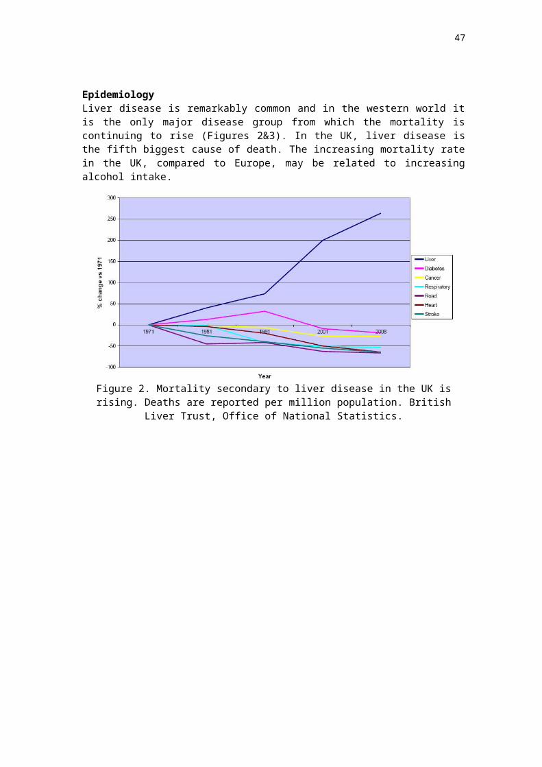
Inhalational agents
Nitrous oxide is safe for use in renal failure.
Hepatic metabolism of volatile agents results in inorganic fluoride, a high concentration of which has been implicated in renal damage. Methoxyflurane and enflurane, which have a high percentage of metabolism to inorganic fluoride, have been associated with reduction in renal function and are best avoided in CKD patients.
Approximately 2-5% of sevoflurane undergoes hepatic metabolism to inorganic fluoride. Additionally, sevoflurane reacts with CO2 absorbents to produce Compound A, which has been shown to be nephrotoxic in rats in a dose-dependent manner. Despite these concerns, studies have not shown adverse renal effects in the clinical use of sevoflurane, and it has been deemed a suitable inhalational agent to use in patients with CKD. Some bodies, but not all, have recommended using fresh gas flows >2L/min to minimise formation of Compound A. 7
There is only minimal hepatic metabolism of desflurane and isoflurane to inorganic fluoride (0.02% and 0.2% respectively). They are not associated with renal toxicity and are safe to use in renal failure. 6
Muscle relaxants
Suxamethonium is metabolized by plasma cholinesterases, so offset of action is not affected by CKD. Suxamethonium increases potassium concentrations transiently by 0.5-1.0 mmol/L, so it is important to check potassium levels prior to use.6
Atracurium and cisatracurium both undergo non-organ dependent metabolism via (temperature and pH dependent) Hoffman degradation, and ester hydrolysis. This makes them ideal for use in patients with CKD. One of the products of Hoffman degradation is laudanosine, which has been shown to induce seizures in animals. The levels of laudanosine are lower with cisatracurium than with atracurium6, however clinical use of atracurium does not produce seizures in CKD patients.
Vecuronium is predominantly metabolized and excreted by the liver, but the kidney may excrete up to 30%. Renal failure results in reduced clearance, increased elimination half-life and prolonged duration of action. Rocuronium has a similar profile, with a prolonged duration of action in patients with CKD. The relaxants pancuronium and tubocurarine both have reduced renal clearance and a prolonged half-life in CKD and should be avoided. Pancuronium also has an active metabolite 3-hydroxypancuronium.
Neostigmine has a reduced clearance and prolonged half-life in CKD. This can lead to bradycardia and AV block, especially if used with atropine rather than with the longer acting glycopyrrolate.
Sugammadex and rocuronium have a decreased renal clearance in CKD, but in a small trial, the reversal of neuromuscular blockade of rocuronium was still rapid and effective in patients with CKD. No patient showed signs of re-curarisation. The sugammadex-rocuronium complex is retained in the body for longer and is renally excreted.8
47
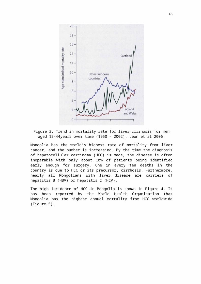
Opioids
Morphine should be used with caution in those with CKD, as it can cause respiratory depression and sedation. It is hepatically metabolized into 2 metabolically active compounds: morphine-3-glucuronide, which actually antagonizes the analgesic effect of morphine and can cause irritability and lowered seizure threshold, and morphine-6-glucuronide, which has potent analgesic properties, and when accumulates can result in over-sedation and respiratory depression. The half-life of this metabolite increases from 2 hours to 27 hours in renal failure. A significant fraction of morphine is removed by haemodialysis.
Fentanyl undergoes extensive hepatic metabolism with no active metabolites, and is a good choice in CKD patients. Haemodialysis has little effect on fentanyl plasma concentration. Alfentanil also has no active metabolites. Remifentanil is metabolized by ester hydrolysis, and not dependent on renal elimination, making these drugs acceptable to use in CKD.6
Pethidine is metabolized to active metabolite norpethidine, which requires renal excretion. Accumulation of this metabolite has been associated with seizures, myoclonus and altered mental state, and is best avoided in CKD.
Thirty percent of Tramadol is excreted unchanged in the urine. O-Demethyl, an active metabolite, also requires renal excretion. The risk of seizures is increased in renal impairment, so the dose must be reduced or the use of tramadol avoided. 6
Oxycodone is hepatically metabolized to noroxycodone and oxymorphone, and all require renal excretion. Oxymorphone has analgesic activity. The dose of oxycodone should be reduced to about half in CKD and the dosing interval increased.6,14 Despite this, accumulation of oxymorphone can still occur. Codeine use is not recommended. It has a significantly prolonged elimination half-life, and conventional doses have been associated with central nervous system depression.14
Adjuvant analgesic agents
Non-Steroidal Anti-Inflammatory Drugs (NSAIDs) should be avoided in CKD. They are nephrotoxic agents that can cause a sharp decrease in glomerular filtration rate. They also contribute to fluid overload, hypertension and hyperkalemia, as well as increase the risk of gastrointestinal bleeding due to platelet inhibition combined with uraemic platelet dysfunction. There is also an increased risk of cardiovascular complications in this at-risk group. COX-2 inhibitors also carry a similar renal risk to non-selective NSAIDs.6
Paracetamol and amitriptyline may be used without any adjustment to dosage.
Gabapentin and pregabalin are renally excreted, and does needs to be modified in renal failure.15
The timing of drugs relative to dialysis is of importance. Also remember that the clearance of drugs might be very different between patients on peritoneal dialysis as compared to those on haemodialysis. Reference materials or expert opinion should be consulted if advice regarding dosing and timing is needed.
Antiemetics
Ondansetron can be administered at the usual dosage. Metoclopramide should be administered with a reduction in dose by half in ESRD.
48
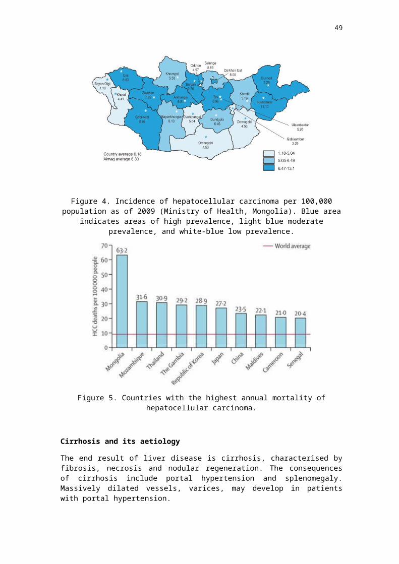
Antimicrobial agents
Aminoglycosides (gentamicin, amikacin, neomycin, tobramycin) and amphotericin B are nephrotoxic. If the use of these agents is necessary then other confounding factors for renal failure need to be managed e.g. hypovolaemia, hypotension, sepsis.5
Other
Radiographic contrast negatively affects renal function from 24 hours after administration, peaking at 4 days. To reduce the impact on the kidneys patients should be well hydrated, contrast load should be minimized, and confounding factors should be avoided. The data on preventative N-acetylcysteine administration is non-conclusive, but tends towards a statistically insignificant trend for a reduction in contrast-induced nephropathy.5
Post-operative management
Post-operative issues for chronic kidney disease patients include:- Nursing in an appropriate environment (e.g. ward care, or HDU/ICU) appropriate
to patient co-morbidities and surgical procedure- Returning to the care of a renal physician- Good analgesia including local anaesthetic infiltration- Aim to avoid sedation/respiratory depression- Appropriate fluid and electrolyte management
References
1. Anesthetic Concerns in Patients Presenting with Renal Failure, Genhard Wagener and Tricia Brentjens. Anesthesiology Clinics 2010 Vol 28(1) p 39-54.
2. Rang, S.T. et al., 2006. Anaesthesia for Chronic Renal Disease and Renal Transplantation. EAU-EBU Update Series, 4(6), pp.246–256.
3. Parham WA, Mehdirad AA, Biermann KM, Fredman CS. Hyperkalemia Revisited. Texas Heart Institute Journal. 2006;33(1):40-47
4. http://www.ncbi.nlm.nih.gov/pmc/articles/PMC1413606/ 5. Moro O Salifu - Perioperative Management of the Patient With Chronic Renal Failure6. http://emedicine.medscape.com/article/284555-overview#showall 7. https://www.openanesthesia.org/renal_disease_anesthesia_text/ 8. Craig, R.G. & Hunter, J.M., 2008. Recent developments in the perioperative
management of adult patients with chronic kidney disease. British Journal of Anaesthesia, 101(3), pp.296–310.
9. Behne, Wilke, Harder 1999. Clinical Pharmacokinetics of Sevoflurane. Clin Pharmacokinet Jan 36(1);13-26.
10. Staals, L.M. et al., 2009. Reduced clearance of rocuronium and sugammadex in patients with severe to end-stage renal failure: a pharmacokinetic study. British Journal of Anaesthesia, 104(1), pp.31–39.
11. Roodenburg, L Roberts. The Alfred Hospital Melbourne Australia 2014 Perioperative Short Course. Topic 11 Perioperative Acute Kidney Injury and Fluid Management.
12. González Cárdenas, V.H. et al., 2013. Pre-anesthesia evaluation in patients with chronic renal disease (emphasis on cardiovascular risk). Colombian Journal of Anesthesiology, 41(2), pp.139–145.
13. Tumurbaatar, B. et al., 2011. Kidney transplantation in Mongolia using effective and economical immunosuppression - a 3-year experience. Transplant International, 25(1), pp.e21–e22.
49
THE ALCOHOLIC PATIENT
Definition of alcohol use disorders Alcohol use disorders range from those with alcohol dependence and abuse to those considered “at risk”. The “at risk” drinker is the person who consumes harmful levels of alcohol but is not alcohol dependent. This amounts to 16 standard drinks per week in men or ten standard drinks per week in women. Harmful drinking is considered by the World Health Organisation to be the level of alcohol use that results in physical or psychological harm, but the patient is not considered to be alcohol dependent. Alcohol abuse is defined as a maladaptive pattern of use associated with one or more of:
A failure to fulfill work and social obligations; recurrent use in physically hazardous situations
Recurrent legal problems (drink-driving for instance) or Continued use in spite of alcohol related social problems.1
Alcohol dependence is defined as a maladaptive pattern of use associated with three or more of:
Tolerance Withdrawal Alcohol taken in larger quantities than intended Persistent desire to cut down use Time spent obtaining, using or recovering form alcohol Social, occupational or recreational tasks being sacrificed Continued use despite physical and psychological problems.1
Alcohol withdrawal syndrome Alcoholics are susceptible to withdrawal symptoms when there is an abrupt cessation of sustained alcohol intake. However, withdrawal is not usually seen because most people drink in an episodic rather than a sustained fashion.
Because of the central nervous system depressant effects of alcohol, abrupt cessation of drinking unmasks compensatory overactivity of certain parts of the central nervous system including sympathetic autonomic outflow.2
Minor withdrawal occurs after 6 to 36 hours after intake and includes tremor, insomnia, mild anxiety, gastrointestinal upset, headache, diaphoresis and anorexia. Withdrawal seizures are generalized tonic-clonic convulsions that occur within 48 hours after the last drink. 3 percent of chronic alcoholics have seizures and of these 3 percent develop status epilepticus. Treatment is controversial and most of these seizures are self-limiting. If the patient develops status epilepticus, short-term phenytoin and benzodiazepines can be used. Alcoholic hallucinosis is characterized by visual, auditory or tactile hallucinations. They can occur 12-48 hours after cessation of drinking and do not define delirium tremens unless there is a clouding of the sensorium.
50

Delirium tremens (DTs) will occur in up to 5 percent of those who undergo withdrawal from alcohol. Its onset is usually 48 hours after the last drink and is characterized by hallucinations, disorientation, tachycardia, hypertension, low-grade fever, agitation and diaphoresis. DTs usually lasts one to five days. The risk factors for DTs are a sustained drinking history, previous episodes of DTs, age greater than 30, concurrent illness and more than 48 hours after the last drink. The DTs will create a catabolic state with increased cardiac output and oxygen consumption.
Hyperventilation will decrease pH and reduce cerebral blood flow. The patients are frequently dehydrated and may suffer electrolyte disturbances including hypokalaemia, hypomagnesaemia and hypophosphataemia. Low phosphate may contribute to cardiac failure and rhabdomyolysis. The stress of surgery may predispose or exacerbate alcohol withdrawal syndrome and this may increase morbidity.3 The identification of alcohol withdrawal syndrome may be delayed in the perioperative period. It is therefore important to identify the patient at risk for withdrawal by taking a careful history of alcohol consumption and previous alcohol withdrawal syndromes.
These patients will then need to receive prophylaxis. Prophylaxis for alcohol withdrawal will need to begin upon cessation of drinking. It should not be delayed until after surgery, because it is possible to develop the withdrawal syndrome intra- operatively. This will compound any surgical stress response and contribute to a worse outcome.
The first line drug for prophylaxis is a benzodiazepine. The use of diazepam is favoured because it has a long half-life and active metabolites. In the patients with advanced cirrhosis, an agent with a shorter half-life such as oxazepam may be more prudent. Diazepam is given in doses of 5 to 10 mg intravenously every five minutes until the patient is calm but awake. Thereafter, a symptom- triggered approach can be used. This involves the administration of benzodiazepine based on the patient’s clinical condition after initial loading with diazepam.
Refractory delirium tremens can be treated with phenobarbitone in addition to diazepam, or with propofol. Alcohol should not be used in the patient with acute withdrawal. Phenothiazines and butyrophenones lower the seizure threshold and are not recommended.2
51
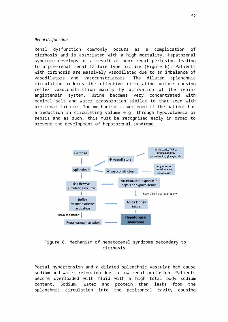
Short and long term effects of alcohol use The systemic effects of chronic alcohol misuse are summarised in the table below:
Central nervous system • Wernicke-Korsakoff syndrome• Peripheral Neuropathy• Autonomic dysfunction
Cardiovascular system • Cardiomyopathy• Heart Failure• Hypertension• Arrhythmias
Gastrointestinal system • Liver Disease• Pancreatitis• Gastritis• Oesophageal and bowel cancer
Metabolic Obesity Hyperlipidaemia Hypokalaemia Hypomagnesaemia Hyperuricaemia
Haematological Macrocytosis Thrombocytopaenia Leucopaenia
Musculo-skeletal Myopathy Osteoporosis Osteomalacia
Nervous system
Acute intoxication depresses the central nervous system, which will lead to blunting and loss of motor, sensory and cognitive function. There also depression of inhibitory pathways leading to behavioural disinhibition.
Chronic alcohol use is associated with peripheral and central nervous system effects. Peripheral neuropathy can lead to “stocking” distribution of sensory effects and there can be proximal myopathy leading to weakness and muscle wasting. Long-term use of ethanol is associated with a higher incidence of dementia.
Psychosis is related to nutritional deficiencies, primarily of B1 (Thiamine). Wernicke’s encephalopathy is an acute disorder that consists of ocular-motor disorders, ataxia and an altered mental state. Korsakoff’s psychosis consists of amnesia (both retrograde and anterograde), disorientation and confabulation. Metabolic stress in conjunction with a glucose load typically triggers Wernicke-Korsakoff’s syndrome so it is recommended that these patients receive thiamine before receiving glucose.4
The other long-term effects of alcohol abuse relate to withdrawal syndromes, which have already been discussed. Cardiovascular system
Long-term alcohol use will cause a gradual increase in blood pressure. Atrial fibrillation can occur in association with acute binge drinking as well as with chronic alcohol abuse. There is a 34% increased risk of developing atrial fibrillation in the chronic alcoholic.4
52

A dilated left ventricle and a small reduction in ejection fraction characterize alcoholic cardiomyopathy. There is a rise in pulmonary pressures, right heart failure and arrhythmias. Thiamine deficiency will cause wet beriberi or high output cardiac failure. The high cardiac output is thought to be due to systemic vasodilatation and increased venous return.
The patient who is withdrawing from alcohol has heightened sympathetic nervous system outflow which can trigger heart failure. Respiratory system
Because of immunosuppression, the alcoholic patient is at increased risk for atypical bacterial pneumonia (such as the gram negative, Klebsiella pneumoniae). Aspiration pneumonia can also occur due to a depression of protective reflexes with intoxication. Gastrointestinal and Hepatobiliary system
Gastric pathology is more common with alcohol use. Acute gastritis, oesophagitis and pancreatitis can occur with binge drinking. Chronic use increases gastric acid reflux. Alcoholic liver disease occurs in three major forms, which commonly overlap. They are fatty liver, alcoholic hepatitis and cirrhosis. The risk factors for the development of severe alcoholic liver disease are the ingestion of over 5-7 standard drinks per day (12g of alcohol per standard drink) for 10 years in men and over 2-4 drinks per day in women.
Fatty liver is the most common response to toxins. Fatty liver is most commonly asymptomatic but it may present with right upper quadrant pain, hepatomegaly, nausea and jaundice. It will occur in over 90% of binge and chronic drinkers and is potentially reversible with the cessation of drinking. Laboratory investigations usually show moderately elevated aspartate aminotransferase (AST), alanine aminotransferase (ALT) and gamma-glutamyl transpeptidase (GGT), high cholesterol and triglycerides.5
Alcoholic hepatitis is characterised by hepatocyte injury and is thought to be a precursor to cirrhosis. It may be potentially reversible with the cessation of drinking, whereas cirrhosis is not. Fortunately only 15% of alcoholics develop alcoholic liver disease. The main features of hepatitis are fever, jaundice and abdominal pain. Some patients are asymptomatic. The AST and ALT are markedly elevated (2 to 7 times normal) and high bilirubin is common. Coagulopathy, anaemia and low albumin indicate severe disease. If the patient becomes critically ill with ascites, variceal haemorrhage, encephalopathy or hepatorenal syndrome, then the prognosis is poor with a mortality of 70%. Treatment is mostly supportive, but glucocorticoids have been used. Abstinence from alcohol is important for those with potentially reversible disease.5 Preoperative assessment of the patient with alcohol abuse Preoperative assessment involves the diagnosis of an alcohol use disorder with careful questioning, the detection of end organ damage and the assessment of the potential for alcohol withdrawal syndrome. Baseline investigations include, electrocardiography (ECG), electrolytes (hypokalemia, hypomagnesaemia, hypophosphatemia, hypoglycaemia), full blood count (anaemia, low platelets), coagulation (elevated prothrombin time), and liver function testing where hepatitis is suspected. Where there is a potential for withdrawal syndrome, diazepam should be administered
53
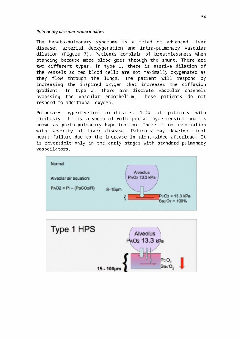
according to symptoms. If there is enough time, the patient should abstain from alcohol pre operatively. This may involve referral for counselling and a program of detoxification. A study from Denmark involving 32 patients for colorectal surgery with an alcohol consumption of at least 60g per day, showed that one month of abstinence reduced postoperative morbidity (31% versus 74% P=0.02) compared to continuous drinkers. The most common complications were infection, cardiopulmonary insufficiency and bleeding.
The mechanism is probably a reduction in organ dysfunction and a reduction of the exaggerated response to surgical stress.6
Chronic alcohol abusers should receive multivitamins and high dose thiamine (100mg) to prevent stress-induced Wernicke-Korsakoff syndrome. The acutely intoxicated patient The acutely intoxicated patient is more sensitive to the effects of barbiturates, benzodiazepines and opioids. There is generally a lesser anaesthetic requirement. The intoxicated patients can be assumed to have an increased risk for pulmonary aspiration and therefore will require a rapid sequence induction after premedication with a non-particulate antacid such as sodium citrate and ranitidine. In addition, the intoxicated patient will have an inability to withstand acute blood loss and a decreased brain tolerance to hypoxia. Alcohol interferes with platelet function and increases the concentration of plasma catecholamines.7
If the procedure is elective, it is best to postpone the operation until the patient is sober so that adequate fasting is achieved and there is less interaction with anaesthetic medication. There is a tendency to hypoglycaemia in the intoxicated patient, so blood glucose should be monitored. In an emergency, precautions to prevent aspiration will need to be taken and invasive blood pressure and central venous pressure monitoring may be required where significant blood loss is expected. The sober chronic alcoholic After careful pre operative assessment, as outlined previously, routine monitoring should be established (blood pressure, ECG, pulse oximetry, end tidal carbon dioxide and agent monitoring) and invasive monitoring may be required depending on the presence of end organ damage and the surgical procedure. Due to increased resistance through the portal vein, patients with liver disease rely on hepatic arterial flow for perfusion and are at risk for hepatic ischaemia if there is hypotension, an arterial line may be required. The assessment of volume status is improved in the presence of an in dwelling urinary catheter and central venous pressure catheter. Those with severe cardiac disease may require a pulmonary artery catheter to monitor filling pressures and cardiac output. In the sober patient, there is less risk for aspiration compared to the intoxicated patient unless there is reflux oesophagitis or raised intra abdominal pressure due to ascites.
54
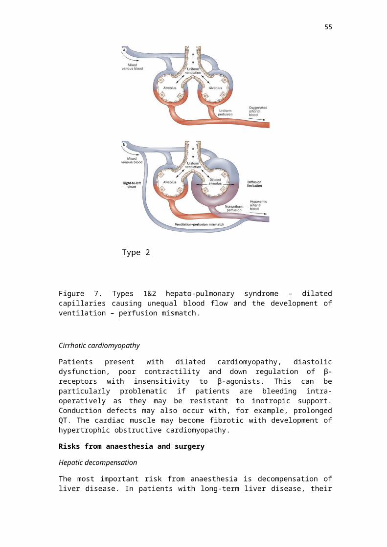
It is important to realize that the presence of oesophageal varices may preclude the use of oro- or nasogastric tubes. There is no evidence that one anaesthetic technique is better than any other but it needs to be remembered that coagulopathy and platelet dysfunction will be a contraindication to spinal or epidural anaesthesia due to the risk of spinal haematoma. The cardiac depressant effects of the volatile agents may be exaggerated in the presence of cardiomyopathy. There have been several studies to show that there is an increased requirement for anaesthesia and analgesia in the patient with chronic alcohol use. 8,9 This increase in anaesthesia and analgesia requirements is thought to be due to cross-tolerance with alcohol but it may be a pharmacokinetic phenomenon. In the study assessing the dose of propofol required for loss of consciousness in the chronic alcoholism, 26 chronic alcoholics and 20 non-habitual drinkers were studied. The dose for loss of consciousness in the alcoholic group was 4.2 mg per kg compared to 2.2 mg per kg in the control group (P<0.001). The blood concentration at loss of consciousness between these groups was not significant. (8). The dose requirement of thiopentone is not increased.7 Muscle relaxants that require organ based (liver or renal) metabolism and excretion should be avoided. Suxamethonium may have a slightly prolonged duration of action if there is liver dysfunction and reduction in plasma cholinesterase. There is a larger volume of distribution in the presence of liver cirrhosis, which may necessitate larger initial doses of non-depolarising muscle relaxants. Atracurium is a good choice for muscle relaxation in the patient who does not require a rapid sequence induction because it is not metabolised in the liver. The surgical stress response is enhanced in the alcoholic patient. This may increase postoperative morbidity due to increased cardiovascular demand and immunosuppression. In addition, it is difficult to distinguish between the stress response and alcohol withdrawal syndrome under anaesthesia. The two may occur simultaneously producing additive effects Postoperative care Alcoholic patients show a two to five fold higher rate of postoperative complications so deserve special postoperative attention. There is a two-fold increase in bleeding complications after surgery and immune suppression results in higher rates of infection (wound, chest and urinary infection).10
Opioid analgesic requirements may be increased in the alcoholic patient, so the use of adjuvants for analgesia is attractive, however, the patient with liver disease is more susceptible to overdose and hepatic necrosis when taking paracetamol and non-steroidal drugs will further impair platelet function. Withdrawal occurs after alcohol intake ceases and symptoms should be sought post operatively given that severe alcohol withdrawal syndrome symptoms typically peak within four days.
55
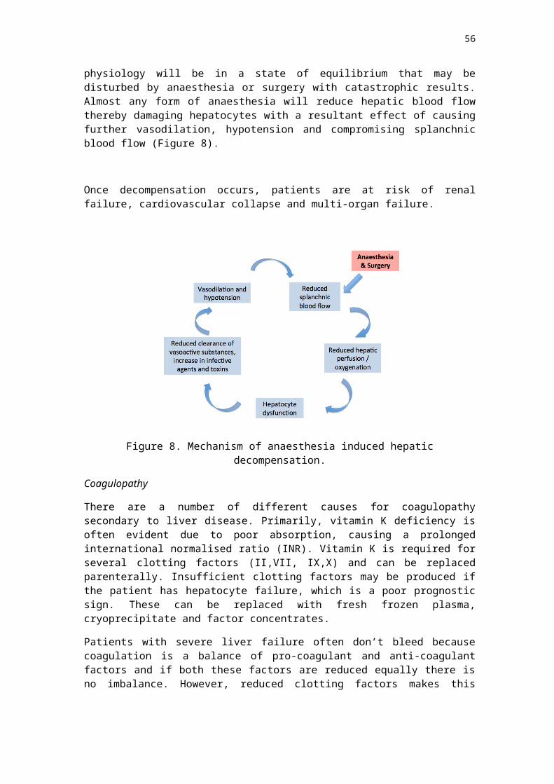
Conclusion Alcohol use disorder is common among surgical patients. The prevalence is about 20%. The alcoholic patient is at risk for major postoperative morbidity including prolonged intensive care and hospital stay, respiratory and cardiovascular complications, infection and surgical bleeding.
Of significant concern, is the risk of alcohol withdrawal syndrome, which carries significant mortality. The challenge for the anaesthetist is to identify the patient who has alcohol use disorder, detect any end organ damage and assess for the risk of withdrawal. Ideally, the patient will be seen some time prior to surgery to enable a referral to an addiction specialist and undergo a period of abstinence. This is not always practical, and indeed it may be necessary to anaesthetise an intoxicated patient who has suffered trauma. Preoperative assessment will need to include a detailed history and examination, looking for the multiple complications of chronic alcohol use including hepatic, pulmonary, cardiac and neurological complications. Investigation will need to include a search for metabolic and electrolyte abnormality, coagulopathy and anaemia and an ECG to detect cardiac arrhythmias.
There is no anaesthetic technique that is ideal for the chronic drinker, but the anaesthetist should bear in mind the risk of aspiration, the change in sensitivity to anaesthetic medications and the risk of coagulopathy. Postoperatively, the patient will need to be carefully monitored. Ideally this will be in a high dependency area where he can be observed for signs of withdrawal and cardiac, respiratory and infectious complications.
56
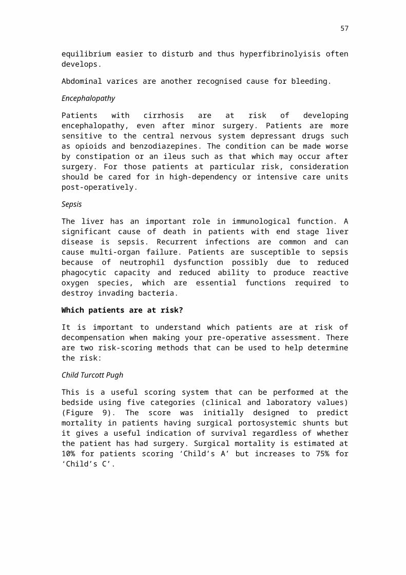
References
1. American Psychiatric Association. In: Diagnostic and Statistical Manual of Mental Disorders, American Psychiatric Association, Washington 1985
2. Weinhouse GL, Manaker S. Alcohol withdrawal syndromes in UpToDate at www.uptodate.com 2006
3. Spies C. Tonnesen H, Andreasson S et al. Peri-operative morbidity and mortality in chronic alcoholic patients. Alcohol Clin Exp Res 2001; 25:164S.
4. Moss M, Burnham EL. Alcohol abuse in the critically ill patient. The Lancet 2006; 23/30.
5. Chapter 288. Alcoholic liver disease in Harrison’s Internal Medicine. McGraw Hill 2006.
6. Tonnesen H, Rosenberg J, Nielsen HJ et al. Effect of preoperative abstinence on poor postoperative outcome in alcohol misusers: randomised controlled trial. BMJ 1999; 318:1311- 6.
7. Stoelting RK, Dierdorf SF. “Diseases of the Liver and Biliary Tract” in Anesthesia and Co-existing Disease 3rd Edition. Churchill Livingstone USA; 1983: p 251-275
8. Fassoulaki A, Farinotti R, Servin F, Desmonts JM. Chronic alcoholism increases the induction dose of propofol in humans. Anesth Analag 1993; 77:553.
9. Tammisto T, Tigerstedt I. The need for fentanyl supplementation of N2O-O2 relaxant anaesthesia in chronic alcoholics. Acta Anesthesiol Scand 1977; 21:216
10. Sander M, Neumann T, von Dossow V et al. Alcohol use disorder: risks in anaesthesia and intensive care medicine. Internist, 2006 47(4): 332, 334-6, 338.
11. Chapman R & Plaat F. Alchohol and Anaesthesia. Continuing Education in Anaesthesia, Critical Care & Pain. 2009; 9: 10-13
57
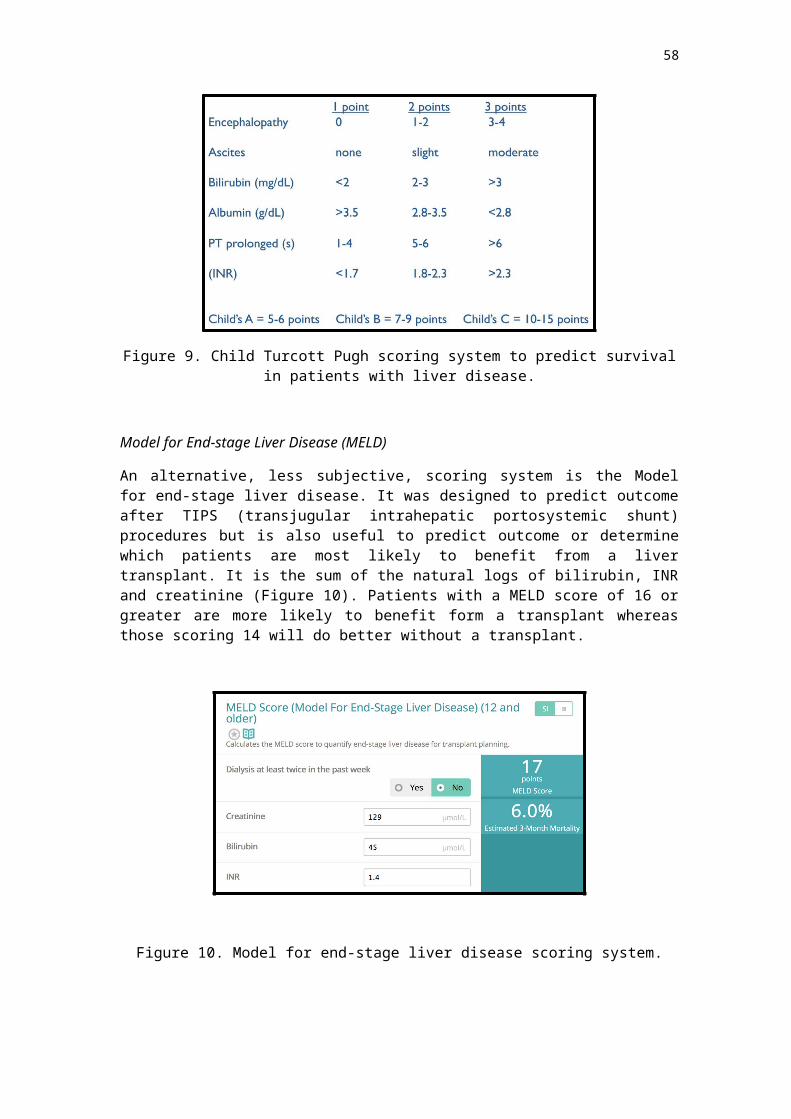
CHRONIC OBSTRUCTIVE PULMONARY DISEASE
Introduction
Chronic obstructive pulmonary disease (COPD) is an extremely common respiratory disorder that affects many people across the world. The long-term survival of patients with severe COPD undergoing any type of surgery is poor and they are at significant risk of postoperative morbidity, especially pulmonary complications. However with risk identification, pre-operative optimisation and appropriate anaesthetic management, the risks of developing postoperative pulmonary complications can be reduced - even in patients with severe disease.
Pathophysiology of COPD
COPD is a generally progressive inflammatory disease of the lungs that is characterised by airflow limitation that is not fully reversible, and is often complicated by significant systemic manifestations and comorbidities. The inflammation is usually secondary to inhaled noxious particles or gases, the most common of which worldwide is cigarette smoke.
The lung pathology in COPD is a combination of inflammatory small airways disease (obstructive bronchiolitis) and parenchymal destruction (emphysema). The relative contribution of these two processes and thus the resultant physiological abnormalities varies from person to person.
The small airways disease leads to obstruction and air trapping. This occurs chronically, but importantly also occurs dynamically under stress. The resulting dynamic hyperinflation adversely affects ventilation / perfusion (VQ) mismatching, the loading and mechanics of the respiratory muscles, and the sensation of dyspnoea.
The loss of lung parenchyma decreases gas transfer, reduces the pulmonary capillary bed and worsens VQ mismatching. Reducing the parenchymal support of the walls of the small airways contributes to the airflow limitation.
The end result of the VQ mismatching, decreased gas transfer and alveolar hypoventilation is hypoxaemia and sometimes hypercarbia. The hyperinflation causes marked dyspnoea even without a fall in PO2.
Importantly, COPD patients can have significant extrapulmonary disease. This can be a direct result of COPD such as cor pulmonale, respiratory and skeletal muscle wasting and weight loss, or comorbid diseases, especially cardiovascular, which are more prevalent in this population.
Clinical Features of COPD
The typical features of COPD are dyspnoea, wheeze and cough with or without sputum production. The onset of COPD is insidious and most patients are symptomatic either with cough or progressive dyspnoea long before they present to medical services. Therefore a diagnosis of COPD should be considered in all patients over forty years old with a significant smoking history (>10 pack years) who should be questioned about symptoms.
58
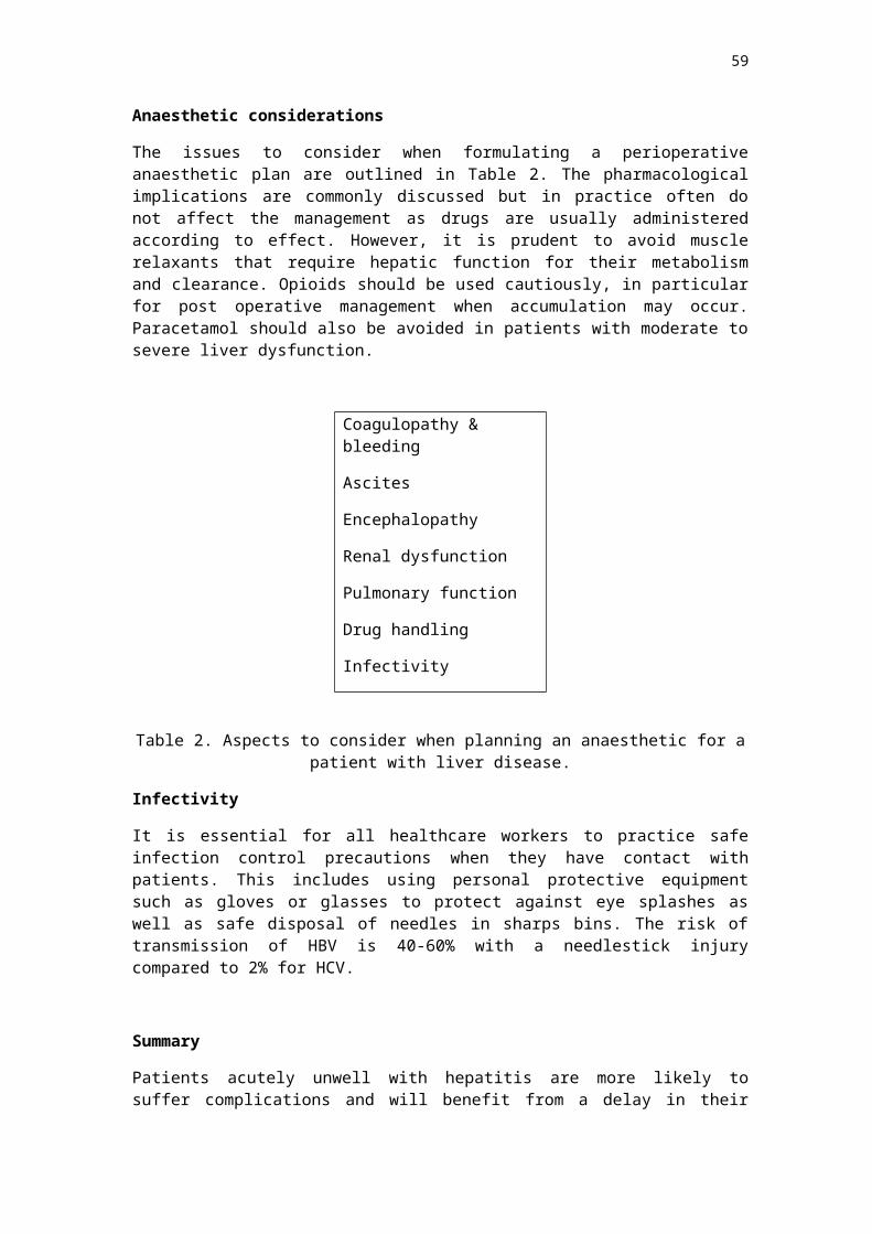
In the early stages of COPD the dyspnoea is usually associated with exertion. The breathlessness is characteristically differentiated from asthma by the lack of nocturnal symptoms (in early COPD), a lack of significant diurnal variability, a lack of association with allergy and its persistent and progressive nature. Be aware that asthma and COPD can co-exist in the same patient.
Patients with significant hyperinflation can often develop significant dyspnoea even with relatively normal spirometry. The dyspnoea is often accompanied by significant anxiety. In exacerbation “breath-stacking” and dynamic hyperinflation results, which, unless it is rapidly treated, can quickly lead to respiratory failure. Conversely some patients have very poor respiratory function and have surprisingly few symptoms. They tolerate a significant degree of chronic hypoxia and often chronic hypercapnoea. These patients are often very oxygen sensitive and can rapidly develop type II respiratory failure if given too much supplemental oxygen.
The mainstay of treatment of COPD is bronchodilation both for maintenance and for exacerbations. Both -agonists and anticholinergics (ipratropium bromide andβ tiotropium bromide) are used. The latter are proposed to have an additional effect of relieving air trapping. Methylxanthines are controversial in COPD but they are widely used. Long-term inhaled steroids are usually only indicated in patients with severe COPD and repeated exacerbations or who have co-existent asthma. Oral steroids have almost no role in maintenance of COPD but are important and beneficial in the treatment of exacerbations.
Preoperative assessment History and examination
Establish exercise tolerance, particularly hills and stairs (although reported levels of dyspnoea and disability may be inaccurate).
Enquire about frequency of exacerbations, hospital admissions and previous requirements for invasive and non-invasive ventilation.
A smoking history is vital. Current and recent smokers are much more likely to develop postoperative pulmonary complications.
Cough and particularly sputum production has been shown to be an independent risk factor for postoperative pulmonary complications in COPD.
A clear history regarding co-morbid conditions is vital. Symptoms and signs of active infection should be sought including green or
purulent sputum, increased dyspnoea, wheeze and signs of consolidation. Delay purely elective surgery for patients with acute exacerbations.
• Nutritional status should be assessed as patients with both high and low BMI have increased risk.
Investigations
For patients with known asthma or COPD, routine preoperative pulmonary function tests are not necessary - a thorough history and exam can assess severity.
Spirometry should be reserved for patients who are suspected of—but have not yet been diagnosed with—chronic obstructive pulmonary disease or patients with a significant change in functional status since previous testing.
59
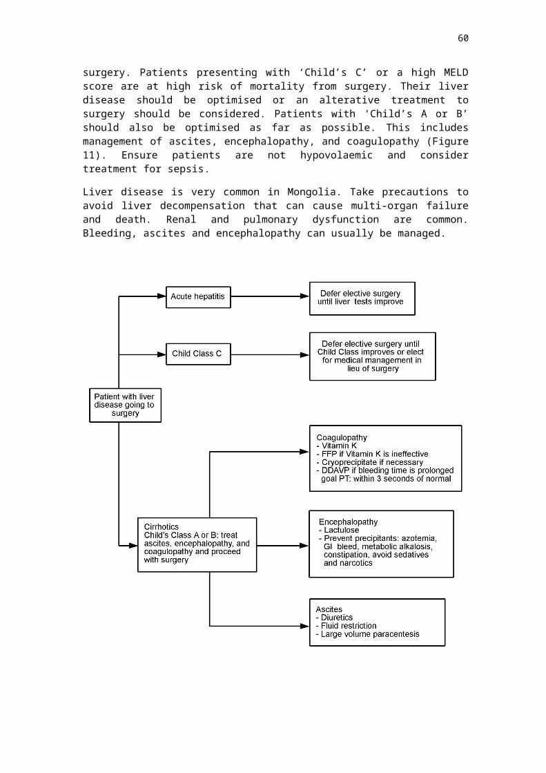
A chest X-ray may be useful to exclude active infection and occult malignancy, but it is generally not necessary to order routine chest radiographs in patients with stable COPD or asthma unless guided by symptoms or physical examination findings suggestive of new or worsening disease. Reviews of the utility of chest radiographs have generally found that while abnormal CXR findings are not unusual, only rarely do such findings influence preoperative management. The presence of extensive bullous disease highlights the potential risk of pneumothorax.
ECG may reveal right heart disease (right ventricular hypertrophy or strain).
Consider echocardiography if clinically indicated.
Spirometry is used to clarify diagnosis and assess severity:
GOLD Classification of COPD (based on post bronchodilator FEV1)
Stage I: Mild FEV1/FVC <0.70 FEV1 ≥ 80% predicted
Stage II: Moderate FEV1/FVC < 0.70FEV1 50 - 80% predicted
Stage III: Severe FEV1/FVC <0.70FEV1 30 - 50% predicted
Stage IV: Very SevereFEV1/FVC 0.70FEV1 <30% predicted or FEV1 < 50% predicted and chronic respiratory failure
Gas transfer measurements can give additional information, especially if significant emphysema is suspected. Simple exercise tests such as stair climbing and the 6-minute walk test are safe and simple to perform, and correlate well with more formal exercise testing.
Arterial blood gas measurement is useful: PaCO2 > 5.9 kPa and PaO2 <7.9 kPa (on room air) are both associated with a worse prognosis.
Preoperative optimisation
Preoperative optimisation can be divided into four main areas: smoking cessation, optimization of drug therapy, treatment of infection, and respiratory physiotherapy.
Smoking cessation. Current smokers are at far greater risk of developing postoperative pulmonary complications. Smoking should be stopped at least eight weeks before surgery in order to obtain maximum benefit.
Optimal drug treatment. Some patients with COPD have a degree of reversible airways disease and even in those patients with no demonstrable reversibility bronchodilators are indicated as they increase exercise tolerance even if there is no change in FEV1. Almost all patients with COPD benefit from at least one dose of nebulised bronchodilator preoperatively. Some caution is needed as high doses of nebulised -agonists can cause or exacerbate tachyarrhythmias and causeβ hypokalaemia. Nebulised anticholinergics can increase sputum viscosity.
60
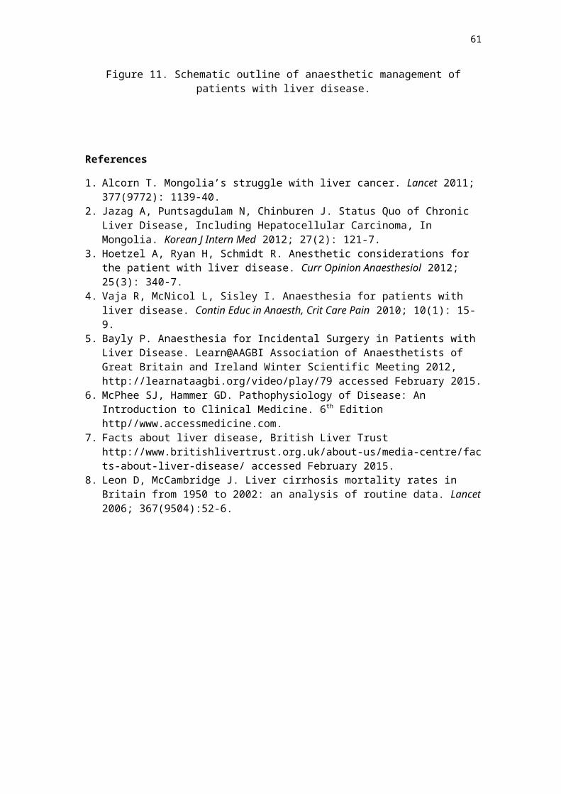
Treatment of infection/ exacerbation. Current infection or exacerbations are a contraindication to anaesthesia. They should be treated with both -agonist andβ anticholinergic therapy, preferably in nebulised form, and a short course of systemic steroids. If the patient has signs of active infection such as viscous, green or brown sputum or worsening cough or dyspnoea this should be treated preoperatively with antibiotics. Prophylactic antibiotics over the operative course may be beneficial. Oral steroids are not recommended in stable COPD.
Physiotherapy. Preoperative physiotherapy is important in sputum-producing COPD patients to clear any retained sputum that may cause intraoperative bronchial plugging or pneumonitis. There may also be a role for pulmonary rehabilitation, involving exercise training, patient education and psychological and behavioural interventions. This has been shown to improve symptoms and reduce length of hospital admission for exacerbation in COPD patients, but it is not yet as established as the above interventions.
Lung volume reduction surgery may be considered in selected patients with emphysema who have regions of overdistended, poorly functioning lung tissue.
Principles of perioperative anaesthetic management
Airway and ventilation
Bronchospasm may occur on induction, during airway instrumentation and on extubation. Whilst in some patients this may be mild and transient, others may need aggressive administration of bronchodilators to prevent or treat acute hypoxaemia and hypercarbia. Avoidance of endotracheal intubation, if possible, may reduce the risk of bronchospasm. Extubation should be carried out with the patient awake and sitting up.
V/Q mismatch increases under general anaesthesia and in the supine position due to cephalad movement of the diaphragm, which reduces the FRC towards closing capacity. Combined with atelectasis, this leads to worsening hypoxaemia. This is especially pronounced if the patient is obese or is in the Trendelenburg or lithotomy position. Supplemental oxygen and positive pressure ventilation may be necessary.
Sputum plugging can be caused by excessive sputum production in an anaesthetised patient. This may lead to lobar collapse, causing ventilatory failure and high airway pressure. Saline nebulisation, endobronchial suctioning, and physiotherapy to clear the sputum plug are the treatment strategies. Inadequate analgesia and insufficient postoperative physiotherapy can lead to ineffective sputum clearance also causing sputum plugging and nosocomial chest infection. Patients with a tendency to produce sputum need to be identified preoperatively, given physiotherapy and adequate postoperative facilities, such as the ICU or HDU. Postoperative chest infection must be promptly diagnosed and treated with antibiotics.
Pneumothorax is more likely in patients with COPD; mechanical ventilation should be carefully set to minimise high airway pressure and avoid breath stacking. The respiratory rate should be low to allow increased expiratory time. The use of PEEP should be minimal.
61
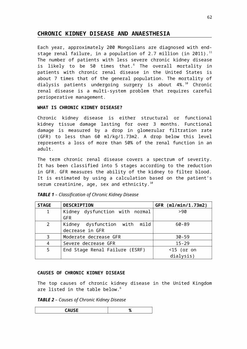
Postoperatively
Patients may require increased monitoring and continued ventilatory support. Residual effects of general anaesthesia and sedation may result in hypoventilation causing hypercarbia and hypoxaemia. Whilst increasing inspired oxygen concentration may suffice in most patients, some will require regular arterial blood gas monitoring and further ventilatory support (non-invasive or invasive). Some patients have ventilatory drive, which is dependent on hypoxia. In these patients high-inspired oxygen levels may suppress respiratory drive causing hypoventilation and hypercarbia. Ideally patients at such risk should be placed in ICU/HDU. The issue of postoperative chest infection is already discussed under ‘sputum plugging’.
←Cardiovascular issues
Some patients with COPD develop cor pulmonale, characterised by right ventricular hypertrophy, dilatation and failure. In these patients adequate oxygen therapy is required to prevent any further increase in right ventricular afterload caused by hypoxic pulmonary vasoconstriction. Fluid balance is crucial as appropriate right ventricular preload is essential to produce adequate cardiac output where the right ventricular afterload is high. Conversely, in an overfilled right ventricle, the left ventricular filling will be compromised thereby affecting systemic output. Central venous pressure monitoring may be useful, although in the presence of tricuspid valve incompetence may not reflect the true right sided filling status. Patients with right-sided heart failure are more likely to encounter ventricular and atrial arrhythmias.
Regional anaesthesia
Regional anaesthesia avoids many of the respiratory problems associated with general anaesthesia and has the added advantage of providing good postoperative analgesia. However patients must be able to tolerate lying flat. Most blocks can safely be performed with or without sedation. Interscalene block should be avoided as the resultant phrenic nerve palsy may further compromise respiratory function.
Analgesia
Effective analgesia is vital in order to optimise respiratory function, especially for patients who have undergone major abdominal or thoracic surgery. Lack of effective pain relief may result in diminished chest expansion and ineffective cough, leading to basal atelectasis, hypoxaemia and nosocomial infection. Epidural analgesia is particularly beneficial in patients with COPD, as it may prevent the problems described above and avoids the drowsiness and respiratory depression associated with the use of systemic opioids.
Thromboprophylaxis
Patients with COPD have an increased risk of developing venous thromboembolism, so appropriate thromboprophylaxis is important, along with early mobilisation and adequate hydration.
Summary
Patients with COPD vary in their presentation - from the asymptomatic and undiagnosed to those with a severely disabling systemic disease. All COPD patients undergoing surgery are at increased risk of postoperative complications and benefit from active management throughout the pre-, intra- and post-operative phases.
62
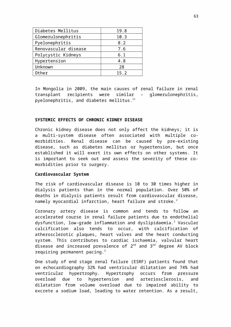
References
Edrich, T., & Sadovnikoff, N. (2010). Anesthesia for patients with severe chronic obstructive pulmonary disease. Current Opinion in Anaesthesiology, 23(1), 18–24. doi:10.1097/ACO.0b013e328331ea5b
1. Darkwater Studios, L. (2015). GOLD - the Global initiative for chronic Obstructive Lung Disease. [online] Goldcopd.org. Available at: http://www.goldcopd.org [Accessed 6 Apr. 2015].
2. Licker, M., Schweizer, A., Ellenberger, C., Tschopp, J.-M., Diaper, J., & Clergue, F. (2007). Perioperative medical management of patients with COPD. International Journal of Chronic Obstructive Pulmonary Disease, 2(4), 493–515.
3. Stoelting, R., Hines, R. and Marschall, K. (2012). Stoelting's anesthesia and co-existing disease. Philadelphia: Saunders/Elsevier.
4. Wakatsuki, M., & Havelock, T. (2015). Anesthesia for patients with severe chronic obstructive pulmonary disease. Web Publication, 1–7.
63
SMOKING
A WHO report published in 2012 showed that the prevalence of cigarette smoking in Mongolia was 48% of adult males, and 6.9% of adult females, and that the majority of smokers were daily users. The prevalence of cigarette smoking in male and female youth (13 to 15 years old) was 9.2% and 2% respectively.
These figures in Mongolia may have decreased now that that laws banning smoking in all public areas, including schools, universities, government facilities, offices, restaurants, pubs, bars, cafes, hotel lobbies, and public transport, were passed in March 2013. The laws also prohibit the sale of cigarettes within 500 metres of schools, and to anyone less than 21 years of age. Health warnings on cigarette boxes were also increased from 33% to 50% of the packaging.11,12
SMOKING - MORTALITY
Tobacco kills nearly 6 million people each year, mostly the result of direct tobacco use. 600,000 deaths occur in non-smokers who have been exposed to second-hand smoke. Up to half of the people who use tobacco will eventually die of a tobacco-related disease, and nearly 80% of the more than 1 billion smokers worldwide live in low- and middle-income countries.14
In the USA, life expectancy for smokers is at least 10 years shorter than for non-smokers. Men who smoke increase their risk of dying from bronchitis and emphysema by 17 times, and women by 12 times. There is an increased risk of death from cancer of the trachea, lung and bronchus by more than 23 times in men, and by 12 times in women. Smoking increases the risk of dying from coronary heart disease among middle-aged men by almost 4 times, and middle-aged women by almost 5 times.13
SMOKING - MORBIDITY
The principal active ingredient in a cigarette is nicotine. In addition to this there are over 4700 chemical compounds, including at least 43 carcinogens in a cigarette. These have effects on various body systems to produce disease.
Respiratory System
Smoking causes hypersecretion of a large volume of thick mucous, as a result of goblet cell hyperplasia. There is impaired tracheobronchial clearance of this mucous due to inactivation of cilia in the lower airways from cilio-toxins in smoke.2 Increased reactivity of the lower and upper airways results from disruption of the epithelial lining of the lung and increased pulmonary permeability. This allows irritants to penetrate the epithelium more easily and stimulate the subepithelial irritant receptors. Increased reactivity leads to small airways narrowing, which results in an increased closing capacity and tendency to ventilation-perfusion mismatch.2 Loss of elastic recoil and emphysema are caused by an increase in lung proteolytic enzymes.4 Lung infection is increased – 25% of smokers suffer from chronic bronchitis, 5 times more than non-smokers.4
Cardiovascular System (CVS)
CVS effects of smoking are related to nicotine. It is a potent adrenergic agonist, and stimulates the adrenal medulla to secrete adrenaline. This leads to increases in heart rate and blood pressure, and increases peripheral vascular resistance.
64
Myocardial contractility is increased, leading to an increase in oxygen demand and consumption. The half-life of nicotine is 30-60 minutes. Three to four hours of abstinence will improve myocardial oxygen supply:demand ratio.9
Heavy smokers may have up to 15% of the available oxygen binding sites of haemoglobin occupied by carbon monoxide (carboxyhaemoglobin COHb), which results in about a 25% reduction in oxygen carrying capacity. The affinity of carbon monoxide for haemoglobin is 250 times greater than oxygen. Left shift of the oxygen haemoglobin dissociation curve also results in reduced oxygen delivery to the tissues and myocardium. Carbon monoxide binds with cytochrome oxidase and myoglobin and inactivates mitochondrial enzymes in cardiac muscle. There is impaired intracellular oxygen transport and utilization, and a negative inotropic effect leading to global tissue hypoxia.(9) The half-life of carboxyhaemoglobin is about 4-6 hours.9 Administering 100% oxygen will significantly expedite the removal of carbon monoxide. With 100% O2 the half-life of carbon monoxide is 40-80 minutes.
Smoking also increases coronary vascular resistance especially at the site of atheroma, so worsens symptoms of angina. It does this by multiple pathways including direct endothelial damage, haematological, metabolic and biochemical pathways.2 Coronary artery vascular resistance improves on cessation of smoking.1
Immune function
Smoking has potent influence on the immune system, including a reduction in neutrophil activity and immunoglobulin concentrations and natural killer cell activity. Many aspects of immune function return to normal after 6 weeks abstinence. There is some improvement in tracheobronchial clearance and reversible small airways disease.1
Cancer
Smoking is by far the most important preventable cause of cancer in the world. Smoking causes more than 4 in 5 cases of lung cancer. Smoking increases the risk of many other cancers including cancer of the larynx, oesophagus, mouth and pharynx, bladder, pancreas, kidney, liver, stomach, bowel, cervix, ovary, nose and sinuses.15
Wound and bone healing
There is a higher incidence of complications related to tissue healing in smokers due to a combination of effects on tissue oxygenation, the vascular system, the healing process and the immune system. Global tissue hypoxia exists due to nicotine-induced vasoconstriction and binding of carbon monoxide to haemoglobin. Smokers are also deficient in Vitamin C, which plays an important part in wound healing. Immunological studies show enhanced inflammatory responses that release tissue-destructive enzymes in smokers.5 Smoking is a major risk for osteoporosis and impairs the healing of fractures.16
Other
There is induction of hepatic microsomal enzymes leading to increased metabolism of various drugs.
Smoking can cause gastro-oesophageal sphincter incompetence, allowing reflux. However this usually resolves within 8 minutes of stopping smoking.16
65
PERIOPERATIVE RISK FROM SMOKING
There is overwhelming evidence that smokers have increased operative risk.1
A Cochrane review showed that patients randomized to cessation of smoking were 30% less likely to have a post-operative complication (RR 0.7, 95% CI 0.6-0.9). (5) A large cohort study of over 80000 patients in each arm (smokers vs. non-smokers) by Turan in 2011 found that thirty day mortality was 1.3 times higher in smokers (95% CI 1.2-1.5)
In terms of respiratory events, studies have shown a six-fold increase in the post-operative respiratory morbidity in patients smoking more than ten cigarettes a day.1
Complications such as atelectasis and pneumonia are consistently 2 to 6 times more frequent in smokers.2 Intra-operative airway events such as re-intubation, laryngospasm, bronchospasm, aspiration, hypoventilation and hypoxemia occur more frequently in smokers than non-smokers, the relative risk being 1.8 times in smokers. In young smokers the relative risk was 2.3 times, and in obese smokers it was 6.3 times that of non-smokers. The relative risk of bronchospasm in young smokers with chronic bronchitis was 25.7 times that of non-smokers.
The study by Turan in 2011 found that unplanned intubation was 1.6 times higher, pneumonia 1.8 times higher (95%CI 1.1 to 2.9), and prolonged ventilation (>48hrs) 1.7 times higher (95%CI 1.2–2.5). It has been shown that children who are exposed to passive smoking suffer from more post-operative oxygen desaturation.2
In terms of the cardiovascular system the findings have been mixed. Despite the disturbances of physiology caused by smoking, some studies demonstrate no increase in perioperative myocardial infarction or unstable angina in smokers.2 Other studies have found that smokers have increased odds of post-operative myocardial infarction (OR 2.1, 95% CI 1.8-2.4) and stroke (OR 1.5, 95% CI 1.3 to 1.8).5
Impaired wound healing and surgical infection, across numerous types of surgical specialties is higher in smokers. The difference between smokers and non-smokers was so great in one study that one author likened it to being as though the patient had not been given their prophylactic antibiotics. 5 Turan’s study showed that smokers had higher odds of superficial wound infection (OR 1.3 95% CI 1.2-1.4), deep wound infection (OR 1.4 95% CI 1.2 to 1.7), and sepsis (OR 1.3 95% CI 1.2-1.5).5 In one study, an effective smoking intervention program applied 6 to 8 weeks before orthopaedic surgery more than halved the frequency of postoperative complications, with the greatest effect on wound-related and cardiovascular complications.3
The one positive perioperative outcome for smokers is a lower incidence of post-operative nausea and vomiting (6%) compared to non-smokers (15%). It is postulated that hepatic enzyme induction, leading to a faster recovery from anaesthetic drugs, is a likely reason for this effect.6
WHEN SHOULD PATIENTS STOP SMOKING?
This has been a controversial point, as previously there were concerns that post-operative pulmonary complications may actually be higher in recent quitters (who stop smoking for less than 8 weeks) than in those who continue to smoke. This was thought to be due to loss of smoker’s cough before there was a reduction in mucous hypersecretion, leading to more pulmonary complications. However data supporting this is flawed and both limited in quality and quantity, and certainly not convincing enough to make the recommendation that if a patient cannot stop smoking 8 weeks before their procedure, they should continue to smoke. Patients should always be encouraged to cease smoking at any stage.
66
In 2011 Myers et al conducted a meta-analysis of 9 studies to address the issue of timing of cessation of smoking pre-operatively. The results suggested that for recent quitters there was a decreased risk of total complications (RR 0.8, 95% CI 0.6 to 1.1), and an increased risk for pulmonary complications (RR 1.2, 95% CI 0.9 to 1.5), however both these results do not reach statistical significance.5 Firm conclusions regarding timing of cessation of smoking currently cannot be drawn and larger studies need to be done. What can be said is that people who stop smoking less than 8 weeks before surgery are no worse off in terms of pulmonary complications, but also may be no better off. Current available evidence should not dissuade the anaesthetist and surgeon from advising patients to quit anytime before surgery.5
According to the 2014 Australian and New Zealand College of Anaesthetists guidelines on smoking in the perioperative period, the longer a patient can abstain from smoking before an operation, the better.7 Abstaining from smoking for one day prior to surgery will lower carboxyhaemoglobin and nicotine levels, improving oxygen delivery. If patients continue to smoke up to the point of surgery, COHb levels may be up to 15%, meaning that oxygen saturations might be 15% lower than what is indicated on the pulse oximeter. They will also have increased sympathetic drive from nicotine. With increased oxygen demand from the heart, and reduced oxygen delivery/supply, the margin of safety is much reduced. (2) Because of the relatively short half-life of nicotine (30-60 minutes) and carboxyhaemoglobin (4 hours), a period of abstinence as little as 12 hours improves oxygen supply to tissues and decreases oxygen consumption.1,2 Abstaining for 4-6 days improves recovery in ciliary action.
Abstaining for 3 weeks may help improve wound healing. Abstaining for 6 to 8 weeks results in sputum volumes that are equivalent to non-smokers, as well as improved pulmonary function. Abstaining for 6 months leads to recovery of immune function.7
ANAESTHETIC MANAGEMENT IN SMOKERS
Pre-operatively
Encourage abstinence from smoking for as long as possible before the surgery.
If the patient continues to smoke, ensure that they do abstain for 12 hours pre-operatively to decrease the cardiac and haemodynamic effects of nicotine, and the decreased oxygen carrying effects of COHb.
Lung function should be optimized. Any lung infections should be treated, and symptomatic smokers should be given breathing exercises, chest physiotherapy ad prescribed bronchodilators. Blood gases should be taken for baseline PaO2 and PaCO2 if major surgery is being performed.
Choice of anaesthesia may be dependent on the surgery, however local or regional anaesthesia may possibly be better as it avoids instrumentation of the airway.
Intraoperative management
Pre-oxygenation with FiO2 100% is imperative to decrease the level of carboxyhaemoglobin. Use of intravenous lignocaine may help prevent laryngospasm
In terms of inhalational agents, sevoflurane is the gas least irritant to the airway.
Avoid light anaesthesia as it may pre-dispose to bronchospasm.
Minute volume needs to be increased to maintain PaCO2 at normal levels.
67

In terms of monitoring, the pulse oximeter will give an overestimation of oxygenated haemoglobin, as carboxyhaemoglobin is also read as oxygenated haemoglobin. The ECG should be monitored for arrhythmias and ischaemia. PaCO2 levels may be a lot higher than end tidal CO2 (ETCO2) levels.
Extubation should occur on high inspired FiO2. Cough, breath-holding, laryngospasm and bronchospasm occur with higher frequency on extubation in smokers.
Postoperative management
Oxygen should be administered in the recovery room. Analgesic requirements may be higher in smokers, as metabolism of some drugs, including opioids may be altered by hepatic enzyme induction. Breathing exercises and chest physiotherapy are important to prevent chest infection post-operatively.
68

APPENDIX 1 – National Anaesthesia Day 2014 Stop Smoking Poster(18)
69
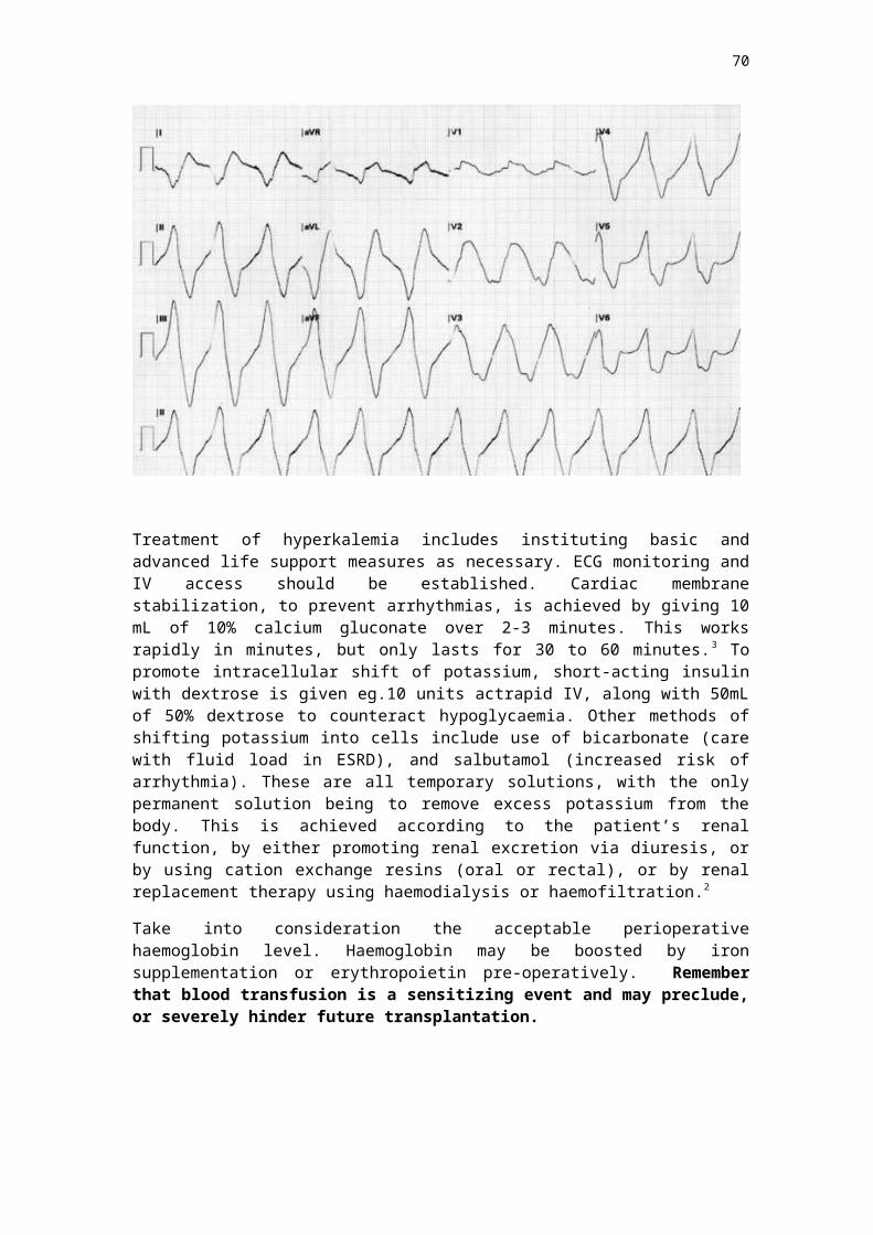
References
1. Editorial: Smoking and anaesthesia. Anaesthesia 1987 vol 42 pages 1-2.2. Editorial: Smoking and Anaesthesia revisited. Anaesthesia 1996, Vol 51, Pg 309-311.3. Møller, A.M. et al., 2002. Effect of preoperative smoking intervention on
postoperative complications: a randomised clinical trial. The Lancet, 359(9301), pp.114–117.
4. Rodrigo, C., 2000. The effects of cigarette smoking on anesthesia. Anesthesia progress, 47(4), p.143.
5. Webb, A., 2012. Smoking and surgery: time to clear the air. Australia Anaesthesia 2011.
6. Chimbira, W. & Sweeney, B.P., 2000. The effect of smoking on postoperative nausea and vomiting. Anaesthesia, 55(6), pp.540–544.
7. Lodewyckx M. 2014. PS12 Guidelines on Smoking as Related to the Perioperative Period. Australian and New Zealand College of Anaesthetists
8. Myers, K. et al., 2011. Stopping Smoking Shortly Before Surgery and Postoperative Complications. Archives of Internal Medicine, 171(11).
9. Thiagarajan N. Smoking and Anaesthesia. Anaesthesia Tutorial of the Week10. http://www.frca.co.uk/Documents/221%20Smoking%20and%20Anaesthesia.pdf 11. http://www.escapeartistes.com/2013/07/27/smoking-in-mongolia/ 12. http://www.wpro.who.int/mongolia/mediacentre/smoking/en/ 13. http://www.who.int/tobacco/surveillance/policy/country_profile/mng.pdf?ua=1 14. http://www.cdc.gov/tobacco/data_statistics/fact_sheets/health_effects/
tobacco_related_mortality/15. http://www.who.int/mediacentre/factsheets/fs339/en/ 16. http://www.cancerresearchuk.org/cancer-info/healthyliving/smoking-and-
cancer/smoking-and-cancer17. Canet, J. & Paluzie, G., 2013. Anaesthesia in smokers. Trends in Anaesthesia and
Critical Care, 3(1), pp.3–7.18. http://www.anzca.edu.au/communications/Media/pdfs/NAD%20Cover%20Letter
%20A4%20P5%20FINAL.pdf/view?searchterm=smoking
70
PERIOPERATIVE MANAGMENENT OF A PATIENT WITH A RENAL TRANSPLANT
As both the short and long term outcomes after renal transplantation continue to improve, an increasing number of recipients require medical care including elective or emergency surgery.
Although the principles of perioperative management are the same in transplant and non-transplant patients, there are a few important differences. The transplant centre or teams should be consulted prior to any elective surgical intervention.
GENERAL CONSIDERATIONS
The anaesthetist must integrate information from the history, physical examination, and laboratory tests to develop an initial estimate of perioperative risk and anaesthetic plan. Significant coronary artery disease could be a common finding among renal transplant recipients.
Adrenal suppression related to the higher maintenance doses of steroids used to be a more significant problem before but currently, with the advent of more potent maintenance immunosuppressive agents such as Cyclosporine, Tacrolimus, Mycophenolate mofetil, and Sirolimus, the issues are fewer. It is, however, likely to be a consideration for patients who are currently or recently on steroids.
SPECIFIC SURGICAL ISSUES
The following illustrates a few specific clinical considerations that may be particularly important in the transplant recipient.
Gastrointestinal surgical emergencies
Signs and symptoms of acute abdominal pathology may be masked in transplant patients due to chronic immunosuppressive therapy. They have high mortality rate (30%), which is principally attributed to immunosuppression and sepsis.
Acute appendicitis
The diagnosis of acute appendicitis may be particularly difficult in a renal transplant patient as the transplanted kidney is usually placed in the right lower quadrant. (It can however, also be placed on the left.) The appendix is generally displaced superiorly; thus, the symptoms and signs are uncharacteristic. The appendix is frequently perforated by the time of diagnosis.
Cardiac surgery
Cardiac surgery can affect a kidney transplant recipient in the following ways:
Perfusion of the allograft may be compromised with cardiopulmonary bypass. The ischemic insult may range from a minimal reversible rise in the serum creatinine concentration to complete shutdown of the organ.
In the unstable cardiac surgical patient who requires an intra-aortic balloon pump; placement of the pump on the side of the renal allograft can result in arterial thrombosis.
71
Abdominal aortic aneurysm
Surgical repair of an abdominal aortic aneurysm (AAA) routinely requires cross clamping of the abdominal aorta. To avoid interruption of the arterial blood supply to the transplanted kidney, temporary bypass may be required. It may not be necessary with the endovascular AAA repair.
IMMUNOSUPPRESSIVE DRUG SIDE EFFECTS AND INTERACTIONS
The immunosuppressive drugs commonly used in renal transplantation are cyclosporine, tacrolimus, azathioprine, mycofenolate mofetil and steroids.
Calcineurin inhibitors: Cyclosporine and Tacrolimus
The important side effects generally seen with long-term use are hypertension, hyperlipidemia, nephrotoxicity, neurotoxicity, and hepatotoxicity. Hypertension is due to increase in systemic vascular resistance and calcium channel blockers are preferred for its treatment. Nephrotoxicity is the major complication due to renal arteriolar vasoconstriction leading to reduction of glomerular filtration rate and creatinine clearance. Thorough neurological examination is important as these drugs contribute to tremors, seizures, and paresthesia. Documentation of paresthesia is important if regional anesthesia is planned.
Azathioprine and Mycofenolate mofetil (MMF)
The major complication is bone marrow suppression especially leucopenia and elevation of liver enzymes. Complete blood examination is necessary before surgery. Glucocorticoids
The major perioperative concern is adrenal suppression and the need for supplemental steroids. Side effects of glucocorticoids include sodium retention, hypertension, diabetes, peptic ulcer disease, Cushingoid syndrome, poor skin integrity, osteoporosis, and delayed wound healing.
Care of patients includes gentle handling to prevent skin damage and fractures, antacid prophylaxis, and thorough airway examination.
mTor inhibitors: Sirolimus and Everolimus
Impairs wound healing. If possible, avoid in the perioperative setting.
PERIOPERATIVE ISSUES
Pathophysiology
The immunocompetence of post transplant patients can be affected by direct and hormonal effect of anesthetic drugs, immunological consequences of other drugs used, type of surgery, and coincidental infection.
72

Exposure to anesthesia and surgery depresses both T cell and B cell responsiveness. The incidence of postoperative infection is related to surgical trauma and associated release of cortisol and catecholamines that inhibit phagocytosis. This hormonal response is mediated through the sympathetic nervous system, which can be attenuated by regional anesthesia or by deepening the plane of GA and by inhibiting the stress response.
The choice of anesthetic technique
It will depend on functional status of transplanted kidney, cardiovascular status, haematological status, and the site of operation. Central neuraxial blocks are not contraindicated. Renal transplant recipients with functioning kidney grafts may have normal creatinine levels. However, glomerular filtration rate and effective plasma flow are likely to be low, and drugs excreted via kidney alone should be avoided as much as possible.
Antibiotic prophylaxis
In general, post renal transplant patients may be considered as higher risk to develop infectious complications after surgical, endoscopic, or dental procedures. Prophylactic antibiotics are given within two-hour "window" period before the initial incision as it lowers rate of surgical site infection.
Two macrolides, erythromycin and clarithromycin, should be avoided in patient having cyclosporine or tacrolimus as these antibiotics antagonize the CYP 3A4 enzyme system, leading to elevated levels of cyclosporine and tacrolimus.
Infection Control
Strict aseptic precautions should be maintained during intravascular access, intubation or while performing regional techniques and the use of disposable accessories is recommended.
Drug Interaction
Immunosuppressive drugs can affect the pharmacology of many anesthetic drugs. Amongst intravenous anesthetic agents, propofol does not alter cyclosporine concentration. Amongst inhalational agents, enflurane should be avoided in patients with renal transplantation because of toxicity of fluoride metabolite. However, sevoflurane is thought to be safe. Choice of nondepolarizing muscle relaxant depends on renal status of patient. Drugs that rely least on renal elimination like atracurium are the optimal choice. The solubility agent of cyclosporine (cremaphor) has been shown to augment the action of neuromuscular blocking agents. Clinically relevant dose of azathioprine do not antagonize neuromuscular blocking drugs. Neuromuscular function should be monitored.
Fluid and Electrolyte
Meticulous perioperative fluid balance avoiding overload is essential. Hypotension can cause the transplanted kidney vulnerable to acute tubular necrosis. Special care must be taken to keep patient well hydrated and maintain urine output more than 1 ml/kg/h.
Postoperative pain management
Adequate post-operative pain control is achieved by using opioids and local anaesthesia by regional, epidural or spinal route or by parenteral opioids if general anaesthesia is given. Non-steroidal anti-inflammatory drugs should be avoided as they increase the risk of gastrointestinal bleeding, reduce renal blood flow through prostaglandin inhibition and exacerbate cyclosporine toxicity.
73
The glomerular filtration rate (GFR) may vary from > 60 ml/min/1.73m2 to severely reduced. The choice of analgesics should take into account the GFR. (See chapter on Chronic Renal Disease and Anaesthesia).
In conclusion, a clear understanding of physiological changes secondary to renal transplantation and changes caused by surgery and anaesthesia on renal allograft recipient would contribute for safe management of these patients.
References
1. Issues relating to the renal allograft recipient undergoing non-transplant surgery, Ron Shapiro, MD, Up to date; Sep 04, 2013
2. Anesthesia for parturient with renal transplantation, Beena K Parikh et al ,J Anaesthesiol Clin Pharmacol. 2012 Oct-Dec; 28(4): 524–527
3. Chas Newstead, Management of the peri-operative and critically ill renal transplant patient, Current Anaesthesia & Critical Care, Volume 21, Issue 2, April 2010, Pages 75-77
74
ANAESTHESIA FOR PATIENTS WITH MYASTHENIA GRAVIS & MULTIPLE SCLEROSIS
This chapter will focus on the neuromuscular disorders myasthenia gravis and multiple sclerosis. Patients with neuromuscular disease may develop multi-system complications as a result of the disease process. A clear understanding of the pathophysiology is needed in order to be aware of the impact on the anaesthetic management.
Myasthenia Gravis
Myasthenia gravis, an autoimmune disease, is the commonest disorder affecting the neuromuscular junction with an incidence of 1 in 10,000. Autoantibodies against the nicotinic acetylcholine (Ach) receptor in the post-synaptic membrane of the neuromuscular junction are present in about 90% of patients. These antibodies reduce the number of available receptors for effective neurotransmission (Figure 1).
Clinical features of the disease are weakness and rapid fatigability of striated muscle with repetitive use, followed by partial recovery with rest. Commonly affected muscles include extra-ocular and bulbar, whilst limb and truncal muscles are less commonly involved. Therefore, patients will often present with ptosis, diplopia and dysphagia. Eaton-Lambert syndrome, by contrast, is characterised by muscle weakness that improves on exertion, sparing ocular and bulbar muscles. It is associated with small-cell carcinoma of the bronchus and like myasthenic patients, these patients are particularly sensitive to non-depolarising muscle relaxants.
Women are affected twice as often as men and myasthenia gravis can occur at any age with a bimodal distribution. Women tend to be affected in their thirties whereas men are more likely to be affected in their sixties and seventies. The disease is classified according to severity (Table 1). About 75% of patients have an associated abnormality of the thymus gland and there is an association with other autoimmune conditions such as thyroid disease and pernicious anaemia (Table 2).
Anaesthetists may be asked to care for patients with myasthenia during surgery, especially for thymectomy, or in critical care when the patient may present in myasthenic crisis.
75
Figure 1. Antibodies at the post-synaptic membrane block acetylcholine receptors preventing nerve conduction.
Type Clinical Features
I
IIA
IIB
III
IV
Ocular signs and symptoms only
Generalised mild muscle weakness responding well to therapy
Generalised moderate muscle weakness responding less well to therapy
Acute fulminating presentation and/or respiratory dysfunction
Myasthenic crisis requiring artificial ventilation
Table 1. Clinical classification of myasthenia gravis
Muscle weakness
Bulbar / oropharyngeal - pooling of secretions and saliva
Respiratory obstruction and aspiration
Respiratory failure
Associated conditions
Thymus hyperplasia (75%), thymoma (10-25%) & other malignancies
Thyroid disease (3-15%)
Systemic lupus erythematosus (2%)
Rheumatoid arthritis (4%)
Ankylosing spondylitis
Crohn’s disease
Hypertension
Diabetes mellitus
Myocarditis, cardiomyopathy, dysrhythmias
Seizures
Table 2. Associated problems in patients with myasthenia gravis
Myasthenic and cholinergic crisis
Myasthenic crisis
This is a severe life threatening worsening of myasthenia gravis that can progress rapidly to respiratory failure requiring intubation and ventilation. A myasthenic crisis can be precipitated by any of the factors outlined in Table 3. The differential diagnosis of an acutely weak patient with myasthenia gravis includes a cholinergic crisis.
76
Cholinergic crisis
A cholinergic crisis is caused by an excess of Ach available at the neuromuscular junction and usually follows excessive administration of anticholinergics. Patients may present with respiratory failure and it may be difficult to differentiate from a myasthenic crisis. Patients may have an excess of secretions, abdominal pain, diarrhoea and blurred vision.
The differential diagnosis may be made by giving a small dose of edrophonium. Patients with a myasthenic crisis should improve whereas those with a cholinergic crisis will deteriorate.
Environmental / Other Drugs
Infection
Exertion
Menstruation
Emotional stress
Acute illness
Pain
Excessive heat / cold
Surgery / trauma
Under-dosing anticholinesterase medication
Malnutrition
Hyper- / Hypothermia
Antibiotics – aminoglycosides, tetracycline, erythromycin
Antiarrhythmics – -blockers, calciumβ channel blockers
Magnesium
-agonistsβ
Thyroxine
Penicilliamine
Lithium
Phenytoin
Diuretics – furosemide
Corticosteroids
Chloroquine
Table 3. Predisposing factors that exacerbate myasthenia gravis. (The benefit of taking any of the above drugs must be balanced with the risk of worsening myasthenia gravis symptoms.)
Investigations
Diagnosis is made by the clinical history and a combination of tests: Ach antibody assay, edrophonium test, and electromyography.
Edrophonium test
Classically, this test has been used to contribute to the diagnosis of myasthenia gravis in individuals demonstrating muscle weakness with exercise and improvement with rest. The diagnosis is supported when muscle strength improves after giving the drug edrophonium, a cholinesterase inhibitor, that increases the amount of Ach at the neuromuscular junction.
77
The test is rarely performed nowadays due to safety reasons. Muscarinic effects of Ach such as bradycardia, asystole and bronchoconstriction can occur and therefore full resuscitation facilities must be available.
Electromyography
Repetitive supramaximal motor nerve stimulation by electrical stimulation shows characteristic fade in amplitude of the evoked action potential in affected individuals.
Acetylcholine antibody assay
Serum Ach receptor binding antibodies are present in 85% of myasthenic patients. Negative results more likely to occur in those patients with mild or only ocular disease.
Treatment
Anticholinesterases
Anticholinesterases enhance neuromuscular transmission by delaying degradation of Ach at the neuromuscular junction. Pyridostigmine bromide four times a day is increased until an optimal response is achieved.
Anticholinergics
Anticholinergic medication is not used routinely but may be required to control side effects of anticholinesterase medication such as salivation, colic and diarrhoea.
Immunosuppressants
Patients with only ocular symptoms may benefit from corticosteroids. They can also be added when treatment with anticholinesterase therapy is suboptimal.
Plasma Exchange
This can be used to provide a short term, rapid response to symptoms for patients with severe myasthenic crisis or to allow weaning from ventilation.
Thymectomy
Thymectomy results in clinical improvement in about 80% of all myasthenic patients. It produces a rapid onset of remission and is associated with a lower mortality than medical therapy alone. Respiratory function should be optimised pre-operatively for example with plasma exchange or steroids. Pyridostigmine therapy should be reduced providing there is no compromise in respiratory function. This is because patients are more sensitive to anticholinesterases post-thymectomy whilst intraoperatively, neuromuscular blockade is easier to manage in the presence of a mildly myasthenic patient.
Avoidance of precipitating factors
The course of the disease is highly variable but usually progressive, although exacerbations and remissions do occur. Exacerbations may occur spontaneously or due to predisposing factors as outlined in Table 3.
78
Anaesthetic management
Pre-operative
Assessment of muscle function is essential. A useful test is to assess the patient’s ability to sustain a head lift off the pillow or generate a negative inspiratory pressure > 25cmH 2O. An arterial blood gas can be used to confirm adequate minute ventilation and provides a useful baseline. Arrangements should be made for post-operative ventilation for patients with a forced vital capacity < 2.9L, a long history of the disease or severe disease with types III or IV myasthenia gravis. An ECG should be recorded as there are reports of myocardial necrosis.
Patients should be assessed for other autoimmune conditions such as thyroid dysfunction, which can be present in up to 15% of patients (Table 2). A careful airway assessment is also required especially if the patient has rheumatoid arthritis or a large thymoma.
It is controversial whether anticholinesterase medication should be continued or discontinued pre-operatively. Patients are at risk of vagal responses, inhibition of plasma cholinesterase (and thus prolongation of ester local anaesthetics and succinylcholine) and possible cholinergic crisis when the medication is continued. However, it is better to continue for patients physically or psychologically dependent or those who have more than ocular symptoms.
Corticosteroids should be continued with consideration made to an additional dose of hydrocortisone. Antimuscarinic drugs, such as glycopyrronium can help reduce secretions. Sedative premedication is safe providing the patient has no respiratory compromise but saturation monitoring is essential.
It is useful to perform a control train-of-four to assess pre-operative muscle strength before administering anaesthetic drugs.
Intra-operative
Routine monitoring is needed but specifically if a muscle relaxant is used then neuromuscular transmission must be monitored. Invasive blood pressure monitoring is indicated for patients undergoing a thymectomy via a median sternotomy incision.
Local and regional anaesthesia should be used where possible as they are less likely to cause opioid-induced respiratory depression, although, when intrathecal and epidural opioids are used, patients ought to be monitored for post-operative respiratory depression. Ester local anaesthetics may have a prolonged half-life due to decreased cholinesterase activity in patients on anticholinesterase medication. This increases the risk of local anaesthetic toxicity or extensive epidural block. Therefore, amide local anaesthetics are preferred.
If general anaesthesia is chosen, almost all patients will require tracheal intubation and assisted ventilation. Ketamine, thiopentone and propofol have been used successfully for induction of general anaesthesia in these patients. An anaesthetic technique should be chosen that minimises or avoids the use of neuromuscular blocking drugs. If a non-depolarising drug is used, atracurium or cisatracurium are the preferred drugs using one tenth to one quarter of the usual dose. The response must be monitored with a nerve stimulator. Neuromuscular function should be allowed to return spontaneously as the administration of reversal agents may cause confusion in the presence of
79
continued weakness. The pharmacological considerations in relation to the anaesthetic technique are outlined in Table 4.
Drug Interaction Comments
Non-depolarising neuromuscular blocking agents
Marked sensitivity Avoid use if possible. Start with 10% normal dosage. Always monitor neuromuscular function. Use short-and intermediate-acting agents only
Suxamethonium Resistance to depolarisation and delayed onset of action
No reported clinical adverse effects using 1.5mg/kg. Delayed recovery in patients with induced esterase deficiency (plasmapheresis anticholinesterase treatment). Follow with non-depolarising agents only when full recovery of neuromuscular function seen
Inhalational anaesthetics
All inhalational agents reduce neuromuscular transmission by up to 50%
Avoid need for neuromuscular blocking agents
IV anaesthetics No apparent clinical effect on neuromuscular transmission
Total IV anaesthesia with propofol may be useful if neuromuscular function unstable
Local anaesthetics
Prolonged action and increased toxicity in ester-linked (e.g. amethocaine) agents with anticholinesterase therapy and plasmapheresis. Exacerbation of myasthenia reported
Use minimum dosage required for adequate block. Monitor respiratory function as with general anaesthesia
Drugs dependent on esterases for elimination
Prolonged effect and increased toxicity if patient on plasmapheresis or (theorectically) anticholinesterase therapy
Suxamethonium, remifentanil, mivacurium, ester-linked local anaesthetics, esmolol etc…
Antibiotics Neuromuscular blocking effects may become clinically important
Avoid aminoglycosides (e.g. gentamicin). Similar effects reported with erythromycin and ciprofloxacin
Miscellaneous All the following agents have a reported effect on neuromuscular transmission: procainamide, -blockers β(especially propranolol), phenytoin, magnesium
Table 4. Specific drugs of interest in myasthenia gravis.
80

Postoperative
If the patient’s symptoms are well controlled pre-operatively, it is usually safe to extubate at the end of the procedure. High risk patients should be managed in a high dependency unit. Adequate analgesia is required to prevent an exacerbation of myasthenia secondary to pain, both non-steroidal anti-inflammatory drugs and opioids are safe to use. Anticholinesterase medication should be restarted as soon as possible post-operatively, initially with a smaller dose until the patient is able to ambulate when they can resume their normal dose.
Multiple Sclerosis
Multiple sclerosis is a chronic disease characterised by relapsing and remitting exacerbations. The condition is more common among women of child bearing age with 70% of presentations occuring between the ages of 20 and 40 years and nearly 10% of cases presenting initially in pregnancy.
In 20-30% of cases, the disease progression is benign, but in 5%, affected individuals deteriorate rapidly.
Demyelination of white matter occurs in the brain and spinal cord leading to retrobulbar neuritis, and upper motor neuron and sensory deficits (Figure 2). Ocular, cerebellar and bladder dysfunction are also common. Respiratory failure occurs in end-stage of disease. Symptoms may be exacerbated by stress, pyrexia, infection, trauma and exertion.
Figure 2. Demyelination of white matter interfers with passage of nerve impulse.
81
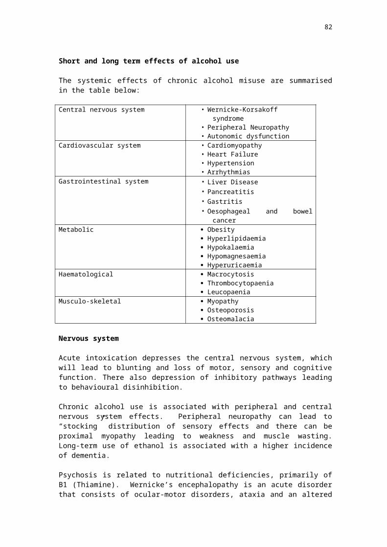
Figure 3. Proposed immune mediated mechanism causing demyelination.
The aetiology is unclear, with environmental, genetic and immunological factors suspected (Figure 3). The incidence varies dramatically with geographical latitude and is most common in Europe, North America and New Zealand.
MRI, with gadolinium enhancement, demonstrates plaques of demyelination and has replaced CSF gamma-globin and visual evoked responses as the best investigation.
Treatment
The condition is not curable but steroids and interferon can prolong the duration between symptomatic episodes. Baclofen and dantrolene are useful to relieve painful muscle spasms.
Anaesthetic management
Pre-operative
A thorough clinical history is required, in particular, evaluation for potential bulbar palsy when aspiration becomes a risk. A detailed neurological examination is also required to enable post-operative comparison. The patient’s respiratory status must also be assessed and if there are concerns, facilities for post-operative venilation should be available.
Intra-operative
Cardiovascular monitoring is essential as patients are at risk of autonomic instability and sensitive to hypovolaemia. Introperative hypotension, for example due to spinal anaesthesia, may be resistant to vasopressors but must be aggressively managed to prevent further spinal cord injury secondary to ischaemia.
General anaesthesia
There is no evidence that general anaesthesia has adverse effects on multiple sclerosis. If laryngeal and pharyngeal muscles are involved, a rapid sequence induction is indicated. In severe cases with muscle wasting, up-regulation of nicotinic Ach receptors occurs, such that succinylcholine should be avoided due to the risk of hyperkalemia. Non-depolarising neuromuscular blocking drugs may be used in normal doses providing the patient does not have a disability where reduced doses are recommended.
82
Temperature maintence is important as hyperthermia may exacerbate the neurological deficit whilst hypothermia may delay recovery. A temperature rise of 0.5C may cause significant deterioration and therefore, anticholinergics should be avoided as they may increase temperature.
Regional anaesthesia
There has previously been a reluctance to provide regional anaesthesia to patients because of a concern regarding the neurotoxicity of local anaesthetics. Spinal anaestheia may expose demyelinated areas of the spinal cord to the potential neurotoxic effects of local anaesthetics, but there is no evidence that this is a real risk. The concentration of local anaesthetic in the subarachnoid space is lower with epidural anaesthesia and therefore theoretically is less of a risk unless multiple doses are administered.
A survey of UK anaesthetists in 2006 demonstrated that many are willing to proceed with regional anaesthesia providing that the patient understands the potential for relapse and is carefully followed up. A combined spinal-epidural technique allows for the minimal use of local anaesthetic in the subarachnoid space and augmentation of anaesthesia if necessary.
Specifically in relation to obstetric anaesthesia, the multiple sclerosis sufferer must be carefully assessed in the postpartum period as they may interpret symptoms such as paraesthesia or bladder dysfunction as a relapse. The Pregnancy In Multiple Sclerosis (PRIMS) study reported a small reduction in relapse rates during preganncy but a significant increase in relapse in the first three months postpartum. This was regardless of anaethetic technique or mode of delivery. Of note, there was no difference in relapse rates between multiple sclerosis patients who received epidural analgesia and those who had not.
Post-operative
Good pain control is required post-operatively since symptoms may be exacerbated by stress. High risk patients for respiratory compromise should be managed in a high dependency or intensive care unit. If the patient has received regional anaesthesia careful follow up is required.
Summary
Patients with neurological disease must be thoroughly assessed for complications and associated co-morbidities. Each case must be considered on an individual basis. Successful management often requires multidisciplinary assessment and planning with consideration of the impact of the neurological condition on the safe provision of anaesthesia.
83
References
1. Drachman D. Myasthenia Gravis. N Engl J Med 1994; 330: 1797-1810.2. Allman K, Wilson I. Ch 11: Neurological and muscular disorders. Oxford Handbook of
Anaesthesia, 3rd Edition, Oxford University Press, 2011.3. Gambling D, Douglas M, McKay R. Obstetric Anaesthesia and Uncommon Disorders, 2nd
edition, Cambridge. Cambridge University Press, 2008. Section 3: Nervous System Disorders, Ch 9: 167-89.
4. Drake E, Drake M, Bird J, Russell R. Obstetric regional blocks for women with multiple sclerosis: a survey of UK experience. Int J Obstet Anesth 2006: 15: 115-23.
5. Confavreux C, Hutchinson M, Hours M, Cortinovis-Tourniaire P, Moreau T. For the Pregnancy in Multiple Sclerosis Group. Rate of pregnancy-related relapse in multiple sclerosis. N Engl J Med 1998; 339: 285-91.
84
STROKE
Stroke is defined as an ischaemic/embolic or haemorrhagic cerebrovascular event that impairs neurologic function. Perioperative stroke is a stroke that occurs in the period up to thirty days after surgery.
Transient Ischaemic Attack (TIA) occurs when there is a change to the blood supply of an area of the brain resulting in a change in neurologic function lasting for less than 24 hours. If the neurological dysfunction persists for up to 72 hours, but resolves, this is termed a Reversible Ischaemic Neurological Defect (RIND). When the neurological dysfunction persists for more than 72 hours, it is categorised as a stroke.
Introduction
The incidence of stroke in the general population is 1.5-3 per 1000. Perioperative stroke in the non-vascular surgery population occurs in 0.08-4 per 1000. The risk is higher for the neurosurgical, cardiac surgical, carotid surgery and other major head and neck surgery patients. Stroke is associated with a high rate of morbidity and mortality. The estimated 30-day fatality rate after a primary ischaemic stroke ranges from 16-23%. In the perioperative stroke patient, it is 18-26%.
The majority of strokes associated with surgery will occur in the postoperative period when the patient is in a hypercoagulable state. The peak incidence is on day 7 and is associated with postoperative hypotension and the occurrence of atrial fibrillation.
Risk factors
The risk factors for ischaemic stroke in the surgical population are similar to those in the general community. They include increased age (over 62), male gender, hypertension, diabetes, renal disease, cardiac and vascular disease, smoking and respiratory disease. The most consistent among these are advanced age, renal failure and a history of stroke or TIA. Cardiac surgery, carotid and neurosurgery and a previous stroke or myocardial infarct are important additional risk factors for perioperative stroke.
Risk per year (%)
Atrial Fibrillation 2-18
85
Risk per year (%)
Hypertension 2-3
Smoking 1-2
Diabetes 1-2
Cardiovascular disease 1-2
Hypercholesterolaemia Not significant
Carotid stenosis >70% 1-2
86
Mechanisms
Stroke can be largely grouped into ischemic or haemorrhagic stroke. Ischemia is the mechanism for most strokes. Ischaemic stroke is further broken down into large vessel disease (artery to artery embolism, which accounts for 30% of strokes), cardio-embolic (20%), small vessel disease (15%), other causes include rare conditions such as arterial dissection, venous thrombosis and vasculopathy (5%). In 15% of ischemic strokes, the mechanism is not known. Haemorrhagic strokes are grouped into intracerebral haemorrhage (which accounts for 10% of strokes) and subarachnoid haemorrhage (5% of strokes).
The mechanism of perioperative stroke is similar to the non-surgical patient. The largest proportion is due to ischaemia. Cerebral arterial thrombosis, low flow states, cardioembolism, embolism of air, fat and cement and dissection of neck arteries are considered to be important causes of stroke in the perioperative period. Haemorrhage, anaemia and hypotension are believed to be significant contributing factors to the occurrence of stroke. Intraoperative hypotension is important but is not the most frequent cause of stroke. It will certainly worsen the outcome of ischaemic stroke if it occurs due to compromised flow to what is known as the "ischaemic penumbra", an area in the periphery of affected brain that is at risk of cell death if perfusion is not restored and remains compromised.
Patients undergoing aortoiliac surgery are especially vulnerable due to generalized atherosclerosis and intraoperative hypotension, and can have a perioperative stroke rate of up to 1 percent.
Prevention and anaesthetic management of patients with previous stroke
Ischemic cerebral tissue will have impaired auto-regulation and patients who have suffered a recent stroke will be at further risk of extension of that stroke or conversion to haemorrhagic stroke. It is not known how long one should delay surgery after a stroke but it would be reasonable to wait for 3 months before performing elective surgery. In more urgent situations, it is reasonable to wait 2-3 weeks, but the urgency of surgery must be balanced against the risk of further stroke. A Danish cohort study by Jorgensen reported that this risk decreased from time of stroke, with elevated risk leveling off at 9 months.
In contrast, the patient who has suffered transient ischaemic attacks due to carotid artery disease is at particularly high risk of suffering a stroke within 2 weeks and should undergo endarterectomy within two weeks, possibly within the first three days of a TIA.
The patient with non-valvular atrial fibrillation on anticoagulation will require assessment with regard to his stroke risk to determine the risk of discontinuation of his anticoagulants prior to surgery. In minor surgery, it is recommended that oral anticoagulation be continued. For major surgery in a patient on warfarin, the risks of haemorrhage and stroke need to be considered. Some of these patients will require "bridging" anticoagulation with unfractionated or low molecular weight heparin in the perioperative period. Most of the data are based on observational studies, and it is uncertain to what degree antiplatelet or anticoagulation therapy should be ceased prior to surgery.
Commencement of a beta blocker or cholesterol-lowering statin (HMG-CoA reductase inhibitor) is not recommended to avoid perioperative stroke, but if the patient is already
87
receiving therapy with either a beta blocker or statin, then it is recommended that these be continued in the perioperative period. There is an association between the administration of intraoperative intravenous metoprolol and perioperative stroke. Beta-blockers with a short duration of action such as esmolol should be used intraoperatively to manage hypertension and/or tachycardia.
If a patient is considered to be at increased risk of stroke, it may be possible to minimise risk of harm resulting from cerebral ischaemia. The choice of medication for anaesthesia has not been conclusively proven to alter outcome. There is weak evidence that propofol and the use of epidural anaesthesia will reduce hypercoagulability in the perioperative period and therefore reduce stroke extension. A regional technique is recommended for the patients undergoing hip or knee replacements. It may be more important to closely monitor the patient and provide good basic care.
The maintenance of cerebral perfusion is important, as is the maximization of oxygen delivery. Intraoperative blood pressure management for patients at risk of stroke will involve close monitoring via direct invasive blood pressure monitoring and the avoidance of a drop in blood pressure below 30% of baseline. Care must be taken to avoid hypertension, as uncontrolled hypertension will increase the risk of cerebral oedema and intracranial haemorrhage.
The POISE study revealed that the commencement of metoprolol before surgery in those considered at risk of cardiac events improved cardiac outcome but at the expense of increased mortality from stroke and an increase in intra and postoperative hypotension.
Hyperglycaemia and hypoglycaemia will worsen the outcome if a patient suffers a stroke. Intensive glycaemic control is not beneficial and may increase the risk of hypoglycemia. Hyperthermia is also undesirable, so temperature monitoring is required.
Hyperventilation causes cerebral vasoconstriction and it is best to ventilate the patient to normal carbon-dioxide levels to avoid reduction in cerebral perfusion.
Oxygen delivery is important and monitoring with pulse oximetry will help avoid hypoxaemia. Severe anaemia will impair oxygen delivery and should be managed with transfusion to levels of 9g/dL.
Stroke patients usually have reduced mobility and bulbar dysfunction. They are therefore at risk of deep venous thrombosis (DVT) and aspiration after extubation. Calf compression devices and prophylactic heparin will be required to minimize the risk of DVT.
Poor nutritional intake following the stroke may lead to malnutrition and may be associated with poor survival and functional outcomes after the surgery. Electrolyte abnormalities are commonly associated with strokes and may lead to dysrhythmias both peri- and post-operatively.
Close clinical monitoring in the postoperative period will be required in order to detect neurologic complications.
Discovery of an asymptomatic carotid bruit happens quite commonly, in up to 4 percent of the population older than 40 years. A bruit alone is a poor predictor of underlying carotid stenosis. An increase in perioperative stroke has not been demonstrated in patients with asymptomatic carotid bruits undergoing non-cardiac surgery.
88
As a result, further evaluation of the carotid arteries in patients with an asymptomatic carotid bruit is not warranted.
Carotid artery disease in patients undergoing coronary artery bypass grafting has been the subject of considerable study. A carotid Doppler study appears to be warranted in patients undergoing coronary artery bypass grafting (CABG) who have a carotid bruit because of the reduced risk of combined CEA and CABG compared with CABG alone with severe carotid stenosis (>70-80 percent). Patients with a history of stroke or TIA should also undergo a noninvasive evaluation of the carotid arteries prior to CABG.
Diagnosis
The diagnosis of intraoperative and early postoperative stroke is challenging as the classical signs are masked by impaired consciousness and medications given for analgesia. A diagnosis of stroke is considered in the patient who experiences prolonged emergence, an altered conscious state, delirium or new focal neurologic signs. The patient at high risk will require regular clinical assessment by experienced staff. Several parameters are considered, including the presence of facial asymmetry, abnormal speech, and sensory and motor signs in the limbs.
The patient at risk will require closer physiologic monitoring in order to reduce the risk of and reduce harm from cerebral ischaemia. Blood pressure monitoring to detect hypotension and damaging severe hypertension is important, as is oxygenation, temperature monitoring, and blood count and coagulation studies.
Once a stroke is suspected clinically, an urgent CT or MRI scan is required. Early reperfusion is required if there is no contraindication. The differential diagnosis of stroke includes tumour, subdural haematoma, migraine, hypoglycaemia, post-ictal paralysis and cerebral abscess. In the postoperative period, shock, drug effects, hypoxia and electrolyte abnormalities can be added to this list. It is important to consider these, particularly hypoglycaemia in the postoperative period, as it requires urgent management.
Management of acute stroke
Stroke induces ischaemia, necrosis and apoptosis of brain that suffers infarction. It is useful to consider a larger surrounding area around the central infarction that may be suffering reversible ischemia or a change in perfusion as a result of an interruption to blood supply. This is termed the "ischaemic penumbra". In an ischemia stroke, this area may be salvaged by restoration of blood flow. This is the rationale for thrombolytic therapy in the first three hours after symptoms appear.
Thrombolysis can reduce the size of a stroke and therefore the degree of disability and mortality. It is not without risk and indeed, 6% of patients treated will suffer haemorrhagic conversion and will suffer a worse outcome. The risk of bleeding increases if reperfusion occurs longer than 3.5 hours after the onset of symptoms.
Patients who are treated in a specialised stroke unit will have an improved outcome compared with those who are managed in an ordinary ward. Stroke patients require specialised nursing care and extensive rehabilitation. Early mobilization, physical therapy, speech therapy and swallowing assessment is required to prevent the major complications of stroke.
89
In the immediate post stroke period the priority is to thrombolyse if appropriate, so CT imaging is required to exclude haemorrhagic stroke. The administration of recombinant Tissue Plasminogen Activator (rTPA) is relatively contraindicated after surgery, so consultation with the surgeons is required to discuss the risk of bleeding. Neurosurgery and spinal surgery are the most risky and the patient who has had such surgery is not likely to benefit from thrombolysis, and may suffer great harm from uncontrolled bleeding into the central nervous system. An alternative to pharmacological thrombolysis is mechanical thrombolysis. Post-thrombolysis, blood pressure should be managed as hypertension is undesirable – aim for systolic blood pressure < 180 mmHg and diastolic blood pressure < 105 mmHg. Aspirin as an adjunctive therapy is not recommended in the first 24 hours post thrombolysis.
An early neurology consultation is also required. The patient with an acute stroke is at risk of arrhythmia in the early period and will benefit from cardiac monitoring as well as support of his oxygenation and circulation. Management falls into two phases: protection of the brain and early reperfusion, as discussed followed by the prevention of further neurologic injury by supporting cerebral perfusion, maintaining normothermia (hyperthermia will worsen injury), normalizing blood glucose and maintaining adequate oxygenation.
The airway may be at risk if there is a depressed conscious state. If the GCS is below 8, intubation and ventilation will be required. A normal arterial carbon dioxide level is desirable and hypoxia needs to be treated with oxygen therapy.
Cerebral autoregulation will be reduced in the area of and surrounding the infarct. Increased intracranial pressure may occur due to cerebral oedema. Hypotension will require treatment to increase cerebral perfusion pressure. The recommended cerebral perfusion pressure will be 90-130 mmHg and this may require the use of vasopressors to support the circulation. General measures to reduce cerebral oedema are recommended including slight head up positioning, avoidance of extremes of neck flexion and rotation, controlling seizures, sedation if the patient is intubated and avoidance of tracheostomy tapes that cause compression of venous return from the head.
Severe hypertension however, will lead to increased intracranial pressure and puts the patient at risk of intracerebral haemorrhage. Hypertension needs to be addressed and treated, particularly for a systolic pressure above 180 mmHg and a diastolic pressure above 105 mmHg.
If thrombolysis is contra indicated or not available, the administration of aspirin is recommended within 48 hours of the stroke.
Seizures can occur and need to be treated as prolonged seizures increase cerebral metabolic rate of oxygen consumption. Options include benzodiazepines (diazepam, clonazepam), phenytoin, phenobarbital, valproate and levetiracetam.
Subarachnoid haemorrhage (SAH)
Subarachnoid haemorrhage typically occurs after the rupture of a cerebral aneurysm. The presentation is usually one of sudden onset of headache with or without altered conscious state. The cause of the headache and neurologic deterioration is a sudden and severe increase in intracerebral pressure. The degree of neurologic impairment and reduction in consciousness will determine the outcome.
90
The classification of subarachnoid haemorrhage by the World Federation of Neurological Surgeons rates the severity of SAH.
Grade Glasgow Coma Score
Focal Deficit Prognosis - Poor Outcome
1 15 - 14.8%2 13-14 - 29.4%3 13-14 + 52.5%4 7-12 +or - 58.3%5 3-6 + or - 92.7%
Only 58% of patients admitted within 3 days of subarachnoid haemorrhage have a good outcome. This proportion may be increased with surgery to clip the aneurysm; however there are several problems the patient faces after SAH. They include; vasospasm, recurrent bleeding and hydrocephalus.
Vasospasm occurs in response to extravascular blood and occurs several days after a subarachnoid haemorrhage. It can lead to focal neurologic deficit and ischaemia. In the acute phase, it may be treated with nitroprusside and nitroglycerin. Nimodipine is also used. It is a calcium channel blocker and has the potential to cause hypotension by reducing systemic vascular resistance. It will increase cardiac output. Hypervolaemia is another strategy to prevent vasospasm. It is achieved with the infusion of crystalloid and albumin and a central pressure of 10-12mmHg is targeted.
Recurrent bleeding occurs in 20-30% of patients within 2 weeks of SAH and can be prevented with surgical clipping of the aneurysm.
Preoperative concerns for a patient presenting for surgical clipping of a cerebral aneurysm include:
1. Assessment of the neurologic state.This is done with the grading scale WFNS. The worse the grade of SAH, the worse the rise in intracerebral pressure, degree of disruption of cerebral autoregulation and carbon dioxide responsiveness
2. Cardiac arrhythmias and ECG abnormalitiesBleeding will increase sympathetic nervous system activation and cause QT prolongation, T wave inversion, ST depression and U waves on the ECG. The patient is at risk of arrhythmia and there can be true cardiac ischaemia. Close monitoring is required, but surgery should not be delayed unless there is a reduction in cardiac contractility.
3. Intravascular volume statusWith the use of hypervolaemia to reduce vasospasm, hypovolaemia is less of a problem, but overload and pulmonary oedema is possible, as is the syndrome of inappropriate anti-diuretic hormone release.
4. The use of calcium channel blockers to reduce vasospasmHypotension is possible in the intraoperative period, but can generally be managed with fluids and vasopressors such as phenylephrine.
Premedication of patients presenting for clipping of cerebral aneurysm may be required. The aim is to reduce hypertension resulting from high anxiety levels, but the use of sedative agents may compromise respiratory function and worsen hypoxia and hypercarbia, so the use of anxiolytics is restricted to those with a WFNS grade of 1-2 and with strict monitoring.
91
Intraoperative monitoring is achieved with standard monitoring and the addition of arterial line monitoring, as variation in blood pressure is expected.
The transducer is kept at the level of the brain, rather than the heart. A urinary catheter will allow for urine output monitoring intraoperatively, although the use of mannitol and frusemide may interfere with accurate volume status monitoring. Central venous pressure monitoring will be necessary for more accurate volume status monitoring and for the administration of infusions.
One of the greatest risks of aneurysm surgery is the risk of rupture intraoperatively. The control of blood pressure is essential to avoid rupture and deliberate hypotension may be required for surgical access. Aneurysmal rupture may also lead to large blood loss.
Induction of anaesthesia needs to be controlled. The stress response to intubation and neurosurgical pin insertion will cause hypertension and risk rupture of the aneurysm. Maintaining an adequate depth of anaesthesia and the use of high dose opioid, such as fentanyl, controls blood pressure. Similarly extubation and emergence needs to be controlled to avoid coughing and blood pressure surges. An opioid and intravenous lignocaine are options to consider at this time.
Surgical access to deep aneurysms can be difficult. Controlled hypotension (aiming for a mean arterial pressure of 70 mmHg followed by 50 mmHg during direct manipulation of the aneurysm) is desirable. In some patients, the surgeon will place a temporary clip on a feeding blood vessel to allow for a higher blood pressure, but this will risk ischaemia if left on for long periods of time. The time allowed for a temporary clip is 20 minutes, after which ischaemia may occur, leading to stroke. Mannitol is another option for reducing intracerebral volume and therefore pressure, so allowing better surgical access.
Intraoperative haemorrhage occurs in 15-20% of cases and needs to be planned for. The consequences of haemorrhage due to aneurysmal rupture include death, haemorrhagic shock and most importantly, cerebral ischaemia. The cerebral ischaemia occurs due to several factors including surgical retraction and prolonged hypotension. Haemorrhage is managed by increasing the intravenous infusion rate (via large bore intravenous access), allowing for a temporary reduction in mean arterial pressure to 50 mmHg (measured at the level of the brain), maintenance of normovolaemia and sometimes, the placement of a temporary clip or manual compression of the carotid arteries.
A smooth but prompt emergence from anaesthesia is sought to allow for a rapid return to normal consciousness and assessment of neurologic state.
Focal deficit after surgery can occur due to cerebral vasospasm. This can occur even after clipping of the aneurysm. It is managed with hypertensive therapy and hypervolaemia, after scans have ruled out a bleed. The aim for blood pressure is 160mmHg systolic and this can be achieved with a vasopressor infusion such as dopamine. The aim for central venous pressure of 10-12mmHg is achieved with infusion of crystalloid and albumin. Hetastarches are not recommended as they can interfere with coagulation. Hypervolaemic therapy can be complicated by pulmonary oedema, which will worsen outcome, as the patient will become hypoxaemic without treatment.
92
References
1. Macellari, F et al. Perioperative Stroke Risk in Nonvascular Surgery. Cerebrovasc Dis 2012; 34:175-181
2. Mashour GA et al. Perioperative Care of Patients at High Risk for Stroke during or after Non-Neurologic Surgery: Consensus Statement from the Society for Neuroscience in Anesthesiology and Critical Care. J Neurosurg Anesthesiol 2014
3. Engelhard K. Anaesthetic techniques to prevent perioperative stroke. Curr Opin Anesthesiol 2013, 26:368-374
4. eTherapeutic Guidelines. Cardiovascular 2014.5. Morris John & Burkey Dell. Evaluation of the patient with neuropsychiatric disease
Ch 10 in Longnecker DE et al Anesthesiology, McGraw Hill, New York, 2008.6. Todd M, Warner DS, Maktabi MA, Vagnerova K. Neuroanesthesia Ch 50 in Longnecker
DE et al Anesthesiology, McGraw Hill, New York, 2008.7. Annemarie W. van Heuven, MD; Sanne M. Dorhout Mees, MD; Ale Algra, MD; Gabriel
J.E. Rinkel, MD. Validation of a Prognostic Subarachnoid Hemorrhage Grading Scale Derived Directly From the Glasgow Coma Scale. Stroke. 2008;39:1347-1348.
8. Merli, GJ and Bell, RD. Perioperative care of the surgical patient with neurologic disease. Available from http://www.uptodate.com/contents/perioperative-care-of-the-surgical-patient-with-neurologic-disease. Accessed 1/4/15
9. Jorgensen ME, Torp-Pedersen C, Gislason GH, et al. Time elapsed after ischemic stroke and risk of adverse cardiovascular events and mortality following elective noncardiac surgery. JAMA 2014; 312:269
93
SEPSIS
Introduction
Severe sepsis and septic shock affects millions of people around the world each year. The incidence in the USA and UK is 66–132 per 100 000 respectively, with mortality of 25 – 40%.1,15 In developing countries the burden of sepsis is understudied, but likely to be high. Ninety percent of the worldwide deaths from pneumonia, meningitis or other infections occur in less developed countries.15 The recently updated ‘Surviving Sepsis’ guidelines have defined the standard of care for patients with severe sepsis in the developed world, but do not incorporate the realities of health care in resource-constrained settings. The following aims to present a guide to management of severe sepsis and septic shock in general terms, and in the perioperative setting as current best evidence shows, and also to present where possible some alternative strategies which have been used where resources have been limited.
Definitions:
Sepsis: The presence (probable or documented) of infection together with systemic manifestations of infection (some of the following): 1
General Variables:
Fever (>38.3°C) or Hypothermia (<36°C)
Heart rate >90/min Tachypnoea Altered mental status Significant oedema / positive fluid
balance (>20ml/kg over 24hrs) Hyperglycaemia (plasma glucose
>140mg/dL or 7.7 mmol/L in the absence of diabetes)
Inflammatory Variables
Leukocytosis (WBC count > 12,000/µL), or
Leukopenia (WBC count <4,000/µL), or greater than 10% immature forms.
Plasma CRP > 2 SD above the normal value
Plasma procalcitonin more than 2 SD above the normal value.
Haemodynamic Variables:
SBP < 90mmHg, MAP < 70mmHg or an SBP decrease > 40mmHg
Organ dysfunction Variables:
Arterial hypoxaemia (PaO2/FiO2 < 300) Acute oliguria (urine output <
0.5ml/kg/hr for at least 2 hrs despite adequate fluid resuscitation.)
Creatinine increase >0.5mg/dL, or 44.2µmol/L
Coagulation abnormalities (INR >1.5 or aPTT > 60sec)
Ileus (absent bowel sounds) Platelet count < 100,000/µL Plasma total bilirubin > 4mg/dL or
70µmol/L
Tissue Perfusion Variables:
Lactate > 1 mmol/L
Decreased capillary refill, or mottling
WBC = white blood cell, CRP = C-reactive protein, SBP = systolic blood pressure, MAP = mean arterial blood pressure, INR = international normalised ratio, aPTT = activated partial thromboplastin time.
94
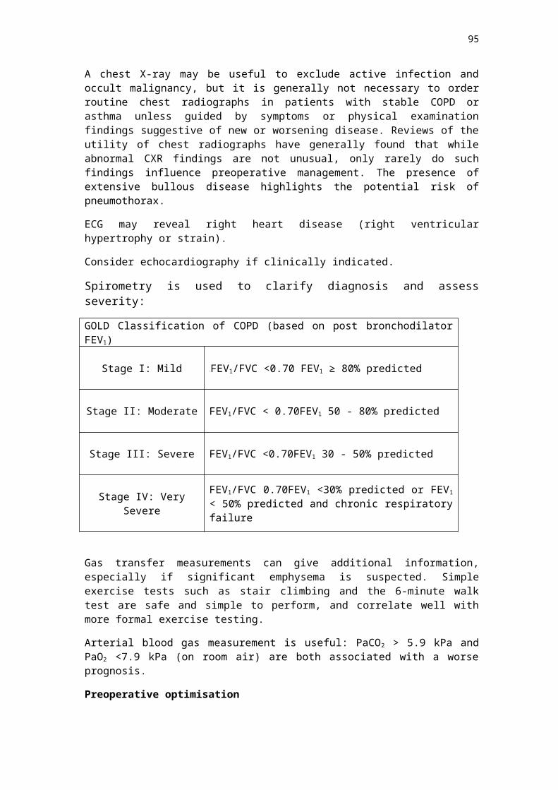
Severe sepsis: Sepsis plus sepsis-induced organ dysfunction or tissue hypoperfusion (any of the following):1
Sepsis-induced hypotension
Lactate above upper limits of laboratory normal
Urine output <0.5mL/kg/hr for more than 2 hrs despite adequate fluid resuscitation
Acute lung injury with PaO2/FiO2 < 250 in the absence of pneumonia as infection source
Acute lung injury with PaO2/FiO2 < 200 in the presence of pneumonia as infection source
Creatinine > 2.0 mg/dL (176.8 mol/L) μ Bilirubin > 2mg/dL (34.2µmol/L)
Platelet count <100,000/µL
Coagulopathy (INR > 1.5)
Sepsis-induced hypotension: A systolic blood pressure (SBP) < 90 mm Hg or mean arterial pressure (MAP) < 70 mm Hg or a SBP decrease > 40 mm Hg in the absence of other causes of hypotension.
Septic shock: Sepsis-induced hypotension persisting despite adequate fluid resuscitation.
Sepsis-induced tissue hypoperfusion: Infection-induced hypotension persisting after initial fluid challenge, elevated lactate, or oliguria.1
Causes
The causes of sepsis are many and beyond the scope of this document. In summary, causative organisms may be bacterial, viral, fungal, or parasitic. Different organisms may be implicated in community acquired versus nosocomial infections, and in different regions around the globe. Many host factors for example immune competence will influence the cause and presentation of sepsis. Any organ system may be affected including central nervous system, cardiac, pulmonary, urinary, gastro-intestinal, gynaecological, or musculoskeletal.
Perioperative management of severe sepsis and septic shock
Assessment
A systematic examination is required to identify the source of sepsis. The primary focus may be obvious (e.g. recent surgery, trauma), or more difficult to identify (e.g. pancreatitis, gynaecological sepsis, osteomyelitis), particularly in agitated patients. Diagnostic imaging to confirm site of infection may be required where possible. It is important that therapeutic and resuscitation measures are not interrupted while this takes place.
The pre-operative examination should focus on the severity of the systemic manifestations of sepsis, intravascular volume status, presence of shock or multi-organ dysfunction and the adequacy of haemodynamic resuscitation.2
95
Symptoms and signs of low intravascular volume status and/or tissue hypoperfusion include altered mental state, decreased capillary refill, tachycardia, hypotension, tachypnea, hypoxia, gastrointestinal disturbance, oliguria (urine output < 0.5ml/kg/hr). There are many biochemical derangements associated with severe sepsis. Serum lactate is most widely used as a marker of tissue hypoperfusion, and as a predictor of mortality.
Other more organ-specific investigations may be helpful in identifying end organ dysfunction; such as serum creatinine, liver transaminases, coagulation profile, chest x-ray, and echocardiogram.
From a practical standpoint, sepsis is largely a clinical diagnosis, and evaluation of a patient’s condition both as an initial assessment and monitoring of response to therapy should be feasible even in resource-challenged areas where supportive radiographic imaging, laboratory measurements, and invasive monitoring devices are not available.14
Early resuscitation and management
Early Antibiotics
Intravenous (i.v.) antibiotics should be started as early as possible after the diagnosis of severe sepsis and septic shock, and within one hour is strongly recommended.1 There is a significant association between a delay in antibiotic administration of greater than 2 hours from diagnosis and an increase in mortality.4 Appropriate cultures including blood cultures should be taken prior, as long as this does not result in significant delay in administration of antimicrobial therapy (> 45 minutes).1 Obtaining at least two sets of blood cultures (aerobic and anaerobic) is recommended. (Can be drawn at the same time if drawn from different sites.) Antimicrobial therapy should not be delayed for the start of the surgical procedure, or to wait until culture results are available.1
It is recommended that initial empiric anti-infective therapy include one or more drugs that have activity against ALL likely pathogens (bacterial and/or fungal and/or protozoal and/or viral) and that penetrate in adequate concentrations into the tissues presumed to be the source of the sepsis.1 Antibiotic choices should be guided by local prevalence patterns and relevant patient factors such as underlying disease and immune status, recent antibiotic treatment, and susceptibility patterns of pathogens in the community and hospital.1
Initial Resuscitation
The primary goal of preoperative resuscitation is to maintain major organ perfusion with intravenous fluid therapy, vasopressors and inotropes. Early haemodynamic optimisation before the development of organ failure can reduce mortality.2
Blood Pressure Target
For the majority of patients in septic shock a mean arterial pressure (MAP) of >65mmHg should be targeted.1 However, in patients with chronic hypertension it may be beneficial to target a higher MAP. Although this has not been shown to reduce mortality, it may reduce the severity of kidney injury and need for renal replacement therapy.
Fluid resuscitation
In patients with sepsis-induced hypoperfusion and a suspicion of hypovoloaemia, an initial fluid challenge is recommended to achieve a minimum of 30ml/kg of crystalloid. 1
Some patients may require greater amounts of fluid, e.g. dehydration due to prolonged reduced intake, or increased losses. Further fluid challenges can be applied as long as there is evidence of haemodynamic improvement. There is no evidence to support the use of one i.v. fluid over another with regard to ICU stay, duration of mechanical ventilation or 28 day mortality. However use of colloid with pentastarch was associated with higher rates of acute renal failure.2
96
The addition of albumin to crystalloids is safe, but does not provide a survival advantage over crystalloids alone, and is more expensive and less readily available.13
Clinical endpoints for volume resuscitation include central venous pressure (CVP) measurement of 8 – 12mmHg, MAP ≥ 65mmHg, urine output ≥ 0.5mL/kg/hr, central venous oxygen saturation > 70%, but as there is no single accurate indicator of volume status, a focused clinical assessment incorporating all available relevant signs of tissue perfusion, such as improvement in capillary refill and mental state, should be performed.15 Other tools where available would include pulse pressure variation or stroke volume variation, bedside cardiovascular ultrasound, or dynamic assessment of fluid responsiveness such as passive leg raise or a fluid challenge.12
Vasopressors and inotropes
Further i.v. fluid administration should be ceased when filling pressures are high and/or no further clinical improvement in tissue perfusion is seen. Vasopressor support should then be commenced if required to maintain MAP ≥65mmHg. The addition of vasopressors may be considered even before optimal i.v. fluid loading is achieved.2 Noradrenaline is recommended as first line.1 Low dose vasopressin (0.03 units/min) may be added to either additionally raise MAP, or to reduce noradrenaline dosage.1 Adrenaline may be added to, or substituted for noradrenaline when an additional agent is required to maintain MAP. All patients receiving vasopressors should have an arterial catheter placed as soon as practicable.
If there is evidence of myocardial dysfunction (elevated filling pressures and low cardiac output), or ongoing clinical signs of hypoperfusion despite adequate intravascular volume and MAP, then a trial of dopamine infusion (up to 20µ/kg/min) is recommended.
Where equipment, drugs, or training for insertion of a central venous catheter and noradrenaline infusion is not available, adrenaline, or dopamine may be used as first line and infused through a peripheral venous cannula placed in a large bore vein, or an intra-osseous cannula can be used.15 Frequent monitoring of the site for signs of extravasation is important as severe skin necrosis may occur. If infusion pumps are unavailable, or there are frequent power cuts, dilute solutions of adrenaline (5-10mg in 500mL) or dopamine (250mg in 500mL) may be infused via a drop regulator or micro-infusion set. Where intra-arterial blood pressure monitoring is not available, non-invasive monitoring should be performed frequently at 5-15 minutely while adrenaline or dopamine is being infused.15
Source Control
Removal of the septic focus such as surgical drainage of an abscess, debridement of necrotic tissue, or removal of an infected device, together with early effective antimicrobial therapy is key to the successful treatment of a patient with severe sepsis. Intervention should be undertaken for source control within the first 12 hours after diagnosis is made.1 When source control in a severely septic patient is required, the intervention with the least physiologic insult should be used (e.g. percutaneous rather than surgical drainage of an abscess).
Central Venous Pressure (CVP) and Central Venous Oxygen Saturation (ScvO2)
Central venous pressure and ScvO2 measured via a CVC can be used as additional measures of tissue hypoperfusion to guide resuscitation in patients with septic shock. However in patients where timely antibiotics and fluid resuscitation have been received, superiority to control has not been demonstrated12.
97
Routine placement of a CVC purely for monitoring purposes is not recommended due to inherent risk of the procedure, however if required, and available for administration of vasopressors or volume resuscitation, CVP and ScvO2 may add to the clinical picture when assessing volume status.
Surviving Sepsis Campaign Bundles
The Surviving Sepsis Campaign (SSC) was established in 2002 to develop evidence based guidelines for the management of severe sepsis and septic shock. The most recent update was in April 2015, and the initial management is summarised as follows:
To be completed within 3 hours:
1. Measure lactate level2. Obtain blood cultures prior to administration of antibiotics3. Administer broad spectrum antibiotics4. Administer 30 mL/kg crystalloid for hypotension or lactate ≥ 4 mmol/L
To be completed within 6 hours:
5. Apply vasopressors (for hypotension that does not respond to initial fluid resuscitation) to maintain a MAP ≥ 65mmHg
6. In the event of persistent hypotension after initial fluid administration (MAP < 65mmHg) or if initial lactate was ≥ 4 mmol/L, reassess volume status and tissue perfusion.
7. Re-measure lactate if initial lactate elevated.
Reassessment of volume status and tissue perfusion with:
Either:
Repeat focused exam (after initial fluid resuscitation), including vital signs, examination, capillary refill, pulse and skin findings.
Or:
Two of the following: Measure CVP Measure ScvO2
Bedside cardiovascular ultrasound Dynamic assessment of fluid responsiveness with passive leg raise or fluid
challenge.1
Other adjunctive therapy
Corticosteroids
There has not been a survival benefit demonstrated from administering hydrocortisone to adult patients with severe septic shock.6 It may lead to harm from superinfection. It does however lead to reversal of shock quicker than placebo.6 Hydrocortisone should not be routinely given to adult patients with severe septic shock, but it may be worth considering when adequate fluid resuscitation and vasopressor therapy are unable to restore hemodynamic stability.1 (Dose 200mg IV per day and tapered over several days once vasopressor/inotropic therapy is withdrawn.) There remains considerable global uncertainty on the role of low-dose corticosteroids in septic shock. A trial is currently underway which will hopefully clarify this question further.7
98
Blood Transfusion
Red blood cell transfusion is recommended when the haemoglobin concentration decreases to ≤ 7.0g/dL.8 Avoiding unnecessary blood transfusion removes the risk of infectious, immune and non-immune related complications as well as conserving this limited resource. Higher transfusion thresholds should be considered for patients with evidence of myocardial ischaemia, severe hypoxaemia, active haemorrhage, and a history of ischaemic coronary artery disease.1
Glucose Control
Hypoglycaemia and hyperglycaemia > 180mg/dL (10mmol/L) should be avoided. Insulin dosing should commence when 2 consecutive blood glucose measurements are > 180mg/dL. Tight glucose control, particularly in resource-limited settings, is associated with a higher incidence of severe hypoglycaemia events, morbidity and mortality. If frequent glucose monitoring is not available, insulin should not be given.1,15
Intraoperative Management
Preparation
Many source control procedures are emergency cases performed out of hours. It is important that the anaesthetist ensure adequate equipment and staff are available. A multidisciplinary team approach, in which the anaesthetist plays a central role, is required to maximise patient resuscitation and optimisation while minimising delays to definitive surgical management. The appropriate surgeon or interventional radiologist with experience in dealing with critically ill patients should be involved. Clear communication between other clinicians involved in patient care such as the pathologist, infectious disease physician and intensivist needs to be maintained. Post operative disposition and planning should start early, with early referral to an intensive care unit if available.
Mode of anaesthesia
The majority of surgical source control procedures are best carried out in the operating theatre under general anaesthesia. But clinicians should remain mindful of the options of local or regional anaesthesia and sedation in appropriate patients.11 Limitations of regional techniques in septic patients include presence of coagulopathy, local or systemic spread of infection, and the fact that acidosis reduces the effectiveness of local anaesthetic.2 Neuraxial blockade is not absolutely contraindicated in severe sepsis.16 If being considered, a careful risk benefit analysis needs to be performed. Infectious complications such as meningitis and epidural abscess as a result of neuraxial blockade in septic patients are rare but potentially serious. Most patients with severe sepsis would not tolerate the haemodynamic effect of a sympathetic blockade, and underlying coagulopathy or thrombocytopenia may increase the risk of epidural haematoma.
Monitoring
Patients undergoing source control procedures are in an inherently unstable cardiovascular state due to the combined effects of sepsis, anaesthesia, intravascular volume loss, bleeding and surgical stress.2 Invasive haemodynamic monitoring is likely to be indicated in addition to standard intraoperative monitoring with intra-arterial blood pressure monitoring if available. Cardiac output monitoring may also be useful to guide therapy. Serial lactate and glucose measurements should be readily available from “near- patient” testing equipment. Urine output should be measured hourly. Core temperature should be monitored.
99
Induction of Anaesthesia
Emergency anaesthesia is often associated with delayed gastric emptying and so a rapid sequence induction (RSI) including modifications to the classical RSI, is arguably the gold standard technique to secure the airway and avoid aspiration. However if there is a predicted difficult airway, an awake technique to secure the airway should be considered.11 A neuromuscular blocking agent such as suxamethonium or rocuronium, and preferably an agent without histamine release is used.11
There are many options for induction agent. Most IV or inhalational anaesthetic agents cause vasodilation and impaired ventricular contractility. Induction of anaesthesia is ideally a step wise process administering small doses titrated to desired response and simultaneously treating haemodynamic disturbance.2 Ketamine, etomidate, or midazolam may provide more haemodynamic stability, though there are concerns regarding the safety of etomidate due to it’s adrenal suppressant effect. Reduced dose thiopentone or propofol are also commonly used.2 Propofol doses should be reduced to 10-20% of an ‘elective’ dose.11 Vagotonic effects of phenylpiperidine derivatives such as alfentanil and remifentanil may be poorly tolerated in patients who require maintenance of a high heart rate in order to maintain adequate cardiac output.
Management of Ventilation
Sepsis, fluid resuscitation, and pulmonary infection are all risk factors for developing acute respiratory distress syndrome (ARDS). Lung protective ventilation strategies should therefore be employed. Avoid volutrauma by using tidal volumes of 6-8ml/kg predicted body weight, limit plateau pressures to <30cm H2O to avoid barotrauma. Positive end expiratory pressure (PEEP) to be applied to avoid alveolar collapse at end expiration.1 Higher rather than lower PEEP may be required for optimal oxygenation (10 – 15cmH2O), but may be limited by degree of associated haemodynamic instability. Recruitment manoeuvres can be used in patients with severe refractory hypoxaemia. Administer FiO2 to maintain SaO2 > 90%. Permissive hypercapnoea may be practised, but when there is pre-existing acidosis, renal impairment, hyperkalaemia or elevated intracranial pressure this may be inappropriate, and risk:benefit should be considered for each case.11 A conservative fluid strategy for patients with established sepsis-induced ARDS who do not have evidence of tissue hypoperfusion.1
Maintenance of Anaesthesia
There is no evidence of benefit whether anaesthesia is maintained by the i.v. or inhalational route, and the anaesthetist should choose the technique they believe most appropriate given their assessment of the patient’s risk factors, and their own clinical expertise.2 The MAC of inhalational agents is reduced in severe sepsis.
During surgery, intravascular fluid resuscitation, including blood transfusion if required, and administration of vasopressor and inotropic therapy should continue in order to optimise tissue oxygen delivery as directed by ongoing clinical assessment. Attention should be paid around the time of surgical manipulation of infective site as further haemodynamic instability may occur. Care should be taken that all required antibiotics are given at their due time. Hypo and hyperglycaemia should be avoided, and core temperature monitored carefully with active warming measures if required to avoid hypothermia and its associated impaired platelet and coagulation dysfunction.2
100
Postoperative management
Ideally, if equipment is available, and medical staff adequately trained, in all severely ill patients, and those who will require further surgery, sedation, analgesia and mechanical ventilation should be maintained and the patient transferred to an Intensive Care Unit for one on one nursing and ongoing critical care support.2 In addition to initial treatment strategies described above, further management of severe sepsis includes the following:1
Reassess the effectiveness of the antimicrobial regimen regularly. Rationalize antibiotic therapy once a causative organism and antimicrobial sensitivities are identified to select the most appropriate antimicrobial that covers the pathogen, and is safe and cost-effective.
Commence oral or enteral nutrition as tolerated
Give deep vein thrombosis (DVT) prophylaxis in the form of combination of pharmacologic therapy and intermittent pneumatic compression devices whenever possible.
Provide stress ulcer prophylaxis in the form of a proton pump inhibitor (preferred), or H2 receptor blocker.
Minimise use of sedation and neuromuscular blockade to reduce duration of mechanical ventilation.
Additional measures to reduce risk of ventilator associated pneumonia (VAP) including nursing in a head up position, regular tracheal suctioning, and oral chlorhexidine gluconate to be used for oropharyngeal decontamination.
Discuss with family regarding prognosis and goals of care.
Conclusion
Severe sepsis is a major healthcare issue with persistent high mortality worldwide, particularly in developing countries. The mainstays of initial treatment are early appropriate antibiotic therapy, source control, early aggressive haemodynamic resuscitation with ongoing re-asessment of tissue perfusion, the most important aspects of which can be achieved in resource-limited clinical environments. When surgical source control is required, the anaesthetist plays a central role in a multidisciplinary team. Choice of anaesthesia technique is made on a case by case basis using a risk benefit strategy for an individual patient’s risk profile. Intraoperative management requires careful induction of anaesthesia, using lowest effective doses of a range of agents. Maintenance of anaesthesia is challenging, requiring achievement of optimal volume status, avoidance of lung injury during mechanical ventilation, and ongoing monitoring of arterial blood gases, and indices of end organ perfusion. 2 Postoperative care overlaps with ongoing management and is ideally continued in an environment of high level care such as an intensive care unit.
101

References
1. Dellinger et al. Surviving Sepsis Campaign: International Guidelines for Management of Severe Sepsis and Septic Shock: 2012. Crit Care Med 2013; 41:580-637
2. Eissa et al. Anaesthetic management of patients with severe sepsis. BJA 2010; 105: 734–43
3. Thompson et al. Known knowns, known unknowns, and unknown unknowns: can systems medicine provide a new approach to sepsis? BJA 2015 Advance Access. First published online 22nd April 2015.
4. Ferrer et al. Empiric antibiotic treatment reduces mortality in severe sepsis and septic shock from the first hour: results from a guideline-based performance improvement program. Crit Care Med 2014; 38:367-374
5. Asfar et al. High versus Low Blood-Pressure Target in Patients with Septic Shock: NEJM 2014; 370:1583-1593
6. Sprung et al. Hydrocortisone Therapy for Patients with Septic Shock: NEJM 2008; 358:111-24.
7. Venkatesh et al. The ADRENAL study protocol: adjunctive corticosteroid treatment in critically ill patients with septic shock. Crit Care Resusc. 2013 Jun; 15(2):83-8.
8. Lars et al. Lower versus Higher Hemoglobin Threshold for Transfusion in Septic Shock. N Engl J Med 2014; 371:1381-1391
9. Caironi et al. Albumin Replacement in Patients with Severe Sepsis or Septic Shock. N Engl J Med 2014; 370: 1412–21
10. Marco Ranieri et al. Drotrecogin Alfa (Activated) in Adults with Septic Shock. N Engl J Med 2012;366: 2055-64.
11. Grey et al. The principles and conduct of anaesthesia for emergency surgery. Anaesthesia 2013; 68 (Suppl. 1), 14–29
12. Surviving Sepsis Campaign: International Guidelines for Management of Severe Sepsis and Septic Shock: 2012: Surviving Sepsis Campaign Bundles: Revised April 2015
13. The SAFE study investigators. A comparison of albumin and saline for fluid resuscitation in the intensive care unit. N Engl J Med 2004; 351:1905-1908
14. Cheng et al. Strategies to Reduce Mortality from Bacterial Sepsis in Adults in Developing Countries. PLOS Medicine: August 2008
15. Dunser et al. Recommendations for sepsis management in resource limited settings. Intensive Care Med 2012; 38: 5557-574
16. Neuraxial block and Septicaemia. Pattullo et al. Australasian Anaesthesia 2011: 27-30
102