Embed Size (px)
Citation preview
1
Muscular SystemDr. Gary Mumaugh
2
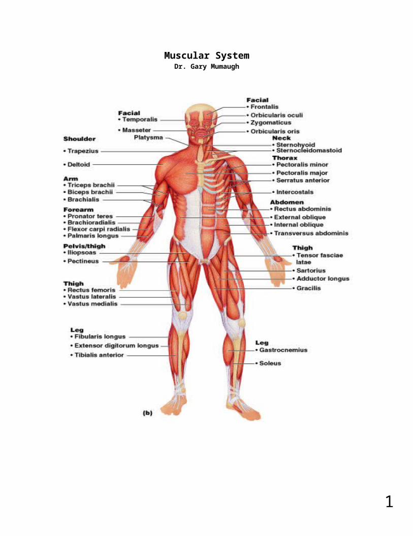
Organization of Muscles about 600 human skeletal muscles constitute about half of our body weight three kinds of muscle tissue
o skeletal, cardiac, smooth specialized for one major purpose
o converting the chemical energy in ATP into the mechanical energy of motion myology – the study of the muscular system
3
The Functions of Muscles Movement
o move from place to place, movement of body parts and body contents in breathing, circulation, feeding and digestion, defecation, urination, and childbirth
o role in communication – speech, writing, and nonverbal communications Stability
o Maintain posture by preventing unwanted movementso antigravity muscles – resist the pull of gravity and prevent us from falling or
slumping overo stabilize joints
Control of openings and passagewayso sphincters – internal muscular rings that control the movement of food, bile,
blood, and other materials Heat production by skeletal muscles
o as much as 85% of our body heat
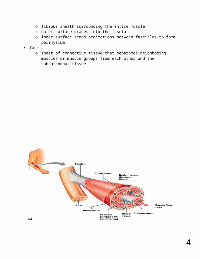
Connective Tissues of a Muscle endomysium
o thin sleeve of loose connective tissue surrounding each muscle fibero allows room for capillaries and nerve fibers to reach each muscle fiber
perimysiumo slightly thicker layer of connective tissueo fascicles – bundles of muscle fibers wrapped in perimysiumo carry larger nerves and blood vessels, and stretch receptors
epimysiumo fibrous sheath surrounding the entire muscleo outer surface grades into the fasciao inner surface sends projections between fascicles to form perimysium
fasciao sheet of connective tissue that separates neighboring muscles or muscle
groups from each other and the subcutaneous tissue
4
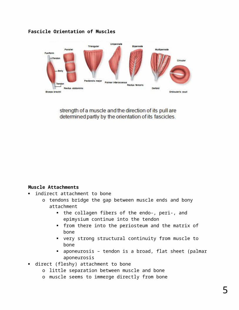
Fascicle Orientation of Muscles
5
Muscle Attachments indirect attachment to bone
o tendons bridge the gap between muscle ends and bony attachment the collagen fibers of the endo-, peri-, and epimysium continue into the
tendon from there into the periosteum and the matrix of bone very strong structural continuity from muscle to bone aponeurosis – tendon is a broad, flat sheet (palmar aponeurosis
direct (fleshy) attachment to boneo little separation between muscle and boneo muscle seems to immerge directly from boneo margins of brachialis, lateral head of triceps brachii
some skeletal muscles do not insert on bone, but in dermis of the skin – muscles of facial expression
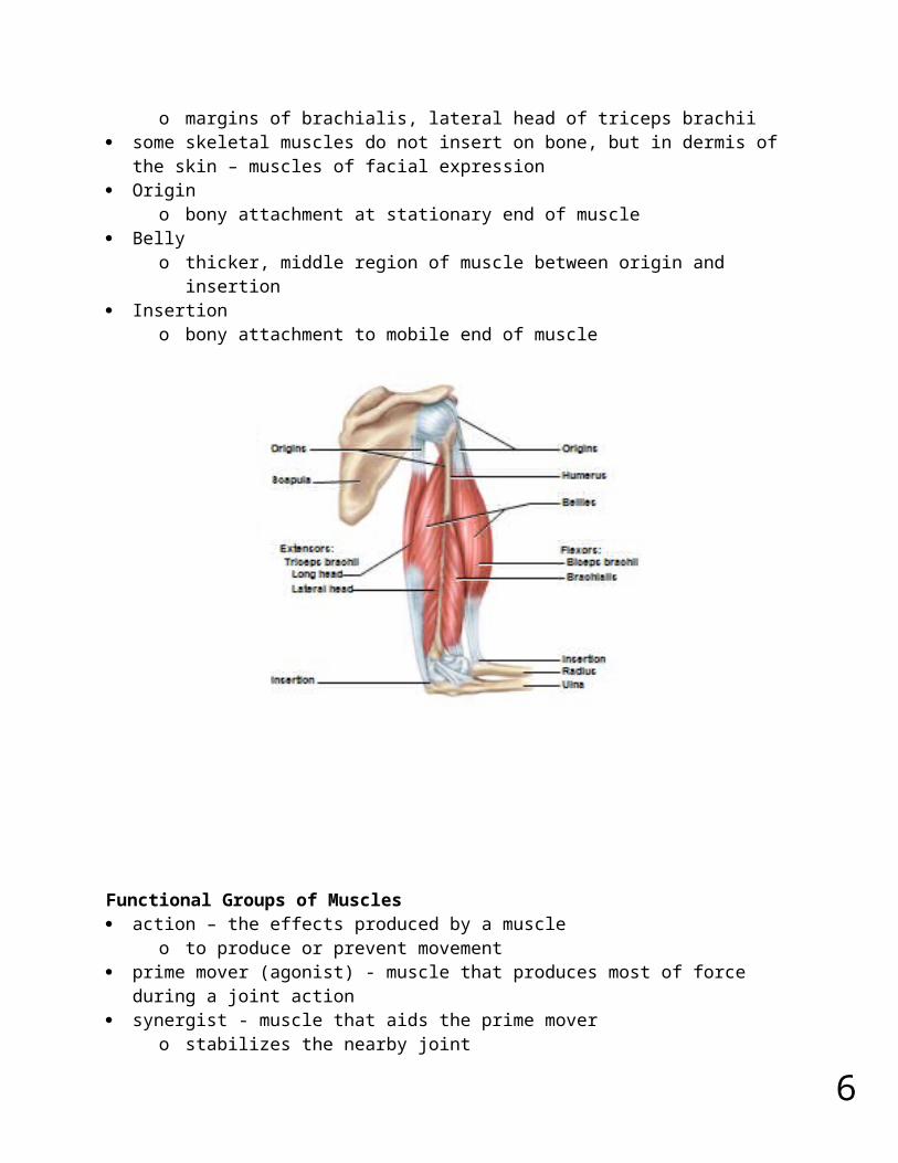
Origino bony attachment at stationary end of muscle
Bellyo thicker, middle region of muscle between origin and insertion
Insertiono bony attachment to mobile end of muscle
6
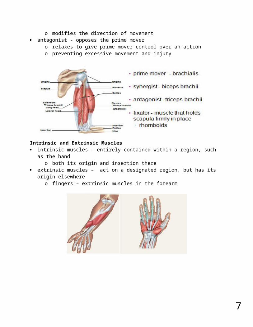
Functional Groups of Muscles action – the effects produced by a muscle
o to produce or prevent movement prime mover (agonist) - muscle that produces most of force during a joint action synergist - muscle that aids the prime mover
o stabilizes the nearby jointo modifies the direction of movement
antagonist - opposes the prime movero relaxes to give prime mover control over an actiono preventing excessive movement and injury
Intrinsic and Extrinsic Muscles intrinsic muscles – entirely contained within a region, such as the hand
o both its origin and insertion there extrinsic muscles – act on a designated region, but has its origin elsewhere
o fingers – extrinsic muscles in the forearm
7
Muscle Innervation innervation of a muscle – refers to the identity of the nerve that stimulates it
o enables the diagnosis of nerve, spinal cord, and brainstem injuries from their effects on muscle function
spinal nerves arise from the spinal cordo emerge through intervertebral foraminao immediately branch into a posterior and anterior ramuso innervate muscles below the neck
cranial nerves arise from the base of the braino emerge through skull foraminao innervate the muscles of the head and necko numbered I to XII
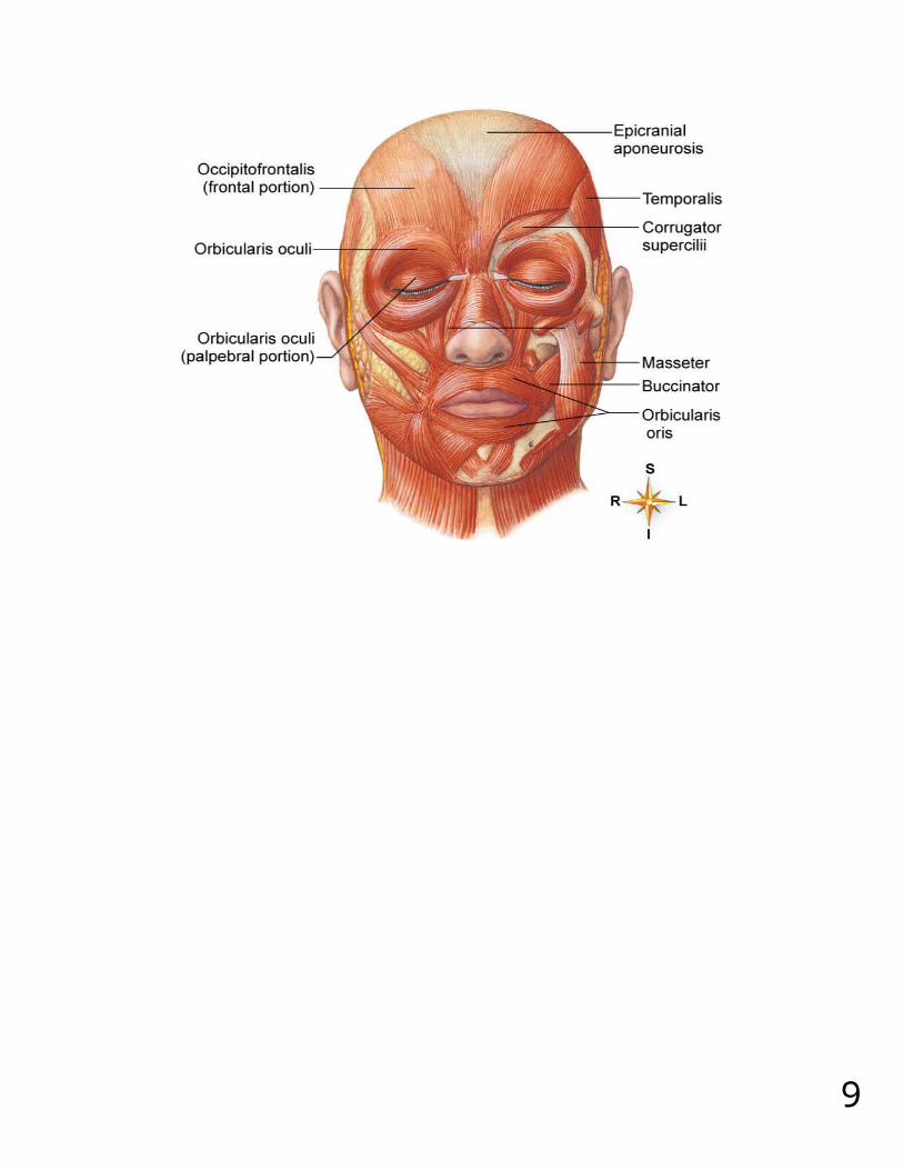
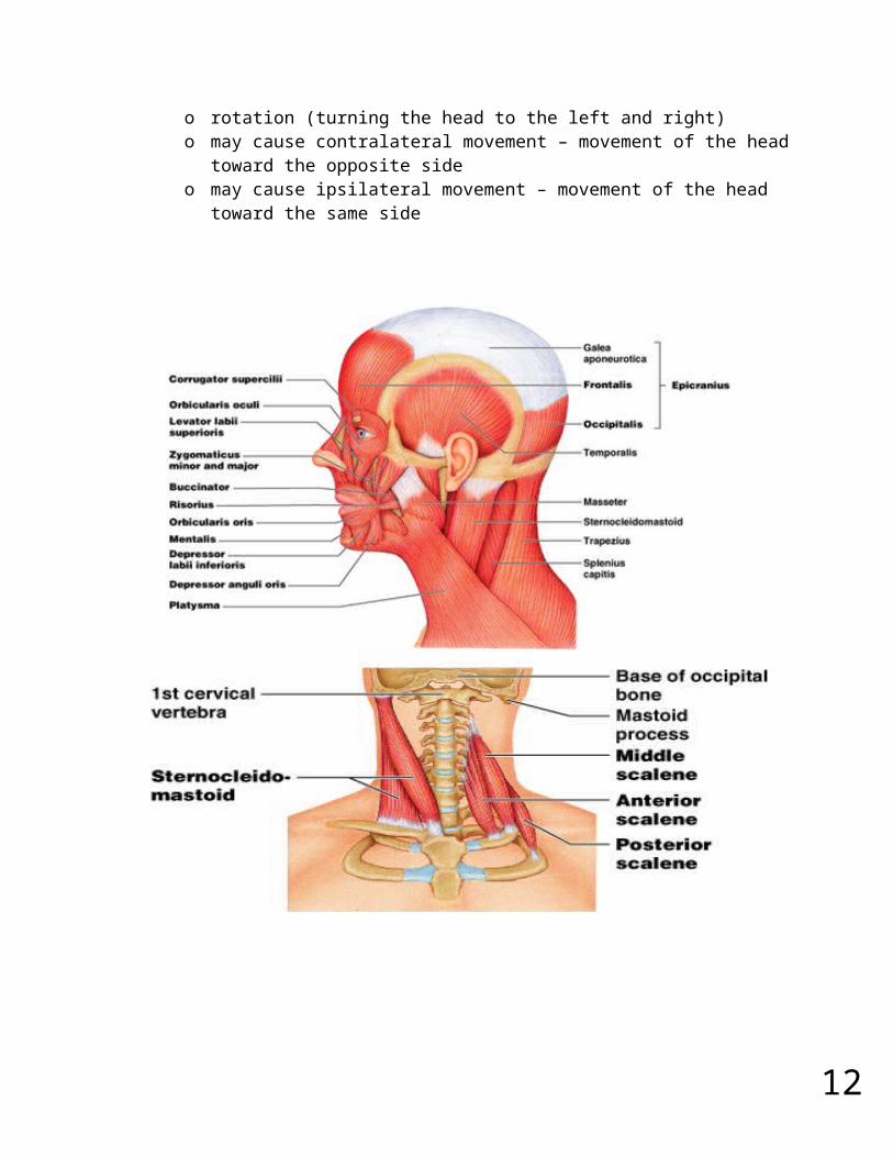
Muscles of Facial Expression muscles that insert in the dermis and subcutaneous tissues tense the skin and produce facial expressions innervated by facial nerve (CN VII) paralysis causes face to sag found in scalp, forehead, around the eyes, nose and mouth, and in the neck
8
9
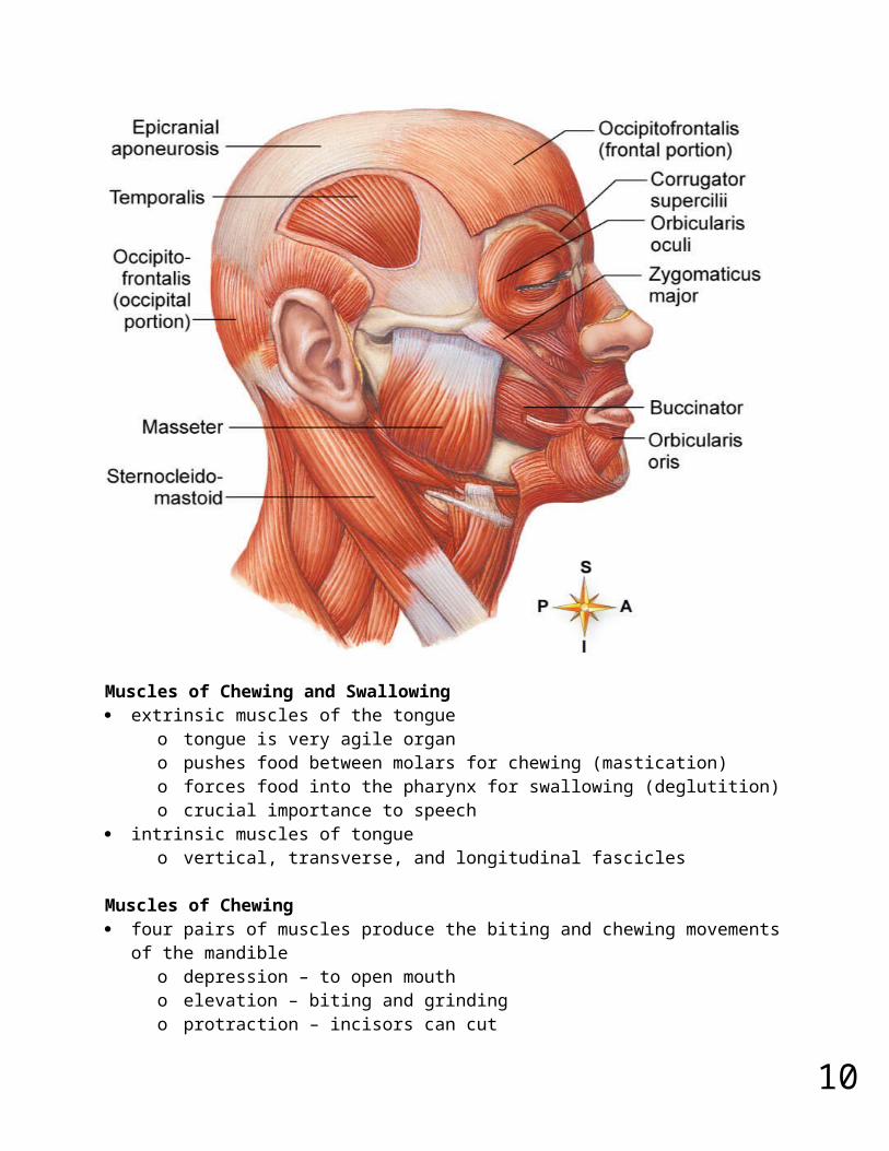
Muscles of Chewing and Swallowing extrinsic muscles of the tongue
o tongue is very agile organo pushes food between molars for chewing (mastication)o forces food into the pharynx for swallowing (deglutition)o crucial importance to speech
intrinsic muscles of tongueo vertical, transverse, and longitudinal fascicles
Muscles of Chewing four pairs of muscles produce the biting and chewing movements of the mandible
o depression – to open moutho elevation – biting and grindingo protraction – incisors can cuto retraction – make rear teeth meeto lateral and medial excursion – grind food
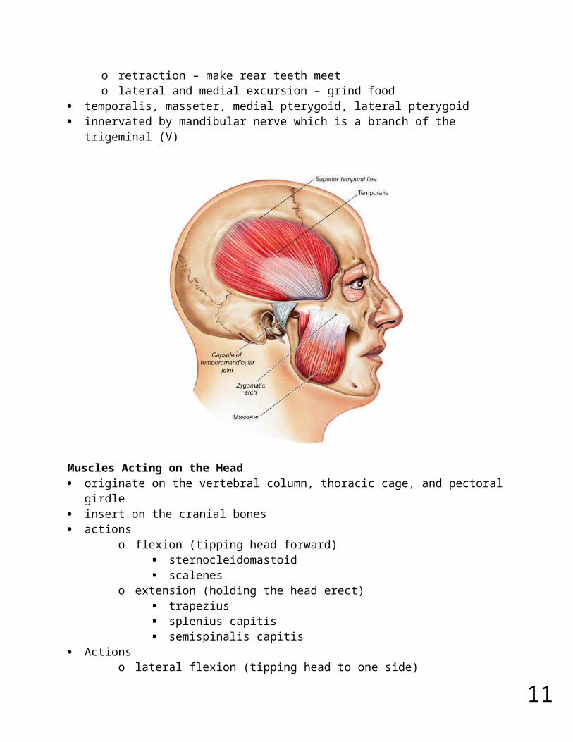
temporalis, masseter, medial pterygoid, lateral pterygoid
10
innervated by mandibular nerve which is a branch of the trigeminal (V)
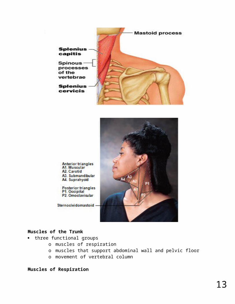
Muscles Acting on the Head originate on the vertebral column, thoracic cage, and pectoral girdle insert on the cranial bones actions
o flexion (tipping head forward) sternocleidomastoid scalenes
o extension (holding the head erect) trapezius splenius capitis semispinalis capitis
Actionso lateral flexion (tipping head to one side)o rotation (turning the head to the left and right)o may cause contralateral movement – movement of the head toward the
opposite sideo may cause ipsilateral movement – movement of the head toward the
same side
11
12
Muscles of the Trunk three functional groups
o muscles of respirationo muscles that support abdominal wall and pelvic flooro movement of vertebral column
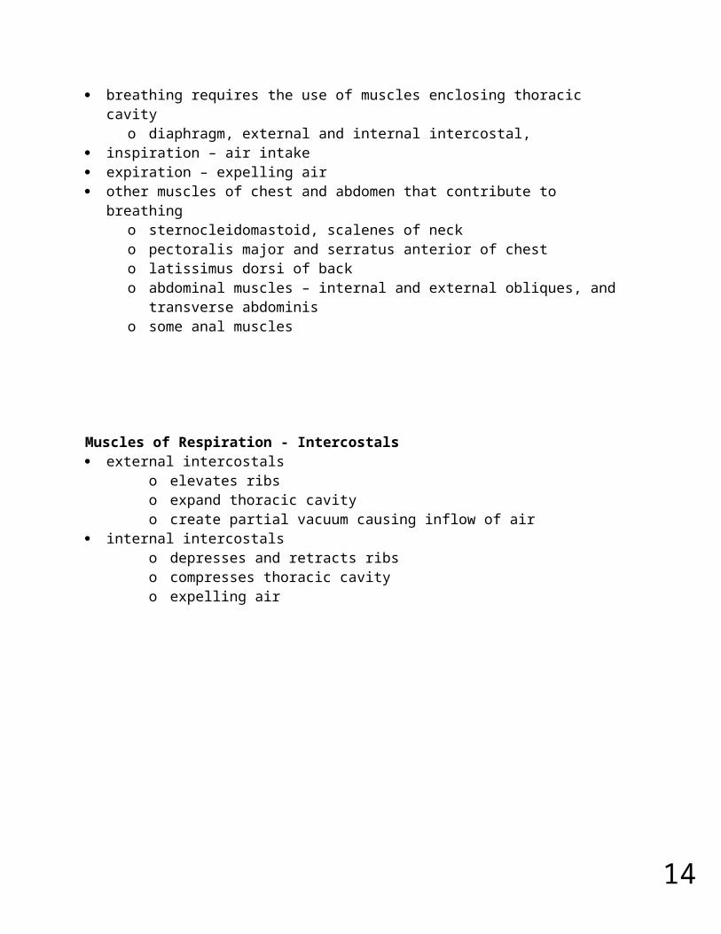
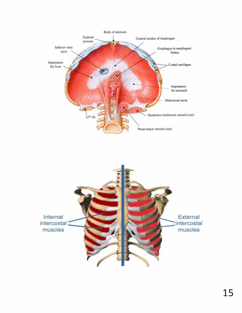
Muscles of Respiration breathing requires the use of muscles enclosing thoracic cavity
o diaphragm, external and internal intercostal, inspiration – air intake expiration – expelling air other muscles of chest and abdomen that contribute to breathing
o sternocleidomastoid, scalenes of necko pectoralis major and serratus anterior of chesto latissimus dorsi of backo abdominal muscles – internal and external obliques, and transverse
abdominiso some anal muscles
13
Muscles of Respiration - Intercostals external intercostals
o elevates ribso expand thoracic cavityo create partial vacuum causing inflow of air
internal intercostalso depresses and retracts ribso compresses thoracic cavityo expelling air
14
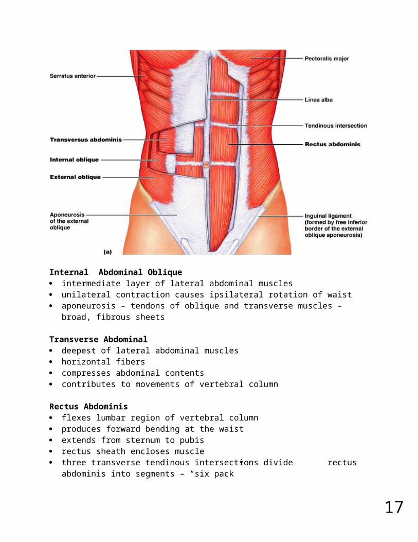
Muscles of the Anterior Abdominal Wall four pairs of sheetlike muscles
o external abdominal obliqueo internal abdominal obliqueo transverse abdominalo rectus abdominis
strengthen abdominal wall
External Abdominal Oblique most superficial of lateral abdominal muscles supports abdominal viscera against pull of gravity stabilizes vertebral column during heavy lifting maintains posture compresses abdominal organs aids in forced expiration rotation at waist
15
Internal Abdominal Oblique intermediate layer of lateral abdominal muscles unilateral contraction causes ipsilateral rotation of waist aponeurosis – tendons of oblique and transverse muscles –broad, fibrous sheets
Transverse Abdominal deepest of lateral abdominal muscles horizontal fibers compresses abdominal contents contributes to movements of vertebral column
Rectus Abdominis flexes lumbar region of vertebral column produces forward bending at the waist extends from sternum to pubis rectus sheath encloses muscle three transverse tendinous intersections divide rectus abdominis into segments –
“six pack”
Superficial Back Muscles
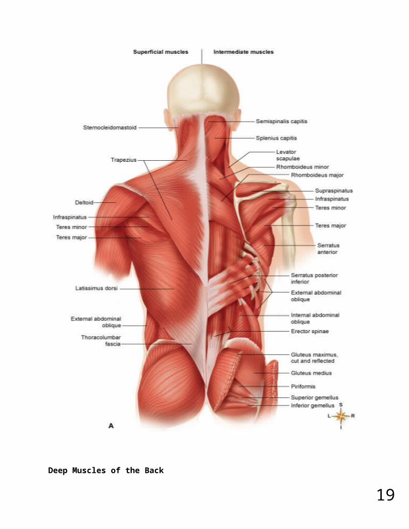
16Deep Muscles of the Back
17
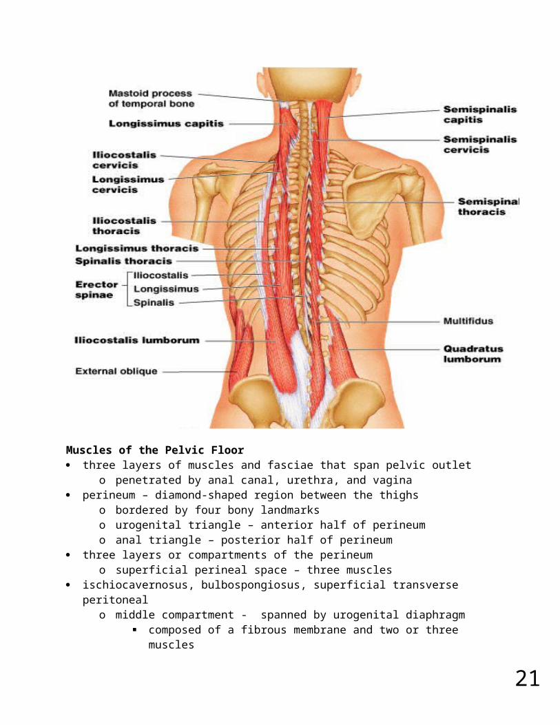
erector spinaeo iliocostalis, longissimus, spinaliso from cranium to sacrumo extension and lateral flexion of vertebral column
semispinalis thoraciso extension and contralateral rotation of vertebral column
quadratus lumborumo aids respirationo ipsilateral flexion of lumbar vertebral column
multifiduso stabilizes adjacent vertebraeo maintains posture
Deep Muscles of the Back
18
Muscles of the Pelvic Floor three layers of muscles and fasciae that span pelvic outlet
o penetrated by anal canal, urethra, and vagina perineum – diamond-shaped region between the thighs
o bordered by four bony landmarkso urogenital triangle – anterior half of perineumo anal triangle – posterior half of perineum
three layers or compartments of the perineumo superficial perineal space – three muscles
ischiocavernosus, bulbospongiosus, superficial transverse peritonealo middle compartment - spanned by urogenital diaphragm
composed of a fibrous membrane and two or three muscles deep transverse perineal muscle, external urethral and anal sphincters compressor urethrae in females only
o pelvic diaphragm – deepest layer consists of two muscle pairs levator ani and coccygeus
Superficial Perineal Space muscles found just deep to the skin ischiocavernosus – maintains erection bulbospongiosus – aids in erection, expels remaining urine
Muscles of Pelvic Diaphragm deepest compartment of the perineum pelvic diaphragm – two muscle pairs
o levator ani - supports viscera and defecationo coccygeus - supports and elevates pelvic floor
19
Hernias hernia – any condition in which the viscera protrudes through a weak point in the
muscular wall of the abdominopelvic cavity inguinal hernia
o most common type of hernia (rare in women)o viscera enter inguinal canal or even the scrotum
hiatal herniao stomach protrudes through diaphragm into thoraxo overweight people over 40
umbilical herniao viscera protrude through the nave
Muscles Acting on Shoulder and Upper Limb compartments – spaces in which muscles are organized and are separated by
fibrous connective tissue sheets (fasciae)o each compartment contains one or more functionally related muscles
along with their nerve and blood supplies muscles of upper limbs divided into anterior and posterior compartments muscles of lower limbs divided into anterior, posterior, medial, and lateral
compartments compartment syndrome – one of the muscles or blood vessels in a compartment is
injured
Compartment Syndrome fasciae of arms and legs enclose muscle compartments very snugly if a blood vessel in a compartment is damaged, blood and tissue fluid accumulate in
the compartment fasciae prevent compartment from expanding with increasing pressure compartment syndrome – mounting pressure on the muscles, nerves and blood
vessel triggers a sequence of degenerative eventso blood flow to compartment is obstructed by pressureo if ischemia (poor blood flow) persists for more than 2 – 4 hours, nerves
begin to dieo after 6 hours, muscles begin to die
nerves can regenerate after pressure relieved, but muscle damage is permanent myoglobin in urine indicates compartment syndrome treatment – immobilization of limb and fasciotomy – incision to relieve compartment
pressure
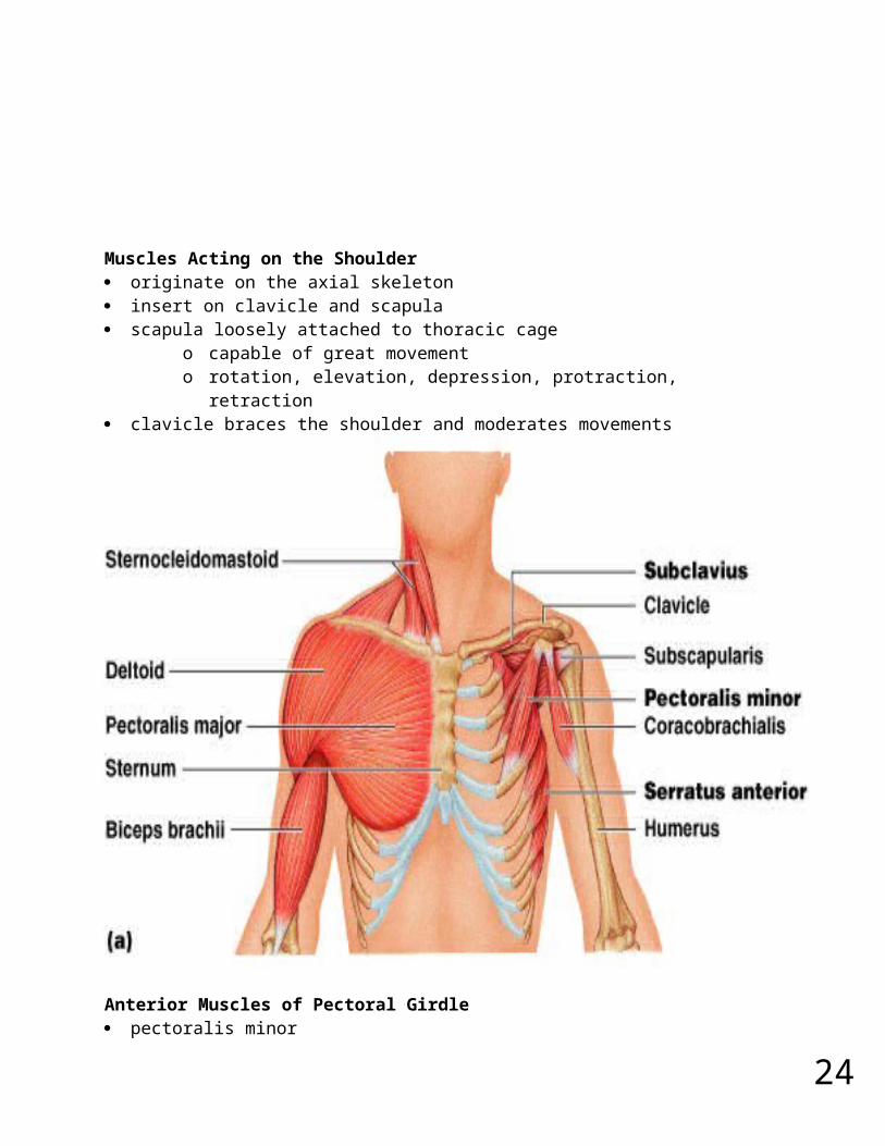
Muscles Acting on the Shoulder
20
originate on the axial skeleton insert on clavicle and scapula scapula loosely attached to thoracic cage
o capable of great movemento rotation, elevation, depression, protraction, retraction
clavicle braces the shoulder and moderates movements
Anterior Muscles of Pectoral Girdle pectoralis minor
o ribs 3-5 to coracoid process of scapulao draws scapula laterally
serratus anterioro ribs 1-9 to medial border of scapulao abducts and rotates or depresses scapula
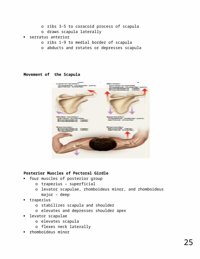
Movement of the Scapula
21
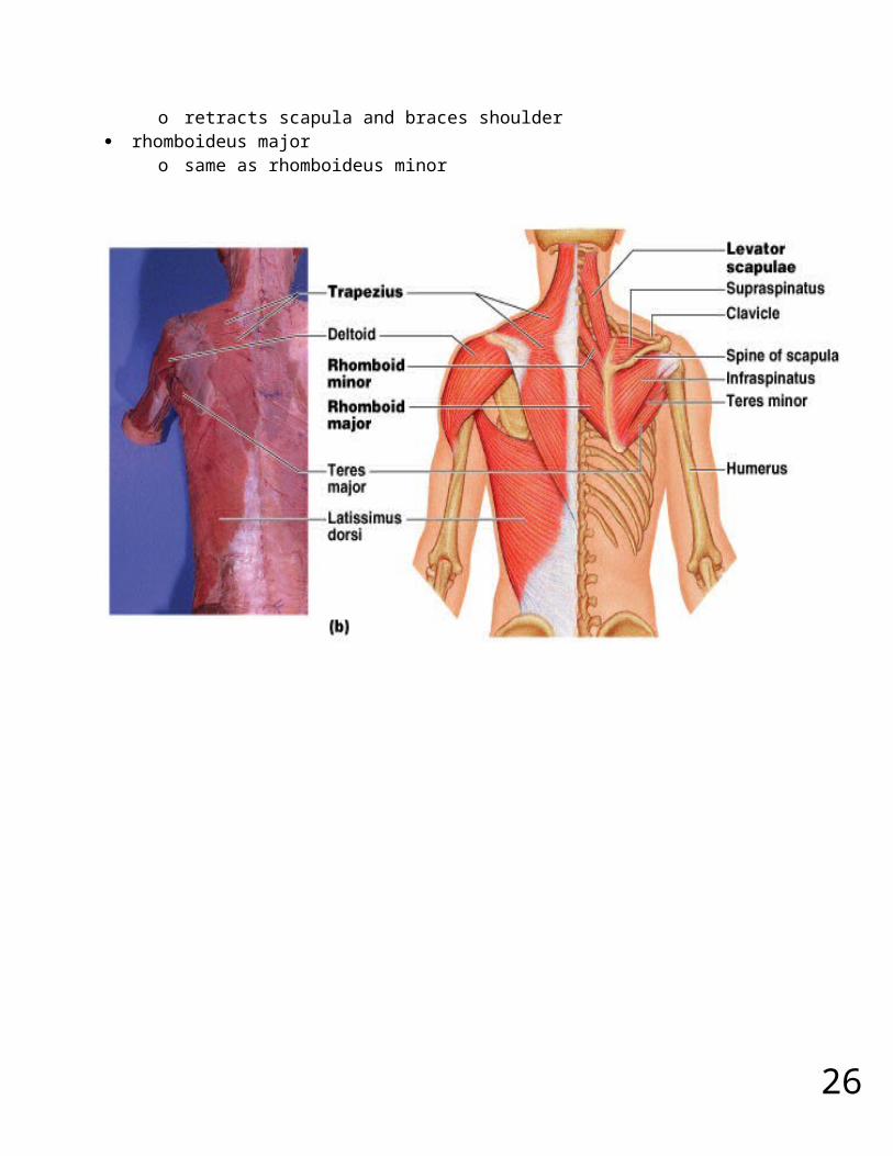
Posterior Muscles of Pectoral Girdle four muscles of posterior group
o trapezius – superficialo levator scapulae, rhomboideus minor, and rhomboideus major – deep
trapeziuso stabilizes scapula and shouldero elevates and depresses shoulder apex
levator scapulae o elevates scapulao flexes neck laterally
rhomboideus minoro retracts scapula and braces shoulder
rhomboideus majoro same as rhomboideus minor

22
23
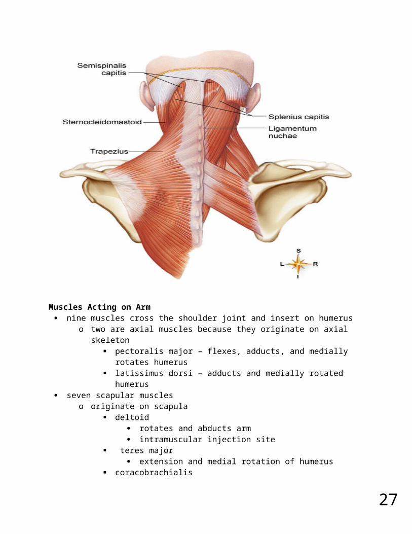
Muscles Acting on Arm nine muscles cross the shoulder joint and insert on humerus
o two are axial muscles because they originate on axial skeleton pectoralis major – flexes, adducts, and medially rotates humerus latissimus dorsi – adducts and medially rotated humerus
seven scapular muscleso originate on scapula
deltoid rotates and abducts arm intramuscular injection site
teres major extension and medial rotation of humerus
coracobrachialis flexes and medially rotates arm
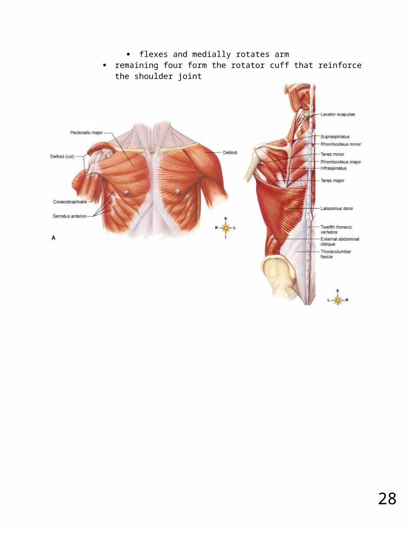
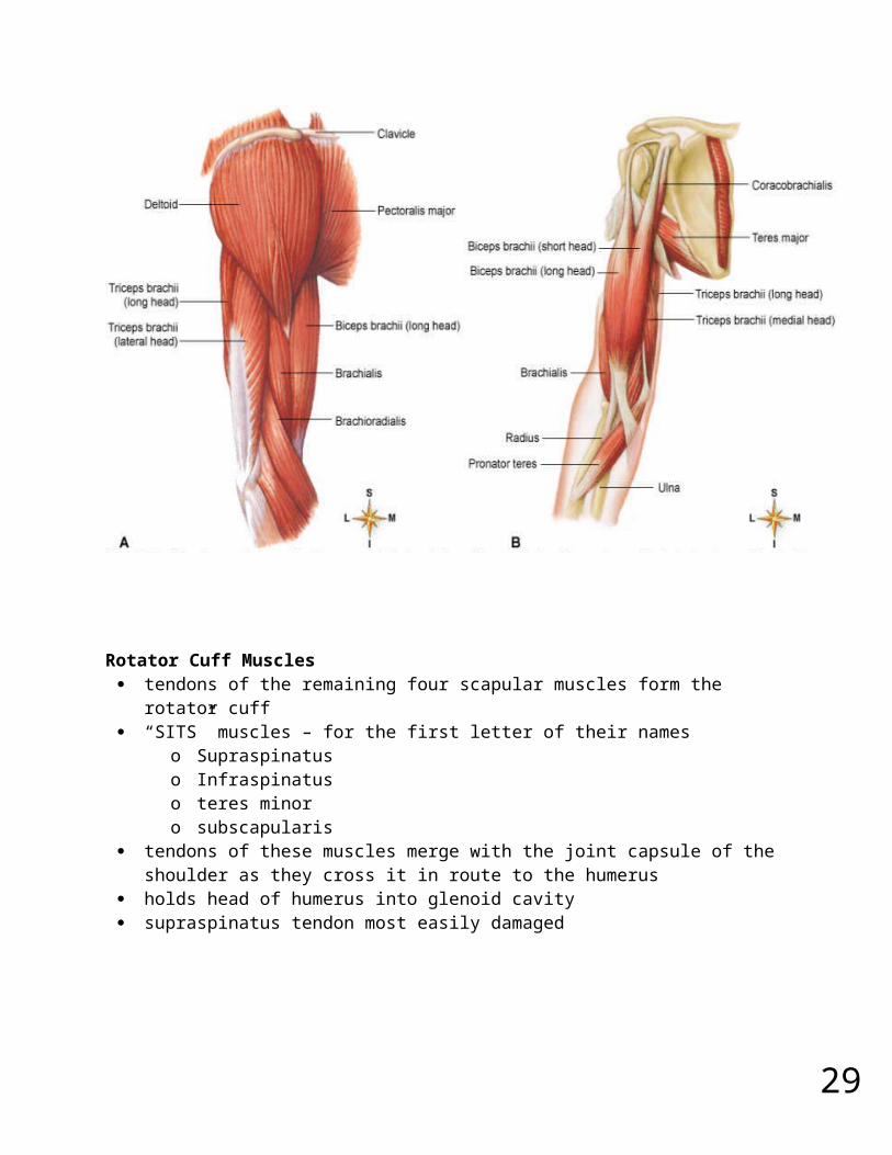
remaining four form the rotator cuff that reinforce the shoulder joint
24
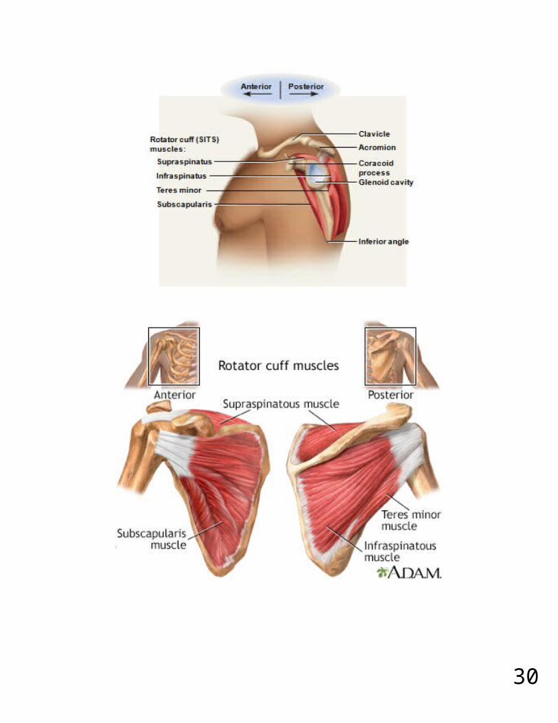
Rotator Cuff Muscles tendons of the remaining four scapular muscles form the rotator cuff “SITS” muscles – for the first letter of their names
o Supraspinatuso Infraspinatuso teres minoro subscapularis
tendons of these muscles merge with the joint capsule of the shoulder as they cross it in route to the humerus
holds head of humerus into glenoid cavity supraspinatus tendon most easily damaged
25
26
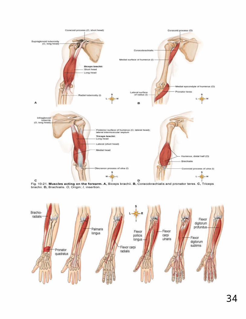
Muscles Acting on Forearm elbow and forearm capable of flexion, extension, pronation, and supination
o carried out by muscles in both brachium (arm) and antebrachium (forearm) muscles with bellies in the arm (brachium)
o principal elbow flexors – anterior compartment brachialis and biceps brachii brachialis produces 50% more power than biceps brachii brachialis is prime mover of elbow flexion
o principal elbow extensor – posterior compartment triceps brachii - prime mover of elbow extension
muscles with bellies in the forearm (antebrachium)o most forearm muscles act on the hand and wrist
brachioradialis – flexes elbow anconeus – extends elbow pronator quadratus – prime mover in forearm pronation pronator teres – assists pronator quadratus in pronation supinator – supinates the forearm
27
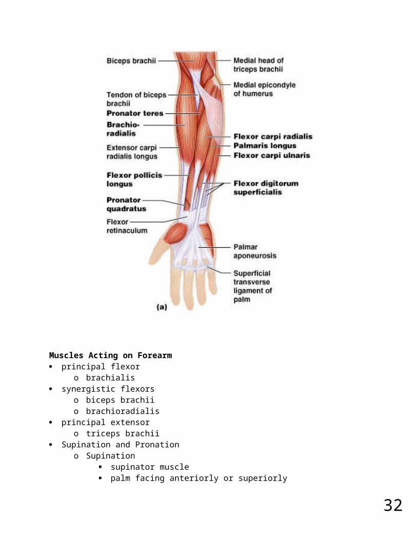
Muscles Acting on Forearm principal flexor
o brachialis synergistic flexors
o biceps brachii o brachioradialis
principal extensoro triceps brachii
Supination and Pronationo Supination
supinator muscle palm facing anteriorly or superiorly
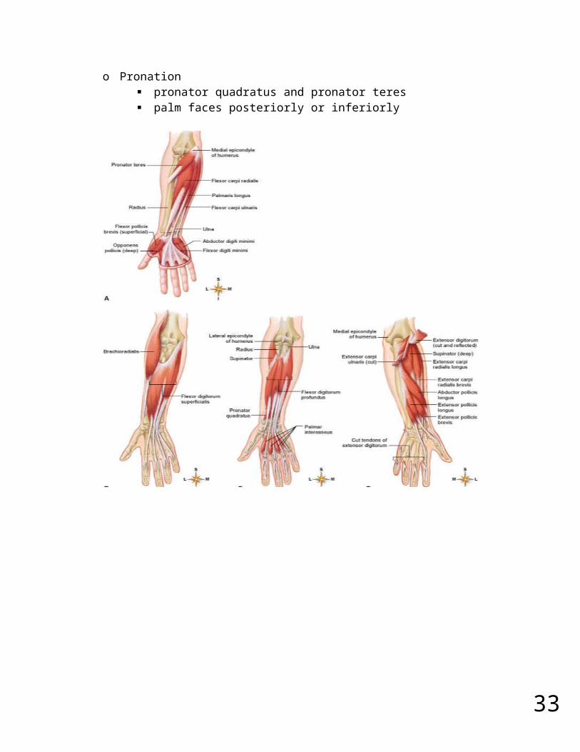
o Pronation pronator quadratus and pronator teres palm faces posteriorly or inferiorly
28
29
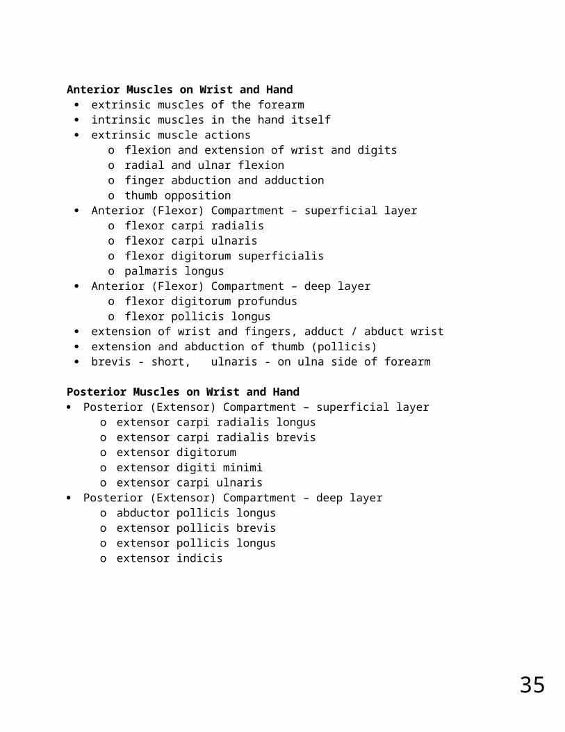
Anterior Muscles on Wrist and Hand extrinsic muscles of the forearm intrinsic muscles in the hand itself extrinsic muscle actions
o flexion and extension of wrist and digitso radial and ulnar flexiono finger abduction and adductiono thumb opposition
Anterior (Flexor) Compartment – superficial layero flexor carpi radialiso flexor carpi ulnariso flexor digitorum superficialiso palmaris longus
Anterior (Flexor) Compartment – deep layero flexor digitorum profunduso flexor pollicis longus
extension of wrist and fingers, adduct / abduct wrist extension and abduction of thumb (pollicis) brevis - short, ulnaris - on ulna side of forearm
Posterior Muscles on Wrist and Hand Posterior (Extensor) Compartment – superficial layer
o extensor carpi radialis longuso extensor carpi radialis breviso extensor digitorumo extensor digiti minimio extensor carpi ulnaris
Posterior (Extensor) Compartment – deep layero abductor pollicis longuso extensor pollicis breviso extensor pollicis longuso extensor indicis
31
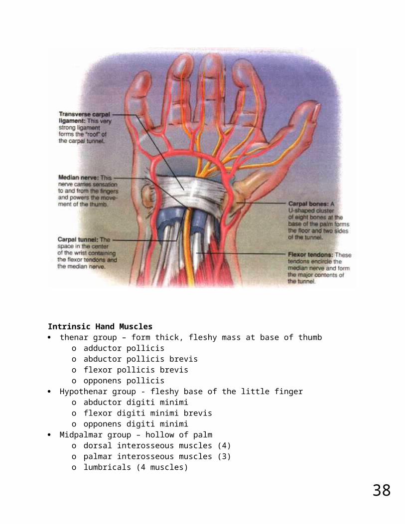
Carpal Tunnel Syndrome flexor retinaculum – bracelet-like fibrous sheet that the flexor tendons of the
extrinsic muscles that flex the wrist pass on their way to their insertions carpal tunnel – tight space between the flexor retinaculum and the carpal bones
o flexor tendons passing through the tunnel are enclosed in tendon sheathso enable tendons to slide back and forth quite easily
carpal tunnel syndrome - prolonged, repetitive motions of wrist and fingers can cause tissues in the carpal tunnel to become inflamed, swollen, or fibrotic
o puts pressure on the median nerve of the wrist that passes through the carpal tunnel along with the flexor tendons
o tingling and muscular weakness in the palm and medial side of the hando pain may radiate to arm and shouldero treatment – anti-inflammatory drugs, immobilization of the wrist, and
sometimes surgery to remove part or all of flexor retinaculum
32
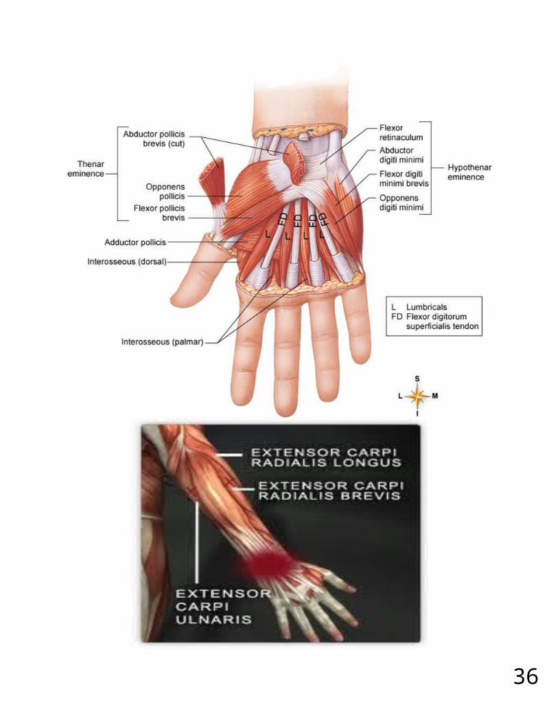
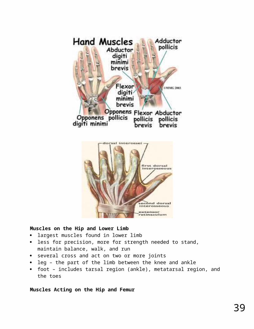
Intrinsic Hand Muscles thenar group – form thick, fleshy mass at base of thumb
o adductor polliciso abductor pollicis breviso flexor pollicis brevis o opponens pollicis
Hypothenar group - fleshy base of the little fingero abductor digiti minimio flexor digiti minimi breviso opponens digiti minimi
Midpalmar group – hollow of palmo dorsal interosseous muscles (4)o palmar interosseous muscles (3)o lumbricals (4 muscles)
33
Muscles on the Hip and Lower Limb largest muscles found in lower limb less for precision, more for strength needed to stand, maintain balance, walk, and
run several cross and act on two or more joints leg – the part of the limb between the knee and ankle foot – includes tarsal region (ankle), metatarsal region, and the toes
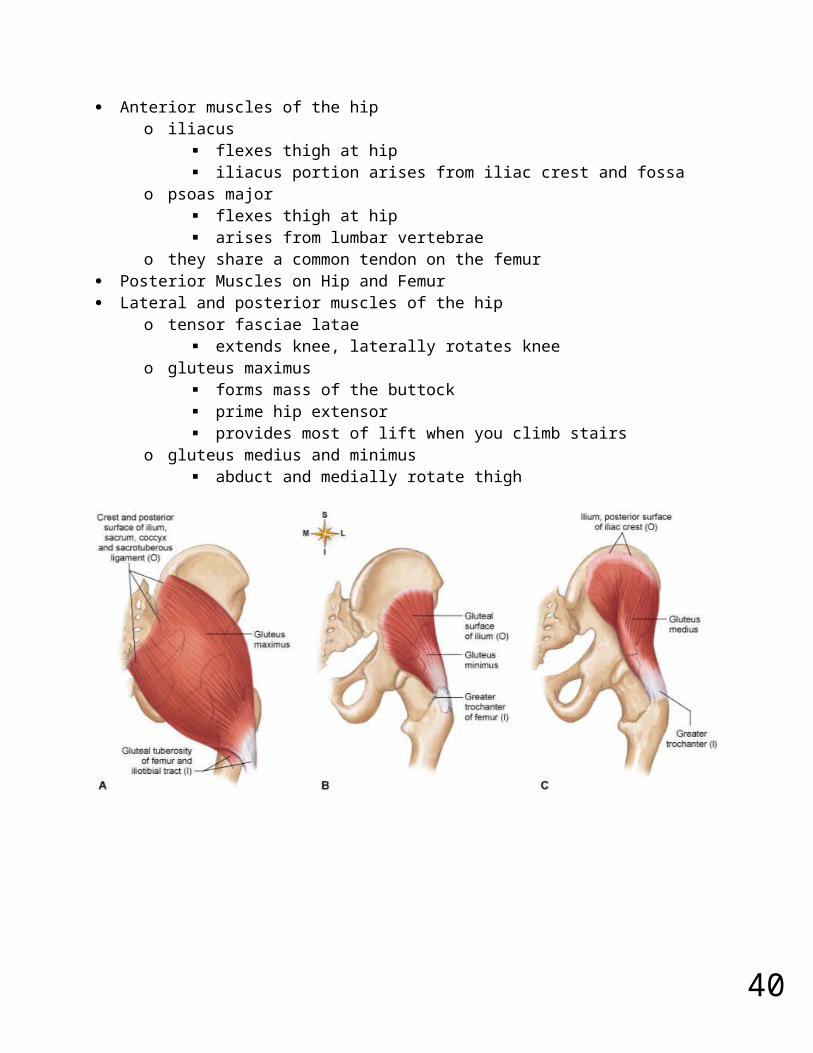
Muscles Acting on the Hip and Femur Anterior muscles of the hip
o iliacus flexes thigh at hip iliacus portion arises from iliac crest and fossa
o psoas major flexes thigh at hip arises from lumbar vertebrae
o they share a common tendon on the femur Posterior Muscles on Hip and Femur Lateral and posterior muscles of the hip
o tensor fasciae latae extends knee, laterally rotates knee
o gluteus maximus forms mass of the buttock prime hip extensor provides most of lift when you climb stairs
o gluteus medius and minimus abduct and medially rotate thigh
34
Posterior Muscles on Hip and Femur lateral rotators - six muscles inferior to gluteus minimus deep to the two other gluteal muscles
35
o gemellus superioro gemellus inferioro obturator externuso obturator internuso piriformiso quadratus femoris
Muscles Acting on Hip and Femur medial (adductor) compartment of thigh five muscles act as primary adductors of the thigh
o adductor breviso adductor longuso adductor magnuso graciliso pectineus
36
Muscles on the Knee and Leg anterior (extensor) compartment of the thigh
o contains large quadriceps femoris muscle prime mover of knee extension most powerful muscle in the body has four heads – rectus femoris, vastus lateralis, vastus
medialis, and vastus intermedius all converge on single quadriceps (patellar) tendon extends to patella
37
then continues as patellar ligament inserts on tibial tuberosity
o sartorius – longest muscle in the body tailor’s muscle
38
Muscles Acting on the Knee and Leg Posterior (flexor) compartment of the thigh
o contains hamstring muscles from lateral to medial; biceps femoris, semitendinosus,
semimembranosus Anterior Compartment of Leg
o anterior (extensor) compartment of the leg dorsiflex the ankle prevent toes from scuffing when walking fibularis (peroneus) tertius extensor digitorum longus extensor hallucis longus tibialis anterior
Posterior Compartment of Leg - Superficial Groupo three muscles of the superficial group
gastrocnemius - plantar flexes foot, flexes knee soleus – plantar flexes foot plantaris - weak synergist of triceps
39
Muscles Acting on the Knee and Leg Posterior Compartment of Leg - Deep Group
o four muscles in the deep group flexor digitorum longus – flexes phalanges flexor hallucis longus – flexes great toe tibialis posterior – inverts foot popliteus – acts on knee
Lateral (Fibular) Compartment of the Lego two muscles in this compartment
fibularis longus fibularis brevis both plantar flex and evert the foot provides lift and forward thrust
40
Intrinsic Muscles of Foot support for arches
o abduct and adduct the toeso flex the toes
one dorsal muscleo extensor digitorum brevis extends toes
Athletic Injuries muscles and tendons are vulnerable to sudden and intense stress proper conditioning and warm-up needed common injuries;
o compartment syndromeo shinsplintso pulled hamstringso tennis elbowo pulled groin o rotator cuff injury
treat with rest, ice, compression and elevation “no pain, no gain” is a dangerous misconception