Embed Size (px)
Citation preview

Micro/nano-bubble-assisted ultrasound to enhance the EPR effect and potential theranostic applicationsLei Duan, ‡, § Li Yang,†,§Juan Jin†, Fang Yang,†, * Dong Liu , Ke Hu‡, Qinxin Wang‡, Yuanbin Yue‡, and Ning Gu †, ‡ ,*
Lei Duan, Ke Hu, Qinxin Wang, Yuanbin Yue, Prof. Ning Gu‡ School of Biomedical Engineering and Informatics, Nanjing Medical University, Nanjing, 211166, P. R. China.
Li Yang, Juan Jin, Prof. Fang Yang, Prof. Ning Gu† State Key Laboratory of Bioelectronics, Jiangsu Key Laboratory for Biomaterials and Devices,School of Biological Science and Medical Engineering, Southeast University, Nanjing 210096, P. R. China.
Dong Liu1. West Anhui University, Lu’an, P.R. China 2. Anhui Engineering Laboratory for Conservation and Sustainable Utilization of Traditional Chinese Medicine Resources
§These authors contributed equally to this work.
* Address correspondence toFang Yang, Email: [email protected] Gu, Email: [email protected];

AbstractDrug delivery for tumor theranostics involves the extensive use of the enhanced
permeability and retention (EPR) effect. Previously, various types of nanomedicines have
been demonstrated to accumulate in solid tumors via the EPR effect. However, EPR is a
highly variable phenomenon because of tumor heterogeneity, resulting in low drug
delivery efficacy in clinical trials. Because ultrasonication using micro/nanobubbles as
contrast agents can disrupt blood vessels and enhance the specific delivery of drugs, it is
an effective approach to improve the EPR effect for the passive targeting of tumors. In
this review, the basic thermal effect, acoustic streaming, and cavitation mechanisms of
ultrasound, which are characteristics that can be utilized to enhance the EPR effect, are
briefly introduced. Second, micro/nanobubble-enhanced ultrasound imaging is discussed
to understand the validity and variability of the EPR effect. Third, because the tumor
microenvironment is complicated owing to elevated interstitial fluid pressure and the
deregulated extracellular matrix components, which may be unfavorable for the EPR
effect, few new trends in smart bubble drug delivery systems, which may improve the
accuracy of EPR-mediated passive drug targeting, are summarized. Finally, the
challenging and major concerns that should be considered in the next generation of
micro/nanobubble-contrast-enhanced ultrasound theranostics for EPR-mediated passive
drug targeting are also discussed.
Keywords: EPR effect, ultrasound, micro/nanobubbles, controlled drug delivery, tumor
theranostics

1. IntroductionSolid tumors exhibit a special microenvironment featuring tortuous vascularity,
angiogenesis, and hypoxia [1-3]. Abnormal tumor vasculature, high interstitial fluid
pressure, growth-induced solid stress. and solid stress from the abnormal stromal matrix
are the main limitations that prevent drugs from penetrating the solid tumor [4, 5].
Therefore, the accurate and efficient delivery of theranostic agents into tumor lesions in a
controlled manner is critical and remains a significant challenge till date. However, the
absence of vascular supportive tissues in tumors results in the formation of leaky vessels
with pores (200 nm to 1.2 μm in diameter) [6] and leads to poor lymphatic drainage,
which is the structural basis of the enhanced permeability and retention (EPR) effect. The
EPR effect is considered an effective method to achieve the passive targeting of tumor
tissues [7, 8]. In preclinical studies, targeted delivery systems based on EPR have
demonstrated a remarkable improvement in anticancer efficacy when compared with the
efficacy of traditional chemotherapeutic drugs [7-9].
However, numerous parameters (such as vascular permeability and mutation, tumor
cell density, and tumor stromal heterogeneity) prevent the successful application of EPR-
based theranostics in clinical applications because of tumor heterogeneity [10-15].
Different patients with the same type of tumor, different types and sizes of tumor, and
different developmental stages of the same tumor may show different EPR effects [16,
17]. For example, hepatocellular carcinoma and renal cell carcinoma both have high
vascular density and exhibit a strong EPR effect, whereas pancreatic and prostate cancers
are characterized by low vascular density, resulting in a lower EPR effect [18]. Metastatic
tumors have a high degree of necrosis at the central site relative to the primary tumor
because of the lack of blood vessels due to the decrease in the EPR effect. Therefore,
accurately monitoring and evaluating the EPR conditions of different tumors is essential
to formulate personalized EPR-mediated tumor treatment [19].
In recent years, various nanomaterials based on the EPR effect have been used
because the sizes of these nanomaterials can be modulated according to the pore sizes in
tumor blood vessels. Because different types of tumors have different sized gaps in the
endothelial cells, a suitable nanoparticle size must be selected according to the type of

tumor. Furthermore, the interception by biological barriers when nanomaterials enter the
circulation system also needs to be considered. Particularly, nanoparticles should not be
destroyed by the reticuloendothelial system (increased particle size) while maintaining
the ability to penetrate the tumor (reduced particle size) [20, 21]. Although the gaps and
pores in the wall of tumor vessels may provide a pathway for the exudation of
nanoparticles, the intermittence and disorder of the vascular structure reduce the blood
flow and intravascular pressure. This may result in the reduction of macromolecular
drugs or nanocarriers entering the stroma through the vascular gaps by diffusion [22].
Without further increasing the movement of these drugs via diffusion, the efficiency of
delivery will not be significantly improved.
In view of these challenges, to best leverage the EPR effect in nanomedicine
delivery, various diagnostic and therapeutic strategies have been developed (Figure 1). To
monitor and evaluate the heterogeneity of the EPR effect in a particular patient, non-
invasive imaging techniques (such as ultrasound, magnetic resonance imaging (MRI),
and positron emission tomography (PET)) have been applied. With the development of
contrast agents such as micro/nanobubbles (MNBs) [23-25], magnetic nanoparticles [26,
27], radionuclides, and fluorophores, the EPR effect can be accurately visualized and
quantified [28-31]. Nanomaterial-based EPR targeting strategies mainly focus on
controlling the size of the drug or carrier and/or using ligands that bind to molecules that
are related to the EPR effect. An appropriately sized material can pass through the
permeable tumor vascular system and passively accumulate in the tumor tissue via
passive targeting. The modification of the ligands on the surface leads to active targeting.
The transport of the micro-materials and nanomaterials in tumor tissue is shown in Figure
2. Perfluorocarbon (PFC) nanodroplets can accumulate in the tumor tissue via passive
targeting, and the targeted microbubbles and nanobubbles are used to realize active
targeting.
To enhance the efficacy of delivery, three approaches based on the EPR effect have
been developed. The first approach involves exerting physical exogenous factors (light
[32-34], heat [35-37], ultrasound [38-41], and radiation [42, 43]) to promote the
permeability of tumor blood vessels and tissues. The second is to develop tumor
microenvironment-targeted smart responsive nanomaterials to improve the permeability

and retention of drugs at the tumor site [44-48]. The third is to combine the internal
biochemical response with external regulatory factors. Some micro-nano materials have
special properties because of which they can respond to internal and external triggers.
Using a series of biological effects generated during such a response, the drug delivery
efficiency at the tumor site can be further improved. In this review, we summarize the
EPR effects based on ultrasound and MNBs and the enhancement of these effects. First,
the physical effects of ultrasound on EPR are briefly introduced. The interaction between
EPR- and MNB-assisted acoustic energy is highlighted. Finally, some new trends in
smart bubble drug delivery systems are summarized and speculated for future cancer
theranostics.
2. Ultrasonic effects and enhancement on EPR Acoustic transducers are used to generate transverse or longitudinal waves with
frequencies greater than 20 kHz. Medical ultrasound is characterized by a broad
frequency range of 0.1-50 MHz and peak negative pressures of 0.01-10 MPa. It can be
classified into focused and unfocused ultrasound depending on the radiation distribution
of transducers. It can also be characterized into continuous and pulsed ultrasound
depending on the waveform and with parameters such as pulse length and pulse repetition
frequency. When used in biomedical applications, the acoustic densitometry of ultrasound
can be expressed in the following manner. Sonic intensity can be defined as the time-
averaged rate of sonic energy-flow through unit area [49]. Ultrasound with a sonic
intensity higher than 5 W/cm2 is generally referred to as high-intensity ultrasound, while
that with an intensity between 0.5 and 1 W/cm2 is usually considered to be low-intensity
ultrasound. In clinical ultrasound imaging, the output levels of the ultrasound equipment
can be quantified using the mechanical index (MI) defined by the U.S. Food and Drug
Administration (FDA). MI is expressed as the ratio of in situ peak negative pressure
(PnP) to the square root of the center frequency (Fc), as shown in Equation (1):
MI=Pn P
√F c (1)
In general, the maximum output levels of diagnostic devices are limited to an MI of
1.9, which is the maximum allowed value for clinical imaging applications without

microbubbles [50]. Incompressible drug carriers such as micelles and liposomes can be
applied with the maximum MI of 1.9, while compressible material such as microbubbles
should be applied with the maximum allowable MI of 0.8 [51].
When ultrasonic energy is applied for the diagnosis and treatment of tumors,
acoustic waves can be used to enhance the EPR in two ways: drug release and bioeffects.
During drug release, ultrasound can stimulate the carrier to release its cargo and increase
the distribution concentration of drugs in the tumor. The efficiency of drug release is
controlled by acoustic parameters such as ultrasound frequency, power density, and pulse
duration [52, 53]. Alexander et al. [54] compared the effects of continuous wave (CW)
and pulsed ultrasound on doxorubicin (DOX) uptake by HL-60 cells. The drug uptake
increased with a pulse duration in the range 0.2–2 s, and was comparable with CW
ultrasound (10 s pulse) when a pulse with a duration of 2 s was applied. High-frequency
ultrasound exhibits sharper focusing than low-frequency ultrasound, whereas low-
frequency ultrasound penetrates the interior of the body deeper than high-frequency
ultrasound. The typical penetration depth of 1-MHz ultrasound for various tissues is
generally a few millimeters. In contrast, low-frequency ultrasound (20–100 kHz) can
penetrate depths reaching tens of centimeters in some types of tissues. High-frequency
ultrasound has, therefore, been advantageous for use in the targeted delivery of drugs to
small superficial tumors, whereas low-frequency ultrasound is beneficial for treating
large and deeply located tumors [55]. For all frequencies studied [52], the drug release
increases with increasing power density.
For bioeffects, the use of acoustic energy with EPR mainly focuses on the
enhancement of cell membrane permeability [51]. For example, Liu et al. [56] found that
compared with other treatment intensities, ultrasonic exposure at 1 MHz and 0.25 W/cm2
can promote the platelet penetration of gold nanoparticles (GNPs). The results indicated
that ultrasound can enhance membrane permeability, which has been proved using
scanning electron microscopy (SEM) and transmission electron microscopy (TEM).
Besides, acoustic waves interact with drug carriers, body tissues, and cell
membranes via a combination of thermal and mechanical effects [53]. The key
mechanisms by which EPR interacts with acoustic waves are mechanical and thermal
energy. Low-intensity ultrasound (0.51 W/cm2) is known to be non-thermal, and this may

be regarded as the boundary between mechanical and thermal effects.
2.1 Mechanical effects The mechanical effects of ultrasound and MNB-assisted ultrasound on EPR are
based on both acoustic radiation forces and mechanical bioeffects.
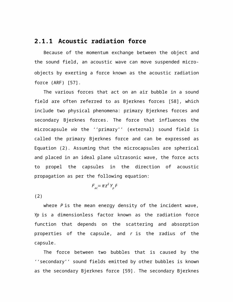
2.1.1 Acoustic radiation forceBecause of the momentum exchange between the object and the sound field, an
acoustic wave can move suspended micro-objects by exerting a force known as the
acoustic radiation force (ARF) [57].
The various forces that act on an air bubble in a sound field are often referred to as
Bjerknes forces [58], which include two physical phenomena: primary Bjerknes forces
and secondary Bjerknes forces. The force that influences the microcapsule via the
‘‘primary’’ (external) sound field is called the primary Bjerknes force and can be
expressed as Equation (2). Assuming that the microcapsules are spherical and placed in
an ideal plane ultrasonic wave, the force acts to propel the capsules in the direction of
acoustic propagation as per the following equation:
Fac=π r2 Y p P (2)
where P is the mean energy density of the incident wave, Yp is a dimensionless
factor known as the radiation force function that depends on the scattering and absorption
properties of the capsule, and r is the radius of the capsule.
The force between two bubbles that is caused by the ‘‘secondary’’ sound fields
emitted by other bubbles is known as the secondary Bjerknes force [59]. The secondary
Bjerknes force is given by Equation (3):
FB=¿F12≥−ρ
4 π d2 <V̇ 1 V̇ 2>er (3)
where < > denotes the time average, ρ is the density of the liquid, V1 and V2 describe the
volume of the first and second bubbles, respectively, and er denotes the radial unit vector.
The mutual interaction forces between two small coupled gas bubbles (R0<10 μm) in a

strong low-frequency acoustic field (Pa>1 bar, f = 20 kHz) have been investigated in a
previous study [59] using the Keller-Miksis equations. The nonlinear resonance of
bubbles leads to secondary Bjerknes forces between the bubbles that are stronger by a
factor of 103–106 than that expected from linear approximations.
The EPR effect would be considerably increased if the agent was initially
concentrated in a particular region, and if the velocity of the agent passing the potential
binding site was decreased. Dayton et al. [60] demonstrated that the radiation force of
ultrasound can displace a microbubble contrast agent to the wall of a 50-mm blood
vessel, significantly reduce the velocity of the flowing contrast agents, and produce
reversible aggregation. Such microbubble aggregates in large vessels were observed
optically under the influence of acoustic radiation force by Wang et al. [61]. The first
acoustic radiation force intravascular ultrasound transducer was designed to translate
microbubbles to the vessel wall under flow velocities comparable with that in the human
common carotid artery to enhance the binding efficiency of intravascular microbubbles
and drug delivery [62].
2.1.2 Acoustic streamingAcoustic streaming is defined as the radiation force from reflectors and scatterers in
an ultrasonic field that can cause local particle displacements and fluid currents. The
movement of localized fluid currents in the direction of the propagating ultrasound
radiation forces that are induced by sound waves is termed bulk streaming.
Microstreaming involves localized eddies or currents that are generated next to cavitating
bodies. The differences between bulk streaming and microstreaming are shown in Figure
3. The acoustic streaming effect can be used to displace particles in the blood [63] or
change the motion of blood flow [64]. These mechanical actions generated by ultrasound
may result in the release of drugs from carriers and the associated movement of the drug
into the targeted tissue [65]. Microstreaming with bubbles or particles plays an important
role in in vitro low-frequency sonophoresis [66].
With the same acoustic-pressure amplitude, the maximum streaming velocity of
ultrasound contrast agent (UCA) microbubbles is achieved under the action of resonance
frequency. The velocity fields of the streaming sonicated UCA microbubbles were

measured using particle image velocimetry (PIV) in a blood vessel model [67]. The
maximum streaming velocity was approximately 60 mm/s at a resonance frequency of
2.25 MHz, and the streaming velocity decreased to 15 mm/s at 1.0 MHz for the same
amplitude of acoustic pressure.
2.1.3 Stable and inertial cavitationGas-filled bubbles show different oscillation performances when acoustic
parameters such as the pulse length, amplitude, and frequency are varied. The stable
cavitation (SC) of UCAs is usually accomplished at low sound pressures (<0.1 MPa).
When exposed to sufficiently large acoustic rarefactional pressure amplitudes, UCAs
undergo a violent collapse known as inertial cavitation (IC) [68]. This phenomenon
results in the generation of shock-waves, broadband acoustic emissions [69], jetting [70],
and microstreaming [69]. These effects have been identified as key mechanical triggers
for various medical [40, 71] and industrial applications [72-74].
The difference between SC and IC is due to the different degrees of bubble
deformation under ultrasonic irradiation. Many models have been developed to describe
the deformations that can occur on the surface of a coated bubble because of oscillation.
Marmottant et al. [75] presented a model that incorporates the effect of coating on the
response of a microbubble to ultrasound. Based on the US-induced SC or IC, UCAs have
wide applications in medical diagnosis and treatment such as contrast harmonic imaging
and ultrasound-induced gene/drug delivery [76-78].
2.1.4 Mechanical-effect-enhanced EPR For US-induced SC or IC of MNBs, MNBs are either excited, causing them to
oscillate and mechanically massage the vascular wall, or are destroyed, generating micro-
jets. These mechanical effects can lead to the creation of pores in the cell membrane. The
opening of the inter-endothelial junctions contributes to an enhancement in the vessel
permeability and improves the extravasation of co-administered drugs [79].
MNB-mediated sonoporation occurs because of the physical perturbation of cellular
structures [80]. Marmottant et al. [81] controlled vesicle deformation and lysis by a single

oscillating bubble. They found that gentle bubble oscillations are sufficient to achieve the
rupture of lipid membranes.
The enhanced efficiency can be evaluated through the accurate measurement of the
spatial and temporal scales of these pores. Some methods are available to measure these
pores. SEM and atomic force microscopy (AFM) have both been employed to obtain
information about pores [82-85]. The transmembrane current (TMC) of a single cell
under a voltage clamp can also be used for monitoring sonoporation in real-time [86].
2.2 Thermal effects When a blood vessel is located near the focal spot, a part of the acoustic energy is
absorbed and converted to thermal energy. Tissues may have different sensitivities to
ultrasound-induced damage. This sensitivity to the effects of ultrasound-induced heating
has primarily been studied by Stanley et al. [87]. Thermal conversion efficiency is related
to protein content and blood perfusion. For example, it has been reported that the
absorption coefficient increases as a function of the protein content, which has a positive
effect on the ultrasound-induced increase in temperature. Furthermore, biological tissue
with poor blood perfusion can also be significantly heated by ultrasound because of slow
heat dissipation [87].
2.2.1 Thermal-effect-enhanced EPR Ultrasound energy can be absorbed and transformed into thermal energy in tissues as
the acoustic waves propagate through the tissue and generate friction [51]. Mild
hyperthermia in a tumor (41–43 °C for 10–60 min) may improve the therapeutic efficacy
of drugs by acting on tumor hemodynamics [88]. The mild hyperthermia induced by
focused ultrasound may, therefore, enhance drug or drug carrier extravasation in tumor
tissue [65]. In a previous study, a thermo-sensitive liposome was designed [89], and the
results showed that drug release can be triggered by mild hyperthermia when the
temperature is just above the normal physiological temperature.
Estimating the efficiency of the temperature rise caused by radiation of ultrasound is
critical. When the distance between the surface of the blood vessel and the focal point is
2.5 mm, the temperature at the focal point was saturated after approximately 50 s of
radiation, and the maximum cooling caused by the blood flow was approximately 0.5 °C
when the supply of radiation was ceased [90]. Based on the thermal wave model of
bioheat transfer, Tan et al. numerically investigated the influence of blood vessels on
temperature during high-intensity focused ultrasound hyperthermia [91]. The results
showed that a longer duration of thermal relaxation leads to the production of smaller
thermal lesions.
3. MNB-assisted ultrasonic evaluation and its enhancement on
EPR MNBs comprise a gaseous core encapsulated in a shell of biocompatible materials
such as lipids or proteins, typically ranging from 0.1 to 10 μm in diameter. When exposed
to ultrasound, MNBs can contract and expand, which may result in backscattering of the
ultrasound, cavitation, or even bursting of the shell. Because of these acoustic behaviors,
MNBs have been extensively used as UCAs and have shown increasing potential for drug
delivery. Under low-intensity ultrasound irradiation, MNBs can be used as contrast
agents in ultrasound imaging for EPR evaluation. Through high-intensity ultrasound-
mediated cavitation, EPR mediated therapy can be improved with enhanced drug
delivery.
3.1 MNBs and ultrasound response of MNBs
3.1.1 MNBsThe materials of the encapsulated gas and shell together determine the
physicochemical properties of a bubble. The transport properties of the encapsulated core
gas mainly affect the stability of the bubbles. Air and bioinert heavy gases such as sulfur
hexafluoride or perfluorocarbons are often used in commercial UCAs found in clinics.
One approach to increase the stability of UCAs is to use insoluble gases. For example,
perfluorocarbons have a higher resistance to water permeation than to air [92, 93].
Recently, bioactive gases that have significant therapeutic potential, including nitric oxide
[94, 95] and oxygen [96, 97], have been encapsulated in UCAs for parenteral delivery.
The performance of MNBs primarily depends on the physicochemical properties of
the shell material. Proteins, polymers, and lipids are mainly used as shell materials for
bubbles. The echogenicity of MBs and NBs is significantly different because of the size
difference. MBs possess good echogenicity and have been widely used for ultrasound
imaging. A reduction in the size reduces the acoustic response of the bubble[98]. The
microbubbles may be more useful in inducing vessel permeability than nanobubbles due
to their larger volume change and higher acoustic response under the ultrasound
radiation. The echogenicity of NBs can be improved via shell modification and bubble
aggregation. Research has shown that NBs are more stable and exhibit a longer
circulation time than MBs [99]. However, extravasation is different in MBs and NBs. The
size of MBs is usually in the order of a few microns. MBs are used as blood pool contrast
agents because they are transported after intravenous injection into the bloodstream
[100], where they either remain in circulation until taken up by the spleen or the liver or
are dissolved over short periods. Typically, tumor vessels are permeable to particles
smaller than 1 μm [101]. Based on these premises, MBs cannot escape the capillaries and
enter the defective tumor microcirculation via the EPR effect. Compared with MBs, NBs
can penetrate the sites where tumor vascular leakage takes place via EPR and can
accumulate in the tumor. The gaps between the endothelial cells in tumors, therefore,
allow passive targeting with NBs. Fluorescent dye-labeled NBs have been found in the
intracellular spaces of tumor cells [99]. NBs are also more stable and exhibit longer
circulation time than MBs, which leads to further accumulation in tumors. In vivo studies
have confirmed the ability of NBs to passively target tumor tissues via the EPR effect
[99]. The accumulation of NBs in the tumor area can lead to higher quality imaging and
more effective drug delivery. Cavalli et al. showed that amphiphilic drugs and negatively
charged complexes can be simultaneously loaded into a NB, which indicates their
theranostic capability [102].
3.1.2 Vascular permeability enhancement through ultrasound-
mediated MNBsThe mechanical effects that ultrasound-mediated MNBs have on the EPR are
associated with the process of cavitation, which is mainly due to the oscillation of gas-
filled bubbles in an acoustic field. MNBs have different responses to external stimulation

based on the features of the applied ultrasound, as shown in Section 2 in the review.
Under diagnostic frequency and low sound pressure (<0.1 MPa), bubbles expand and
contract with a small amplitude, and the generated energy is low enough to form an SC.
This regular oscillation can echo the ultrasonic wave utilized by ultrasound imaging. The
SC can create a circulating flow of fluid around the bubble, which is known as
microstreaming. Under higher sound pressures (>0.1 MPa), the oscillation of the bubble
becomes nonlinear and the expanded diameter is much larger, causing the microbubbles
to oscillate drastically. When the sound pressure exceeds a certain threshold, the bubbles
instantaneously collapse to release strong jets, shock waves, and shear force (IC). Both
SC and IC lead to the creation of pores in the cell membrane and the opening of
interendothelial junctions, thus enhancing vessel permeability and improving the
extravasation of co-administered drugs [38].
Inducing cavitation via MNBs changes the vascular and cell membrane permeability
of a tumor. Both SC and IC can cause mechanical disturbance to the vessel walls and cell
membranes in cancer tissue, thus improving the EPR effects in the tumor. The application
of ultrasound to vessels containing MNBs can change the permeability of the blood
vessel wall, resulting in the extravasation of drugs into the interstitial space. The change
in the permeability of the capillaries depends on various factors, including shell
composition, bubble size, the ratio of capillary diameter to bubble diameter, and
ultrasound parameters. In addition to changing the permeability of the blood-vessel wall,
the cavitation of MNBs can enhance the permeability of a cell membrane. The collapse of
a bubble and the associated production of a jet can instantly break adjacent cell
membranes. Small holes are produced within a cell membrane, resulting in either
repairable or non-repairable sonoporation. With different ultrasound parameters, short-
lived pores are produced within the cell membrane and exogenous materials can thus be
transported into the cytoplasm. The collapse of MNBs can also cause cell death in the
tumor tissue, which further alleviates solid stress and can reduce the barriers to deeper
penetration. Studies have shown that cavitation effects can alter vascular and cell
membrane permeability through three different mechanisms: (1) During the regular
mechanical disturbance of the oscillating bubbles undergoing SC, the cell membrane
potential is changed to promote endocytic uptake [103, 104]. (2) The volume of the

oscillating bubble changes during the transition from SC to IC. The gap between the
vascular endothelial cells is temporarily increased and the integrity of the vascular
endothelium is destroyed, thereby allowing enhanced diffusion of the active substances,
which can then enter the tissue [71, 105, 106]. (3) Based on the sonoporation produced by
IC, transient pores are generated within the vascular endothelial cells, which promote the
uptake of intracellular macromolecules [86, 107-109].
Cavitation is directly affected by various factors, such as ultrasound frequency,
intensity, and the length of US pulses, which in turn influence the EPR in the biological
tissues or cells. Low-frequency ultrasound (20–100 kHz) can penetrate the tissues deeper
than high-frequency ultrasound (1–3 MHz). Transdermal transport and cell penetration
are enhanced as the US frequency decreases [110, 111]. Yang et al. [112] experimentally
demonstrated the cavitation of polymer shelled MBs mediated by ultrasound fields of
different frequencies and sound pressures. MBs loaded with Fe3O4 nanoparticles ruptured
and released the nanoparticles into the cells under exposure to ultrasound. The results
showed that cells internalized the Fe3O4 nanoparticles released by the MBs most
efficiently at 1 MHz and 0.25 MPa. The pores in the cell membrane can be
simultaneously repaired by the lysosomal membrane associated protein (LAMP-1). When
the sound pressure exceeded 0.25 MPa, the severe nonlinear oscillation of the
microbubbles may have caused permanent damage to the cells. It has been found that
high-intensity focused ultrasound combined with microbubbles can open the blood-brain
barrier (BBB), which provides a means for treating brain tumors. Pulsed US causes less
negative biological interactions with healthy cells and tissues not intended for treatment
[113]. For medical purposes, the use of pulsed US is, therefore, more favorable than the
use of CW. High-intensity focused ultrasound (HIFU), which can focus US intensity at
the target site, has been developed for ablating various types of live tissue [114]. This
treatment is known to have irreversible thermal effects, including hemostasis and blood
vessel occlusion. However, the pulsed-HIFU (pHIFU) treatment can reduce these
irreversible thermal effects by reducing the average intensity of US at the target tissue.
This pulsed-HIFU (pHIFU) method has been developed to enhance the extravasation and
tissue penetration of nanoparticles in the femoral tissue of mice [115].
In addition to cavitation, MNBs can also generate other biological effects, such as

thermal effects, under the guidance of high-intensity ultrasonic energy. These biological
effects work together to temporarily enhance the permeability of biological tissues and
blood vessels, allowing the active substances carried in the MNBs to enter the tumor
tissue more easily through the EPR effect, which is the theoretical basis of ultrasound-
assisted MNB-enhanced EPR.
3.2 MNB-assisted ultrasonic evaluation of EPR The high heterogeneity of EPR among individual patients is considered a major
barrier to the clinical application of nanomedicines. In preclinical validation, drug
delivery systems generally work very well, with significant improvements in both target
site accumulation and therapeutic efficacy. Clinically, however, the efficacy of passive
tumor-targeted nanomedicines is compromised because of the large inter- and intra-
individual variability in EPR. Significant increases in tolerability but hardly any increases
in efficacy have been seen in clinical therapy [10, 11, 19]. Consequently, it is necessary to
develop methods to visualize and characterize the EPR effect before treatment. Based on
the assessment of individual EPR, patients presenting with sufficiently high levels of
EPR, pre-stratify responders, and non-responders could be pre-selected to individualize
and improve nano-chemotherapeutic treatments.
Ultrasound contrast imaging is a simple, non-invasive imaging modality used to
evaluate vascular conditions. However, tumors are characterized by tortuous vascularity,
angiogenesis, and hypoxia. To address these problems, different strategies for MNB-
enhanced ultrasound imaging based on EPR have been developed. One important factor
to be considered in ultrasound imaging is the accumulation of MNBs in tumor tissue. As
mentioned above, NBs of an appropriate size can penetrate the leakage sites of tumor
vascular tissue. EPR-mediated passive targeting can result in the accumulation of NBs in
the tumor. However, the enhancement of ultrasound imaging by nanobubbles is reduced
relative to microbubbles [115] in clinical applications. Therefore, it is important to
maintain its ability to enhance ultrasound imaging while the bubble size is reduced. A
sufficient aggregation of NBs within the tumor is beneficial for a high quality image and
can be used for enhanced ultrasound imaging of a tumor [116]. The shell composition of
the bubbles can also be adjusted to increase the echo signals of bubbles in an ultrasonic

field. Studies have shown that the incorporation of magnetic nanoparticles into bubble
shells can enhance ultrasound imaging [117, 118], which could be used to improve the
imaging ability of NBs.
Although MBs cannot pass through blood vessels and enter tumor tissue, efforts can
be made to allow them to accumulate around the blood vessels of the tumor.
Angiogenesis is a remarkable characteristic in the tumor microenvironment. Endothelial
cells and receptors associated with angiogenesis are readily accessible for targeting with
MBs. The conjugation of ligands onto the surface of MBs has already been proven
successful for MB accumulation in tumors for imaging purposes. It has been reported that
a better ultrasound signal was detected from MBs in angiogenic areas after the
conjugation of arginylglycylaspartic acid (RGD) peptides onto the MB surfaces [119].
Another important factor in contrast ultrasound imaging for EPR characterization is
to identify the correlation between the imaging parameters of tumor blood vessels and
drug accumulation in tumors. A US-based assessment of tumor vascularization can
potentially be used to predict the efficiency of passive tumor targeting. EPR assessment
based on ultrasound imaging is simple, general, and easily transformed for clinical use. In
an exemplary preclinical study [28], the relative blood volume (rBV) in tumors was
assessed using contrast-enhanced US imaging. The rBV values were correlated with the
tumor accumulation of HPMA-based polymeric drug carriers. A good positive correlation
was observed, supporting the fact that imaging vascular parameters such as rBV may be
useful in predicting EPR-mediated tumor targeting. Applying multi-modal imaging (MRI,
μCT, US, and microscopy), Sulheim correlated the accumulation of polystyrene
nanoparticles in different tumor models with parameters such as the functionality of
tumor vessels measured via the inflow of MBs using US [31]. The density of functional
blood vessels measured by fluorescence microscopy was significantly correlated (p =
0.0056) with nanoparticle accumulation, and the inflow of microbubbles measured with
ultrasound also showed a moderate, but significant (p = 0.041), correlation with
nanoparticle accumulation, indicating that both the number of vessels and the vessel
morphology and perfusion can predict nanoparticle accumulation. This indicates that
blood vessel characterization using contrast-enhanced ultrasound imaging or other
methods could be valuable in patient stratification for treatment with nanomedicines.

3.3 MNB-assisted ultrasonic enhancement of EPR MNBs can act as carriers for therapeutic payload delivery. Therapeutic payloads
including therapeutic gas, targeting ligands, drug molecules, and nanoparticles can be
loaded into the bubble.
The delivery of drugs used for chemotherapy suffers from many problems including
severe side effects and the resistance of tumor cells [120]. MNBs can be developed as
multifunctional carriers for drug transport and delivery. The drugs encapsulated in MNBs
are protected from intracellular reactions. MNBs can mediate drug delivery in tumors
through EPR effects or active targeting. Particularly, when combined with ultrasound,
drug delivery can be controlled and enhanced in tumors. A fluid flow is created when
MNBs undergo oscillation mediated by ultrasound [121]. This phenomenon increases the
drug convection inside the blood vessels and elevates the accumulation of a drug in the
surrounding tumor tissues through the EPR effect, resulting in the increased therapeutic
index of a drug [121, 122]. Local US application can further induce a transient increase in
the endothelial cell membrane permeability to enhance the uptake of therapeutic agents
by the target cells. Fan et al. proposed a boron-polymer/microbubble complex (B-MB)-
assisted focused ultrasound (FUS) treatment for brain glioma [123]. B-MBs can
simultaneously achieve the safe opening of the blood-brain tumor barrier and enhance
boron drug delivery into the tumor tissue using FUS sonication. Cao et al. reported the
release of an anticancer drug (DOX) from phase-changeable nanodroplets triggered by
low-intensity FUS (LIFU) [124]. They found that the intratumoral accumulation and
distribution of a drug with LIFU exposure was significantly enhanced. The combination
of ultrasound, microbubbles, and gemcitabine is now under phase-II clinical trials for the
treatment of pancreatic cancer [125].
Nanoparticles (NPs) such as gold and superparamagnetic iron oxide NPs are widely
used in tumor diagnosis and treatment. The delivery of these NPs at the tumor-specific
site is a challenging task. Small nanoparticles (<5 nm) can be rapidly removed from
vessels by renal clearance or uptake by the liver, whereas large particles (>200 nm) are
removed by the spleen or the reticuloendothelial system (RES) [126, 127]. NPs can be
delivered to specific cancer tissue only by designing a proper carrier system and avoiding

the delivery of the drug to normal tissues. MNBs can be used as carriers to deliver NPs to
the tumor tissue site via the enhanced EPR effect. NPs are often loaded into the shell of
MNBs and delivered to tumor cells in a process controlled by ultrasound. A typical
example is the delivery of superparamagnetic iron oxide (SPIO) NPs through MNBs.
Different types of bubble related carriers including magnetic microbubbles [128] and
magnetic nanoliposomes [129] have been developed for in situ microbubble generation.
Combined with US, SPIO NPs were locally released at the tumor and taken up by the
tumor cells though EPR effects. These bubble-based magnetic carriers offer an attractive
possibility for clinical MR imaging, hyperthermia, and the magnetically targeted release
of therapeutic agents for cancer.
Gaseous molecules including oxygen (O2), nitric oxide (NO), hydrogen sulfide, and
carbon monoxide have important physiological roles or can elicit biological responses.
Therefore, they have significant therapeutic potential [130]. These bioactive gases are
much smaller than classical drugs and can easily diffuse across the barriers within vessels
and cell membranes [130]. While the therapeutic effect of a gas is concentration
dependent, a major hurdle in biomedical application is the controlled and site-specific
delivery of the gas. Compared with solid carriers, MNBs are good containers for gas,
with effective loading and controlled release. It is estimated that at least 50–60% of
advanced solid tumors contain hypoxic tissue, which is a significant factor leading to the
resistance of tumors to treatment [131]. Vaupel et al. developed O2 nanobubbles for
spontaneous oxygenation in response to the tumor microenvironment [131]. The
intratumoral O2 level can be significantly increased by the O2 released from O2
nanobubbles that have been passively accumulated within the tumor because of the EPR
effect. NO plays a critical role in vascular physiology and cancer biology. At low
concentrations, NO promotes tumor cell growth by stimulating angiogenesis, while at
high concentrations, NO is cytotoxic and may be a useful chemotherapeutic agent [132].
MNBs can protect NO from endogenous scavengers and provide an opportunity for
targeted delivery, thus providing a therapeutic potential for cancer.
4. Advanced ultrasonic carriers and manipulationMNBs can effectively enhance the ultrasound echo signals for evaluating the EPR

effect of a tumor; at the same time, modifications can be made to the membrane shell
structure, allowing the easy loading or coupling of other contrast agents, drugs, and
targets. Mediated by an ultrasonic field, MNBs can also lead to various useful biological
effects such as cavitation and hyperthermia to enhance EPR and achieve a synergistic
interaction. Therefore, studies on MNB-based theranostic drug-loading systems have
received extensive attention in recent years.
4.1 Adjustable size modulation strategyThe EPR effect is strongly dependent on the size of nanomedicines. If the
nanomedicines are too large, they will not be able to leak through the tumor blood vessels
into the solid tumor. If they ate too small, they will be quickly removed by the kidneys.
Even if the nanoparticles reach the tumor tissue through the blood vessels, the difference
in size affects their ability to spread further. Therefore, the design of a drug carrier with
adjustable size can promote the penetration and enrichment of a drug inside a tumor. The
special properties by which MNBs respond to an external field can be used to change the
size of MNBs under the guidance of an external field such as ultrasound and temperature.
This can be achieved by devising ingenious designs of the membrane and the gas core
component, thus forming an effective diagnostic and therapeutic carrier. At present, two
strategies are applied for size adjustment of MNBs: one is "small to large" and the other
is "large to small".
The “small to large” strategy is mainly used in EPR targeting to overcome the
contradiction between particle size and the sound intensity of the contrast agent.
Nanobubbles exhibit in vivo stability and can even reach the interstitial space through the
vascular endothelium, providing enhanced tissue imaging and obtaining more abundant
imaging information. Thus, nanobubbles are useful to evaluate the EPR effect. However,
the clinical practicality of using ultrasound imaging with small-sized bubbles is
considerably lower [133]. It is, therefore, important to maintain the imaging ability when
the bubble size is reduced. One typical class of “small to large” bubbles are phase-
inversion UCAs, as seen in Figure 4 [134, 135]. Nanoparticles or nanodroplets containing
liquid fluorocarbons have been extensively explored and designed. The nanoscale and
internal liquid environment promotes prolonged stability and tissue penetration. When the

external temperature changes under the trigger of ultrasonic energy, the liquid
fluorocarbon inside undergoes a liquid-to-gas phase transition and the volume increases
accordingly, resulting in enhanced ultrasonic imaging at the lesion site. Finally, the
bubble bursts, and the drug that is released in the target region may exert a further
synergistic therapeutic effect. Min et al. developed tumor-homing echogenic chitosan-
based nanoparticles, in which bioinert perfluoropentane (PFP) was encapsulated into
glycol chitosan nanoparticles [136]. These particles showed prolonged echogenicity via
the sustained microbubble formation process in the liquid-phase PFP at body
temperature. A significant increase was found in the tumor-homing ability through the
EPR effect of the nanoparticles, and this was used for cancer theranostics. Liu et al.
prepared folic-acid-modified nanodroplets (FA-NDs) with phase-change ability, which
undergo a liquid-gas phase transition under LIFU irradiation [137]. FA-NDs were found
to be enriched in the tumor area under LIFU, and the resulting micron-sized MBs could
enhance the ultrasound imaging signals.
The "big to small" strategy is mainly used to help microbubbles bypass the EPR
effect and release drugs at the tumor site. Micro-sized bubbles are good angiographic
contrast agents and drug carriers, but they cannot penetrate blood vessels to reach the
tissues. If MBs can somehow be made smaller to become nanobubbles after reaching the
tumor blood vessels and completing the imaging function, they could spread across the
vascular barrier and into the tumor tissue to be used for further tissue imaging and drug
release. Huynh et al. reported a method used for the conversion of microbubbles to
nanoparticles using low-frequency ultrasound, as shown in Figure 5 [138]. Porphyrin
loaded microbubbles can be used as tri-modality contrast agents for ultrasound,
photoacoustic, and fluorescent imaging. During exposure to ultrasound, the microbubbles
burst and formed smaller nanoparticles, which maintains the imaging and therapeutic
properties and allows drug delivery to the tumor. With this conversion, microbubbles can
potentially be used to bypass the EPR effect when delivering drugs to tumors.
Therefore, through the delicate design of the bubble structure and the appropriate
introduction of external triggers, a new size-controllable theranostic agent with
ultrasound enhancement effect and tissue penetration ability can be constructed to
evaluate and enhance the EPR effect.
4.2 In situ generationDifferent types of stimuli have been reported to trigger the in-situ generation of
bubbles in a solid tumor. These types of stimuli are usually endogenous and are closely
related to changes in the tumor microenvironment. Agents that are responsive to such
stimuli are encapsulated in nanocarriers, which accumulate at the tumor site through the
EPR effect. Stimuli are then applied, thus triggering the generation of gas free bubbles at
the tumor tissue. The advantages of this in situ generation strategy are as follows: (1) The
bubbles generated in situ at the tumor site do not need to be transported through blood.
They can, therefore, avoid various biological barriers in the human body and can also
bypass the requirement for EPR in changing the bubble size. (2) The generation of
bubbles enhances the ultrasound imaging effect at the tumor site, which is conducive to
diagnosis. If the generated gas can also be used for treatment, targeting, diagnosis, and
treatment can be integrated.
For example, the production of reactive oxygen species (ROS) is much higher in
cancer tissues than in normal tissues. H2O2 is an ROS and a diagnostic marker in the
development of certain diseases. Studies have found that Prussian blue nanoparticles have
a catalase-like function in neutral environments, catalyzing H2O2 to produce oxygen
[139]. When the oxygen generated exceeds the saturation concentration, it can form free
oxygen bubbles, enhancing ultrasound imaging. Together with the MRI properties of
Prussian blue nanoparticles, this could be used as an ultrasound/magnetic resonance
bimodal imaging system which is triggered by the disease environment [140]. Similarly,
Wang et al. [141] constructed a multifunctional carrier system loaded with two catalase
enzymes and SPIO. The two enzymes catalyze the reaction of the ROS in the body under
pathological conditions to produce oxygen that can be utilized for ultrasound imaging, as
can be in Figure 6. The changes to the reaction environment also enhance the MRI
capabilities of SPIO within the system.
4.3 Multi-gradient targetingIn the above studies, the aggregation of bubbles and release of drugs in the target
region were mainly dependent on various exogenous and endogenous stimuli and were
not caused by specific active targeting. EPR and the (over-) expression of angiogenesis-
related surface receptors are key features of tumor blood vessels. As a consequence, EPR-
mediated passive and ligand-based active targeting have attracted considerable attention.
Kunjachan et al. visualized and quantified passive and active tumor targeting using RGD-
and NGR-modified polymeric nanomedicines [142]. Wu et al. developed paclitaxel-
loaded and A10-3.2 aptamer-targeted poly(lactide-co-glycolic acid) nanobubbles, which
can be aimed specifically at prostate cancer cells to sustainably release the loaded PTX
via the EPR effect and US-triggered drug delivery [143]. Li et al. reported the use of
neuropeptide Y Y1 receptor-mediated biodegradable photoluminescent nanobubbles as
UCAs for targeted breast cancer imaging [144].
Rapid and efficient early binding of MNBs to the blood vessels of the tumor occurred
through vascular targeting, but passive targeting was significantly more efficient over
time. These results indicate that a combination of passive and active targeting is required
for effective tumor imaging and therapy. Based on the above ideas, a multi-gradient
targeting strategy has emerged. Multi-gradient targeting strategies refer to the further
coupling of specific targeting factors on MNBs to combine active targeting with EPR-
mediated passive targeting. When such MNBs are intravenously injected into the body,
they become actively enriched in the tumor blood vessels under the action of specific
targeting ligands, contributing to clearer ultrasound imaging. Following the application of
an ultrasound field or various other internal and external sources of stimulation, the
structure and/or the size of the MNBs changes. On the one hand, this change triggers
various biological effects, which affect the permeability of the tumor blood vessels and
enhance the EPR effect. On the other hand, the drug is released, forming a high drug
concentration gradient in the microcirculation of the tumor tissue, efficiently allowing the
drug to enter the tumor tissue for treatment by virtue of the EPR effect.
Several classes of ligands have been conjugated onto the surface of UCAs to achieve
better targeting of tumor cells, including peptides, aptamers, and affibody molecules.
These targeting ligands allow the UCAs to remain at a size at which the EPR effects can
be exploited [145]. Cell membranes are a new type of shell material that have been used
in the construction of bubbles. The inherent properties of natural lipids and protein
components on a membrane can be used for natural targeting. For example, platelet

membranes are rich in natural vessel adhesive components such as α2β1, α5β1, α6β1, and
αbβ3, and the glycoproteins, GPIb-IX-V and GPV, which can naturally target injured
vasculature [146]. Mingxi Li et al. used platelet membranes to fabricate bio-nanobubbles
and demonstrated their lesion-targeting ability [147]. The abnormal vasculature within
tumors provides the possibility of using bio-shelled bubbles for the targeting of tumors.
Active targeting can be performed by modifying bubble shells using a range of
targeting ligands; such bubbles can therefore be applied in different types of cancer.
There are several strategies that allow the binding of ligands to the bubble shell. First,
avidin or streptavidin is commonly used for non-covalent attachment of biotinylated
ligands onto the shell materials [148]. Yang et al. used the avidin-biotin method to
combine NBs with biotinylated anti-ErbB2 Affibody® molecules [149]. The resulting
NB-Affibody had a specific affinity for human epidermal growth factor receptor type 2
(HER2)-over-expressing tumors. Second, the covalent binding of ligands to the shell has
also been explored. One approach involves the binding of activated carboxylic groups to
shells with amino-groups from the lysine residues of binding ligands [145]. In a third
approach, the ligands are coupled with the shell components (e.g., to phospholipids),
followed by the formation of MBs. Chung-Hsin Wang et al. conjugated sgc8c aptamers to
functional lipids and prepared DOX-loaded acoustic droplets [150]. The fully vaporized
droplets resulted in the highest DOX uptake by cancer cells.
Multi-gradient targeting can be applied to both microscale and nanoscale bubbles.
Nanoscale bubbles modified with targeting ligands are good carriers. Li et al. reported
that the soft biodegradable glycine/PEG/RGD-modified poly (methacrylic acid)
nanobubbles can be applied as intelligent theranostic vehicles for drug delivery [144].
Gao et al. developed the targeted ultrasound-triggered phase transition nanodroplets for
Her2-overexpressing breast cancer diagnosis and gene transfection [151]. For micron-
sized bubbles, Duan et al. [128] demonstrated a multi-gradient continuous targeting
strategy based on the RGD-l-TRAIL-labeled magnetic microbubbles shown in Figure 7
for cancer therapy. The designed RGD-l-TRAIL@MMB system had a complex but
elaborate multilayer structure: gas in the core for US imaging, SPIO on the shell for MRI
imaging and magnetic targeting control, and RGD-l-TRAIL ligands for an enhanced
combination of the tumor-targeted delivery of TRAIL. First, the RGD-l-TRAIL protein
on the MMB surface enables the microbubbles to actively target angiogenesis of a tumor.
The size of the micro-scaled bubbles around the tumor is beneficial for real-time US
imaging and MRI to clearly delineate the tumor margin. Therefore, it is micron-scale
targeting and diagnosis. Second, after US imaging, the microbubbles in situ around the
tumor rupture and the nanoscale SPIOs with associated RGD-l-TRAIL molecules can
travel through the blood vessels and enter the tumor tissue because of the tumor ligand
induced integrin αvβ3-receptor-mediated endocytosis. At this stage, the tumor tissue can
also be imaged using MRI. With the accumulation of RGD-l-TRAIL and SPIOs in the
tumor tissue and cells, tumor apoptosis induced by TRAIL molecules can also be
observed during this process; therefore, this process involves nanoscale targeting and
diagnosis. The multi-gradient targeting drug delivery system has a reasonable structure,
an adjustable drug dosage, and a clear target. It is a typical example of applying a multi-
gradient targeting strategy for in vivo diagnosis and treatment.
4.4 Multi-field associationIn addition to the ultrasound field, external factors including light, magnetic fields,
radiation, and microwaves affect EPR. Internal factors such as pH, high concentrations of
active oxygen, and various special cytokines are provided by the special pathological
environment of the lesion. The mechanism by which each factor affects the EPR is
different. Combining multiple energy fields can exert the combined effects of all of the
different mechanisms to obtain an optimized and enhanced EPR effect.
Local heating of the tumor site tends to increase the accumulation of nanomedicine,
resulting in more effective intratumoral drug delivery. Hyperthermia enhances the
increase in nano drug delivery within tumors because of the increased blood flow to the
tumor caused by vasodilation. This increased vascular permeability is due to the
increased intercellular space between endothelial cells [152-154]. Through the
photothermal conversion effect, the irradiation of a tumor with a laser can also change the
physiological state of the tumor and enhance the penetration of the nano drug into the
tumor. By constructing a versatile theranostics carrier that responds to light and other
external fields, thermal effects can be combined with other effects to improve vascular
permeability. Ke et al. [155] constructed a multifunctional nanocapsule (NC) by loading

perfluorooctylbromide (PFOB) and superparamagnetic iron oxide nanoparticles into
poly(lactic acid) (PLA) NCs, followed by the formation of PEGylated gold nanoshells on
the surface. Such a theranostic agent could efficiently kill tumor cells using NIR laser
irradiation under the guidance of contrast-enhanced US/MRI to achieve great therapeutic
effectiveness without systemic toxicity to the patient. Above all, the bimodal imaging
guidance could display dynamic complementary information about a tumor to help alter
the treatment on an individual basis with high efficiency.
The strategy of combining internal and external stimuli is also beneficial. Yang et al.
[156, 157] constructed a composite microcapsule with internally encapsulated L-arginine,
SPIO in the membrane shell, and glucose oxidase on the surface, which is a double-
response, microcapsule transport system that reacts to both glucose and external magnetic
fields, as shown in Figure 8. First, the glucose oxidase on the surface of the microcapsule
specifically reacts with glucose in diabetic patients, reducing the glucose concentration
and generating H2O2 and gluconic acid. The magnetic nanoparticles in the microcapsule
shell then move under the action of the external magnetic field to generate gaps in the
membrane, and the internally loaded L-arginine further reacts with the H2O2 to form NO.
Therefore, it is apparent that through the dual response of the microcapsule structure to
glucose and magnetic fields, high concentrations of glucose can be reduced and the
generated NO gas can enhance the ultrasound imaging and the EPR effect, thus treating
the disease. In addition to responding to external magnetic fields, the SPIO in the system
also functions for MRI imaging. This system can be used as a multifunctional carrier
platform for the effective regulation of blood glucose.
Liu Yang et al. [129] designed a liposome drug delivery system, as seen in Figure 9,
which is a complex structure of a therapeutic gas prodrug, anthranil trisulfide, and
magnetic nanoparticles, which is efficiently concentrated at the targeted tumor site by the
EPR effect under the control of an external magnetic field. Under the catalysis of
biological enzymes such as CBS and CSE in the tumor, hydrogen sulfide is produced,
which has antitumor properties. The liposome-loaded magnetic nanoparticles respond
specifically to the external magnetic field. The intratumoral targeted aggregation of
liposome can monitored and regulated by MRI; and the hydrogen sulfide microbubbles
catalyzed by the enzyme enhance the intratumoral ultrasound contrast, leading to a

greater intensity of ultrasound energy. The microbubble then ruptures, ultrasonic
cavitation leads to physical tumor ablation, and the antitumor properties of the hydrogen
sulfide itself lead to necrosis of the original dense intratumoral tissue, creating favorable
conditions for the use of nanocarriers. This liposome drug system can achieve better
theranostic destruction of a tumor under ultrasound/magnetic resonance bimodal image
monitoring.
There are also multifunctional carrier systems with sophisticated designs that
combine lasers, heat, radiation, and other external and ultrasonic fields to improve the
permeability of tumor blood vessels and tissues and deliver drugs effectively.
The ideal process for tumor diagnosis and treatment based on the EPR effect is
shown in Figure 10. Before starting treatment, an accurate individualized assessment of
the EPR level should be performed for each individual patient, and different treatment
options should be developed based on the results of the assessment. For patients with
high EPR levels, an appropriate drug delivery system can be selected for treatment. For
patients with low EPR levels, the EPR effect should be enhanced using various physical
and pharmacological methods before treatment. Would it be possible to construct a
multifunctional theranostics carrier that can play a role in all aspects of the evaluation,
enhancement, and treatment, realizing the integration of EPR evaluation, enhancement,
and EPR-based tumor treatments on a single platform? MNB-based drug delivery
systems have become a potential choice because of their special properties such as
imaging, drug release, changing structures, and production of various biological effects
that can be mediated with ultrasound fields.
Around the process of tumor diagnosis and treatment and applying the four strategies
mentioned, the potential of MNBs can be described in detail, as seen below in Figure 10.
Firstly, MNBs can be used as contrast enhancers to visually and quantitatively evaluate
the EPR levels of a patient. The accuracy of the assessment depends on two key
processes: (1) Whether MNBs can efficiently reach tumor blood vessels through blood
circulation, and (2) whether a strong echo signal can be generated after reaching the
tumor site. The various strategies mentioned can have a positive effect on both of these
processes. As described previously, the "small to large" size control strategy has been
applied to construct small-sized nanodroplets that have higher stability in the blood

circulation than MNBs. A multi-level targeting strategy can also be applied to couple the
targeting factors on the surface of the nanodroplets to reach the tumor blood vessels more
efficiently. In the latter process, through the mediation of an external energy field, the
liquid droplets within the nano-droplet core can be transformed, thus increasing the
volume to generate a stronger echo signal which is beneficial to ultrasound imaging. It is
also possible to use the "in situ generation" strategy to generate bubbles in situ using the
specific microenvironment of the tumor to create ultrasound contrast with which to
evaluate the EPR.
Second, for patients with low EPR levels, MNBs can be combined with internal
biochemical triggers and external energy fields to enhance the EPR effect using various
strategies, as shown in Figure 10. For example, the "large to small" size control strategy
facilitates the micron-sized bubbles to bypass the EPR effect and become smaller
nanobubbles that can penetrate the vascular endothelial cell gap, allowing movement into
the tumor tissue. The “multi-field association” strategy comprehensively uses light, heat,
ultrasound, radiation, and various endogenous factors to increase vascular permeability
and enhance the ability of the drug components carried by MNBs to spread out of the
blood vessels. The “multi-gradient targeting” strategy allows the drug released after the
rupture of a bubble to utilize molecular targeting to efficiently reach the tumor tissue
through the blood vessel.
In addition, while enhancing the EPR effect, various energy fields can mediate the
gradual disintegration of the structure of MNBs, releasing the various active substances
the MNBs carry, including drugs, other nanoparticles, and therapeutic gases. These active
substances are enriched in tumor tissues by the enhanced EPR effect and lead to further
therapeutic or real-time imaging functions.
5. Summary and future perspectivesIn the complex and variable micro-environment of a living organism, the key to
effective treatment is drug components reaching a lesion efficiently and exerting
continuous therapeutic effect. The nano carrier targeted drug delivery system, based on
EPR, is one way to effectively improve the anticancer effect of drugs. In recent decades,
various forms of multifunctional carrier materials such as liposomes, MNBs, micelles,

nanoemulsions, magnetic nanoparticles, polymer nanoparticles, and dendrimers have
been designed and developed to improve the aggregation and permeability of the drug at
the tumor site. In this review, we focused on the effects of ultrasound and MNBs on EPR,
discussed in detail the potential mechanisms by which their respective roles and synergies
affect EPR, and illustrated new strategies that have been developed to enhance EPR.
Although these new strategies do show promise, further research is needed in this area to
demonstrate effective drug delivery and therapeutic effects. In this study, we discuss the
advantages of MNB-assisted ultrasound to enhance the EPR effect, the current problems,
and any possible countermeasures.
5.1 AdvantagesMNBs are distinctly distinguished from other carrier materials in that they have
multiple forms of response to the ultrasound field, and the rich biological effects of these
responses have a potential role in both EPR imaging and enhancement processes. Under
ultrasound energy mediation, the MNB theranostic carrier can affect the EPR in
combination with various favorable conditions. First, ultrasound and MNBs can both
affect the EPR. The ultrasonic streaming and hyperthermia effect from the ultrasound
itself can improve the ability of a drug to diffuse into a tissue, promote the extravasation
of drugs, and enhance the permeability of blood vessels. MNBs can also be used as drug
carriers with certain EPR effects using various strategies such as structural design, size
control, and modification of the membrane shell. Second, ultrasound can be used to
further assist MNBs, which are imaged by low-intensity ultrasound energy to evaluate
EPR. MNBs can also rupture under high-intensity ultrasound energy and produce
biological effects such as cavitation, which leads to the more efficient transport of a drug
into the tumor tissue through the vascular endothelial cells. Through sophisticated design,
the internal and external source energy fields can be combined, making full use of
“adjustable size modulation”, “in situ generation”, “multi-gradient targeting”, and “multi-
field association” strategies, which can further improve the penetration ability and
delivery efficiency of drugs at the tumor site.
5.2 Challenges and countermeasures

5.2.1 Size barrierThe size of the MNBs is still a barrier to the EPR effect. The efficacy of a diagnostic
and therapeutic agent through the EPR effect is size-dependent. Related research has
shown that liposomes with a hydrodynamic particle size between 100 and 200 nm
showed a 4-fold higher accumulation in tumors than liposomes with a particle size of less
than 50 nm and above 300 nm [158][188]. The preparation of nanobubbles with a size of
less than 200 nm remains challenging. Although studies have shown that ultrasound can
rupture lipid microbubbles in the body and form smaller nanoparticles with the same
optical properties as the original microbubbles, meaning that micron-sized bubbles can
bypass the EPR effect when delivering drugs to the tumor, there is still a need to develop
small-sized nanobubbles with the ability of enhanced accumulation. Therefore, it is
critical that new preparation processes are developed, and that various adverse problems
such as the weak imaging effects caused by small-sized nanobubbles and the decreased
drug-loading capacity are dealt with. The combination of NBs with other nanocarriers is
one possible way to increase the efficiency of these bubbles in terms of drug delivery.
5.2.2 Structural complexityMulti-modal imaging, in which targeting factors are coupled with the response to
various energy fields and other functions, makes the structure of MNBs complex, thus
increasing the difficulty in constructing carriers. Therefore, it is necessary to further study
and optimize the preparation method of MNBs theranostics carriers, improve the
fineness, controllability, and repeatability, and finally to achieve standardization. It is also
necessary to further study the effects of MNB carrier structures on their function, such as
the material from which membrane shells are built, size, drug-loading mode, and the
effects of target type on imaging and targeting. In addition, in the complex and variable
microenvironment of the body, it is very challenging to simultaneously achieve a targeted
system with high diagnostic sensitivity, high specificity, and good therapeutic effect. The
more complex a structure is, the more risk there is, so it is difficult to anticipate the
realization of a function and to achieve clinical transformation, especially when using
relatively large targeting systems (hundred nanometers and above) such as MNBs. The
sensitivity of different biological tissues to ultrasound is quite different, so the applied

ultrasound parameters will need to be changed accordingly. The ultrasound parameters
used to stimulate the MNBs would also need to be adjusted for use with the different
kinds of bubbles.
More in vivo experiments are needed to clarify the toxicity, distribution, and
pharmacokinetics of a carrier within the body and to provide abundant evidence for
clinical application. It is foreseeable that with the continuous development of biomedical
nanotechnology based on ultrasound and MNBs, more innovative and more powerful
theranostics carriers will be designed and developed to further promote tumor-targeted
therapy.
5.2.3 Potential safety concernsOne of the important principles of drug delivery system development is safety. The
safety issues for micro/nano-bubble-assisted ultrasound to enhance EPR effects, such as
micro/nano-bubble toxicity [159-161] and damage to vessels through acoustic energy still
need to be fully investigated on priority for future clinical applications [162, 163]. The
safety issues related to ultrasound have been addressed in Section 2 of this review. The
biodegradability and metabolites of ultrasound contrast agents should be investigated.
Although some studies have investigated the short-time toxicity, safety tests for
biocompatibility, acute toxicity, subacute toxicity, carcinogenicity, developmental
toxicity, immunotoxicology, genotoxicity, and irritation to blood vessel should be
conducted in the future, especially when MNBs are coupled with ultrasound energy.
AbbreviationsEPR: permeability and retention; UCA: Ultrasound contrast agent; TI: thermal index; MI:
mechanical index; FDA: Drug Administration; PnP: negative pressure; CW: continuous
wave; DOX: Doxorubicin; GNPs: gold nanoparticles; DDS: drug delivery system; PIV:
particle image velocimetry; SC: stable cavitation; IC: inertial cavitation; SEM: scanning
electron microscopy; AFM: atomic force microscopy; TMC: Transmembrane current;
CAD: computer-aided detection; UOM: ultrasound-modulated optical microscopy;
CEUS: contrast-enhanced US; IVUS: intravascular ultrasound; BFR: blood flow
resistance; MNBs: micro/ nanobubbles; UCAs: ultrasound contrast agents; PLGA: poly-

(lactic-co-glycolic acid); rBV: relative blood volume; BBB: blood-brain barrier; B-MB:
boron-polymer/microbubble complex; FUS: focused ultrasound; LIFU: low-intensity
focused ultrasound; NPs: nanoparticles; RES: reticuloendothelial system; PFP:
perfluoropentane; FA-NDs: folic-acid-modified nanodroplets; pMBs: porphyrin
microbubbles; pNPs: porphyrin nanoparticles; ROS: reactive oxygen species; PFOB:
perfluorooctylbromide; PLA: poly(lactic acid); NCs: nanocapsules; PSV: peak systolic
velocity; SGC: SPIO@GCS/acryl/biotin-CAT/SOD-gel; PFC: perfluocarbon; AML: the
anethole dithiolethiones (ADT)-loaded magnetic nanoliposomes; CBS: cystathionine-β
synthase; CSE: cystathionine-γ-lyase ; MMB: magnetic microbubbles
Acknowledgements This investigation was financially funded by the National Key Research and
Development Program of China (2017YFA0104302, 2018YFA0704103), the National
Natural Science Foundation of China (51832001, 61821002, 81701833 and 81971750),
Natural Science Foundation of Jiangsu Province (BK20191266), and the Natural Science
Foundation of Anhui Province (1808085QH233). Funding also partially comes from the
Six Talent Peaks Project of Jiangsu Province (2017-SWYY-006) and Zhong Ying Young
Scholar of Southeast University. The authors also would like to thank the support from
the Fundamental Research Funds for the Central Universities.
Competing InterestsThe authors have declared that no competing interest exists.
References
1. Papetti M, Herman IM. Mechanisms of normal and tumor-derived angiogenesis. Am J Physiol Cell Physiol. 2002; 282: C947-C70.2. Ziyad S, Iruela-Arispe ML. Molecular mechanisms of tumor angiogenesis. Genes & cancer. 2011; 2: 1085-96.3. Fukumura D, Duda DG, Munn LL, Jain RK. Tumor microvasculature and microenvironment: novel insights through intravital imaging in pre-clinical models. Microcirculation. 2010; 17: 206-25.4. Nakamura Y, Mochida A, Choyke PL, Kobayashi H. Nanodrug delivery: is the enhanced permeability and retention effect sufficient for curing cancer? Bioconjug Chem. 2016; 27: 2225-38.5. Kobayashi H, Turkbey B, Watanabe R, Choyke PL. Cancer drug delivery: considerations in the rational

design of nanosized bioconjugates. Bioconjug Chem. 2014; 25: 2093-100.6. Tosetti F, Ferrari N, De Flora S, Albini A. ‘Angioprevention’: angiogenesis is a common and key target for cancer chemopreventive agents. FASEB J. 2002; 16: 2-14.7. Torchilin V. Tumor delivery of macromolecular drugs based on the EPR effect. Adv Drug Deliv Rev. 2011; 63: 131-5.8. Prabhakar U, Maeda H, Jain RK, Sevick-Muraca EM, Zamboni W, Farokhzad OC, et al. Challenges and key considerations of the enhanced permeability and retention effect for nanomedicine drug delivery in oncology. Cancer Res. 2013; 73: 2412-7.9. Maeda H, Nakamura H, Fang J. The EPR effect for macromolecular drug delivery to solid tumors: Improvement of tumor uptake, lowering of systemic toxicity, and distinct tumor imaging in vivo. Adv Drug Deliv Rev. 2013; 65: 71-9.10. Jain RK, Stylianopoulos T. Delivering nanomedicine to solid tumors. Nat Rev Clin Oncol. 2010; 7: 653-64.11. Lammers T, Kiessling F, Hennink WE, Storm G. Drug targeting to tumors: principles, pitfalls and (pre-) clinical progress. J Control Release. 2012; 161: 175-87.12. Taurin S, Nehoff H, Greish K. Anticancer nanomedicine and tumor vascular permeability; Where is the missing link? J Control Release. 2012; 164: 265-75.13. Kwon IK, Lee SC, Han B, Park K. Analysis on the current status of targeted drug delivery to tumors. J Control Release. 2012; 164: 108-14.14. Bae YH, Park K. Targeted drug delivery to tumors: myths, reality and possibility. J Control Release. 2011; 153: 198-205.15. Maeda H, Khatami M. Analyses of repeated failures in cancer therapy for solid tumors: poor tumor-selective drug delivery, low therapeutic efficacy and unsustainable costs. Clin Transl Med. 2018; 7: 11.16. Harrington KJ, Mohammadtaghi S, Uster PS, Glass D, Peters AM, Vile RG, et al. Effective targeting of solid tumors in patients with locally advanced cancers by radiolabeled pegylated liposomes. Clin Cancer Res. 2001; 7: 243-54.17. Tanaka N, Kanatani S, Tomer R, Sahlgren C, Kronqvist P, Kaczynska D, et al. Whole-tissue biopsy phenotyping of three-dimensional tumours reveals patterns of cancer heterogeneity. Nat Biomed Eng. 2017; 1: 796-806.18. Kalyane D, Raval N, Maheshwari R, Tambe V, Kalia K, Tekade RK. Employment of enhanced permeability and retention effect (EPR): nanoparticle-based precision tools for targeting of therapeutic and diagnostic agent in cancer. Mater Sci Eng C Mater Biol Appl. 2019; 98: 1252-76.19. Lammers T. Smart drug delivery systems: back to the future vs. clinical reality. Int J Pharm. 2013; 454: 527-9.20. Chauhan VP, Stylianopoulos T, Martin JD, Popovic Z, Chen O, Kamoun WS, et al. Normalization of tumour blood vessels improves the delivery of nanomedicines in a size-dependent manner. Nat Nanotechnol. 2012; 7: 383-8.21. Mitragotri S, Lahann J. Physical approaches to biomaterial design. Nat Mater. 2009; 8: 15-23.22. Carmeliet P, Jain RK. Principles and mechanisms of vessel normalization for cancer and other angiogenic diseases. Nat Rev Drug Discov. 2011; 10: 417-27.23. Kotopoulis S, Dimcevski G, Gilja OH, Hoem D, Postema M. Treatment of human pancreatic cancer using combined ultrasound, microbubbles, and gemcitabine: a clinical case study. Med Phys. 2013; 40: 072902.24. Dimcevski G, Kotopoulis S, Bjanes T, Hoem D, Schjott J, Gjertsen BT, et al. A human clinical trial using ultrasound and microbubbles to enhance gemcitabine treatment of inoperable pancreatic cancer. J Control Release. 2016; 243: 172-81.25. Nofiele JIT, Karshafian R, Furukawa M, Al Mahrouki A, Giles A, Wong S, et al. Ultrasound-activated microbubble cancer therapy: ceramide production leading to enhanced radiation effect in vitro. Technol Cancer Res Treat. 2013; 12: 53-60.26. Kim J, Cho HR, Jeon H, Kim D, Song C, Lee N, et al. Continuous O2-evolving MnFe2O4 nanoparticle-anchored mesoporous silica nanoparticles for efficient photodynamic therapy in hypoxic cancer. J Am Chem Soc. 2017; 139: 10992-5.27. Chen L, Zang F, Wu H, Li J, Xie J, Ma M, et al. Using PEGylated magnetic nanoparticles to describe the

EPR effect in tumor for predicting therapeutic efficacy of micelle drugs. Nanoscale. 2018; 10: 1788-97.28. Theek B, Gremse F, Kunjachan S, Fokong S, Pola R, Pechar M, et al. Characterizing EPR-mediated passive drug targeting using contrast-enhanced functional ultrasound imaging. J Control Release. 2014; 182: 83-9.29. Ehling J, Theek B, Gremse F, Baetke S, Moeckel D, Maynard J, et al. Micro-CT imaging of tumor angiogenesis quantitative measures describing micromorphology and vascularization. Am J Pathol. 2014; 184: 431-41.30. Karageorgis A, Dufort S, Sancey L, Henry M, Hirsjaervi S, Passirani C, et al. An MRI-based classification scheme to predict passive access of 5 to 50-nm large nanoparticles to tumors. Sci Rep. 2016; 6: 21417.31. Sulheim E, Kim J, van Wamel A, Kim E, Snipstad S, Vidic I, et al. Multi-modal characterization of vasculature and nanoparticle accumulation in five tumor xenograft models. J Control Release. 2018; 279: 292-305.32. Sano K, Nakajima T, Choyke PL, Kobayashi H. The effect of photoimmunotherapy followed by liposomal daunorubicin in a mixed tumor model: a demonstration of the super-enhanced permeability and retention effect after photoimmunotherapy. Mol Cancer Ther. 2014; 13: 426-32.33. Sano K, Nakajima T, Choyke PL, Kobayashi H. Markedly enhanced permeability and retention effects induced by photo-immunotherapy of tumors. ACS Nano. 2013; 7: 717-24.34. Chen B, Pogue BW, Luna JM, Hardman RL, Hoopes PJ, Hasan T. Tumor vascular permeabilization by vascular-targeting photosensitization: effects, mechanism, and therapeutic implications. Clin Cancer Res. 2006; 12: 917-23.35. Kong G, Braun RD, Dewhirst MW. Characterization of the effect of hyperthermia on nanoparticle extravasation from tumor vasculature. Cancer Res. 2001; 61: 3027-32.36. Dewey WC, Thrall DE, Gillette EL. Hyperthermia and radiation - selective thermal effect on chronically hypoxic tumor-cells invivo. Int J Radiat Oncol Biol Phys. 1977; 2: 99-103.37. Burd R, Dziedzic TS, Xu Y, Caligiuri MA, Subjeck JR, Repasky EA. Tumor cell apoptosis, lymphocyte recruitment and tumor vascular changes are induced by low temperature, long duration (fever-like) whole body hyperthermia. J Cell Physiol. 1998; 177: 137-47.38. Theek B, Baues M, Ojha T, Moeckel D, Veettil SK, Steitz J, et al. Sonoporation enhances liposome accumulation and penetration in tumors with low EPR. J Control Release. 2016; 231: 77-85.39. Grainger SJ, Serna JV, Sunny S, Zhou Y, Deng CX, El-Sayed MEH. Pulsed ultrasound enhances nanoparticle penetration into breast cancer spheroids. Mol Pharm. 2010; 7: 2006-19.40. Carlisle R, Choi J, Bazan-Peregrino M, Laga R, Subr V, Kostka L, et al. Enhanced tumor uptake and penetration of virotherapy using polymer stealthing and focused ultrasound. J Natl Cancer Inst. 2013; 105: 1701-10.41. Wang S, Shin IS, Hancock H, Jang B-s, Kim H-s, Lee SM, et al. Pulsed high intensity focused ultrasound increases penetration and therapeutic efficacy of monoclonal antibodies in murine xenograft tumors. J Control Release. 2012; 162: 218-24.42. Lammers T, Subr V, Peschke P, Kuehnlein P, Hennink WE, Ulbrich K, et al. Image-guided and passively tumour-targeted polymeric nanomedicines for radiochemotherapy. Br J Cancer. 2008; 99: 900-10.43. Mi Y, Shao Z, Vang J, Kaidar-Person O, Wang AZ. Application of nanotechnology to cancer radiotherapy. Cancer Nanotechnol. 2016; 7: 11.44. Xing H, Hwang K, Lu Y. Recent developments of liposomes as nanocarriers for theranostic applications. Theranostics. 2016; 6: 1336-52.45. Yu B, Goel S, Ni D, Ellison PA, Siamof CM, Jiang D, et al. Reassembly of Zr-89-labeled cancer cell membranes into multicompartment membrane-derived liposomes for PET-trackable tumor-targeted theranostics. Adv Mater. 2018; 30: e1704934.46. Hansen AE, Petersen AL, Henriksen JR, Boerresen B, Rasmussen P, Elema DR, et al. Positron emission tomography based elucidation of the enhanced permeability and retention effect in dogs with cancer using Copper-64 liposomes. ACS Nano. 2015; 9: 6985-95.47. Johnstone TC, Kulak N, Pridgen EM, Farokhzad OC, Langer R, Lippard SJ. Nanoparticle encapsulation of mitaplatin and the effect thereof on in vivo properties. ACS Nano. 2013; 7: 5675-83.48. Cho H, Kwon GS. Polymeric micelles for neoadjuvant cancer therapy and tumor-primed optical imaging. ACS Nano. 2011; 5: 8721-9.

49. Kim YS, Rhim H, Choi MJ, Lim HK, Choi D. High-intensity focused ultrasound therapy: an overview for radiologists. Korean J Radiol. 2008; 9: 291-302.50. Dalecki D. Mechanical bioeffects of ultrasound. Annu Rev Biomed Eng. 2004; 6: 229-48.51. Sirsi SR, Borden MA. State-of-the-art materials for ultrasound-triggered drug delivery. Adv Drug Deliv Rev. 2014; 72: 3-14.52. Husseini GA, Myrup GD, Pitt WG, Christensen DA, Rapoport NAY. Factors affecting acoustically triggered release of drugs from polymeric micelles. J Control Release. 2000; 69: 43-52.53. Ahmed SE, Moussa HG, Martins AM, Abbas Y, Al-Sayah MH, Husseini GA. Factors affecting the acoustic in vitro release of calcein from pegylated liposomes. J Nanosci Nanotechnol. 2019; 19: 6899-906.54. Alexander Marin MM, Natalya Rapoport. Acoustic activation of drug delivery from polymeric micelles: effect of pulsed ultrasound. J Control Release. 2001; 71: 239-249.55. Alexandre Marin HS, Ghaleb A. Husseini , William G. Pitt , Douglas A. Christensen NYRaa. Drug delivery in pluronic micelles: effect of high-frequency ultrasound on drug release from micelles and intracellular uptake. J Control Release. 2002; 84: 39-47.56. Liu T, Li M, Tang J, Li J, Zhou Y, Liu Y, et al. An acoustic strategy for gold nanoparticle loading in platelets as biomimetic multifunctional carriers. J Mater Chem B. 2019; 7: 2138-44.57. Li Y, Qiu C, Xu S, Ke M, Liu Z. Theoretical study of large-angle bending transport of microparticles by 2d acoustic half-bessel beams. Sci Rep. 2015; 5: 13063.58. Crum LA. Bjerknes forces on bubbles in a stationary sound field. J Acoust Soc Am. 1975; 57: 1363-70.59. Mettin R, Akhatov I, Parlitz U, Ohl CD, Lauterborn W. Bjerknes forces between small cavitation bubbles in a strong acoustic held. Phys Rev E. 1997; 56: 2924-31.60. Dayton P, Klibanov A, Brandenburger G, Ferrara K. Acoustic radiation force in vivo: a mechanism to assist targeting of microbubbles. Ultrasound Med Biol. 1999; 25: 1195-201.61. Wang S, Wang CY, Unnikrishnan S, Klibanov AL, Hossack JA, Mauldin FW, Jr. Optical verification of microbubble response to acoustic radiation force in large vessels with in vivo results. Invest Radiol. 2015; 50: 772-84.62. Kilroy JP, Klibanov AL, Wamhoff BR, Hossack JA. Intravascular ultrasound catheter to enhance microbubble-based drug delivery via acoustic radiation force. IEEE Trans Ultrason Ferroelectr Freq Control. 2012; 59: 2156-66.63. Shi XG, Martin RW, Vaezy S, Crum LA. Quantitative investigation of acoustic streaming in blood. J Acoust Soc Am. 2002; 111: 1110-21.64. Solovchuk MA, Thiriet M, Sheu TWH. Computational study of acoustic streaming and heating during acoustic hemostasis. Appl Therm Eng. 2017; 124: 1112-22.65. Rapoport N. Ultrasound-mediated micellar drug delivery. Int J Hyperthermia. 2012; 28: 374-85.66. Peng H, Zhu P, Lu P. Acoustic streaming simulation and analyses in in vitro low frequency sonophoresis. Sens Actuators A Phys. 2017; 263: 744-53.67. Cho E, Chung SK, Rhee K. Streaming flow from ultrasound contrast agents by acoustic waves in a blood vessel model. Ultrasonics. 2015; 62: 66-74.68. Leighton TG. The acoustic bubble. London: Academic Press; 1994.69. Coussios CC, Roy RA. Applications of acoustics and cavitation to noninvasive therapy and drug delivery. Annu Rev Fluid Mech. 2008; 40: 395-420.70. Sankin GN, Simmons WN, Zhu SL, Zhong P. Shock wave interaction with laser-generated single bubbles. Phys Rev Lett. 2005; 95: 034501.71. Chen H, Kreider W, Brayman AA, Bailey MR, Matula TJ. Blood vessel deformations on microsecond time scales by ultrasonic cavitation. Phys Rev Lett. 2011; 106: 034301.72. Bang JH, Suslick KS. Applications of ultrasound to the synthesis of nanostructured materials. Adv Mater. 2010; 22: 1039-59.73. Rivas DF, Prosperetti A, Zijlstra AG, Lohse D, Gardeniers HJGE. Efficient sonochemistry through microbubbles generated with micromachined surfaces. Angew Chem. 2010; 49: 9699-701.74. Kwan JJ, Graham S, Myers R, Carlisle R, Stride E, Coussios CC. Ultrasound-induced inertial cavitation from gas-stabilizing nanoparticles. Phys Rev E. 2015; 92: 023019.75. Marmottant P, van der Meer S, Emmer M, Versluis M, de Jong N, Hilgenfeldt S, et al. A model for large amplitude oscillations of coated bubbles accounting for buckling and rupture. J Acoust Soc Am. 2005; 118:
3499-505.76. Radhakrishnan K, Haworth KJ, Peng T, McPherson DD, Holland CK. Loss of echogenicity and onset of cavitation from echogenic liposomes: pulse repetition frequency independence. Ultrasound Med Biol. 2015; 41: 208-21.77. Pouliopoulos AN, Bonaccorsi S, Choi JJ. Exploiting flow to control the in vitro spatiotemporal distribution of microbubble-seeded acoustic cavitation activity in ultrasound therapy. Phys Med Biol. 2014; 59: 6941-57.78. Lo CW, Desjouy C, Chen SR, Lee JL, Inserra C, Bera JC, et al. Stabilizing in vitro ultrasound-mediated gene transfection by regulating cavitation. Ultrason Sonochem. 2014; 21: 833-9.79. Guevener N, Appold L, de Lorenzi F, Golombek SK, Rizzo LY, Lammers T, et al. Recent advances in ultrasound-based diagnosis and therapy with micro- and nanometer-sized formulations. Methods. 2017; 130: 4-13.80. Fan P, Zhang Y, Guo X, Cai C, Wang M, Yang D, et al. Cell-cycle-specific cellular responses to sonoporation. Theranostics. 2017; 7: 4894-908.81. Marmottant P, Hilgenfeldt S. Controlled vesicle deformation and lysis by single oscillating bubbles. Nature. 2003; 423: 153-6.82. Mehier Humbert S, Bettinger T, Yan F, Guy RH. Plasma membrane poration induced by ultrasound exposure: implication for drug delivery. J Control Release; 104: 213-22.83. Ross JP, Cai X, Chiu JF, Yang J, Wu JR. Optical and atomic force microscopic studies on sonoporation. J Acoust Soc Am. 2002; 111: 1161-4.84. Schlicher RK, Radhakrishna H, Tolentino TP, Apkarian RP, Zarnitsyn V, Prausnitz MR. Mechanism of intracellular delivery by acoustic cavitation. Ultrasound Med Biol. 2006; 32: 915-24.85. Zhao YZ, Luo YK, Lu CT, Xu JF, Tang J, Zhang M, et al. Phospholipids-based microbubbles sonoporation pore size and reseal of cell membrane cultured in vitro. J Drug Target. 2008; 16: 18-25.86. Zhou Y, Kumon RE, Cui J, Deng CX. The size of sonoporation pores on the cell membrane. Ultrasound Med Biol. 2009; 35: 1756-60.87. Stanley B. Barnett HDD, Gail R. Ter Haar, i, Marvin C. Ziskin aKM. The sensitivity of biological tissue to ultrasound. Ultrasound Med Biol. 1997; 23: 805-812.88. Lammertink BH, Bos C, Deckers R, Storm G, Moonen CT, Escoffre JM. Sonochemotherapy: from bench to bedside. Front Pharmacol. 2015; 6: 138.89. Needham D, Anyarambhatla G, Kong G, Dewhirst MW. A new temperature-sensitive liposome for use with mild hyperthermia: characterization and testing in a human tumor xenograft model. Cancer Res. 2000; 60: 1197-201.90. Shimizu K, Tsuchiya T, Fukasawa K, Hatano Y, Endoh N. Analysis of cooling effect by blood vessel on temperature rise due to ultrasound radiation in tissue phantom. Jpn J Appl Phys. 2015; 54: 07HF22.91. Tan Q, Zou X, Dong H, Ding Y, Zhao X. Influence of blood vessels on temperature during high-intensity focused ultrasound hyperthermia based on the thermal wave model of bioheat transfer. Adv Condens Matter Phys. 2018; 4: 1-10.92. Kabalnov A, Klein D, Pelura T, Schutt E, Weers J. Dissolution of multicomponent microbubbles in the bloodstream: 1. Theory. Ultrasound Med Biol. 1998; 24: 739-49.93. Kabalnov A, Bradley J, Flaim S, Klein D, Pelura T, Peters B, et al. Dissolution of multicomponent microbubbles in the bloodstream: 2. Experiment. Ultrasound Med Biol. 1998; 24: 751-60.94. Huang SL, Kee PH, Kim H, Moody MR, Chrzanowski SM, MacDonald RC, et al. Nitric oxide-loaded echogenic liposomes for nitric oxide delivery and inhibition of intimal hyperplasia. J Am Coll Cardiol. 2009; 54: 652-9.95. Cavalieri F, Finelli I, Tortora M, Mozetic P, Chiessi E, Polizio F, et al. Polymer microbubbles as diagnostic and therapeutic gas delivery device. Chem Mater. 2008; 20: 3254-8.96. Cavalli R, Bisazza A, Rolfo A, Balbis S, Madonnaripa D, Caniggia I, et al. Ultrasound-mediated oxygen delivery from chitosan nanobubbles. Int J Pharm 2009; 378: 215-7.97. Eisenbrey JR, Albala L, Kramer MR, Daroshefski N, Brown D, Liu JB, et al. Development of an ultrasound sensitive oxygen carrier for oxygen delivery to hypoxic tissue. Int J Pharm. 2015; 478: 361-7.98. Pellow C, Acconcia C, Zheng G, Goertz DE. Threshold-dependent nonlinear scattering from porphyrin nanobubbles for vascular and extravascular applications. Phys Med Biol. 2018; 63: 215001.
99. Yin T, Wang P, Zheng R, Zheng B, Cheng D, Zhang X, et al. Nanobubbles for enhanced ultrasound imaging of tumors. Int J Nanomed. 2012; 7: 895-904.100. Ferrara KW, Borden MA, Zhang H. Lipid-shelled vehicles: engineering for ultrasound molecular imaging and drug delivery. Acc Chem Res. 2009; 42: 881-92.101. Maeda H, Bharate GY, Daruwalla J. Polymeric drugs for efficient tumor-targeted drug delivery based on EPR-effect. Eur J Pharm Biopharm 2009; 71: 409-19.102. Cavalli R, Argenziano M, Vigna E, Giustetto P, Torres E, Aime S, et al. Preparation and in vitro characterization of chitosan nanobubbles as theranostic agents. Colloids Surf B. 2015; 129: 39-46.103. Hauser J, Ellisman M, Steinau H-U, Stefan E, Dudda M, Hauser M. Ultrasound enhanced endocytotic activity of human fibroblasts. Ultrasound Med Biol. 2009; 35: 2084-92.104. Meijering BDM, Juffermans LJM, van Wamel A, Henning RH, Zuhorn IS, Emmer M, et al. Ultrasound and microbubble-targeted delivery of macromolecules is regulated by induction of endocytosis and pore formation. Circ Res. 2009; 104: 679-U226.105. Caskey CF, Stieger SM, Qin S, Dayton PA, Ferrara KW. Direct observations of ultrasound microbubble contrast agent interaction with the microvessel wall. J Acoust Soc Am. 2007; 122: 1191-200.106. Dayton PA, Allen JS, Ferrara KW. The magnitude of radiation force on ultrasound contrast agents. J Acoust Soc Am. 2002; 112: 2183-92.107. Tran TA, Roger S, Le Guennec JY, Tranquart F, Bouakaz A. Effect of ultrasound-activated microbubbles on the cell electrophysiological properties. Ultrasound Med Biol. 2007; 33: 158-63.108. Hassan MA, Campbell P, Kondo T. The role of Ca2+ in ultrasound-elicited bioeffects: progress, perspectives and prospects. Drug Discov Today. 2010; 15: 892-906.109. Zhou Y, Yang K, Cui J, Ye JY, Deng CX. Controlled permeation of cell membrane by single bubble acoustic cavitation. J Control Release. 2012; 157: 103-11.110. Goss SA, Johnston RL, Dunn F. Comprehensive compilation of empirical ultrasonic properties of mammalian-tissues. J Acoust Soc Am. 1978; 64: 423-57.111. Frenkel V. Ultrasound mediated delivery of drugs and genes to solid tumors. Adv Drug Deliv Rev. 2008; 60: 1193-208.112. Yang F, Gu N, Chen D, Xi X, Zhang D, Li Y, et al. Experimental study on cell self-sealing during sonoporation. J Control Release. 2008; 131: 205-10.113. van Wamel A, Kooiman K, Harteveld M, Emmer M, ten Cate FJ, Versluis M, et al. Vibrating microbubbles poking individual cells: Drug transfer into cells via sonoporation. J Control Release. 2006; 112: 149-55.114. Crouzet S, Chapelon JY, Rouviere O, Mege-Lechevallier F, Colombel M, Tonoli-Catez H, et al. Whole-gland ablation of localized prostate cancer with high-intensity focused ultrasound: oncologic outcomes and morbidity in 1002 patients. Eur Urol. 2014; 65: 907-14.115. You DG, Yoon HY, Jeon S, Um W, Son S, Park JH, et al. Deep tissue penetration of nanoparticles using pulsed-high intensity focused ultrasound. Nano Converg. 2017; 4: 30.116. Li X, Xin H, Yang S, Zhao M, Wang X, Zhang N. Breast osteoblastoma and recurrence after resection: Demonstration by color Doppler ultrasound. J Xray Sci Technol. 2017; 25: 787-91.117. Brismar TB, Grishenkov D, Gustafsson B, Harmark J, Barrefelt A, Kothapalli SVVN, et al. Magnetite nanoparticles can be coupled to microbubbles to support multimodal imaging. Biomacromolecules. 2012; 13: 1390-9.118. Poehlmann M, Grishenkov D, Kothapalli SVVN, Harmark J, Hebert H, Philipp A, et al. On the interplay of shell structure with low- and high-frequency mechanics of multifunctional magnetic microbubbles. Soft Matter. 2014; 10: 214-26.119. Leong-Poi H, Christiansen J, Klibanov AL, Kaul S, Lindner JR. Noninvasive assessment of angiogenesis by ultrasound and microbubbles targeted to alpha(v)-integrins. Circulation. 2003; 107: 455-60.120. Rapoport N, Gao Z, Kennedy A. Multifunctional nanoparticles for combining ultrasonic tumor imaging and targeted chemotherapy. J Natl Cancer Inst. 2007; 99: 1095-106.121. Kooiman K, Vos HJ, Versluis M, de Jong N. Acoustic behavior of microbubbles and implications for drug delivery. Adv Drug Deliv Rev. 2014; 72: 28-48.122. Khokhlova TD, Haider Y, Hwang JH. Therapeutic potential of ultrasound microbubbles in gastrointestinal oncology: recent advances and future prospects. Ther Adv Gastroenterol. 2015; 8: 384-94.

123. Fan CH, Wang TW, Hsieh YK, Wang CF, Gao Z, Kim A, et al. Enhancing boron uptake in brain glioma by a boron-polymer/microbubble complex with focused ultrasound. ACS Appl Mater Interfaces. 2019; 11: 11157-11166.124. Cao Y, Chen Y, Yu T, Guo Y, Liu F, Yao Y, et al. Drug release from phase-changeable nanodroplets triggered by low-intensity focused ultrasound. Theranostics. 2018; 8: 1327-1339.125. Dimcevski G, Kotopoulis S, Bjånes T, Hoem D, Schjøtt J, Gjertsen BT, et al. A human clinical trial using ultrasound and microbubbles to enhance gemcitabine treatment of inoperable pancreatic cancer. J Control Release. 2016; 243: 172-81.126. Lajos P Balogh. Nanomedicine in Cancer. Pan Stanford Publishing; 2017.127. Bertrand N, Wu J, Xu X, Kamaly N, Farokhzad OC. Cancer nanotechnology: the impact of passive and active targeting in the era of modern cancer biology. Adv Drug Deliv Rev. 2014; 66: 2-25.128. Duan L, Yang F, He W, Song L, Qiu F, Xu N, et al. A multi-gradient targeting drug delivery system based on RGD-L-TRAIL-labeled magnetic microbubbles for cancer theranostics. Adv Funct Mater. 2016; 26: 8313-24.129. Liu Y, Yang F, Yuan C, Li M, Wang T, Chen B, et al. Magnetic nanoliposomes as in situ microbubble bombers for multimodality image-guided cancer theranostics. ACS nano. 2017; 11: 1509-19.130. Fix SM, Borden MA, Dayton PA. Therapeutic gas delivery via microbubbles and liposomes. J Control Release. 2015; 209: 139-49.131. Vaupel P, Höckel M, Mayer A. Detection and characterization of tumor hypoxia using pO2 histography. Antioxid Redox Signal. 2007; 9: 1221-36.132. Mocellin S, Bronte V, Nitti D. Nitric oxide, a double edged sword in cancer biology: searching for therapeutic opportunities. Med Res Rev. 2007; 27: 317-52.133. Son S, Min HS, You DG, Kim BS, Kwon IC. Echogenic nanoparticles for ultrasound technologies: Evolution from diagnostic imaging modality to multimodal theranostic agent. Nano Today. 2014; 9: 525-40.134. Lee JY, Carugo D, Crake C, Owen J, de St Victor M, Seth A, et al. Nanoparticle-loaded protein-polymer nanodroplets for improved stability and conversion efficiency in ultrasound imaging and drug delivery. Adv Mater. 2015; 27: 5484-92.135. Paproski RJ, Forbrich A, Huynh E, Chen J, Lewis JD, Zheng G, et al. Porphyrin nanodroplets: sub-micrometer ultrasound and photoacoustic contrast imaging agents. Small. 2016; 12: 371-80.136. Min HS, You DG, Son S, Jeon S, Park JH, Lee S, et al. Echogenic glycol chitosan nanoparticles for ultrasound-triggered cancer theranostics. Theranostics. 2015; 5: 1402-1418.137. Liu J, Shang T, Wang F, Cao Y, Hao L, Ren J, et al. Low-intensity focused ultrasound (LIFU)-induced acoustic droplet vaporization in phase-transition perfluoropentane nanodroplets modified by folate for ultrasound molecular imaging. Int J Nanomedicine. 2017; 12: 911-923.138. Huynh E, Leung BY, Helfield BL, Shakiba M, Gandier J-A, Jin CS, et al. In situ conversion of porphyrin microbubbles to nanoparticles for multimodality imaging. Nat Nanotechnol. 2015; 10: 325.139. Zhang W, Hu S, Yin J-J, He W, Lu W, Ma M, et al. Prussian blue nanoparticles as multienzyme mimetics and reactive oxygen species scavengers. J Am Chem Soc. 2016; 138: 5860-5.140. Yang F, Hu S, Zhang Y, Cai X, Huang Y, Wang F, et al. A hydrogen peroxide-responsive O2 nanogenerator for ultrasound and magnetic-resonance dual modality imaging. Adv Mater. 2012; 24: 5205-11.141. Wang X, Niu D, Li P, Wu Q, Bo X, Liu B, et al. Dual-enzyme-loaded multifunctional hybrid nanogel system for pathological responsive ultrasound imaging and T2-weighted magnetic resonance imaging. ACS Nano. 2015; 9: 5646-56.142. Kunjachan S, Pola R, Gremse F, Theek B, Ehling J, Moeckel D, et al. Passive versus active tumor targeting using RGD-and NGR-modified polymeric nanomedicines. Nano Lett. 2014; 14: 972-81.143. Wu M, Wang Y, Wang Y, Zhang M, Luo Y, Tang J, et al. Paclitaxel-loaded and A10-3.2 aptamer-targeted poly (lactide-co-glycolic acid) nanobubbles for ultrasound imaging and therapy of prostate cancer. Int J Nanomedicine. 2017; 12: 5313-5330.144. Li Y, Wan J, Zhang Z, Guo J, Wang C. Targeted soft biodegradable glycine/PEG/RGD-modified poly (methacrylic acid) nanobubbles as intelligent theranostic vehicles for drug delivery. ACS Appl Mater Interfaces. 2017; 9: 35604-12.145. Shi D, Guo L, Duan S, Shang M, Meng D, Cheng L, et al. Influence of tumor cell lines derived from

different tissue on sonoporation efficiency under ultrasound microbubble treatment. Ultrason Sonochem. 2017; 38: 598-603.146. Ruggeri ZM. Platelets in atherothrombosis. Nat Med. 2002; 8: 1227.147. Li M, Liu Y, Chen J, Liu T, Gu Z, Zhang J, et al. Platelet bio-nanobubbles as microvascular recanalization nanoformulation for acute ischemic stroke lesion theranostics. Theranostics. 2018; 8: 4870-4883.148. Klibanov AL. Ligand-carrying gas-filled microbubbles: ultrasound contrast agents for targeted molecular imaging. Bioconjug Chem. 2005; 16: 9-17.149. Yang H, Cai W, Xu L, Lv X, Qiao Y, Li P, et al. Nanobubble–affibody: novel ultrasound contrast agents for targeted molecular ultrasound imaging of tumor. Biomaterials. 2015; 37: 279-88.150. Wang C-H, Kang S-T, Lee Y-H, Luo Y-L, Huang Y-F, Yeh C-K. Aptamer-conjugated and drug-loaded acoustic droplets for ultrasound theranosis. Biomaterials. 2012; 33: 1939-47.151. Gao D, Gao J, Xu M, Cao Z, Zhou L, Li Y, et al. Targeted ultrasound-triggered phase transition nanodroplets for Her2-overexpressing breast cancer diagnosis and gene transfection. Mol Pharm. 2017; 14: 984-98.152. Dudar TE, Jain RK. Differential response of normal and tumor microcirculation to hyperthermia. Cancer Res. 1984; 44: 605-12.153. Kong G, Braun RD, Dewhirst MW. Hyperthermia enables tumor-specific nanoparticle delivery: Effect of particle size. Cancer Res. 2000; 60: 4440-5.154. Chen BG, Zhou MJ, Xu LX. Study of vascular endothelial cell morphology during hyperthermia. J Therm Biol. 2005; 30: 111-7.155. Ke H, Wang J, Tong S, Jin Y, Wang S, Qu E, et al. Gold nanoshelled liquid perfluorocarbon magnetic nanocapsules: a nanotheranostic platform for bimodal ultrasound/magnetic resonance imaging guided photothermal tumor ablation. Theranostics. 2014; 4: 12-23.156. Yang F, Chen P, He W, Gu N, Zhang X, Fang K, et al. Bubble microreactors triggered by an alternating magnetic field as diagnostic and therapeutic delivery devices. Small. 2010; 6: 1300-5.157. Yang F, Li M, Liu Y, Wang T, Feng Z, Cui H, et al. Glucose and magnetic-responsive approach toward in situ nitric oxide bubbles controlled generation for hyperglycemia theranostics. J Control Release. 2016; 228: 87-95.158. Charrois GJR, Allen TM. Drug release rate influences the pharmacokinetics, biodistribution, therapeutic activity, and toxicity of pegylated liposomal doxorubicin formulations in murine breast cancer. Biochim Biophys Acta Biomembr. 2004; 1663: 167-77.159. Unga J, Kageyama S, Suzuki R, Omata D, Maruyama K. Scale-up production, characterization and toxicity of a freeze-dried lipid-stabilized microbubble formulation for ultrasound imaging and therapy. J Liposome Res. 2019, DOI: 10.1080/08982104.2019.1649282.160. Paradossi G, Oddo L, Cerroni B, Ben-Harush C, Ariel E, Di Meco F, et al. In vivo toxicity study of engineered lipid microbubbles in rodents. ACS Omega. 2019; 4: 5526-33.161. Park J, Park D, Shin U, Moon S, Kim C, Kim HS, et al. Synthesis of laboratory ultrasound contrast agents. Molecules. 2013; 18: 13078-95.162. Iwasaki R, Nagaoka R, Yoshizawa S, Umemura Si. Selective detection of cavitation bubbles by triplet pulse sequence in high-intensity focused ultrasound treatment. Jpn J Appl Phys. 2018; 57: 7S1.163. Cheng M, Li F, Han T, Yu ACH, Qin P. Effects of ultrasound pulse parameters on cavitation properties of flowing microbubbles under physiologically relevant conditions. Ultrason Sonochem. 2019; 52: 512-21.164. Zhang C, Huang PT, Zhang Y, Chen J, Shentu WH, Sun Y, et al. Anti-tumor efficacy of ultrasonic cavitation is potentiated by concurrent delivery of anti-angiogenic drug in colon cancer. Cancer Lett. 2014; 347: 105-13.165. Zhao YZ, Lin Q, Wong HL, Shen XT, Yang W, Xu HL, et al. Glioma-targeted therapy using Cilengitide nanoparticles combined with UTMD enhanced delivery. J Control Release. 2016; 224: 112-25.166. Park J, Aryal M, Vykhodtseva N, Zhang Y-Z, McDannold N. Evaluation of permeability, doxorubicin delivery, and drug retention in a rat brain tumor model after ultrasound-induced blood-tumor barrier disruption. J Control Release. 2017; 250: 77-85.167. Lin L, Fan Y, Gao F, Jin L, Li D, Sun W, et al. UTMD-promoted co-delivery of gemcitabine and MIR-21 inhibitor by dendrimer-entrapped gold nanoparticles for pancreatic cancer therapy. Theranostics. 2018; 8: 1923-39.
168. Fan C-H, Cheng Y-H, Ting C-Y, Ho Y-J, Hsu P-H, Liu H-L, et al. Ultrasound/magnetic targeting with spio-dox-microbubble complex for image-guided drug delivery in brain tumors. Theranostics. 2016; 6: 1542-56.169. Liu L, Chang S, Sun J, Zhu S, Yin M, Zhu Y, et al. Ultrasound-mediated destruction of paclitaxel and oxygen loaded lipid microbubbles for combination therapy in ovarian cancer xenografts. Cancer Lett. 2015; 361: 147-54.170. Wang B, Zhai Y, Shi J, Zhuang L, Liu W, Zhang H, et al. Simultaneously overcome tumor vascular endothelium and extracellular matrix barriers via a non-destructive size-controlled nanomedicine. J Control Release. 2017; 268: 225-36.171. Zhang K, Xu H, Chen H, Jia X, Zheng S, Cai X, et al. CO2 bubbling-based 'Nanobomb' System for targetedly suppressing PANC-1 pancreatic tumor via low intensity ultrasound-activated inertial cavitation. Theranostics. 2015; 5: 1291-302.172. Zhang K, Xu H, Jia X, Chen Y, Ma M, Sun L, et al. Ultrasound-triggered nitric oxide release platform based on energy transformation for targeted inhibition of pancreatic tumor. ACS Nano. 2016; 10: 10816-28.
TableTable 1 Summary of ultrasound classification and acoustic vehicles for tumor
theranostics.
Payload classification
Ultrasound Classification
Therapeutic Substance Vehicle Tumor
TypeAuthor, Year, Publishing
Periodicals, Ref.
Chemicals
Low-frequency Ultrasound
Paclitaxel (PTX)poly(lactide-co-
glycolic acid) nanobubbles
prostate cancer
Wu, 2017, Int J Nanomedicine, [143]
Endostar lipid microbubbles colon cancer
Zhang, 2014, Cancer Lett,[164]
High-frequency Ultrasound
Cilengitide nanoparticles
(CGT-NP)
phospholipid-based microbubbles gliomas Zhao, 2016, J Controlled
Release, [165]
Focused Ultrasound
boron drug polymer microbubbles
brain glioma
Fan , 2019, ACS Appl Mater Interfaces, [123]
Doxorubicin (DOX)
phase-changeable nanodroplets
breast cancer
Cao, 2018, Theranostics, [124]
Doxorubicin(DOX) lipid microbubbles brain
tumorPark, 2017, J Control
Release, [166]
Nanoparticles (NPs)
Low-frequency Ultrasound
Gold nanoparticles
and Gemcitabine and miR-21
Inhibitor
Dendrimer-Entrapped GoldNanoparticles
pancreatic cancer
Lin, 2018, Theranostics, [167]
High-frequency Ultrasound
superparamagnetic
nanoparticles (MNPs) and
Anethole dithiolethione
(ADT)
magnetic nanoliposome
(AML)HepG2 Liu ,2017, ACS nano, [129]
superparamagnetic iron oxide nanoparticles
(SPIO) and RGD l TRAIL‐ ‐
magnetic microbubbles( Polymer microbubbles)
.
colon cancer
Duan, 2016, Adv Funct Mater, [128]
Focused Ultrasound
superparamagnetic iron oxide nanoparticles
(SPIO)- Doxorubicin
(DOX)
lipid microbubblesrat
cerebral glioma
Fan , 2016, Theranostics , [168]
Therapeutic gases
Low-frequency Ultrasound
O 2, Paclitaxel (PTX) Lipid Microbubble ovarian
cancer Liu, 2015 Cancer Lett, [169]
NO/DOXUltrasound responsiveliposome
breast cancer/
pancreatic cancer
Wang, 2017, J Control Releae [170]
CO2
Hollow Mesoporous Silica
Nanoparticle
pancreatic cancer Zhang, 2015, Theranostics,
[171]

Focused Ultrasound NO Hollow Mesoporous
Silica Nanoparticlespancreatic
cancerZhang, 2016, Acs Nano,
[172]

Figures
Figure 1. Various diagnostic and therapeutic strategies that make the best use of the EPR effect.

Figure 2. Transportation of micro/nanobubbles in the tumor tissue.
Figure 3. Bulk streaming and microstreaming in the blood vessel.

Figure 4. The “small to large” strategy. Schematic of the method used to phase-change
porphyrin nanodroplets into microbubbles, and the imaging contrast provided by both agents.
Porphyrin nanodroplets, which provide photoacoustic contrast because of the strong optical
absorption of porphyrin, can absorb acoustic or photonic energy which causes the liquid
perfluorobutane core to phase-change into a gas microbubble, thereby providing ultrasound
contrast due to its hyper-echogenic and nonlinear acoustic properties [135].

Figure 5. The "big to small" strategy. Schematics of porphyrin microbubbles (pMBs) and their
micro-to-nano conversion. A. The pMBs consist of a BChl-lipid shell encapsulating
perfluorocarbon gas. B. Conversion of pMBs to porphyrin nanoparticles (pNPs) via sonication
with low-frequency, high-duty-cycle ultrasound (conversion ultrasound) [138].

Figure 6. In vivo US imaging. A US imaging and B the corresponding average gray values of
VX2 tumors on rabbit livers before and after intravenous injection of SGC
(SPIO@GCS/acryl/biotin-CAT/SOD-gel) at various times. C. Schematic mechanism of responsive
bubble generation for US imaging. D. Yellow arrows indicate outlines of the VX2 tumors. The bars
in A correspond to 0.5 cm. The standard deviation in B is derived from a group within the region of
interest (ROI) (n = 3) [141].

Figure 7. Schematic diagram showing multi-gradient targeting strategy of RGD-l-TRAIL@MMBs for tumor diagnostics and therapy [128].

Figure 8. Schematic diagram of microvesicles encapsulated with magnetic nanoparticles and glucose oxidase for the dual-stimuli responsive programmable delivery model. The
encapsulated glucose-specific enzyme catalyzes glucose into gluconic acid and H2O2. The
subsequent alternating magnetic field increases the porosity of the polymer shell, leading to the
reaction between L-arginine and H2O2 to produce nitric oxides [157].

Figure 9. Concepts and schematics of AMLs and their nano to micro conversion for US/MR dual modal imaging and the spatiotemporal-bombed combination tumor accurate therapy [129].

Figure 10. Ideal process for tumor diagnosis and treatment based on the EPR effect by ultrasound and its contrast agents.