Embed Size (px)
Citation preview

Wear evaluation of the human enamel opposing differentY-TZP dental ceramics and other porcelains
Mi-Jin Kim a, Sun-Hee Oh b, Ji-Hwan Kim a, Sung-Won Ju c, Deog-Gyu Seo d, Sang-Ho Jun b,Jin-Soo Ahn c,*, Jae-Jun Ryu b
aDepartment of Dental Laboratory Science and Engineering, College of Health Science, Korea University, Jeongneung 3-dong, Seongbuk-gu,
Seoul 136-703, Republic of KoreabDepartment of Medical Science, Graduate School, Korea University Medical Center, 126-1 Anam-dong 5ga, Seongbuk-gu, Seoul 136-705,
Republic of KoreacDental Research Institute and Department of Biomaterials Science, School of Dentistry, Seoul National University, 101 Daehak-ro, Jongno-gu,
Seoul 110-749, Republic of KoreadDental Research Institute and Department of Conservative Dentistry, School of Dentistry, Seoul National University, 101 Daehak-ro,
Jongno-gu, Seoul 110-749, Republic of Korea
j o u r n a l o f d e n t i s t r y 4 0 ( 2 0 1 2 ) 9 7 9 – 9 8 8
a r t i c l e i n f o
Article history:
Received 17 April 2012
Received in revised form
30 July 2012
Accepted 4 August 2012
Keywords:
Zirconia
Ceramic
Wear
Tooth
Y-TZP
a b s t r a c t
Purpose: This study examined the wear resistance of human enamel and feldspathic
porcelain after simulated mastication against 3 zirconia ceramics, heat-pressed ceramic
and conventional feldspathic porcelain.
Materials and methods: Human teeth and feldspathic porcelain cusp were tested against
ceramic discs. 5 brands were tested – 3 monolithic zirconia, Prettau, Lava, and Rainbow, one
lithium disilicate, IPS e.max Press, and one feldspathic porcelain, Vita-Omega 900. The
surface was polished using a 600 grit and 1200 grit SiC paper. Each group was loaded for
300,000 cycles in a chewing simulator. The wear resistance was analyzed by measuring the
volume of substance lost. The wear surfaces were observed by scanning electron micros-
copy to determine the wear characteristics.
Results: Vita-Omega 900 led to the greatest amount of enamel wears followed by IPS e.max
Press, Prettau, Lava and Rainbow. There was a significant difference between Vita-Omega
900 and IPS e.max Press ( p < 0.05). The wear values for human enamel were significantly
greater than those for feldspathic porcelain, regardless of the surface roughness of the
ceramic specimens ( p < 0.05).
Conclusion: The wear behaviour of human enamel and feldspathic porcelain varies accord-
ing to the type of substrate materials. On the other hand, 3 zirconia ceramics caused less
wear in the abrader than the conventional ceramic.
Clinical significance: Dental professionals should be aware of the wear effect of dental
restorations on the opposing teeth or restorations. The amount of enamel wear was highest
in feldspathic porcelains whereas zirconia ceramics caused less wear on the opposing teeth.
# 2012 Elsevier Ltd. All rights reserved.
* Corresponding author. Tel.: +82 2 740 8691; fax: +82 2 740 8694.
Available online at www.sciencedirect.com
journal homepage: www.intl.elsevierhealth.com/journals/jden
E-mail address: [email protected] (J.-S. Ahn).
0300-5712/$ – see front matter # 2012 Elsevier Ltd. All rights reserved.http://dx.doi.org/10.1016/j.jdent.2012.08.004

j o u r n a l o f d e n t i s t r y 4 0 ( 2 0 1 2 ) 9 7 9 – 9 8 8980
Table 1 – Y-TZP dental zirconia and ceramic materialsexamined.
Materials Type Manufacturer
Prettau Y-TZP Zirkonzahn GmbH, Bruneck, Italy
Lava Y-TZP 3M ESPE, St. Paul, MN, USA
Rainbow Y-TZP Dentium, Seoul, Korea
e.max Press Lithium
disilicate
glass ceramic
Ivoclar Vivadent, Liechtenstein
Vita-Omega
900
Low fusing
porcelain
Vita Zahnfabrik,
Bad Sackingen, Germany
1. Introduction
Dental prosthetic materials should have good mechanical
properties that will enable them to withstand a repetitive
masticatory pressure; be biocompatible; chemically durable so
that they can be used for a long time in the oral environment;
have aesthetic properties that are similar to those of natural
teeth; and have a high level of wear resistance, while
minimizing the wear of the opposing teeth.1 Despite the
biocompatibility and aesthetic advantages of ceramic crowns
in the early days, they had limitations in clinical use due to
their susceptibility to fracture and their excessive wear of
opposing teeth.2
First introduced to dental clinics in the late 1990s, Y-TZP
(Yttria partially stabilized tetragonal zirconia) has consider-
ably higher bending and fracture strength than other
ceramics.3,4 Generally, a zirconia ceramic crown consists of
a zirconia core in the lower part and a feldspathic porcelain in
the upper part, as in most all-ceramic crowns.
This ceramic crown has good physical properties in the
lower core as well as improved physical properties and good
aesthetic features due to the condensation of the upper
ceramic.5 A ceramic crown has disadvantages such as the
susceptibility of the upper veneer ceramic to fracture, the
possibility of structural weakening of the veneer ceramic
during the construction, and the need to remove a larger
portion of teeth for the abutment preparation. Sailer et al.6
recently reported that the most common failure of ceramic
crowns was fracture of the veneer ceramic lining, with a
failure rate of 15.2%. Such fractures are attributed to the
difference in the thermal expansion coefficients of the lower
core and upper ceramic, and to mechanical error during
condensation of the ceramic. Hence, the possibility of a
fracture is unavoidable.
All-ceramic crowns made from zirconia can replace metal-
ceramic restorative materials in terms of their mechanical
strength by employing the physical advantages of zirconia.
Despite this, ceramic crowns made only of zirconia, mono-
lithic zirconia crowns are not used widely in clinical practice
because of the absence of a sound standard and the possibility
of wear of the opposing teeth due to the hardness of zirconia.
Particularly, in relation to the possibility of wear of the
opposing teeth, the selection of an appropriate restorative
material is important for preserving the function of normal
opposing teeth and the balance of articulation.7 Among the
restorative materials, the type III gold alloy is ideal in terms of
the possibility of wear of the opposing teeth with its minimal
wear of the enamel of opposing teeth, unlike ceramics that
have a disadvantage, such as the high likelihood of wear of the
opposing teeth, despite their good aesthetic features and
biocompatibility, which limits their clinical applications.8,9
Studies on the wear of opposing teeth in relation to ceramic
crowns have been conducted. Many studies reported that the
ceramic destroys the enamel.10
These same studies also reported that although dental
ceramic is resistant to wear, it is extremely hard and causes
wear of the enamel and other restorative materials.8,9,11–14
Monasky and Taylor reported that ceramic with a rough
surface causes excessive wear of the natural opposing teeth;
more than gold alloy, amalgam, composite resin and enam-
el.15 Krejci et al.14 concluded that a ground surface causes less
wear than a glazed surface, and that the rate of enamel wear
depends on the hardness, texture and surface finish of the
opposing restorative materials. The surface roughness of
restorative materials is clinically important in relation to the
wear of opposing teeth, and has attracted considerable
attention from studies of dental restorative materials.16 The
rough surface of restorative material may cause gingivitis,
periodontitis, physical irritation of the surrounding gingiva, or
aesthetic problems due to the deposition of dental plaque or
increased retention.17,18
Turssi et al.19 reported that surface roughness plays an
important role in the wear pattern of the restorative material
itself or of the opposing teeth. Oh et al.7 reported that the
hardness and strength of the ceramic are not related strongly
to the wear of enamel, and that enamel wear is strongly
related to the microstructure of the ceramic, roughness of the
contact surface, and the environmental effect.
Many studies have reported the effect of different finishing
and grinding methods in relation to the surface roughness of
dental restorative materials.10,20,21 Although many studies
have examined the possibility of wear of enamel in relation to
the effect of veneering porcelain or composite resin on the
possibility of wear of the enamel, few studies have investigat-
ed the influence of zirconia ceramic directly on enamel wear.
Therefore, this study evaluated the possibility of wear of
the opposing teeth to establish structural criteria for the safe
intraoral use of zirconia monolithic crowns for dental
restorations. To achieve this goal, specimens were made
using three types of ceramics (zirconia, heat-pressed ceramic,
and feldspathic porcelain), and the degree of enamel wear due
to the surface roughness were observed and compared.
2. Materials and methods
Five kinds of substrates were tested. Three types of commer-
cially available zirconia blocks and heat-pressed ceramic and
low-fusing feldspathic porcelain were used for the wear test.
The three types of zirconia blocks were Prettau (Zirkonzahn
GmbH, Bruneck, Italy), Lava (3M ESPE, St. Paul, MN, USA) and
Rainbow (Dentium, Seoul, Korea). The heat-pressed ceramic
was IPS e.max Press (Ivoclar Vivadent, Liechtenstein) and the
low-fusing feldspathic porcelain was Vita-Omega 900 (Vita
Zahnfabrik, Bad Sackingen, Germany) (Table 1). The extracted
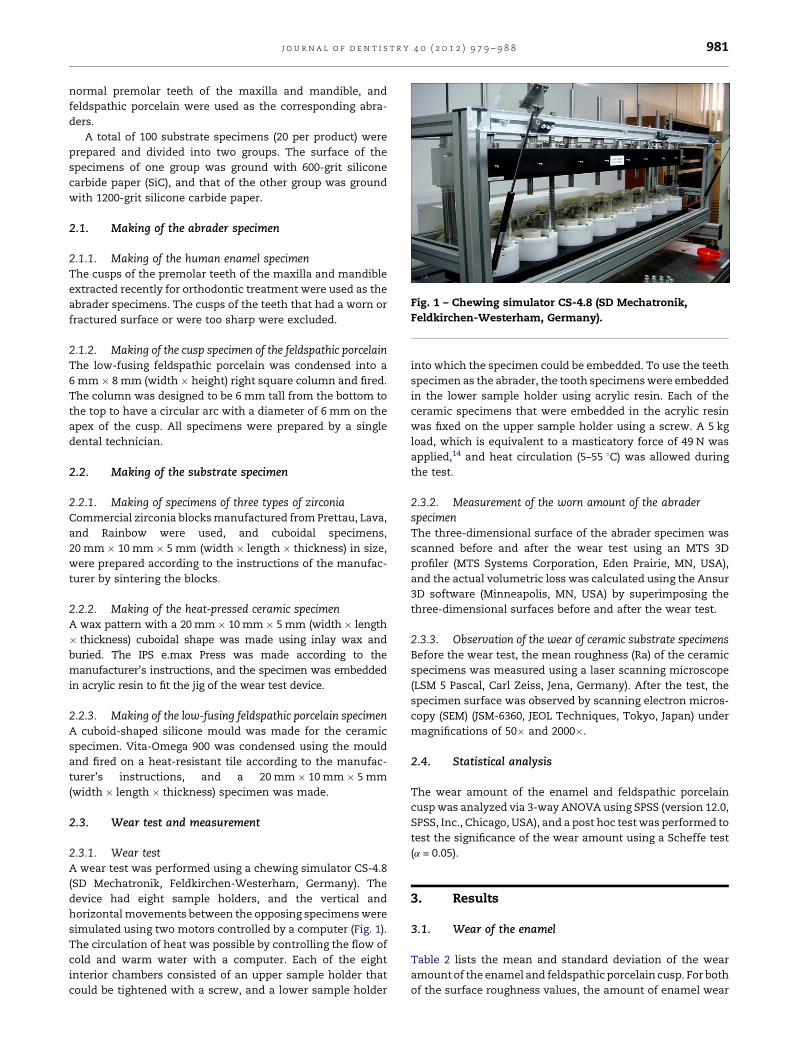
Fig. 1 – Chewing simulator CS-4.8 (SD Mechatronik,
Feldkirchen-Westerham, Germany).
j o u r n a l o f d e n t i s t r y 4 0 ( 2 0 1 2 ) 9 7 9 – 9 8 8 981
normal premolar teeth of the maxilla and mandible, and
feldspathic porcelain were used as the corresponding abra-
ders.
A total of 100 substrate specimens (20 per product) were
prepared and divided into two groups. The surface of the
specimens of one group was ground with 600-grit silicone
carbide paper (SiC), and that of the other group was ground
with 1200-grit silicone carbide paper.
2.1. Making of the abrader specimen
2.1.1. Making of the human enamel specimenThe cusps of the premolar teeth of the maxilla and mandible
extracted recently for orthodontic treatment were used as the
abrader specimens. The cusps of the teeth that had a worn or
fractured surface or were too sharp were excluded.
2.1.2. Making of the cusp specimen of the feldspathic porcelainThe low-fusing feldspathic porcelain was condensed into a
6 mm � 8 mm (width � height) right square column and fired.
The column was designed to be 6 mm tall from the bottom to
the top to have a circular arc with a diameter of 6 mm on the
apex of the cusp. All specimens were prepared by a single
dental technician.
2.2. Making of the substrate specimen
2.2.1. Making of specimens of three types of zirconiaCommercial zirconia blocks manufactured from Prettau, Lava,
and Rainbow were used, and cuboidal specimens,
20 mm � 10 mm � 5 mm (width � length � thickness) in size,
were prepared according to the instructions of the manufac-
turer by sintering the blocks.
2.2.2. Making of the heat-pressed ceramic specimenA wax pattern with a 20 mm � 10 mm � 5 mm (width � length
� thickness) cuboidal shape was made using inlay wax and
buried. The IPS e.max Press was made according to the
manufacturer’s instructions, and the specimen was embedded
in acrylic resin to fit the jig of the wear test device.
2.2.3. Making of the low-fusing feldspathic porcelain specimenA cuboid-shaped silicone mould was made for the ceramic
specimen. Vita-Omega 900 was condensed using the mould
and fired on a heat-resistant tile according to the manufac-
turer’s instructions, and a 20 mm � 10 mm � 5 mm
(width � length � thickness) specimen was made.
2.3. Wear test and measurement
2.3.1. Wear testA wear test was performed using a chewing simulator CS-4.8
(SD Mechatronik, Feldkirchen-Westerham, Germany). The
device had eight sample holders, and the vertical and
horizontal movements between the opposing specimens were
simulated using two motors controlled by a computer (Fig. 1).
The circulation of heat was possible by controlling the flow of
cold and warm water with a computer. Each of the eight
interior chambers consisted of an upper sample holder that
could be tightened with a screw, and a lower sample holder
into which the specimen could be embedded. To use the teeth
specimen as the abrader, the tooth specimens were embedded
in the lower sample holder using acrylic resin. Each of the
ceramic specimens that were embedded in the acrylic resin
was fixed on the upper sample holder using a screw. A 5 kg
load, which is equivalent to a masticatory force of 49 N was
applied,14 and heat circulation (5–55 8C) was allowed during
the test.
2.3.2. Measurement of the worn amount of the abraderspecimenThe three-dimensional surface of the abrader specimen was
scanned before and after the wear test using an MTS 3D
profiler (MTS Systems Corporation, Eden Prairie, MN, USA),
and the actual volumetric loss was calculated using the Ansur
3D software (Minneapolis, MN, USA) by superimposing the
three-dimensional surfaces before and after the wear test.
2.3.3. Observation of the wear of ceramic substrate specimensBefore the wear test, the mean roughness (Ra) of the ceramic
specimens was measured using a laser scanning microscope
(LSM 5 Pascal, Carl Zeiss, Jena, Germany). After the test, the
specimen surface was observed by scanning electron micros-
copy (SEM) (JSM-6360, JEOL Techniques, Tokyo, Japan) under
magnifications of 50� and 2000�.
2.4. Statistical analysis
The wear amount of the enamel and feldspathic porcelain
cusp was analyzed via 3-way ANOVA using SPSS (version 12.0,
SPSS, Inc., Chicago, USA), and a post hoc test was performed to
test the significance of the wear amount using a Scheffe test
(a = 0.05).
3. Results
3.1. Wear of the enamel
Table 2 lists the mean and standard deviation of the wear
amount of the enamel and feldspathic porcelain cusp. For both
of the surface roughness values, the amount of enamel wear

Table 2 – Mean values and SDs of wear volume of the enamel and feldspathic porcelain cusp (mm3).
Abrader Grit Substrate materials
Prettau Lava Rainbow e.max Press Vita-Omega 900
Enamel 600 0.04 � 0.02 0.04 � 0.02 0.04 � 0.02 0.08 � 0.03 0.13 � 0.05
1200 0.04 � 0.02 0.04 � 0.02 0.04 � 0.02 0.06 � 0.03 0.11 � 0.03
Feldspathic porcelain 600 0.03 � 0.02 0.03 � 0.01 0.02 � 0.01 0.06 � 0.02 0.03 � 0.01
1200 0.03 � 0.02 0.03 � 0.01 0.03 � 0.02 0.02 � 0.01 0.03 � 0.01
j o u r n a l o f d e n t i s t r y 4 0 ( 2 0 1 2 ) 9 7 9 – 9 8 8982
was highest in the Vita-Omega 900 group followed by the IPS
e.max Press, Prettua, Lava, and Rainbow groups, although
there was no significant difference seen between the latter
three groups, i.e. Prettau, Lava and Rainbow groups ( p > 0.05).
A significant difference was seen between Vita-Omega 900 and
IPS e.max Press ( p < 0.05) in terms of enamel wear.
3.2. Wear of the feldspathic porcelain cusp
In the 600-grit group, the wear amount of the feldspathic
porcelain cusp was highest in the IPS e.max Press group,
followed in order by the Vita-Omega 900, Prettau, Lava, and
Rainbow groups. In the 1200-grit group, the amount of wear
was lowest in the e.max group.
From the results of 3-way ANOVA, The amount of wear
significantly differed depending on the type of abrader
materials, the substrate materials, and surface roughness.
Result showed significant interaction between abrader and
substrate, and between substrate and surface roughness
( p < 0.05). However, there was no significant interaction
Table 3 – Scheffe analysis to evaluate the mean enamel volumshowing multiple comparisons (P, Prettau; L, Lava; R, Rainbow
(I) Groups (J) Groups Mean difference (I � J) S
P L 0.0071
R 0.0054
E �0.0175*
V �0.0257*
L P �0.0071
R �0.0016
E �0.0246*
V �0.0328*
R P �0.0054
L 0.0016
E �0.0229*
V �0.0312*
E P 0.0175*
L 0.0246*
R 0.0229*
V �0.0083
V P 0.0258*
L 0.0328*
R 0.0312*
E 0.0083
p < 0.05 indicates significant difference.* Difference in the mean values between group pair ( p < 0.05).
between abrader and surface roughness, and among three
factors ( p > 0.05).
A post hoc test in ANOVA was performed using the
Scheffe test. There were no significant differences in the
wear amounts of the Prettau, Lava, and Rainbow groups,
whereas the wear amounts were significantly different for
the IPS e.max Press and Vita-Omega 900 groups from
those of the Prettau, Lava, and Rainbow groups ( p < 0.05)
(Table 3).
Fig. 2 shows the mean amount of wear of the enamel and
feldspathic porcelain cusp according to the materials with
different degrees of surface roughness. When the abrader
was the enamel, the wear amount was higher in the 600-grit
group than in the 1200-grit group in all ceramic substrate
material groups. The amount of wear of the enamel was
highest in the Vita-Omega 900 group in the 600-grit group,
and lowest in the three types of zirconia groups in the 1200-
grit group. The amount of wear of the enamel group was
higher than that of the feldspathic porcelain cusp group
regardless of their surface roughness ( p < 0.01).
e loss (mm3) differences between the material groups; E, e.max Press; V, Vita-Omega 900).
td. error Sig. 95% confidence interval
Lower bound Upper bound
0.005316 0.778 �0.0098 0.0240
0.005316 0.901 �0.0115 0.0223
0.005316 0.038 �0.0343 �0.0060
0.005316 0.000 �0.0426 �0.0089
0.005316 0.778 �0.0240 0.0098
0.005316 0.999 �0.0185 0.0152
0.005316 0.001 �0.0415 �0.0077
0.005316 0.000 �0.0497 �0.0160
0.005316 0.901 �0.0223 0.0115
0.005316 0.999 �0.0153 0.0185
0.005316 0.002 �0.3983 �0.0060
0.005316 0.000 �0.0481 �0.0143
0.005316 0.038 0.0006 0.0344
0.005316 0.001 0.0077 0.0415
0.005316 0.002 0.0060 0.0340
0.005316 0.663 �0.0251 0.0086
0.005316 0.000 0.0089 0.0426
0.005316 0.000 0.0159 0.0497
0.005316 0.000 0.0142 0.0481
0.005316 0.663 �0.0086 0.0252

Fig. 2 – Mean wear volume of the enamel and porcelain
cusp. Standard deviations are shown by error bars.
Table 4 – Mean values and SDs of the surface roughnessof the ceramic substrate specimens before the wear test(mm).
Materials 600 grit 1200 grit
Prettau 0.784 � 0.069 0.284 � 0.052
Lava 0.785 � 0.123 0.459 � 0.075
Rainbow 0.678 � 0.026 0.411 � 0.034
e.max Press 0.455 � 0.066 0.249 � 0.015
Vita-Omega 900 0.823 � 0.383 0.704 � 0.094
j o u r n a l o f d e n t i s t r y 4 0 ( 2 0 1 2 ) 9 7 9 – 9 8 8 983
3.3. Wear surface of the ceramic substrate specimens
3.3.1. Average RaTable 4 lists the average Ra and standard deviation of each of
the ceramic substrate specimens measured by laser scanning
microscopy before the wear test. In the 600-grit SiC wear
group, the average Ra was lowest in the IPS e.max Press group.
In the 1200-grit SiC wear group, the average Ra was highest in
the Vita-Omega 900 group, and in both the 600-grit and 1200-
grit, the average Ra was lowest in the IPS e.max Press group.
3.3.2. Scanning electron microscopy (SEM)Figs. 3–7 show SEM images of the five ceramic substrate
specimens of 1200-grit group, which was performed after the
wear test (the marks of wear are shown in each of the
specimens). In the IPS e.max Press and three types of zirconia
substrate specimens, the inner structure was consistently
dense and air bubbles were rarely observed, whereas a large
crystalline shape and numerous air bubbles were observed in
the Vita-Omega 900 porcelain substrate specimen.
Fig. 3 – SEM images after wear test of e.max Press (1200 grit SiC
50T, (B) original magnification 2000T.
4. Discussion
Tooth wear is a complex process that involves a range of
factors, such as the food, non-functional habits, neuromus-
cular force, thickness and hardness of enamel, acidity and
other properties of saliva, masticatory pattern, and opposing
restorative materials.10 The progressive wear of natural teeth
is a normal physiological phenomenon.
This physiological wear can be affected if restorative
materials with different wear rates from that of natural teeth
are used for intraoral restorations.15,22 Lambrechts et al.23
reported that vertical wear of enamel is 20–40 mm a year under
normal condition. Seghi et al.24 reported that the wear rates of
intraoral restorative materials should be similar to that of the
human enamel.25 Therefore, it is important to evaluate the
wear resistance of restorative materials against the opposing
natural teeth and the physical properties of restorative
materials. Among the dental restorative materials, ceramic
has the best aesthetic features as well as good compressive
strength and biocompatibility,22 although it presents certain
disadvantages such as relatively low tensile strength and can
cause an excessive wear of the opposing teeth.8,9 Restorative
materials that significantly wear down the opposing teeth can
cause hypersensitivity and articular imbalance by rapidly
wearing down the opposing teeth.26 To overcome this
problem, new ceramics that produce less wear on the
opposing teeth have been developed.27
Among the improved all-ceramic crowns, the zirconia
crown has a feldspathic porcelain lining in its upper part, and
it is used in clinical practice. Recently, the construction of an
all-ceramic crown made only of zirconia has become possible.
polished) against human enamel (A) original magnification

Fig. 4 – SEM images after the wear test of Lava (1200 grit SiC polished) against human enamel (A) original magnification 50T,
(B) original magnification 2000T.
j o u r n a l o f d e n t i s t r y 4 0 ( 2 0 1 2 ) 9 7 9 – 9 8 8984
This has made it possible to overcome the susceptibility of its
veneer ceramic layer to wear, while maintaining its physical
advantages. Most studies on the wear of enamel have focused
on the veneering porcelain and resin,15,28 and few studies have
examined the effect of zirconia ceramic on enamel wear. This
study examined the possibility of wear against the opposing
teeth to establish a structural standard for the safe intraoral
use of zirconia for monolithic ceramic crowns. A clinical study
on wear takes at least 6 months to 1 year, and involves
difficulties in accurate measurements due to many uncon-
trollable variables.29
Although an in vitro study has limitations in perfectly
simulating the intraoral masticatory movement,30 it can
simulate simple movements, such as grinding and clenching
teeth,30 and the mechanism behind the wear resistance of
various materials or the order of susceptibility of various
materials to wear can be assessed at the pre-clinical stage
using specific test variables. Because of the advantages of an
in vitro study, the test devices that can simulate the intraoral
masticatory movement were developed to assess the wear of
natural teeth and restorative materials.13,29,31–34 The 2-axes
wear test device used in this study is currently being used in
many studies, and is known to be practical, durable and cost-
effective. This device has a total of 8 interior chambers, and
simulates the mandibular closing movement that occurs
Fig. 5 – SEM images after the wear test of Prettau (1200 grit SiC
50T, (B) original magnification 2000T.
during matiscatory movement by simulating the occlusal
contact followed by the sliding movement.35 In this study, a 2-
body wear test was performed. This test is used widely in wear
resistance measurements,36 and was used to simulate the
attrition caused by the occlusal contact between restorative
materials and the teeth during bruxism and clenching.
This method could also simulate the friction and fatigue
wear caused by direct contact between the maxilla and
mandibular teeth during swallowing or non-functional dy-
namic occlusal movement.37 The load of the wear test device
was 5 kg, which is the mean physiological occlusal force
without bruxism based on the intraoral occlusal force reported
in previous studies on ceramic wear.38 The subsequent sliding
movement plays an important role in simulating the intraoral
wear as microfatigue occurs during the opposing wear
material slides on the specimen.39 In the present study,
0.3 mm lateral movement was applied. Mair et al.40 reported
that as the load that accompanies the sliding factor causes 10
times greater stress than that caused by a static load, and a
crack may be formed on the surface of the ceramic. During the
test, the 8 interior chambers were filled with water, and the
continuous change of water enabled the removal of wear
particles created by the wear test from the contact surface. In
addition, heat circulation was performed during the wear test
to simulate changes in the intraoral temperature.
polished) against human enamel (A) original magnification

Fig. 6 – SEM images after the wear test of Rainbow (1200 grit SiC polished) against human enamel (A) original magnification
50T, (B) original magnification 2000T.
j o u r n a l o f d e n t i s t r y 4 0 ( 2 0 1 2 ) 9 7 9 – 9 8 8 985
Up to 300,000 masticatory movements were simulated,
which is equivalent to the masticatory movement performed
at 1 year in a clinical environment.37 After the test, the
volumetric loss was measured using an MTS 3D profiler and
Ansur 3D software by superimposing the 3D surfaces before
and after the test. Standardization of the enamel surface of
natural teeth that was used as the opposing wear material has
been a subject of considerable controversy.35 In most studies, a
standardized enamel cusp was used for the wear test of
ceramic and enamel. Krejci et al.41 reported that the non-
standardized enamel cusp of natural teeth is most appropriate
for the opposing wear materials in the wear test, which was
consistent with previous studies by Krejci et al.42 and Lutz
et al.43 In a preliminary experiment, Kunzelmann et al.44 used
an enamel cusp surface that was not treated, and reported that
the standardization of the enamel through grinding prepara-
tion altered the wear properties of the opposing wear material,
unlike that with the non-standardized cusp. Heintze et al.35
reported that a standardized enamel cusp could result in a
range of results, and the results obtained from these cusps
were slightly inconsistent. Therefore, in this study, the enamel
surface of the premolar teeth of the maxilla and mandible,
which were extracted for orthodontic treatment, was used
without standardization.
Fig. 7 – SEM images after the wear test of Vita-Omega 900 (1200
magnification 50T, (B) original magnification 2000T.
In this study, the amount of wear of the opposing abrader
shows a significant difference depending on the type of
ceramic substrate, opposing abrader and surface roughness.
Of the five types of ceramic substrate material, the amount of
enamel wear was the highest in the Vita-Omega 900 group,
followed by the IPS e.max Press, Lava, Rainbow, and Prettau
groups. In particular, the amount of enamel and feldspathic
porcelain cusp wear was the lowest in the Prettau, Lava, and
Rainbow groups, and was significantly higher in the Vita-
Omega 900, IPS e.max Press, and three types of zirconia
substrate groups.
The amount of wear in the Vita-Omega 900 group was
higher when the abrader was the enamel rather than of the
feldspathic porcelain cusp. Unlike the other wear materials,
IPS e.max Press group significantly wore out the abrader at a
600-grit surface. The mean wear amounts of the three types of
zirconia substrate specimen that were measured after the
wear test were all too small to be measured. This suggests that
compared to feldspathic porcelain, the superior physical
properties and surface features of zirconia, such as its
hardness, bending strength, fracture toughness and density,
enabled it to maintain a smooth surface during the wear test,
which is consistent with the results reported by Ghazal et al.
on the wear of a zirconia specimen.37 In this study, the amount
grit SiC polished) against human enamel (A) original

j o u r n a l o f d e n t i s t r y 4 0 ( 2 0 1 2 ) 9 7 9 – 9 8 8986
of the enamel wear of natural teeth was higher than that
of feldspathic porcelain cusp, possibly due to the higher
wear resistance of feldspathic porcelain than the human
enamel.
The lower level of enamel hardness than feldspathic
porcelain could cause greater wear of enamel than feldspathic
porcelain. Enamel generally has a Vickers hardness of 320–
380 kg/mm2 and a fracture toughness of 0.8 Mpa m1/2. Also
these values have variations caused by anatomical character-
istics in enamel such as Hunter–Schreger Band (HSB). It is
known that higher packing densities of HSB are noted in those
areas subjected to greater external forces.45,46 Feldspathic
porcelain, however, has a higher degree of hardness of 500 kg/
mm2 or more, and its wear of the opposing enamel could be
accelerated by a rougher surface and by broken particles
during the matiscatory test. Additionally the wear of enamel
could increase considerably as the fracture toughness of
enamel is significantly lower than that of feldspathic porce-
lain. This superior property of feldspathic porcelain to that of
the human enamel can affect the wear amount, which is
consistent with these results. The surface roughness of
ceramic can vary according to the methods and degree of
surface grinding, and a rough ceramic surface decreases the
physical properties of the materials, or causes an excessive
wear of the opposing teeth, surface discoloration, and
inflammation of soft oral tissue. Schuh et al.47 reported that
a high friction coefficient between low-fusing ceramic and the
opposing wear materials increases the fatigue and ceramic
wear.
The results of this study showed that the amount of enamel
wear in relation to the surface roughness differed significantly
( p < 0.05) and decreased in the 1200-grit grinding group. The
average Ra of the specimen was measured by laser scanning
microscopy before the wear test, and the surface was observed
after the test using SEM. In the SiC grinding group of 600 grit,
the average Ra was the lowest in the IPS e.max Press group and
in the SiC grinding group of 1200 grit, and the degree of
roughness was the highest in the Vita-Omega 900 group. In the
IPS e.max Press group, the average Ra was the lowest both in
the 600-grit group and 1200-grit group. In the three types of
zirconia groups where grinding was performed using 1200-grit
SiC, the mean surface roughness of the Lava, Rainbow,
Prettau, and IPS e.max Press groups were similar. Willems
et al.48 reported that the roughness of the contact surface of
the enamel after wear was 0.64 mm, and Al-Wahadni49
reported that the average Ra of the ceramic after grinding
was 0.26–0.75 mm.
The hardness of metal is related to enamel wear. Based on
the results of various studies, however, Seghi et al.24 Magne
et al.50 and Oh et al.7 reported the hardness of ceramic and the
wear of opposing teeth by ceramic restorative materials. The
hardness and wear of ceramic are closely related to each other,
and that wear is closely related to the microstructure of
ceramic, the roughness of the contact surface, and environ-
mental factors. Although the correlation between the surface
roughness and amount of enamel wear can be expected, the
correlation was not analyzed in the present study.
The effect of the surface roughness on wear was
reconsidered because in the present study, some ceramic
materials with similar degrees of surface roughness showed
significantly different enamel wear after the wear test.
Metzler et al.51 reported that although the surface condition
of ceramic is important at the initial stages of wear, the inner
properties of ceramic affected the wear rate once the effect of
roughness disappeared with wear progression. The surface
roughness is one of the factors that affect wear. This study
examined the possibility of the wear of zirconia ceramic
against the opposing teeth as part of a study to apply a
zirconia monolithic crown to clinical practice. These results
suggest that zirconia ceramic can be used as zirconia
monolithic crown with an advantage as it showed less
enamel wear than feldspathic porcelain and heat-pressed
ceramics.
As this is an in vitro study, which involves many factors,
such as occlusal force, matiscatory habits, type of food, and
location of the teeth in the maxillary and mandibular arch, the
conditions used in this study could differ from the clinical
intra oral conditions. In addition, in this study, only 2-body
wear was evaluated. Therefore, different results could be
achieved if a 3-body wear test is performed using different
wear materials, further studies with well simulated intra oral
condition are required for better clinical implication.
5. Conclusion
(I) The enamel wear was significantly greater than the
feldspathic porcelain abrader wear ( p < 0.05).
(II) The feldspathic porcelain substrate caused the most wear
of the enamel abrader ( p < 0.05).
(III) The zirconia substrates caused the least wear of the
enamel abrader ( p < 0.05) and had no difference among
them ( p > 0.05).
Acknowledgements
First two authors contributed equally to this work. This study
was supported by the Basic Science Research Program through
the National Research Foundation of Korea (NRF) funded by
the Ministry of Education, Science and Technology (2010-
0005090 and 2011-0005559).
r e f e r e n c e s
1. Kadokawa A, Suzuki S, Tanaka T. Wear evaluation ofporcelain opposing gold, composite resin and enamel.Journal of Prosthetic Dentistry 2006;96:258–65.
2. Southan DE, Jorgensen KD. Faulty porcelain jacket crowns.Australian Dental Journal 1972;17:436–40.
3. Piconi C, Maccauro G. Zirconia as a ceramic biomaterial.Biomaterials 1999;20:1–25.
4. Guazzato M, Albakry M, Ringer SP, Swain MV. Strength,fracture toughness and microstructure of a selection of all-ceramic materials. Part I: pressable and alumina glass-infiltrated ceramics. Dental Materials 2004;20:441–8.
5. Sailer I, Feher A, Filser F, Luthy H, Gauckler LJ, Scharer P,et al. Prospective clinical study of zirconia posterior fixedpartial dentures: 3-year follow-up. Quintessence International2006;37:685–93.

j o u r n a l o f d e n t i s t r y 4 0 ( 2 0 1 2 ) 9 7 9 – 9 8 8 987
6. Sailer I, Feher A, Filser F, Gauckler L, Luthy H, Hammerle C.Five-year clinical results of zirconia frameworks forposterior fixed partial dentures. International Journal ofProsthodontics 2007;20:383–8.
7. Oh WS, DeLong R, Anusavice KJ. Factors affecting enameland ceramic wear: a literature review. Journal of ProstheticDentistry 2002;87:451–9.
8. Wiley MG. Effects of porcelain on occluding surfaces ofrestored teeth. Journal of Prosthetic Dentistry 1989;61:133–7.
9. Mahalick JA, Knap FJ, Weiter EJ. Occusal wear inprosthodontics. The Journal of the American Dental Association1971;82:154–9.
10. Elmaria A, Goldstein G, Vijayaraghavan T, Legeros RZ,Hittelman EL. An evaluation of wear when enamel isopposed by various ceramic materials and gold. Journal ofProsthetic Dentistry 2006;96:345–53.
11. Hudson JD, Goldstein GR, Georgescu M. Enamel wear causedby three different restorative materials. Journal of ProstheticDentistry 1995;74:647–54.
12. Hacker CH, Wagner WC, Razzoog ME. An in vitroinvestigation of the wear of enamel on porcelain and gold insaliva. Journal of Prosthetic Dentistry 1996;75:14–7.
13. Ramp MH, Suzuki S, Cox CF, Lacefield WR, Koth DL.Evaluation of wear: enamel opposing three ceramicmaterials and a gold alloy. Journal of Prosthetic Dentistry1997;77:523–30.
14. Krejci I, Lutz F, Reimer M, Heinzmann JL. Wear of ceramicinlays, their enamel antagonists, and luting cements. Journalof Prosthetic Dentistry 1993;69:425–30.
15. Monasky GE, Taylor DF. Studies on the wear of porcelain,enamel, and gold. Journal of Prosthetic Dentistry 1971;25:299–306.
16. Al-Shammery HAO, Bubb NL, Youngson CC, Fasbinder DJ,Wood DJ. The use of confocal microscopy to assess surfaceroughness of two milled CAD–CAM ceramics following twopolishing techniques. Dental Materials 2007;23:736–41.
17. Quirynen M, Bollen CML. The influence of surfaceroughness and surface-free energy on supra- andsubgingival plaque formation in man. Journal of ClinicalPeriodontology 1995;22:1–14.
18. Quirynen M. The clinical meaning of the surface roughnessand the surface free energy of intra-oral hard substrata onthe microbiology of the supra- and subgingival plaque:results of in vitro and in vivo experiments. Journal ofDentistry 1994;22(Suppl. 1):S13–S16.
19. Turssi CP, de Moraes Purquerio B, Serra MC. Wear ofdental resin composites: Insights into underlyingprocesses and assessment methods—a review. Journal ofBiomedical Materials Research Part B Applied Biomaterials2003;65:280–5.
20. Kou W, Molin M, SjOGren G. Surface roughness of fivedifferent dental ceramic core materials after grinding andpolishing. Journal of Oral Rehabilitation 2006;33:117–24.
21. Sara D, Turk T, Elekdag Turk S, Sara YS. Comparison of 3polishing techniques for 2 all-ceramic materials.International Journal of Prosthodontics 2007;20:465–8.
22. Jagger DC, Harrison A. An in vitro investigation into thewear effects of unglazed, glazed, and polished porcelainon human enamel. Journal of Prosthetic Dentistry1994;72:320–3.
23. Lambrechts P, Braem M, Vuylsteke-Wauters M, Vanherle G.Quantitative in vivo wear of human enamel. Journal of DentalResearch 1989;68:1752–4.
24. Seghi RR, Rosenstiel SF, Bauer P. Abrasion of human enamelby different dental ceramics in vitro. Journal of DentalResearch 1991;70:221–5.
25. Sulong MZAM, Aziz RA. Wear of materials used in dentistry:a review of the literature. Journal of Prosthetic Dentistry1990;63:342–9.
26. Jacobi R, Shillingburg JHT, Duncanson JMG. A comparison ofthe abrasiveness of six ceramic surfaces and gold. Journal ofProsthetic Dentistry 1991;66:303–9.
27. Eom S, Oh S, Dong J. A study on the wear of dentalrestorative materials. Journal of Korean Academiy ofProsthodontics 1998;36:69–81.
28. McLaren E, Giodano IIR, Pober R, Abozenada B. Materialtesting and layering techniques of a new two-phase all-glass veneering porcelain for bonded porcelain and high-alumina frameworks. Quintessence of Dental Technology2003;26:69–81.
29. Ratledge DK, Smith BGN, Wilson RF. The effect of restorativematerials on the wear of human enamel. Journal of ProstheticDentistry 1994;72:194–203.
30. Suzuki S. Simulated enamel wear during occlusal contact.American Journal of Dentistry 2004;17:373–7.
31. Yip KH, Smales RJ, Kaidonis JA. Differential wear of teethand restorative materials: clinical implications. InternationalJournal of Prosthodontics 2004;17:350–6.
32. Degee AJ, Pallav P, Davidson CL. Effect of abrasion mediumon wear of stress-bearing composites and amalgam in vitro.Journal of Dental Research 1986;65:654–8.
33. Kaidonis JA, Richards LC, Townsend GC, Tansley GD. Wearof human enamel: a quantitative in vitro assessment.Journal of Dental Research 1998;77:1983–90.
34. Yap AUJ, Teoh SH, Hastings GW, Lu CS. Comparative wearranking of dental restorative materials utilizing differentwear simulation modes. Journal of Oral Rehabilitation1997;24:574–80.
35. Heintze SD, Cavalleri A, Forjanic M, Zellweger G, RoussonV. Wear of ceramic and antagonist—a systematicevaluation of influencing factors in vitro. Dental Materials2008;24:433–49.
36. Heintze SD, Zappini G, Rousson V. Wear of ten dentalrestorative materials in five wear simulators—results of around robin test. Dental Materials 2005;21:304–17.
37. Ghazal M, Yang B, Ludwig K, Kern M. Two-body wear ofresin and ceramic denture teeth in comparison to humanenamel. Dental Materials 2008;24:502–7.
38. Gibbs CH, Mahan PE, Lundeen HC, Brehnan K, Walsh EK,Holbrook WB. Occlusal forces during chewing andswallowing as measured by sound transmission. Journal ofProsthetic Dentistry 1981;46:443–9.
39. Heintze SD. How to qualify and validate wear simulationdevices and methods. Dental Materials 2006;22:712–34.
40. Mair LH, Stolarski TA, Vowles RW, Lloyd CH. Wear:mechanisms, manifestations and measurement. Report of aworkshop. Journal of Dentistry 1996;24:141–8.
41. Krejci I, Albert P, Lutz F. The influence of antagoniststandardization on wear. Journal of Dental Research1999;78:713–9.
42. Krejci I, Reich T, Lutz F, Albertoni M. An in vitro testprocedure for evaluating dental restoration systems. 1: acomputer-controlled mastication simulator. SchweizMonatsschr Zahnmed 1990;100:953–60.
43. Lutz F, Krejci I, Barbakow F. Chewing pressure us. wearcomposites opposing enamel cusps. Journal of Dental Research1992;71:1525–9.
44. Kunzelmann KH, Jelen B, Mehl A, Hickel R. Wearevaluation of MZ100 compared to ceramic CAD/CAMmaterials. International Journal of Computerized Dentistry2001;4:171–84.
45. Lynch CD, O’Sullivan VR, Dockery P, McGillycuddy CT, SloanAJ. Hunter–Schreger Band patterns in human tooth enamel.Journal of Anatomy 2010;217:106–15.
46. Lynch CD, O’sullivan VR, Dockery P, McGillycuddy CT, ReesJS, Sloan AJ. Hunter–Schreger Band patterns and theirimplications for clinical dentistry. Journal of OralRehabilitation 2011;38:359–65.

j o u r n a l o f d e n t i s t r y 4 0 ( 2 0 1 2 ) 9 7 9 – 9 8 8988
47. Schuh C, Kinast EJ, Mezzomo E, Kapczinski MP. Effect ofglazed and polished surface finishes on the frictioncoefficient of two low-fusing ceramics. Journal of ProstheticDentistry 2005;93:245–52.
48. Willems G, Lambrechts P, Braem M, Vuylsteke-Wauters M,Vanherle G. Surface roughness enamel-to-enamel contactareas compared intrinsic roughness dental resincomposites. Journal of Dental Research 1991;70:1299–305.
49. Al-Wahadni A. An in vitro investigation into thesurface roughness of 2 glazed, unglazed, and refinished
ceramic materials. Quintessence International 2006;37:311–7.
50. Magne P, Oh WS, Pintado MR, DeLong R. Wear of enameland veneering ceramics after laboratory and chairsidefinishing procedures. Journal of Prosthetic Dentistry1999;82:669–79.
51. Metzler KT, Woody RD, Miller Iii AW, Miller BH.In vitro investigation of the wear of human enamel bydental porcelain. Journal of Prosthetic Dentistry 1999;81:356–64.

![On the construction of wear maps for Y-TZP dental ceramics ... · unique ductility among other ceramics, Y-TZP material is found to be wear resistant under repeated mastication [3]](https://img.pdfslide.us/doc/110x75/5fb88b69b8d4f26ccb217478/on-the-construction-of-wear-maps-for-y-tzp-dental-ceramics-unique-ductility.jpg)