Embed Size (px)
Citation preview


• We do not have any conflict of interest, nor will we be discussing any off-label product use.
• This presentation has no commercial support or sponsorship, nor is it co-sponsored.

• Describe the role and resources provided by the prenatal clinic
• State how to contact the prenatal clinic
• Understand how to access a prenatal chart in order to identify the postnatal plan of care, including genetic testing
• Describe the genetic testing done prenatally and know the next steps in pediatric evaluation

• Multidisciplinary clinic that brings together obstetric and pediatric specialty care for families when a pregnancy is complicated by a known or suspected condition in the developing fetus
• Diagnostic testing, consultation and fetal intervention
– Education
– Birth and newborn care planning
• Screening echocardiograms
• Referring providers are Maternal-Fetal-Medicine (MFM) specialists and primary OB providers from the WAMI region.

• Improved outcomes:
–Family logistics & support
–Emotional preparedness
–Health outcomes
–Neonatal palliative care
–DC planning

• Location:
– Springbrook Professional Center (above adolescents clinic)
• Contact Us:
– Main Line: x7-5629
– RN Line: x7-0134
– GC Line: x7-7973
– Fax: x7-2962
• Regional Sites

• Core Team:
– Family Service Coordinators
– Program Coordinator
– Nursing
– Nurse Practitioner/Clinic Manager
– Genetic Counselor
– Social Work
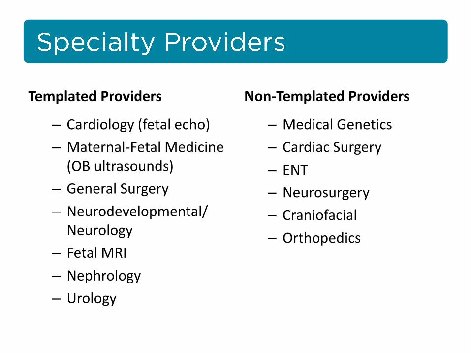
Templated Providers
– Cardiology (fetal echo)
– Maternal-Fetal Medicine (OB ultrasounds)
– General Surgery
– Neurodevelopmental/ Neurology
– Fetal MRI
– Nephrology
– Urology
Non-Templated Providers
– Medical Genetics
– Cardiac Surgery
– ENT
– Neurosurgery
– Craniofacial
– Orthopedics

Cardiology
Neurodevelopmental
General Surgery
Other


• Referrals
• Triage
• Our FSCs call patient’s to schedule
• Relocation/Resource Needs, as needed
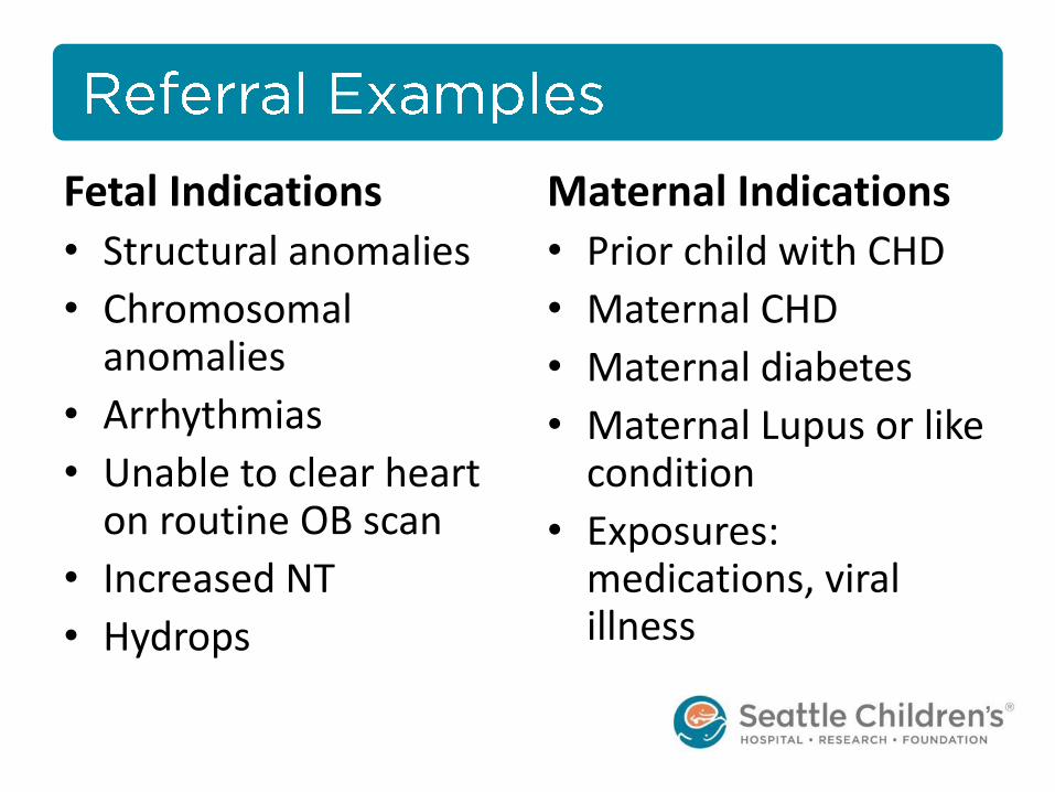
Fetal Indications
• Structural anomalies
• Chromosomal anomalies
• Arrhythmias
• Unable to clear heart on routine OB scan
• Increased NT
• Hydrops
Maternal Indications
• Prior child with CHD
• Maternal CHD
• Maternal diabetes
• Maternal Lupus or like condition
• Exposures: medications, viral illness

Clinical Liaison
Check In Diagnostics Consults Follow-up
Patient’s day lasts about 2-5 hours depending on complexity of case
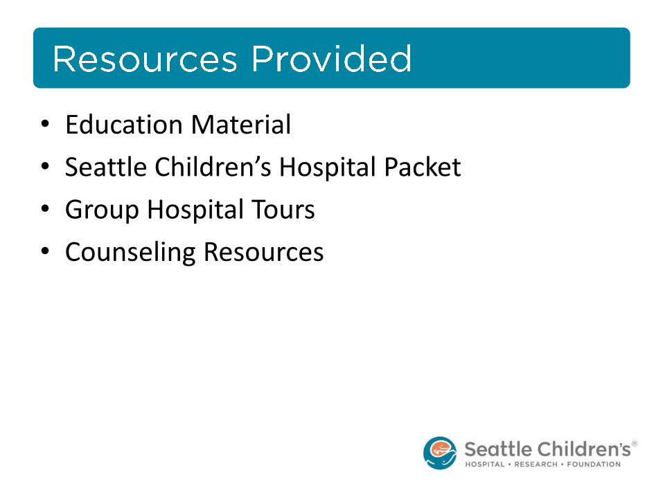
• Education Material
• Seattle Children’s Hospital Packet
• Group Hospital Tours
• Counseling Resources

• Referring provider updated
• Assist with relocation
• Coordinate appointments
• Research study
• Offer continued support and education
• Assist with delivery planning
• Forecast List


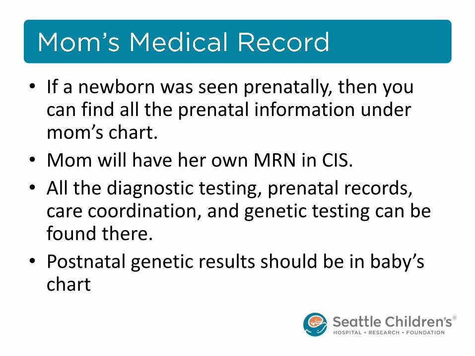
• If a newborn was seen prenatally, then you can find all the prenatal information under mom’s chart.
• Mom will have her own MRN in CIS.
• All the diagnostic testing, prenatal records, care coordination, and genetic testing can be found there.
• Postnatal genetic results should be in baby’s chart


• Background
• Screening – Maternal Serum Screening
– Cell Free DNA screening
• Diagnostic testing – Chorionic Villus Sampling and Amniocentesis
– Karyotype and Microarray
• Overall understanding of types of prenatal testing

Genetic counseling is the process of helping people understand and adapt to the medical, psychological and familial implications of genetic contributions to disease. This process integrates the following:
• Interpretation of family and medical histories to assess the chance of disease occurrence or recurrence.
• Education about inheritance, testing, management, prevention, resources and research.
• Counseling to promote informed choices and adaptation to the risk or condition.
“A new definition of Genetic Counseling: National Society of Genetic Counselors’ Task Force report.”Journal of Genetic Counseling, 2006 Apr;5(2):77-83

• Chromosome anomaly – an extra or missing whole book or huge portion of a book.
• Microdeletion/duplication - a few pages are extra or missing
• Single gene disorder – spelling error

• Trisomy – an extra chromosome (extra book) – T21/Down – hypotonia, mild/mod ID, variety of birth defects,
medical conditions – T18/Edwards – slow growth, heart defects, ID, multiple
congenital anomalies – T13/Patau – heart defects, brain or spinal cord abnormalities,
small or poorly developed eyes – T18/T13 – 5-10% live past their first year
• Microdeletion/duplication – small extra or missing piece of chromosomal material (pages) – 22q11 deletion – heart defects, mild/mod ID, cleft palate, low
calcium
*Not comprehensive discussion of any of these conditions and more detail and nuance given to all families.

• ALL women should be offered aneuploidy screening and diagnostic testing in early pregnancy
• Wide variety of screening options • Varying levels of information and accuracy
• No one screening method is superior to others in all test
characteristics • Relative advantages and disadvantages
• Complex counseling by providers • Complex decision making by patients
AL
ACOG PB 163. Screening for Fetal Aneuploidy. May 2016 Slide courtesy of Shani Delaney

1st trimester 10-14wks
2nd trimester 15-22wks
2nd and 3rd trimester 23-40wks
Serum Integrated, Combined & Sequential screening
NT sono
Quad screen
CVS Amniocentesis
Cell free fetal DNA screening
Anatomy scan
Timeline of genetic screening and testing
Serum screening
Ultrasound
Non-invasive
screening
Invasive testing
Timeline of Genetic Screening and Testing
Slide courtesy of Shani Delaney


• Definition: a test performed on a large number of people to identify those who have or are likely to develop a specified disease or condition
• Simple, generally highly sensitive to not miss potential affected persons
• False positives, hopefully few false negatives
• Indicates suspicion of condition that warrants confirmation through diagnostic testing

• Measure proteins/analytes produced by the fetus or placenta
• (MoM) calculated to population standards
• Compare a number of different factors: age, ethnicity, results from blood tests, gestational age, diabetes, weight, number of fetuses, etc.
• Reported as a 1 in ________ risk number
• Neural tube defects, trisomy 21, trisomy 18, trisomy 13, and more indicators

Serum Screen Results Examples
www.CCLbooks.com

Cell free DNA
• The entire fetal genome is represented in the maternal plasma
• Detected as early at 7 weeks GA
• Disappears rapidly from the maternal circulation after delivery – Undetectable 2 hours after delivery!
• Size difference: – Maternal 167bp
– Fetal 147bp
Lo YM, Corbetta N, Chamberlain PF, Rai V, Sargent IL, Redman CW, et al. Presence of fetal DNA in maternal plasma and serum. Lancet 1997;350:485–7 Slide courtesy of Shani Delaney
Cell Free DNA

Cell free DNA = Apoptosis
Ashoor G, Syngelaki A, Poon LC, Rezende JC, Nicolaides KH. Fetal fraction in maternal plasma cell-free DNA at 11-13 weeks’ gestation: relation to maternal and fetal characteristics. Ultrasound Obstet Gynecol 2013;41:26–32 Slide courtesy of Shani Delaney
~90% of cell free DNA in maternal plasma during pregnancy is maternal in origin
~10% of cell free DNA in maternal plasma during pregnancy is from placental trophoblasts
Cell Free DNA = Apoptosis
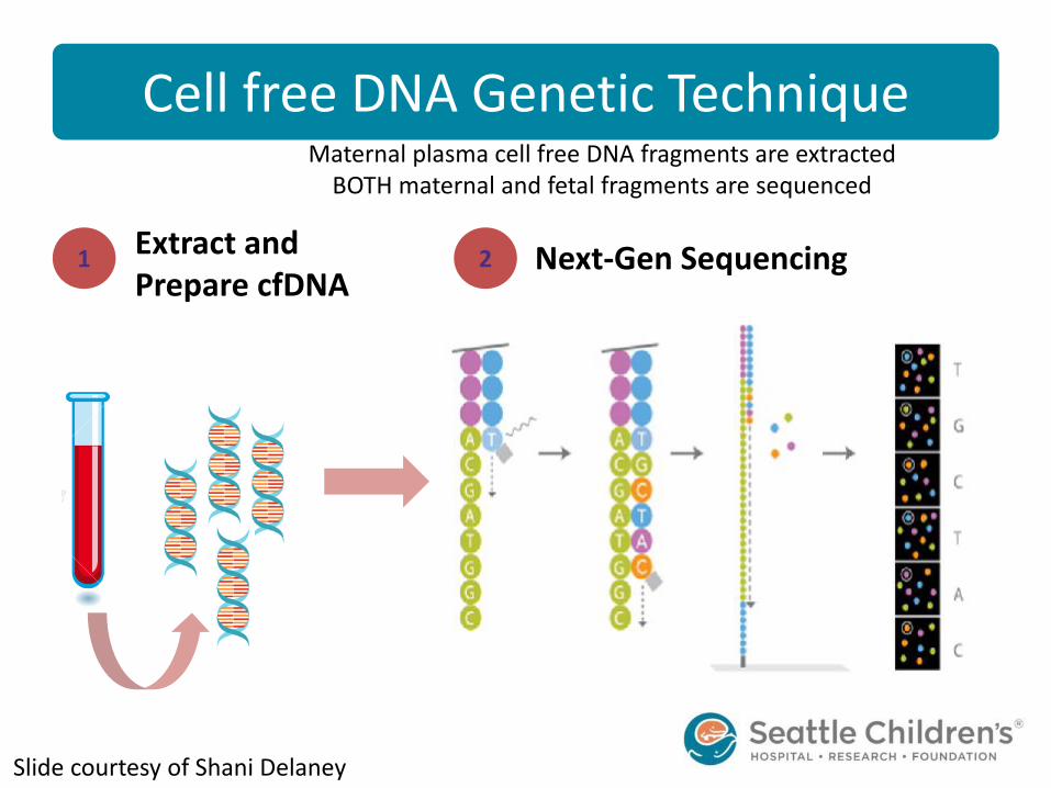
Cell free DNA Genetic Technique
Next-Gen Sequencing 1 2 Extract and Prepare cfDNA
Maternal plasma cell free DNA fragments are extracted BOTH maternal and fetal fragments are sequenced
Slide courtesy of Shani Delaney
Cell free DNA Genetic Technique

Cell free DNA Genetic technique
Alignment of unique cell free DNA sequences 3
CGATTTAACT …ACCACGATTTAACTGGAGTAAAGACTTCCAGGTACCGATCTAGCCT…
Reference Human Genome
GACTTCCAGG
AGGTACCGAT
Human Genome
Slide courtesy of Shani Delaney
Cell free DNA Genetic Technique

Bin Method
Fetal cfDNA
Chromosomes: 1 2 3
……
21 21
VS
Counting 4
Trisomy 21
Slide courtesy of Shani Delaney
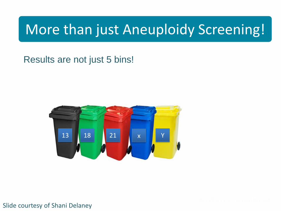
Results are not just 5 bins!
More than just aneuploidy screening
13 18 21 x Y
More than just Aneuploidy Screening!
Slide courtesy of Shani Delaney

cfDNA provides information for genome
1 2 3
4 5 6
7 8 9
10 11 12
13 14 15
16 17 18
19 20 21
22 X Y
Micro- deletions and duplications?
Sub-chromosome deletions and duplications?
Single gene disorders?
cfDNA can provide information on genome
Slide courtesy of Shani Delaney

> cfDNA tells us about gains and losses of DNA
> Quantity but not structure, and not location
> Importance of diagnostic testing
cfDNA and structural rearrangements Cell Free DNA & structural rearrangements
Slide courtesy of Shani Delaney

Microdeletions/duplications
www.sequenom.com Hui, Bianchi. Annu Rev Med (2017);68:459-472
Cell free DNA will miss at least 20% of micro-deletions/duplication syndromes that are detectable by diagnostic prenatal testing
methods
• Current resolution of cfDNA is 5Mb • Many microdeletion/duplication syndromes are <5Mb • Importance of diagnostic testing
Microdeletions/duplications
Slide courtesy of Shani Delaney

Norton ME. AJOG 2016;214(6):727.e1-6
Detection rates of cfDNA vs. Serum Screening (Sequential)
Slide courtesy of Shani Delaney

• Cell free DNA and Serum testing are screening tests, both are very good.
• Maternal blood used.
• If a child has had SCREENING in utero suggesting a likely diagnosis then DIAGNOSTIC testing needs to happen postnatally.
• Best diagnostic test depends on each situation.


• Tests designed to establish the presence/absence of disease/condition
• May be invasive, justifiable as necessary to establish diagnosis
• Chosen towards high specificity (true negatives)
• Result provides a definite diagnosis

Chorionic Villus Sampling
– Transabdominal
– Transcervical
– 11-14 weeks
– Loss rate = 0.1 - 1.1%
Amniocentesis
– ≥ 15 weeks
– Loss rate 0.2 - 0.4%
Enzensberger C. Ultraschall Med 2012; 33(7): E75-E79
Slide courtesy of Shani Delaney
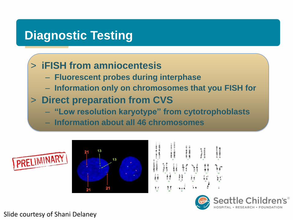
> iFISH from amniocentesis – Fluorescent probes during interphase
– Information only on chromosomes that you FISH for
> Direct preparation from CVS – “Low resolution karyotype” from cytotrophoblasts
– Information about all 46 chromosomes
Diagnostic Testing
Slide courtesy of Shani Delaney

> Karyotype
– Big picture
– Large changes in
structure or number of
chromosomes
– Resolution 5-10Mb
Diagnostic Testing
Slide courtesy of Shani Delaney https://www.pexels.com/photo/pile-of-books-159866/

> Microarray
– Submicroscopic level
– Resolution 50-100kb
– Duplication/deleted sections that differ in size from a
reference human genome = copy number variants (CNV)
> Benign
> Pathogenic
> Variants of unknown significance (VOUS)
> Advantanges of microarray – Higher resolution
– Can be obtained from uncultured cells, does not require actively dividing
cells (IUFD, SAB)
> Limitations of microarray: quantity, not structure – Will not detect balanced translocations
– Low-level mosaicism
Diagnostic Testing
Slide courtesy of Shani Delaney

Patient SDC
• 33-year-old mother and unrelated father. • G2P2002 • Maternal Serum Screen = increased risk for Down
syndrome • Normal Harmony cfDNA testing @13w5d
– 12.1% fetal fraction – <1:10,000 risk for T13, T18, T21
• 38 weeks 3 days gestation
• Repeat cesarean section for breech • 8lbs 13oz 4003 gram /OFC 34cm /Length 51cm • Features of Down syndrome • She had difficulty latching for feeding & sleepy while eating.
Patient Example

FISH Results iFISH Results

Karyotype

47,XX,+21,der(21;21)(q10;q10)x2 in 4 cells 46,XX,+21,der(21;21)(q10;q10) in 16 cells
https://christinamolin.wordpress.com/2007/01/18/translocation-trisomy-21robertsonian-trisomy-21/
Trisomy 21 & Pentasomy 21

> 33 yo G3P1102 @ 25 weeks for MFM consult
> Fetal Tetrology of Fallot and brain anomalies
> cfDNA with MaterniT21= 15Mb loss of 18p
> 18p minus phenotype
– Hearing loss, heart defects, pituitary anomalies, seizures,
cataracts, hypotonia, developmental delay
> Declined amniocentesis
Subchromosome changes and structural
rearrangements Subchromosome changes and structural
rearrangements
Slide courtesy of Shani Delaney

> GC called Maternity T21
> “Smaller deletion also seen at the terminal end of 18q. Below reporting threshold, so not originally reported.”
> Ring chromosome 18 carries a worse prognosis than 18p-minus syndrome
Subchromosome changes and
structural rearrangements Subchromosome changes and Structural
Rearrangements
Slide courtesy of Shani Delaney

> Term SVD, female, weight 2832g (13% for 38 weeks)
> Postnatal karyotype on cord blood confirmed a ring
chromosome 18
– 15Mb loss of 18p
– 1.6Mb loss of 18q
> Baby is now a ~2 year old toddler
– Has undergone repair of TOF
– Slow to meet her neurodevelopmental milestones
– But medical doing well
Subchromosome changes and
structural rearrangements Subchromosome changes and structural
rearrangements
Slide courtesy of Shani Delaney

• We are seeing a decrease in the number of families pursuing diagnostic testing in utero even when a concern arises. This means that more diagnostic testing burden is on the pediatric care team.
• Some of this decrease is due to continued misunderstanding even with counseling that they “think they tested for everything.”

• If a child had a diagnostic test (amniocentesis, cvs) this does not need to be repeated. Need a copy of the result.
• Children with residual concerns of a genetic condition need a genetics consultation – inpatient or outpatient is child/situation dependent.

Contact Us:
– Main Line: x7-5629
– RN Line: x7-0134
– GC Line: x7-7973
– Fax: x7-2962

> Rate of Microdeletions/duplication conditions is
independent of maternal age
> Rate of clinically significant microdeletions and
duplications in sonographically normal fetus = 1.7%
– Among low risk women, collectively microdel/dup
conditions are more common than Trisomy 21
Microdeletions/duplications
Hui, Bianchi. Annu Rev Med (2017);68:459-472 Adapted from Snijders et al. Ultrasound Obstet Gynecol 1999;13:167-170
Microdeletions/duplications
Slide courtesy of Shani Delaney


![One in Messiah Congregation Can we talk? Gal.3 [28] There is neither Jew nor Greek, there is neither bond nor free, there is neither male nor female: for](https://img.pdfslide.us/doc/110x75/56649d645503460f94a46c99/one-in-messiah-congregation-can-we-talk-gal3-28-there-is-neither-jew-nor.jpg)