Embed Size (px)
Citation preview

PARAFFINALIA
IF YOU HAVE NOT ELECTED HOW TO
RECEIVE PARAFFINALIA THIS WILL BE
YOUR LAST EDITION.
ALL MEMBERS NEED TO ELECT HOW TO
RECEIVE PARAFFINALIA.
EMAIL [email protected] PRIOR TO
APRIL EDITION.
SUBJECT: PARAFFINALIA
MESSAGE: YOUR NAME – EMAIL COPY OR YOUR NAME – HARD COPY
IF SELECTING EMAIL COPY, SEND THE EMAIL FROM THE
ADDRESS TO WHICH YOU WISH TO RECEIVE PARAFFINALIA
IF YOU DO NOT HAVE ACCESS TO EMAIL YOU WILL NEED TO
SEND YOUR REQUEST TO:
ATTENTION MEMBERSHIP SECRETARY
HISTOLOGY GROUP OF VICTORIA
PO BOX 1461
COLLINGWOOD 3066
CURRENTLY ONLY 50% OF MEMBERS HAVE
RESPONDED.
Volume 17 Number 1
February 2012
Contents:
________________________________________________________________________________
Editor: Elizabeth Baranyai
“The HGV aims to provide a dynamic continuing education program in which all persons with
an interest in Histology and Histotechnology are freely invited to participate.”
Org. No. A003523F ABN 49 725 623 468
http://www.hgv.org.au
Blurb from the Bush
Scientific Meeting Review-Marking
dyes
2011National Conference Report by
Elizabeth Baranyai
Book Review by Judy Brincat
AIMS (Vic Branch), HGV &
Haematology Discussion Group Joint
meeting 2012
Under the Microscope with Jarrod
Phillips
From the QAP
Next Scientific Meeting
Future Events 2012

Committee Page:
The members of the Histology Group of Victoria 2011-2012 are: Committee Members Name Institution Phone
President Adrian Warmington St. John of God Pathology (Victoria) 5320 1171
Treasurer Judy Brincat Dorevitch Pathology 9244 0351
Editor Elizabeth Baranyai Cabrini Health 9508 1263
Trade representative Kristy De George Austin Pathology 9496 5792
Web Master Sean Phefley RCPA QAP 9024 8608
Social Secretary Maria Chavez Monash Medical Centre 9594 3493
Minutes Secretary Michelle Zammit The Alfred Hospital 9076 3088
Meeting Co-ordinator Nguyen, Nguyen Peter MacCallum Cancer Centre 9656 1431
Mark Bromley Melbourne Pathology 9287 7806
Rebecca Forrester Peter MacCallum Cancer Centre 9656 1431
Rosemary Savino Monash Medical Centre 9594 3494
Kellie Vukovic Peter MacCallum Cancer Centre 9656 1431
Please feel free to contact any of the committee members listed above with any comments or suggestions.
Contributions are always welcome.
Advertising:
All enquiries for trade advertising in the next edition, please contact: Kristy De George - [email protected]
Advertising for the next edition of Paraffinalia closes: 1st April, 2012
Articles & Reports:
Author enquiries and readers wishing to contribute articles or reports can contact the Editor - [email protected]
Please email articles (preferably Microsoft Word format) for inclusion in the next edition to [email protected] All
items submitted for publication will then become the sole property of the Histology Group of Victoria Inc.
Disclaimer: Any opinions expressed in this publication are solely those of the contributing author and are not necessarily reflective of the
Histology Group of Victoria Incorporated or the editor.
NOTE: No responsibility is assumed by the Histology Group of Victoria Incorporated for any injury and/or damage to persons or
property as a matter of products liability, negligence or otherwise, or from any use or operation of any methods, products,
instructions or ideas contained in the material herein. It is the users responsibility to ensure that all procedures are carried out
according to appropriate Health and Safety requirements.
Copyright of this newsletter “Paraffinalia” is held by the Histology Group of Victoria Incorporated. No material may be reproduced
in part or in whole without written consent from the copyright holders. All rights reserved.
Rates:
A4 Electonically Submitted
Single sided B&W $250
Double sided B&W $325
Per page colour $250 (Will be colour for e-newsletter and B&W for hard copy
newsletters)
Used Equipment FREE
50 words – no logos/no pictures
Positions Vacant
No Logo up to 75 words FREE
A4 B&W with logo $150
PARAFFINALIA PACKAGE
One A4 page in remaining 5 editions of 2012 Paraffinalia for the price of 4 $1000
Blurb from the Bush
I trust everyone had a very merry Christmas and a safe New Year celebration. The HGV committee
is going into a year of preparation. We are holding a joint meeting with AIMS in August this year,
which will be held at Cape Schanck on the Mornington Peninsula. The focus will be presentations
for both Haematologists and Histologists. This is an exciting new project that will hopefully bring a
Coonawarra type conference a little closer to home. More information regarding program and
registration will follow soon.
Victoria is holding the next National meeting in 2013. Preliminary work has already commenced
on establishing dates and venues. This will consume a significant amount of committee time
throughout the next year.
As usual we have our scientific meetings. Whilst attendance at these meetings has been waning, we
still consider these the crux of our mission to deliver ongoing education.
This will be the final edition of Paraffinalia in its current form. All members have been informed
that they need to identify how they would like to receive Paraffinalia, either by hard copy or by
email. To date only about 50% have responded. The next edition of Paraffinalia will be sent in
hard copy or by email to those who have identified which format they would like. For those that
have not contacted us, this will be your last Paraffinalia. Should you wish to continue to receive
Paraffinalia, please contact [email protected] and state either hard copy, or email (with the
appropriate email address).
Adrian Warmington
HGV President
PARAFFINALIA ADVERTISING 2012
$250 per page
Paraffinalia Advertising Package: Receive a 1 page advertisement space in each edition of Paraffinalia in 2012
(5 editions) For the discounted rate of $1000!
Only 3 Advertising Packages available for 2012!
Please submit all advertisements electronically to Kristy De George at [email protected]
Any queries or to purchase an advertising package contact Kristy 9496 5792.
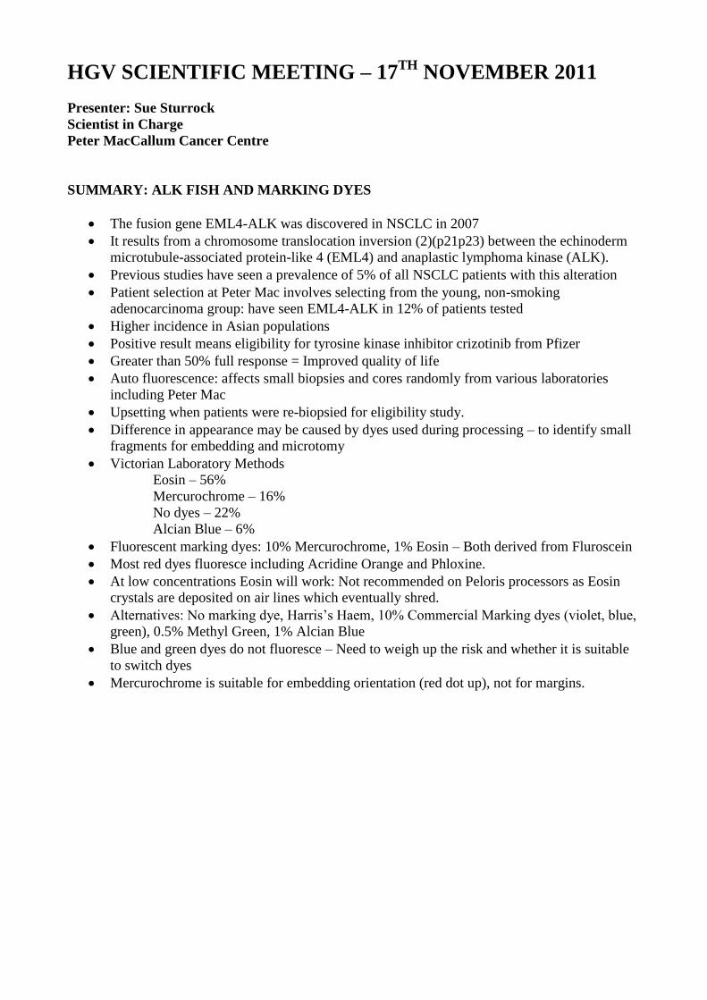
HGV SCIENTIFIC MEETING – 17TH
NOVEMBER 2011
Presenter: Sue Sturrock
Scientist in Charge
Peter MacCallum Cancer Centre
SUMMARY: ALK FISH AND MARKING DYES
The fusion gene EML4-ALK was discovered in NSCLC in 2007
It results from a chromosome translocation inversion (2)(p21p23) between the echinoderm
microtubule-associated protein-like 4 (EML4) and anaplastic lymphoma kinase (ALK).
Previous studies have seen a prevalence of 5% of all NSCLC patients with this alteration
Patient selection at Peter Mac involves selecting from the young, non-smoking
adenocarcinoma group: have seen EML4-ALK in 12% of patients tested
Higher incidence in Asian populations
Positive result means eligibility for tyrosine kinase inhibitor crizotinib from Pfizer
Greater than 50% full response = Improved quality of life
Auto fluorescence: affects small biopsies and cores randomly from various laboratories
including Peter Mac
Upsetting when patients were re-biopsied for eligibility study.
Difference in appearance may be caused by dyes used during processing – to identify small
fragments for embedding and microtomy
Victorian Laboratory Methods
Eosin – 56%
Mercurochrome – 16%
No dyes – 22%
Alcian Blue – 6%
Fluorescent marking dyes: 10% Mercurochrome, 1% Eosin – Both derived from Fluroscein
Most red dyes fluoresce including Acridine Orange and Phloxine.
At low concentrations Eosin will work: Not recommended on Peloris processors as Eosin
crystals are deposited on air lines which eventually shred.
Alternatives: No marking dye, Harris’s Haem, 10% Commercial Marking dyes (violet, blue,
green), 0.5% Methyl Green, 1% Alcian Blue
Blue and green dyes do not fluoresce – Need to weigh up the risk and whether it is suitable
to switch dyes
Mercurochrome is suitable for embedding orientation (red dot up), not for margins.

Figure 1: Colorectal adenocarcinoma – no translocation, useful abundant white tumour cores.
Mucin causes autofluorscence.
Harris haematoxylin contains mercuric oxide oxidizer.
Reported By:
Kellie Vukovic
Medical Scientist
Peter MacCallum Cancer Centre
No Dye
Harris’ Haem
10% Violet
10% Blue
10% Green 1% Alcian
Blue
0.5% Methyl Green
0.1%
Eosin
0.5% Eosin
10% Violet

National Histology Conference
Sydney 4-6 November, 2011 The National Histology Conference had the theme Horses for Courses, which was appropriate as it followed close on the heels of the Melbourne Cup. It was well attended by delegates from all over the country, and well hosted by the Histotechnology Group of NSW. They are to be congratulated for this and for celebrating their 30th year of operation. Friday began with a range of workshops both in the morning and afternoon, covering surgical dissection, histochemistry and histo hypotheticals. On Saturday and Sunday we were treated to a number of interesting guest speakers who covered a broad range of subjects. The first speaker, Mr. René Buesa from Cuba, introduced us to the possibility of eliminating xylene from the laboratory, both in tissue processors and staining protocols. He showed the advantages of this from a health and safety point of view as well as being cost effective. In a nutshell, isopropyl alcohol is used to dehydrate the tissue, a mixture of alcohol and mineral oil is used for clearing the tissue, infiltration uses a mix of mineral oil and paraffin wax, dewaxing uses a 2% solution of dishwashing solution, sections are oven dried prior to coverslipping and cleaning of processors, microtomes etc. can be carried out with the dishwashing solution. He showed a range of pictures that demonstrated clearly that this is possible. Dr. Susan Branford presented a discussion on the use of molecular pathology for patients with Chronic Myeloid Leukaemia. She showed how the outcome for patients with this disease has improved dramatically due to the introduction of targeted therapy. She discussed the genes responsible, the mutations and the development of the targeted therapy using the drug imatinib. The research is ongoing to identify other molecular targets. Dr. Geoffrey O’Brien discussed the various forms of basal cell carcinomas, including the challenges that they can bring to the laboratory. He stressed the importance of orderly cut up of the specimen, processing, cutting (often including multiple levels), staining and immunoprofiling. Colin Gordon’s presentation was on Acid-Fast Staining: Separating the Sprinters from the Stayers. His discussion included the nature of the bacteria we are trying to stain, the chemical nature of the stains involved, their purity, colour index, and solubility in alcohol and water. His consensus was: the absorbance maximum ≥552nm (predominantly new fuchsine) often provided more stable results; some batches of pararosanaline are prone to “bleeding” artefact on smears (but not sections); and that “dye dosage” manipulation will not correct poor staining in a low purity dye. Drs. Fiona Maclean, Julie Schatz and Richard Boyle gave a panel discussion on The Marriage of Orthopaedic Pathology with Clinical and Radiological features. They showed how medical imaging was of use in detection, diagnosis, staging prior to biopsy, and co-ordinated with the surgeon performing the surgery. Then they presented a number of interesting case studies and an investigation into gout and the fall of the Roman Empire! Other crystalline substances were also discussed as well as metal prostheses and the problems they may cause. Dr. Daniel Talmont’s presentation was on Familial Hypercholesterolaemia. He described what it was, the gene involved, the metabolic findings, and clinical presentations. He also gave a number of statistics showing that this genetic condition is highly prevalent in certain communities in Australia, as high as 1:60 compared to 1:500 in the general community. Early detection and management is critical to saving lives. Dr. Manuel B. Graeber spoke about the Glioma Challenge. Gliomas are common brain neoplasms and glioblastoma is the most malignant variant, constituting more than 20%of all adult primary malignant brain

tumours. Classification was discussed in detail along with micrographs of MRI’s, macro and micro photographs demonstrating the different types. He stated that histology remains the “gold standard” for glioma diagnosis whilst molecular markers can be of some help regarding problems caused by tumour heterogeneity, overlapping morphologic features and tumour sampling. A few molecular markers also have prognostic value with regard to patient survival and therapeutic response. This was the final talk for the day and we all eagerly anticipated the Gala dinner with the theme of dressing for the races. Some of the delegates went to a lot of trouble with their outfits and would have blended in nicely at the Melbourne Cup Carnival. Some would have even made great jockeys!

The selection of food and wine waswonderful, and the entertainment enticed many out onto the dance floor. A great time was had by all. Sunday morning began with Mike Rentsch educating us on the Haematoxylin Blues. Sometimes throwing out a batch of stain that isn’t working may not improve the performance of the haematoxylin and eosin stain, especially if the batch of stain has been compromised. He covered basic chemistry, alums, solubility, ripening agents, antioxidants and preservatives. His advice was to investigate the various formulations and select the best one for your site, whilst being prepared to modify the formula if necessary. He also advocated sharing this knowledge with all your staff members. Dr. William Ryman gave an in depth presentation on Moh’s Surgery. He began with the development of Moh’s surgery from its origins in the 1940’s, to the current form using frozen sections under local anaesthetic. The main advantage of this type of surgery is that the entire process from biopsy, diagnosis, ensuring clear margins, to reconstruction can be completed in one day. I think everyone who saw the pictures of the various cases presented were awed by the skills required by all involved from the technicians to the surgeons. The technicians for being so precise with their frozen sections and the surgeons reconstructing the faces that had gaping holes in them, stitching them up like patchwork. The end results after healing were spectacular. Bill Sinai and Penny Whippy presented the intriguing Do You Know What I Did Last Summer?—SPHERE Programme. These two incredible people undertook the huge task of educating and training delegates from 9 participating countries from Asia and the Pacific on the best practice in HER-2 testing in gastric and breast cancer. They developed a set of teaching materials aimed at a multidisciplininary team of surgeons, pathologists, oncologists, scientists and technicians. They covered the entire process from cut-up to processing, IHC, and ISH, including quality control, quality assurance and interpretation. The theory was followed up by practical workshops and instruction on how to troubleshoot. Mr. René Buesa returned to discuss Productivity in the Histology Laboratory. Here he gave us a history lesson on how productivity has improved markedly from the early days due to the introduction of tissue processors, embedding centres and the like. He purports that the histotechnologist’s work output is just a limited aspect of the overall productivity and it should be the manager’s goal to create the conditions for an efficient outcome of the operation. The final presentation was by Dr. Murray Killingsworth on the Emerging Role of Nanotechnology in Immunocytochemistry and Cell Biology. He introduced us to quantum dot nanocrystals that are potential universal markers that can be visualized by both light and electron microscopy in living and fixed cells. This was a new and exciting technology for all to see. And so the conference came to an end. The posters presented were of a high standard, and the trade booths were well visited with little competitions enticing the delegates. Many thanks to the organizers and to all of the sponsors. Elizabeth Baranyai Cabrini Health

SCIENTIFIC MEETING SPONSORSHIP 2012
$250
Meeting Sponsorship Includes: Company Logo on the meeting notice
Company Logo displayed prior to the meeting
Also available at no additional cost: Display table including brochures, posters etc*
5 minute presentation at the start of the meeting
Any queries or to sponsor a meeting contact Kristy De George at [email protected]
or 9496 5792. *If a Trade representative is unable to attend the meeting but
would like brochures or posters displayed please contact Kristy.
Book Review
Fundamentals of Biomedical Science
HISTOPATHOLOGY
Edited by Guy Orchard and Brian Nation
Oxford University Press 2012
This book is one of a series and its introductory pages explain that the series is “written to reflect the
challenges of biomedical science education and training today. It blends essential basic science with
insights into laboratory practice to show how an understanding of the biology of disease is coupled to
the analytical approaches that lead to diagnosis”. The series is produced in collaboration with the
Institute of Biomedical Science (UK) and is supported by an on-line resource centre which has the
potential to provide extra material for students, trainees and lecturers.
Histopathology consists of 11 chapters provided by 14 contributors including the editors. Each chapter
adheres to the following structure: Cases studies are provided (where relevant), additional information
to the main text appears in a variety of boxes: methods; health and safety; clinical correlations. Key
points and summaries as well as key terms and self -check questions appear throughout each chapter.
The answers to the self-check questions should be available in the Online Resource Centre, at the time
of writing they are listed as “forthcoming”. Learning objectives are set out at the beginning of each
chapter, and cross references appear at the end in the form of further reading and useful websites. A
glossary of terms and list of abbreviations are found towards the end of the book, followed by the index.
Chapter 1: What is histopathology? provides a comprehensive introduction and summary of the science
of histopathology. Some of the areas covered such as the Human Tissue Act 2004, the Data Protection
Act, CPA (Clinical Pathology Accreditation) and the COSHH (Control of Substances Hazardous to Health)
are specific to the UK, but the principle of their areas of responsibility is similar to regulatory bodies here
in Australia. The chapter describes: the different type of specimens which may be encountered in a
histopathology laboratory; the passage of the specimen through the laboratory (ie the steps from
fixation to despatch of the final report); the light microscope; introduces the concept of special stains
which are further described in following chapters; provides an overview of laboratory information
management systems and quality assurance and briefly discusses both neoplasia and inflammation as
part of the reporting process. Three cases studies are provided, complete with both macro and micro
photographs.
Chapter 2: Staining-principles and demonstration techniques covers the aims and principles of staining,
the various ‘types’ of staining (eg histochemical, impregnation etc), the components of a stain, staining
mechanisms, and some staining methods. One general H & E protocol is described, together with a
micrograph of both good and poor staining. This format is repeated for examples of connective tissue
staining, cellular products (eg mucins), pigments and micro-organisms. Where necessary the clinical
relevance of the entity being demonstrated (eg fungi) is briefly discussed. The end of the chapter
provides an extensive list for further reading, which immediately expands the reader’s access to a wide
variety of staining methods.
Chapter 3: From specimen to slide commences with a list of standard definitions used within a
histopathology laboratory and proceeds to: cover in depth the submission of a specimen to the
histopathology laboratory; fixation (illustrated succinctly with a case study using a lymph node);
decalcification; specimen dissection (illustrated with a case study); processing and embedding;
sectioning including frozen sections and their application; staining, coverslipping and quality assurance.
Where relevant, examples of equipment used in various procedures eg tissue processors, embedding
centres, microtomes, automated stainers and coverslippers are discussed.
Chapter 4: Stains in action looks closely at special stains and their role in diagnosis. OH &S and quality
control are discussed, followed by an extensive section devoted to carbohydrate staining strongly
supported by micrographs, case studies and clinical correlations. Staining for micro-organisms, the
identification of pigments and minerals, connective tissue (also described as extracellular proteins) and
lipids is similarly explained and illustrated with micrographs, case studies and clinical correlations. The
final section of this extensive chapter deals with liver biopsies and the panel of special stains used to
demonstrate liver function and its pathology, again enhanced with micrographs, case studies and clinical
correlation. The list for further reading and useful websites is extensive and comprehensive, and the
content of this chapter inspires and encourages further reading.
Chapter 5: Immunocytochemistry in diagnostic histopathology or as is commonly used here in Australia
“Immunohistochemistry” introduces the student to the techniques involved in examining the markers
expressed by cells in histological preparations. The history of IHC and its application are discussed, along
with again fixation and processing for optimal results with an emphasis on factors which can affect the
end result, neatly tabulated. Antigen retrieval and detection methods are discussed in depth,
accompanied where appropriate by comprehensive diagrams. The importance of quality control is
explained and illustrated well. Trouble-shooting is well covered in the form of a comprehensive table (a
useful reference for any laboratory performing IHC), mention is made of external quality assessment
programs, and the chapter concludes with a brief discussion about automation, health and safety, the
role of IHC in multi-disciplinary team meetings and possible future developments. I particularly like the
photograph of the statue at St Thomas’ Hospital, which has been used to symbolise the interaction of
IHC and molecular techniques! The case study provided introduces the reader to the application of the
technique of IHC, which is further explored in the next chapter. There is an extensive list of resources for
further reading.
Chapter 6: Immunocytochemistry in action comprehensively outlines the major role that
immunohistochemistry plays in the diagnosis of pathological conditions and the reasons behind its
significance. There is a brief introduction to the pathology and incidence of malignancy followed by a
comprehensive tabulated list of the key antibodies used in the diagnostic laboratory displaying the
antibody name, what it stains in normal tissue, in the pathological state, and the staining pattern (eg
cytoplasmic, membranous, nuclear) another valuable reference for any laboratory. The concept of
antibody panels is introduced, followed by specific investigative protocols for breast, lung and prostate
cancers, lymphoma, sentinel nodes, aetiological agents (eg viral) and autoimmune states, all with
instructive well-illustrated case studies and clinical correlations. The final features of the chapter include
the introduction of algorithmic panels, supported by a case study to demonstrate the identification of
tumours of unknown origin, and the use of IHC to determine the likelihood of a patient’s response to
targeted therapy eg HER-2 and Herceptin. The information contained in this chapter is supported by an
extensive further reading list as well as a series of useful websites.
Chapter 7: Molecular diagnostics in histopathology comprehensively introduces and explains another
series of techniques which can be used to further classify pathological conditions. Essential background
knowledge takes the form of the description of nucleic acids, their basic structure and function, the
enzymes essential for their manipulation and the changes that can occur to genes or part thereof.
Techniques described include In situ hybridisation; Southern blotting and the Polymerase chain reaction.
For each technique specimen requirements, principle of the technique, visualisation of results,
equipment required, quality control and interpretation of results are discussed. The application of
molecular techniques is demonstrated by well-illustrated case studies, and the chapter is closed
following a discussion of the potential developments and applications of the rapidly expanding area of
molecular technology within diagnostic pathology.
Chapter 8: Light Microscopy. Of course all of the material in the preceding chapters is useless without a
light microscope to examine the results! So, Chapter 8 deals with the scientific principles associated with
light microscopy (there had to be a reason for the Physics we are forced to learn!) and the components
of a compound microscope. A vital inclusion is a text box outlining the method for achieving Koehler
illumination. A variety of microscopy techniques are described including phase contrast, polarisation,
dark field, fluorescence and confocal, and several alternative microscope designs are mentioned. The
final concept discussed in this chapter is that of sharing microscopic images with someone in the same
room or an entirely different location.
Chapter 9: Transmission electron microscopy introduces the technique of TEM, discusses the design and
function of an electron microscope, imaging associated with recording what is seen on examination of
the sample, tissue preparation for electron microscopy, including health and safety issues, and the
incorporation of immunocytochemistry into the specimen preparation protocol. The application of
electron microscopy for diagnostic purposes is excellently demonstrated by the use of specific, well-
illustrated cases studies for tumour diagnosis, muscle biopsies, renal disease and virus identification.
Scanning electron microscopy and associated techniques also rate a mention. The list for further reading
is extensive.
Chapter 10: Essentials of laboratory management is a brave chapter heading which is immediately
qualified by the opening paragraph as “a difficult concept to define”! Topics covered include: roles in
laboratory management, quality management systems, clinical governance, risk management, dealing
with complaints, and The Human Tissue Act of 2004 (which is of course specific to the UK). Several of the
topics are supported by case studies in the form of questions posed in response to potential sources of
error eg the receipt of an un-labelled specimen. An extensive list of useful websites and further reading
complements the information contained within the chapter.

Chapter 11: Mortuary Practice As many histopathology laboratories are associated with the provision of
mortuary services, no text book on the subject of Histopathology would be complete without a
comprehensive discussion on the function and facilities of the mortuary, including the many associated
regulations and legislations which again are specific to the UK, but are similar to those in other
countries. Post mortem procedures are described at length, including safety considerations with regard
to infection and other potential risks, observations made and the most common samples taken at
autopsy to aid in the establishment of cause of death. Further information is provided in the form of
websites, reading and various legislative publications.
Any potential biomedical scientist with an interest in histopathology, and sound knowledge of the
contents of this well-written, systematically laid-out and nicely-illustrated text together with appropriate
practical experience would be well on their way to becoming a valuable member of the team that
comprises the core of today’s histopathology laboratory. The text will make a valuable addition to any
library associated with laboratories performing histopathological techniques.
Reviewed by Judy Brincat
AIMS (Vic Branch), Histology Group of Victoria & Haematology Discussion Group
Joint meeting 2012
Dear all
The AIMS (Vic Branch), HGV and HDG have proposed a joint meeting to occur on the weekend of August 18th & 19th 2012 at Cape Schanck Resort. The program of the meeting will include presentations in the first half of the both days, followed by social events in the afternoon. The organising committee are currently looking for potential speakers of both the Haematology and Histology fields who would like to present at this meeting. Possible topics include Lymph node and or bone marrow histology, and or interesting case studies. Anyone who may be interested in presenting at this event and or have come across any topics of interest that they would like to suggest please contact. Maria Chavez Histology Scientist Monash Medical Centre HGV Program coordinator Mobile 0402 467 202 E-mail [email protected] And/or
Peter Gambell
Chair – AIMS Vic Branch Peter MacCallum Cancer Centre Phone +61 3 9656 1531 Fax +61 3 9656 1460 Mobile 0408 556 378 E-mail [email protected]
Speakers will be contacted with all relevant details of this event once they have been finalised.
Thanking you
Maria Chavez, Peter Gambell, Judy Brincat, Steve Valentine, Sheridan Heathcote, Kristy DeGeorge, Patricia Szczurek

Under the Microscope : Jarrod Phillips Laboratory Manager ANATPATH Reported by: Kellie Vukovic and Rebecca Forrester
1. What was your first job? I have the proud CV that reads pharmacy delivery boy and supermarket shelf stacker. Professionally, I completed my traineeship at the Mercy Hospital for Women when it was based in East Melbourne (opposite the beautiful Fitzroy Gardens). 2. How long have you worked in histology? I am approaching my 15th year in the discipline. It has been an extraordinary journey that has taken me across the country and back, meeting many wonderful and talented scientists! 3. When people ask, “So, what do you do?” How do you explain Histology? For many years I used the shock approach – discussing the post-mortem cases or the awesome power of the EM. Now, I like to give people a reality check of the importance of histology in the broader healthcare landscape. Often the most recent and significant tumour case seen in the lab will suffice. This usually follows with a description of the relationship between the pathologist & scientist. 4. What is your all-time favourite movie? Flying High and the Naked Gun trilogy are definitely up there competing with a number of Mel Brooks films. 5. What is your favourite TV series? Currently it is Big Love – a polygamist fundamentalist Mormon and his 3 wives and families living in down-town Utah – need I say more! 5. What is your favourite stain?\ PASM – the delicacy of the silver impregnated glomeruli still amaze me. 6. What is the best conference you have ever attended? 2010 AIMS National Conference in Perth. I am probably a little biased, but after months of organising, it was wonderful to see Histology on an equal representation (quality and quantity) to the other disciplines. 7. What is your favourite beverage? A cold beer or a strong coffee – depending on the mood and the climate! 8. When was your most rewarding moment? Professionally – finally seeing my research paper published…and the speaker invites that followed. A great excuse to attend a few more conferences. Personally – the spontaneous kisses & cuddles from my 2 young girls! 9. What is your dream holiday destination and why? Taking my wife on a cruise sans children. I hear the Scandinavian fjords are quite scenic!

In 2012 the RCPA Anatomical Pathology QAP is launching a pilot Immunohistochemistry EGFR module. This year will also see the introduction of a Neuropathology Immunohistochemistry and Technical Module and a specialist Neuropathology Diagnostic Module. After a successful pilot exercise in 2011, a stand-alone Technical Frozen Section Module has been established. The RCPA QAP welcomes feedback from participants in regards to the content and format of its modules. After discussion and review of the current format, a decision has been made to modify the format of the Immunohistochemistry module and HER2 Bright Field In-Situ Hybridisation module for both breast cancer and gastric adenocarcinoma. This year the order of the immunohistochemistry surveys has changed. The first survey is the Marker Assessment (IH12-1) followed by the Breast Marker Audit (IH12-2) and finally the repeat Marker Assessment (IH12-3) later in the year. The Breast Marker audit will open at the same time as the first assessment. These changes will enable laboratories sufficient time to action any negative results achieved in the IH12 -1 assessments and more time to collate and enter the results for the Breast Marker Audit. This year we will also be opening the Frozen Section Module earlier (7th March) and closing it on the 1st July to give participants time to collect the appropriate material for submission. The specimen type specified in the 2012 Technical Processing exercise will be less rigid than in previous years but the specimen will still be required to adhere to specific tissue elements and ratios for assessment. These specifications relating to adipose content etc will be outlined in the survey case notes sent prior to the survey. RCPA Anatomical Pathology QAP has launched a new website enabling participants to enter results in a more user-friendly manner. The new website brings Anatomical Pathology into line with the rest of the RCPA QAP using a secure server which provides full traceability of result and data entry. New enrolment numbers have been assigned to all individual participants, this allows for the portability of the individual’s enrolment should they move to another institution. We have extended the survey closing dates in most Modules to allow participants more time to return the material to QAP and upload responses online in time for QAP assessment. In 2012 NO LATE SUBMISSIONS will be accepted. Feedback to changes is always welcome and can be sent to Ms Sonya Prasad, Anatomical Pathology Technical Manager [email protected]. Best wishes for the year from the team at RCPA QAP Anatomical Pathology Martyn, Sonya, Sean, Erin, Jey, Ann & Sheryl

Org. No. A0035235F
Histology Group of Victoria Incorporated 1998
A Series of short
Presentations
Date: Thursday 22nd
March, 2012 Time: 6:00 – 6:45 Refreshments 6:45 – 7:45 Presentation Venue: Brockhoff Lecture Theatre Level 3, Smorgan Family Building Peter MacCallum Cancer Centre St. Andrew’s Place, East Melbourne
Attendance at this meeting contributes to APACE points

Org. No. A0035235F
22nd
March
HGV/ASC Scientific Meeting – Student Presentations
Venue – Peter Mac
3rd
May
HGV Scientific Meeting – Julian Richardson (Cabrini) – Kidney Transplantation
Venue – PeterMac
28th
June
HGV Scientific Meeting – TBA
Venue – Peter Mac
27nd
July
Social Event – Trivia Night
Venue – TBA
18th
– 19th
August
HGV/HDV Joint Meeting - Mornington Penninsula
13th
September
Scientific Meeting -TBA
Venue: Peter Mac
24th
– 27th
September
AIMS Conference – Darwin
28th
– 3rd
October
NSH Conference
15th
November
HGV Scientific Meeting/AGM – Paul Kennedy/Veronika Gazdik (VNLS)
Venue – PeterMac
Future Scientific Meetings:
2012

Hello All
The program for the HGQ conference at Sofitel Broadbeach has been finalised and registration
documents are available. Please visit our website at www.hgq.org.au to access all of the
information. Registration and payment can be done on-line (via paypal), by direct deposit or by
cheque. I would please ask if you choose direct deposit that a name is included for our records.
A number of sponsors are already locked in and there are plans for a great social aspect to the
conference.
Please do not hesitate to contact me for any further information.
regards
Tony Reilly
President
HGQ

o
Histotechnology Group of Queensland
State Histotechnology Conference
Program
Friday 4th
May 2012
5:30pm Registration Desk Opens – Level 1 Foyer
6:00pm – 9:00pm Welcome Function & Trade Exhibition – Level 1 Foyer
Saturday 5th
May 2012
8:00am Registration Desk Opens – Level 1 Foyer
1st Plenary Session - Grand Ballroom
9:00am – 9:30am Dr Andrew Dettrick
“C4D and Antibody Mediated Rejection in the Heart”
9:30am – 10:00am Dr Bruce Corney & Amanda De Jong
“Hendra, Horses & Hysteria”
10:00am – 10:30am Christopher Schmidt
“Melanoma Vaccines: Can they work?
10:30am – 11:00am Morning Tea Break
2nd
Plenary Session - Grand Ballroom
11:00am – 11:45am Damien Cass
“Disaster Victim Identification: QLD & Off-Shore Operations”
11:45am – 12:30pm Naomi McCallum & Joshua Masterson
“Digital Imaging Demystified: From Pixels to Pathological Diagnosis”
12:30pm – 1:30pm Lunch Break
3rd
Plenary Session - Grand Ballroom

1:30pm – 2:00pm A/Prof Damien Harkin
“Histology of the Corneal Limbus & Cultivated Tissue Substitutes”
2:00pm – 2:30pm Dr Peter Hopkins
“Lung Transplantation Overview: Covering important aspects of
Immunology & Histology”
2:30pm – 3:00pm Susan Branford
Topic To Be Confirmed
3:00pm – 3:30pm Afternoon Tea Break
4th
Plenary Session - Grand Ballroom
3:30pm – 4:00pm Emma Raymond
“An overview of the Mincom Wesley Research Institute Tissue Bank”
4:00pm – 4:30pm David Gan
Topic To Be Confirmed
5:00pm Trade Exhibition Area closes
6:15pm – 7:00pm Pre-Dinner Function – Level 1 Foyer
7:00pm – 11:30pm Conference Dinner – Grand Ballroom
Sunday 6th
May 2012
10:30am – 11:00am Morning Tea Break
1st Plenary Session - Grand Ballroom
11:00am – 11:30am Anthony Van Zwieten
“The use of Tissue Microarray Technology (TMA) in the Diagnostic
Immunohistochemistry Laboratory”
11:30am – 12:00pm Dr Brian Miller
Topic To Be Confirmed
12:00pm – 12:30pm Dr Robin Cooke
Topic To Be Confirmed
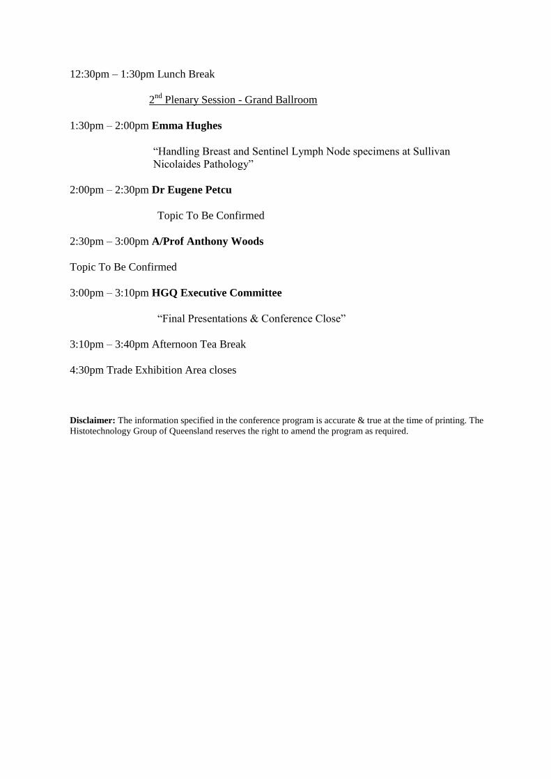
12:30pm – 1:30pm Lunch Break
2nd
Plenary Session - Grand Ballroom
1:30pm – 2:00pm Emma Hughes
“Handling Breast and Sentinel Lymph Node specimens at Sullivan
Nicolaides Pathology”
2:00pm – 2:30pm Dr Eugene Petcu
Topic To Be Confirmed
2:30pm – 3:00pm A/Prof Anthony Woods
Topic To Be Confirmed
3:00pm – 3:10pm HGQ Executive Committee
“Final Presentations & Conference Close”
3:10pm – 3:40pm Afternoon Tea Break
4:30pm Trade Exhibition Area closes
Disclaimer: The information specified in the conference program is accurate & true at the time of printing. The
Histotechnology Group of Queensland reserves the right to amend the program as required. H