Embed Size (px)
Citation preview

Water-soluble chlorophyll protein is involved inherbivore resistance activation during greeningof Arabidopsis thalianaEdouard Boex-Fontvieillea, Sachin Rustgib, Diter von Wettsteinb,1, Steffen Reinbothea,1, and Christiane Reinbothea
aLaboratoire de Génétique Moléculaire des Plantes and Biologie Environnementale et Systémique, Université Joseph Fourier, Laboratoire deBioénergétique Fondamentale et Appliquée, BP53F, 38041 Grenoble Cedex 9, France; and bDepartment of Crop and Soil Sciences, Molecular PlantSciences, School of Molecular Biosciences, and Center for Reproductive Biology, Washington State University, Pullman, WA 99164-6420
Contributed by Diter von Wettstein, April 20, 2015 (sent for review January 29, 2015)
Water-soluble chlorophyll proteins (WSCPs) constitute a small familyof unusual chlorophyll (Chl)-binding proteins that possess a Kunitz-type protease inhibitor domain. In Arabidopsis thaliana, a WSCP hasbeen identified, named AtWSCP, that forms complexes with Chl andthe Chl precursor chlorophyllide (Chlide) in vitro. AtWSCP exhibits aquite unexpected expression pattern for a Chl binding protein andaccumulated to high levels in the apical hook of etiolated plants.AtWSCP expression was negatively light-regulated. Transgenicexpression of AtWSCP fused to green fluorescent protein (GFP)revealed that AtWSCP is localized to cell walls/apoplastic spaces.Biochemical assays identified AtWSCP as interacting with RD21(RESPONSIVE TO DESICCATION 21), a granulin domain-containingcysteine protease implicated in stress responses and defense. Re-constitution experiments showed tight interactions between RD21and WSCP that were relieved upon Chlide binding. Laboratoryfeeding experiments with two herbivorous isopod crustaceans,Porcellio scaber (woodlouse) and Armadillidium vulgare (pillbug),identified the apical hook as Achilles’ heel of etiolated plants andthat this was protected by RD21 during greening. Because Chlide isformed in the apical hook during seedling emergence from thesoil, our data suggest an unprecedented mechanism of herbivore re-sistance activation that is triggered by light and involves AtWSCP.
skotomorphogenesis | plant defense | herbivore deterrence |cysteine proteases | Kunitz protease inhibitor
Higher plants can pass through two different developmentalprograms after seed germination termed skotomorphogenesis
and photomorphogenesis. Dark-grown (etiolated) seedlings under-going skotomorphogenesis have long hypocotyls, an apical hookwith closely apposed, unexpanded cotyledons, and a pale-yellow leafcolor. By contrast, light-grown seedlings undergoing photomor-phogenesis have short hypocotyls, open and fully expanded cot-yledons, and are green. Because of their unique morphology,especially the presence of the apical hook, etiolated seedlings areenabled to grow through the soil without mechanical damage (1).At the cellular level, etioplasts are the predominant plastid
form of dark-grown plants, whereas chloroplasts are found in light-grown seedlings. Etioplasts contain a paracristalline internal mem-brane system termed the prolamellar body (1). In prolamellarbodies of barley and Arabidopsis thaliana etioplasts, two closelyrelated isoforms of NADPH:protochlorophyllide (Pchlide) oxido-reductase, dubbed PORA and PORB, bind Pchlide and NADPH(2). Upon light exposure, Pchlide is converted to chlorophyllide(Chlide), which is subsequently esterified to chlorophyll (Chl)(2). Because the expression of the major light-harvesting Chl a/b-binding proteins (LHCs) is light-induced and because these proteinsare not present during the early hours of greening (3), other pro-teins were suggested to bind the bulk of freshly formed Chlide (4).Early light-induced proteins (ELIPs) are candidates for this func-tion because they are transiently expressed during the develop-mental switch from dark to light growth (5). However, we wereable to show that a distant ortholog of water-soluble chloro-phyll proteins (WSCPs) of Brassicaceae exists in barley, which is
capable of binding Chlide during the transition of etioplasts tochloroplasts (6).WSCPs constitute a small family of proteins that have been
found in only a limited number of plant species belonging toAmaranthaceae, Brassicaceae, Chenopodiaceae, and Polygonaceae.WSCPs differ from the classical Chl-binding proteins of photo-systems I and II in several aspects: (i) they are not constitutivelyexpressed in green leaves, (ii) they are not membrane-bound,(iii) they bind less Chl and Chl a to Chl b at a different ratio, (iv) theydo not interact with carotenoids, and (v) they contain a Kunitzprotease inhibitor motif in their NH2-terminal parts not present inLHCs (7–10).Despite considerable progress made over the last few years,
definite answers on the actual role of WSCPs in planta are stillmissing (7–9). In the present work, we used reverse genetics todefine the role of WSCP in Arabidopsis thaliana. Our data provideevidence for an unprecedented, organ-specific, and developmentallyprogrammed WSCP function in herbivore deterrence.
ResultsAtWSCP Belongs to a Small Family of Kunitz Protease Inhibitors.WSCPs are characterized by the presence of the chlorophyll(ide)-binding signature PFCPLGI (10) and Kunitz trypsin inhibitor sig-nature [LIVM]-x-D-x-[EDNTY]-[DG]-[RKHDENQ]-x-[LIVM]-x(5)-Y-x-[LIVM]) (7). Based on the occurrence of these motifs, aWSCP-related protein was identified in Arabidopsis thaliana, namedAtWSCP, which is encoded by At1g72290. Sequence comparisonrevealed that AtWSCP belongs to a subfamily of Kunitz proteaseinhibitors in Arabidopsis (Fig. S1A). Interestingly, the proteinencoded by At1g72290 lacks a predictable transit sequence forimport into chloroplasts (www.cbs.dtu.dk/services/TargetP andwww.cbs.dtu.dk/services/SignalP/) but contains a predictable signalpeptide typical for proteins entering the secretory pathway(Fig. S1B) (7).
Significance
Herbivory is one of the most important processes in the bio-sphere. When plants germinated underneath the soil or fallenleaves undergo skotomorphogenesis, they are especially proneto a vast range of seed predators and herbivorous arthropods.How greening plants protect themselves against these foeswas thus far largely unknown. Here, we describe a mechanismhow etiolated seedlings deter arthropod devourers. Our articlethus contributes to the understanding of plant survival strat-egies in the natural environment.
Author contributions: E.B.-F., D.v.W., and S. Reinbothe designed research; E.B.-F., S. Rustgi,S. Reinbothe, and C.R. performed research; E.B.-F., S. Rustgi, S. Reinbothe, and C.R. ana-lyzed data; and E.B.-F., S. Rustgi, D.v.W., S. Reinbothe, and C.R. wrote the paper.
The authors declare no conflict of interest.1To whom correspondence may be addressed. Email: [email protected] or [email protected].
This article contains supporting information online at www.pnas.org/lookup/suppl/doi:10.1073/pnas.1507714112/-/DCSupplemental.
www.pnas.org/cgi/doi/10.1073/pnas.1507714112 PNAS | June 9, 2015 | vol. 112 | no. 23 | 7303–7308
PLANTBIOLO
GY
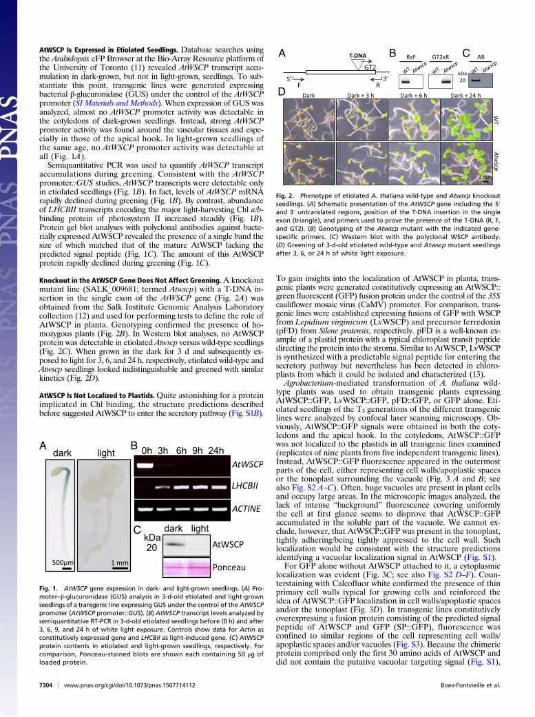
AtWSCP Is Expressed in Etiolated Seedlings. Database searches usingthe Arabidopsis eFP Browser at the Bio-Array Resource platform ofthe University of Toronto (11) revealed AtWSCP transcript accu-mulation in dark-grown, but not in light-grown, seedlings. To sub-stantiate this point, transgenic lines were generated expressingbacterial β-glucuronidase (GUS) under the control of the AtWSCPpromoter (SI Materials and Methods). When expression of GUS wasanalyzed, almost no AtWSCP promoter activity was detectable inthe cotyledons of dark-grown seedlings. Instead, strong AtWSCPpromoter activity was found around the vascular tissues and espe-cially in those of the apical hook. In light-grown seedlings ofthe same age, no AtWSCP promoter activity was detectable atall (Fig. 1A).Semiquantitative PCR was used to quantify AtWSCP transcript
accumulations during greening. Consistent with the AtWSCPpromoter::GUS studies, AtWSCP transcripts were detectable onlyin etiolated seedlings (Fig. 1B). In fact, levels of AtWSCP mRNArapidly declined during greening (Fig. 1B). By contrast, abundanceof LHCBII transcripts encoding the major light-harvesting Chl a/b-binding protein of photosystem II increased steadily (Fig. 1B).Protein gel blot analyses with polyclonal antibodies against bacte-rially expressed AtWSCP revealed the presence of a single band thesize of which matched that of the mature AtWSCP lacking thepredicted signal peptide (Fig. 1C). The amount of this AtWSCPprotein rapidly declined during greening (Fig. 1C).
Knockout in the AtWSCP Gene Does Not Affect Greening.A knockoutmutant line (SALK_009681; termed Atwscp) with a T-DNA in-sertion in the single exon of the AtWSCP gene (Fig. 2A) wasobtained from the Salk Institute Genomic Analysis Laboratorycollection (12) and used for performing tests to define the role ofAtWSCP in planta. Genotyping confirmed the presence of ho-mozygous plants (Fig. 2B). In Western blot analyses, no AtWSCPprotein was detectable in etiolated Atwscp versus wild-type seedlings(Fig. 2C). When grown in the dark for 3 d and subsequently ex-posed to light for 3, 6, and 24 h, respectively, etiolated wild-type andAtwscp seedlings looked indistinguishable and greened with similarkinetics (Fig. 2D).
AtWSCP Is Not Localized to Plastids.Quite astonishing for a proteinimplicated in Chl binding, the structure predictions describedbefore suggested AtWSCP to enter the secretory pathway (Fig. S1B).
To gain insights into the localization of AtWSCP in planta, trans-genic plants were generated constitutively expressing an AtWSCP::green fluorescent (GFP) fusion protein under the control of the 35Scauliflower mosaic virus (CaMV) promoter. For comparison, trans-genic lines were established expressing fusions of GFP with WSCPfrom Lepidium virginicum (LvWSCP) and precursor ferredoxin(pFD) from Silene pratensis, respectively. pFD is a well-known ex-ample of a plastid protein with a typical chloroplast transit peptidedirecting the protein into the stroma. Similar to AtWSCP, LvWSCPis synthesized with a predictable signal peptide for entering thesecretory pathway but nevertheless has been detected in chloro-plasts from which it could be isolated and characterized (13).Agrobacterium-mediated transformation of A. thaliana wild-
type plants was used to obtain transgenic plants expressingAtWSCP::GFP, LvWSCP::GFP, pFD::GFP, or GFP alone. Eti-olated seedlings of the T3 generations of the different transgeniclines were analyzed by confocal laser scanning microscopy. Ob-viously, AtWSCP::GFP signals were obtained in both the coty-ledons and the apical hook. In the cotyledons, AtWSCP::GFPwas not localized to the plastids in all transgenic lines examined(replicates of nine plants from five independent transgenic lines).Instead, AtWSCP::GFP fluorescence appeared in the outermostparts of the cell, either representing cell walls/apoplastic spacesor the tonoplast surrounding the vacuole (Fig. 3 A and B; seealso Fig. S2 A–C). Often, huge vacuoles are present in plant cellsand occupy large areas. In the microscopic images analyzed, thelack of intense “background” fluorescence covering uniformlythe cell at first glance seems to disprove that AtWSCP::GFPaccumulated in the soluble part of the vacuole. We cannot ex-clude, however, that AtWSCP::GFP was present in the tonoplast,tightly adhering/being tightly appressed to the cell wall. Suchlocalization would be consistent with the structure predictionsidentifying a vacuolar localization signal in AtWSCP (Fig. S1).For GFP alone without AtWSCP attached to it, a cytoplasmic
localization was evident (Fig. 3C; see also Fig. S2 D–F). Coun-terstaining with Calcofluor white confirmed the presence of thinprimary cell walls typical for growing cells and reinforced theidea of AtWSCP::GFP localization in cell walls/apoplastic spacesand/or the tonoplast (Fig. 3D). In transgenic lines constitutivelyoverexpressing a fusion protein consisting of the predicted signalpeptide of AtWSCP and GFP (SP::GFP), fluorescence wasconfined to similar regions of the cell representing cell walls/apoplastic spaces and/or vacuoles (Fig. S3). Because the chimericprotein comprised only the first 30 amino acids of AtWSCP anddid not contain the putative vacuolar targeting signal (Fig. S1),
Fig. 1. AtWSCP gene expression in dark- and light-grown seedlings. (A) Pro-moter–β-glucuronidase (GUS) analysis in 3-d-old etiolated and light-grownseedlings of a transgenic line expressing GUS under the control of the AtWSCPpromoter (AtWSCP promoter::GUS). (B) AtWSCP transcript levels analyzed bysemiquantitative RT-PCR in 3-d-old etiolated seedlings before (0 h) and after3, 6, 9, and 24 h of white light exposure. Controls show data for Actin asconstitutively expressed gene and LHCBII as light-induced gene. (C) AtWSCPprotein contents in etiolated and light-grown seedlings, respectively. Forcomparison, Ponceau-stained blots are shown each containing 50 μg ofloaded protein.
A B C
D
Fig. 2. Phenotype of etiolated A. thaliana wild-type and Atwscp knockoutseedlings. (A) Schematic presentation of the AtWSCP gene including the 5′and 3′ untranslated regions, position of the T-DNA insertion in the singleexon (triangle), and primers used to prove the presence of the T-DNA (R, F,and GT2). (B) Genotyping of the Atwscp mutant with the indicated gene-specific primers. (C ) Western blot with the polyclonal WSCP antibody.(D) Greening of 3-d-old etiolated wild-type and Atwscp mutant seedlingsafter 3, 6, or 24 h of white light exposure.
7304 | www.pnas.org/cgi/doi/10.1073/pnas.1507714112 Boex-Fontvieille et al.

we assume that this reporter protein was localized in cell walls/apoplastic spaces, while not necessarily precluding a localizationof the full-length AtWSCP in the vacuole. For LvWSCP::GFP, acompletely different localization in plastids was found in alltransgenic lines examined (Fig. S4 A–C), as was seen for pFd::GFPused as control (Fig. S4 D–F).AtWSCP::GFP fluorescence distribution in the apical hook
was similar to that in the cotyledons and revealed the presence ofthe protein in cell walls/apoplastic spaces (Fig. S5 A–C). In fact,no fluorescence signal was present in plastids (Fig. S5 A–C),whereas LvWSCP::GFP (Fig. S5 D–F) and pFd::GFP (Fig. S5G–I) gave rise to strong plastid signals.
AtWSCP Does Not Operate in Vascular Tissue Formation and HypocotylElongation. One Kunitz trypsin inhibitor in chickpea, termed TPI-1,has been implicated in regulating hypocotyl growth and apical hookformation (14–17). If AtWSCP were to accomplish a similar role,changing its amount should have severe anatomical (phenotypic)effects. To test this hypothesis, we compared seedling morphologyfor wild-type and Atwscp mutant seedlings with that of transgeniclines constitutively overexpressing AtWSCP (35S::AtWSCP) underthe control of the 35S promoter. Several independent 35S::AtWSCPlines were obtained that overexpressed AtWSCP (one is pre-sented in Fig. 4A). However, in none of these AtWSCP over-expressors could differences in hypocotyl length, light-triggeredapical hook straightening, and overall seedling morphology be ob-served (Fig. 4B). When hypocotyl length was measured after 3 and5d, respectively, of growth in the dark provoking skotomopho-genesis, no difference became apparent for wild-type, Atwscp mu-tant, and 35S::AtWSCP overexpressing plants (Fig. 4C).
AtWSCP Interacts with the Granulin Domain-Containing CysteineProteases RD21. Halls and coworkers (8) identified AtWSCP asa potent inhibitor of the recombinant proaleurain maturation
protease (At3g19390) and papain. There is a small family ofgranulin domain-containing proteases in Arabidopsis encoded byAt3g19390, At5g43060, At4g34460 (XYLEM BARK CYSTEINEPEPTIDASE 3, XBCP3), and At1g47120 (RESPONSIVE TODESICCATION 21, RD21). RD21 is most interesting because ithas been implicated in various reactions conferring resistance tobiotic and abiotic foes (18–21). Like many other cysteine endo-proteases, RD21 is encoded as a preproprotein having an NH2-terminal propeptide with autoinhibitory activity and a COOH-terminal granulin-domain containing propeptide with unknownfunction (Fig. S6). The NH2-terminal propeptide is cleaved by ayet-unknown mechanism either requiring an autocatalytic pro-cessing under low pH or activity of a processing enzyme (22).We asked whether RD21 may interact with AtWSCP in etio-
lated plants. To test for such interactions, pull-down assays wereconducted on protein extracts that had been prepared from theapical hook of 4.5-d-old etiolated wild-type plants. Interestingly,both antibodies used, the one against RD21 and the one againstAtWSCP, coprecipitated both proteins from the tissue extract ofetiolated wild-type plants and, thus, confirmed the interaction ofRD21 and AtWSCP in the apical hook. With protein extractsfrom flashed seedlings, however, the RD21 antibody precipitatedonly RD21 and the AtWSCP antibody precipitated only AtWSCP(Fig. 5A). This result showed that RD21 and AtWSCP no longerinteracted in flash-illuminated seedlings.Next, we isolated WSCP-containing complexes from the apical
hook of transgenic plants expressing AtWSCP-(His)6 constitu-tively. The protein complexes in turn were subjected to non-denaturing PAGE and detected by (i) Western blotting using theWSCP and RD21 antisera and (ii) scoring red light-inducedpigment autofluorescence on X-ray films in case the proteinwould be complexed with Chl and/or Chlide. As shown in Fig.5B, both assays revealed the presence and light-induced dis-sociation of higher molecular mass (HMr) complexes containingboth WSCP and RD21 in the apical hook of etiolated plants.Several different WSCP bands of descent size were obtained,which most likely represent dissociation intermediates. Inter-estingly, a pool of monomeric WSCP was present already in thedark but gave rise to an autofluorescing band containing pigmentonly during greening (Fig. 5B, compare a and b).In a final experiment, cDNA-encoded Flag-tagged RD21 con-
taining the granulin domain and His-tagged mature AtWSCPlacking its targeting signal were produced by coupled in vitrotranscription/translation of respective cDNA clones, purified,and reconstituted into larger complexes. These complexes in turnwere supplemented with Chl or Chlide and complex dissociationmonitored by nondenaturing PAGE as described before. Fig. 5C
A B
DC
Fig. 3. Localization of AtWSCP::GFP in planta. (A and B) Fluorescenceanalysis of AtWSCP::GFP accumulation in the cotyledons of etiolated seed-lings of respective transgenic plants of the T3 generation. (C) Localization ofGFP in T3 plants expressing GFP without AtWSCP attached to it. (D) Calco-fluor white stain to visualize cell walls in AtWSCP::GFP plants. Red flashesmark apoplastic spaces, yellow flashes mark GFP fluorescence in cytoplasmand nuclei, whereas blue arrowheads indicate sparing of GFP fluorescenceby plastids.
A B C
Fig. 4. Phenotype of etiolated A. thaliana wild-type and Atwscp mutantplants versus transgenic plants overexpressing AtWSCP (35S::AtWSCP).(A) AtWSCP protein levels in leaves of 3-wk-old wild-type and 35S::AtWSCPplants, as assessed by Western blotting using the AtWSCP antiserum (Upper)and Coomassie staining of a replicate SDS/PAGE gel (Lower). (B) Morphologyof 3-d-old etiolated wild-type, Atwscp and 35S::AtWSCP seedlings. (C) Hy-pocotyl length of 3-d-old etiolated wild-type, Atwscp and 35S::AtWSCPseedlings. The data represent the mean of three independent replicates (n =40; ± defines the SE).
Boex-Fontvieille et al. PNAS | June 9, 2015 | vol. 112 | no. 23 | 7305
PLANTBIOLO
GY

depicts HMr complexes containing RD21 and WSCP and showstheir Chlide-dependent dissociation. Results shown in the sup-porting information proved the specificity of the WSCP-pigmentinteraction and that the dissociation of the HMr complexes ledto an activation of RD21’s proteolytic activity. This dissociationis illustrated in Fig. S7, showing the decline of both WSCP andRD21 to vanishingly low levels after prolonged incubations inwhite light.
Reduced Herbivory on Atwscp Seedlings. The results presented sofar suggested a light-dependent, Chlide- and WSCP-mediatedmechanism of RD21 activation in the apical hook of etiolatedplants during greening. We asked whether such mechanismcould contribute to plant protection against herbivorous ar-thropods that prey on seeds and young seedlings (23). Examplesfor such herbivorous arthropods are provided by Porcellio scaber(woodlouse) and Armadillidium vulgare (pillbug), two largelynocturnal isopod crustaceans that are generally considered torepresent seed predators and detritivores but can also live asfacultative herbivores (23). If deprived for nutrients, woodliceclimbed light-grown plants and severed inflorescence stems andpetioles (23). Both isopod species also attacked etiolated seed-lings and selected the apical hook as primary target. Quantitativeassays revealed that approximately 50% of the etiolated seed-lings analyzed in three replicate experiments were severed by theisopods, breaking the apical hook and dropping the cotyledons(Fig. 6). The dropped cotyledons in turn were consumed, be-ginning at the mid ribs and outer leaf edges, but this feedingactivity was delayed and occurred only 24 h after the first attackbreaking the apical hook.Remarkably, the primary attack at the apical hook, but not
subsequent feeding activity on the cotyledons, was significantlyreduced in the Atwscp mutant containing free RD21 and, in fact,accounted to only 10% of that seen on etiolated wild-typeseedlings (Fig. 6). Interestingly, this percentage was similar tothe value obtained for wild-type seedlings that had been flashedand subsequently transferred to darkness for 2 h. In markedcontrast to these results, no such light effect on the primary andsecondary feeding activity of woodlice and pillbugs was observedfor seedlings constitutively overexpressing AtWSCP (Fig. 6).
DiscussionAtWSCP Does Not Operate in the Establishment of the PhotosyntheticApparatus. Previously, a role of WSCPs as Chlide carrier andputative protease inhibitor was proposed (9, 13). Both possibil-ities are not mutually exclusive. Here, we add more informationon AtWSCP’s temporal and spatial expression, its intracellularlocalization, and its putative function during seedling develop-ment and show the following. First, AtWSCP accumulation is notrestricted to the transmitting tract tissue of developing flowers,as reported in ref. 9. Substantial amounts of AtWSCP were alsofound in etiolated plants. Nevertheless, the expression pattern ofAtWSCP during light-induced greening was not consistent with arole as transient Chlide carrier during the establishment of thephotosynthetic apparatus. For example, AtWSCP promoter ac-tivity and AtWSCP transcript accumulation were confined toetiolated plants and were not detected in light-grown seedlings.Moreover, maximum promoter activity was restricted to theapical hook of etiolated seedlings, but no activity was detected inthe cotyledons where the bulk of Chl synthesis occurs. Second,AtWSCP was not found in plastids (etioplasts) as should beexpected for a protein binding Chl or one of its precursors, butaccumulated in cell walls/apoplastic spaces.AtWSCP and LvWSCP both do not possess predictable chlo-
roplast transit sequences, but contain predictable signal peptidesfor proteins entering the secretory pathway. Interestingly, a class ofchloroplast proteins was identified in Arabidopsis thaliana that aretargeted to their final destination through the endoplasmic re-ticulum (24–26). LvWSCP could be a member of this unique familyof N-glycosylated chloroplast proteins (13). Because also AtWSCPappears to enter the secretory pathway but is not targeted tochloroplasts, differences in the structure of the respective signalpeptides and/or N-glycosylation sites could be responsible for thedifferential targeting of AtWSCP and LvWSCP in planta thatneed to be explored in future work.
AtWSCP Interacts with RD21 in the Apical Hook of Etiolated Seedlings.AtWSCP interacts with RD21, a papain-like cysteine proteinaseinvolved in stress responses and defense (18–21). Localizationstudies using the eFP browser (11) identified both AtWSCP andRD21 to have overlapping spatial expression patterns and toaccumulate in endoplasmic reticulum (ER) bodies, lytic vacu-oles, and cell walls/apoplastic spaces (Fig. S8). Like AtWSCP,RD21 is synthesized as a preproprotein bearing an NH2-terminalsignal peptide (predomain) directing the protein to the secretorypathway (22). RD21 additionally contains a prodomain exhibit-ing autoinhibitory activity and has with the protease domain,proline-rich domain, and granulin domain three supplementary
A B
C
Fig. 5. Interaction of RD21 with WSCP. (A) Pull-down assay to detect RD21–WSCP interactions in plant extracts prepared from the apical hook region of4.5-d-old etiolated seedlings and seedlings that had been flash-illuminatedand kept in the dark for another 2 h. (B) Isolation of high molecular mass(HMr) complexes containing RD21 and WSCP from transgenic plants expressingAtWSCP-(His)6. Protein extracts were prepared from 4.5-d-old etiolated seed-lings and flashed seedlings as before and used for nondenaturing PAGEplus Western blotting with the indicated RD21 and WSCP antisera (a) aswell as pigment fluorescence analysis (b). (C ) In vitro reconstitution ofRD21–WSCP complexes and their Chl- and Chlide-dependent dissociation(b and c), as assessed by nondenaturing PAGE. C, a shows 35S-RD21-Flagand 35S-WSCP-(His)6 that had been produced from respective cDNA clonesby coupled in vitro transcription/translation and analyzed on a denaturingSDS/PAGE gel.
A B
Fig. 6. Apical hook damage by P. scaber (woodlice, white columns) andA. vulgare (pillbug, gray columns) of etiolated wild-type (WT), Atwscpmutant,and 35S::AtWSCP overexpressor (OE) seedlings. Percentages refer to 4.5-d-oldseedlings that had been illuminated for 30 min with white light shifted back todarkness for 2 h (B) or kept in the dark (A). The number of seedlings withdamaged apical hooks was counted and is expressed as percentage of the totalnumber of seedlings analyzed in three independent experiments comprisingeach 120 plantlets and 3 isopods. Error bars are indicated.
7306 | www.pnas.org/cgi/doi/10.1073/pnas.1507714112 Boex-Fontvieille et al.

protein modules (ref. 22; compare Fig. S6). The granulin domainshares homology to granulins/epithelins in animals, which aregrowth hormones that are released upon wounding (27). Acti-vation of RD21 involves removal of the prodomain, giving rise toan intermediate form (iRD21). Then, iRD21 undergoes furtherprocessing/maturation that is associated with the removal of thegranulin domain (Fig. S6). In planta, RD21 isoforms containingor lacking the granulin domain have been detected (28–30).The propeptide regulates RD21 activity by autoinhibition
during seed development, but on germination when pH dropsbelow 5, intramolecular conformational changes take place thatlead to the destabilization and cleavage of the propeptide. Thus,to avoid uncontrolled RD21 activity in both time and space,another layer of regulation exists that consists of protease in-hibitors. AtWSCP is such an inhibitor that expresses in etiolatedseedlings and controls RD21 activity throughout the skotomor-phogenic phase of seedling growth.Molecular modeling and docking analyses were carried out to
suggest a scenario of how AtWSCP and RD21 may interact.Using established methods (refs. 31–33; SI Materials and Methods),individual 3D models were first built for AtWSCP and RD21. The3D structure for AtWSCP most closely resembled that of soybeanKunitz-type trypsin inhibitor and tamarind Kunitz inhibitor (TKI),with an α-turn and 10 antiparallel β-strands that form a barrel-likestructure (Fig. S9A) (34, 35). Similarly, the molecular modeling ofRD21 revealed a typical papain-like structure, with two almostequally sized lobes dubbed R (right) and L (left), divided by anactive site cleft (Fig. S9B) (36). The R domain of RD21 is pre-dominantly comprised of an extended NH2-terminal loop andfour antiparallel β-strands, whereas the L domain is primarilycomposed of α-helices and the COOH-terminal end (Fig. S9B).Based on the 3D models, we next sought to understand the
AtWSCP–RD21 interactions. A clue to this end was provided bystudies on oryzacystatin-I and papain-like proteases and on TKIand its interactions with factor Xa and trypsin (34, 35, 37).Specifically, the second loop (Ala37-Leu46, orange) of AtWSCP,which spans between β-strands 2 and 3, encompasses the LHCIIsignature sequence and the fifth loop (Lys84-Ser95, purple),which connects β-strands 5 and 6 is proposed to be the reactive-site loop (RSL) (Fig. 7A and Fig. S9A). In our interaction model,Try88 and Pro89 in the RSL of AtWSCP are predicted to intrudeinto the active site region of RD21 containing Cys161 and His297and, thereby, to block its proteolytic activity (Fig. 7 A and B).Moreover, one amino acid residue, Lys92 in the RSL, and twoamino acid residues, Leu41 and Pro42 in the LHCII signaturesequence, are predicted to form hydrogen bonds with amino acidresidues Asp154 and Lys227, respectively, in RD21 (Fig. 7B; seealso Fig. S9 C and D). Together, these hydrogen bonds maystabilize the observed AtWSCP–RD21 interaction. However, thepresence and close physical proximity of the LHCII signature ofAtWSCP to the catalytic triad of RD21 could explain the observedlight-triggered, Chlide-dependent dissociation of the AtWSCP–RD21 complex in vitro and in planta.
AtWSCP Is Operative in Herbivore Resistance Activation During Greening.As mentioned, the interaction between AtWSCP and RD21 inthe apical hook appears to be part of a mechanism of keepingRD21 and, perhaps, also other papain-like cysteine proteinasesin an inactive state as long as the seedling etiolates underneaththe soil or fallen leaves. Once the seedling de-etiolates, the light-triggered switch to photomorphogenesis then would releaseRD21 from AtWSCP. Our biochemical studies suggest Chlide astrigger of this dissociation step. How Chlide is transported fromthe developing chloroplast to cell walls/apoplastic spaces is un-known but may involve stromules, providing recently discoveredhighways from plastids to the remainder of the cell (38). Super-impose on this very rapid effect is the light-induced depres-sion of AtWSCP gene expression that depletes AtWSCP frometiolated plants and, thereby, increases the amount of freeRD21 ready of counteracting proteases present in the arthro-pod gut. Presumably in concert with other proteinases, efficient
protection is conferred onto the seedling’s Achilles’ heel, the apicalhook, against arthropod devourers. What destiny the Chlide-complexed WSCP oligomers may have is unclear and should bestudied in future work.
Materials and MethodsPlant Material and Growth Conditions. Seeds of the following Arabidopsisthaliana lines were used for the experiments: ecotype Columbia (Col-0; re-ferred to as wild-type), SALK_009681 (renamed Atwscp) that carries a T-DNAinsertion in the gene At1g72290 encoding AtWSCP (12), transgenic linesexpressing AtWSCP constitutively (wild-type transformed with the plas-mid pB7WG2 containing the coding frame for AtWSCP; referred toas 35S::AtWSCP), lines carrying the promoter of AtWSCP in front of theβ-glucuronidase coding sequence (AtWSCP::GUS), and transgenic linesexpressing AtWSCP, LvWSCP, and pFD fused to green fluorescent protein (GFP)under the control of 35S promoter (35S::AtWSCP/LvWSCP/pFD::GFP). Forcomparison, transgenic seedlings expressing a fusion consisting of thesignal peptide of AtWSCP and GFP (SP::GFP) were produced. Seeds wereplated onto Murashige–Skoog agar medium and germinated in the darkor in white light for appropriate periods.
Whole-Plant Predation Assay. Populations of 120 wild-type, Atwscp mutant,and 35S::AtWSCP overexpressing plants were grown for 4.5 d in darkness onPetri dishes, and the open dishes were then transferred to a large box, filledto a depth of 6 cm with soil, and containing P. scaber and A. vulgare. Cul-tivation of the isopods was performed essentially as described in ref. 23. Forfeeding experiments, P. scaber and A. vulgare were fed with pesticide-freeArabidopsis wild-type plants, then starved for 3 d and placed at low densityof 1–3 per liter of soil into the nutrient chamber. At different times, feeding
A B
Fig. 7. Structural model for the AtWSCP–RD21 interaction, predicted byusing ClusPro. (A) Ribbon diagram of the AtWSCP–RD21 complex (frontview). The fifth or reactive site loop (RSL) that spans between the fifth andsixth β-strands of AtWSCP is shown in purple, and it’s beginning at Lys84 andend at Ser95 are marked by arrows. The Trp88 and Pro89 residue at the RSLof AtWSCP that intrudes between the catalytic triad, i.e., Cys161, His297, andAsn317 at the active site cleft of RD21 are shown by lines. The β-strands andα-helices of RD21 are shown respectively in yellow and red, whereas theβ-strands of AtWSCP are depicted in magenta, an α-turn in cyan, and con-necting loops are shown in deep salmon. (B) An enlarged view of the reactivesite loop showing the intruding amino acid residues and their respective lo-cations in AtWSCP (a). Specific amino acid residues from AtWSCP and RD21 thatform hydrogen bonds are shown in purple or orange and green, respectively.These interactions involve the following: AtWSCP:Lys92-RD21:Asp154 (b), andAtWSCP:Leu41 and Pro42-RD21:Lys227 (c). Amino acid numbering for RD21 isbased on their locations in the full-length preproprotein, and for AtWSCP isbased on the mature protein after removal of the signal peptide.
Boex-Fontvieille et al. PNAS | June 9, 2015 | vol. 112 | no. 23 | 7307
PLANTBIOLO
GY
was measured by counting the number of plants with damaged apical hooksand/or dropped cotyledons.
Protein Analyses. Protein extracts of etiolated seedlings were prepared ac-cording to Hurkman and Tanaka (39). Briefly, tissues were extracted with doublyconcentrated SDS sample buffer (40), separated on 12% (wt/vol) SDS/PAGEgels, and blotted onto nitrocellulose membranes. Western blotting wascarried out according to Towbin et al. (41) by using an alkaline phospha-tase-based system with 5-bromo-4-chloro-3-indolyl phosphate and nitroblue tetrazolium or enhanced chemiluminescence (ECL Western BlottingAnalysis system; Amersham), respectively. Pull-down assays on proteinextracts of the apical hook region were performed with antibodies raisedagainst the bacterially expressed and purified AtWSCP (SI Materials andMethods) and antibodies against RD21, using standard procedures. Isola-tion of HMr AtWSCP complexes was achieved by Ni-NTA agarose chro-matography of apical hook extracts that had been prepared fromtransgenic seedlings overexpressing a AtWSCP(His)6 protein. In vitro re-constitution experiments and pigment autofluorescence screens were
carried out with cDNA-encoded, wheat germ-translated proteins con-taining or lacking [35S]methionine and isolated pigments (Chl, Chlide,Pchlide) (42).
cDNA Synthesis and Semiquantitative PCR. Semiquantitative PCR was carriedout on DNA templates that had been generated by first-strand cDNA synthesis,using appropriate primers, as described in the SI Materials and Methods.
Promoter Studies. AtWSCP promoter activity was scored in transgenic plantsexpressing β-glucuronidase (GUS) by using standard procedures (SI Materials andMethods). Image acquisition was made with an Eclipse E-600 (Nikon) microscopeor a SZX12 (Olympus) binocular and documented with an Olympus DP70 camera.
ACKNOWLEDGMENTS. We thank Dr. Claudia Rossig for a gift of transgenicseeds expressing pFd::GFP fusion proteins and Ikuko Hara-Nishimura (KyotoUniversity) for a gift of the antibody directed against RD21 (see ref. 28). Thiswork was supported by the Chaire d’Excellence Program of the French Min-istry of National Education and Research (to C.R.).
1. von Wettstein D, Gough S, Kannangara CG (1995) Chlorophyll biosynthesis. Plant Cell7(7):1039–1057.
2. Reinbothe C, et al. (2010) Chlorophyll biosynthesis: Spotlight on protochlorophyllidereduction. Trends Plant Sci 15(11):614–624.
3. Apel K, Kloppstech K (1978) The plastid membranes of barley (Hordeum vulgare).Light-induced appearance of mRNA coding for the apoprotein of the light-harvestingchlorophyll a/b protein. Eur J Biochem 85(2):581–588.
4. Reinbothe S, Reinbothe C (1996) The regulation of enzymes involved in chlorophyllbiosynthesis. Eur J Biochem 237(2):323–343.
5. Adamska I (2001) The Elip family of stress proteins in the thylakoid membranes of pro-and eukaryota. Regulation of Photosynthesis, eds Aro E-M, Andersson B (Kluwer Aca-demic Publishers, Dordrecht, The Netherlands), pp 487–505.
6. Reinbothe C, Satoh H, Alcaraz JP, Reinbothe S (2004) A novel role of water-solublechlorophyll proteins in the transitory storage of chlorophyllide. Plant Physiol 134(4):1355–1365.
7. Satoh H, Uchida A, Nakayama K, Okada M (2001) Water-soluble chlorophyll protein inBrassicaceae plants is a stress-induced chlorophyll-binding protein. Plant Cell Physiol42(9):906–911.
8. Halls CE, et al. (2006) A Kunitz-type cysteine protease inhibitor from cauliflower andArabidopsis. Plant Sci 170:1102–1110.
9. Bektas I, Fellenberg C, Paulsen H (2012) Water-soluble chlorophyll protein (WSCP) ofArabidopsis is expressed in the gynoecium and developing silique. Planta 236(1):251–259.
10. Green BR, Kühlbrandt W (1995) Sequence conservation of light-harvesting and stress-response proteins in relation to the three-dimensional molecular structure of LHCII.Photosynth Res 44(1-2):139–148.
11. Winter D, et al. (2007) An “Electronic Fluorescent Pictograph” browser for exploringand analyzing large-scale biological data sets. PLoS ONE 2(8):e718.
12. Alonso JM, et al. (2003) Genome-wide insertional mutagenesis of Arabidopsis thali-ana. Science 301(5633):653–657.
13. Takahashi S, et al. (2013) Molecular cloning, characterization and analysis of the in-tracellular localization of a water-soluble chlorophyll-binding protein (WSCP) fromVirginia pepperweed (Lepidium virginicum), a unique WSCP that preferentially bindschlorophyll b in vitro. Planta 238(6):1065–1080.
14. Jiménez T, Martín I, Labrador E, Dopico B (2007) A chickpea Kunitz trypsin inhibitor islocated in cell wall of elongating seedling organs and vascular tissue. Planta 226(1):45–55.
15. Jiménez T, Martín I, Hernández-Nistal J, Labrador E, Dopico B (2008) The accumula-tion of a Kunitz trypsin inhibitor from chickpea (TPI-2) located in cell walls is increasedin wounded leaves and elongating epicotyls. Physiol Plant 132(3):306–317.
16. Hernández-Nistal J, Martín I, Jiménez T, Dopico B, Labrador E (2009) Two cell wallKunitz trypsin inhibitors in chickpea during seed germination and seedling growth.Plant Physiol Biochem 47(3):181–187.
17. Downing WL, et al. (1992) A Brassica napus transcript encoding a protein related tothe Künitz protease inhibitor family accumulates upon water stress in leaves, not inseeds. Plant J 2(5):685–693.
18. Koizumi M, Yamaguchi-Shinozaki K, Tsuji H, Shinozaki K (1993) Structure and ex-pression of two genes that encode distinct drought-inducible cysteine proteinases inArabidopsis thaliana. Gene 129(2):175–182.
19. Yamaguchi-Shinozaki K, Koizumi M, Urao S, Shinozaki K (1992) Molecular cloningand characterization of 9 cDNAs for genes that are responsive to desiccation inArabidopsis thaliana: Sequence analysis of one cDNA clone that encodes a putativetransmembrane channel protein. Plant Cell Physiol 33(3):217–224.
20. Gepstein S, et al. (2003) Large-scale identification of leaf senescence-associated genes.Plant J 36(5):629–642.
21. Shindo T, Misas-Villamil JC, Hörger AC, Song J, van der Hoorn RA (2012) A role inimmunity for Arabidopsis cysteine protease RD21, the ortholog of the tomato im-mune protease C14. PLoS ONE 7(1):e29317.
22. Gu C, et al. (2012) Post-translational regulation and trafficking of the granulin-containing protease RD21 of Arabidopsis thaliana. PLoS ONE 7(3):e32422.
23. Farmer EE, Dubugnon L (2009) Detritivorous crustaceans become herbivores onjasmonate-deficient plants. Proc Natl Acad Sci USA 106(3):935–940.
24. Nanjo Y, et al. (2006) Rice plastidial N-glycosylated nucleotide pyrophosphatase/phosphodiesterase is transported from the ER-golgi to the chloroplast through thesecretory pathway. Plant Cell 18(10):2582–2592.
25. Villarejo A, et al. (2008) Evidence for a protein transported through the secretorypathway en route to the higher plant chloroplast. Nat Cell Biol 10(2):220–227.
26. Faye L, Daniell H (2006) Novel pathways for glycoprotein import into chloroplasts.Plant Biotechnol J 4(3):275–279.
27. Bateman A, Bennett HPJ (2009) The granulin gene family: From cancer to dementia.BioEssays 31(11):1245–1254.
28. Yamada K, Matsushima R, Nishimura M, Hara-Nishimura I (2001) A slow maturation ofa cysteine protease with a granulin domain in the vacuoles of senescing Arabidopsisleaves. Plant Physiol 127(4):1626–1634.
29. Hayashi Y, et al. (2001) A proteinase-storing body that prepares for cell death orstresses in the epidermal cells of Arabidopsis. Plant Cell Physiol 42(9):894–899.
30. Carter C, et al. (2004) The vegetative vacuole proteome of Arabidopsis thalianareveals predicted and unexpected proteins. Plant Cell 16(12):3285–3303.
31. Roy A, Kucukural A, Zhang Y (2010) I-TASSER: A unified platform for automatedprotein structure and function prediction. Nat Protoc 5(4):725–738.
32. Biasini M, et al. (2014) SWISS-MODEL: Modelling protein tertiary and quaternarystructure using evolutionary information. Nucleic Acids Res 42(Web Server issue):W252–W258.
33. Comeau SR, Gatchell DW, Vajda S, Camacho CJ (2004) ClusPro: A fully automatedalgorithm for protein-protein docking. Nucleic Acids Res 32(Web Server issue):W96–W99.
34. Song HK, Suh SW (1998) Kunitz-type soybean trypsin inhibitor revisited: Refinedstructure of its complex with porcine trypsin reveals an insight into the interactionbetween a homologous inhibitor from Erythrina caffra and tissue-type plasminogenactivator. J Mol Biol 275(2):347–363.
35. Patil DN, Chaudhary A, Sharma AK, Tomar S, Kumar P (2012) Structural basis for dualinhibitory role of tamarind Kunitz inhibitor (TKI) against factor Xa and trypsin. FEBS J279(24):4547–4564.
36. Bethune MT, Strop P, Tang Y, Sollid LM, Khosla C (2006) Heterologous expression,purification, refolding, and structural-functional characterization of EP-B2, a self-activating barley cysteine endoprotease. Chem Biol 13(6):637–647.
37. Benchabane M, Schlüter U, Vorster J, Goulet M-C, Michaud D (2010) Plant cystatins.Biochimie 92(11):1657–1666.
38. Hanson MR, Sattarzadeh A (2013) Trafficking of proteins through plastid stromules.Plant Cell 25(8):2774–2782.
39. Hurkman WJ, Tanaka CK (1986) Solubilization of plant membrane proteins foranalysis by two-dimensional gel electrophoresis. Plant Physiol 81(3):802–806.
40. Laemmli UK (1970) Cleavage of structural proteins during the assembly of the head ofbacteriophage T4. Nature 227(5259):680–685.
41. Towbin H, Staehelin T, Gordon J (1979) Electrophoretic transfer of proteins frompolyacrylamide gels to nitrocellulose sheets: Procedure and some applications. ProcNatl Acad Sci USA 76(9):4350–4354.
42. Reinbothe C, Buhr F, Pollmann S, Reinbothe S (2003) In vitro reconstitution of light-harvesting POR-protochlorophyllide complex with protochlorophyllides a and b.J Biol Chem 278(2):807–815.
7308 | www.pnas.org/cgi/doi/10.1073/pnas.1507714112 Boex-Fontvieille et al.